Isc1 regulates sphingolipid metabolism in yeast mitochondria
Upload
independentCategory
view
5download
0
659Research Article
IntroductionMitochondria play a unique role in cellular physiology. Theyare responsible for energy production, regulation of Ca2+
concentration in the cytoplasm and programmed cell death.These important functions make the study of mitochondrialtransport and localization an important topic in cell biology(Hollenbeck, 1996). Previous research has shown thatmitochondria can move in cells along microtubules and actinfilaments (Morris and Hollenbeck, 1995; Ligon and Steward,2000) and several molecular motors that could be involved inthis movement have been identified (Nangaku et al., 1994;Tanaka et al., 1998).
In many types of cultured cells, mitochondria are distributedthroughout the cytoplasm and this distribution depends on theintegrity of cytoplasmic microtubules (Ball and Singer, 1982).However, often a considerable fraction of these organellesreside in the perinuclear area, whereas at the peripherymitochondria are distributed more sparsely (Tanaka et al.,1998; Trinczek et al., 1999; Collins et al., 2002). The majorityof mitochondria at the periphery remains immobile or movesslowly, and only a smaller fraction moves with relatively highspeed (Trinczek et al., 1999; De Vos et al., 2003). The presence
of immobile organelles is probably explained by theiranchoring at sites in the cytoplasm where local ATP supply isrequired. On the basis of ultrastructural and biochemicalstudies, several mechanisms for mitochondria ‘docking’ tomicrotubules and intermediate filaments have been proposed(Leterrier et al., 1994; Wagner et al., 2003). Thus, the controlof the mitochondrial motility should be considered not only interms of regulation of motor protein activity but also as aswitching between stationary and motile phases.
Substantial progress has been achieved in studies ofmechanisms by which extracellular signals affect thecytoskeleton and change cell morphology. Members of the Rhofamily of small GTPases and their effectors were shown toregulate both actin cytoskeleton re-arrangements andmicrotubule dynamics (for reviews, see Bar-Sagi and Hall,2000; Wittmann and Waterman-Storer, 2001). For example,Rac1 and Cdc42 induce formation of lamellipodia andfilopodia, respectively, by activation of actin polymerization atthe plasma membrane (Nobes and Hall, 1995). At the sametime, Rac1 promotes microtubule growth into advancinglamellipodia of migrating cells (Wittmann et al., 2003), andCdc42 is involved in the orientation of the microtubule-
The distribution of mitochondria is strictly controlled bythe cell because of their vital role in energy supply,regulation of cytosolic Ca2+ concentration and apoptosis.We employed cultured mammalian CV-1 cells andDrosophila BG2-C2 neuronal cells with enhanced greenfluorescent protein (EGFP)-tagged mitochondria toinvestigate the regulation of their movement andanchorage. We show here that lysophosphatidic acid (LPA)inhibits fast mitochondrial movements in CV-1 cells actingthrough the small GTPase RhoA. The action of RhoA ismediated by its downstream effectors: formin-homologyfamily members mDia1 in mammalian cells anddiaphanous in Drosophila. Overexpression of constitutivelyactive mutant forms of formins leads to dramatic loss ofmitochondrial motility and to their anchorage to actinmicrofilaments. Conversely, depletion of endogenous
diaphanous protein in BG2-C2 cells by RNA interference(RNAi) stimulates the mitochondrial movement. Theseeffects are not simply explained by increased cytoplasmviscosity resulting from an increased F-actin concentrationsince stimulators of Arp2/3-dependent actinpolymerization and jasplakinolide do not cause inhibition.The observed effects are highly specific to mitochondriasince perturbations of diaphanous or mDia1 have no effecton movement of other membrane organelles. Thus,mitochondrial movement is controlled by the small GTPaseRhoA and this control is mediated by formins.
Supplementary material available online athttp://jcs.biologists.org/cgi/content/full/119/4/659/DC1
Key words: MDia1, Actin, Microtubules, Mitochondria
Summary
Regulation of mitochondria distribution by RhoA andforminsAlexander A. Minin1,2,*,‡, Alexander V. Kulik1,3,*, Fatima K. Gyoeva1, Ying Li2, Gohta Goshima4 andVladimir I. Gelfand5
1Institute of Protein Research, Russian Academy of Sciences, Moscow 119988, Russia2Department of Cell and Structural Biology, University of Illinois at Urbana-Champaign, Urbana, IL 61801, USA3Moscow Institute of Physics and Technology, Moscow 141700, Russia4Department of Cellular and Molecular Pharmacology, University of California San Francisco, San Francisco, CA 94107, USA5Department of Cell and Molecular Biology, Feinberg School of Medicine, Northwestern University, Chicago, IL 60611, USA*These authors contributed equally to this work‡Author for correspondence (e-mail: [email protected])
Accepted 25 October 2005
Journal of Cell Science 119, 659-670 Published by The Company of Biologists 2006doi:10.1242/jcs.02762
Jour
nal o
f Cel
l Sci
ence
660
organizing center towards the leading edge of migrating cells(Etienne-Manneville and Hall, 2001; Palazzo et al., 2001b).Activation of RhoA induces the formation of focal adhesionsand stress fibers (Ridley and Hall, 1992) and has beenimplicated in the stabilization of microtubules (Cook et al.,1998; Palazzo et al., 2001a).
The action of RhoA on the cytoskeleton is mediated by itsdownstream effectors. One of them, Rho-associated kinase(ROCK), phosphorylates the phosphatase of myosin lightchains, thus inhibiting its activity and increasing myosinactivity (Kimura et al., 1996). However, the enhanced myosincontractility induced by ROCK leads to the formation ofnormal stress fibers only when its action is coordinated withanother RhoA effector, mDia1 (Watanabe et al., 1999). Themolecular mechanism of this coordination remains to beelucidated, but the available data point to the involvement ofmicrotubules in the activity of this protein.
mDia1 is a mammalian homolog of diaphanous, a proteinfirst found in Drosophila, and is a member of the formin-homology family of proteins (Watanabe et al., 1997). Forminsare highly conserved among eukaryotes and are implicated inmany actin-based processes such as cytokinesis, cellpolarization, spermatozoa acrosome formation, and others(reviewed by Zeller et al., 1999). The most important commonfeature of formin proteins is their two proline-rich forming-homology domains, FH1 and FH2 (Castrillon and Wasserman,1994), that participate in control of actin polymerization(Evangelista et al., 1997; Evangelista et al., 2002; Sagot et al.,2002a; Sagot et al., 2002b; Watanabe et al., 1999). The FH1domain binds to G-actin-binding protein profilin (Chang et al.,1997; Evangelista et al., 1997; Watanabe et al., 1997; Krebs etal., 2001; Kovar, 2004), which can deliver actin monomers togrowing ends of actin filaments. Some other proteins such asnon-receptor tyrosine kinase Src also associate with the FH1domain of mammalian diaphanous mDia (Tominaga et al.,2000). The FH2 domain possesses a unique property tonucleate actin filaments in vitro (Pruyne et al., 2002; Sagot etal., 2002b; Li and Higgs, 2003) and maintains their assemblyin cells (Copeland and Treisman, 2002; Evangelista et al.,2002; Pruyne et al., 2002; Sagot et al., 2002a; Sagot et al.,2002b). mDia1, like other diaphanous-related formins (DRFs),also contains a Rho-GTP-binding domain (RBD) at the N-terminus (Evangelista et al., 1997; Watanabe et al., 1997) anda diaphanous autoregulatory domain (DAD) at the C-terminus(Castrillon and Wasserman, 1994; Watanabe et al., 1997;Alberts, 2001). An intramolecular inhibitory interaction occursbetween the RBD and DAD regions of mDia in the absence ofother ligands; RhoA-GTP binds to the RBD terminus,disrupting this interaction and activating mDia (Watanabe etal., 1997; Alberts, 2001). Thus, DRFs can translate differentsignals into Arp2/3-independent formation of actin filaments(reviewed by Zigmond, 2004).
In addition to their role in actin-based processes, mDiaproteins were identified as RhoA effectors involved inmicrotubule stabilization (Palazzo et al., 2001a; Bershadsky etal., 2003). Furthermore, a constitutively active mutant ofmDia1 induces the alignment of microtubules and bundles ofactin filaments, and point mutations in its FH2 domain impedethis alignment (Ishizaki et al., 2001).
Much less is known about the consequences of mDia-induced reorganizations of cytoskeleton on organelle transport.
However, it was reported that mDia3 (hDia2C), activated byRhoD, had an inhibitory effect on the motility of earlyendosomes inducing their alignment with actin filaments(Gasman et al., 2003). Recently, another Rho GTPase, RhoBand its effector Dia1, were implicated in the regulation ofendosome transport (Fernandez-Borja et al., 2005). In bothcases, DRFs were recruited to endosomes by activated RhoGTPases and caused the formation of F-actin coat around theseorganelles.
An important contribution in studies of regulation ofmitochondrial motility was provided by P. Hollenbeck’slaboratory. They have demonstrated that the transport anddistribution of mitochondria in neuronal cells is regulatedconcordantly with growth cone motility: the organelles areactively translocated to the sites of ATP consumption, whereastheir supply to non-motile growth cones is severely restricted(Morris and Hollenbeck, 1993; Chada and Hollenbeck, 2003).In addition to an upregulation of mitochondrial transport to theactive regions of the neurites, their selective retention at thesesites was also observed (Chada and Hollenbeck, 2003).
Here, we show that a new regulatory pathway that includesthe small GTPase RhoA and its downstream effectormDia1/diaphanous is responsible for regulation of motility ofmitochondria in mammalian and Drosophila cells by inducingorganelle binding to the actin cytoskeleton. Inhibition of fastmitochondrial movements was not simply a consequence ofincreased cytoplasm viscosity as a result of enhanced actinpolymerization since other ligands that are known to induceactin polymerization had no effect on mitochondrialmovement. Effects of mDia1/diaphanous were also highlyspecific to mitochondria, as no modulation of transport of suchmembrane organelles as lysosomes or peroxisomes wasobserved, which additionally argues against the viscositymodel. Thus, mDia1 and diaphanous specifically regulate themovement of mitochondria in cells. This regulatorymechanism is conserved between Drosophila and mammals,suggesting its biological significance.
ResultsLysophosphatidic acid (LPA) inhibits the motility ofmitochondria through RhoAMitochondria at the cellular periphery are characterized by twodistinct types of behavior – stationary state and motile state. Inthe stationary state, they are probably anchored to thecytoskeleton; by contrast, in the high-motility state, they aretransported along microtubules. To investigate the mechanismsof transition between these two states, we studied the behaviorof fluorescently tagged individual mitochondria in live cells.To accomplish this goal, we selected two cell lines, mammalianCV-1 cells and Drosophila BG2-C2 neuronal cells stablytransfected with the plasmids pEYFP-Mito and pAc-EGFP-Mito, respectively. These plasmids encode fluorescent proteinprobes targeted to mitochondria. Both probes displayed clearmitochondrial localization, as was demonstrated earlier forEYFP-Mito (Rizzuto et al., 1995), and completely colocalizedwith fluorescent mitochondrial dyes (MitoTracker-Red andRhodamine 123) (data not shown). Expression of fluorescentlytagged mitochondrial markers had no effect on cellularmorphology and behavior. A typical distribution of fluorescentmitochondria in a polarized CV-1 cell and in BG2-C2 cellswith pronounced neurite-like processes is shown in Fig. 1.
Journal of Cell Science 119 (4)
Jour
nal o
f Cel
l Sci
ence
661Regulation of mitochondria distribution
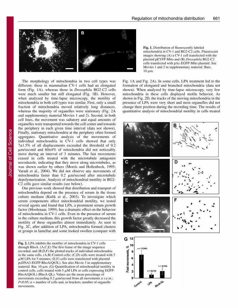
The morphology of mitochondria in two cell types wasdifferent: those in mammalian CV-1 cells had an elongatedform (Fig. 1A), whereas those in Drosophila BG2-C2 cellswere much smaller but still elongated (Fig. 1B). However,when analyzed by time-lapse microscopy, the motility ofmitochondria in both cell types was similar. First, only a smallfraction of mitochondria moved relatively long distances,whereas the majority of organelles were stationary (Fig. 2Aand supplementary material Movies 1 and 2). Second, in bothcell lines, the movement was saltatory and equal amounts oforganelles were transported towards the cell center and towardsthe periphery in each given time interval (data not shown).Finally, stationary mitochondria at the periphery often formedaggregates. Quantitative analysis of the movements ofindividual mitochondria in CV-1 cells showed that only7±1.5% of all displacements exceeded the threshold of 0.2�m/second and 60±4% of mitochondria did not noticeablymove during an interval of 3 minutes. The fast movementsceased in cells treated with the microtubule antagonistnocodazole, indicating that they move along microtubules, aswas shown earlier by others (Morris and Hollenbeck, 1995;Varadi et al., 2004). We did not observe any movements ofmitochondria faster than 0.2 �m/second after microtubuledepolymerization. Analysis of mitochondrial motility in BG2-C2 cells gave similar results (see below).
Our previous work showed that distribution and transport ofmitochondria depend on the presence of serum in the tissueculture medium (Kulik et al., 2003). To investigate whichserum components affect mitochondrial motility, we testedseveral agents and found that LPA, a prominent serum growthfactor (Moolenaar, 1999), has a dramatic effect on the behaviorof mitochondria in CV-1 cells. Even in the presence of serumin the culture medium, this growth factor greatly decreased themotility of these organelles almost immediately. As seen inFig. 2C, after addition of LPA, mitochondria formed clustersor groups in lamellae and some looked swollen (compare with
Fig. 1A and Fig. 2A). In some cells, LPA treatment led to theformation of elongated and branched mitochondria (data notshown). When analyzed by time-lapse microscopy, very fewmitochondria in these cells displayed motile behavior. Asshown in Fig. 2D, the tracks of the moving mitochondria in thepresence of LPA were very short and most organelles did notchange their position during the recording time. The results ofquantitative analysis of mitochondrial motility in cells treated
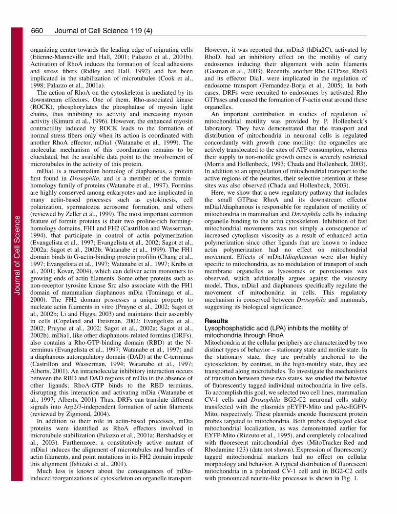
Fig. 1. Distribution of fluorescently labeledmitochondria in CV-1 and BG2-C2 cells. Fluorescentimages showing (A) a CV-1 cell transfected with theplasmid pEYFP-Mito and (B) Drosophila BG2-C2cells transfected with pAc-EGFP-Mito plasmid. SeeMovies 1 and 2 in supplementary material. Bars,10 �m.
Fig. 2. LPA inhibits the motility of mitochondria in CV-1 cellsthrough RhoA. (A,C,E) The first frame of the image sequencerecorded, and (B,D,F) the plotted tracks of individual mitochondriain the same cells. (A,B) Control cells; (C,D) cells were treated with 5�M LPA for 5 minutes; (E,F) cells were transfected with plasmidpcDNA3-EGFP-RhoA(Q63L). See also Movie 3 in supplementarymaterial. Bar, 10 �m. (G) Quantification of mitochondrial motility incontrol cells, cells treated with 5 �M LPA or cells expressing EGFP-RhoA(Q63L) (RhoA-QL). Values are the mean percentage ofmovements exceeding 0.2 �m/second from all movements ± s.e.m.;P<0.05; n = number of cells and, in brackets, number of organellemovements.
Jour
nal o
f Cel
l Sci
ence
662
with LPA are shown in Fig. 2G. These results indicate that LPAstops movement of mitochondria.
LPA has multiple effects on the cytoskeleton. When addedto culture medium, it stimulates the formation of stress fibersand focal adhesions in different cell types, and induces theformation of stable microtubules. LPA acts through a smallGTPase RhoA (Ridley and Hall, 1992; Cook et al., 1998) andits downstream effectors, including Rho-kinase (ROCK)(Matsui et al., 1996; Ishizaki et al., 1997) and mDia (Watanabeet al., 1997; Ishizaki et al., 2001; Palazzo et al., 2001a). We setout to verify whether RhoA is involved in the regulation ofmitochondrial distribution by LPA. We transfected CV-1 cellswith a constitutively active mutant form of RhoA. To visualizecells that co-expressed an enhanced yellow fluorescent protein(EYFP)-labeled mitochondria probe and an enhanced greenfluorescent protein (EGFP) fusion of RhoA(Q63L), we utilizedthe fact that EYFP fluorescence that can be detected both ingreen and red channels.
Expression of a constitutively active mutant of RhoA,EGFP-RhoA(Q63L), in CV-1 cells resulted in a gross alterationof mitochondria morphology (Fig. 2E). Some mitochondriamerged to form long-branched organelles and fused to form theextensive networks filling the entire cytoplasm (data notshown). Neither the large organelles nor those of smaller sizemoved significant distances when analyzed by time-lapsemicroscopy (see the tracks in Fig. 2E and supplementarymaterial Movie 3). The overall motility of mitochondria inthese cells was considerably suppressed (Fig. 2G). Wemicroinjected CV-1 cells with C3 transferase, a potent inhibitorof RhoA (Ridley and Hall, 1992), to confirm its involvementin the regulation of mitochondrial distribution. This led tofragmentation of mitochondria and their collapse to theperinuclear region (supplementary material Fig. S1A,B). Thus,our data suggest that RhoA controls not only the anchoring ofmitochondria to the cytoskeleton but also their morphology.
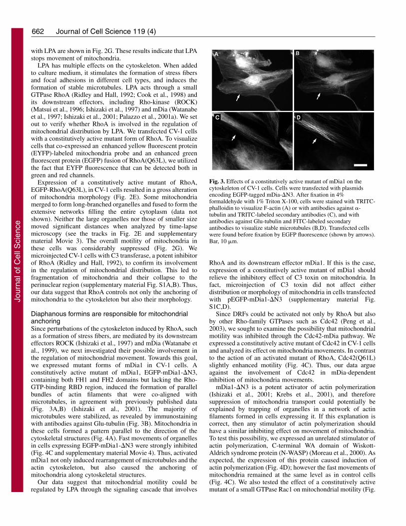
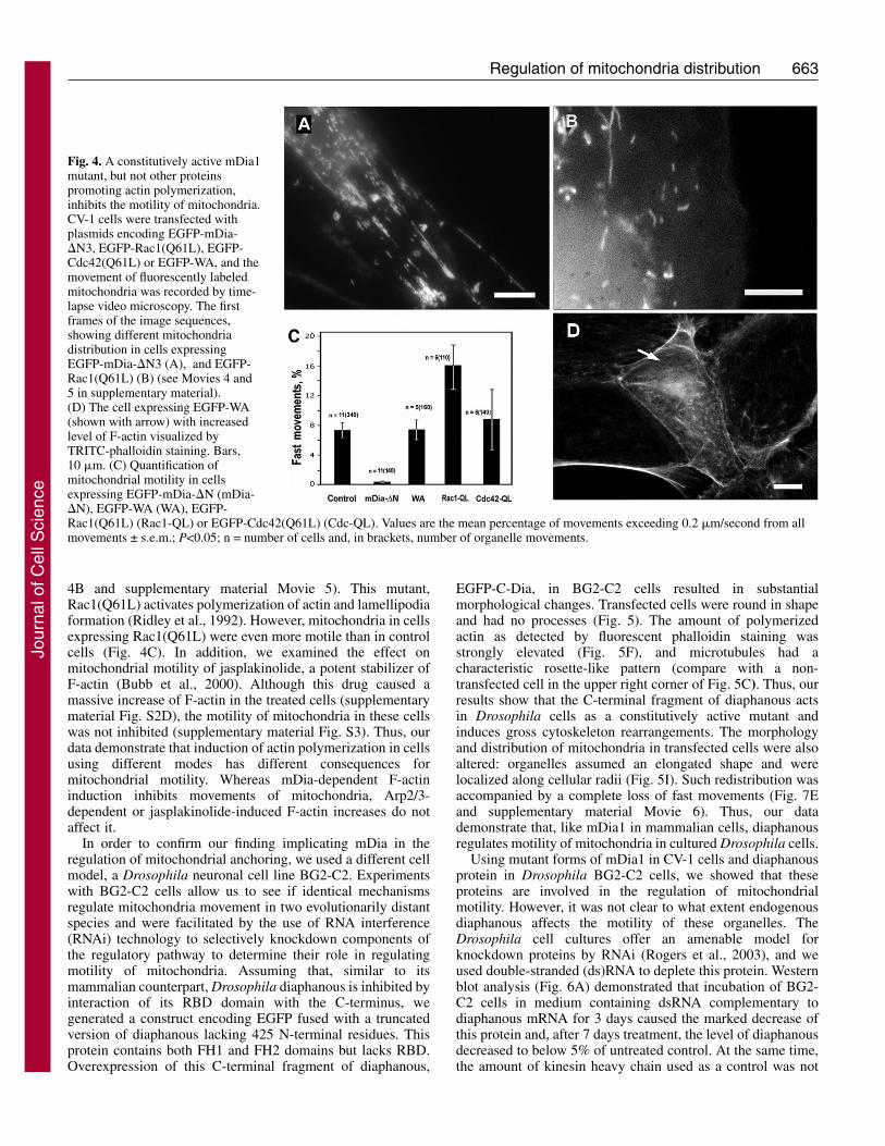
Diaphanous formins are responsible for mitochondrialanchoringSince perturbations of the cytoskeleton induced by RhoA, suchas a formation of stress fibers, are mediated by its downstreameffectors ROCK (Ishizaki et al., 1997) and mDia (Watanabe etal., 1999), we next investigated their possible involvement inthe regulation of mitochondrial movement. Towards this goal,we expressed mutant forms of mDia1 in CV-1 cells. Aconstitutively active mutant of mDia1, EGFP-mDia1-�N3,containing both FH1 and FH2 domains but lacking the Rho-GTP-binding RBD region, induced the formation of parallelbundles of actin filaments that were co-aligned withmicrotubules, in agreement with previously published data(Fig. 3A,B) (Ishizaki et al., 2001). The majority ofmicrotubules were stabilized, as revealed by immunostainingwith antibodies against Glu-tubulin (Fig. 3B). Mitochondria inthese cells formed a pattern parallel to the direction of thecytoskeletal structures (Fig. 4A). Fast movements of organellesin cells expressing EGFP-mDia1-�N3 were strongly inhibited(Fig. 4C and supplementary material Movie 4). Thus, activatedmDia1 not only induced rearrangement of microtubules and theactin cytoskeleton, but also caused the anchoring ofmitochondria along cytoskeletal structures.
Our data suggest that mitochondrial motility could beregulated by LPA through the signaling cascade that involves
RhoA and its downstream effector mDia1. If this is the case,expression of a constitutively active mutant of mDia1 shouldrelieve the inhibitory effect of C3 toxin on mitochondria. Infact, microinjection of C3 toxin did not affect eitherdistribution or morphology of mitochondria in cells transfectedwith pEGFP-mDia1-�N3 (supplementary material Fig.S1C,D).
Since DRFs could be activated not only by RhoA but alsoby other Rho-family GTPases such as Cdc42 (Peng et al.,2003), we sought to examine the possibility that mitochondrialmotility was inhibited through the Cdc42-mDia pathway. Weexpressed a constitutively active mutant of Cdc42 in CV-1 cellsand analyzed its effect on mitochondria movements. In contrastto the action of an activated mutant of RhoA, Cdc42(Q61L)slightly enhanced motility (Fig. 4C). Thus, our data argueagainst the involvement of Cdc42 in mDia-dependentinhibition of mitochondria movements.
mDia1-�N3 is a potent activator of actin polymerization(Ishizaki et al., 2001; Krebs et al., 2001), and thereforesuppression of mitochondria transport could potentially beexplained by trapping of organelles in a network of actinfilaments formed in cells expressing it. If this explanation iscorrect, then any stimulator of actin polymerization shouldhave a similar inhibiting effect on movement of mitochondria.To test this possibility, we expressed an unrelated stimulator ofactin polymerization, C-terminal WA domain of Wiskott-Aldrich syndrome protein (N-WASP) (Moreau et al., 2000). Asexpected, the expression of this protein caused induction ofactin polymerization (Fig. 4D); however the fast movements ofmitochondria remained at the same level as in control cells(Fig. 4C). We also tested the effect of a constitutively activemutant of a small GTPase Rac1 on mitochondrial motility (Fig.
Journal of Cell Science 119 (4)
Fig. 3. Effects of a constitutively active mutant of mDia1 on thecytoskeleton of CV-1 cells. Cells were transfected with plasmidsencoding EGFP-tagged mDia-�N3. After fixation in 4%formaldehyde with 1% Triton X-100, cells were stained with TRITC-phalloidin to visualize F-actin (A) or with antibodies against �-tubulin and TRITC-labeled secondary antibodies (C), and withantibodies against Glu-tubulin and FITC-labeled secondaryantibodies to visualize stable microtubules (B,D). Transfected cellswere found before fixation by EGFP fluorescence (shown by arrows).Bar, 10 �m.
Jour
nal o
f Cel
l Sci
ence
663Regulation of mitochondria distribution
4B and supplementary material Movie 5). This mutant,Rac1(Q61L) activates polymerization of actin and lamellipodiaformation (Ridley et al., 1992). However, mitochondria in cellsexpressing Rac1(Q61L) were even more motile than in controlcells (Fig. 4C). In addition, we examined the effect onmitochondrial motility of jasplakinolide, a potent stabilizer ofF-actin (Bubb et al., 2000). Although this drug caused amassive increase of F-actin in the treated cells (supplementarymaterial Fig. S2D), the motility of mitochondria in these cellswas not inhibited (supplementary material Fig. S3). Thus, ourdata demonstrate that induction of actin polymerization in cellsusing different modes has different consequences formitochondrial motility. Whereas mDia-dependent F-actininduction inhibits movements of mitochondria, Arp2/3-dependent or jasplakinolide-induced F-actin increases do notaffect it.
In order to confirm our finding implicating mDia in theregulation of mitochondrial anchoring, we used a different cellmodel, a Drosophila neuronal cell line BG2-C2. Experimentswith BG2-C2 cells allow us to see if identical mechanismsregulate mitochondria movement in two evolutionarily distantspecies and were facilitated by the use of RNA interference(RNAi) technology to selectively knockdown components ofthe regulatory pathway to determine their role in regulatingmotility of mitochondria. Assuming that, similar to itsmammalian counterpart, Drosophila diaphanous is inhibited byinteraction of its RBD domain with the C-terminus, wegenerated a construct encoding EGFP fused with a truncatedversion of diaphanous lacking 425 N-terminal residues. Thisprotein contains both FH1 and FH2 domains but lacks RBD.Overexpression of this C-terminal fragment of diaphanous,
EGFP-C-Dia, in BG2-C2 cells resulted in substantialmorphological changes. Transfected cells were round in shapeand had no processes (Fig. 5). The amount of polymerizedactin as detected by fluorescent phalloidin staining wasstrongly elevated (Fig. 5F), and microtubules had acharacteristic rosette-like pattern (compare with a non-transfected cell in the upper right corner of Fig. 5C). Thus, ourresults show that the C-terminal fragment of diaphanous actsin Drosophila cells as a constitutively active mutant andinduces gross cytoskeleton rearrangements. The morphologyand distribution of mitochondria in transfected cells were alsoaltered: organelles assumed an elongated shape and werelocalized along cellular radii (Fig. 5I). Such redistribution wasaccompanied by a complete loss of fast movements (Fig. 7Eand supplementary material Movie 6). Thus, our datademonstrate that, like mDia1 in mammalian cells, diaphanousregulates motility of mitochondria in cultured Drosophila cells.
Using mutant forms of mDia1 in CV-1 cells and diaphanousprotein in Drosophila BG2-C2 cells, we showed that theseproteins are involved in the regulation of mitochondrialmotility. However, it was not clear to what extent endogenousdiaphanous affects the motility of these organelles. TheDrosophila cell cultures offer an amenable model forknockdown proteins by RNAi (Rogers et al., 2003), and weused double-stranded (ds)RNA to deplete this protein. Westernblot analysis (Fig. 6A) demonstrated that incubation of BG2-C2 cells in medium containing dsRNA complementary todiaphanous mRNA for 3 days caused the marked decrease ofthis protein and, after 7 days treatment, the level of diaphanousdecreased to below 5% of untreated control. At the same time,the amount of kinesin heavy chain used as a control was not
Fig. 4. A constitutively active mDia1mutant, but not other proteinspromoting actin polymerization,inhibits the motility of mitochondria.CV-1 cells were transfected withplasmids encoding EGFP-mDia-�N3, EGFP-Rac1(Q61L), EGFP-Cdc42(Q61L) or EGFP-WA, and themovement of fluorescently labeledmitochondria was recorded by time-lapse video microscopy. The firstframes of the image sequences,showing different mitochondriadistribution in cells expressingEGFP-mDia-�N3 (A), and EGFP-Rac1(Q61L) (B) (see Movies 4 and5 in supplementary material).(D) The cell expressing EGFP-WA(shown with arrow) with increasedlevel of F-actin visualized byTRITC-phalloidin staining. Bars,10 �m. (C) Quantification ofmitochondrial motility in cellsexpressing EGFP-mDia-�N (mDia-�N), EGFP-WA (WA), EGFP-Rac1(Q61L) (Rac1-QL) or EGFP-Cdc42(Q61L) (Cdc-QL). Values are the mean percentage of movements exceeding 0.2 �m/second from allmovements ± s.e.m.; P<0.05; n = number of cells and, in brackets, number of organelle movements.
Jour
nal o
f Cel
l Sci
ence

664
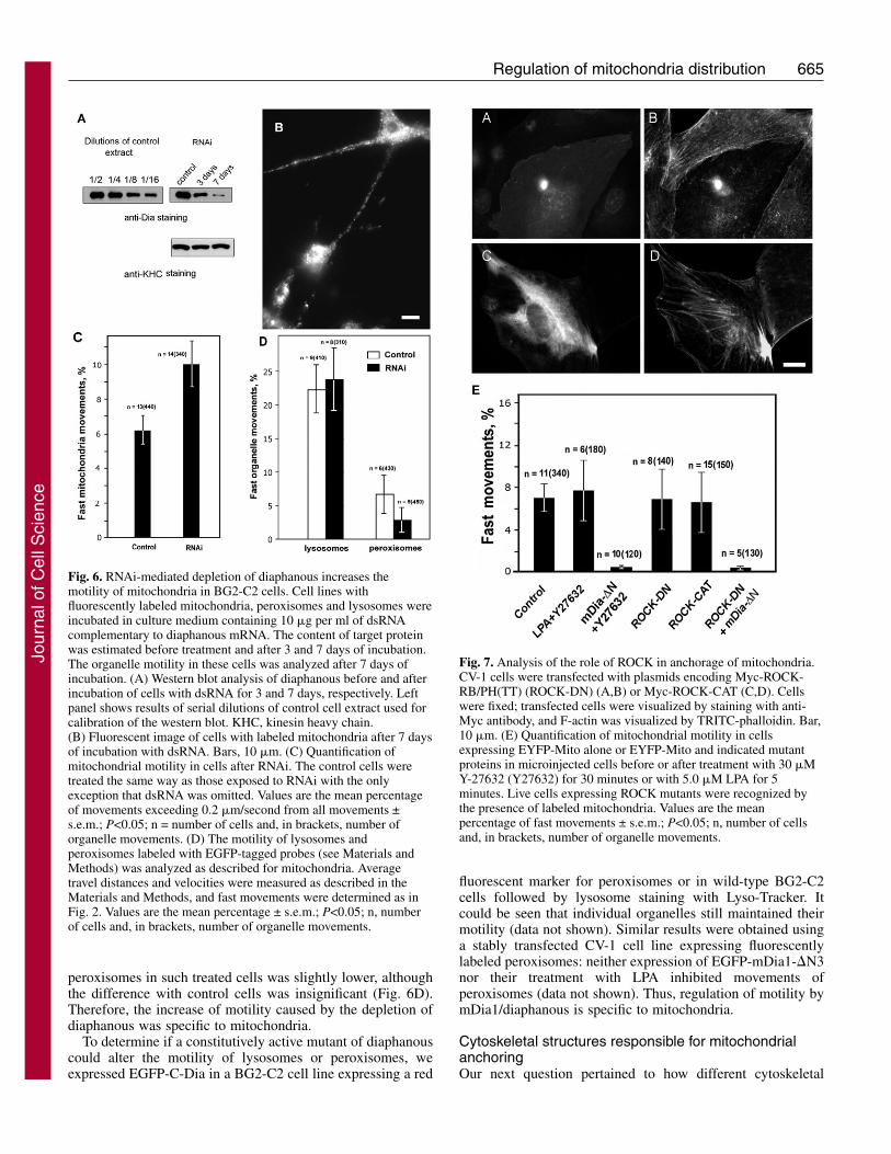
changed (Fig. 6A). The depletion of diaphanous did not changethe morphology of BG2-C2 cells: they remained attached tothe substrate and had normal processes (Fig. 6B). However, inagreement with our data obtained in mammalian andDrosophila cells using mutant constructs, knockdown ofdiaphanous led to an increased motility of the mitochondria(Fig. 6C). Thus, our data indicate that diaphanous inhibits themotility of mitochondria.
Role of ROCK in mitochondrial anchoringThe second downstream effector of RhoA that acts on the actincytoskeleton together with mDia is ROCK (Ishizaki et al.,1997). Activation of this kinase by RhoA is implicated in theformation of stress fibers and focal contacts (Amano et al.,1997). ROCK increases the phosphorylation of myosin lightchains and thus increases acto-myosin-based contractility by
directly phosphorylating myosin light chains and by negativelyregulating myosin phosphatase (Kureishi et al., 1997)(reviewed by Fukata et al., 2001). In the experiments describedabove, we demonstrate that mDia1/diaphanous regulatesmitochondrial motility. It is therefore important to understandwhether movement of mitochondria is similarly regulated bythe second RhoA effector (ROCK). We first examined theeffect of a chemical inhibitor of ROCK, Y-27632 (Uehata etal., 1997), on the inhibition of mitochondrial movements byLPA. These experiments demonstrate that Y-27632 restoresnormal motility of mitochondria blocked by LPA (Fig. 7E).Thus, our data could argue for the participation of ROCKtogether with mDia1 in the regulation of mitochondrialmotility. To test if the activity of ROCK is required formitochondrial anchoring induced by an active mutant ofmDia1, we treated cells expressing EGFP-mDia1-�N3 with Y-
27632. The results of these experiments show thatinhibition of Rho-kinase does not restore movementof mitochondria inhibited by constitutively activemDia1 (Fig. 7E).
To explore the role of ROCK further, we expresseddominant-negative (RB/PH) and constitutively active(CAT) forms of ROCK (Amano et al., 1998) in CV-1cells. As was expected, expression of ROCK-CATresulted in strongly developed actin stress fibers (Fig.7B), whereas ROCK-RB/PH prevented theirformation at the central part of the cells (Fig. 7D).However, the motility of mitochondria in these cellsdid not change (Fig. 7E). In addition, when ROCK-RB/PH was co-expressed with EGFP-mDia1-�N3,microfilament bundles became wavy and less straightcompared with cells expressing EGFP-mDia1-�N3alone, similar to the effect of Y-27632 on these cells(see supplementary material Fig. S2E,F). However,this co-expression did not suppress the inhibition ofmitochondrial movements caused by the mDia1mutant (Fig. 7E). Thus, activation of mDia1 wasnecessary and sufficient for mitochondrial anchoringat the cell periphery. We believe that modulation ofLPA effects by Y-27632 could not be explained byinhibition of ROCK, but is probably mediated by itsaction on a different protein kinase; for a discussionof Y-27632 specificity, see Davies et al. (Davies et al.,2000)].
The motility of lysosomes and peroxisomes isnot affected by diaphanousWe next asked if the affects of diaphanous are specificfor mitochondria or if it affects the transport of otherorganelles as well. To address this question, weselected stably transfected BG2-C2 cell linesexpressing fluorescently labeled lysosomes orperoxisomes. The movements of both types oforganelles could be easily observed in the cell bodyas well as in the processes (data not shown). Similarto mitochondria, both peroxisomes and lysosomes canbe in either motile or stationary states, althoughlysosomes showed a much higher level of motilitythan mitochondria (Fig. 6D). However, depletion ofdiaphanous in these cell lines had no effect on themovement of lysosomes (Fig. 6D). The motility of
Journal of Cell Science 119 (4)
Fig. 5. The constitutively active mutant of diaphanous induces mitochondrialanchoring in Drosophila BG2-C2 cells. BG2-C2 cells were transfected with aplasmid encoding EGFP-C-Dia. Cells were either fixed for cytoskeletonstaining (A-F) or incubated with 100 nM Texas Red Mitotracker for theanalysis of mitochondrial motility (G-I). (A,D,G) Fluorescent images thatshow the transfected cells. (C) Cells were stained with anti-tubulin antibodyand TRITC-labeled secondary antibody. Microtubules in transfected cellsform the characteristic rosette-like pattern. (F) Cells were stained withTRITC-phalloidin; the higher level of F-actin could be seen in transfectedcells. (B,E,H) Phase-contrast images of cells shown in A,D,G, and (I) the firstframe in the video sequence (Movie 6 in supplementary material) showing thedistribution of elongated mitochondria in the transfected cell shown in G.Bars, 10 �m.
Jour
nal o
f Cel
l Sci
ence
665Regulation of mitochondria distribution
peroxisomes in such treated cells was slightly lower, althoughthe difference with control cells was insignificant (Fig. 6D).Therefore, the increase of motility caused by the depletion ofdiaphanous was specific to mitochondria.
To determine if a constitutively active mutant of diaphanouscould alter the motility of lysosomes or peroxisomes, weexpressed EGFP-C-Dia in a BG2-C2 cell line expressing a red
fluorescent marker for peroxisomes or in wild-type BG2-C2cells followed by lysosome staining with Lyso-Tracker. Itcould be seen that individual organelles still maintained theirmotility (data not shown). Similar results were obtained usinga stably transfected CV-1 cell line expressing fluorescentlylabeled peroxisomes: neither expression of EGFP-mDia1-�N3nor their treatment with LPA inhibited movements ofperoxisomes (data not shown). Thus, regulation of motility bymDia1/diaphanous is specific to mitochondria.
Cytoskeletal structures responsible for mitochondrialanchoringOur next question pertained to how different cytoskeletal
Fig. 6. RNAi-mediated depletion of diaphanous increases themotility of mitochondria in BG2-C2 cells. Cell lines withfluorescently labeled mitochondria, peroxisomes and lysosomes wereincubated in culture medium containing 10 �g per ml of dsRNAcomplementary to diaphanous mRNA. The content of target proteinwas estimated before treatment and after 3 and 7 days of incubation.The organelle motility in these cells was analyzed after 7 days ofincubation. (A) Western blot analysis of diaphanous before and afterincubation of cells with dsRNA for 3 and 7 days, respectively. Leftpanel shows results of serial dilutions of control cell extract used forcalibration of the western blot. KHC, kinesin heavy chain.(B) Fluorescent image of cells with labeled mitochondria after 7 daysof incubation with dsRNA. Bars, 10 �m. (C) Quantification ofmitochondrial motility in cells after RNAi. The control cells weretreated the same way as those exposed to RNAi with the onlyexception that dsRNA was omitted. Values are the mean percentageof movements exceeding 0.2 �m/second from all movements ±s.e.m.; P<0.05; n = number of cells and, in brackets, number oforganelle movements. (D) The motility of lysosomes andperoxisomes labeled with EGFP-tagged probes (see Materials andMethods) was analyzed as described for mitochondria. Averagetravel distances and velocities were measured as described in theMaterials and Methods, and fast movements were determined as inFig. 2. Values are the mean percentage ± s.e.m.; P<0.05; n, numberof cells and, in brackets, number of organelle movements.
Fig. 7. Analysis of the role of ROCK in anchorage of mitochondria.CV-1 cells were transfected with plasmids encoding Myc-ROCK-RB/PH(TT) (ROCK-DN) (A,B) or Myc-ROCK-CAT (C,D). Cellswere fixed; transfected cells were visualized by staining with anti-Myc antibody, and F-actin was visualized by TRITC-phalloidin. Bar,10 �m. (E) Quantification of mitochondrial motility in cellsexpressing EYFP-Mito alone or EYFP-Mito and indicated mutantproteins in microinjected cells before or after treatment with 30 �MY-27632 (Y27632) for 30 minutes or with 5.0 �M LPA for 5minutes. Live cells expressing ROCK mutants were recognized bythe presence of labeled mitochondria. Values are the meanpercentage of fast movements ± s.e.m.; P<0.05; n, number of cellsand, in brackets, number of organelle movements.
Jour
nal o
f Cel
l Sci
ence
666
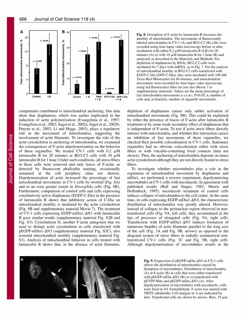
components contributed to mitochondrial anchoring. Our datashow that diaphanous, which was earlier implicated in theinduction of actin polymerization (Evangelista et al., 1997;Evangelista et al., 2002; Sagot et al., 2002a; Sagot et al., 2002b;Pruyne et al., 2002; Li and Higgs, 2003), plays a regulatoryrole in the movement of mitochondria, suggesting theinvolvement of actin filaments. To investigate the role of theactin cytoskeleton in anchoring of mitochondria, we examinedthe consequences of F-actin depolymerization on the behaviorof these organelles. We treated CV-1 cells with 0.2 �Mlatrunculin B for 20 minutes or BG2-C2 cells with 10 �Mlatrunculin B for 1 hour. Under such conditions, all stress fibersin these cells were removed and only traces of F-actin, asdetected by fluorescent phalloidin staining, occasionallyremained at the cell periphery (data not shown).Depolymerization of actin increased the percentage of fastmitochondrial movements in CV-1 cells by twofold (Fig. 8A)and to an even greater extent in Drosophila cells (Fig. 8B).Furthermore, comparison of control cells and cells expressingconstitutively active diaphanous (EGFP-C-Dia) in the presenceof latrunculin B shows that inhibitory action of C-Dia onmitochondrial motility is mediated by the actin cytoskeleton(Fig. 8B and supplementary material Movie 7). The treatmentof CV-1 cells expressing EGFP-mDia1-�N3 with latrunculinB gave similar results (supplementary material Fig. S2B andFig. S3). Cytochalasin D, another F-actin antagonist that weused to disrupt actin cytoskeleton in cells transfected withpEGFP-mDia1-�N3 (supplementary material Fig. S2C), alsorestored mitochondrial motility (supplementary material Fig.S3). Analysis of mitochondrial behavior in cells treated withlatrunculin B shows that, in the absence of actin filaments,
depletion of diaphanous causes only subtle activation ofmitochondrial movements (Fig. 8B). This could be explainedby either the presence of traces of F-actin after latrunculin Btreatment or by some weak secondary effect of diaphanous thatis independent of F-actin. To test if actin stress fibers directlyinteract with mitochondria, and whether this interaction causesan inhibition of fast movements of these organelles, wechecked their possible colocalization in CV-1 cells. Stationaryorganelles had no obvious colocalization either with stressfibers or with vinculin-containing focal contacts (data notshown). Thus, the anchoring of mitochondria depends on intactactin cytoskeleton although they are not directly bound to stressfibers.
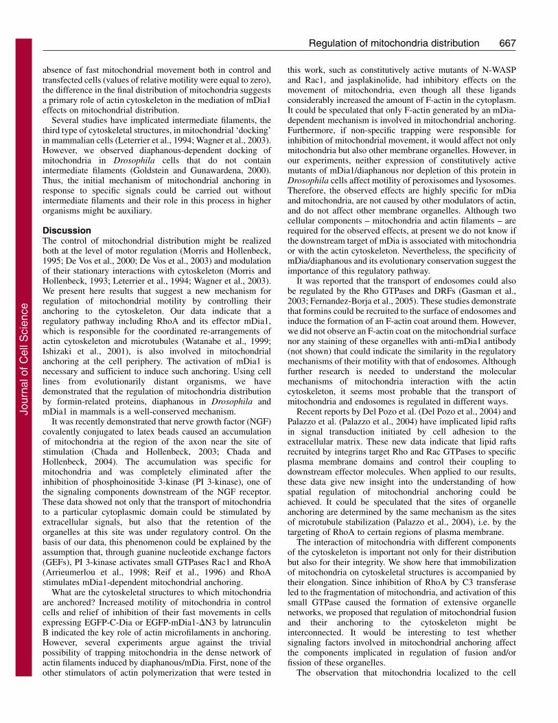
To investigate whether microtubules play a role in theregulation of mitochondrial movement by diaphanous andmDia1, we performed a reverse experiment, depolymerizingmicrotubules in CV-1 cells with nocodazole. In agreement withpublished results (Ball and Singer, 1982; Morris andHollenbeck, 1995), nocodazole treatment of control cellsinduces collapse of mitochondria to the cell center. At the sametime, in cells expressing EGFP-mDia1-�N3, the characteristicdistribution of mitochondria was greatly altered. However,instead of collapse to the perinuclear region observed in non-transfected cells (Fig. 9A, left cell), they accumulated at thetips of processes of elongated cells (Fig. 9A, right cell).Transfection with EGFP-mDia1-�N3 induces formation ofnumerous bundles of actin filaments parallel to the long axisof the cell (Fig. 3A and Fig. 9B, arrows) as opposed to thediagonal system of stress fibers in radially symmetrical non-transfected CV-1 cells (Fig. 3C and Fig. 9B, right cell).Although depolymerization of microtubules results in the
Journal of Cell Science 119 (4)
Fig. 8. Disruption of F-actin by latrunculin B increases themotility of mitochondria. The movements of fluorescentlylabeled mitochondria in CV-1 (A) and BG2-C2 (B) cells wererecorded using time-lapse video microscopy before or afterincubation with either 0.2 �M latrunculin B (LB) for 20minutes (A) or with 10 �M latrunculin B for 1 hour (B) andanalyzed, as described in the Materials and Methods. Fordepletion of diaphanous by RNAi, BG2-C2 cells wereincubated for 7 days with dsRNA, as in Fig. 7. For the analysisof mitochondrial motility in BG2-C2 cells transfected withEGFP-C-Dia (GFP-C-Dia), they were incubated with 100 nMTexas Red Mitotracker for 40 minutes, and mitochondrialmovements were recorded by time-lapse video microscopyusing red fluorescence filter set (see also Movie 7 insupplementary material). Values are the mean percentage offast mitochondria movements ± s.e.m.; P<0.05; n, number ofcells and, in brackets, number of organelle movements.
Fig. 9. Expression of pEGFP-mDia-�N3 in CV-1 cellsaffects the distribution of mitochondria caused bydisruption of microtubules. Distribution of mitochondria(A) or F-actin (B) in cells that were either transfectedwith pEGFP-mDia-�N3 (B) or co-transfected withpEYFP-Mito and pEGFP-mDia-�N3 (A). Afterdepolymerization of microtubules with nocodazole, cellswere fixed in 4% formaldehyde. F-actin was stained withTRITC-phalloidin. Cell margins in A are indicated bydots. Transfected cells are shown by arrows. Bars, 10 �m.
Jour
nal o
f Cel
l Sci
ence
667Regulation of mitochondria distribution
absence of fast mitochondrial movement both in control andtransfected cells (values of relative motility were equal to zero),the difference in the final distribution of mitochondria suggestsa primary role of actin cytoskeleton in the mediation of mDia1effects on mitochondrial distribution.
Several studies have implicated intermediate filaments, thethird type of cytoskeletal structures, in mitochondrial ‘docking’in mammalian cells (Leterrier et al., 1994; Wagner et al., 2003).However, we observed diaphanous-dependent docking ofmitochondria in Drosophila cells that do not containintermediate filaments (Goldstein and Gunawardena, 2000).Thus, the initial mechanism of mitochondrial anchoring inresponse to specific signals could be carried out withoutintermediate filaments and their role in this process in higherorganisms might be auxiliary.
DiscussionThe control of mitochondrial distribution might be realizedboth at the level of motor regulation (Morris and Hollenbeck,1995; De Vos et al., 2000; De Vos et al., 2003) and modulationof their stationary interactions with cytoskeleton (Morris andHollenbeck, 1993; Leterrier et al., 1994; Wagner et al., 2003).We present here results that suggest a new mechanism forregulation of mitochondrial motility by controlling theiranchoring to the cytoskeleton. Our data indicate that aregulatory pathway including RhoA and its effector mDia1,which is responsible for the coordinated re-arrangements ofactin cytoskeleton and microtubules (Watanabe et al., 1999;Ishizaki et al., 2001), is also involved in mitochondrialanchoring at the cell periphery. The activation of mDia1 isnecessary and sufficient to induce such anchoring. Using celllines from evolutionarily distant organisms, we havedemonstrated that the regulation of mitochondria distributionby formin-related proteins, diaphanous in Drosophila andmDia1 in mammals is a well-conserved mechanism.
It was recently demonstrated that nerve growth factor (NGF)covalently conjugated to latex beads caused an accumulationof mitochondria at the region of the axon near the site ofstimulation (Chada and Hollenbeck, 2003; Chada andHollenbeck, 2004). The accumulation was specific formitochondria and was completely eliminated after theinhibition of phosphoinositide 3-kinase (PI 3-kinase), one ofthe signaling components downstream of the NGF receptor.These data showed not only that the transport of mitochondriato a particular cytoplasmic domain could be stimulated byextracellular signals, but also that the retention of theorganelles at this site was under regulatory control. On thebasis of our data, this phenomenon could be explained by theassumption that, through guanine nucleotide exchange factors(GEFs), PI 3-kinase activates small GTPases Rac1 and RhoA(Arrieumerlou et al., 1998; Reif et al., 1996) and RhoAstimulates mDia1-dependent mitochondrial anchoring.
What are the cytoskeletal structures to which mitochondriaare anchored? Increased motility of mitochondria in controlcells and relief of inhibition of their fast movements in cellsexpressing EGFP-C-Dia or EGFP-mDia1-�N3 by latrunculinB indicated the key role of actin microfilaments in anchoring.However, several experiments argue against the trivialpossibility of trapping mitochondria in the dense network ofactin filaments induced by diaphanous/mDia. First, none of theother stimulators of actin polymerization that were tested in
this work, such as constitutively active mutants of N-WASPand Rac1, and jasplakinolide, had inhibitory effects on themovement of mitochondria, even though all these ligandsconsiderably increased the amount of F-actin in the cytoplasm.It could be speculated that only F-actin generated by an mDia-dependent mechanism is involved in mitochondrial anchoring.Furthermore, if non-specific trapping were responsible forinhibition of mitochondrial movement, it would affect not onlymitochondria but also other membrane organelles. However, inour experiments, neither expression of constitutively activemutants of mDia1/diaphanous nor depletion of this protein inDrosophila cells affect motility of peroxisomes and lysosomes.Therefore, the observed effects are highly specific for mDiaand mitochondria, are not caused by other modulators of actin,and do not affect other membrane organelles. Although twocellular components – mitochondria and actin filaments – arerequired for the observed effects, at present we do not know ifthe downstream target of mDia is associated with mitochondriaor with the actin cytoskeleton. Nevertheless, the specificity ofmDia/diaphanous and its evolutionary conservation suggest theimportance of this regulatory pathway.
It was reported that the transport of endosomes could alsobe regulated by the Rho GTPases and DRFs (Gasman et al.,2003; Fernandez-Borja et al., 2005). These studies demonstratethat formins could be recruited to the surface of endosomes andinduce the formation of an F-actin coat around them. However,we did not observe an F-actin coat on the mitochondrial surfacenor any staining of these organelles with anti-mDia1 antibody(not shown) that could indicate the similarity in the regulatorymechanisms of their motility with that of endosomes. Althoughfurther research is needed to understand the molecularmechanisms of mitochondria interaction with the actincytoskeleton, it seems most probable that the transport ofmitochondria and endosomes is regulated in different ways.
Recent reports by Del Pozo et al. (Del Pozo et al., 2004) andPalazzo et al. (Palazzo et al., 2004) have implicated lipid raftsin signal transduction initiated by cell adhesion to theextracellular matrix. These new data indicate that lipid raftsrecruited by integrins target Rho and Rac GTPases to specificplasma membrane domains and control their coupling todownstream effector molecules. When applied to our results,these data give new insight into the understanding of howspatial regulation of mitochondrial anchoring could beachieved. It could be speculated that the sites of organelleanchoring are determined by the same mechanism as the sitesof microtubule stabilization (Palazzo et al., 2004), i.e. by thetargeting of RhoA to certain regions of plasma membrane.
The interaction of mitochondria with different componentsof the cytoskeleton is important not only for their distributionbut also for their integrity. We show here that immobilizationof mitochondria on cytoskeletal structures is accompanied bytheir elongation. Since inhibition of RhoA by C3 transferaseled to the fragmentation of mitochondria, and activation of thissmall GTPase caused the formation of extensive organellenetworks, we proposed that regulation of mitochondrial fusionand their anchoring to the cytoskeleton might beinterconnected. It would be interesting to test whethersignaling factors involved in mitochondrial anchoring affectthe components implicated in regulation of fusion and/orfission of these organelles.
The observation that mitochondria localized to the cell
Jour
nal o
f Cel
l Sci
ence
668
periphery and interacting with the cytoskeleton have a highertransmembrane potential than those residing at the perinuclearregion (Collins et al., 2002) indicate the possibleinterdependence of their anchoring with multiple properties ofthese organelles. It will be interesting to examine whether theenergization of mitochondria could be regulated by interactionswith the cytoskeleton.
Materials and MethodsCell culture, treatments and transfectionsCV-1 cells stably transfected with pEYFP-Mito or with pEYFP-PEX3 were selectedusing G-418 (Sigma) and maintained in DMEM supplemented with 10% fetal calfserum, 100 U/ml penicillin and 100 �g/ml streptomycin, without G-418.Transfections of CV-1 cells were performed using Unifectin M reagent (kindlyprovided by A. Surovoy, Institute of Bioorganic Chemistry, Moscow, Russia) using1 �g of plasmid DNA per 35 mm plate in serum-containing medium. Forcotransfection with several plasmids, CV-1 cells were plated on cover slips withphotoetched grids and microinjected into nuclei with plasmid solutions in 50 mMglutamate buffer (pH 7.4) as described previously (Minin, 1997). For microscopy,cells were plated at least 18 hours before experiments. To depolymerizemicrotubules, cells were treated with 10 �M nocodazole (Calbiochem-Behring) for1-3 hours. F-actin in CV-1 cells was depolymerized by incubation with 0.2 �Mlatrunculin B (Calbiochem-Behring) for 20 minutes. Treatments of cells withlysophosphatidic acid (LPA; Sigma) and Y-27632 (Calbiochem-Behring) wereperformed as indicated in the figure legends.
Drosophila BG2-C2 cells were kindly provided by K. Ui-Tei (University ofTokyo, Japan) and maintained in Shields and Sang ME3 (Shields and Sang M3)insect medium (M3 medium) (Sigma) supplemented with 10% heat-inactivated fetalcalf serum, 100 U/ml of penicillin/streptomycin, and 50 �g/ml of insulin at 26°Cas described previously (Ui et al., 1994). For microscopy, cells were plated on acid-washed coverslips that were coated with 0.5 mg/ml of concanavalin A (ConA) inmedium conditioned for 24 hours by a dense culture of BG2-C2 cells. Under theseconditions, many cells develop long processes that are convenient for microscopy.BG2-C2 cells were used for microscopy 16-48 hours after plating. F-actin in BG2-C2 cells was depolymerized with 10 �M latrunculin B for 1 hour.
BG2-C2 cells were transfected using Cellfectin (Invitrogen) in M3 mediumcontaining 5% heat-inactivated serum for 5 hours and then the medium was changedto one with 10% serum. Stably transfected cell lines expressing EGFP-tagged probesfor mitochondria, lysosomes and peroxisomes, and red fluorescent protein (RFP)-tagged probe for peroxisomes, were created by co-transfecting with selection vectorpCoHygro (Invitrogen), and selecting with 300 �g/ml hygromycin.
For RNAi, BG2-C2 cells in a 35 mm plate were incubated for 1 hour in 1 ml ofM3 medium containing 5% heat-inactivated fetal calf serum and 30 �g of dsRNA.Then 2 ml of M3 medium with 10% serum were added, and cells were cultured for3 days. On the fourth day, a fresh portion of dsRNA was added and the cells wereincubated for an additional 3 days.
Plasmids, RNAi and antibodiesMitochondria in CV-1 cells were tagged using the pEYFP-Mito plasmid vector(Clontech). For labeling of mitochondria in Drosophila BG2-C2, cells weretransfected with plasmid pAc-EGFP-Mito that was prepared by subcloning asequence encoding mitochondria localization signal from pEYFP-Mito into pAc-EGFP vector (a gift of S.-C. Ling, Northwestern University, Chicago, IL). Forperoxisome labeling, plasmids pAc-EGFP-SKL and pAc-RFP-SKL were made byattaching the Ser-Lys-Leu sequence to the C-terminus of EGFP and RFP. Thetandem dimer2(12) version of mRFP was kindly provided by R. Tsien (UCSD, LaJolla, CA) (Campbell et al., 2002). Lysosomes were labeled using plasmid pAc-LAMP1-EGFP, which was made by fusion of putative Drosophila LAMP1 withEGFP. Peroxisomes in CV-1 cells were labeled with plasmid pEYFP-PEX3 kindlyprovided by A. Akhmanova (Erasmus University, Rotterdam, Netherlands).Plasmids pcDNA3-EGFP-Rac1(Q61L), pcDNA3-EGFP-Cdc42(Q61L) andpcDNA3-EGFP-RhoA(Q63L) were a gift from K. Hahn (University of NorthCarolina, Chapel Hill, NC). Plasmid encoding the mDia1 mutant pEGFP-mDia1-�N3 was obtained from S. Narumiya (Kyoto University, Kyoto, Japan). Plasmidencoding C3 transferase from Clostridium botulinum was a gift from A. Hall(University College London, London, UK). Plasmid CB6-EGFP-WA encoding theC-terminus of rat N-WASP was a gift from M. Way (London Research Institute,London, UK). Plasmids pEF-BOS-Myc-Rho-kinase-RB/PH(TT) and pEF-BOS-Myc-Rho-kinase-CAT encoding dominant-negative and dominant-active mutants ofRho-kinase were kindly provided by K. Kaibuchi (Nagoya University, Japan).
Plasmid pAc-EGFP-C-Dia, encoding the truncated version of Drosophiladiaphanous (accession number NM-165341), containing only FH1 and FH2domains but not the RBD domain, was prepared by inserting the 1297-3276 cDNAfragment into the pAc-EGFP vector between the BamHI and AgeI restriction sites.The fragment was amplified by PCR from a Drosophila embryonic phage cDNAlibrary (gift of S. Dzitoeva, University of Illinois College of Medicine, Chicago, IL)
using primers: 5�-CAAACCGGTCACAAGGGTTACTGTGATCCGA-3� and 5�-CCGGATCCCTACGCGGAGCCTAGAACC-3�. By analogy with its mammaliancounterparts, this truncation is assumed to encode a constitutively active version ofdiaphanous.
To prepare dsRNA complementary to diaphanous mRNA, a 4-1316 cDNAfragment was amplified by PCR from the same cDNA library using primers 5�-GAGCAACAACTAAATAAAATGTCTCGTCACGAG-3� and 5�-GGATCACAG-TAACCCTTGTGGAAGAC-3� and inserted into the TOPO-Vector (Invitrogen).Then, using primers 5�-TAATACGACTCACTATAGGGTCGTTCTGCATTGTC-TATGAGC-3� and 5�-TAATACGACTCACTATAGGGGGATCACAGTAACCCTT-GTGGA-3� as in (Rogers et al., 2003), the template containing promoters for T7-polymerase and the 584 bp fragment of diaphanous cDNA was prepared. In vitrotranscription was carried out using a MEGAScript kit (Ambion).
A polyclonal antibody against Glu-tubulin was a gift of C. Bulinski (ColumbiaUniversity, New York, NY); monoclonal antibody DM1� against �-tubulin wasfrom Sigma; polyclonal rabbit antibodies against the motor domain of the kinesinheavy chain (HD) were prepared and characterized earlier (Rodionov et al., 1993);polyclonal rabbit antibodies against human mDia1 were purchased from BethylLaboratories; polyclonal rabbit antibodies against diaphanous (Afshar et al., 2000)was kindly provided by S. Wasserman (University of California, San Diego, CA).Monoclonal antibody against vinculin was from Y. M. Vasiliev (Moscow StateUniversity, Moscow). Secondary anti-mouse and anti-rabbit antibodies conjugatedwith horseradish peroxidase or fluorescent dyes were purchased from JacksonImmuno Research Laboratories.
Time-lapse fluorescent microscopyTime-lapse epi-fluorescent microscopy of CV-1 cells was performed using Axiophotmicroscope (Zeiss) equipped with a Plan-Apochromat 63� 1.4 NA objective.Images were captured with Micromax 782Y cooled CCD camera (Roper Scientific)driven by WinView32 software. The frames were collected every 4 seconds with anexposure time of 0.5-1.0 second. For video microscopy of BG2-C2 cells a NikonDiaphot 200 inverted microscope equipped with a Planapo 60� 1.4 NA objectivewas used. Images were collected with an Orca II cooled CCD camera (HamamatsuPhotonics) driven by Isee software (Isee Imaging Systems). To avoid phototoxiceffects a 100 W halogen lamp was used for epi-fluorescent illumination.
SDS-PAGE and immunoblottingElectrophoretic separation of proteins was performed using discontinuous 7.5%SDS-PAGE (Laemmli, 1970). For comparison of the content of diaphanous andKHC in cells treated with dsRNAs, the samples first were run on gels that werestained with Coomassie R250. The gels were scanned, and the load for western blotswas normalized according to the scan data. KHC was detected using HD antibodyat a dilution of 1:1000. Anti-diaphanous antibody was used at 1:5000. Blots weredeveloped using SuperSignal (Pierce Chemical Co.). To determine an amount ofresidual diaphanous after RNAi we compared intensities of bands on blots with thatobtained for serially diluted control cellular extract.
Quantitative analysis of mitochondrial motilityImage analysis was done with the open source image analysis software ImageJ usingavailable plug-ins and macros, and with Photoshop (Adobe Systems). Because ofthe diverse morphology of mitochondria we plotted the coordinates of one end forlonger organelles and the center of mass for shorter ones. Mitochondrial motilitywas expressed in terms of the displacement distances, and velocities. To define fastorganelle movements we applied a threshold of 200 nm/second. The values wereexpressed as mean percentages of fast movements in all recorded displacements ±s.e.m. The significance of differences was estimated statistically by paired-sampleStudent’s t-test. Variability of the values calculated for different cells in sampleswas analyzed by the same method and was insignificant.
We are grateful to S. Narumiya, A. Hall, S. Wasserman, K. Hahn,R. Tsien, S. Dzitoeva, A. Akhmanova, M. Way, K. Kaibuchi and S.-C. Ling for generously providing reagents; K. Ui-Tei for sending theBG2-C2 cell line; C. Waterman-Storer for stimulating discussions;and M. Gelfand and S. Deacon for critical reading of the manuscript.This study was partially funded by CRDF grant RB1-2506-PU-03 toA.A.M. and V.I.G.; NIH grant GM-52111 to V.I.G.; HHMI grant55000311, and RFBR grant 02-04-48548 to F.K.G.; Long-termfellowship from HFSP to G.G.; and Program of Molecular andCellular Biology of Russian Academy of Sciences.
ReferencesAfshar, K., Stuart, B. and Wasserman, S. A. (2000). Functional analysis of the
Drosophila Diaphanous FH protein in early embryonic development. Development127, 1887-1897.
Journal of Cell Science 119 (4)
Jour
nal o
f Cel
l Sci
ence
669Regulation of mitochondria distribution
Alberts, A. S. (2001). Identification of a carboxyl-terminal diaphanous-related forminhomology protein autoregulatory domain. J. Biol. Chem. 276, 2824-2830.
Amano, M., Chihara, K., Kimura, K., Fukata, Y., Nakamura, N., Matsuura, Y. andKaibuchi, K. (1997). Formation of actin stress fibers and focal adhesions enhanced byRho-kinase. Science 75, 1308-1311.
Amano, M., Chihara, K., Nakamura, N., Fukata, Y., Yano, T., Shibata, M., Ikebe,M. and Kaibuchi, K. (1998). Myosin II activation promotes neurite retraction duringthe action of Rho and Rho-kinase. Genes Cells 3, 177-188.
Arrieumerlou, C., Donnadieu, E., Brennan, P., Keryer, G., Bismuth, G., Cantrell, D.and Trautmann, A. (1998). Involvement of phosphoinositide 3-kinase and Rac inmembrane ruffling induced by IL-2 in T cells. Eur. J. Immunol. 28, 1877-1885.
Ball, E. H. and Singer, S. J. (1982). Mitochondria are associated with microtubules andnot with intermediate filaments in cultured fibroblasts. Proc. Natl. Acad. Sci. USA 79,123-126.
Bar-Sagi, D. and Hall, A. (2000). Ras and Rho GTPases: a family reunion. Cell 103,227-238.
Bershadsky, A. D., Balaban, N. Q. and Geiger, B. (2003). Adhesion-dependent cellmechanosensitivity. Annu. Rev. Cell Dev. Biol. 19, 677-695.
Bubb, M. R., Spector, I., Beyer, B. B. and Fosen, K. M. (2000). Effects of jasplakinolideon the kinetics of actin polymerization. An explanation for certain in vivo observations.J. Biol. Chem. 275, 5163-5170.
Campbell, R. E., Tour, O., Palmer, A. E., Steinbach, P. A., Baird, G. S., Zacharias,D. A. and Tsien, R. Y. (2002). A monomeric red fluorescent protein. Proc. Natl. Acad.Sci. USA 99, 7877-7882.
Castrillon, D. H. and Wasserman, S. A. (1994). Diaphanous is required for cytokinesisin Drosophila and shares domains of similarity with the products of the limb deformitygene. Development 120, 3367-3377.
Chada, S. R. and Hollenbeck, P. J. (2003). Mitochondrial movement and positioning inaxons: the role of growth factor signaling. J. Exp. Biol. 206, 1985-1992.
Chada, S. R. and Hollenbeck, P. J. (2004). Nerve growth factor signaling regulatesmotility and docking of axonal mitochondria. Curr. Biol. 14, 1272-1276.
Chang, F., Drubin, D. and Nurse, P. (1997). cdc12p, a protein required for cytokinesisin fission yeast, is a component of the cell division ring and interacts with profilin. J.Cell Biol. 137, 169-182.
Collins, T. J., Berridge, M. J., Lipp, P. and Bootman, M. D. (2002). Mitochondria aremorphologically and functionally heterogeneous within cells. EMBO J. 21, 1616-1627.
Cook, T. A., Nagasaki, T. and Gundersen, G. G. (1998). Rho guanosine triphosphatasemediates the selective stabilization of microtubules induced by lysophosphatidic acid.J. Cell Biol. 141, 175-185.
Copeland, J. W. and Treisman, R. (2002). The Diaphanous-related formin mDia1controls serum response factor activity through its effects on actin polymerization. Mol.Biol. Cell 13, 4088-4099.
Davies, S. P., Reddy, H., Caivano, M. and Cohen, P. (2000). Specificity and mechanismof action of some commonly used protein kinase inhibitors. Biochem. J. 351, 95-105.
Del Pozo, M. A., Alderson, N. B., Kiosses, W. B., Chiang, H. H., Anderson, R. G. andSchwartz, M. A. (2004). Integrins regulate rac targeting by internalization ofmembrane domains. Science 303, 839-842.
De Vos, K., Severin, F., Van Herreweghe, F., Vancompernolle, K., Goossens, V.,Hyman, A. and Grooten, J. (2000). Tumor necrosis factor induceshyperphosphorylation of kinesin light chain and inhibits kinesin-mediated transport ofmitochondria. J. Cell Biol. 149, 1207-1214.
De Vos, K. J., Sable, J., Miller, K. E. and Sheetz, M. P. (2003). Expression ofphosphatidylinositol (4,5) bisphosphate-specific pleckstrin homology domains altersdirection but not the level of axonal transport of mitochondria. Mol. Biol. Cell 14, 3636-3649.
Etienne-Manneville, S. and Hall, A. (2001). Integrin-mediated activation of Cdc42controls cell polarity in migrating astrocytes through PKCzeta. Cell 106, 489-498.
Evangelista, M., Blundell, K., Longtine, M. S., Chow, C. J., Adames, N., Pringle, J.R., Peter, M. and Boone, C. (1997). Bni1p, a yeast formin linking cdc42p and theactin cytoskeleton during polarized morphogenesis. Science 276, 118-122.
Evangelista, M., Pruyne, D., Amberg, D. C., Boone, C. and Bretscher, A. (2002).Formins direct Arp2/3-independent actin filament assembly to polarize cell growth inyeast. Nat. Cell Biol. 4, 32-41.
Fernandez-Borja, M., Janssen, L., Verwoerd, D., Hordijk, P. and Neefjes, J. (2005).RhoB regulates endosome transport by promoting actin assembly on endosomalmembranes through Dia1. J. Cell Sci. 118, 2661-2670.
Fukata, Y., Amano, M. and Kaibuchi, K. (2001). Rho-Rho-kinase pathway in smoothmuscle contraction and cytoskeletal reorganization of non-muscle cells. TrendsPharmacol. Sci. 22, 32-39.
Gasman, S., Kalaidzidis, Y. and Zerial, M. (2003). RhoD regulates endosome dynamicsthrough Diaphanous-related Formin and Src tyrosine kinase. Nat. Cell Biol. 5, 195-204.
Goldstein, L. S. and Gunawardena, S. (2000). Flying through the drosophilacytoskeletal genome. J. Cell Biol. 150, F63-F68.
Hollenbeck, P. J. (1996). The pattern and mechanism of mitochondrial transport in axons.Front. Biosci. 1, D91-D102.
Ishizaki, T., Naito, M., Fujisawa, K., Maekawa, M., Watanabe, N., Saito, Y. andNarumiya, S. (1997). p160ROCK, a Rho-associated coiled-coil forming proteinkinase, works downstream of Rho and induces focal adhesions. FEBS Lett. 404, 118-124.
Ishizaki, T., Morishima, Y., Okamoto, M., Furuyashiki, T., Kato, T. and Narumiya,S. (2001). Coordination of microtubules and the actin cytoskeleton by the Rho effectormDia1. Nat. Cell Biol. 3, 8-14.
Kimura, K., Ito, M., Amano, M., Chihara, K., Fukata, Y., Nakafuku, M., Yamamori,
B., Feng, J., Nakano, T., Okawa, K. et al. (1996). Regulation of myosin phosphataseby Rho and Rho-associated kinase (Rho-kinase). Science 273, 245-248.
Kovar, D. R. and Pollard, T. D. (2004). Progressing actin: Formin as a processiveelongation machine. Nat. Cell Biol. 6, 1158-1159.
Krebs, A., Rothkegel, M., Klar, M. and Jockusch, B. M. (2001). Characterization offunctional domains of mDia1, a link between the small GTPase Rho and the actincytoskeleton. J. Cell Sci. 114, 3663-3672.
Kulik, A. V., Gioeva, F. K. and Minin, A. A. (2002). Study of the mitochondriamovement using video microscopy. Russ. J. Dev. Biol. 33, 299-305.
Kureishi, Y., Kobayashi, S., Amano, M., Kimura, K., Kanaide, H., Nakano, T.,Kaibuchi, K. and Itoh, M. (1997). Rho-associated kinase directly induces smoothmuscle contraction through myosin light chain phosphorylation. J. Biol. Chem. 272,12257-12260.
Laemmli, U. K. (1970). Cleavage of structural proteins during the assembly of the headof bacteriophage T4. Nature 227, 680-685.
Leterrier, J. F., Rusakov, D. A., Nelson, B. D. and Linden, M. (1994). Interactionsbetween brain mitochondria and cytoskeleton: evidence for specialized outermembrane domains involved in the association of cytoskeleton-associated proteins tomitochondria in situ and in vitro. Microsc. Res. Tech. 27, 233-261.
Li, F. and Higgs, H. N. (2003). The mouse formin, mDia1, is a potent actin nucleationfactor regulated by auto-inhibition. Curr. Biol. 13, 1335-1340.
Ligon, L. A. and Steward, O. (2000). Role of microtubules and actin filaments in themovement of mitochondria in the axons and dendrites of cultured hippocampalneurons. J. Comp. Neurol. 427, 351-361.
Matsui, T., Amano, M., Yamamoto, T., Chihara, K., Nakafuku, M., Ito, M., Nakano,T., Okawa, K., Iwamatsu, A. and Kaibuchi, K. (1996). Rho-associated kinase, anovel serine/threonine kinase, as a putative target for small GTP binding protein Rho.EMBO J. 15, 2208-2216.
Minin, A. (1997). Despersal of Gogli apparatus in nocodazole-treated fibroblasts is akinesin-driven process. J. Cell Sci. 110, 2495-2505.
Moolenaar, W. H. (1999). Bioactive lysophospholipids and their G protein-coupledreceptors. Exp. Cell Res. 253, 230-238.
Moreau, V., Frischknecht, F., Reckmann, I., Vincentelli, R., Rabut, G., Stewart, D.and Way, M. (2000). A complex of N-WASP and WIP integrates signalling cascadesthat lead to actin polymerization. Nat. Cell Biol. 2, 441-448.
Morris, R. L. and Hollenbeck, P. J. (1993). The regulation of bidirectional mitochondrialtransport is coordinated with axonal outgrowth. J. Cell Sci. 104, 917-927.
Morris, R. L. and Hollenbeck, P. J. (1995). Axonal transport of mitochondria alongmicrotubules and F-actin in living vertebrate neurons. J. Cell Biol. 131, 1315-1326.
Nangaku, M., Sato-Yoshitake, R., Okada, Y., Noda, Y., Takemura, R., Yamazaki, H.and Hirokawa, N. (1994). KIF1B, a novel microtubule plus end-directed monomericmotor protein for transport of mitochondria. Cell 79, 1209-1220.
Nobes, C. D. and Hall, A. (1995). Rho, rac and cdc42 GTPases: regulators of actinstructures, cell adhesion and motility. Biochem. Soc. Trans. 23, 456-459.
Palazzo, A. F., Cook, T. A., Alberts, A. S. and Gundersen, G. G. (2001a). mDiamediates Rho-regulated formation and orientation of stable microtubules. Nat. CellBiol. 3, 723-729.
Palazzo, A. F., Joseph, H. L., Chen, Y. J., Dujardin, D. L., Alberts, A. S., Pfister, K.K., Vallee, R .B. and Gundersen, G. G. (2001b). Cdc42, dynein, and dynactin regulateMTOC reorientation independent of Rho-regulated microtubule stabilization. Curr.Biol. 11, 1536-1541.
Palazzo, A. F., Eng, C. H., Schlaepfer, D. D., Marcantonio, E. E. and Gundersen, G.G. (2004). Localized stabilization of microtubules by integrin- and FAK-facilitated Rhosignaling. Science 303, 836-839.
Peng, J., Wallar, B. J., Flanders, A., Swiatek, P. J. and Alberts, A. S. (2003). Disruptionof the Diaphanous-related formin Drf1 gene encoding mDia1 reveals a role for Drf3as an effector for Cdc42. Curr. Biol. 13, 534-545.
Pruyne, D., Evangelista, M., Yang, C., Bi, E., Zigmond, S., Bretscher, A. and Boone,C. (2002). Role of formins in actin assembly: nucleation and barbed-end association.Science 297, 612-615.
Reif, K., Nobes, C. D., Thomas, G., Hall, A. and Cantrell, D. A. (1996).Phosphatidylinositol 3-kinase signals activate a selective subset of Rac/Rho-dependenteffector pathways. Curr. Biol. 6, 1445-1455.
Ridley, A. J. and Hall, A. (1992). The small GTP-binding protein rho regulates theassembly of focal adhesions and actin stress fibers in response to growth factors. Cell70, 389-399.
Ridley, A. J., Paterson, H. F., Johnston, C. L., Diekmann, D. and Hall, A. (1992). Thesmall GTP-binding protein rac regulates growth factor-induced membrane ruffling.Cell 70, 401-410.
Rizzuto, R., Brini, M., Pizzo, P., Murgia, M. and Pozzan, T. (1995). Chimeric greenfluorescent protein as a tool for visualizing subcellular organelles in living cells. Curr.Biol. 5, 635-642.
Rodionov, V. I., Gyoeva, F. K., Tanaka, E., Bershadsky, A. D., Vasiliev, J. M. andGelfand, V. I. (1993). Microtubule-dependent control of cell shape and pseudopodialactivity is inhibited by the antibody to kinesin motor domain. J. Cell Biol. 123, 1811-1820.
Rogers, S. L., Wiedemann, U., Stuurman, N. and Vale, R. D. (2003). Molecularrequirements for actin-based lamella formation in Drosophila S2 cells. J. Cell Biol.162, 1079-1088.
Sagot, I., Klee, S. K. and Pellman, D. (2002a). Yeast formins regulate cell polarity bycontrolling the assembly of actin cables. Nat. Cell Biol. 4, 42-50.
Sagot, I., Rodal, A. A., Moseley, J., Goode, B. L. and Pellman, D. (2002b). An actinnucleation mechanism mediated by Bni1 and profilin. Nat. Cell Biol. 4, 626-631.
Tanaka, Y., Kanai, Y., Okada, Y., Nonaka, S., Takeda, S., Harada, A. and Hirokawa,
Jour
nal o
f Cel
l Sci
ence
670
N. (1998). Targeted disruption of mouse conventional kinesin heavy chain, kif5B,results in abnormal perinuclear clustering of mitochondria. Cell 93, 1147-1158.
Tominaga, T., Sahai, E., Chardin, P., McCormick, F., Courtneidge, S. A. and Alberts,A. S. (2000). Diaphanous-related formins bridge Rho GTPase and Src tyrosine kinasesignaling. Mol. Cell 5, 13-25.
Trinczek, B., Ebneth, A., Mandelkow, E. M. and Mandelkow, E. (1999). Tau regulatesthe attachment/detachment but not the speed of motors in microtubule-dependenttransport of single vesicles and organelles. J. Cell Sci. 112, 2355-2367.
Uehata, M., Ishizaki, T., Satoh, H., Ono, T., Kawahara, T., Morishita, T., Tamakawa,H., Yamagami, K., Inui, J., Maekawa, M. et al. (1997). Calcium sensitization ofsmooth muscle mediated by a Rho-associated protein kinase in hypertension. Nature389, 990-994.
Ui, K., Nishihara, S., Sakuma, M., Togashi, S., Ueda, R., Miyata, Y. and Miyake, T.(1994). Newly established cell lines from Drosophila larval CNS express neuralspecific characteristics. In Vitro Cell Dev. Biol. Anim. 30, 209-216.
Varadi, A., Johnson-Cadwell, L. I., Cirulli, V., Yoon, Y., Allan, V. J. and Rutter, G.A. (2004). Cytoplasmic dynein regulates the subcellular distribution of mitochondriaby controlling the recruitment of the fission factor dynamin-related protein-1. J. CellSci. 117, 4389-4400.
Wagner, O. I., Lifshitz, J., Janmey, P. A., Linden, M., McIntosh, T. K. and Leterrier,
J. F. (2003). Mechanisms of mitochondria-neurofilament interactions. J. Neurosci. 23,9046-9058.
Watanabe, N., Madaule, P., Reid, T., Ishizaki, T., Watanabe, G., Kakizuka, A., Saito,Y., Nakao, K., Jockusch, B. M. and Narumiya, S. (1997). p140mDia, a mammalianhomolog of Drosophila diaphanous, is a target protein for Rho small GTPase and is aligand for profilin. EMBO J. 16, 3044-3056.
Watanabe, N., Kato, T., Fujita, A., Ishizaki, T. and Narumiya, S. (1999). Cooperationbetween mDia1 and ROCK in Rho-induced actin reorganization. Nat. Cell Biol. 1, 136-143.
Wittmann, T. and Waterman-Storer, C. M. (2001). Cell motility: can Rho GTPases andmicrotubules point the way? J. Cell Sci. 114, 3795-3803.
Wittmann, T., Bokoch, G. M. and Waterman-Storer, C. M. (2003). Regulation ofleading edge microtubule and actin dynamics downstream of Rac1. J. Cell Biol. 161,845-851.
Zeller, R., Haramis, A. G., Zuniga, A., McGuigan, C., Dono, R., Davidson, G.,Chabanis, S. and Gibson, T. (1999). Formin defines a large family ofmorphoregulatory genes and functions in establishment of the polarizing region. CellTissue Res. 296, 85-93.
Zigmond, S. H. (2004). Formin-induced nucleation of actin filaments. Curr. Opin. CellBiol. 16, 99-105.
Journal of Cell Science 119 (4)
Jour
nal o
f Cel
l Sci
ence
Copyright © 2022 FDOKUMEN