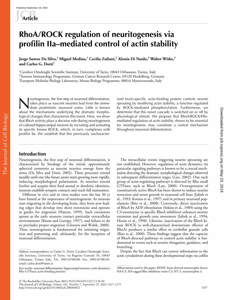
RhoA/ROCK regulation of neuritogenesis via profilin IIa-mediated control of actin stability
13
The Journal of Cell Biology The Rockefeller University Press, 0021-9525/2003/09/1267/13 $8.00 The Journal of Cell Biology, Volume 162, Number 7, September 29, 2003 1267–1279 http://www.jcb.org/cgi/doi/10.1083/jcb.200304021 JCB Article 1267 RhoA/ROCK regulation of neuritogenesis via profilin IIa–mediated control of actin stability Jorge Santos Da Silva, 1 Miguel Medina, 1 Cecilia Zuliani, 2 Alessia Di Nardo, 3 Walter Witke, 3 and Carlos G. Dotti 1 1 Cavalieri Ottolenghi Scientific Institute, University of Turin, 10043 Orbassano, Torino, Italy 2 Tumour Immunology Programme, German Cancer Research Centre, 69120 Heidelberg, Germany 3 European Moleular Biology Laboratory, Mouse Biology Programme, 00016 Monterotondo, Italy euritogenesis, the first step of neuronal differentiation, takes place as nascent neurites bud from the imme- diate postmitotic neuronal soma. Little is known about the mechanisms underlying the dramatic morpho- logical changes that characterize this event. Here, we show that RhoA activity plays a decisive role during neuritogenesis of cultured hippocampal neurons by recruiting and activating its specific kinase ROCK, which, in turn, complexes with profilin IIa. We establish that this previously uncharacter- N ized brain-specific actin-binding protein controls neurite sprouting by modifying actin stability, a function regulated by ROCK-mediated phosphorylation. Furthermore, we determine that this novel cascade is switched on or off by physiological stimuli. We propose that RhoA/ROCK/PIIa- mediated regulation of actin stability, shown to be essential for neuritogenesis, may constitute a central mechanism throughout neuronal differentiation. Introduction Neuritogenesis, the first step of neuronal differentiation, is characterized by breakage of the initial, approximately spherical symmetry as nascent neurites emerge from the soma (Da Silva and Dotti, 2002). These processes extend steadily until one (the future axon) starts growing more rapidly, inducing morphological polarization. As neurites extend further and acquire their final axonal or dendritic identities, neurons establish synaptic contacts and reach full maturation. Different in vivo and in vitro studies over the last 30 yr have hinted at the importance of neuritogenesis. As neurons start migrating in the developing brain, they form new lead- ing edges that develop into short extensions and operate as guides for migration (Hatten, 1999). Such extensions sprout as the early neurons contact particular extracellular environments (Baum and Garriga, 1997), and failure to do so precludes proper migration (Gleeson and Walsh, 2000). Thus, neuritogenesis is fundamental for initiating migra- tion and patterning and, ultimately, for the inception of neuronal differentiation. The intracellular events triggering neurite sprouting are not established. However, regulation of actin dynamics via particular signaling pathways is known to be a crucial mech- anism directing the dramatic morphological changes observed in subsequent differentiation stages (Luo, 2002). One such group of actin-regulating pathways is directed by Rho small GTPases, such as RhoA (Luo, 2000). Overexpression of constitutively active RhoA has been shown to induce neurite retraction and arrest growth in neuronal cell lines (Jalink et al., 1993; Kozma et al., 1997) and in primary neuronal pop- ulations (Bito et al., 2000). Conversely, direct inactivation of RhoA by ADP-ribosylation (Sekine et al., 1989) using the C3-exoenzyme (a specific RhoA inhibitor) enhances neurite extension and growth cone movement (Jalink et al., 1994; Hirose et al., 1998). Likewise, inactivation of the RhoA ki- nase ROCK (a well-characterized downstream effector of RhoA) produces a similar effect in cerebellar granule cells (Bito et al., 2000). These findings suggest that the capacity of RhoA-directed pathways to control actin stability is fun- damental to events such as neurite elongation, guidance, and branching. Despite the fact that RhoA can convey information to the actin cytoskeleton during these developmental steps via cofilin Address correspondence to Carlos G. Dotti, Cavalieri Ottolenghi Scien- tific Institute, University of Turin, via Regione Gonzole 10, 10043 Orbassano, Torino, Italy. Tel.: 390116708180. Fax.: 390116708149. email: [email protected] Key words: neuronal differentiation; hippocampal neurons; actin dynamics; Rho GTPases; actin-binding proteins Abbreviations used in this paper: BDNF, brain-derived neurotrophic factor; HA-C3, HA-tagged Rho inhibitory toxin C3; NT-3, neurotrophin 3. on May 23, 2014 jcb.rupress.org Downloaded from Published September 29, 2003
-
Upload
independent -
Category
Documents
-
view
1 -
download
0
Transcript of RhoA/ROCK regulation of neuritogenesis via profilin IIa-mediated control of actin stability
The
Jour
nal o
f Cel
l Bio
logy
The Rockefeller University Press, 0021-9525/2003/09/1267/13 $8.00The Journal of Cell Biology, Volume 162, Number 7, September 29, 2003 1267–1279http://www.jcb.org/cgi/doi/10.1083/jcb.200304021
JCB
Article
1267
RhoA/ROCK regulation of neuritogenesis via profilin IIa–mediated control of actin stability
Jorge Santos Da Silva,
1
Miguel Medina,
1
Cecilia Zuliani,
2
Alessia Di Nardo,
3
Walter Witke,
3
and Carlos G. Dotti
1
1
Cavalieri Ottolenghi Scientific Institute, University of Turin, 10043 Orbassano, Torino, Italy
2
Tumour Immunology Programme, German Cancer Research Centre, 69120 Heidelberg, Germany
3
European Moleular Biology Laboratory, Mouse Biology Programme, 00016 Monterotondo, Italy
euritogenesis, the first step of neuronal differentiation,takes place as nascent neurites bud from the imme-diate postmitotic neuronal soma. Little is known
about the mechanisms underlying the dramatic morpho-logical changes that characterize this event. Here, we showthat RhoA activity plays a decisive role during neuritogenesisof cultured hippocampal neurons by recruiting and activatingits specific kinase ROCK, which, in turn, complexes withprofilin IIa. We establish that this previously uncharacter-
N
ized brain-specific actin-binding protein controls neuritesprouting by modifying actin stability, a function regulatedby ROCK-mediated phosphorylation. Furthermore, wedetermine that this novel cascade is switched on or off byphysiological stimuli. We propose that RhoA/ROCK/PIIa-mediated regulation of actin stability, shown to be essentialfor neuritogenesis, may constitute a central mechanismthroughout neuronal differentiation.
Introduction
Neuritogenesis, the first step of neuronal differentiation, ischaracterized by breakage of the initial, approximatelyspherical symmetry as nascent neurites emerge from thesoma (Da Silva and Dotti, 2002). These processes extendsteadily until one (the future axon) starts growing more rapidly,inducing morphological polarization. As neurites extendfurther and acquire their final axonal or dendritic identities,neurons establish synaptic contacts and reach full maturation.
Different in vivo and in vitro studies over the last 30 yrhave hinted at the importance of neuritogenesis. As neuronsstart migrating in the developing brain, they form new lead-ing edges that develop into short extensions and operateas guides for migration (Hatten, 1999). Such extensionssprout as the early neurons contact particular extracellularenvironments (Baum and Garriga, 1997), and failure to doso precludes proper migration (Gleeson and Walsh, 2000).Thus, neuritogenesis is fundamental for initiating migra-tion and patterning and, ultimately, for the inception ofneuronal differentiation.
The intracellular events triggering neurite sprouting arenot established. However, regulation of actin dynamics viaparticular signaling pathways is known to be a crucial mech-anism directing the dramatic morphological changes observedin subsequent differentiation stages (Luo, 2002). One suchgroup of actin-regulating pathways is directed by Rho smallGTPases, such as RhoA (Luo, 2000). Overexpression ofconstitutively active RhoA has been shown to induce neuriteretraction and arrest growth in neuronal cell lines (Jalink etal., 1993; Kozma et al., 1997) and in primary neuronal pop-ulations (Bito et al., 2000). Conversely, direct inactivationof RhoA by ADP-ribosylation (Sekine et al., 1989) using theC3-exoenzyme (a specific RhoA inhibitor) enhances neuriteextension and growth cone movement (Jalink et al., 1994;Hirose et al., 1998). Likewise, inactivation of the RhoA ki-nase ROCK (a well-characterized downstream effector ofRhoA) produces a similar effect in cerebellar granule cells(Bito et al., 2000). These findings suggest that the capacityof RhoA-directed pathways to control actin stability is fun-damental to events such as neurite elongation, guidance, andbranching.
Despite the fact that RhoA can convey information to theactin cytoskeleton during these developmental steps via cofilin
Address correspondence to Carlos G. Dotti, Cavalieri Ottolenghi Scien-tific Institute, University of Turin, via Regione Gonzole 10, 10043Orbassano, Torino, Italy. Tel.: 390116708180. Fax.: 390116708149.email: [email protected]
Key words: neuronal differentiation; hippocampal neurons; actin dynamics;Rho GTPases; actin-binding proteins
Abbreviations used in this paper: BDNF, brain-derived neurotrophic factor;
HA-C3, HA-tagged Rho inhibitory toxin C3; NT-3, neurotrophin 3.
on May 23, 2014
jcb.rupress.orgD
ownloaded from
Published September 29, 2003
The
Jour
nal o
f Cel
l Bio
logy
1268 The Journal of Cell Biology
|
Volume 162, Number 7, 2003
(Bamburg, 1999), other actin-binding proteins, such asthe profilins, may be involved as well. Profilins have beenimplicated in the maintenance of cytoskeletal integrity ina variety of organisms such as
Dictyostelium
discoideum
(Haugwitz et al., 1994), yeast (Haarer et al., 1990), and
Drosophila
melanogaster
(Verheyen and Cooley, 1994). Inthe fly, the sole form of profilin (
chickadee
) has beenshown to play a role in motor neuron axon extension as
chickadee
mutations arrest growth (Wills et al., 1999). Inmammals, there are different profilin isoforms that, whilesharing similar biochemical properties, have diverse tissuedistributions. Profilin I (PI) is ubiquitous, whereas bothprofilin II isoforms (PIIa and PIIb) are largely restrictedto the brain (Witke et al., 2001). A third profilin has beendescribed recently and its expression detected exclusivelyin kidney and testis (Hu et al., 2001). Apart from the factthat PIIa is a brain-specific profilin isoform, the only ac-tin-binding protein specifically interacting with the RhoAdownstream kinase ROCK in brain extracts is PIIa (Witkeet al., 1998). This led us to hypothesize that PIIa may be akey player in neuronal-specific events directed by theRhoA–ROCK pathway.
In this work, we show that RhoA and its specific down-stream effector ROCK are essential during neuritogenesis inmammalian hippocampal neurons by modulating actin sta-bility. Consistent with the aforementioned hypothesis, suchmodulation is dependent on the downstream effector profi-lin IIa, a protein of previously undetermined function. Fur-thermore, we show that this early neuronal program is regu-lated by different physiological stimuli. These findings reveala central regulatory mechanism directing the, as yet, unchar-acterized process of initial neuronal symmetry breakage.
Results
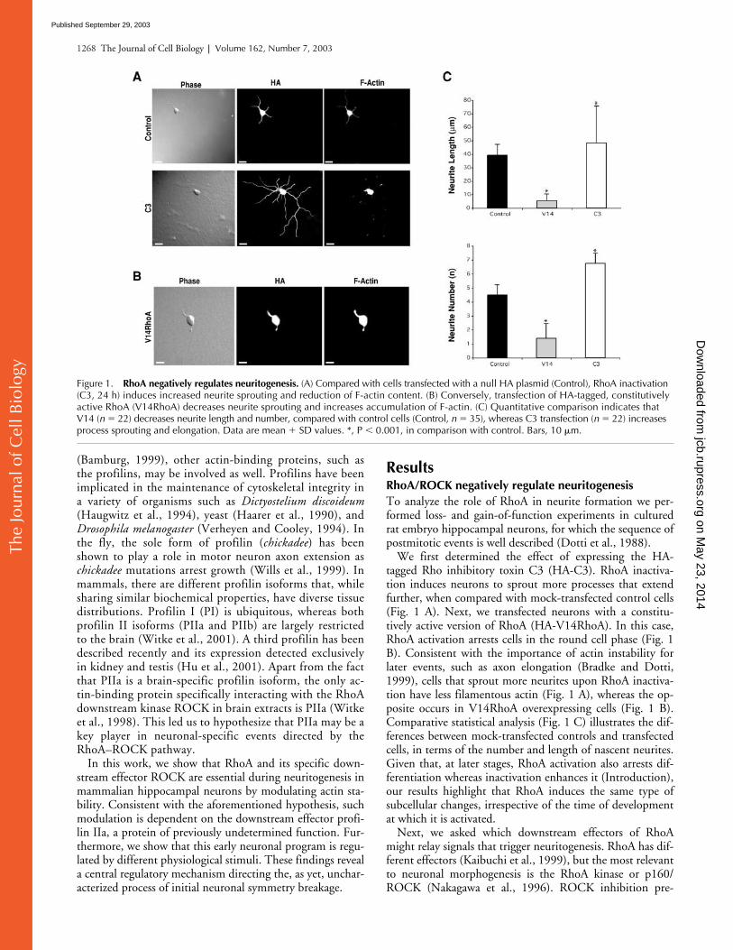
RhoA/ROCK negatively regulate neuritogenesis
To analyze the role of RhoA in neurite formation we per-formed loss- and gain-of-function experiments in culturedrat embryo hippocampal neurons, for which the sequence ofpostmitotic events is well described (Dotti et al., 1988).
We first determined the effect of expressing the HA-tagged Rho inhibitory toxin C3 (HA-C3). RhoA inactiva-tion induces neurons to sprout more processes that extendfurther, when compared with mock-transfected control cells(Fig. 1 A). Next, we transfected neurons with a constitu-tively active version of RhoA (HA-V14RhoA). In this case,RhoA activation arrests cells in the round cell phase (Fig. 1B). Consistent with the importance of actin instability forlater events, such as axon elongation (Bradke and Dotti,1999), cells that sprout more neurites upon RhoA inactiva-tion have less filamentous actin (Fig. 1 A), whereas the op-posite occurs in V14RhoA overexpressing cells (Fig. 1 B).Comparative statistical analysis (Fig. 1 C) illustrates the dif-ferences between mock-transfected controls and transfectedcells, in terms of the number and length of nascent neurites.Given that, at later stages, RhoA activation also arrests dif-ferentiation whereas inactivation enhances it (Introduction),our results highlight that RhoA induces the same type ofsubcellular changes, irrespective of the time of developmentat which it is activated.
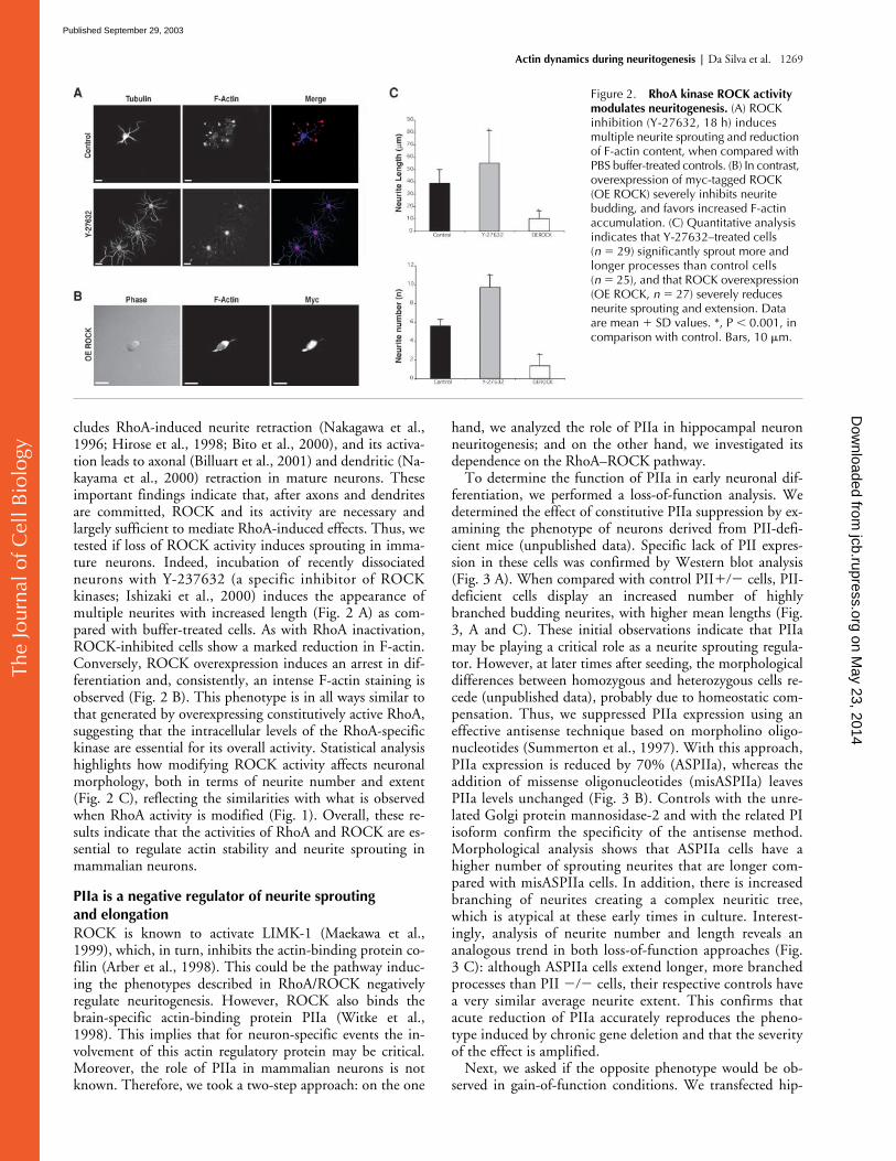
Next, we asked which downstream effectors of RhoAmight relay signals that trigger neuritogenesis. RhoA has dif-ferent effectors (Kaibuchi et al., 1999), but the most relevantto neuronal morphogenesis is the RhoA kinase or p160/ROCK (Nakagawa et al., 1996). ROCK inhibition pre-
Figure 1. RhoA negatively regulates neuritogenesis. (A) Compared with cells transfected with a null HA plasmid (Control), RhoA inactivation (C3, 24 h) induces increased neurite sprouting and reduction of F-actin content. (B) Conversely, transfection of HA-tagged, constitutively active RhoA (V14RhoA) decreases neurite sprouting and increases accumulation of F-actin. (C) Quantitative comparison indicates that V14 (n � 22) decreases neurite length and number, compared with control cells (Control, n � 35), whereas C3 transfection (n � 22) increases process sprouting and elongation. Data are mean � SD values. *, P � 0.001, in comparison with control. Bars, 10 �m.
on May 23, 2014
jcb.rupress.orgD
ownloaded from
Published September 29, 2003
The
Jour
nal o
f Cel
l Bio
logy
Actin dynamics during neuritogenesis |
Da Silva et al. 1269
cludes RhoA-induced neurite retraction (Nakagawa et al.,1996; Hirose et al., 1998; Bito et al., 2000), and its activa-tion leads to axonal (Billuart et al., 2001) and dendritic (Na-kayama et al., 2000) retraction in mature neurons. Theseimportant findings indicate that, after axons and dendritesare committed, ROCK and its activity are necessary andlargely sufficient to mediate RhoA-induced effects. Thus, wetested if loss of ROCK activity induces sprouting in imma-ture neurons. Indeed, incubation of recently dissociatedneurons with Y-237632 (a specific inhibitor of ROCKkinases; Ishizaki et al., 2000) induces the appearance ofmultiple neurites with increased length (Fig. 2 A) as com-pared with buffer-treated cells. As with RhoA inactivation,ROCK-inhibited cells show a marked reduction in F-actin.Conversely, ROCK overexpression induces an arrest in dif-ferentiation and, consistently, an intense F-actin staining isobserved (Fig. 2 B). This phenotype is in all ways similar tothat generated by overexpressing constitutively active RhoA,suggesting that the intracellular levels of the RhoA-specifickinase are essential for its overall activity. Statistical analysishighlights how modifying ROCK activity affects neuronalmorphology, both in terms of neurite number and extent(Fig. 2 C), reflecting the similarities with what is observedwhen RhoA activity is modified (Fig. 1). Overall, these re-sults indicate that the activities of RhoA and ROCK are es-sential to regulate actin stability and neurite sprouting inmammalian neurons.
PIIa is a negative regulator of neurite sprouting and elongation
ROCK is known to activate LIMK-1 (Maekawa et al.,1999), which, in turn, inhibits the actin-binding protein co-filin (Arber et al., 1998). This could be the pathway induc-ing the phenotypes described in RhoA/ROCK negativelyregulate neuritogenesis. However, ROCK also binds thebrain-specific actin-binding protein PIIa (Witke et al.,1998). This implies that for neuron-specific events the in-volvement of this actin regulatory protein may be critical.Moreover, the role of PIIa in mammalian neurons is notknown. Therefore, we took a two-step approach: on the one
hand, we analyzed the role of PIIa in hippocampal neuronneuritogenesis; and on the other hand, we investigated itsdependence on the RhoA–ROCK pathway.
To determine the function of PIIa in early neuronal dif-ferentiation, we performed a loss-of-function analysis. Wedetermined the effect of constitutive PIIa suppression by ex-amining the phenotype of neurons derived from PII-defi-cient mice (unpublished data). Specific lack of PII expres-sion in these cells was confirmed by Western blot analysis(Fig. 3 A). When compared with control PII
���
cells, PII-deficient cells display an increased number of highlybranched budding neurites, with higher mean lengths (Fig.3, A and C). These initial observations indicate that PIIamay be playing a critical role as a neurite sprouting regula-tor. However, at later times after seeding, the morphologicaldifferences between homozygous and heterozygous cells re-cede (unpublished data), probably due to homeostatic com-pensation. Thus, we suppressed PIIa expression using aneffective antisense technique based on morpholino oligo-nucleotides (Summerton et al., 1997). With this approach,PIIa expression is reduced by 70% (ASPIIa), whereas theaddition of missense oligonucleotides (misASPIIa) leavesPIIa levels unchanged (Fig. 3 B). Controls with the unre-lated Golgi protein mannosidase-2 and with the related PIisoform confirm the specificity of the antisense method.Morphological analysis shows that ASPIIa cells have ahigher number of sprouting neurites that are longer com-pared with misASPIIa cells. In addition, there is increasedbranching of neurites creating a complex neuritic tree,which is atypical at these early times in culture. Interest-ingly, analysis of neurite number and length reveals ananalogous trend in both loss-of-function approaches (Fig.3 C): although ASPIIa cells extend longer, more branchedprocesses than PII
���
cells, their respective controls havea very similar average neurite extent. This confirms thatacute reduction of PIIa accurately reproduces the pheno-type induced by chronic gene deletion and that the severityof the effect is amplified.
Next, we asked if the opposite phenotype would be ob-served in gain-of-function conditions. We transfected hip-
Figure 2. RhoA kinase ROCK activity modulates neuritogenesis. (A) ROCK inhibition (Y-27632, 18 h) induces multiple neurite sprouting and reduction of F-actin content, when compared with PBS buffer-treated controls. (B) In contrast, overexpression of myc-tagged ROCK (OE ROCK) severely inhibits neurite budding, and favors increased F-actin accumulation. (C) Quantitative analysis indicates that Y-27632–treated cells (n � 29) significantly sprout more and longer processes than control cells (n � 25), and that ROCK overexpression (OE ROCK, n � 27) severely reduces neurite sprouting and extension. Data are mean � SD values. *, P � 0.001, in comparison with control. Bars, 10 �m.
on May 23, 2014
jcb.rupress.orgD
ownloaded from
Published September 29, 2003
The
Jour
nal o
f Cel
l Bio
logy
1270 The Journal of Cell Biology
|
Volume 162, Number 7, 2003
Figure 3.
PIIa is a negative regulator of neurite sprouting.
(A) Western blot of heterozygous (
���
) and PIIa-null mice (
���
) brain lysates probed for actin, PIIa, and PI showing that only PIIa expression is affected in PII
���
mice. Compared with PII
���
neurons, at 24 h, PII
���
cells exhibit multiple sprouting neurites with increased lengths. (B) PIIa-morpholino antisense oligonucleotides (ASPIIa) reduce PIIa levels by 70% in rat hippocampal neurons in culture, whereas PIIa-missense oligonucleotides (misASPIIa) do not alter PIIa expression, compared with untreated control cells (C). The unrelated Golgi protein mannosidase-2 (Mann2) and the closely related PI are unaffected by antisense treatment. At 24 h, misASPIIa neurons present a typical morphology, whereas ASPIIa cells display increased number of sprouting neurites and extreme elongation of most processes. (C) Quantitative analysis shows that mouse PII
���
and rat misASPIIa neurons display similar neurite number and length averages. PII
���
cells have a clear increase in the number, branching (up to quaternary, 4ry), and length of emergent processes (*, P
�
0.001, PII
���
in comparison with PII
���
; PII
���
,
n
�
23; PII
���
,
n
�
21). Similarly, but more strikingly, ASPIIa rat hippocampal neurons exhibit increased neurite number, branching (up to quintenary, 5ry), and length when compared with misASPIIa cells
on May 23, 2014
jcb.rupress.orgD
ownloaded from
Published September 29, 2003
The
Jour
nal o
f Cel
l Bio
logy
Actin dynamics during neuritogenesis |
Da Silva et al. 1271
pocampal neurons with a PIIa-GFP fusion construct, result-ing in the detectable expression of the tagged protein (Fig. 3D). PIIa-GFP expression produces cells with greatly reducedneurite number and length, in contrast with cells transfectedwith the equivalent GFP-null vector (Fig. 3, D and E). Thesame phenotype was observed with a construct containing asmaller epitope tag derived from Sendai virus and with anuntagged PIIa vector (unpublished data). This ensures thatthe GFP moiety did not affect PIIa activity as expected be-cause this construct has the ability to bind actin, without in-fluence of the tag on the dynamics of polymerization (DiNardo et al., 2000). Differences between the loss- and gain-of-function phenotypes are illustrated by camera lucida mor-phological reconstructions (see Materials and methods; Fig.3 F). Our findings indicate that the intracellular availabilityof this 1:1 actin monomer-binding protein is essential forcontrolled neurite sprouting and suggest a novel role of PIIaas a key regulator of neuritogenesis.
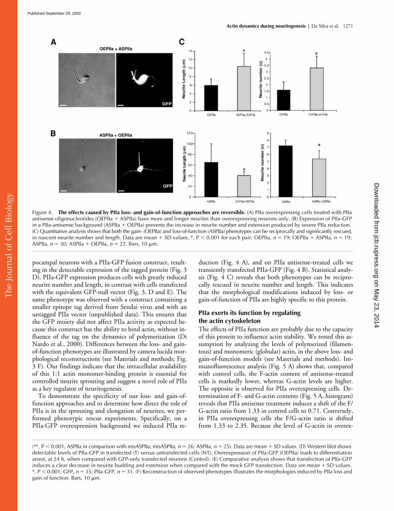
To demonstrate the specificity of our loss- and gain-of-function approaches and to determine how direct the role ofPIIa is in the sprouting and elongation of neurites, we per-formed phenotypic rescue experiments. Specifically, on aPIIa-GFP overexpression background we induced PIIa re-
duction (Fig. 4 A), and on PIIa antisense-treated cells wetransiently transfected PIIa-GFP (Fig. 4 B). Statistical analy-sis (Fig. 4 C) reveals that both phenotypes can be recipro-cally rescued in neurite number and length. This indicatesthat the morphological modifications induced by loss- orgain-of-function of PIIa are highly specific to this protein.
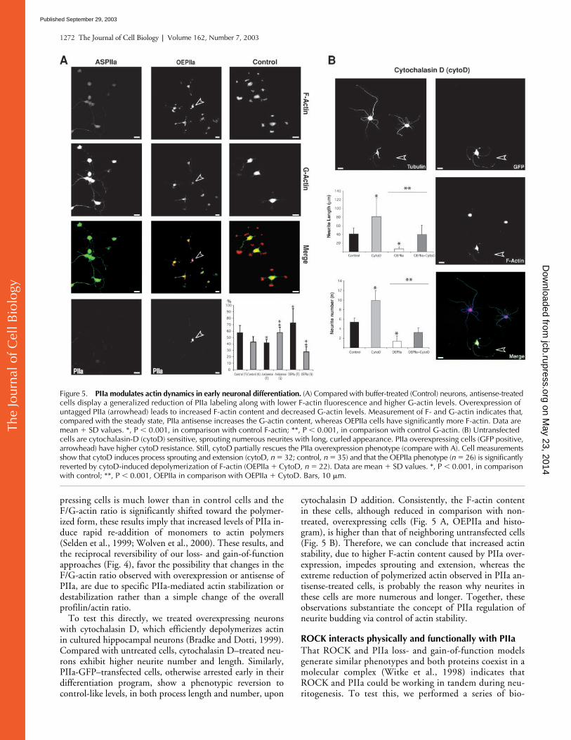
PIIa exerts its function by regulating the actin cytoskeleton
The effects of PIIa function are probably due to the capacityof this protein to influence actin stability. We tested this as-sumption by analyzing the levels of polymerized (filamen-tous) and monomeric (globular) actin, in the above loss- andgain-of-function models (see Materials and methods). Im-munofluorescence analysis (Fig. 5 A) shows that, comparedwith control cells, the F-actin content of antisense-treatedcells is markedly lower, whereas G-actin levels are higher.The opposite is observed for PIIa overexpressing cells. De-termination of F- and G-actin contents (Fig. 5 A, histogram)reveals that PIIa antisense treatment induces a shift of the F/G-actin ratio from 1.33 in control cells to 0.71. Conversely,in PIIa overexpressing cells the F/G-actin ratio is shiftedfrom 1.33 to 2.35. Because the level of G-actin in overex-
(**, P
�
0.001, ASPIIa in comparison with misASPIIa; misASPIIa,
n
�
26; ASPIIa,
n
�
25). Data are mean
�
SD values. (D) Western blot shows detectable levels of PIIa-GFP in transfected (T) versus untransfected cells (NT). Overexpression of PIIa-GFP (OEPIIa) leads to differentiation arrest, at 24 h, when compared with GFP-only transfected neurons (Control). (E) Comparative analysis shows that transfection of PIIa-GFP induces a clear decrease in neurite budding and extension when compared with the mock GFP transfection. Data are mean
�
SD values. *, P
�
0.001;
GFP,
n
�
35; PIIa-GFP,
n
�
31. (F) Reconstruction of observed phenotypes illustrates the morphologies induced by PIIa loss and gain of function. Bars, 10
�
m.
Figure 4. The effects caused by PIIa loss- and gain-of-function approaches are reversible. (A) PIIa overexpressing cells treated with PIIa antisense oligonucleotides (OEPIIa � ASPIIa) have more and longer neurites than overexpressing neurons only. (B) Expression of PIIa-GFP in a PIIa-antisense background (ASPIIa � OEPIIa) prevents the increase in neurite number and extension produced by severe PIIa reduction. (C) Quantitative analysis shows that both the gain- (OEPIIa) and loss-of-function (ASPIIa) phenotypes can be reciprocally and significantly rescued, in nascent neurite number and length. Data are mean � SD values. *, P � 0.001 for each pair; OEPIIa, n � 19; OEPIIa � ASPIIa, n � 19; ASPIIa, n � 30; ASPIIa � OEPIIa, n � 22. Bars, 10 �m.
on May 23, 2014
jcb.rupress.orgD
ownloaded from
Published September 29, 2003
The
Jour
nal o
f Cel
l Bio
logy
1272 The Journal of Cell Biology
|
Volume 162, Number 7, 2003
pressing cells is much lower than in control cells and theF/G-actin ratio is significantly shifted toward the polymer-ized form, these results imply that increased levels of PIIa in-duce rapid re-addition of monomers to actin polymers(Selden et al., 1999; Wolven et al., 2000). These results, andthe reciprocal reversibility of our loss- and gain-of-functionapproaches (Fig. 4), favor the possibility that changes in theF/G-actin ratio observed with overexpression or antisense ofPIIa, are due to specific PIIa-mediated actin stabilization ordestabilization rather than a simple change of the overallprofilin/actin ratio.
To test this directly, we treated overexpressing neuronswith cytochalasin D, which efficiently depolymerizes actinin cultured hippocampal neurons (Bradke and Dotti, 1999).Compared with untreated cells, cytochalasin D–treated neu-rons exhibit higher neurite number and length. Similarly,PIIa-GFP–transfected cells, otherwise arrested early in theirdifferentiation program, show a phenotypic reversion tocontrol-like levels, in both process length and number, upon
cytochalasin D addition. Consistently, the F-actin contentin these cells, although reduced in comparison with non-treated, overexpressing cells (Fig. 5 A, OEPIIa and histo-gram), is higher than that of neighboring untransfected cells(Fig. 5 B). Therefore, we can conclude that increased actinstability, due to higher F-actin content caused by PIIa over-expression, impedes sprouting and extension, whereas theextreme reduction of polymerized actin observed in PIIa an-tisense-treated cells, is probably the reason why neurites inthese cells are more numerous and longer. Together, theseobservations substantiate the concept of PIIa regulation ofneurite budding via control of actin stability.
ROCK interacts physically and functionally with PIIa
That ROCK and PIIa loss- and gain-of-function modelsgenerate similar phenotypes and both proteins coexist in amolecular complex (Witke et al., 1998) indicates thatROCK and PIIa could be working in tandem during neu-ritogenesis. To test this, we performed a series of bio-
Figure 5. PIIa modulates actin dynamics in early neuronal differentiation. (A) Compared with buffer-treated (Control) neurons, antisense-treated cells display a generalized reduction of PIIa labeling along with lower F-actin fluorescence and higher G-actin levels. Overexpression of untagged PIIa (arrowhead) leads to increased F-actin content and decreased G-actin levels. Measurement of F- and G-actin indicates that, compared with the steady state, PIIa antisense increases the G-actin content, whereas OEPIIa cells have significantly more F-actin. Data are mean � SD values. *, P � 0.001, in comparison with control F-actin; **, P � 0.001, in comparison with control G-actin. (B) Untransfected cells are cytochalasin-D (cytoD) sensitive, sprouting numerous neurites with long, curled appearance. PIIa overexpressing cells (GFP positive, arrowhead) have higher cytoD resistance. Still, cytoD partially rescues the PIIa overexpression phenotype (compare with A). Cell measurements show that cytoD induces process sprouting and extension (cytoD, n � 32; control, n � 35) and that the OEPIIa phenotype (n � 26) is significantly reverted by cytoD-induced depolymerization of F-actin (OEPIIa � CytoD, n � 22). Data are mean � SD values. *, P � 0.001, in comparison with control; **, P � 0.001, OEPIIa in comparison with OEPIIa � CytoD. Bars, 10 �m.
on May 23, 2014
jcb.rupress.orgD
ownloaded from
Published September 29, 2003

The
Jour
nal o
f Cel
l Bio
logy
Actin dynamics during neuritogenesis |
Da Silva et al. 1273
chemical and functional experiments. First, we tested ifboth molecules interact physically. Indeed, ROCK andPIIa interact as observed by reciprocal co-immunoprecipi-tations (Fig. 6 A). Second, we determined if the intrinsickinase activity of ROCK is important for complex forma-tion. Extracts treated with Y-27632 do not display anychange in the efficiency of PIIa immunoprecipitation byROCK (Fig. 6 B), indicating that the kinase activity is not
essential for PIIa binding. Finally, we assessed if the intra-cellular levels of ROCK influence complex formation.This is the case as ROCK overexpression increases the effi-ciency of PIIa recruitment (Fig. 6 B). Noticeably, ROCKdoes not immunoprecipitate profilin I (unpublished data),indicating that, at least in the very early steps of neuronaldifferentiation, the ROCK pathway is selective for thebrain-specific form of profilin.
Figure 6. ROCK physically and functionally interacts with PIIa. (A) Brain cell homogenates immunoprecipitated with anti-PIIa antibody followed by blotting with anti-ROCK antibody (top) or immunoprecipitated with anti-ROCK antibody followed by blotting with anti-PIIa antibody (bottom). Both ROCK and PIIa are reciprocally immunoprecipitated. No signal is detected when protein G beads are incubated without antibodies (B). (B) Western blotting with the PIIa antibody of untransfected cells treated with the ROCK inhibitor (Y-27632, 6 h) or buffer-treated (control) cells, reveals that the amount of immunoprecipitated PIIa does not depend on the phosphorylation activity of ROCK (middle, PIIa, IP). Western blotting with the PIIa antibody of transfected (myc-ROCK) and un-transfected (control) cells, reveals that the amount of immunoprecipitated PIIa is higher in cells with higher levels of ROCK (middle, PIIa, IP). ROCK expression is detected with a myc-specific antibody (top, myc, IP). No PIIa signal is detected when extracts are incubated with beads without ROCK antibody (middle, PIIa, IP). To control for the presence and levels of PIIa, preimmunoprecipitation lysates were blotted with PIIa antibody (bottom, PIIa, lysate). (C) Western blots of 2D-SDS-PAGE samples from neuronal extracts blotted with anti-PIIa antibody. PIIa is present in two clearly distinguishable spots (control). The more negative form (� to � labels the isoelectrical focusing direction) corresponds to a more phosphorylated form (AP-sensitive, histogram). Y-27632 (6 h) increases the percentage of dephosphorylated PIIa versus control (43% increase, histogram), whereas the more phosphorylated form is lower than in controls (histogram: only �% for the more dephosphorylated form is shown; data are mean � SD values; n � 3; P � 0.001 for AP and Y-27632 in comparison with control; 100% is the sum of both forms of PIIa). (D) Inhibition of ROCK activity (18 h) increases neurite sprouting (Y-27632 reconstruction and unmarked cells). This phenotype is significantly prevented by PIIa overexpression (OEPIIa � Y-27632 reconstruction and GFP-positive cells, arrowheads) and accompanied by an increase of the F-actin content. (E) ROCK overexpression induces a dramatic arrest of differentiation (Fig. 2 B and OE ROCK reconstruction). PIIa antisense on a ROCK overexpression background results in partial reversion of the ROCK phenotype, with longer neurites and reduced F-actin (arrowhead and OE ROCK � ASPIIa reconstruction). (F) Quantitative analysis indicates that overexpression of PIIa (OEPIIa, n � 21) causes an arrest in neurite budding and elongation. Inhibition of ROCK, in this experimental background (OEPIIa � Y-27632, n � 22), leads to a very significant recovery with neurites reaching control-like number and length, but not to the levels observed for ROCK inhibition alone (compare with Fig. 2 C). Overexpression of ROCK (OE ROCK, n � 27) inhibits neurite sprouting and extension. PIIa antisense in this case (OE ROCK � ASPIIa, n � 26) results in a partial phenotype rescue, as processes elongate further, but cells do not sprout more neurites. Data are mean � SD values. *, P � 0.001, OEPIIa � Y-27632 in comparison with OEPIIa; **, P � 0.001, OEROCK � ASPIIa in comparison with OEROCK; , P � 0.05, OEROCK � ASPIIa in comparison with OEROCK. Bars, 10 �m.
on May 23, 2014
jcb.rupress.orgD
ownloaded from
Published September 29, 2003
The
Jour
nal o
f Cel
l Bio
logy
1274 The Journal of Cell Biology | Volume 162, Number 7, 2003
These results imply that ROCK interacts with PIIa in astoichiometric fashion and suggest that its kinase activity isnot essential for recruitment efficiency, though it might un-derlie the mechanism by which it regulates PIIa. To addressthis, we analyzed neuronal extracts in two-dimensional gels.In control conditions, PIIa is present in two distinguishableforms (Fig. 6 C, Control). One of these forms is more phos-phorylated than the other because it is more negative in theisoelectric focusing direction and is reduced when extractsare previously treated with the phosphorylation inhibitor al-kaline phosphatase (Fig. 6 C, AP and histogram). Signifi-cantly, extracts of cells treated with Y-27632 present an in-crease in the more dephosphorylated form of PIIa (Fig. 6 C,Y-27632 and histogram), similar to that observed with alka-line phosphatase (43%). These results indicate that ROCKregulates PIIa via its serine/threonine kinase activity.
To examine the functional relevance of these findings weasked whether the increased budding and extension of neu-rites, observed when ROCK is inactivated (Fig. 2 A), couldbe prevented by excess PIIa, which increases the concentra-tion of ROCK–PIIa complexes. In fact, when we overex-pressed PIIa-GFP and inhibited ROCK activity, cells wereless differentiated than untransfected, ROCK-inhibited cells(Fig. 6 D and Fig. 2 A). At the same time, these cells weresignificantly more differentiated than PIIa overexpressingcells alone (Fig. 6 D and Fig. 3 D) and presented a reductionin F-actin content (Fig. 5 A), indicating that these neuronswere sensitive to the absence of ROCK activity. Thus, thekinase activity of ROCK is essential to regulate the neurito-genic arrest effects of PIIa. To further substantiate this func-tional link, we tested whether reducing the pool of PIIacould, to any extent, rescue the effects of ROCK overexpres-sion. Indeed, reduction of PIIa levels in ROCK overexpress-ing cells resulted in a milder ROCK phenotype (Fig. 6 E).No reversion on the ROCK phenotype was observed whenPIIa missense oligonucleotides were added (unpublisheddata). Comparison of average neurite number and length be-tween the aforementioned phenotypes (Fig. 6 F) furthersubstantiates our conclusion that ROCK and PIIa interactfunctionally. Altogether, these results establish that PIIa is amajor downstream target of ROCK during the initial stepsof neuronal differentiation.
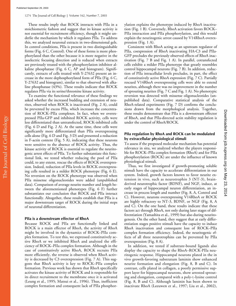
PIIa is a downstream effector of RhoABecause ROCK and PIIa are functionally linked andROCK is a main effector of RhoA, the activity of RhoAmight be involved in the dynamics of ROCK–PIIa com-plex formation. To test this, we expressed constitutively ac-tive RhoA or we inhibited RhoA and analyzed the effi-ciency of ROCK–PIIa complex formation. Although in thecase of constitutively active RhoA, ROCK recruits PIIamore efficiently, the reverse is observed when RhoA activ-ity is decreased by C3 overexpression (Fig. 7 A). This sug-gests that RhoA activity is key to ROCK–PIIa complexformation. Previous work has shown that RhoA specificallyactivates the kinase activity of ROCK and is responsible forits direct recruitment to the membrane once it is activated(Leung et al., 1995; Matsui et al., 1996). Thus, inefficientcomplex formation and consequent lack of PIIa phosphor-
ylation explains the phenotype induced by RhoA inactiva-tion (Fig. 1 B). Conversely, RhoA activation favors ROCK–PIIa interaction and PIIa phosphorylation, and this wouldexplain the neuritogenic arrest caused by V14RhoA overex-pression (Fig. 1 A).
Consistent with RhoA acting as an upstream regulator ofPIIa, coexpression of RhoA inactivating HA-C3 and PIIa-GFP precludes the previously observed effects of RhoA inac-tivation (Fig. 7 B and Fig. 1 A). In parallel, cotransfectedcells exhibit a milder PIIa phenotype that greatly resemblescontrol hippocampal neurons (Fig. 7 B). In addition, reduc-tion of PIIa intracellular levels precludes, in part, the effectof constitutively active RhoA expression (Fig. 7 C). Partiallyrescued V14RhoA overexpressing cells were able to extendneurites, although there was no improvement in the numberof sprouting neurites (Fig. 7 C and Fig. 1 A). No phenotypicreversion was detected with missense oligonucleotides (un-published data). Comparative statistical analysis of theRhoA-related experiments (Fig. 7 D) confirms the conclu-sions drawn from the morphological analysis. Together,these results demonstrate that PIIa is a downstream effectorof RhoA, and that PIIa-directed actin stability regulation isunder the control of RhoA/ROCK.
PIIa regulation by RhoA and ROCK can be modulated by extracellular physiological stimuliTo assess if the proposed molecular mechanism has potentialrelevance in situ, we analyzed whether the players responsi-ble for ROCK–PIIa complex formation (RhoA) and for PIIaphosphorylation (ROCK) are under the influence of knownphysiological stimuli.
Thus, we first investigated if growth-promoting solublestimuli have the capacity to accelerate differentiation in oursystem. Indeed, growth factors known to favor neurite ex-tension in vivo, such as neurotrophin 3 (NT-3), brain-derived neurotrophic factor (BDNF), and NGF, induce, atearly stages of hippocampal neuron differentiation, an in-crease in process length and number (Fig. 8, A, Control andC). However, neurons overexpressing V14RhoA or ROCKare highly refractory to NT-3, BDNF, or NGF (Fig. 8, Aand C). On the one hand, these results indicate that thesefactors act through RhoA, not only during later stages of dif-ferentiation (Yamashita et al., 1999) but also during neurito-genesis. On the other hand, they suggest that at early differ-entiation stages positive stimuli have the capacity to induceRhoA inactivation and consequent loss of ROCK–PIIacomplex formation efficiency. Indeed, the neuritogenic ef-fects of all three neurotrophins can be prevented by PIIaoverexpression (Fig. 8 A).
In addition, we tested if substrate-bound ligands alsodisplay the capacity to shape the RhoA–ROCK-PIIa neu-ritogenic response. Hippocampal neurons plated in the invivo growth-favoring substratum laminin show enhancedneurite formation and elongation (Fig. 8, B and C). Incontrast, cells plated in collagen, a poorly permissive sup-port layer for hippocampal neurons, show arrested sprout-ing and elongation, compared with a poly-L-lysine control(Fig. 8, B and C). Although laminin has been shown toinactivate RhoA (Leeuwen et al., 1997; Liu et al., 2002),
on May 23, 2014
jcb.rupress.orgD
ownloaded from
Published September 29, 2003
The
Jour
nal o
f Cel
l Bio
logy
Actin dynamics during neuritogenesis | Da Silva et al. 1275
collagen has been found to activate RhoA in other celltypes (Schoenwaelder et al., 2002). Supporting our find-ings, recently plated neurons with excess V14RhoA orROCK are highly refractory to laminin (Fig. 8, B and C).On the other hand, the collagen effect on sprouting wasless obvious in cells expressing C3 or in cells treated withY-27632 (Fig. 8, B and C). Furthermore, PIIa overex-pressing cells prevent the positive effects of laminin,whereas reduction of PIIa levels in collagen-plated cellspartly reverts the arrest stimulus (Fig. 8, B and C). Thislast series of results show that extracellular stimuli can in-duce modifications in the activity of the RhoA–ROCKpathway by allowing an adaptative regulation of PIIafunction and point out the physiological relevance of thefindings described here.
DiscussionIn this work, we show that the activity of RhoA and its down-stream effector ROCK control initial sprouting: activationleads to neuritogenic arrest, whereas inactivation acceleratesneuritogenesis. RhoA and ROCK have been described as neg-ative regulators of neuritic growth in later differentiationstages (Introduction). Our results are consistent with such arole, and favor the emerging view that similar molecularmechanisms can be used throughout the entire neuronal mor-phogenesis program (Da Silva and Dotti, 2002; Luo, 2002).
The observed RhoA and ROCK activity phenotypes areaccompanied by changes in F-actin content, suggesting thatdownstream actin-related proteins are implicated in neurito-genesis. We demonstrate that the brain-specific protein PIIa
Figure 7. PIIa is a downstream effector of RhoA. (A) ROCK immunoprecipitation of cell extracts overexpressing constitutively active RhoA (V14) or inactive Rho (C3, 24 h), followed by Western blotting with PIIa antibody. V14RhoA increases the amount of PIIa immunoprecipitated by ROCK (middle, PIIa, IP), whereas C3 reduces the amount of PIIa immunoprecipitated by ROCK (middle, PIIa, IP). V14RhoA and C3 expression is detected with an HA-specific antibody (top, HA, IP). No signal is detected (B) when extracts are precipitated without ROCK antibody (middle, PIIa, IP). To control for the presence and levels of PIIa, preimmunoprecipitation lysates were blotted with PIIa antibody (bottom, PIIa, lysate). (B) C3 expression (24 h) enhances neurite sprouting (Fig. 1 A and C3 reconstruction). Co-transfection with PIIa-GFP (C3 � OEPIIa, 24 h) prevents the increased neurite sprouting and elongation characteristic of C3 expression (C3 � OEPIIa reconstruction) and favors actin polymerization at the distal fractions of budding neurites (arrowhead). Conversely, the overall F-actin content is reduced in comparison with PIIa overexpression alone (compare with Fig. 5 A). (C) Constitutively active RhoA induces a differentiation-arrested phenotype (Fig. 1 A and V14RhoA reconstruction). Reduction of PIIa levels partially reverts this arrest (V14RhoA � ASPIIa reconstruction); these cells show a less polymerized actin cytoskeleton, namely along elongated processes (arrowhead) but do not sprout more neurites (compare with Fig. 1 B). (D) Statistical analysis shows that PIIa antisense (V14 � ASPIIa, n � 28) incompletely rescues the V14-induced (V14, n � 22) phenotype as neurons extend longer neurites, but there is no detectable change in neurite number. On the contrary, compared with single HA-C3 transfection (C3, n � 22), co-transfection of HA-C3 and PIIa-GFP (C3 � OEPIIa, n � 24) results in a decrease of neurite number and length. Data are mean � SD values. *, P � 0.001, V14 � ASPIIa in comparison with V14; **, P � 0.001, C3 � OEPIIa in comparison with C3; , P � 0.05, V14 � ASPIIa in comparison with V14. Bars, 10 �m.
on May 23, 2014
jcb.rupress.orgD
ownloaded from
Published September 29, 2003

The
Jour
nal o
f Cel
l Bio
logy
1276 The Journal of Cell Biology | Volume 162, Number 7, 2003
Figure 8. Extracellular stimuli mimic the RhoA/ROCK modulation of PIIa. (A) Neurons treated with different neurotrophins (NT-3, BDNF or NGF) at 50 ng/ml sprout numerous long neurites (Control) compared with untreated cells (compare with Fig. 1 A). Cells treated and transfected with HA-V14RhoA, myc-ROCK, or PIIa-GFP are refractory to these growth-promoting effects. (B) Neurons overexpressing ROCK (arrowhead and myc inset in the myc-ROCK panel), constitutively active RhoA (arrowhead and HA inset in the HA-V14 panel), or PIIa (arrowhead and GFP inset in the PIIa-GFP panel) interfere with the effect of the growth-promoting substratum laminin (unmarked cells in the respective panels). Cells either overexpressing inactive Rho (arrowhead and inset in the HA-C3 [24 h] panel), or treated with Y-27632 (18 h) or with low PIIa levels
on May 23, 2014
jcb.rupress.orgD
ownloaded from
Published September 29, 2003
The
Jour
nal o
f Cel
l Bio
logy
Actin dynamics during neuritogenesis | Da Silva et al. 1277
is directly involved in neuritogenesis, as PIIa deficiencyinduces actin depolymerization and multiple sprouting,whereas overexpression of PIIa hinders neuritogenesis due tohyperstabilization of the actin cytoskeleton. These results in-dicate that PIIa is required to modulate actin polymeriza-tion, thus hindering or facilitating neurite budding. This issupported by recent studies in yeast, where profilin plays animportant role in actin cable formation (Evangelista et al.,
2002; Sagot et al., 2002). Noticeably, there is an obvious re-semblance between yeast budding and neurite sproutingfrom an initially spherical soma (Da Silva and Dotti, 2002).However, our results contrast with findings in Drosophila,where chickadee favors neurite extension (Wills et al., 1999).It is possible that different tissue-specific isoforms allowmammalian cells a finer control of the actin system.
PIIa overexpression and deficiency, respectively, pheno-copy RhoA/ROCK gain and loss of function. This reflects atrue molecular interaction between ROCK and PIIa be-cause: (a) both proteins can be reciprocally coimmunopre-cipitated, as suggested by affinity chromatography (Witke etal., 1998); (b) ROCK recruits PIIa in a stoichiometricfashion; (c) the kinase activity of ROCK is necessary forPIIa phosphorylation, probably in a PIP2-dependent man-ner because PIIa binds PIP2 (Lambrechts et al., 2000) andROCK controls its intracellular availability (Yamamoto et al.,2001); and (d) ROCK activation/inactivation phenotypescan be prevented by changing intracellular PIIa levels, andthus its availability to associate with ROCK. Because rescuesare not complete, alternative ROCK downstream effectorsmight also play a role in neuritogenesis. One of these couldbe LIMK-1, a target known to be important in the laterstages of axon outgrowth (Bito et al., 2000). However, pre-vention of ROCK inactivation effects through manipulationof LIMK-1 is less robust (Bito et al., 2000), indicating thatPIIa is a key actin-binding protein, at least during the firststages of differentiation.
Our work also reveals that RhoA activity regulates the for-mation of the ROCK–PIIa complex. RhoA is the upstreamregulator of ROCK and target of various physiological stimuliinfluencing neuronal differentiation (Introduction). That lossof RhoA activity can rescue PIIa overexpression and that PIIareduction reverts the enhanced RhoA activity phenotype illus-trates the functional relevance of our findings. Such significantbut incomplete morphological reversions hint at potential syn-ergies between the effects of RhoA on PIIa and on other actinstabilization targets, such as myosin. ROCK phosphorylatesmyosin light chain phosphatase to regulate the formation of ac-tin fibers (for review see Tapon and Hall, 1997). It is temptingto speculate that myosin-related proteins might be members ofthe ROCK–PIIa complex, where both the regulatory kinaseand an actin monomer-binding protein are present.
Neurons have different actin-binding proteins; which andhow each is activated depends on the momentary propertiesof the extracellular milieu that activate specific intracellularpathways. Consistent with this idea, stimuli known to pro-mote growth via inactivation of RhoA were not able to do soin the presence of excess ROCK or PIIa, whereas stimulithat activate RhoA could not prevent neurite formation incells with inactivated ROCK or reduced levels of PIIa.
(ASPIIa) are able to differentiate in the presence of a growth unfavorable collagen-rich substratum that normally induces reduced sprouting and elongation (exemplified by unmarked cells in the HA-C3 panel). Transfected cells in individual insets are either labeled with specific antibodies against myc (inset in myc-ROCK panel) and HA (insets in HA-V14 and HA-C3 panels) or detected by GFP fluorescence (inset in PIIa-GFP panel). (C) Quantitative analysis of experiments shown in A and B. Measurements of neurite length and number shows that the effects of different extracellular stimuli on neurite sprouting and initial elongation can be modulated by modifying the activity of RhoA, ROCK and PIIa. The same number of cells was used for each experimental condition tested for every external stimuli: NT-3, n � 31; BDNF, n � 29; NGF, n � 34; Laminin, n � 23; and Collagen, n � 18. Data are mean � SD values; *, P � 0.001, in comparison with respective controls presented with same extracellular stimuli (grouped by lower horizontal bar). Cells were labeled with phalloidin. Bars, 10 �m.
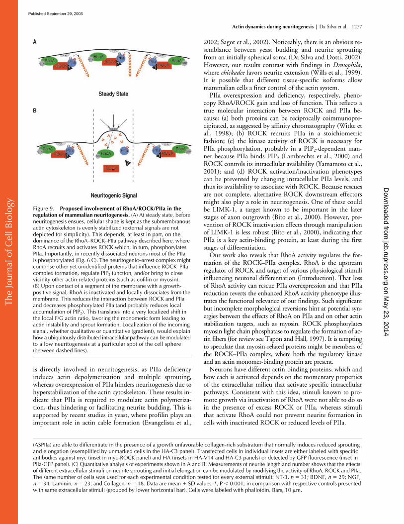
Figure 9. Proposed involvement of RhoA/ROCK/PIIa in the regulation of mammalian neuritogenesis. (A) At steady state, before neuritogenesis ensues, cellular shape is kept as the submembranous actin cytoskeleton is evenly stabilized (external signals are not depicted for simplicity). This depends, at least in part, on the dominance of the RhoA–ROCK–PIIa pathway described here, where RhoA recruits and activates ROCK which, in turn, phosphorylates PIIa. Importantly, in recently dissociated neurons most of the PIIa is phosphorylated (Fig. 6 C). The neuritogenic–arrest complex might comprise other yet unidentified proteins that influence ROCK–PIIa complex formation, regulate PIP2 function, and/or bring to close vicinity other actin-related proteins (such as cofilin or myosin). (B) Upon contact of a segment of the membrane with a growth-positive signal, RhoA is inactivated and locally dissociates from the membrane. This reduces the interaction between ROCK and PIIa and decreases phosphorylated PIIa (and probably reduces local accumulation of PIP2). This translates into a very localized shift in the local F/G actin ratio, favoring the monomeric form leading to actin instability and sprout formation. Localization of the incoming signal, whether qualitative or quantitative (gradient), would explain how a ubiquitously distributed intracellular pathway can be modulated to allow neuritogenesis at a particular spot of the cell sphere (between dashed lines).
on May 23, 2014
jcb.rupress.orgD
ownloaded from
Published September 29, 2003
The
Jour
nal o
f Cel
l Bio
logy
1278 The Journal of Cell Biology | Volume 162, Number 7, 2003
Previous work on neuritogenesis has largely relied on de-rived cell lines (Hirose et al., 1998), where ROCK is knownto impair process extension usually induced by in vitro ma-nipulations such as serum starvation. By analyzing the timespan in which mammalian neurons naturally sprout neurites(that later will become specialized axons and dendrites), wenow show that ROCK, together with RhoA, PIIa, and actin,is part of an important mechanism, in-built in the differenti-ation program of hippocampal neurons and regulated byknown in vivo physiological stimuli. Such mechanism eluci-dates how one cell can respond to simultaneous, neighbor-ing signals that favor or discourage actin polymerization.This, in turn, would determine if a sprout forms at a givenlocation of the round, undeveloped neuron (Fig. 9). It istempting to hypothesize that RhoA/ROCK/PIIa–mediatedevents may also be key for the different architectural modifi-cations occurring at later stages of neuronal differentiation,such as axon formation and elongation.
Materials and MethodsCell culturePrimary cultures of rat embryo hippocampal neurons were prepared as de-scribed previously (Goslin and Banker, 1991). For biochemical analysis,cells were plated onto dishes coated with 0.1 mg/ml poly-L-lysine (PLL)and for morphological studies onto PLL-, laminin- (2 �g/ml), or collagen-treated (25 �g/ml) glass coverslips. Primary cultures of PII��� and PII���mice were prepared following the same protocol. All times (hours) referredto are after seeding. Cytochalasin D was used at 1 �M (Sigma-Aldrich) andY-27632 at 37 nmol/ml (BIOMOL Research Laboratories, Inc.).
Transient transfectionsNeurons in suspension were electroporated (20 �g DNA/400 �l suspen-sion) using a GenePulser (Bio-Rad Laboratories) set at 250 V, 250 �F, and200 . Cells were analyzed at 24 h in culture. Where stated, lipofectionwas used (Effectene; QIAGEN). The following plasmids were used: pPIIa-EGFP (termed PIIa-GFP), null GFP vector pEGFP-N1 (CLONTECH Labora-tories, Inc.), pIRES-neo/Sendai/PIIa (Di Nardo et al., 2000), untaggedpIRES-neo/PIIa (Di Nardo et al., 2000), empty vector pIRES-neo (CLON-TECH Laboratories, Inc.), pRc/HA-V14 and pRc/HA-C3 (a gift from J. Set-tleman, Massachusetts General Hospital, Charlestown, MA), empty vectorpRc/HA (CLONTECH Laboratories, Inc.), and pEF-Bos-myc/ROCK (a giftfrom K. Kaibuchi, Nagoya University, Nagoya, Japan).
Antisense treatmentsTreatments were performed using antisense (5�-CCACGTAGCTCTGC-CAACCGGCCAT-3’) and control 4-missense (5�-CCACcTAGCaCTGC-CtACCGcCCAT-3’; lowercase for missense nucleotides) Morpholino oligo-nucleotides (Gene Tools). These, prepaired with partially complementaryDNA, were incubated with delivery solution (EPEI) for 30 min and added3 h after seeding. Final Morpholino and EPEI concentrations were 0.25nmol/ml and 0.03 nmol/ml, respectively. Cells were analyzed after 24 h inculture.
Western blotting, 2D gels, and immunoprecipitationNeuronal extracts were loaded onto SDS-PAGE gels (equal proteinamounts following spectrophotometric determination) and proteins weretransferred to nitrocellulose filters. These were then incubated with rabbitpolyclonal PIIa-specific antibody (Witke et al., 2001), mouse monoclonal�-tubulin–specific antibody (N536; Amersham Biosciences), mouse mono-clonal mannosidase2-specific antibody (a gift from G. Griffiths, EMBL,Heidelberg, Germany), and with rabbit polyclonal PI-specific antibody(Witke et al., 2001). After secondary antibody incubation, nitrocellulosemembranes were developed using an ECL system (Amersham Bio-sciences). Quantification was performed by densitometry of autoradio-grams, using NIH Image 1.62 software and Microsoft Excel v. X. Quanti-fied Western blots were plotted for PIIa expression levels in arbitrary units(a.u.) of optical density, normalized against the unrelated Golgi proteinmannosidase-2 in the population. In all cases, quantification was based onfour independent experiments. 2D gel separation of neuronal extracts was
followed by the same Western blotting procedure. For immunoprecipita-tions, extracts were obtained with lysis buffer (1% Triton X-100, 100 mMNaCl, 2 mM EDTA, 10 mM Tris/HCl, pH 7.5, and protease inhibitors) andincubated at 4C overnight with anti-PIIa or anti–ROCK-II (clone 21; BDBiosciences) antibodies coupled to protein G–Sepharose beads (Roche).Beads without antibodies incubated with homogenates were used as con-trols. Samples were analyzed following the same Western Blotting pro-tocol.
Morphological analysisNeurons were analyzed by immunofluorescence using the following anti-bodies: anti–mouse �-tubulin antibody, mouse monoclonal 12CA5 anti-HA antibody (Boehringer), mouse monoclonal 9E10 anti-myc antibody(American Type Culture Collection), and rabbit polyclonal anti-PIIa anti-body. F-actin was detected with Alexa 568–conjugated phalloidin (Molec-ular Probes). Alexa-conjugated secondary antibodies (Molecular Probes)were used. Cells were observed with a microscope equipped with 40 ,63 , and 100 objectives (model DMIRE2; Leica), and images were cap-tured using Qfluoro software (Leica). Neurite length and number weremeasured using this software, by two observers, from at least three inde-pendent experiments. Obtained values were exported to Excel v. X for sta-tistical analysis. Student’s t tests were based on our results’ two-tailed dis-tribution and two-sample unequal variance.
F/G-actin ratiosDetermination of F- and G-actin fluorescence, labeled with Alexa 568phalloidin and Alexa 488 DNaseI (Molecular Probes), respectively, wasdone with the ProbeMeter plug-in of the Qfluoro software. Images from F-and G-actin channels were captured and merged. A user-determined 5 5-pixel area was placed at 10 different points along each neurite (control,n � 90; antisense, n � 88; overexpression, n � 35), and the percentage ofsignal from each channel was calculated by the software against the un-modified background. The results obtained were statistically analyzed withExcel v. X.
Morphological reconstructionsFor the different experimental backgrounds, phase-contrast images of indi-vidual cells were obtained. Preparation of camera lucida drawings foreach particular image ensued, after which a montage of camera lucida re-constructions was produced. This resulted in drawings of average cells,each representative of individual experimental perturbations and respec-tive controls. Thicker lines depicted thicker processes and the cell body,whereas thinner processes were drawn with increasingly finer lines. De-gree of branching was represented by a variable number of stemming pro-cesses.
We thank Bianca Hellias and Etienne Cassin for technical assistance andJose Abad-Rodriguez, Maria Dolores Ledesma, and Vanessa Schubert forhelpful suggestions.
J.S. Da Silva is supported by an FCT/PRAXIS XXI scholarship (Portu-guese Ministry of Science and Technology).
Submitted: 3 April 2003Accepted: 14 August 2003
ReferencesArber, S., F.A. Barbayannis, H. Hanser, C. Schneider, C.A. Stanyon, O. Bernard,
and P. Caroni. 1998. Regulation of actin dynamics through phosphorylationof cofilin by LIM-kinase. Nature. 393:805–809.
Bamburg, J.R. 1999. Proteins of the ADF/cofilin family: essential regulators of ac-tin dynamics. Annu. Rev. Cell Dev. Biol. 15:185–230.
Baum, P.D., and G. Garriga. 1997. Neuronal migrations and axon fasciculation aredisrupted in ina-1 integrin mutants. Neuron. 19:51–62.
Billuart, P., C.G. Winter, A. Maresh, X. Zhao, and L. Luo. 2001. Regulating axonbranch stability: the role of p190 RhoGAP in repressing a retraction signal-ing pathway. Cell. 107:195–207.
Bito, H., T. Furuyashiki, H. Ishihara, Y. Shibasaki, K. Ohashi, K. Mizuno, M.Maekawa, T. Ishizaki, and S. Narumiya. 2000. A critical role for a Rho-asso-ciated kinase, p160ROCK, in determining axon outgrowth in mammalianCNS neurons. Neuron. 26:431–441.
Bradke, F., and C.G. Dotti. 1999. The role of local actin instability in axon forma-tion. Science. 283:1931–1934.
on May 23, 2014
jcb.rupress.orgD
ownloaded from
Published September 29, 2003
The
Jour
nal o
f Cel
l Bio
logy
Actin dynamics during neuritogenesis | Da Silva et al. 1279
Da Silva, J.S., and C.G. Dotti. 2002. Breaking the neuronal sphere: regulation ofthe actin cytoskeleton in neuritogenesis. Nat. Rev. Neurosci. 3:694–704.
Di Nardo, A., R. Gareus, D. Kwiatkowski, and W. Witke. 2000. Alternative splic-ing of the mouse profilin II gene generates functionally different profilin iso-forms. J. Cell Sci. 113:3795–3803.
Dotti, C.G., C.A. Sullivan, and G.A. Banker. 1988. The establishment of polarityby hippocampal neurons in culture. J. Neurosci. 8:1454–1468.
Evangelista, M., D. Pruyne, D.C. Amberg, C. Boone, and A. Bretscher. 2002.Formins direct Arp2/3-independent actin filament assembly to polarize cellgrowth in yeast. Nat. Cell Biol. 4:32–41.
Gleeson, J.G., and C.A. Walsh. 2000. Neuronal migration disorders: from geneticdiseases to developmental mechanisms. Trends Neurosci. 23:352–359.
Goslin, K., and G. Banker. 1991. Rat hippocampal neurons in low-density culture.In Culturin Nerve Cells. K. Goslin and G. Banker, editors. MassachusettsInstitute of Technology Press, Cambridge, MA. 251–281.
Haarer, B.K., S.H. Lillie, A.E. Adams, V. Magdolen, W. Bandlow, and S.S.Brown. 1990. Purification of profilin from Saccharomyces cerevisiae and anal-ysis of profilin-deficient cells. J. Cell Biol. 110:105–114.
Hatten, M.E. 1999. Central nervous system neuronal migration. Annu. Rev. Neu-rosci. 22:511–539.
Haugwitz, M., A.A. Noegel, J. Karakesisoglou, and M. Schleicher. 1994. Dictyoste-lium amoebae that lack G-actin-sequestering profilins show defects in F-actincontent, cytokinesis, and development. Cell. 79:303–314.
Hirose, M., T. Ishizaki, N. Watanabe, M. Uehata, O. Kranenburg, W.H.Moolenaar, F. Matsumura, M. Maekawa, H. Bito, and S. Narumiya. 1998.Molecular dissection of the Rho-associated protein kinase (p160ROCK)–regulated neurite remodeling in neuroblastoma N1E-115 cells. J. Cell Biol.141:1625–1636.
Hu, E., Z. Chen, T. Fredrickson, and Y. Zhu. 2001. Molecular cloning and char-acterization of profilin-3: a novel cytoskeleton-associated gene expressed inrat kidney and testes. Exp. Nephrol. 9:265–274.
Ishizaki, T., M. Uehata, I. Tamechika, J. Keel, K. Nonomura, M. Maekawa, and S.Narumiya. 2000. Pharmacological properties of Y-27632, a specific inhibi-tor of rho-associated kinases. Mol. Pharmacol. 57:976–983.
Jalink, K., T. Eichholtz, F.R. Postma, E.J. van Corven, and W.H. Moolenaar.1993. Lysophosphatidic acid induces neuronal shape changes via a novel, re-ceptor-mediated signaling pathway: similarity to thrombin action. CellGrowth Differ. 4:247–255.
Jalink, K., E.J. van Corven, T. Hengeveld, N. Morii, S. Narumiya, and W.H.Moolenaar. 1994. Inhibition of lysophosphatidate- and thrombin-inducedneurite retraction and neuronal cell rounding by ADP ribosylation of thesmall GTP-binding protein Rho. J. Cell Biol. 126:801–810.
Kaibuchi, K., S. Kuroda, and M. Amano. 1999. Regulation of the cytoskeleton andcell adhesion by the Rho family GTPases in mammalian cells. Annu. Rev.Biochem. 68:459–486.
Kozma, R., S. Sarner, S. Ahmed, and L. Lim. 1997. Rho family GTPases and neu-ronal growth cone remodelling: relationship between increased complexityinduced by Cdc42Hs, Rac1, and acetylcholine and collapse induced byRhoA and lysophosphatidic acid. Mol. Cell. Biol. 17:1201–1211.
Lambrechts, A., A. Braun, V. Jonckheere, A. Aszodi, L.M. Lanier, J. Robbens, I.Van Colen, J. Vandekerckhove, R. Fassler, and C. Ampe. 2000. Profilin II isalternatively spliced, resulting in profilin isoforms that are differentially ex-pressed and have distinct biochemical properties. Mol. Cell. Biol. 20:8209–8219.
Leeuwen, F.N., H.E. Kain, R.A. Kammen, F. Michiels, O.W. Kranenburg, andJ.G. Collard. 1997. The guanine nucleotide exchange factor Tiam1 affectsneuronal morphology; opposing roles for the small GTPases Rac and Rho. J.Cell Biol. 139:797–807.
Leung, T., E. Manser, L. Tan, and L. Lim. 1995. A novel serine/threonine kinasebinding the Ras-related RhoA GTPase which translocates the kinase to pe-ripheral membranes. J. Biol. Chem. 270:29051–29054.
Liu, R.Y., R.S. Schmid, W.D. Snider, and P.F. Maness. 2002. NGF enhances sen-
sory axon growth induced by laminin but not by the L1 cell adhesion mole-cule. Mol. Cell. Neurosci. 20:2–12.
Luo, L. 2000. Rho GTPases in neuronal morphogenesis. Nat. Rev. Neurosci.1:173–180.
Luo, L. 2002. Actin cytoskeleton regulation in neuronal morphogenesis and struc-tural plasticity. Annu. Rev. Cell Dev. Biol. 18:601–635.
Maekawa, M., T. Ishizaki, S. Boku, N. Watanabe, A. Fujita, A. Iwamatsu, T. Obi-nata, K. Ohashi, K. Mizuno, and S. Narumiya. 1999. Signaling from Rho tothe actin cytoskeleton through protein kinases ROCK and LIM-kinase. Sci-ence. 285:895–898.
Matsui, T., M. Amano, T. Yamamoto, K. Chihara, M. Nakafuku, M. Ito, T. Na-kano, K. Okawa, A. Iwamatsu, and K. Kaibuchi. 1996. Rho-associated ki-nase, a novel serine/threonine kinase, as a putative target for small GTPbinding protein Rho. EMBO J. 15:2208–2216.
Nakagawa, O., K. Fujisawa, T. Ishizaki, Y. Saito, K. Nakao, and S. Narumiya.1996. ROCK-I and ROCK-II, two isoforms of Rho-associated coiled-coilforming protein serine/threonine kinase in mice. FEBS Lett. 392:189–193.
Nakayama, A.Y., M.B. Harms, and L. Luo. 2000. Small GTPases Rac and Rho inthe maintenance of dendritic spines and branches in hippocampal pyramidalneurons. J. Neurosci. 20:5329–5338.
Sagot, I., A.A. Rodal, J. Moseley, B.L. Goode, and D. Pellman. 2002. An actin nu-cleation mechanism mediated by Bni1 and profilin. Nat. Cell Biol. 4:626–631.
Schoenwaelder, S.M., S.C. Hughan, K. Boniface, S. Fernando, M. Holdsworth,P.E. Thompson, H.H. Salem, and S.P. Jackson. 2002. RhoA sustains inte-grin alpha IIbbeta 3 adhesion contacts under high shear. J. Biol. Chem. 277:14738–14746.
Sekine, A., M. Fujiwara, and S. Narumiya. 1989. Asparagine residue in the rhogene product is the modification site for botulinum ADP-ribosyltransferase.J. Biol. Chem. 264:8602–8605.
Selden, L.A., H.J. Kinosian, J.E. Estes, and L.C. Gershman. 1999. Impact of profi-lin on actin-bound nucleotide exchange and actin polymerization dynamics.Biochemistry. 38:2769–2778.
Summerton, J., D. Stein, S.B. Huang, P. Matthews, D. Weller, and M. Partridge.1997. Morpholino and phosphorothioate antisense oligomers compared incell- free and in-cell systems. Antisense Nucleic Acid Drug Dev. 7:63–70.
Tapon, N., and A. Hall. 1997. Rho, Rac and Cdc42 GTPases regulate the organi-zation of the actin cytoskeleton. Curr. Opin. Cell Biol. 9:86–92.
Verheyen, E.M., and L. Cooley. 1994. Profilin mutations disrupt multiple actin-dependent processes during Drosophila development. Development. 120:717–728.
Wills, Z., L. Marr, K. Zinn, C.S. Goodman, and D. Van Vactor. 1999. Profilinand the Abl tyrosine kinase are required for motor axon outgrowth in theDrosophila embryo. Neuron. 22:291–299.
Witke, W., A.V. Podtelejnikov, A. Di Nardo, J.D. Sutherland, C.B. Gurniak, C.Dotti, and M. Mann. 1998. In mouse brain profilin I and profilin II associ-ate with regulators of the endocytic pathway and actin assembly. EMBO J.17:967–976.
Witke, W., J.D. Sutherland, A. Sharpe, M. Arai, and D.J. Kwiatkowski. 2001. Pro-filin I is essential for cell survival and cell division in early mouse develop-ment. Proc. Natl. Acad. Sci. USA. 98:3832–3836.
Wolven, A.K., L.D. Belmont, N.M. Mahoney, S.C. Almo, and D.G. Drubin.2000. In vivo importance of actin nucleotide exchange catalyzed by profilin.J. Cell Biol. 150:895–904.
Yamamoto, M., D.H. Hilgemann, S. Feng, H. Bito, H. Ishihara, Y. Shibasaki, andH.L. Yin. 2001. Phosphatidylinositol 4,5-bisphosphate induces actin stress-fiber formation and inhibits membrane ruffling in CV1 cells. J. Cell Biol.152:867–876.
Yamashita, T., K.L. Tucker, and Y.A. Barde. 1999. Neurotrophin binding to thep75 receptor modulates Rho activity and axonal outgrowth. Neuron. 24:585–593.
on May 23, 2014
jcb.rupress.orgD
ownloaded from
Published September 29, 2003