Specific phosphorylation of p120-catenin regulatory domain differently modulates its binding to RhoA
13
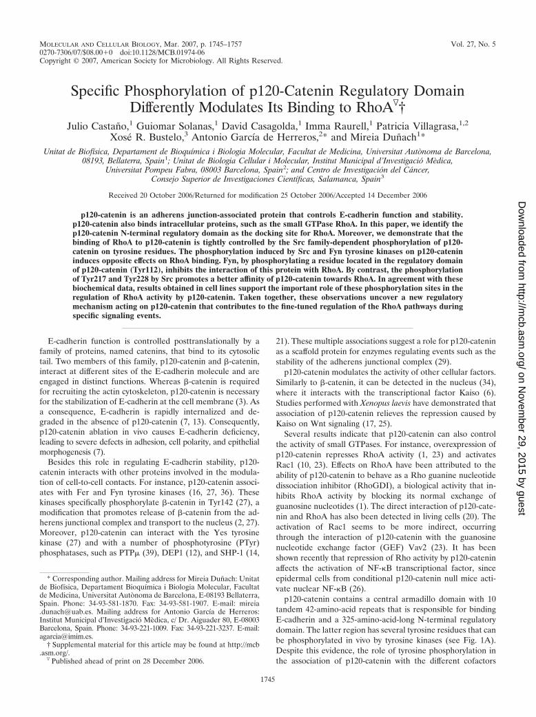
MOLECULAR AND CELLULAR BIOLOGY, Mar. 2007, p. 1745–1757 Vol. 27, No. 5 0270-7306/07/$08.000 doi:10.1128/MCB.01974-06 Copyright © 2007, American Society for Microbiology. All Rights Reserved. Specific Phosphorylation of p120-Catenin Regulatory Domain Differently Modulates Its Binding to RhoA † Julio Castan ˜o, 1 Guiomar Solanas, 1 David Casagolda, 1 Imma Raurell, 1 Patricia Villagrasa, 1,2 Xose ´ R. Bustelo, 3 Antonio Garcı ´a de Herreros, 2 * and Mireia Dun ˜ach 1 * Unitat de Biofı ´sica, Departament de Bioquı ´mica i Biologia Molecular, Facultat de Medicina, Universitat Auto `noma de Barcelona, 08193, Bellaterra, Spain 1 ; Unitat de Biologia Cellular i Molecular, Institut Municipal d’Investigacio ´ Me `dica, Universitat Pompeu Fabra, 08003 Barcelona, Spain 2 ; and Centro de Investigacio ´n del Ca ´ncer, Consejo Superior de Investigaciones Cientı ´ficas, Salamanca, Spain 3 Received 20 October 2006/Returned for modification 25 October 2006/Accepted 14 December 2006 p120-catenin is an adherens junction-associated protein that controls E-cadherin function and stability. p120-catenin also binds intracellular proteins, such as the small GTPase RhoA. In this paper, we identify the p120-catenin N-terminal regulatory domain as the docking site for RhoA. Moreover, we demonstrate that the binding of RhoA to p120-catenin is tightly controlled by the Src family-dependent phosphorylation of p120- catenin on tyrosine residues. The phosphorylation induced by Src and Fyn tyrosine kinases on p120-catenin induces opposite effects on RhoA binding. Fyn, by phosphorylating a residue located in the regulatory domain of p120-catenin (Tyr112), inhibits the interaction of this protein with RhoA. By contrast, the phosphorylation of Tyr217 and Tyr228 by Src promotes a better affinity of p120-catenin towards RhoA. In agreement with these biochemical data, results obtained in cell lines support the important role of these phosphorylation sites in the regulation of RhoA activity by p120-catenin. Taken together, these observations uncover a new regulatory mechanism acting on p120-catenin that contributes to the fine-tuned regulation of the RhoA pathways during specific signaling events. E-cadherin function is controlled posttranslationally by a family of proteins, named catenins, that bind to its cytosolic tail. Two members of this family, p120-catenin and -catenin, interact at different sites of the E-cadherin molecule and are engaged in distinct functions. Whereas -catenin is required for recruiting the actin cytoskeleton, p120-catenin is necessary for the stabilization of E-cadherin at the cell membrane (3). As a consequence, E-cadherin is rapidly internalized and de- graded in the absence of p120-catenin (7, 13). Consequently, p120-catenin ablation in vivo causes E-cadherin deficiency, leading to severe defects in adhesion, cell polarity, and epithelial morphogenesis (7). Besides this role in regulating E-cadherin stability, p120- catenin interacts with other proteins involved in the modula- tion of cell-to-cell contacts. For instance, p120-catenin associ- ates with Fer and Fyn tyrosine kinases (16, 27, 36). These kinases specifically phosphorylate -catenin in Tyr142 (27), a modification that promotes release of -catenin from the ad- herens junctional complex and transport to the nucleus (2, 27). Moreover, p120-catenin can interact with the Yes tyrosine kinase (27) and with a number of phosphotyrosine (PTyr) phosphatases, such as PTP (39), DEP1 (12), and SHP-1 (14, 21). These multiple associations suggest a role for p120-catenin as a scaffold protein for enzymes regulating events such as the stability of the adherens junctional complex (29). p120-catenin modulates the activity of other cellular factors. Similarly to -catenin, it can be detected in the nucleus (34), where it interacts with the transcriptional factor Kaiso (6). Studies performed with Xenopus laevis have demonstrated that association of p120-catenin relieves the repression caused by Kaiso on Wnt signaling (17, 25). Several results indicate that p120-catenin can also control the activity of small GTPases. For instance, overexpression of p120-catenin represses RhoA activity (1, 23) and activates Rac1 (10, 23). Effects on RhoA have been attributed to the ability of p120-catenin to behave as a Rho guanine nucleotide dissociation inhibitor (RhoGDI), a biological activity that in- hibits RhoA activity by blocking its normal exchange of guanosine nucleotides (1). The direct interaction of p120-cate- nin and RhoA has also been detected in living cells (20). The activation of Rac1 seems to be more indirect, occurring through the interaction of p120-catenin with the guanosine nucleotide exchange factor (GEF) Vav2 (23). It has been shown recently that repression of Rho activity by p120-catenin affects the activation of NF-B transcriptional factor, since epidermal cells from conditional p120-catenin null mice acti- vate nuclear NF-B (26). p120-catenin contains a central armadillo domain with 10 tandem 42-amino-acid repeats that is responsible for binding E-cadherin and a 325-amino-acid-long N-terminal regulatory domain. The latter region has several tyrosine residues that can be phosphorylated in vivo by tyrosine kinases (see Fig. 1A). Despite this evidence, the role of tyrosine phosphorylation in the association of p120-catenin with the different cofactors * Corresponding author. Mailing address for Mireia Dun ˜ach: Unitat de Biofı ´sica, Departament Bioquı ´mica i Biologia Molecular, Facultat de Medicina, Universitat Auto `noma de Barcelona, E-08193 Bellaterra, Spain. Phone: 34-93-581-1870. Fax: 34-93-581-1907. E-mail: mireia [email protected]. Mailing address for Antonio Garcı ´a de Herreros: Institut Municipal d’Investigacio ´ Me `dica, c/ Dr. Aiguader 80, E-08003 Barcelona, Spain. Phone: 34-93-221-1009. Fax: 34-93-221-3237. E-mail: [email protected]. † Supplemental material for this article may be found at http://mcb .asm.org/. Published ahead of print on 28 December 2006. 1745 on November 29, 2015 by guest http://mcb.asm.org/ Downloaded from
-
Upload
independent -
Category
Documents
-
view
1 -
download
0
Transcript of Specific phosphorylation of p120-catenin regulatory domain differently modulates its binding to RhoA
MOLECULAR AND CELLULAR BIOLOGY, Mar. 2007, p. 1745–1757 Vol. 27, No. 50270-7306/07/$08.00�0 doi:10.1128/MCB.01974-06Copyright © 2007, American Society for Microbiology. All Rights Reserved.
Specific Phosphorylation of p120-Catenin Regulatory DomainDifferently Modulates Its Binding to RhoA�†
Julio Castano,1 Guiomar Solanas,1 David Casagolda,1 Imma Raurell,1 Patricia Villagrasa,1,2
Xose R. Bustelo,3 Antonio Garcıa de Herreros,2* and Mireia Dunach1*Unitat de Biofısica, Departament de Bioquımica i Biologia Molecular, Facultat de Medicina, Universitat Autonoma de Barcelona,
08193, Bellaterra, Spain1; Unitat de Biologia Cellular i Molecular, Institut Municipal d’Investigacio Medica,Universitat Pompeu Fabra, 08003 Barcelona, Spain2; and Centro de Investigacion del Cancer,
Consejo Superior de Investigaciones Cientıficas, Salamanca, Spain3
Received 20 October 2006/Returned for modification 25 October 2006/Accepted 14 December 2006
p120-catenin is an adherens junction-associated protein that controls E-cadherin function and stability.p120-catenin also binds intracellular proteins, such as the small GTPase RhoA. In this paper, we identify thep120-catenin N-terminal regulatory domain as the docking site for RhoA. Moreover, we demonstrate that thebinding of RhoA to p120-catenin is tightly controlled by the Src family-dependent phosphorylation of p120-catenin on tyrosine residues. The phosphorylation induced by Src and Fyn tyrosine kinases on p120-catenininduces opposite effects on RhoA binding. Fyn, by phosphorylating a residue located in the regulatory domainof p120-catenin (Tyr112), inhibits the interaction of this protein with RhoA. By contrast, the phosphorylationof Tyr217 and Tyr228 by Src promotes a better affinity of p120-catenin towards RhoA. In agreement with thesebiochemical data, results obtained in cell lines support the important role of these phosphorylation sites in theregulation of RhoA activity by p120-catenin. Taken together, these observations uncover a new regulatorymechanism acting on p120-catenin that contributes to the fine-tuned regulation of the RhoA pathways duringspecific signaling events.
E-cadherin function is controlled posttranslationally by afamily of proteins, named catenins, that bind to its cytosolictail. Two members of this family, p120-catenin and �-catenin,interact at different sites of the E-cadherin molecule and areengaged in distinct functions. Whereas �-catenin is requiredfor recruiting the actin cytoskeleton, p120-catenin is necessaryfor the stabilization of E-cadherin at the cell membrane (3). Asa consequence, E-cadherin is rapidly internalized and de-graded in the absence of p120-catenin (7, 13). Consequently,p120-catenin ablation in vivo causes E-cadherin deficiency,leading to severe defects in adhesion, cell polarity, and epithelialmorphogenesis (7).
Besides this role in regulating E-cadherin stability, p120-catenin interacts with other proteins involved in the modula-tion of cell-to-cell contacts. For instance, p120-catenin associ-ates with Fer and Fyn tyrosine kinases (16, 27, 36). Thesekinases specifically phosphorylate �-catenin in Tyr142 (27), amodification that promotes release of �-catenin from the ad-herens junctional complex and transport to the nucleus (2, 27).Moreover, p120-catenin can interact with the Yes tyrosinekinase (27) and with a number of phosphotyrosine (PTyr)phosphatases, such as PTP� (39), DEP1 (12), and SHP-1 (14,
21). These multiple associations suggest a role for p120-cateninas a scaffold protein for enzymes regulating events such as thestability of the adherens junctional complex (29).
p120-catenin modulates the activity of other cellular factors.Similarly to �-catenin, it can be detected in the nucleus (34),where it interacts with the transcriptional factor Kaiso (6).Studies performed with Xenopus laevis have demonstrated thatassociation of p120-catenin relieves the repression caused byKaiso on Wnt signaling (17, 25).
Several results indicate that p120-catenin can also controlthe activity of small GTPases. For instance, overexpression ofp120-catenin represses RhoA activity (1, 23) and activatesRac1 (10, 23). Effects on RhoA have been attributed to theability of p120-catenin to behave as a Rho guanine nucleotidedissociation inhibitor (RhoGDI), a biological activity that in-hibits RhoA activity by blocking its normal exchange ofguanosine nucleotides (1). The direct interaction of p120-cate-nin and RhoA has also been detected in living cells (20). Theactivation of Rac1 seems to be more indirect, occurringthrough the interaction of p120-catenin with the guanosinenucleotide exchange factor (GEF) Vav2 (23). It has beenshown recently that repression of Rho activity by p120-cateninaffects the activation of NF-�B transcriptional factor, sinceepidermal cells from conditional p120-catenin null mice acti-vate nuclear NF-�B (26).
p120-catenin contains a central armadillo domain with 10tandem 42-amino-acid repeats that is responsible for bindingE-cadherin and a 325-amino-acid-long N-terminal regulatorydomain. The latter region has several tyrosine residues that canbe phosphorylated in vivo by tyrosine kinases (see Fig. 1A).Despite this evidence, the role of tyrosine phosphorylation inthe association of p120-catenin with the different cofactors
* Corresponding author. Mailing address for Mireia Dunach: Unitatde Biofısica, Departament Bioquımica i Biologia Molecular, Facultatde Medicina, Universitat Autonoma de Barcelona, E-08193 Bellaterra,Spain. Phone: 34-93-581-1870. Fax: 34-93-581-1907. E-mail: [email protected]. Mailing address for Antonio Garcıa de Herreros:Institut Municipal d’Investigacio Medica, c/ Dr. Aiguader 80, E-08003Barcelona, Spain. Phone: 34-93-221-1009. Fax: 34-93-221-3237. E-mail:[email protected].
† Supplemental material for this article may be found at http://mcb.asm.org/.
� Published ahead of print on 28 December 2006.
1745
on Novem
ber 29, 2015 by guesthttp://m
cb.asm.org/
Dow
nloaded from

remains unknown except in the case of E-cadherin: phosphor-ylation of p120-catenin by Src increases the in vitro associationof these two proteins (30). In this article, we present newresults demonstrating that Src and Fyn can phosphorylate theregulatory domain of p120-catenin with different functionaloutcomes. Moreover, we have identified Tyr112, a residue ofp120-catenin specifically phosphorylated by Fyn, as a key reg-ulator of the p120-catenin–RhoA interaction both in vitro andin vivo.
MATERIALS AND METHODS
Generation of p120-catenin mutants. The full-length coding region of mousep120Cas1B (obtained from Albert Reynolds, Vanderbilt University) was clonedinto the EcoRV site of cDNA3.1 (Invitrogen). A DNA fragment encoding theentire open reading frame of mouse p120-catenin was released from pcDNA3.1-p120-catenin(1–911) (fragment comprising amino acids 1 to 911) by EcoRI andNotI digestion and cloned into EcoRI/NotI-linearized pGEX-6P3 (GE Health-care). To generate the bacterial expression plasmid pGEX, encoding a glutathi-one S-transferase (GST) protein fused to the p120-catenin region encompassingamino acids 234 to 911, the cDNA fragment was released from pcDNA3-Cas1plasmid by digestion with SmaI/NotI and cloned into SmaI/NotI-linearizedpGEX-6P1. The armadillo repeat domain of p120-catenin (amino acids 350 to824) was amplified by PCR using as a template the pcDNA3.1-p120-cateninplasmid and primers annealing to the nucleotide sequences 1050 to 1064 and2457 to 2472. To allow the subsequent cloning step, these oligonucleotides weremodified to include EcoRI and EcoRV sites at their respective 5� ends. The1.4-kbp-long cDNA fragment obtained was then digested with those enzymesand cloned into pGEX-6P1 previously digested with EcoRI/SmaI. The 0.8-kbp-long p120-catenin cDNA fragment encompassing amino acids 350 to 911 wasgenerated by cutting the pGEX-p120-catenin(1-911) plasmid with XhoI andNotI and subsequently ligating it to the XhoI/NotI-digested p120-catenin(350-824) plasmid. A DNA fragment encoding amino acids 1 to 234 of p120-cateninwas amplified by PCR from pcDNA3.1-p120-catenin by using appropriate prim-ers containing either EcoRI or BamHI sites at their 5� ends. The 0.7-kbp am-plification fragment was then digested with EcoRI and BamHI and cloned intothe same sites of pGEX-6P2. To generate p120-catenin(1-96), the pGEX-p120-catenin(1-911) plasmid was digested with NotI and BsrGI, filled in, and religated.The fragment of p120-catenin from amino acids 102 to 911 was obtained by PCRusing oligonucleotides including restriction sites for EcoRI and SacII at their 5�ends. The PCR product was digested with SacII, and overhang ends were filledwith Klenow polymerase. The product was then digested with EcoRI and ligatedin pGEX-6P1 that was digested with EcoRI and SmaI. The pGEX-6P3-RhoAplasmid was generated from pGEX-4T3-RhoA, containing the full-length humanRhoA sequence, by standard cloning procedures using the flanking EcoRI andBamHI sites present in the plasmid polylinkers.
p120-catenin point mutants (Y96E, Y96F, Y112E, Y112F, Y217E, Y217F,Y228E, and Y228F mutants) were obtained using a QuikChange site-directedmutagenesis kit (Stratagene) according to the manufacturer’s instructions andeither phrGFP-p120-catenin wild type (where GFP is green fluorescent protein)or p120-catenin(1-234) as the template. The sense primers used for the gener-ation of the above-described mutants were, respectively, 5�-AACCACCTTCTGGAAAGCACTATCCCC-3�, 5�-AACCACCTTCTGTTTAGCACTATCCCC-3�,5�-ATTGTGGAAACCGAAACCGAG GAGGAC-3�, 5�-ATTGTGGAAACCTTTACCGAGGAGGAC-3�, 5�-GGGTATGGCC GACACGAAGAAGATGGTTATCC-3� , 5�-GGGTATGGCCGACACTTTGAAGA TGGTTATCC-3�, 5�-TGGCAGTGACAACGAAGGCAGTCTGTCC-3�, and 5�-TGGCAGTGACAACTTTGGCAGTCTGTCC-3�. Nucleotides modified with respect to the p120-cateninsequence (GenBank accession number Z17804) are indicated in bold.
Generation of GFP-p120-catenin constructs. A DNA fragment encodingamino acid 8 to the C terminus of p120-catenin was obtained from pGEX-p120-catenin(1–911) by using oligonucleotides corresponding to nucleotide sequences22 to 36 and 2717 to 2733 that contained EcoRI and SacII sites at the respective5� ends. The 2.7-kbp amplification fragment was digested with EcoRI and SacIIand cloned in the same sites of phrGFP-N1 (Stratagene). phrGFP-p120-cate-nin(234–911) was generated from pGEX-p120-catenin(1–911) by using oligonu-cleotides corresponding to nucleotide sequences 693 to 704 and 2717 to 2733 thatcontained EcoRI and SacII sites at the respective 5� ends. The 2-kbp amplifica-tion fragment was digested with EcoRI and SacII and cloned in the same sites ofphrGFP-N1.
Preparation of recombinant RhoA and loading with GDP or GTP. GST fusionproteins containing full-length human RhoA were expressed in Escherichia coliand purified by affinity chromatography on glutathione-Sepharose. GST-RhoAwas first incubated with 200 �l of binding buffer (20 mM Tris, pH 7.5, 100 mMEDTA, 100 mM NaCl, 0.1% Triton X-100, 1 mM dithiothreitol [DTT], 10 �g/mlaprotinin, and 1 mM phenylmethylsulfonyl fluoride) for 30 min at room temper-ature in order to release the bound nucleotides. For GDP or GTP binding, thesame buffer was supplemented with 50 mM MgCl2 plus GDP or GTP-�-S (450�M) (Sigma) and proteins were incubated for 25 min at room temperature.Loaded RhoA bound to Sepharose beads was washed with binding buffer inorder to eliminate unbound nucleotides. Beads containing the loaded GTPasewere used for binding assays to p120-catenin as described below. When required,the fusion protein was cleaved with PreScission protease (GE Healthcare) toremove the GST tag.
Protein binding assays. GST fusion proteins corresponding to the indicatedforms of p120-catenin, the cytoplasmic domain of E-cadherin (cytoEcad), or�-catenin were expressed in E. coli and purified by affinity chromatography onglutathione-Sepharose (30). Binding assays were carried out by incubating theindicated protein with a constant amount of either GST fusion protein used asbait or the nonchimeric GST used as a control. Details of these assays havealready been described (27, 30). Pull-down assays were performed by usingpurified recombinant proteins fused to GST and cell extracts from RWP-1 cells,NIH 3T3 cells, and IEC-18 cells overexpressing K-ras oncogene (IEC-K-Rascells), as reported previously (30). Glutathione-Sepharose-bound proteins wereanalyzed by Western blotting with specific monoclonal antibodies (MAbs)against p120-catenin, RhoA, E-cadherin, Fyn, PTyr, p120-catenin pY96, p120-catenin pY228 (all from Transduction Laboratories), AU5 tag (Covance), andGFP (Clontech). The polyclonal antibody to the GST protein was from GEHealthcare. Serial immunoblot assays were performed after stripping the mem-branes. All binding assays were repeated three times. In order to quantify theamount of bound protein, the autoradiograms were scanned in a densitometer.
Cell fractionation and immunoprecipitation. Cytosolic fractions were pre-pared by homogenizing cells in buffer C (25 mM Tris-HCl, pH 7.6, 200 mM NaCl,1 mM EGTA, 1 mM DTT, 5 mM MgCl2) supplemented with protease inhibitors(0.3 �M aprotinin, 1 �M leupeptin, 1 �M pepstatin,1 mM Pefabloc) and phos-phatase inhibitors (10 mM NaF, 0.1 mM sodium orthovanadate). Cell homoge-nates were centrifuged at 20,000 � g for 5 min at 4°C to obtain the cytosolicfractions. Pellets were resuspended in the same volume of radioimmunoprecipi-tation assay buffer (25 mM Tris-HCl, pH 7.6, 200 mM NaCl, 1 mM EGTA, 0.5%sodium deoxycholate, 0.1% sodium dodecyl sulfate, 1% Nonidet P-40) supple-mented with protease and phosphatase inhibitors. After passing cells 10 times bya 20-gauge syringe, extracts were left on ice for 20 min and centrifuged at 20,000 �g for 5 min at 4°C. Supernatants constituted the membrane-associated fraction.Alternatively, cell extracts were prepared by resuspending cells in buffer D (50mM Tris-HCl, pH 7.6, 200 mM NaCl, 5 mM MgCl2, 0.1% Nonidet P-40, 1 mMDTT) supplemented with protease and phosphatase inhibitors. Lysates werecentrifuged at 12,000 � g in a microfuge for 15 min at 4°C.
Proteins were immunoprecipitated from cell extracts (100 to 300 �g) by using4 �g/ml of the appropriate antibody for 16 h at 4°C and then collected using 30�l of protein A-agarose (Sigma). Immunoprecipitates were washed three timeswith lysis buffer and proteins bound to beads directly eluted with electrophoresissample buffer and analyzed by Western blotting. When appropriate, mouseTrueBlot reagent (BD Biosciences) was used as a secondary antibody in order toeliminate interferences in the autoradiogram.
Fluorescence microscopy. NIH 3T3 cells were transfected with GFP-p120-catenin wild type, GFP-p120-catenin Y112E, GFP-p120-catenin Y217E, andGFP-p120-catenin(234-end) by using Lipofectamine (Life Technologies) accord-ing to the manufacturer’s instructions. Cells were plated on glass coverslips andfixed with 4% paraformaldehyde 2 days after transfection. After 20 min, cover-slips were washed with phosphate-buffered saline for 5 min and with water for 3min. Coverslips were mounted onto glass slides and cells visualized by confocalmicroscopy. Rhodamine-conjugated phalloidin (Sigma) was used to visualizeF-actin distribution.
Kinase assays. c-Src and recombinant Fyn kinase were purchased from Up-state Biotechnology. Fer kinase was cloned in pcDNA3-His(C) (Invitrogen) thatlabeled the protein with Xpress and poly-His tags, transfected to RWP-1 cells,and purified by nickel-agarose chromatography (27). Phosphorylation assayswere performed in a final volume of 50 �l of kinase buffer (25 mM Tris-HCl, pH6.8, 25 mM MgCl2, 5 mM MnCl2, 0.5 mM EGTA, 1 mM DTT, 0.25 mM sodiumorthovanadate, 0.1 mM ATP) for either 2 h at 23°C (in the case of Src assays) or1 h at 30°C (in the case of Fer and Fyn assays). pEF-Bos-active Fyn was a kindgift of A. Carrera (Centro Nacional de Biotecnologıa, Madrid, Spain).
1746 CASTANO ET AL. MOL. CELL. BIOL.
on Novem
ber 29, 2015 by guesthttp://m
cb.asm.org/
Dow
nloaded from

GDP dissociation assays. The effect of purified GST-p120-catenin on thedissociation of [3H]GDP from RhoA was examined by preloading GST-RhoA(20 pmol) with [3H]GDP (1.5 �M, 1,000 cpm/pmol) for 10 min at 30°C. Incu-bations were performed in the presence of 25 mM Tris-HCl, pH 7.8, 120 mMNaCl, 1 mM EDTA, and 3 mM MgCl2 in a final volume of 100 �l. [3H]GDP-RhoA was incubated with equimolar amounts of p120-catenin at 30°C for 10 min.An excess of GTP (10 �M) was then added to allow the exchange of bound[3H]GDP-RhoA for GTP for 10 additional minutes. [3H]GDP-bound RhoA wasseparated from free nucleotide by filtration, and the amount of RhoA bound to[3H]GDP was determined and compared to the radionucleotide bound beforethe excess of unlabeled GTP was added.
RESULTS
RhoA binds to the regulatory domain of p120-catenin. Inorder to study the binding of p120-catenin to RhoA and E-cadherin, we expressed several p120-catenin deletion mutantsas GST fusion proteins (Fig. 1A). Binding assays demonstratedthat RhoA interacts similarly with a p120-catenin fragmentcomprising amino acids 1 to 234 and the full-length protein(Fig. 1B, lanes 2 and 5). No association with a protein contain-
FIG. 1. RhoA interacts with the N-terminal regulatory domain of p120-catenin. (A) Diagram of the different domains of p120-catenin.Alternative splicing in the N-terminal domain gives rise to isoforms 1, 2, 3, and 4, each initiating at distinct ATG start codons (1, 55, 102, and 340,respectively) (arrows). The regulatory domain contains nine tyrosines (Y96, Y112, Y217, Y228, Y257, Y280, Y291, Y296, and Y302) capable ofbeing phosphorylated in vitro (15, 17). The length and composition of the protein fragments used in this work are shown. (B) GST fusion proteins(1.5 pmol) were incubated with 5 pmol of RhoA. Protein complexes were affinity purified with glutathione-Sepharose and analyzed by sodiumdodecyl sulfate-polyacrylamide gel electrophoresis and Western blotting (WB) with anti-RhoA MAb. RhoA (1.2 pmol) was included as an internalreference standard (St). When indicated, the assay was performed in the presence of a 100-fold molecular excess of cytoEcad. Blots werereanalyzed with anti-GST to ensure that similar amounts of fusion proteins were present in all cases. (C) GST-p120-catenin fusion proteins orGST-�-catenin (7 pmol) was incubated with 270 �g of whole-cell extracts from RWP-1 cells. Protein complexes were affinity purified as describedabove and analyzed with anti-E-cadherin and anti-GST MAbs. In the Input lane, 5% of the total cell extract used for the assay was loaded.(D) Either GST-cytoEcad or GST (1.2 pmol) was incubated with 30 pmol of RhoA in the presence of 3 pmol of p120-catenin-1 when indicated.Protein complexes were affinity purified with glutathione-Sepharose and analyzed with specific MAbs. p120-catenin (0.6 pmol) was included as aninternal reference standard. In panels B, C, and D, the numbers below the lanes correspond to the amounts of bound RhoA, E-cadherin, orp120-catenin, respectively, determined after densitometry, compared to the standard or to the amount bound to full-length GST-p120-catenin. Theresults presented in this figure correspond to an experiment representative of three performed.
VOL. 27, 2007 REGULATION OF p120-CATENIN RhoGDI ACTIVITY 1747
on Novem
ber 29, 2015 by guesthttp://m
cb.asm.org/
Dow
nloaded from

ing the sequence from amino acid 234 to the end or the first 96amino acids of p120-catenin was observed (Fig. 1B, lanes 4 and6). A p120-catenin form comprising amino acids 102 to theend, which corresponds to isoform 3, bound RhoA similarly tothe full-length protein (Fig. 1B, right panel, lanes 2 and 3).Therefore, these results suggest that the binding site for RhoAin p120-catenin was mapped in the regulatory domain of p120-catenin, probably between amino acids 102 and 234. As hasbeen proposed previously, E-cadherin bound to a protein frag-ment comprising amino acids 350 to 824, including the arma-dillo repeat domain (Fig. 1C, lane 5). The interaction of E-cadherin with this domain was twofold stronger than itsassociation with the full-length p120-catenin (Fig. 1C, lanes 3and 5), similarly to that shown for �-catenin (Fig. 1C, lanes 2and 5). As expected, E-cadherin did not bind to p120-cate-nin(1–234) (data not shown). E-cadherin and RhoA could bedetected interacting simultaneously with p120-catenin, furthersuggesting that those proteins bind to different regions of p120-catenin (Fig. 1B, lane 3). Additional binding experiments dem-onstrated that RhoA associated to GST-cytoEcad only whenp120-catenin was present (Fig. 1D, lane 3), indicating that acomplex of the three proteins can be formed.
Phosphorylation of the regulatory domain by different ki-nases affects binding of p120-catenin to RhoA and E-cadherin.Our results indicate that RhoA associates to the regulatorydomain of p120-catenin, a sequence known to be heavily phos-phorylated by several tyrosine kinases (21, 22, 24). These ob-servations led us to check whether this interaction might bemodulated by tyrosine phosphorylation. To this end, we ana-lyzed the effect of recombinant Src and Fyn tyrosine kinasesand of immunopurified Fer tyrosine kinase on the p120-cate-nin–RhoA interaction. Surprisingly, whereas phosphorylationby Fer and Src kinase stimulated p120-catenin–RhoA binding(Fig. 2A, upper panel, compare lane 2 with lanes 4 and 5), Fyntotally prevented it (Fig. 2A, upper panel, lane 3). Fyn had adominant effect on this regulation, since simultaneous phos-phorylation of p120-catenin by Src and Fyn diminished theamount of RhoA associated to this protein with respect to thecontrol, unphosphorylated protein (Fig. 2A, upper panel, com-pare lanes 2 and 6). Similar results were observed when theeffects of Fyn and Src on the binding of RhoA to the N-terminal p120-catenin(1–234) fragment were analyzed (Fig.2B, upper panel, lanes 2, 3, and 4), indicating that the relevanttyrosine residues under the control of this interaction wereplaced within this sequence. Effects of the kinases were depen-dent on the addition of ATP (data not shown), demonstratingthat although Fyn interacts with this domain, repression ofRhoA binding is not due to steric competition.
The effect of the phosphorylation of p120-catenin by thesekinases on E-cadherin binding was also determined. In thethree cases studied, phosphorylation promoted an increasedassociation between p120-catenin and E-cadherin (Fig. 2A,second panel from top, compare lane 2 with lanes 3, 4, and 5).This effect was not observed when binding of E-cadherin to thearmadillo repeat domain of p120-catenin (amino acids 350 to824) was analyzed, since this domain was not phosphorylatedby any of these kinases under our experimental conditions(data not shown).
Fyn-mediated phosphorylation of Tyr112 of p120-catenininterferes with its binding to RhoA. Since the effect of Fyn on
p120-catenin–RhoA binding was dominant, we reasoned thatthis kinase was probably targeting a Tyr residue not recognizedby Src. Therefore, we searched for a Tyr residue in the p120-catenin(1–234) amino acid sequence modified differently byFyn and Src kinases. Four Tyr residues have previously beenreported to be phosphorylated in this region: Tyr96, Tyr112,Tyr217, and Tyr228 (Fig. 3A) (21, 24). Other putative phos-phorylation site tyrosines of the regulatory domain are locateddownstream of amino acid residue 234. Taking advantage ofthe availability of monoclonal antibodies specific for PTyr96and PTyr228, we first checked whether either of these tworesidues was phosphorylated differently by Fyn and Src. Thesetwo residues were modified similarly by both kinases (see Fig.SA1 in the supplemental material), ruling out the possibilitythat modification of either of these two amino acids is respon-sible for the different effects of Fyn.
FIG. 2. Tyrosine phosphorylation of p120-catenin modulates its in-teraction with RhoA. Full-length GST-p120-catenin (A) or GST-p120-catenin(1–234) (B) was phosphorylated with Fyn, Fer, Src, or Fyn andSrc tyrosine kinases, as described in Materials and Methods. Controland phosphorylated GST-p120-catenin proteins or GST as a control(1.5 pmol) were incubated with 5 pmol of RhoA (upper panel) or with2 pmol cytoEcad (middle panel) in a final volume of 200 �l. Proteincomplexes were affinity purified with glutathione-Sepharose and ana-lyzed by sodium dodecyl sulfate-polyacrylamide gel electrophoresisand Western blotting (WB) with specific MAbs. RhoA (1 pmol) (Aand B) or cytoEcad (0.1 pmol) (A) were included as internal referencestandards (St). The numbers below the lanes correspond to theamounts of RhoA or E-cadherin bound to GST-p120-catenin fusionproteins compared to the standard. The results presented in this figurecorrespond to an experiment representative of at least three per-formed.
1748 CASTANO ET AL. MOL. CELL. BIOL.
on Novem
ber 29, 2015 by guesthttp://m
cb.asm.org/
Dow
nloaded from

To further characterize the phosphorylation sites involved inthis regulatory step, we mutated tyrosines 112 and 217 to phe-nylalanine. We performed these mutations in a p120-cate-nin(1–234) fragment in which the tyrosine residues at positions
96 and 228 had been replaced previously by phenylalanineresidues in order to eliminate the background phosphorylationinduced by Fyn and Src in those amino acids. As shown in Fig.3B, the Tyr112Phe and Tyr217Phe p120 mutations differently
FIG. 3. Phosphorylation of Tyr112 inhibits the p120-catenin–RhoA interaction. (A) Diagram of the N-terminal regulatory domain of p120-catenin(1–347). Tyrosine residues phosphorylated by tyrosine kinases are indicated (Y96, Y112, Y217, Y228, Y257, Y280, Y291, Y296, and Y302).(B) Mapping of the residues involved in Src and Fyn phosphorylation of p120-catenin. Various amounts (2 pmol in the case of Fyn and 4 pmolin the case of Src) of different GST-p120-cat(1–234) point mutants were phosphorylated with Fyn (left panel) or Src (right panel). Phosphorylationwas analyzed by Western blotting (WB) with an anti-PTyr MAb. The membrane was stripped and reanalyzed against GST to check that similarlevels of the different GST-p120-cat(1–234) point mutants were used. (C) RhoA-binding assays were performed with 5 pmol of RhoA and 5 pmolof GST or GST-p120-cat(1–234) fusion proteins corresponding to the wild-type (WT) form, the Y96F Y228F double mutant, or the Y96F Y228FY112F triple mutant. When indicated, the fusion proteins were phosphorylated with recombinant Fyn or Src tyrosine kinases. Protein complexeswere affinity purified with glutathione-Sepharose and analyzed by Western blotting with anti-RhoA. RhoA (1 pmol [left panel] or 0.4 pmol [rightpanel]) was included as an internal reference standard (St). (D) GST-p120-catenin and Y112E, Y217E, and Y228E point mutants (1.5 pmol) wereincubated with 5 pmol of RhoA in a final volume of 200 �l, as indicated above. RhoA (1 pmol [left panel] or 2.5 pmol [right panel]) was includedas an internal reference standard. (E) Pull-down assays were performed by incubating 6 pmol of the different GST-p120-catenin point mutants with300 �g of whole-cell extracts from RWP-1 cells; protein complexes were affinity purified with glutathione-Sepharose and analyzed by sodiumdodecyl sulfate-polyacrylamide gel electrophoresis and Western blotting with anti-E-cadherin. Blots were reanalyzed with anti-GST antibodies toensure equal loading of samples. In the Input lane, a sample containing 5% of the total cell extracts used for the assay was loaded. (F) RWP-1cells were cotransfected with different GFP-p120-catenin fusion proteins and pEF-Bos-active Fyn or the empty plasmid. After 48 h, cell extractswere prepared, 300 �g of each cell extract was immunoprecipitated with anti-GFP, and the associated proteins were analyzed with specific MAbsagainst GFP, RhoA, and Fyn. The numbers below the lanes correspond to the amounts of RhoA or E-cadherin bound to GST-p120-catenin fusionproteins, compared to the standard or to the amount bound to wild-type GST-p120-catenin. The results presented in this figure correspond to anexperiment representative of at least three performed.
VOL. 27, 2007 REGULATION OF p120-CATENIN RhoGDI ACTIVITY 1749
on Novem
ber 29, 2015 by guesthttp://m
cb.asm.org/
Dow
nloaded from

affected the phosphorylation of the p120-catenin(1–234) frag-ment catalyzed by Fyn (Fig. 3B, left panel) and Src (Fig. 3B,right panel). Thus, the Tyr112Phe mutation abolished almostcompletely the phosphorylation of this fusion protein by Fynbut it affected only slightly the phosphorylation promoted bySrc (Fig. 3B, lane 2, left and right panels). By contrast, thebehavior of the Tyr217Phe mutation was the opposite of thatfound with the Tyr112Phe replacement (Fig. 3B, lane 3, leftand right panels). These results indicate that p120-cateninTyr112 is phosphorylated more actively by Fyn than by Src,suggesting that the posttranslational modification of this resi-due is responsible for the inhibition of the interaction of p120-catenin with RhoA.
In order to demonstrate the relevance of Tyr112 in theinhibition of the RhoA–p120-catenin interaction by Fyn, weperformed binding assays using the p120-catenin(1–234) form,where Tyr96 and Tyr228 had been replaced by Phe. As shownin Fig. 3C, this mutant protein interacted with RhoA withaffinities similar to that of its wild-type counterpart (Fig. 3C,right panel, compare lanes 2 and 3). Phosphorylation by Fynprevented the interaction, whereas Src enhanced it (Fig. 3C,right panel, lanes 3, 4, and 5), similarly to what happened with
the wild-type form, further supporting the irrelevance of Tyr96and Tyr228 in the effects of Fyn. When the same assays wereperformed with a mutant containing the additional replace-ment of Tyr112 by Phe, phosphorylation by Fyn increased itsaffinity towards RhoA (Fig. 3C, left panel). These results sug-gest that the Fyn-dependent phosphorylation of p120-cateninon Tyr112 prevents RhoA binding, whereas the Src/Fyn-de-pendent phosphorylation of the Try217 residue induces theopposite effect.
To further check the relevance of the phosphorylation ofthese residues, we replaced each of them with glutamate res-idues to mimic the effect of the negative charge introduced bythe phosphate groups. As shown in Fig. 3D, binding of RhoAto the p120-catenin Tyr112Glu mutant was completely abol-ished. Instead, the p120-catenin Tyr217Glu mutant interactedwith RhoA significantly better than the wild-type form (ap-proximately 2.05-fold � 0.14-fold [n 3]). A Tyr228Glu mu-tation also upregulated the binding of p120-catenin to RhoA,although to a lower extent than the Tyr217Glu mutation (Fig.3D). These mutant proteins were also tested for E-cadherinbinding by use of a similar experimental approach. TheTyr112Glu and Tyr217Glu p120-catenin mutants presented abetter association with E-cadherin than did the wild-type coun-terpart (Fig. 3E) (2.55-fold � 0.24-fold and 2.85-fold � 0.31-fold [n 3], respectively, for Tyr112Glu and Tyr217Glu). Amutant protein harboring both mutations interacted with E-cadherin slightly better than each of the single mutants alone(data not shown).
The relevance of the p120-catenin Tyr112 phosphorylationby Fyn was also determined in RWP-1 epithelial cells. Ectopicexpression of Fyn greatly downregulated the amount of RhoAcapable of coimmunoprecipitating with cotransfected p120-catenin (Fig. 3F, lane 2). A similar block in the interaction wasobserved when the Tyr112Glu mutant was introduced in cells
FIG. 4. p120-catenin interacts better with the RhoA-bound GDPform. (A) p120-catenin (1 pmol) was incubated with 2 pmol of GDP-or GTP-loaded GST-RhoA. Protein complexes were affinity purifiedwith glutathione-Sepharose and analyzed by sodium dodecyl sulfate-polyacrylamide gel electrophoresis and Western blotting (WB) with ananti-p120-catenin MAb. p120-catenin (0.25 pmol) was included as aninternal reference standard (St). (B) NIH 3T3 cells were transfectedwith two RhoA mutants: AU5-RhoAQ63L (which is always active) orAU5-RhoAT19N (inactive). After 48 h, cell extracts were preparedand incubated (300 �g) with GST-p120-catenin. Protein complexeswere purified with glutathione-Sepharose and associated proteins an-alyzed with a monoclonal antibody specific to the AU5 tag. Blots werereanalyzed with anti-GST to ensure that similar levels of fusion proteinforms were present in all samples. In the Input lane, a sample con-taining 5% of the total cell extracts used for the assay was loaded. Theresults presented in this figure correspond to an experiment represen-tative of three (A) or two (B) performed.
FIG. 5. Phosphorylation of p120-catenin on Tyr112 blocks itsRhoGDI activity. [3H]GDP-bound RhoA (20 pmol) was incubatedwith equimolar amounts of the different GST-p120-catenin fusion pro-teins for 10 min. An excess of GTP (10 �M) was then added to allowthe exchange of bound [3H]GDP-RhoA for GTP, and samples wereincubated for an additional 10 min. [3H]GDP-bound RhoA was sep-arated from free nucleotide by filtration, and the amount of [3H]GDPbound to RhoA was determined and compared to the radionucleotidebound at time zero. The figure shows the averages � standard devia-tions of three experiments performed in duplicate.
1750 CASTANO ET AL. MOL. CELL. BIOL.
on Novem
ber 29, 2015 by guesthttp://m
cb.asm.org/
Dow
nloaded from

in the absence of ectopic Fyn (Fig. 3F, lane 3). However, whenTyr112Phe p120-catenin was used, Fyn was unable to inhibitthe association between this p120-catenin form and RhoA(Fig. 3F, lanes 4 and 5). Actually, Fyn increased this bindingevent slightly, although significantly, a result probably derivedfrom its ability to phosphorylate tyrosine residues in additionto Tyr112 (e.g., Tyr217).
Phosphorylation of Tyr112 blocks p120-catenin effects onRhoA activity. It is known that p120-catenin binds better to theinactive than to the active form of RhoA. To test the behaviorof our mutant proteins in this biochemical property, we per-formed binding assays with RhoA proteins that were loadedwith either GDP or GTP-�-S (see Materials and Methods).p120-catenin was pulled down with a higher efficiency by aGST-RhoA loaded with GDP, approximately 4.5-fold (�0.52-fold) (n 3) better than by GST-RhoA loaded with GTP (Fig.4A). Assays using different mutants of RhoA confirmed ourconclusion: p120-catenin interacted better with the RhoA
Thr19Asn mutant, an inactive RhoA mutant (4, 32), than withthe RhoA Gln63Leu mutant, a RhoA mutant with impairedGTPase activity and, therefore, constitutively bound to GTPmolecules (15) (Fig. 4B, lanes 3 and 4). Consistent with thisresult, we also observed that p120-catenin acts as a RhoGDI-like molecule (Fig. 5). The inhibition of the exchange of GDPwith GTP was dependent on p120-catenin binding, since adeletion mutant lacking the RhoA-binding domain [p120-cate-nin(234-end)] was unable to retard the GDP release. As ex-pected from the results described above, the p120-cateninTyr112Glu mutant was totally inactive in this assay, confirmingagain that this residue is important for a stable interaction withRhoA. The binding domain by itself (amino acids 1 to 234) wasnot functional either, indicating that additional sequences inp120-catenin were required for stabilization. The use of addi-tional controls demonstrated that the assay worked properly(Fig. 5).
As an additional way of corroborating our biochemical ob-
FIG. 6. Phosphorylation of p120-catenin on Tyr112 by Fyn prevents repression of RhoA activity by p120-catenin. SW480 cells were cotrans-fected with 7.5 �g of pcDNA3.1-Myc-His-RhoA wild type (WT) and (A) with 7.5 �g of pEF-Bos-active Fyn, pEF-Bos-active v-Src, or pEF-Bosalone, (B) with pEF-Bos-active Fyn and 7.5 �g of GFP, GFP-p120-catenin wild type, or GFP-p120-catenin Y112F, or (C) with the same amountsof GFP, GFP-p120-catenin wild type, or GFP-p120-catenin Y112E. After 48 h, cell extracts were prepared and added to a suspension ofGST-rhotekin–glutathione-Sepharose. The amount of active RhoA was analyzed by Western blotting (WB) with a specific MAb against RhoA.Blots were reanalyzed with anti-Fyn and -Src (A), with anti-Fyn and anti-GFP (B), or with anti-GFP (C). In the Input lane, a sample of 5% of thetotal cell extract used for the assay was loaded. The right panels show the representation of active Rho with respect to the total amount of thisprotein. The averages � standard deviations of the three different experiments performed in each case are presented.
VOL. 27, 2007 REGULATION OF p120-CATENIN RhoGDI ACTIVITY 1751
on Novem
ber 29, 2015 by guesthttp://m
cb.asm.org/
Dow
nloaded from
servations, we analyzed the role of Tyr112 phosphorylation inp120-catenin activity in vivo. To this end, we tested the effectsof Src and Fyn on the levels of GTP-bound RhoA by usingtransient transfections in RWP-1 cells coupled to pull-downexperiments with GST-rhotekin, which specifically retains theactivated form of this GTPase. These experiments indicatedthat the overexpression of Src decreased the total levels ofactivated, GTP-bound RhoA (Fig. 6A, upper panel, comparelanes 4 and 5). In contrast, Fyn promoted RhoA stimulationdue to the probable elimination of the RhoGDI activity ofp120-catenin through phosphorylation of Tyr112 and the dis-ruption of the p120-catenin–RhoA interaction (Fig. 6A, upperpanel, lane 6). To further test this hypothesis, Fyn and p120-catenin were overexpressed in these cells. In the presence ofFyn, wild-type p120-catenin only slightly decreased the levelsof active RhoA; however, the Tyr112Phe mutant decreased by90% the amount of active RhoA in the presence of Fyn (Fig.6B). The ectopic expression of p120-catenin mutants also con-firmed our conclusions, since the transfection of wild-typep120-catenin markedly downregulated the levels of activeRhoA whereas its Tyr112Glu mutant was totally ineffective insuch action (Fig. 6C).
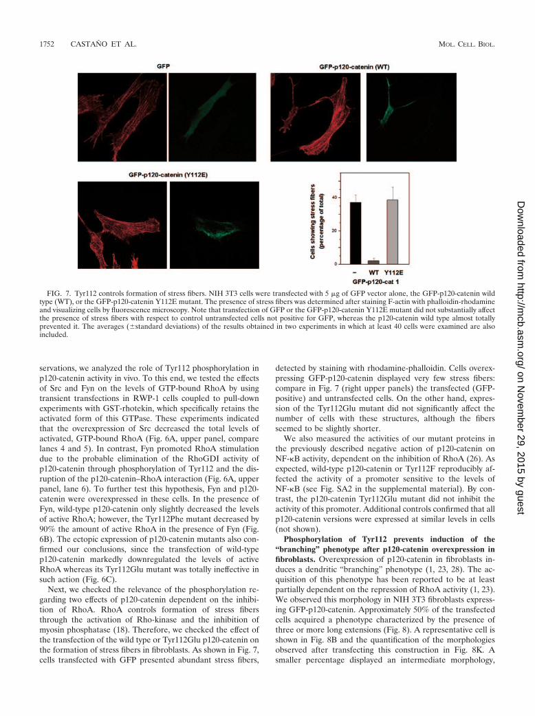
Next, we checked the relevance of the phosphorylation re-garding two effects of p120-catenin dependent on the inhibi-tion of RhoA. RhoA controls formation of stress fibersthrough the activation of Rho-kinase and the inhibition ofmyosin phosphatase (18). Therefore, we checked the effect ofthe transfection of the wild type or Tyr112Glu p120-catenin onthe formation of stress fibers in fibroblasts. As shown in Fig. 7,cells transfected with GFP presented abundant stress fibers,
detected by staining with rhodamine-phalloidin. Cells overex-pressing GFP-p120-catenin displayed very few stress fibers:compare in Fig. 7 (right upper panels) the transfected (GFP-positive) and untransfected cells. On the other hand, expres-sion of the Tyr112Glu mutant did not significantly affect thenumber of cells with these structures, although the fibersseemed to be slightly shorter.
We also measured the activities of our mutant proteins inthe previously described negative action of p120-catenin onNF-�B activity, dependent on the inhibition of RhoA (26). Asexpected, wild-type p120-catenin or Tyr112F reproducibly af-fected the activity of a promoter sensitive to the levels ofNF-�B (see Fig. SA2 in the supplemental material). By con-trast, the p120-catenin Tyr112Glu mutant did not inhibit theactivity of this promoter. Additional controls confirmed that allp120-catenin versions were expressed at similar levels in cells(not shown).
Phosphorylation of Tyr112 prevents induction of the“branching” phenotype after p120-catenin overexpression infibroblasts. Overexpression of p120-catenin in fibroblasts in-duces a dendritic “branching” phenotype (1, 23, 28). The ac-quisition of this phenotype has been reported to be at leastpartially dependent on the repression of RhoA activity (1, 23).We observed this morphology in NIH 3T3 fibroblasts express-ing GFP-p120-catenin. Approximately 50% of the transfectedcells acquired a phenotype characterized by the presence ofthree or more long extensions (Fig. 8). A representative cell isshown in Fig. 8B and the quantification of the morphologiesobserved after transfecting this construction in Fig. 8K. Asmaller percentage displayed an intermediate morphology,
FIG. 7. Tyr112 controls formation of stress fibers. NIH 3T3 cells were transfected with 5 �g of GFP vector alone, the GFP-p120-catenin wildtype (WT), or the GFP-p120-catenin Y112E mutant. The presence of stress fibers was determined after staining F-actin with phalloidin-rhodamineand visualizing cells by fluorescence microscopy. Note that transfection of GFP or the GFP-p120-catenin Y112E mutant did not substantially affectthe presence of stress fibers with respect to control untransfected cells not positive for GFP, whereas the p120-catenin wild type almost totallyprevented it. The averages (�standard deviations) of the results obtained in two experiments in which at least 40 cells were examined are alsoincluded.
1752 CASTANO ET AL. MOL. CELL. BIOL.
on Novem
ber 29, 2015 by guesthttp://m
cb.asm.org/
Dow
nloaded from

with one or two long extensions (Fig. 8C and K). Simultaneousoverexpression of E-cadherin and p120-catenin prevented theinduction of this phenotype (Fig. 8D and K), as previouslyreported (10, 23). The branching phenotype was not observedwhen the two deletion mutants [p120-catenin(1–234) andp120-catenin(234-end)] were expressed (Fig. 8G, H, and K).Curiously, these two fragments presented a cellular distribu-tion different from those of the rest of the GFP-p120-cateninproteins, which were associated mostly to the cell periphery;p120-catenin was mainly cytosolic, whereas p120-catenin(234-end) was located also in the nucleus (Fig. 8G). The presence ofthe characteristic dendritic morphology was also observed aftertransfection of the p120-catenin Tyr217Glu mutant (Fig. 8Eand K), which shows an enhanced interaction with RhoA.However, the Tyr112Glu mutant did not promote the acquisi-tion of this phenotype (Fig. 8F): only 10% of the cells pre-sented a morphology similar to that shown in Fig. 8B or E (Fig.8K). When Fyn was cotransfected with GFP-p120-catenin, theacquisition of this phenotype was also partially prevented, fur-ther indicating that phosphorylation of Tyr112 in this proteinprevents branching (Fig. 8J). Curiously, Fyn by itself promoted
significant morphological changes, with a higher percentage ofelongated cells (Fig. 8I and K). Therefore, these results suggestthat interaction of p120-catenin with RhoA is necessary for theacquisition of the dendritic phenotype in fibroblasts.
Phosphorylation of Tyr112 contributes to the regulation ofp120-catenin–RhoA binding and RhoA GTPase activity by K-ras in IEC-18 cells. Cellular effects of p120-catenin on RhoAactivity and branching phenotype have been attributed to thecytosolic fraction of this protein (23). However, activity of Fynkinase is normally restricted to the membrane. We approachedthis problem by analyzing the cytosolic or membrane distribu-tion of p120-catenin, RhoA, and Fyn in intestinal epithelialIEC-18 cells and in IEC-18 cells overexpressing K-ras onco-gene (IEC-K-ras cells). In this cell system, K-ras oncogeneexpression has been shown to induce the phosphorylation ofp120-catenin (19, 27) and the activation of Fyn kinase (27).Compared to control IEC cells, IEC-K-ras cells present lowlevels of active RhoA, as determined by GST-rhotekin pull-down assays (Fig. 9A). Cellular fractionation of IEC cellsindicated that most of the endogenous p120-catenin wasassociated to the membrane fraction, although a significant
FIG. 8. Phosphorylation of p120-catenin on Tyr112 prevents its RhoA-dependent effects in vivo. NIH 3T3 cells were transfected with 1 �g ofthe different phrGFP vectors in order to express the indicated GFP fusion proteins. When appropriate, 5 �g of pCFP-E-cadherin (D) or pEF-Fyn(I and J) was also transfected. After 24 h, cells were plated on glass coverslips, fixed with 4% paraformaldehyde, and washed with phosphate-buffered saline. Coverslips were mounted on glass slides and the morphology of transfected cells visualized by confocal microscopy. Arrows indicatethe membrane protrusions typically induced by the expression of wild-type (WT) p120-catenin. The most predominant morphology observed ineach case is shown; other relevant phenotypes are also displayed in the case of cells transfected with GFP-p120-catenin (wild type). Thepercentages of cells showing the different phenotypes are included (K); this diagram corresponds to the result of one experiment representativeof the three performed. Fifty transfected cells were observed under each condition. A cell was scored as having a branched phenotype (examplesin panels B and E) if it presented three or more extensions longer than the cell body. Cells presenting one or two extensions at least twice the cellbody were scored as having a partial branched phenotype (intermediate phenotype, examples in panels C and I).
VOL. 27, 2007 REGULATION OF p120-CATENIN RhoGDI ACTIVITY 1753
on Novem
ber 29, 2015 by guesthttp://m
cb.asm.org/
Dow
nloaded from

pool was also detected in the cytosol (Fig. 9B). The distri-butions of p120-catenin were not significantly different inIEC and IEC-K-ras cells. Ectopically expressed p120-cate-nin was distributed similarly to the endogenous protein (Fig.9C). K-Ras overexpression increased the extent of tyrosinephosphorylation on p120-catenin, as observed by analyzingwith an anti-PTyr MAb p120-catenin immunoprecipitates.This increase was detected to occur exclusively in the mem-brane-associated fraction, probably because Fyn was presentonly in this fraction (Fig. 9B).
RhoA cellular distribution was sensitive to K-Ras overex-pression. In IEC cells, similar levels of RhoA were found in themembrane and cytosolic fractions (Fig. 9B). K-ras transfec-tants presented lower levels of RhoA in the membrane fractionand higher levels in the cytosol, indicative of a release of RhoAfrom the membrane. Association of RhoA to p120-catenin wasdetected only in the cytosol by coimmunoprecipitation exper-iments (Fig. 9B). The amount of RhoA associated to p120-catenin in this fraction was increased in IEC-K-ras cells withrespect to IEC, probably reflecting the fact that higher levels ofRhoA are found in the cytosol in these cells.
We reasoned that the inability to detect RhoA associated top120-catenin in the membrane might be due to the rapid ac-tivation of this GTPase and the low affinity of GTP-boundRhoA to p120-catenin. Therefore, we transfected an inactive(Thr19Asn) mutant of RhoA to IEC and IEC-K-ras cells andanalyzed its interaction with wild-type p120-catenin or with amutant where phosphorylation by Fyn was prevented(Tyr112Phe). As shown in Fig. 9C, binding of p120-catenin toRhoA was detected in the membrane fraction only when phos-phorylation of Tyr112 was prevented.
Therefore, Fyn activation and p120-catenin phosphorylationin Tyr112 correlate with a diminished ability of RhoA to bindto the membrane-associated p120-catenin, promoting the sub-sequent release of this GTPase to the cytosol and its bindingand inhibition by the unphosphorylated form of this p120-catenin present in this fraction.
DISCUSSION
In this work we have shown that the N-terminal regulatorydomain of p120-catenin is the region responsible for the inter-action with RhoA. Perhaps more importantly, we have dem-onstrated that the association between p120-catenin and RhoAor E-cadherin, two proteins binding to different elements, ismodulated by Fyn, Src, and Fer tyrosine kinases. As we havedescribed previously for Src (30), phosphorylation of p120-catenin increased the association of p120-catenin to E-cad-herin. It is remarkable that the armadillo domain of p120-catenin, the region responsible for E-cadherin association, isnot phosphorylated by these kinases. This result suggeststhat the armadillo and regulatory domains are in close spa-tial proximity and that, similarly to what happens in otherproteins containing these repeats (5, 33), the binding capa-bility of the armadillo domain is regulated by distal ele-ments. Experiments performed with p120-catenin deletionmutants further support this conclusion: a fragment com-prising only the armadillo repeats binds E-cadherin betterthan the full-length protein (Fig. 1C).
Other results suggest that both domains are functionally
FIG. 9. Effect of K-Ras overexpression in IEC-18 cells on RhoAdistribution and activity. (A) Activity of RhoA in IEC or IEC-K-rascells was determined using chromatography with GST-rhotekin–gluta-thione-Sepharose as indicated in Materials and Methods. Bound pro-teins were analyzed with a RhoA-specific MAb. The lower panel showsthe representation of active Rho with respect to the total amount ofthis protein. The averages � standard deviations of the three differentexperiments performed in each case are presented. (B) Cytosolic andmembrane-associated fractions from IEC and IEC-K-ras cells wereprepared as detailed in Materials and Methods. Fractions were pre-cipitated with an anti-p120-catenin MAb and analyzed by Westernblotting (WB) with the indicated MAbs. In parallel, an aliquot of thefour different inputs was also analyzed. (C) IEC and IEC-K-ras cellswere cotransfected with inactive AU5-RhoA(T19N) and GFP-p120-catenin, either the wild type or the Y112F mutant, or with GFP as acontrol. Cytosolic and membrane fractions were prepared as describedabove, and the exogenous p120-catenin was immunoprecipitated (IP)with an anti-GFP antibody. The immunoprecipitates and inputs wereanalyzed by Western blotting with the indicated antibodies. The resultspresented in panels B and C correspond to an experiment represen-tative of three performed.
1754 CASTANO ET AL. MOL. CELL. BIOL.
on Novem
ber 29, 2015 by guesthttp://m
cb.asm.org/
Dow
nloaded from

interdependent. As shown in our in vitro nucleotide exchangeassays, p120-catenin acts as a GDI for RhoA. This activityrequires the binding sequence in p120-catenin but also addi-tional elements, as inferred from the observation that the frag-ment comprising amino acids 1 to 234 interacts with RhoA butis not sufficient to induce the inhibition of nucleotide ex-change. This result suggests that the armadillo domain isnecessary to protect the binding site for GDP, precludingthe release of this nucleotide (Fig. 10). Our data also indi-cate that binding of E-cadherin and RhoA to p120-catenincan occur simultaneously. These results are not contradic-tory to observations by several labs indicating that the inhi-bition by p120-catenin of RhoA activity is blocked by E-cadherin overexpression (1, 10, 23). According to ourmodel, binding to E-cadherin would not prevent p120-cate-nin association to RhoA but it might either induce a con-formational change that would prevent the GDI activity(Fig. 10) or, alternatively, recruit a GEF responsible for theactivation of RhoA, as it has been proposed previously (1).
Our work has also demonstrated that the phosphorylation ofdifferent tyrosine residues of p120-catenin promotes distinctiveeffects on RhoA binding. Thus, whereas phosphorylation ofTyr112 totally blocks this association, the similar modificationof Tyr217 (or Tyr228 to a lower extent) increases it. We con-sider it likely that the negative effect of phospho-Tyr112 is dueto the interference in the association of RhoA. However, thepositive effect might be more indirect (i.e., caused by thepartial unfolding of the regulatory domain) and presentanalogies with the stimulation of the association of p120-
catenin to E-cadherin. In this case, we have evidence indi-cating that the phosphorylation of several tyrosine residuespromotes an accumulative effect because (i) the Tyr112,217Glu double mutant binds E-cadherin better than any ofthe single mutants and (ii) phosphorylation of this doublemutant by Src can still increase affinity for E-cadherin (datanot shown). It is likely, therefore, that the phosphorylationof several tyrosines cooperates in the modification of thestructure of p120-catenin.
Phosphorylation by Fyn kinase exerts an effect opposite thanthat of Src or Fer kinases on RhoA–p120-catenin binding.Both Fer and Fyn are detected constitutively bound to p120-catenin through its regulatory domain (16, 27). It has to bedetermined whether both kinases can associate simultaneouslyto the same complex, but if this is not the case, as seems likely,the balance between the levels of these active protein kinasesbound to p120-catenin would control the phosphorylation sta-tus of Tyr112 and, consequently, the association of RhoA top120-catenin. The identification of the molecular mechanismscontrolling the interactions of p120-catenin with these kinasesis an interesting topic of research, as is the characterization ofthe signaling pathways regulating them.
We have also demonstrated that the interaction of RhoAwith p120-catenin can be detected in vivo, since in intestinalIEC-18 cells, cytosolic p120-catenin coimmunoprecipitatedwith RhoA. Most of the cellular effects observed after trans-fection of p120-catenin are promoted by the cytosolic fractionof this protein, as has been demonstrated previously for theinhibition of RhoA activity (23). However, we have also shown
FIG. 10. Proposed model for the regulation of RhoA activity by p120-catenin. p120-catenin protein is composed by the regulatory and thearmadillo domains (1). After binding of RhoA to the regulatory domain, the armadillo repeats are disposed in the close vicinity of the nucleotidebinding site of RhoA, hindering the release of GDP and therefore contributing to the GDI effect of p120-catenin (3). In cells, most p120-cateninis located at the membrane due to its interaction with the juxtamembrane domain (JMD) of E-cadherin (2). This membrane-associatedp120-catenin is unable to work as a GDI due to either a conformational change induced in the armadillo domain by E-cadherin or the recruitmentof a RhoA GEF (not depicted) by this protein. Therefore, GDP bound to RhoA is exchanged by GTP, and the affinity of the GTPase byp120-catenin decreases and is rapidly released (4). This effect of E-cadherin or E-cadherin-associated factor is not present in the cytosol; therefore,only cytosolic p120-catenin can work as a GDI (3). Activation of Fyn tyrosine kinase leads to phosphorylation of tyrosines 112, 228, and others inthe regulatory domain of the p120-catenin located in the membrane fraction, where Fyn is normally present (5), and not in the cytosol (7).Phosphorylation of Tyr112 prevents interaction of GDP-bound RhoA with p120-catenin (6), therefore precluding the activation of this protein andincreasing the levels of this GTPase at the cytosol, where it can be bound and inhibited by unphosphorylated p120-catenin (8).
VOL. 27, 2007 REGULATION OF p120-CATENIN RhoGDI ACTIVITY 1755
on Novem
ber 29, 2015 by guesthttp://m
cb.asm.org/
Dow
nloaded from

that the interaction of RhoA with p120-catenin could be de-tected in the membrane fraction when we transfected an inac-tive form of this GTPase (Fig. 9). We have explained this resulton the basis of the different affinities of p120-catenin by GDP-and GTP-bound RhoA and the lack of effect of membrane-associated p120-catenin on RhoA activity. Therefore, althoughGDP-RhoA also interacts with membrane-bound p120-cate-nin, it would be activated to GTP-RhoA by a local RhoA GEFand therefore it would be rapidly released. A model presentingthis hypothesis is depicted in Fig. 10.
We have reported previously the activation of Fyn tyrosinekinase in IEC-18 cells transfected with K-ras oncogene (27).This tyrosine kinase is located in the membrane fraction; there-fore, only the p120-catenin present in this fraction is phosphor-ylated under these conditions (Fig. 9). Phosphorylation pre-vents association of inactive RhoA to membrane p120-catenin,enhancing the levels of RhoA in the cytosol, where it caninteract and be inhibited by the active form of p120-catenin.Therefore, phosphorylation of Tyr112 in membrane-associatedp120-catenin would exert an inhibitory effect over RhoA ac-tivity, contributing to the diminished levels of active RhoAobserved to occur in these cells after transfection of K-ras (Fig.10). The different levels of stimulation of Fer, Fyn, or Srctyrosine kinases by this oncogene might help to explain thedivergent results observed for RhoA activity in cells trans-fected with K-ras (9, 31, 38).
A recent report has described the involvement of Src ty-rosine kinase in the regulation of Rho by another GDI, a25-kDa RhoGDI (8). Phosphorylation of this protein by Srcpromotes its translocation to the plasma membrane and de-creases its interaction with RhoA. On the contrary, on p120-catenin Src stimulates its binding to RhoA and enhances theinteraction of this catenin with the membrane, probablythrough its binding to E-cadherin. It is possible that this low-molecular-mass GDI also participates in the inactivation ofRhoA in the cytosol, playing the same role we proposed forp120-catenin in this compartment (Fig. 10). The relative affin-ities of RhoA for these two GDIs are an interesting matter ofstudy, either when both GDIs are not modified or after theaction of Src or Fyn tyrosine kinases.
Furthermore, different tyrosine kinases can also phosphor-ylate and activate another effector of RhoA, p190RhoGAP(11). This protein can be coupled to the adherens junctionalcomplex through its association with p120-catenin (35). Al-though it has not been demonstrated formally, it has beenproposed that tyrosine phosphorylation of p190RhoGAP facil-itates its association with p120-catenin and the inactivation ofRhoA in the membrane (35). The molecular details of thiscomplex remain to be characterized, for instance, whetherp120-catenin directly interacts with p190RhoGAP and whetherthis association can take place simultaneously with the bindingof RhoA to both proteins. In any case, it is possible thatphosphorylation of specific residues of p120-catenin may alsoaffect its interaction with p190RhoGAP, adding a new elementto the control of RhoA activity.
Different cellular roles for p120-catenin have been identifiedin past years. Some of these roles require inhibition of RhoAby p120-catenin. For instance, the induction of a dendritic,branched phenotype by p120-catenin has been attributed tothe ability to block RhoA activity and activate Rac1 (1, 10, 23).
The characterization of p120-catenin mutants unable to bindto RhoA described in this article provides new evidence sup-porting the relevance of the interaction of both proteins in theacquisition of the branched phenotype and introduces newtools to dissect the molecular pathways triggered by this pleio-tropic protein.
ACKNOWLEDGMENTS
We especially thank Neus Ontiveros for technical assistance.This study was supported by grants awarded by the Spanish Minis-
terio de Ciencia y Tecnologıa (BMC2003-00410 and BFU2006-03203to M.D. and SAF2003-02324 and SAF2006-00339 to A.G.H.). Fundingby Generalitat de Catalunya (2005SGR00970) is also appreciated.G.S., I.R., J.C., D.C., and P.V. were recipients of predoctoral fellow-ships awarded by DGR-UAB (Generalitat de Catalunya) (to G.S.),Instituto de Salud Carlos III (Ministerio de Sanidad) (to I.R.), andMinisterio de Educacion y Ciencia (to J.C., D.C., and P.V.).
REFERENCES
1. Anastasiadis, P. Z., S. Y. Moon, M. A. Thoreson, D. J. Mariner, H. C.Crawford, Y. Zheng, and A. B. Reynolds. 2000. Inhibition of RhoA by p120catenin. Nat. Cell Biol. 2:637–644.
2. Brembeck, F. H., T. Schwarz-Romond, J. Bakkers, S. Wilhelm, M. Hammer-schmidt, and W. Birchmeier. 2004. Essential role of BCL9-2 in the switchbetween beta-catenin’s adhesive and transcriptional functions. Genes Dev.18:2225–2230.
3. Bryant, D. M., and J. L. Stow. 2004. The ins and outs of E-cadherin traf-ficking. Trends Cell. Biol. 14:427–434.
4. Carnac, G., M. Primig, M. Kitzmann, P. Chafey, D. Tuil, N. Lamb, and A.Fernandez. 1998. RhoA GTPase and serum response factor control selec-tively the expression of MyoD without affecting Myf5 in mouse myoblasts.Mol. Biol. Cell 9:1891–1902.
5. Castano, J., I. Raurell, J. Piedra, S. Miravet, M. Dunach, and A. Garcıa deHerreros. 2002. �-Catenin N-and C-tails modulate the coordinated bindingof adherens junction proteins to �-catenin. J. Biol. Chem. 277:31541–31550.
6. Daniel, J. M., and A. B. Reynolds. 1999. The catenin p120ctn interacts withKaiso, a novel BTB/POZ domain zinc finger transcription factor. Mol. Cell.Biol. 19:3614–3623.
7. Davis, M. A., and A. B. Reynolds. 2006. Blocked acinar development, E-cadherin reduction, and intraepithelial neoplasia upon ablation of p120-catenin in the mouse salivary gland. Dev. Cell 10:21–31.
8. DerMardirossian, C., G. Rocklin, J. Y. Seo, and G. Bokoch. 2006. Phosphor-ylation of RhoGDI by Src regulates RhoGTPase binding and cytosol-mem-brane cycling. Mol. Biol. Cell 17:4760–4768.
9. Dreissigacker, U., M. M. Mueller, M. Unger, P. Siegert, F. Genze, P. Gier-schik, and K. Giehl. 2006. Oncogenic K-ras down-regulates Rac1 and RhoAactivity and enhances migration and invasion of pancreatic carcinoma cellsthrough activation of p38. Cell. Signal. 18:1156–1168.
10. Grosheva, I., M. Shtutman, M. Elbaum, and A. D. Bershadsky. 2001. p120catenin affects cell motility via modulation of activity of Rho-family GTPases: alink between cell-cell contact formation and regulation of cell locomotion.J. Cell Sci. 114:695–707.
11. Hernandez, S. E., J. Settlemen, and A. J. Koleske. 2004. Adhesion-depen-dent regulation of p190RhoGAP in the developing brain by the Abl-relatedgene tyrosine kinase. Curr. Biol. 14:691–696.
12. Holsinger, L. J., K. Ward, B. Duffield, J. Zachwieja, and B. Jallal. 2002. Thetransmembrane receptor protein tyrosine phosphatase DEP1 interacts withp120(ctn). Oncogene 21:7067–7076.
13. Ireton, R. C., M. A. Davis, J. van Hengel, D. J. Mariner, K. Barnes, M. A.Thoreson, P. Z. Anastasiadis, L. Matrisian, L. M. Bundy, L. Sealy, B.Gilbert, F. van Roy, and A. B. Reynolds. 2002. A novel role for p120 cateninin E-cadherin function. J. Cell Biol. 159:465–476.
14. Keilhack, H., U. Hellman, J. van Hengel, F. van Roy, J. Godovac-Zimmer-mann, and F. D. Bohmer. 2000. The protein-tyrosine phosphatase SHP-1binds to and dephosphorylates p120 catenin. J. Biol. Chem. 275:26376–26384.
15. Khoshravi-Far, R., P. A. Solski, G. J. Clark, M. S. Kinch, and C. J. Der.1995. Activation of Rac1, RhoA, and mitogen-activated protein kinases isrequired for Ras transformation. Mol. Cell. Biol. 15:6443–6453.
16. Kim, L., and T. W. Wong. 1995. The cytoplasmic tyrosine kinase FER isassociated with the catenin-like substrate pp120 and is activated by growthfactors. Mol. Cell. Biol. 15:4553–4561.
17. Kim, S. W., J. I. Park, C. M. Spring, A. K. Sater, H. Ji, A. A. Otchere, J. M.Daniel, and P. D. McCrea. 2004. Non-canonical Wnt signals are modulatedby the Kaiso transcriptional repressor and p120-catenin. Nat. Cell Biol.6:1212–1220.
18. Kimura, K., M. Ito, M. Amano, K. Chihara, Y. Fukata, M. Nakafuku, B.
1756 CASTANO ET AL. MOL. CELL. BIOL.
on Novem
ber 29, 2015 by guesthttp://m
cb.asm.org/
Dow
nloaded from
Yamamori, J. H. Feng, T. Nakano, K. Okawa, A. Iwamatsu, and K. Kaibuchi.1996. Regulation of myosin phosphates by Rho and Rho-associated kinase(Rho-kinase). Science 273:245–248.
19. Kinch, M. S., G. J. Clark, C. J. Der, and K. Burridge. 1995. Tyrosinephosphorylation regulates the adhesion of ras-transformed breast epithelia.J. Cell Biol. 130:461–471.
20. Magie, C. R., D. Pinto-Santini, and S. M. Parkhurst. 2002. Rho1 interactswith p120ctn and alpha-catenin, and regulates cadherin-based adherensjunction components in Drosophila. Development 129:3771–3782.
21. Mariner, D. J., P. Anastasiadis, H. Keilhack, F. D. Bohmer, J. Wang, andA. B. Reynolds. 2001. Identification of Src phosphorylation sites in thecatenin p120ctn. J. Biol. Chem. 276:28006–28013.
22. Mariner, D. J., M. A. Davis, and A. B. Reynolds. 2004. EGFR signaling top120-catenin through phosphorylation at Y228. J. Cell Sci. 117:1339–1350.
23. Noren, N. K., B. P. Liu, K. Burridge, and B. Kreft. 2000. p120 cateninregulates the actin cytoskeleton via Rho family GTPases. J. Cell Biol. 150:567–580.
24. Ozawa, M., and T. Ohkubo. 2001. Tyrosine phosphorylation of p120(ctn) inv-Src transfected L cells depends on its association with E-cadherin andreduces adhesion activity. J. Cell Sci. 114:503–512.
25. Park, J. I., S. W. Kim, J. P. Lyons, H. Ji, T. T. Nguyen, K. Cho, M. C. Barton,T. Deroo, K. Vleminckx, R. T. Moon, and P. D. McCrea. 2005. Kaiso/p120-catenin and TCF/beta-catenin complexes coordinately regulate canonicalWnt gene targets. Dev. Cell 8:843–854.
26. Perez-Moreno, M., M. A. Davis, E. Wong, H. A. Pasolli, A. B. Reynolds, andE. Fuchs. 2006. p120-catenin mediates inflammatory responses in the skin.Cell 124:631–644.
27. Piedra, J., S. Miravet, J. Castano, H. G. Palmer, N. Heisterkamp, A. Garciade Herreros, and M. Dunach. 2003. p120 catenin-associated Fer and Fyntyrosine kinases regulate beta-catenin Tyr-142 phosphorylation and beta-catenin-alpha-catenin interaction. Mol. Cell. Biol. 23:2287–2297.
28. Reynolds, A. B., J. M. Daniel, Y. Y. Mo, and Z. Zhang. 1996. The novelcatenin p120cas binds classical cadherins and induces an unusual morpho-logical phenotype in NIH-3T3 fibroblasts. Exp. Cell Res. 225:328–337.
29. Reynolds, A. B., and A. Roczniak-Ferguson. 2004. Emerging roles for p120-catenin in cell adhesion and cancer. Oncogene 23:7947–7956.
30. Roura, S., S. Miravet, J. Piedra, A. Garcia de Herreros, and M. Dunach.1999. Regulation of E-cadherin/catenin association by tyrosine phosphory-lation. J. Biol. Chem. 274:36734–36740.
31. Sahai, E., M. Olson, and C. J. Marshall. 2001. Cross-talk between Ras andRho signalling pathways in transformation favours proliferation and in-creased motility. EMBO J. 20:755–766.
32. Shaw, R. J., M. Henry, F. Solomon, and T. Jacks. 1998. RhoA-dependentphosphorylation and relocalization of ERM proteins into apical membraneactin protrusions in fibroblasts. Mol. Biol. Cell 9:403–419.
33. Solanas, G., S. Miravet, D. Casagolda, J. Castano, I. Raurell, A. Corrionero,A. G. de Herreros, and M. Dunach. 2004. Beta-catenin and plakoglobin N-and C-tails determine ligand specificity. J. Biol. Chem. 279:49849–49856.
34. van Hengel, J., P. Vanhoenacker, K. Staes, and F. van Roy. 1999. Nuclearlocalization of the p120(ctn) armadillo-like catenin is counteracted by anuclear export signal and by E-cadherin expression. Proc. Natl. Acad. Sci.USA 96:7980–7985.
35. Wildenberg, G. A., M. R. Dohn, R. Carnahan, M. A. Davis, N. A. Lobdell,J. Settleman, and A. B. Reynolds. 2006. p120-catenin and p190RhoGAPregulate cell-cell adhesion by coordinating antagonism between Rac andRho. Cell 127:1027–1039.
36. Xu, G., A. W. Craig, P. Greer, M. Miller, P. Z. Anastasiadis, J. Lilien, andJ. Balsamo. 2004. Continuous association of cadherin with beta-cateninrequires the non-receptor tyrosine-kinase Fer. J. Cell Sci. 117:3207–3219.
37. Reference deleted.38. Zondag, G. C., E. E. Evers, J. P. ten Klooster, L. Janssen, R. A. van der
Kammen, and J. G. Collard. 2000. Oncogenic ras downregulates Rac activ-ity, which leads to increased RhoA activity and epithelial-mesenchymal tran-sition. J. Cell Biol. 149:775–781.
39. Zondag, G. C., A. B. Reynolds, and W. H. Moolenaar. 2000. Receptorprotein-tyrosine phosphatase RPTP� binds to and dephosphorylates thecatenin p120(ctn). J. Biol. Chem. 275:11264–11269.
VOL. 27, 2007 REGULATION OF p120-CATENIN RhoGDI ACTIVITY 1757
on Novem
ber 29, 2015 by guesthttp://m
cb.asm.org/
Dow
nloaded from