On the mechanism of autoinhibition of the RhoA-specific nucleotide exchange factor PDZRhoGEF
14
BioMed Central Page 1 of 14 (page number not for citation purposes) BMC Structural Biology Open Access Research article On the mechanism of autoinhibition of the RhoA-specific nucleotide exchange factor PDZRhoGEF Meiying Zheng 1,2 , Tomasz Cierpicki 1 , Ko Momotani 1 , Mykhaylo V Artamonov 1 , Urszula Derewenda 1 , John H Bushweller 1 , Avril V Somlyo 1 and Zygmunt S Derewenda* 1 Address: 1 Department of Molecular Physiology and Biological Physics, University of Virginia, PO Box 800736, Charlottesville, Virginia. 22908- 0736, USA and 2 Present address : Monsanto Company, 800 North Lindbergh Boulevard, St Louis, MO 63167 Email: Meiying Zheng - [email protected]; Tomasz Cierpicki - [email protected]; Ko Momotani - [email protected]; Mykhaylo V Artamonov - [email protected]; Urszula Derewenda - [email protected]; John H Bushweller - [email protected]; Avril V Somlyo - [email protected]; Zygmunt S Derewenda* - [email protected] * Corresponding author Abstract Background: The Dbl-family of guanine nucleotide exchange factors (GEFs) activate the cytosolic GTPases of the Rho family by enhancing the rate of exchange of GTP for GDP on the cognate GTPase. This catalytic activity resides in the DH (Dbl-homology) domain, but typically GEFs are multidomain proteins containing other modules. It is believed that GEFs are autoinhibited in the cytosol due to supramodular architecture, and become activated in diverse signaling pathways through conformational change and exposure of the DH domain, as the protein is translocated to the membrane. A small family of RhoA-specific GEFs, containing the RGSL (regulators of G-protein signaling-like) domain, act as effectors of select GPCRs via Gα 12/13 , although the molecular mechanism by which this pathway operates is not known. These GEFs include p115, LARG and PDZRhoGEF (PRG). Results: Here we show that the autoinhibition of PRG is caused largely by an interaction of a short negatively charged sequence motif, immediately upstream of the DH-domain and including residues Asp706, Glu708, Glu710 and Asp712, with a patch on the catalytic surface of the DH-domain including Arg867 and Arg868. In the absence of both PDZ and RGSL domains, the DH-PH tandem with additional 21 residues upstream, is 50% autoinhibited. However, within the full-length protein, the PDZ and/or RGSL domains significantly restore autoinhibition. Conclusion: Our results suggest a mechanism for autoinhibition of RGSL family of GEFs, in which the RGSL domain and a unique sequence motif upstream of the DH domain, act cooperatively to reduce the ability of the DH domain to bind the nucleotide free RhoA. The activation mechanism is likely to involve two independent steps, i.e. displacement of the RGSL domain and conformational change involving the autoinhibitory sequence motif containing several negatively charged residues. Published: 21 May 2009 BMC Structural Biology 2009, 9:36 doi:10.1186/1472-6807-9-36 Received: 13 February 2009 Accepted: 21 May 2009 This article is available from: http://www.biomedcentral.com/1472-6807/9/36 © 2009 Zheng et al; licensee BioMed Central Ltd. This is an Open Access article distributed under the terms of the Creative Commons Attribution License (http://creativecommons.org/licenses/by/2.0 ), which permits unrestricted use, distribution, and reproduction in any medium, provided the original work is properly cited.
Transcript of On the mechanism of autoinhibition of the RhoA-specific nucleotide exchange factor PDZRhoGEF

BioMed CentralBMC Structural Biology
ss
Open AcceResearch articleOn the mechanism of autoinhibition of the RhoA-specific nucleotide exchange factor PDZRhoGEFMeiying Zheng1,2, Tomasz Cierpicki1, Ko Momotani1, Mykhaylo V Artamonov1, Urszula Derewenda1, John H Bushweller1, Avril V Somlyo1 and Zygmunt S Derewenda*1Address: 1Department of Molecular Physiology and Biological Physics, University of Virginia, PO Box 800736, Charlottesville, Virginia. 22908-0736, USA and 2Present address : Monsanto Company, 800 North Lindbergh Boulevard, St Louis, MO 63167
Email: Meiying Zheng - [email protected]; Tomasz Cierpicki - [email protected]; Ko Momotani - [email protected]; Mykhaylo V Artamonov - [email protected]; Urszula Derewenda - [email protected]; John H Bushweller - [email protected]; Avril V Somlyo - [email protected]; Zygmunt S Derewenda* - [email protected]
* Corresponding author
AbstractBackground: The Dbl-family of guanine nucleotide exchange factors (GEFs) activate the cytosolicGTPases of the Rho family by enhancing the rate of exchange of GTP for GDP on the cognateGTPase. This catalytic activity resides in the DH (Dbl-homology) domain, but typically GEFs aremultidomain proteins containing other modules. It is believed that GEFs are autoinhibited in thecytosol due to supramodular architecture, and become activated in diverse signaling pathwaysthrough conformational change and exposure of the DH domain, as the protein is translocated tothe membrane. A small family of RhoA-specific GEFs, containing the RGSL (regulators of G-proteinsignaling-like) domain, act as effectors of select GPCRs via Gα12/13, although the molecularmechanism by which this pathway operates is not known. These GEFs include p115, LARG andPDZRhoGEF (PRG).
Results: Here we show that the autoinhibition of PRG is caused largely by an interaction of a shortnegatively charged sequence motif, immediately upstream of the DH-domain and including residuesAsp706, Glu708, Glu710 and Asp712, with a patch on the catalytic surface of the DH-domainincluding Arg867 and Arg868. In the absence of both PDZ and RGSL domains, the DH-PH tandemwith additional 21 residues upstream, is 50% autoinhibited. However, within the full-length protein,the PDZ and/or RGSL domains significantly restore autoinhibition.
Conclusion: Our results suggest a mechanism for autoinhibition of RGSL family of GEFs, in whichthe RGSL domain and a unique sequence motif upstream of the DH domain, act cooperatively toreduce the ability of the DH domain to bind the nucleotide free RhoA. The activation mechanismis likely to involve two independent steps, i.e. displacement of the RGSL domain and conformationalchange involving the autoinhibitory sequence motif containing several negatively charged residues.
Published: 21 May 2009
BMC Structural Biology 2009, 9:36 doi:10.1186/1472-6807-9-36
Received: 13 February 2009Accepted: 21 May 2009
This article is available from: http://www.biomedcentral.com/1472-6807/9/36
© 2009 Zheng et al; licensee BioMed Central Ltd. This is an Open Access article distributed under the terms of the Creative Commons Attribution License (http://creativecommons.org/licenses/by/2.0), which permits unrestricted use, distribution, and reproduction in any medium, provided the original work is properly cited.
Page 1 of 14(page number not for citation purposes)

BMC Structural Biology 2009, 9:36 http://www.biomedcentral.com/1472-6807/9/36
BackgroundRho (Ras-homology) cytosolic GTPases function asmolecular switches that, in the GTP-bound form, interactwith a multitude of effectors that exert control overcytoskeletal elements, gene transcription, and other bio-logical phenomena [1-3]. Spatial and temporal controlover these GTPases is exercised by GEFs (guanine nucle-otide exchange factors), which load up GTP and activatecognate GTPases, and by GAPs (GTPase activating pro-teins) which are required by the GTPase for efficienthydrolysis of GTP to GDP [4,5]. Most of Rho GEFs belongto the Dbl-homology family of large, multidomain pro-teins [6]. There are approximately 70 of these proteins inthe human proteome, some highly specific and some acti-vating indiscriminately two or more different RhoGTPases [5,7]. The catalytic step is executed by the Dbl-homology (DH) domain, often assisted by a pleckstrin-homology (PH) domain, which is invariably locatedimmediately downstream of the DH domain [6,7]. TheDH domain, either alone or synergistically with the PH-domain, binds the cognate GTPase in its nucleotide-freeform. Upon release, the GTPase forms a biologically activecomplex with the more abundant GTP nucleotide [8]. TheGEFs are thought to be inactive in their nascent form dueto a 'folded', or 'closed' conformation, in which specificdomains or motifs outside the DH-PH tandem bind tothose surfaces on the DH domain which are involved inthe binding of the GTPase [7]. The activation of specificGEFs requires extra- or intracellular stimuli that directly orindirectly induce conformational changes in GEFs leadingto release of autoinhibition and expression of full catalyticpotential. While this paradigm may be generally con-served, details vary depending on the architecture of a par-ticular GEF.
Recently, structural studies elucidated the mechanism ofautoinhibition in several Dbl-homology GEFs. The proto-oncogene product Vav, which activates Rac1, is autoinhib-ited by an N-terminal, helical extension of the DH-domain which lies in the GTPase interaction site. Thisautoinhibition is relieved by Src-mediated phosphoryla-tion of Tyr174, which is in the center of the autoinhibitoryhelix [9]. In Asef, a Rac-specific exchange factor, the activ-ity of its DH domain is suppressed through an intramo-lecular interaction with an SH3 domain, foundimmediately upstream of the DH module [10,11]. It ispossible that a similar mechanism is also in operation inother GEFs that show a similar architecture of SH3-DH-PH modules. In contrast, the Rho-specific p63RhoGEFcontains no identifiable domains other than the DH-PHtandem, and the autoinhibition is mediated by a con-served, α-helical C-terminal extension of the PH domain[12]. Analogous extensions are found in Trio and Kalirin,and the autoinhibition mode of these GEFs may be verysimilar to that of p63RhoGEF [12].
A family of three RhoA-specific GEFs act downstream ofthe Gα12/13-coupled receptors (GPCRs). They are:p115RhoGEF [13], PDZRhoGEF, henceforth referred to asPRG [14], and LARG [15,16]. This family is distinguishedby the presence of the RGSL (regulator of G-protein sign-aling-like) domain, upstream of the DH-PH tandem. Fur-ther, both PRG and LARG contain additional N-terminalextensions which include a PDZ (PSD-95, Disc-large,ZO1) domain. Finally, downstream of the DH-PH tan-dem, all three GEFs contain a ~400 residue long, largelyunstructured region which include a coiled-coil fragmentthat mediates homo- and heterodimerization [17].
There is considerable evidence that Gα12/13 activates theRGSL-family of Dbl-homology GEFs via direct interactionwith the RGSL domain. Crystal structures of thesedomains from both p115 and PRG have been determined[18,19], and additionally the structure of the complex ofthe p115 RGSL domain with the Gα12/i3 chimera eluci-dated the nature of the interaction of both proteins [20].However, this did not explain how RGSL-domain con-taining GEFs are activated. Further, it appears that merephysical interaction does not necessarily result in up-reg-ulation of GEF activity. For example, the RGSL domain ofp115 has been shown to bind to both Gα12 and Gα13, butonly Gα13, stimulates the GEF activity for p115RhoGEFand non-phosphorylated LARG [21]. Gα12 will stimulateLARG when the latter is phosphorylated, although it is notknown why [22]. An excellent review of the interactions ofRGSL-family GEFs with G proteins was published recently[23].
There is also evidence that the PDZ domain may beimportant for the activation of LARG and PRG. Typically,these relatively small domains are expected to play a rolein targeting proteins to large membrane-associated pro-tein complexes [24-26]. However, there is evidence thatinteractions mediated by the PDZ domains of both LARGand PRG may also activate their nucleotide exchangefunction. For example, plexin-B family of receptors bindthe PDZ domains of both LARG and PRG leading to RhoAactivation [27-29]. It has also been reported that the lyso-phosphatidic acid receptors, LPA1 and LPA2, bind thePDZ domain of PRG, and that this interaction leads toactivation of the RhoA pathway in HEK293 cells [30]. Thesame PDZ domain was also shown to interact with themicrotubule associated protein 1 (MAP1) light chain andthis interaction modulated the GEF activity [31]. Finally,the PDZ domain of LARG was shown to bind to CD44(major hyaluronan receptor), also upregulating RhoA[32].
Since the mechanism of autoinhibition of the RGSL-familyof GEFs has not been elucidated, it is difficult to speculatehow the interactions mediated by RGSL and PDZ domains
Page 2 of 14(page number not for citation purposes)

BMC Structural Biology 2009, 9:36 http://www.biomedcentral.com/1472-6807/9/36
may lead to activation of LARG or PRG, and if they act syn-ergistically or through alternative and independent mecha-nisms. In the present study we investigated the question ofthe molecular basis of autoinhibition of the RGSL family ofGEFs. We find that the recombinant fragment encompass-ing residues 37–1081 and including all four key domains(PDZ, RGSL, DH and PH) of PRG is indeed autoinhibited,and shows only ~15% of the intrinsic catalytic activity ofthe isolated DH-PH tandem. Unexpectedly, the removal ofthe PDZ domain, or of a fragment containing both the PDZand RGSL domains, has limited effect on the catalytic rates,which are 18% and 35% of that of the isolated DH-PH tan-dem, respectively. We identify an autoinhibitory elementwithin the sequence immediately upstream of the DH-PHtandem, and we trace the autoinhibition of PRG primarilyto an interaction between a negatively charged sequencemotif, including Asp706, Glu708, Glu710 and Asp712,and a positive patch on the DH domain that is involved inthe recognition and binding of RhoA. Our results reveal acomplex autoinhibitory mechanism and suggest the possi-bility that in addition to relief of autoinhibition throughprotein-protein interactions, actual up-regulation of theintrinsic nucleotide exchange activity of GEFs by interactingpartners may constitute a hitherto unrecognized regulatorymechanism.
Results and discussionIdentification of an autoinhibitory element in the RGSL-DH linker regionMultidomain fragments of the human PRG were expressedin E. coli as described in Materials and Methods. We werenot able to express the full-length protein encompassingresidues 1–1522, but we were able to express the core frag-ment containing the four functional domains, i.e. PDZ,RGSL, DH and PH, with the connecting linkers, i.e. residues37–1081. Initial experiments showed that constructs con-taining a single His6 tag at the N-terminus resulted inexpression of heterogeneous mixtures of products, proba-bly due to incomplete translation. The problem was effec-tively solved by replacing the N-terminal His6 tag with aGST tag and adding a His8 tag at the C-terminus, so thatfull-length products could be readily purified using tan-dem-affinity chromatography. Figure 1 shows a diagram ofthe expression plasmid, the constructs which wereexpressed and purified in this study and the results of thenucleotide exchange assay for each, while figure S1 (Addi-tional File 1) illustrates the purity of the final protein sam-ples. The shortest fragment comprised only the DH-PHtandem (PRG712–1081), previously shown by us to havemaximum catalytic efficiency on recombinant RhoA (resi-dues 1–181) with a nucleotide exchange enhancement ofapp. 130-fold compared to spontaneous rates [33].
As judged by the fluorescence assay (see figure 2 for repre-sentative raw data), all constructs longer than the DH-PH
tandem exchanged GDP for mant-GTP far less efficiently,consistent with the notion that structural elementslocated upstream of the DH-PH tandem inhibit the cata-lytic function. The construct containing all four domains(PDZ-RGSL-DH-PH, or PRG37–1081) enhanced the nucle-otide exchange only 21-fold, and the RGSL-DH-PH con-struct (PRG277–1081), lacking the PDZ domain, showedessentially identical activity on RhoA, consistent ~80%autoinhibition. This baseline activity is comparable to thelevels observed for other GEFs: for example, the C-termi-nal extension on p63RhoGEF confers 90% inhibition[12]; for Dbl, the full length protein retains about 10% ofthe activity of the isolated DH-PH domain [34]; fulllength TIM enhances nucleotide exchange on RhoA 5-fold, and the removal of the N-terminal 22 residuesincreases the effect to 30-fold [35]. A recent study ele-gantly demonstrated that the baseline activity of Vav1 isdue to an equilibrium between the inhibited, groundstate, and an excited state which is transiently active, high-lighting the importance of protein dynamics in cell regu-lation [36].
Unexpectedly, the removal of the RGSL domain (PRG496–
1081) did not relieve autoinhibition, and the resulting pro-tein showed only 33-fold enhancement in nucleotideexchange on RhoA, i.e. ~65% autoinhibition. Thus, weconcluded that key elements responsible for autoinhibi-tion reside within the 200 amino acid linker sequencebetween the RGSL and DH domains. Secondary structureprediction suggests that the linker sequence has propen-sity to form localized helical structures, although morethan 50% of the sequence is predicted to harbor unstruc-tured regions (not shown). We were able to express thelinker region (residues 476–711) and a CD (circulardichroism) spectrum (not shown) was consistent with arandom coil structure. Given that the sequence gave noclue as to the identity of the functionally important frag-ment, we expressed additional, progressively shorter con-structs, i.e. PRG524–1081, PRG630–1081, PRG672–1081 andPRG691–1081. All retained a significant level of autoinhibi-tion. We then measured the nucleotide exchange rate onRhoA in the presence of both the isolated DH-PH tandem(i.e. PRG712–1081), and the isolated fragment containingthe RGSL domain with the C-terminal linker (i.e. PRG277–
711), or just the recombinant linker (i.e. PRG496–711). Bothexperiments gave results identical to those obtained forthe DH-PH tandem alone, indicating that the neither theRGSL domain, nor the RGSL domain with its C-terminalextension are capable of interacting with the DH-PH tan-dem in the absence of a covalent link.
Chemical shift assignment of DH-PHTo probe the molecular mechanism of PRG autoinhibi-tion we used NMR spectroscopy. The 1H-15N TROSY-HSQC spectrum of triple-labeled, DH-PH tandem
Page 3 of 14(page number not for citation purposes)

BMC Structural Biology 2009, 9:36 http://www.biomedcentral.com/1472-6807/9/36
(PRG712–1081) recorded at 800 MHz is shown in figure 3.Due to the large size of the protein (43 kDa) the spectrumshows considerable signal overlap. To obtain chemicalshift assignment for the DH-PH resonances, we firstrecorded separate spectra for isolated DH and PHdomains. We designed the corresponding constructsbased on the crystal structure of the DH-PH/RhoA com-plex [37], so that the domains were separated between res-idues 932 and 933, within the interdomain α-helix. Thetwo isolated domains, i.e. DH (residues 712–932) and PH(residues 933–1081), yield spectra of sufficient quality toallow for backbone assignment using TROSY-based tripleresonance experiments. We achieved nearly complete,
92% assignment of chemical shifts for the isolated DHdomain, and a 70% assignment for the PH domain, whichrequired 0.6 M NaCl to reduce protein aggregation andconsequent peak broadening.
When we compared the spectra of the DH-PH tandemwith the spectra of the isolated domains, we found that avast majority of signals were only slightly shifted, allow-ing us to use the assignment for the isolated domains todirectly assign the chemical shifts for the tandem. In addi-tion, we used the sequential connectivities derived fromHNCA and 1H-15N HSQC-NOESY experiments measuredfor the tandem. Overall, we assigned 72% of backbone
Multidomain fragments and mutants of PRG generated in this study and their functional characterization; (A) diagrammatic rep-resentation of the expression vector used for all constructs; (B) The constructs used in this study, the kcat values and fold-enhancement of nucleotide exchange compared to uncatalyzed RhoAFigure 1Multidomain fragments and mutants of PRG generated in this study and their functional characterization; (A) diagrammatic representation of the expression vector used for all constructs; (B) The constructs used in this study, the kcat values and fold-enhancement of nucleotide exchange compared to uncatalyzed RhoA.
Page 4 of 14(page number not for citation purposes)

BMC Structural Biology 2009, 9:36 http://www.biomedcentral.com/1472-6807/9/36
amides in the DH-PH tandem: 84% for the DH domainbut only 55% for the PH domain, due to resonance broad-ening.
Interactions of the autoinhibitory element with the DH domain and with RhoATo assess if the DH-PH tandem is structurally perturbed bythe presence of the N-terminal extensions (i.e. the RGSL-DH linker fragments), in the autoinhibited constructsPRG630–1081 and PRG672–1081, we studied both proteins insolution using heteronuclear NMR. Despite the highmolecular weight of the proteins (>43 kDa), protein deu-teration allowed us to record high quality 1H-15N TROSY-HSQC spectra. We compared them to those obtained byus for the PRG712–1081 fragment (i.e. the isolated DH-PHtandem). All three spectra are similar, except for addi-tional peaks due to the unstructured residues from thelinker in the longer constructs (data not shown). Thepeaks associated with the DH-PH tandem amides weresuperimposable for the two longer constructs, indicatingthat the linker does not significantly perturb the structureof the tandem; this observation is consistent with thenucleotide exchange assay. However, a detailed analysis of
the 1H-15N TROSY-HSQC spectra measured at 900 MHzfor PRG672–1081 clearly identified a group of amides withmarkedly perturbed chemical shifts compared to PRG712–
1081 (Fig. 4A) [38]. We assigned these resonances based onthe assignment obtained for PRG712–1081 and found thatthe corresponding residues are within the DH domainand cluster into three segments: 712–745, 761–779 and842–878 (see Fig. 4B). The first cluster is located at thevery N-terminus of the DH domain and the changesobserved in this region are likely to be caused simply bythe presence of additional upstream residues. However,the most interesting changes are observed for the thirdcluster of residues, which overlaps with a patch involvedin the interaction with RhoA (Fig. 4C) [37]. This suggeststhat the interaction of the linker with the functionallyimportant surfaces of the DH domain leads to steric inter-ference and may constitute the basis of the autoinhibitorymechanism. We did not identify any peaks in 1H-15NTROSY-HSQC that would indicate the presence of struc-tured elements within the linker sequence, arising fromthe linker sequestered between other structural elements;however, the crowded spectrum leaves such possibilityopen. We conclude that the linker-DH interaction is not
Representative results of the nucleotide exchange assayFigure 2Representative results of the nucleotide exchange assay. Only half of experimental points are visualized in the graph. For other details refer to Materials and Methods.
Page 5 of 14(page number not for citation purposes)

BMC Structural Biology 2009, 9:36 http://www.biomedcentral.com/1472-6807/9/36
accompanied by the formation of any distinct structuralelements in the linker (e.g. α-helix). However, given theoverall negative charge of the linker and the positivecharges in the proximity of the relevant DH surfaces, theinteraction of the linker with the DH domain is mostlikely electrostatic in nature.
In order to determine the position of the linker relative tothe DH domain, we mapped residues with affected chem-ical shifts onto the crystal structure of the complex. Inter-estingly, we found that the linker could adopt aconformation, in which it can interact with RhoA. To testthis we measured spectra of 2H,15N-labeled RhoA in com-plex with unlabeled DH-PH and PRG712–1081. In bothcases we observed formation of tight complexes, as evi-denced by slow exchange on NMR timescale. Further, wealso found that the presence of the linker affects chemicalshifts of several amides within RhoA (Fig. 4D). However,the magnitude of the chemical shift changes is smallerthan that for the DH domain. Therefore, the interaction ofthe linker with RhoA is less intimate than that with theDH domain. Due to the lack of the chemical shift assign-ments of RhoA in the complex, the detailed analysis ofperturbed residues was not possible.
The nature of the interdomain interactionsThe observation that Arg867 and Arg868 in the DHdomain may be affected by interaction with the linker,prompted us to look at two negatively charged clusters inthe linker: one made up of Glu698, Asp699 and Asp700,and the second which includes Asp706, Glu708, Glu710and Asp712. These clusters are reminiscent of the so-called acidic region of Vav1, harboring the autoinhibitoryhelix [39]. A triple mutation E698A, D699A, D700A hadlittle effect on the catalytic rate, as did truncation resultingin the PRG701–1081 construct (see above). In contrast, acharge reversal mutant E698R, D699R, D700R (hereafterreferred to as PRG3R) showed catalytic activity slightlyabove that of the isolated DH-PH tandem, suggestingcomplete relief of autoinhibition. Further, a chargereversal quadruple mutant of the second cluster D706R,E708R, E710R, D712R (denoted PRG4R) showed a signif-icant increase in catalytic activity, 3.3 fold over the isolatedDH-PH tandem. A possible explanation is that the kinet-ics of RhoA binding by the DH-PH tandem are altered, i.e.the off and on rates could be elevated, with no concomi-tant change in binding affinity for the nucleotide freeRhoA, leading to higher turnover of nucleotide free RhoAto RhoA•GTP.
The 800 MHz 1H-15N TROSY-HSQC spectrum of the 0.3 mM DH-PH tandem at 30°C; assignment is shown for two selected fragments of the spectrumFigure 3The 800 MHz 1H-15N TROSY-HSQC spectrum of the 0.3 mM DH-PH tandem at 30°C; assignment is shown for two selected fragments of the spectrum.
Page 6 of 14(page number not for citation purposes)

BMC Structural Biology 2009, 9:36 http://www.biomedcentral.com/1472-6807/9/36
Page 7 of 14(page number not for citation purposes)
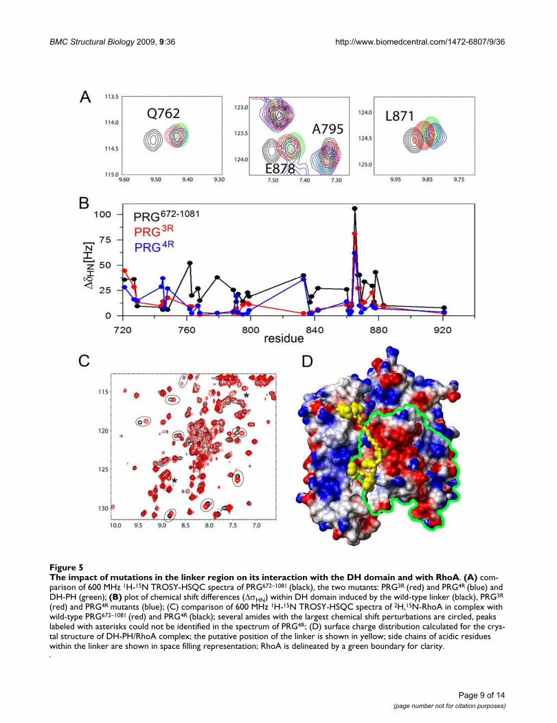
The fragment containing residues 672–712, upstream of the DH-PH tandem, interacts with residues within the DH domain and with RhoA in the binary complexFigure 4The fragment containing residues 672–712, upstream of the DH-PH tandem, interacts with residues within the DH domain and with RhoA in the binary complex. (A) comparison of 900 MHz 1H-15N TROSY-HSQC spectra of DH-PH (blue) and PRG672–1081 (red) showing chemical shift changes within DH domain induced by the linker; only select fragments of the spectrum are shown for clarity (B) a graph showing the magnitude of linker induced chemical shift changes (ΔσHN [Hz]) within the DH domain (blue) compared to the surface area of DH residues buried upon formation of the complex with RhoA (red); ΔσHN [Hz] were calculated as a differences between chemical shifts for PRG672–1081and DH-PH; the surface buried upon complex formation was calculated using the crystal structure of DHPH-RhoA complex and the program MOLMOL [38]; (C) crystal structure of the DH-PH/RhoA complex (PDB code 1XCG), showing the residues within the DH domain with the larg-est amide chemical shift changes induced by the linker (>50 Hz, red; 25 – 50 Hz, orange); the hypothetical position of the linker, postulated on the basis of chemical shift perturbations, is shown in yellow; side chains of acidic residues within the linker are shown in space filling representation; RhoA is shown as blue ribbon; (D) comparison of 600 MHz 1H-15N TROSY-HSQC spectra of RhoA in complex with DH-PH (blue) and PRG672–1081 (red); several RhoA amides with chemical shifts perturbed by the presence of the linker are circled.

BMC Structural Biology 2009, 9:36 http://www.biomedcentral.com/1472-6807/9/36
To better understand the role of individual amino acids,we generated four single-site mutants of the PRG712–1081
construct, i.e. D706R, E708R, E710R and D712R (seeabove). The E710R mutant shows significant autoinhibi-tion (it is actually less active in the in vitro assay thanPRG701–1081), but each of the remaining mutants exhibitscatalytic activity comparable to the isolated DH-PH tan-dem. It is therefore possible that the observed enhancedcatalytic potential of the PRG4R is due to additive effects ofindividual mutations. Interestingly, within the context ofthe intact four-domain construct (i.e. PRG37–1081) most ofthe autoinhibition is effectively restored, indicating thatboth the PDZ and RGSL domains play an important role.
To understand how mutations within the linker modulateGEF activity, we measured NMR spectra of both the tripleand quadruple charge reversal mutants of PRG672–1081
(henceforth denoted PRG3R and PRG4R) and analyzed thechemical shifts within the DH domains. We found thatboth mutants qualitatively show similar patterns of chemi-cal shift perturbations as observed for the wild-typePRG672–1081. However, the magnitude of chemical shiftchanges is significantly decreased compared to the wild-type protein (Figs. 5A &5B). To quantify this effect, we cal-culated the total chemical shift change, ΔσHN-total, i.e. a sumof chemical shift changes observed for all amides assignedto the DH domain, compared to the isolated DH-PH tan-dem. For PRG672–1081, PRG3R and PRG4R the values of ΔσHN-
total were 875, 480 and 404 Hz, respectively. Therefore, themutations within the linker decreased the chemical shiftchanges in the DH domain by approximately 50% com-pared to the wild-type protein. This is consistent with theobserved relief of the inhibitory effect, although it does notexplain the enhanced catalytic activity of PRG4R mutant.
The next step was to evaluate the effect of the mutations inthe linker on the direct interaction of the PRG712–1081 withRhoA. We measured 1H-15N TROSY-HSQC spectra of 15N-labeled RhoA in complexes with each of the two mutants,i.e. PRG3R, and PRG4R. The spectrum of RhoA in the pres-ence of PRG3R is essentially identical to that of RhoA in thepresence of the wild-type PRG672–1081 indicating no signif-icant effect of the mutations in the linker on the interac-tion with RhoA (data not shown). However, noticeablechanges are observed for RhoA in the presence of thePRG4R with over a dozen resonances shifted by more than25 Hz (Fig. 5C). As inferred from the NMR analysis, thelinker of the PRG4R mutant appears to interact with nega-tively charged surfaces of RhoA (Fig. 5D). Reversing thecharge of acidic residues within the linker appears to havea pronounced effect on the kinetics of DH-PH/RhoA inter-action and ultimately, the rate of nucleotide exchange.
The role of PDZ-RGSL domainsTo evaluate the impact of the mutations in the linkerregion on the autoinhibited core fragment of PRG con-
taining all four functional domains, we carried out thenucleotide exchange assay for PRG37–1081 with the D706R,E708R, E710R and D712R mutations. The rate ofexchange was increased from ~20, in the wild-type PRG37–
1081, to 43, i.e. only two-fold. Interestingly, this is equiva-lent to eliminating the PDZ and RGSL domains fromwild-type PRG37–1081. This result shows that autoinhibi-tion in the wild-type, full-length protein is an additiveeffect of a number of different interactions.
Functional assays are consistent with the in vitro modelIn order to assess if the in vitro studies are representative ofbiological properties of PDZRhoGEF in smooth muscle,we used a contractility assay of a permeabilized rabbit pul-monary artery (Fig. 6). In this assay, which measures forceunder isometric conditions, we are able to introducerecombinant protein directly into a smooth muscle strip,after contractile force is induced at pCa of 6.7. As weshowed elsewhere [37], the addition of the wild-typerecombinant DH-PH domain substantially increases thecontractile force via RhoA activation. When the PRG37–1081
fragment is added instead, the contractile force increasesmarginally, consistent with the autoinhibition of thatfragment, while the same fragment with the mutationsD706R, E708R, E710R, D712R, causes a significantlystronger contraction, albeit well below the DH-PH level,in full agreement with the in vitro assays.
We then conducted a rhotekin-based assay to measureRhoA activation in NIH 3T3 cells transfected with full-length, wild-type human PRG, as well as 3R and 4Rmutants of the full-length protein, and the isolated DH-PH tandem (Fig. 7). As expected, overexpression of theisolated DH-PH tandem resulted in significant increase inRhoA•GTP over the control. Lower but equal expressionlevels of wild-type and the 3R and 4R mutants in unstim-ulated serum starved cells resulted in greater Rho activa-tion over control with the 3R and 4R having the greatesteffect. These findings are consistent with significantautoinhibition of the full length PRG and also show thatthe 3R and 4R mutants are biologically active in cells.
The model of autoinhibitionOn the basis of the experiments described in this paper wepropose a mechanism for the autoinhibition of PDZRho-GEF (Fig. 8). Unlike in other GEFs for which similarmechanisms were rationalized at the structural level,[11,12,40], the DH domain of PRG is not stericallyobstructed by a single structural element, but instead sev-eral elements act synergistically within the autoinhibited,full-length protein. A fragment that spans all functionaldomains of PRG, i.e. PRG37–1081, retains only ~15% of thecatalytic activity of the isolated DH-PH tandem, and so weconcluded that intramolecular interactions within thisfragment are primarily responsible for autoinhibition.This was expected, because RGSL and PDZ domains are
Page 8 of 14(page number not for citation purposes)
BMC Structural Biology 2009, 9:36 http://www.biomedcentral.com/1472-6807/9/36
Page 9 of 14(page number not for citation purposes)
The impact of mutations in the linker region on its interaction with the DH domain and with RhoAFigure 5The impact of mutations in the linker region on its interaction with the DH domain and with RhoA. (A) com-parison of 600 MHz 1H-15N TROSY-HSQC spectra of PRG672–1081 (black), the two mutants: PRG3R (red) and PRG4R (blue) and DH-PH (green); (B) plot of chemical shift differences (ΔσHN) within DH domain induced by the wild-type linker (black), PRG3R
(red) and PRG4R mutants (blue); (C) comparison of 600 MHz 1H-15N TROSY-HSQC spectra of 2H,15N-RhoA in complex with wild-type PRG672–1081 (red) and PRG4R (black); several amides with the largest chemical shift perturbations are circled, peaks labeled with asterisks could not be identified in the spectrum of PRG4R; (D) surface charge distribution calculated for the crys-tal structure of DH-PH/RhoA complex; the putative position of the linker is shown in yellow; side chains of acidic residues within the linker are shown in space filling representation; RhoA is delineated by a green boundary for clarity.

BMC Structural Biology 2009, 9:36 http://www.biomedcentral.com/1472-6807/9/36
thought to play a role in the autoinhibition/activationmechanism. Surprisingly, our in vitro catalytic assayrevealed that removal of both of these domains results inonly 2-fold increase in activity, indicating that other fac-tors are involved. We traced the primary autoinhibitoryelement to a sequence motif upstream of the DH domain,harboring Asp706, Glu708 and Asp712, and Glu710. ThePRG fragment encompassing residues 691 to 1081, i.e.with only 21 amino acids upstream of the DH domain,
showed less than 50% of the activity of the DH-PH tan-dem. Replacing the four negatively charged residues witharginines not only abolished autoinhibition but gener-ated a 3.3 fold enhancement of the nucleotide exchangeactivity, as compared to the isolated DH-PH tandem.
A model of autoinhibition and activation of PDZRhoGEFFigure 8A model of autoinhibition and activation of PDZRhoGEF. In the inhibited protein the linker between the RGSL and DH domains is sequestered by the two modules, and the DH domain is not accessible to RhoA. Interaction with the Gα subu-nit, and possibly direct interaction with relevant GPCRs via the PDZ domain, relieve this inhibition by first dislodging the RGSL domain and then the inhibitory portion of the linker.
Increase in the endogenous RhoA activation in NIH 3T3 cells monitored by the Rhotekin assayFigure 7Increase in the endogenous RhoA activation in NIH 3T3 cells monitored by the Rhotekin assay. Upper panel: Rhotekin pull-down of RhoA•GTP from cells ectopi-cally expressing full-length wild type (wt) PRG, the isolated DH-PH tandem and the 3R and 4R mutants of the full-length form. The cells were under serum free conditions. Bottom panel: quantitative assessment of RhoA activation using a set of three independent experiments. The isolated DH-PH tan-dem is not included because the expression level was signifi-cantly higher than that of the three full-length PRG variants, and while the increased endogenous RhoA activity as meas-ured by the Rhotekin assay (see Methods Section), demon-strating that these expressed constructs are biologically active in cells. Mutant PRG-RhoGEFS were FLAG tagged. Western blotting for FLAG indicated that the mutants were expressed at an equal level while the DH-PH domain was overexpressed (data not shown) accounting for the greater level of active RhoA.
Contractility assayFigure 6Contractility assay. Wild type (wt) and 4R mutant of the core fragment of PRG were added to β-escin permeabilized pulmonary artery precontracted with pCa 6.7 solution con-taining 1 μM calmodulin and 2 μM GTP. Concomitant increase in force is consistent with activation of the RhoA pathway. The 4R mutant, 10–15 μM, induced significantly greater Ca2+-sensitized force than the wild type protein (p = 0.002; n = 11. The magnitudes of the responses are normal-ized to pCa 6.7-induced force developed before addition of the recombinant PRG variants.
Page 10 of 14(page number not for citation purposes)

BMC Structural Biology 2009, 9:36 http://www.biomedcentral.com/1472-6807/9/36
However, when the mutations were introduced into thePRG37–1081 core fragment containing all four functionaldomains, autoinhibition is largely restored, presumablyby interactions mediated by the RGSL and/or PDZdomains which may stabilize the autoinhibitory elementin its functional conformation in spite of the mutations.
Based on these results, we suggest that the activation ofPRG, and probably also of the two other remaining mem-bers of this family of GEFs, i.e. LARG and p115, proceedsby a multistep mechanism. The primary autoinhibitoryelement resides upstream of the DH-PH tandem, but maynot be accessible due to secondary interactions with RGSLand/or PDZ domains. This is why a conformationalchange involving the latter domains is essential, but notsufficient for full GEF activation. In agreement with thismodel, activated, GTP-bound Gα13 can bind directly to afragment containing both the linker and DH-PH tandemof the p115 GEF, but without concomitant activation ofthe nucleotide exchange activity [41]. Thus, both theRGSL domain and the 'activation box' may act synergisti-cally. Also, activation of GEF activity can be partiallyuncoupled from binding of Gα13 to the RGSL domain inp115 GEF: point mutations in the latter that reduce affin-ity for Gα13 have no effect on the activation of nucleotideexchange of p115 GEF [42]. Analogous mutants do notaffect translocation of p115 GEF to particulate fractions inthe presence of Gα13, suggesting that translocation ofp115 GEF is due to complex mechanisms involving ele-ments other than the RGSL domain [43].
ConclusionWe here propose that in the closed, autoinhibited form, theRGSL domain interacts with both the RGSL-DH linker andthe DH domain, or perhaps only with the linker, whichserves as the direct inhibitory element (Fig. 7). When theRGSL domain binds to Gα12/13, the inhibitory elementremains in place, but becomes exposed and accessible tosecondary interactions which in consequence remove thelinker from the functional DH surfaces. The dramatic effectof the charge reversal mutant also suggests that electrostaticinteractions could play a major role, not only in relievingthe autoinhibition, but also in positive regulation of thekinetics of RhoA binding and nucleotide exchange. It is typ-ically assumed that the intrinsic catalytic properties of theDH domain, or more commonly the DH-PH tandem,reflect the maximum theoretical nucleotide exchange rateof the respective GEF. However, this is not always true. Theisolated DH domain of Vav enhances the nucleotideexchange rate on Rac1 73-fold, but the addition of the PH-CRD fragment produces a further 13-fold enhancement[44]. Thus, it is possible that an activating interaction, e.g.involving Gα subunits, may not only relieve the autoinhi-bition, but also directly up-regulate the GEF activity. In thatcontext, we note that the human Gα12 and Gα13 subunitsdiffer with regard to their pI values, i,e. 9.8 and 8.2, respec-
tively, and the positively charged surfaces could be impor-tant in the interaction with the DH/RhoA complex.
Finally, we note that the proposed mechanism might berelevant to all three GEFs of the RGSL-family. The aminoacid sequences of both LARG and p115 show similar neg-atively charged patches in nearly identical locations:LARG contains a Glu-Asp-Glu tripeptide, while p115 GEFhas an Asp-Glu-Gly-Glu motif (Additional File 1: FigureS2). Further work, underway in our laboratories, willhopefully establish the details of these pathways.
MethodsProtein Expression and PurificationFull-length and truncated versions of human PDZRhoGEFwere PCR-amplified and subcloned into pDEST15 (Invit-rogene) vector containing a glutathione S-transferase(GST) tag at the N-terminus, an rTEV protease sitebetween GST and the insert, and a non-cleavable His8 tagat the C terminus. All constructs were verified by directsequencing prior to protein expression. The proteins wereexpressed in the E. coli strain BL21 (DE3) RIPL (Strate-gene). Cell cultures were grown at 37°C in TB/ampicillin(100 μg/ml) and chloramphenicol (34 μg/ml), andinduced with 1 mM isopropyl β-D-thiogalactopyranoside(IPTG) overnight at 18°C. Cell pellets were resuspendedin 50 mM Tris pH 7.8, 300 mM NaCl, 0.5 mM EDTA, 1mM dithiothreitol (DTT) (Buffer A), lysed using Sonifier450 (VWR), and clarified by centrifugation at 40,000 × gfor 45 min at 4°C. Supernatant was loaded on a glutath-ione-Sepharose 4B column (Pharmacia), pre-equilibratedwith Buffer A. Protein was bound to the resin by gentlyrocking the column at 4°C for 1 hour and eluted with 20mM glutathione in 50 mM Tris, pH 8.0, 50 mM NaCl, 0.5mM EDTA, 1 mM DTT (Buffer B). Fractions containingpure fusion protein were pooled, digested overnight withrTEV protease in the same buffer and then dialyzedagainst Buffer B to remove glutathione. After rTEV cleav-age, the protein solution was loaded again on glutath-ione-Sepharose 4B column and rocked for 1 hour toremove GST and the uncut fusion protein. Flow-throughwas collected and diluted with 50 mM Tris pH 8.0, 150mM NaCl (Buffer C). The solution was then loaded ontoNi-NTA column pre-equilibrated with buffer C and elutedwith 200 mM imidazole in Buffer C. Fractions containingpure protein were pooled and dialyzed against 20 mM TrispH 7.5, 150 mM NaCl, 1 mM DTT. Pure protein was con-centrated and stored at -80°C for the fluorescence assay.
Proteins labeled with stable isotopes (2H, 13C, 15N) wereexpressed in minimal media with (15NH4)2SO4 as solesource of nitrogen and 13C- or 2H,13C-glucose as a sourceof carbon. The media were enhanced by the addition oflabeled BioExpress (Cambridge Isotope Labs.). Four typesof labeling scheme were used: 15N, 13C 15N, 2H 15N and 2H13C 15N.
Page 11 of 14(page number not for citation purposes)

BMC Structural Biology 2009, 9:36 http://www.biomedcentral.com/1472-6807/9/36
The expression and purification of human RhoAF25N (res-idues 1–181) was carried out as described previously [45].
Guanine Nucleotide Exchange AssayFluorescence spectroscopy analysis of N-methylan-thraniloyl (mant)-GTP incorporation into RhoA was car-ried out using a FluoroMax-3 (Jobin Yvon Inc.)spectrofluorimeter. The exchange reaction was carried outat 22°C for 5 min in a 1.5 ml Eppendorf tube containing1 μM GDP-preloaded RhoA and 0.5 μM mant-GTP in 20mM Tris-HCl, (pH 7.5), 50 mM NaCl, 5 mM MgCl2 and 1mM DTT. After equilibration, assayed proteins wereadded at 0.1 μM concentration and increases in mant-GTPfluorescence was monitored (λex = 356 nm, λem = 450nm). The control experiment, in the absence of other pro-teins, reflects intrinsic exchange activity of RhoA, meas-ured after equivalent equilibration time. Each exchangeexperiment was carried out three times independently.The initial rates of guanine nucleotide exchange weredetermined by regression analysis. The average variationamong measured rate did not exceed 10%. Fold stimula-tion values were calculated as the ratio of the initialexchange rate of DH-PH-stimulated reaction to the intrin-sic rate of exchange for the wild type RhoA.
Optimization of DH-PH samples for NMR experimentsIn order to optimize both the stability of protein samplesand the sensitivity of NMR experiments, we screened a largenumber of different buffers. We prepared 0.5 mM solution ofDH-PH in several buffers with varied salt concentrations andkept the samples at 4 – 35°C with periodic monitoring of theconcentration and the level of sample degradation usingSDS-PAGE. We found the best solubility and stability of DH-PH samples in 50 mM TRIS buffer, pH 7.5 and 150 mMNaCl. However, such conditions do not provide optimalsensitivity for cryogenic probes due to high salt concentra-tion [46]. Therefore, to achieve the highest possible sensitiv-ity of NMR experiments we tested several salt-free buffers andfound a mixture of 200 mM MOPS/TRIS, pH 7.5, to be opti-mal. In such conditions DH-PH can remain in solution at aconcentration above 0.3 mM for over a week at 25°C. Highertemperature, ~30°C, reduces the lifetime of the sample toapproximately 3–4 days.
Because backbone assignment of perdeuterated proteinsdepends on efficient back-exchange of amide protons, wedissolved 15N-labeled DH-PH in D2O based buffer andmonitored H/D exchange using the HSQC spectra. Weobserved complete exchange of amide protons over oneweek.
Chemical shift assignmentTo assign the backbone amide chemical resonances of iso-lated DH and PH domains, we used standard triple reso-nance experiments (HNCO, HNCA, HN(CO)CA,HNCACB, CBCA(CO)NH and 3D 15N-edited NOESY
with 200 ms mixing time). The spectra for 0.3 mM2H,13C,15N-labeled DH domain in 200 mM MOPS/TRISbuffer, pH 7.5, and 2 mM DTT were collected using VarianInova 600 MHz at 25°C. The spectra for 13C,15N-labelledPH domain were recorded using Varian Inova 500 MHzfor 0.5 – 0.7 mM protein concentrations in 600 mM NaCl,50 mM TRIS pH 7.0 and 2 mM DTT buffer at 25°C.
To assign the resonances in the intact DH-PH tandem, weused 2H,13C,15N labeled protein at 0.3–0.4 mM concen-tration in 200 mM MOPS/TRIS buffer at pH 7.5 and 2 mMDTT. The NMR spectra were collected at 30°C using Var-ian Inova 800 MHz spectrometer equipped with cryogenicprobe. The following TROSY-based experiments were con-ducted: TROSY-HNCA, TROSY-HNCO, 3D 15N-editedTROSY-NOESY with 250 ms mixing time.
Chemical shift perturbation experimentsIn order to analyze chemical shift perturbations we meas-ured series of experiments using Varian Inova 900 MHz, Var-ian Inova 600 MHz and Bruker Avance 600 MHzspectrometers equipped with cryogenic probes. Samples forNMR contained 0.2 to 0.3 mM protein concentrations in200 mM MOPS/TRIS buffer, pH 7.5 with 1 mM DTT. All ofthe 1H-15N TROSY-HSQC spectra were collected at 25°C.
Tissue Preparation and Force MeasurementsRabbits were anesthetized and killed with isoflurane.Small strips (100–150 μm thick, 150–200 μm wide, and2–3 mm long) of rabbit pulmonary artery were dissectedand freed from connective tissue. Single strips were tiedwith monofilament silk to the fine tips of two tungstenneedles, one of which was connected to a force transducerand mounted in a well on a bubble plate, as described pre-viously [47]. After measuring contractions induced byhigh 154 mM K+, the strips were incubated at room tem-perature (21–22°C) in Ca2+ free relaxing solution (G1)containing 4.5 mM MgATP and 1 mM EGTA for severalminutes and permeabilized with 75 uM β-escin for 25min. This permeabilization protocol permits penetrationof molecules up to 150 kDa [48]. To deplete the sarcoplas-mic reticulum of calcium [47]. and inhibit NO-synthaseactivity, every strip was also treated with 10 μM A23187(Calbiochem) and 10 μM L-NAME (Sigma) (for addi-tional 10 min in the G1 solution. Buffer in protein sam-ples of wild type and 4R mutant PRG core fragment(PRG37–1081) was exchanged against pCa 6.7, 1 μM cal-modulin and 2 μM GTP. Samples of ~10–15 μM of recom-binant protein were used to stimulate the force responses.
The Rhotekin assayThe cDNA of full-length wild-type PDZ-RhoGEF, as wellas 3R and 4R mutants and the isolated DH-PH tandemwere PCR amplified with flanking restriction sites andintroduced into p3XFLAG-myc-CMV-24 mammalianexpression vector (Sigma-Aldrich, St. Louis MO). NIH 3T3
Page 12 of 14(page number not for citation purposes)

BMC Structural Biology 2009, 9:36 http://www.biomedcentral.com/1472-6807/9/36
cells were cultured on a 10 cm culture plate in Dulbecco'sModified Eagle Medium (DMEM; Invitrogen-Gibco) sup-plemented with 10% fetal bovine serum (FBS; Invitrogen-Gibco) at 37°C in 5% CO2. The plasmids for the ectopicexpression with N-terminal 3XFLAG and C-terminal myc-tag, and coding sequences for the full-length wild-type, 3Ror 4R mutant PDZ-RhoGEF or the DH-PD tandem weretransfected into cells using Lipofectamine 2000 (Invitro-gen) following manufacture's standard protocol. The cellswere treated for transfection, cultured overnight, treatedfor transfection for the second time and cultured inDMEM without FBS, changed 8 hours after the secondtransfection, overnight. RhoA activation was determinedby precipitation of active GTP-bound RhoA (RhoA•GTP)with a glutathione S-transferase (GST)-fusion protein ofthe Rho-binding domain (RBD) of the Rho effectorrhotekin as previously described [49]. Briefly, the cells ona 10 cm plate were lysed in 500 μl of ice cold lysis buffer(25 mM HEPES, pH 7.5; 150 mM NaCl; 1% NP-40; 10mM MgCl2; 1 mM EDTA; 6% Glycerol; 1% Proteaseinhibitor cocktail (Sigma-Aldrich, St. Louis MO)), andcleared by centrifugation at 14,000 g for 10 minutes at4°C. Ectopic expression of full-length wild-type, 3R and4R mutant PDZ-RhoGEF at an equal level and the DH-PDtandem over-expression were confirmed by subjecting thesupernatant to Western blotting for FLAG epitope tag(data not shown). The supernatant was added to GST-RBD agarose beads (Millipore, Billerica MA) and affinityprecipitated active RhoA•GTP was detected by Westernblotting for RhoA using a mouse monoclonal antibody(Santa Cruz Biotechnology Inc., Santa Cruz CA). Detec-tion and quantification of the signal was performed usingthe Odyssey system software (Li-Cor, Lincoln NE).
Authors' contributionsMZ expressed and purified all protein constructs and con-ducted the nucleotide exchange assays. TC conducted theNMR experiments, analyzed the data and drafted relevantsections of the manuscript. KM carried out the Rhotekinassay. MVA carried out force measurements. UD, JHB, AVSand ZSD conceived the project, analyzed the data anddrafted the manuscript. All authors read and approved thefinal manuscript.
Additional material
AcknowledgementsThis work was supported by NIH Grant PO1 HL48807. We thank Natalya Olekhnovich for technical assistance and Dr. J. Otlewski (University of Wrocław, Poland) for discussions. This research benefited from access to the Southeast Collaboratory for High-Field Biomolecular NMR, a resource at the University of Georgia, funded by the National Institute of General Medical Sciences (GM66340) and the Georgia Research Alliance. We thank Dr. John Glushka and Dr. Yizhou Liu for assistance in setting up NMR experiments.
References1. Jaffe AB, Hall A: RHO GTPASES: Biochemistry and Biology.
Annu Rev Cell Dev Biol 2005, 21:247-269.2. Raftopoulou M, Hall A: Cell migration: Rho GTPases lead the
way. Dev Biol 2004, 265:23-32.3. Etienne-Manneville S, Hall A: Rho GTPases in cell biology. Nature
2002, 420:629-635.4. Kandpal RP: Rho GTPase activating proteins in cancer pheno-
types. Curr Protein Pept Sci 2006, 7:355-365.5. Rossman KL, Der CJ, Sondek J: GEF means go: turning on RHO
GTPases with guanine nucleotide-exchange factors. Nat RevMol Cell Biol 2005, 6:167-180.
6. Whitehead IP, Campbell S, Rossman KL, Der CJ: Dbl family pro-teins. Biochim Biophys Acta 1997, 1332:F1-23.
7. Erickson JW, Cerione RA: Structural elements, mechanism,and evolutionary convergence of Rho protein-guanine nucle-otide exchange factor complexes. Biochemistry 2004,43:837-842.
8. Worthylake DK, Rossman KL, Sondek J: Crystal structure of Rac1in complex with the guanine nucleotide exchange region ofTiam1. Nature 2000, 408:682-688.
9. Aghazadeh B, Lowry WE, Huang XY, Rosen MK: Structural basisfor relief of autoinhibition of the Dbl homology domain ofproto-oncogene Vav by tyrosine phosphorylation. Cell 2000,102:625-633.
10. Mitin N, Betts L, Yohe ME, Der CJ, Sondek J, Rossman KL: Releaseof autoinhibition of ASEF by APC leads to CDC42 activationand tumor suppression. Nat Struct Mol Biol 2007, 14:814-823.
11. Murayama K, Shirouzu M, Kawasaki Y, Kato-Murayama M, Hanawa-Suetsugu K, Sakamoto A, Katsura Y, Suenaga A, Toyama M, Terada T,et al.: Crystal structure of the rac activator, Asef, reveals itsautoinhibitory mechanism. J Biol Chem 2007, 282:4238-4242.
12. Rojas RJ, Yohe ME, Gershburg S, Kawano T, Kozasa T, Sondek J: Gal-phaq directly activates p63RhoGEF and Trio via a conservedextension of the Dbl homology-associated pleckstrin homol-ogy domain. J Biol Chem 2007, 282:29201-29210.
13. Kozasa T, Jiang X, Hart MJ, Sternweis PM, Singer WD, Gilman AG,Bollag G, Sternweis PC: p115 RhoGef, a Gtpase activating pro-tein for galpha12 and galpha13 [in Process Citation]. Science1998, 280:2109-2111.
14. Fukuhara S, Murga C, Zohar M, Igishi T, Gutkind JS: A novel PDZdomain containing guanine nucleotide exchange factor linksheterotrimeric G proteins to Rho. J Biol Chem 1999,274:5868-5879.
15. Fukuhara S, Chikumi H, Gutkind JS: Leukemia-associated Rhoguanine nucleotide exchange factor (LARG) links heterot-rimeric G proteins of the G(12) family to Rho. FEBS Lett 2000,485:183-188.
16. Kourlas PJ, Strout MP, Becknell B, Veronese ML, Croce CM, Theil KS,Krahe R, Ruutu T, Knuutila S, Bloomfield CD, Caligiuri MA: Identifi-cation of a gene at 11q23 encoding a guanine nucleotideexchange factor: evidence for its fusion with MLL in acutemyeloid leukemia. Proc Natl Acad Sci USA 2000, 97:2145-2150.
17. Chikumi H, Barac A, Behbahani B, Gao Y, Teramoto H, Zheng Y, Gut-kind JS: Homo- and hetero-oligomerization of PDZ-RhoGEF,LARG and p115RhoGEF by their C-terminal region regu-lates their in vivo Rho GEF activity and transforming poten-tial. Oncogene 2004, 23:233-240.
18. Longenecker KL, Lewis ME, Chikumi H, Gutkind JS, Derewenda ZS:Structure of the RGS-like domain from PDZ-RhoGEF: link-ing heterotrimeric g protein-coupled signaling to RhoGTPases. Structure (Camb) 2001, 9:559-569.
19. Chen Z, Wells CD, Sternweis PC, Sprang SR: Structure of thergRGS domain of p115RhoGEF. Nat Struct Biol 2001, 8:805-809.
Additional file 1Supplementary figures. Figure S1 Coomassie stained SDS-PAGE gel illustrating the purity of protein samples used for functional studies. Fig-ure S2 Sequence alignment of the putative regulatory motifs upstream of the DH domains of PRG, LARG and p115 guanine exchange factors for RhoA.Click here for file[http://www.biomedcentral.com/content/supplementary/1472-6807-9-36-S1.doc]
Page 13 of 14(page number not for citation purposes)
http://www.ncbi.nlm.nih.gov/entrez/query.fcgi?cmd=Retrieve&db=PubMed&dopt=Abstract&list_uids=9061011
http://www.ncbi.nlm.nih.gov/entrez/query.fcgi?cmd=Retrieve&db=PubMed&dopt=Abstract&list_uids=9061011
http://www.ncbi.nlm.nih.gov/entrez/query.fcgi?cmd=Retrieve&db=PubMed&dopt=Abstract&list_uids=9641915

BMC Structural Biology 2009, 9:36 http://www.biomedcentral.com/1472-6807/9/36
Publish with BioMed Central and every scientist can read your work free of charge
"BioMed Central will be the most significant development for disseminating the results of biomedical research in our lifetime."
Sir Paul Nurse, Cancer Research UK
Your research papers will be:
available free of charge to the entire biomedical community
peer reviewed and published immediately upon acceptance
cited in PubMed and archived on PubMed Central
yours — you keep the copyright
Submit your manuscript here:http://www.biomedcentral.com/info/publishing_adv.asp
BioMedcentral
20. Chen Z, Singer WD, Sternweis PC, Sprang SR: Structure of thep115RhoGEF rgRGS domain-Galpha13/i1 chimera complexsuggests convergent evolution of a GTPase activator. NatStruct Mol Biol 2005, 12:191-197.
21. Kreutz B, Hajicek N, Yau DM, Nakamura S, Kozasa T: Distinctregions of Galpha13 participate in its regulatory interactionswith RGS homology domain-containing RhoGEFs. Cell Signal2007, 19:1681-1689.
22. Suzuki N, Nakamura S, Mano H, Kozasa T: Galpha 12 activatesRho GTPase through tyrosine-phosphorylated leukemia-associated RhoGEF. Proc Natl Acad Sci USA 2003, 100:733-738.
23. Sternweis PC, Carter AM, Chen Z, Danesh SM, Hsiung YF, SingerWD: Regulation of Rho guanine nucleotide exchange factorsby G proteins. Adv Protein Chem 2007, 74:189-228.
24. Jemth P, Gianni S: PDZ domains: folding and binding. Biochemis-try 2007, 46:8701-8708.
25. Nourry C, Grant SG, Borg JP: PDZ domain proteins: plug andplay! Sci STKE 2003, 2003:RE7.
26. Hung AY, Sheng M: PDZ domains: structural modules for pro-tein complex assembly. J Biol Chem 2002, 277:5699-5702.
27. Swiercz JM, Kuner R, Behrens J, Offermanns S: Plexin-B1 DirectlyInteracts with PDZ-RhoGEF/LARG to Regulate RhoA andGrowth Cone Morphology. Neuron 2002, 35:51-63.
28. Aurandt J, Vikis HG, Gutkind JS, Ahn N, Guan KL: The semaphorinreceptor plexin-B1 signals through a direct interaction withthe Rho-specific nucleotide exchange factor, LARG. Proc NatlAcad Sci USA 2002, 99:12085-12090.
29. Perrot V, Vazquez-Prado J, Gutkind JS: Plexin B regulates Rhothrough the guanine nucleotide exchange factors Leukemia-associated RhoGEF (LARG) and PDZ-RhoGEF. J Biol Chem2002, 277:43115-43120.
30. Yamada T, Ohoka Y, Kogo M, Inagaki S: Physical and functionalinteractions of the lysophosphatidic acid receptors with PDZdomain-containing Rho guanine nucleotide exchange factors(RhoGEFs). J Biol Chem 2005, 280:19358-19363.
31. Longhurst DM, Watanabe M, Rothstein JD, Jackson M: Interactionof PDZRhoGEF with microtubule-associated protein 1 lightchains: link between microtubules, actin cytoskeleton, andneuronal polarity. J Biol Chem 2006, 281:12030-12040.
32. Bourguignon LY, Gilad E, Brightman A, Diedrich F, Singleton P:Hyaluronan-CD44 interaction with leukemia-associatedRhoGEF and epidermal growth factor receptor promotesRho/Ras co-activation, phospholipase C epsilon-Ca2+ signal-ing, and cytoskeleton modification in head and neck squa-mous cell carcinoma cells. J Biol Chem 2006, 281:14026-14040.
33. Oleksy A, Opalinski L, Derewenda U, Derewenda ZS, Otlewski J:The molecular basis of RhoA specificity in the guanine nucle-otide exchange factor PDZ-RhoGEF. J Biol Chem 2006,281:32891-32897.
34. Bi F, Debreceni B, Zhu K, Salani B, Eva A, Zheng Y: Autoinhibitionmechanism of proto-Dbl. Mol Cell Biol 2001, 21:1463-1474.
35. Yohe ME, Rossman KL, Gardner OS, Karnoub AE, Snyder JT, Gersh-burg S, Graves LM, Der CJ, Sondek J: Auto-inhibition of the Dblfamily protein Tim by an N-terminal helical motif. J Biol Chem2007, 282:13813-13823.
36. Li P, Martins IR, Amarasinghe GK, Rosen MK: Internal dynamicscontrol activation and activity of the autoinhibited Vav DHdomain. Nat Struct Mol Biol 2008, 15:613-618.
37. Derewenda U, Oleksy A, Stevenson AS, Korczynska J, Dauter Z,Somlyo AP, Otlewski J, Somlyo AV, Derewenda ZS: The crystalstructure of RhoA in complex with the DH/PH fragment ofPDZRhoGEF, an activator of the Ca(2+) sensitization path-way in smooth muscle. Structure (Camb) 2004, 12:1955-1965.
38. Koradi R, Billeter M, Wuthrich K: MOLMOL: a program for dis-play and analysis of macromolecular structures. J Mol Graph1996, 14:51-55.
39. Amarasinghe GK, Rosen MK: Acidic region tyrosines provideaccess points for allosteric activation of the autoinhibitedVav1 Dbl homology domain. Biochemistry 2005, 44:15257-15268.
40. Lutz S, Shankaranarayanan A, Coco C, Ridilla M, Nance MR, Vettel C,Baltus D, Evelyn CR, Neubig RR, Wieland T, Tesmer JJ: Structure ofGalphaq-p63RhoGEF-RhoA complex reveals a pathway forthe activation of RhoA by GPCRs. Science 2007, 318:1923-1927.
41. Wells CD, Liu MY, Jackson M, Gutowski S, Sternweis PM, RothsteinJD, Kozasa T, Sternweis PC: Mechanisms for reversible regula-
tion between G13 and Rho exchange factors. J Biol Chem 2002,277:1174-1181.
42. Chen Z, Singer WD, Wells CD, Sprang SR, Sternweis PC: Mappingthe Galpha13 binding interface of the rgRGS domain ofp115RhoGEF. J Biol Chem 2003, 278:9912-9919.
43. Bhattacharyya R, Wedegaertner PB: Galpha 13 requires palmi-toylation for plasma membrane localization, Rho-dependentsignaling, and promotion of p115-RhoGEF membrane bind-ing. J Biol Chem 2000, 275:14992-14999.
44. Brooun A, Foster SA, Chrencik JE, Chien EY, Kolatkar AR, Streiff M,Ramage P, Widmer H, Weckbecker G, Kuhn P: Remedial strate-gies in structural proteomics: expression, purification, andcrystallization of the Vav1/Rac1 complex. Protein Expr Purif2007, 53:51-62.
45. Ihara K, Muraguchi S, Kato M, Shimizu T, Shirakawa M, Kuroda S, Kai-buchi K, Hakoshima T: Crystal structure of human RhoA in adominantly active form complexed with a GTP analogue. JBiol Chem 1998, 273:9656-9666.
46. Kelly AE, Ou HD, Withers R, Dotsch V: Low-conductivity buffersfor high-sensitivity NMR measurements. J Am Chem Soc 2002,124:12013-12019.
47. Kitazawa T, Kobayashi S, Horiuti K, Somlyo AV, Somlyo AP: Recep-tor-coupled, permeabilized smooth muscle. Role of thephosphatidylinositol cascade, G-proteins, and modulation ofthe contractile response to Ca2+. J Biol Chem 1989,264:5339-5342.
48. Iizuka K, Ikebe M, Somlyo AV, Somlyo AP: Introduction of highmolecular weight (IgG) proteins into receptor coupled, per-meabilized smooth muscle. Cell Calcium 1994, 16:431-445.
49. Ren XD, Kiosses WB, Schwartz MA: Regulation of the smallGTP-binding protein Rho by cell adhesion and the cytoskel-eton. Embo J 1999, 18:578-585.
Page 14 of 14(page number not for citation purposes)
http://www.ncbi.nlm.nih.gov/entrez/query.fcgi?cmd=Retrieve&db=PubMed&dopt=Abstract&list_uids=8744573
http://www.ncbi.nlm.nih.gov/entrez/query.fcgi?cmd=Retrieve&db=PubMed&dopt=Abstract&list_uids=8744573
http://www.ncbi.nlm.nih.gov/entrez/query.fcgi?cmd=Retrieve&db=PubMed&dopt=Abstract&list_uids=9545299
http://www.ncbi.nlm.nih.gov/entrez/query.fcgi?cmd=Retrieve&db=PubMed&dopt=Abstract&list_uids=9545299
http://www.ncbi.nlm.nih.gov/entrez/query.fcgi?cmd=Retrieve&db=PubMed&dopt=Abstract&list_uids=2494163
http://www.ncbi.nlm.nih.gov/entrez/query.fcgi?cmd=Retrieve&db=PubMed&dopt=Abstract&list_uids=2494163
http://www.ncbi.nlm.nih.gov/entrez/query.fcgi?cmd=Retrieve&db=PubMed&dopt=Abstract&list_uids=2494163
http://www.ncbi.nlm.nih.gov/entrez/query.fcgi?cmd=Retrieve&db=PubMed&dopt=Abstract&list_uids=7712537
http://www.ncbi.nlm.nih.gov/entrez/query.fcgi?cmd=Retrieve&db=PubMed&dopt=Abstract&list_uids=7712537
http://www.ncbi.nlm.nih.gov/entrez/query.fcgi?cmd=Retrieve&db=PubMed&dopt=Abstract&list_uids=7712537
http://www.ncbi.nlm.nih.gov/entrez/query.fcgi?cmd=Retrieve&db=PubMed&dopt=Abstract&list_uids=9927417
http://www.ncbi.nlm.nih.gov/entrez/query.fcgi?cmd=Retrieve&db=PubMed&dopt=Abstract&list_uids=9927417