A nucleotide-level coarse-grained model of RNA
25
arXiv:1403.4180v1 [physics.bio-ph] 17 Mar 2014 A nucleotide-level coarse-grained model of RNA Petr ˇ Sulc, 1 Flavio Romano, 2 Thomas E. Ouldridge, 1 Jonathan P. K. Doye, 2 and Ard A. Louis 1 1) Rudolf Peierls Centre for Theoretical Physics, University of Oxford, 1 Keble Road, Oxford, OX1 3NP, United Kingdom 2) Physical and Theoretical Chemistry Laboratory, Department of Chemistry, University of Oxford, South Parks Road, Oxford, OX1 3QZ, United Kingdom (Dated: 18 March 2014) We present a new, nucleotide-level model for RNA, oxRNA, based on the coarse-graining methodology recently developed for the oxDNA model of DNA. The model is designed to reproduce structural, mechanical and thermodynamic properties of RNA, and the coarse-graining level aims to retain the relevant physics for RNA hybridization and the structure of single- and double-stranded RNA. In order to explore its strengths and weaknesses, we test the model in a range of nanotechnological and biological settings. Applications explored include the folding thermodynamics of a pseudoknot, the formation of a kissing loop complex, the structure of a hexagonal RNA nanoring, and the unzipping of a hairpin motif. We argue that the model can be used for efficient simulations of the structure of systems with thousands of base pairs, and for the assembly of systems of up to hundreds of base pairs. The source code implementing the model is released for public use. I. INTRODUCTION RNA (ribonucleic acid) strands are polymers that play crucial cellular roles in gene expression, translation and regulation. 1 RNA is composed of units called nucleotides, which consist of a phosphate and ribose sugar backbone to which one of four different bases is attached: adenine (A), guanine (G), cytosine (C) or uracil (U). DNA has a similar chemical structure, but thymine (T) is present in place of uracil and the backbone includes deoxyribose sugars instead of ribose sugars. Both RNA and DNA can form double-helical molecules, stabilized by hydrogen bonds between complementary Watson-Crick base pairs: AU (AT in case of DNA) and GC. For RNA, wobble base pairs (GU) can also stabilize the duplex form and are commonly found. While DNA typically forms a B-helix, double-stranded RNA is typically found in a related, but different A-helix geometry. 2 DNA’s role in biological systems is to store genetic information and it is most often found in its double- helical form. By contrast, most naturally occurring RNA molecules are single-stranded and fold into com- plex structures that contain double-helical segments as well as loops, junctions and numerous tertiary struc- ture interactions that further stabilize the molecule. The lengths of RNA strands found in living cells can range from a few nucleotides to several thousands or even tens of thousands. 3 DNA nanotechnology 4 uses DNA molecules to create nanoscale structures and active devices. Examples of experimentally realized systems include DNA motors, 5,6 self-assembled nanostructures such as DNA origamis 7 and the use of DNA strands for computation. 8 Since RNA molecules are more difficult to handle and preserve than DNA in experimental settings, developing RNA- based nanodevices has been a more challenging task. However, the emerging field of RNA nanotechnology 9 of- fers promising applications of RNA-based nanodevices both in vitro and in vivo. The general design principles from DNA nanotechnology can be applied to RNA, but because specific RNA sequences form functional struc- tures that are known to interact with proteins and other RNA molecules in the cell, RNA molecules would be the natural choice for nanotechnology devices operating in- side cells. 10 Successful examples of experimentally real- ized RNA nanotechnology include self-assembling RNA nanocubes 11 and RNA tiles. 12 Recently, an RNA strand displacement reaction cascade was proposed that could be used for conditional gene silencing inside the cell. 13 Because of its importance for biological systems, RNA has been the subject of intensive study. In addition to nu- merous experimental studies, a range of theoretical and computational methods have been applied to the study of its properties. Currently, there are multiple tools and computational approaches that can describe RNA struc- ture at various levels of detail. 14,15 In an important series of works 16–22 the thermodynam- ics of RNA secondary structure (i.e., the list of base pairs in the folded state of RNA) was characterized in terms of a nearest-neighbor model for calculating the free ener- gies of various secondary-structure motifs. The nearest- neighbor model is the basis of various tools for the pre- diction of the secondary structure. Such tools typically use dynamic programming approaches to find the sec- ondary structure with minimal free energy. 23–25 Further- more, some tools have been extended by adding simple kinetic descriptions to the nearest-neighbor thermody- namics, allowing folding transitions to be modeled. 26,27 Although these methods are typically very fast, the fun- damentally discrete nature of the description and the lack of structural and mechanical detail places a limit on what they can treat. At the highest level of detail, quantum chemistry meth- ods have been employed to study the interactions be- tween nucleotides. 28,29 Due to the complexity of the cal- culations, such methods remain limited to interactions between nearest-neighbor base pairs in vacuum. Fully atomistic molecular dynamics simulation packages such as Amber 30 or CHARMM 31 include the solvent, and use
-
Upload
independent -
Category
Documents
-
view
1 -
download
0
Transcript of A nucleotide-level coarse-grained model of RNA

arX
iv:1
403.
4180
v1 [
phys
ics.
bio-
ph]
17
Mar
201
4
A nucleotide-level coarse-grained model of RNAPetr Sulc,1 Flavio Romano,2 Thomas E. Ouldridge,1 Jonathan P. K. Doye,2 and Ard A. Louis11)Rudolf Peierls Centre for Theoretical Physics, University of Oxford, 1 Keble Road, Oxford, OX1 3NP,
United Kingdom2)Physical and Theoretical Chemistry Laboratory, Department of Chemistry, University of Oxford, South Parks Road,
Oxford, OX1 3QZ, United Kingdom
(Dated: 18 March 2014)
We present a new, nucleotide-level model for RNA, oxRNA, based on the coarse-graining methodology recentlydeveloped for the oxDNA model of DNA. The model is designed to reproduce structural, mechanical andthermodynamic properties of RNA, and the coarse-graining level aims to retain the relevant physics for RNAhybridization and the structure of single- and double-stranded RNA. In order to explore its strengths andweaknesses, we test the model in a range of nanotechnological and biological settings. Applications exploredinclude the folding thermodynamics of a pseudoknot, the formation of a kissing loop complex, the structureof a hexagonal RNA nanoring, and the unzipping of a hairpin motif. We argue that the model can be used forefficient simulations of the structure of systems with thousands of base pairs, and for the assembly of systemsof up to hundreds of base pairs. The source code implementing the model is released for public use.
I. INTRODUCTION
RNA (ribonucleic acid) strands are polymers that playcrucial cellular roles in gene expression, translation andregulation.1 RNA is composed of units called nucleotides,which consist of a phosphate and ribose sugar backboneto which one of four different bases is attached: adenine(A), guanine (G), cytosine (C) or uracil (U). DNA hasa similar chemical structure, but thymine (T) is presentin place of uracil and the backbone includes deoxyribosesugars instead of ribose sugars. Both RNA and DNAcan form double-helical molecules, stabilized by hydrogenbonds between complementary Watson-Crick base pairs:AU (AT in case of DNA) and GC. For RNA, wobble basepairs (GU) can also stabilize the duplex form and arecommonly found. While DNA typically forms a B-helix,double-stranded RNA is typically found in a related, butdifferent A-helix geometry.2
DNA’s role in biological systems is to store geneticinformation and it is most often found in its double-helical form. By contrast, most naturally occurringRNA molecules are single-stranded and fold into com-plex structures that contain double-helical segments aswell as loops, junctions and numerous tertiary struc-ture interactions that further stabilize the molecule. Thelengths of RNA strands found in living cells can rangefrom a few nucleotides to several thousands or even tensof thousands.3
DNA nanotechnology4 uses DNA molecules to createnanoscale structures and active devices. Examples ofexperimentally realized systems include DNA motors,5,6
self-assembled nanostructures such as DNA origamis7
and the use of DNA strands for computation.8 SinceRNA molecules are more difficult to handle and preservethan DNA in experimental settings, developing RNA-based nanodevices has been a more challenging task.However, the emerging field of RNA nanotechnology9 of-fers promising applications of RNA-based nanodevicesboth in vitro and in vivo. The general design principles
from DNA nanotechnology can be applied to RNA, butbecause specific RNA sequences form functional struc-tures that are known to interact with proteins and otherRNA molecules in the cell, RNA molecules would be thenatural choice for nanotechnology devices operating in-side cells.10 Successful examples of experimentally real-ized RNA nanotechnology include self-assembling RNAnanocubes11 and RNA tiles.12 Recently, an RNA stranddisplacement reaction cascade was proposed that couldbe used for conditional gene silencing inside the cell.13
Because of its importance for biological systems, RNAhas been the subject of intensive study. In addition to nu-merous experimental studies, a range of theoretical andcomputational methods have been applied to the studyof its properties. Currently, there are multiple tools andcomputational approaches that can describe RNA struc-ture at various levels of detail.14,15
In an important series of works16–22 the thermodynam-ics of RNA secondary structure (i.e., the list of base pairsin the folded state of RNA) was characterized in termsof a nearest-neighbor model for calculating the free ener-gies of various secondary-structure motifs. The nearest-neighbor model is the basis of various tools for the pre-diction of the secondary structure. Such tools typicallyuse dynamic programming approaches to find the sec-ondary structure with minimal free energy.23–25 Further-more, some tools have been extended by adding simplekinetic descriptions to the nearest-neighbor thermody-namics, allowing folding transitions to be modeled.26,27
Although these methods are typically very fast, the fun-damentally discrete nature of the description and the lackof structural and mechanical detail places a limit on whatthey can treat.At the highest level of detail, quantum chemistry meth-
ods have been employed to study the interactions be-tween nucleotides.28,29 Due to the complexity of the cal-culations, such methods remain limited to interactionsbetween nearest-neighbor base pairs in vacuum. Fullyatomistic molecular dynamics simulation packages suchas Amber30 or CHARMM31 include the solvent, and use

2
classical effective interaction potentials between atomsto represent systems with high resolution. However, thestudy of rare events such as the formation or break-ing of individual base pairs remains a very challengingtask. Simulations of several folding pathways of a shortRNA hairpin32 and tetraloops33 provide examples of thelimit of what is currently possible. At present, atomisticmolecular dynamics simulations can attain timescales ofthe order of µs. Moreover, while the forcefields are im-proving, they are still under development so that differentversions can generate different behavior.34–36 A recentlydeveloped approach37 combines fully atomistic represen-tation with hierarchical Monte Carlo sampling, where dif-ferent series of moves are used to move whole sections ofa molecule (such as all atoms contained in one stem) atonce. Such methods have for instance been used to studythe effects of mutations in a sequence on the conforma-tional freedom of a tRNA molecule and of a nanosquarecomposed of four tRNAs.38
In order to access longer timescales relevant to rareevents, such as the breaking of base pairs or the forma-tion of large structures, one needs to use a more coarse-grained description. In this approach, atoms are incor-porated into a reduced set of degrees of freedom thatexperience effective interactions. Solvent molecules areoften integrated out. Such models always present a com-promise between accuracy, efficiency and the level of de-tail, which determines their scope. Several coarse-grainedRNA models have been developed in recent years.39–51
Knowledge-based coarse-graining uses the informationextracted from experimentally determined crystal struc-tures to develop potentials, usually with the goal to pre-dict the folded structure for an RNA sequence, either denovo or with some additional input of data from the user.An example of such an approach is the NAST model39
which represents each nucleotide as a single pseudoatomin the simulation and uses a statistical potential, in-ferred from known structures of RNA molecules, thatdepends on the distances and angles between the nu-cleotides. This model requires the secondary structureand tertiary contacts of the final folded RNA structureas an input for the folding simulation. It has been usedto study RNA structures of up to 158 nucleotides. Whileit was able to reproduce the structures of folded RNA, itwas not parametrized to reproduce their thermodynamicproperties.
Similarly, the model of Xia et al.,40 which uses 6 beadsto represent a nucleotide, has interactions parametrizedto reproduce known RNA structures. Xia et al. were ableto predict the tertiary structure of several RNA moleculesof lengths up to 122 nucleotides by using simulated an-nealing to attempt to find the global potential energyminimum of the structures in their coarse-grained model.The resulting structures were then refined by a simulationwith a fully atomic representation. The thermodynamicproperties of the coarse-grained model were not reported.
Finally, some knowledge-based methods combine to-gether various structural motifs from database of experi-
mentally determined RNA structures to predict foldedRNA structure for a given sequence. The algorithmsmatch fragments with a particular section in the RNAstrand. For example, Parisien and Major41 used such anapproach to predict the secondary and tertiary structureof RNA strands with up to 50 nucleotides. The FARFARmethod42 further uses sampling with a fully atomisticrepresentation of the respective RNA residues in orderto obtain the final structure. It successfully predicted de
novo folded structures for RNA sequences of size up to20 nucleotides.
An alternative approach for model development is touse fully atomistic simulations to parametrize the effec-tive interactions between coarse-grained representationsof groups of atoms. Such an approach was adopted, forexample, by Paliy et al.,43 who presented different levelsof coarse-graining, using either one or three beads pernucleotide. The interactions between beads were fittedto reproduce the probability distribution of their mutualorientations and distances, calculated from from a simu-lation with a fully atomistic representation. The authorswere then able to simulate the conformations of an RNAnanoring structure which consisted of 330 nucleotides.
The HiRe-RNA model44,45 represents each nucleotideas 6 or 7 beads with empirically chosen interactions basedon a combination of atomistic simulations and knownstructures. It reproduces the structure of RNA duplexesand was used to simulate the association and dissocia-tion of small oligonucleotides (16 base pairs). The modelfurther allows the reconstruction of a fully atomistic rep-resentation of an RNA molecule from its coarse-grainedrepresentation. The model was also used to study sometransitions in RNA, although a direct link between theparameters and experimental melting temperatures hasnot yet been made.
The above mentioned models were parametrized tostructure, either through comparison to experiment, or toatomistic simulations from which thermodynamic quan-tities are hard to extract. While that is useful for thestructure of folded RNA complexes, it makes it hard tocompare with available experimental data on RNA ther-modynamics, or to simulate reactions involving multipleRNA strands. The next set of models do include explicitthermodynamic information in their parametrization.
The coarse-grained model of Ding et al.46 uses threebeads (sugar, phosphate and base) to represent each nu-cleotide and has been used to study the folding of vari-ous RNA structures, including tRNA and pseudoknots,of sizes up to 100 nucleotides. The parametrization ofthe interactions combines a knowledge-based approachwith a parametrization of the interaction strengths tothe free energies of base pairs taken from the nearest-neighbor model. Their simulation algorithm furthermoretakes into account explicitly the free-energy cost for clos-ing a loop as predicted by the nearest-neighbor model.This added free-energy contribution does not come fromthe model’s interactions and hence ties the use of themodel to this particular simulation algorithm.

3
The model of Hyeon and Thirumalai47 also uses threebeads per nucleotide. Its interaction strengths are basedon nearest-neighbor model parameters. The model wasused to study mechanical unfolding of hairpins. Recently,the model was extended by Denesyuk and Thirumalai48
with interactions parametrized using thermodynamicdata from pseudoknot and hairpin melting experimentscombined with the free energies in the nearest-neighbormodel for RNA thermodynamics.19 The new model hasbeen used to study the thermodynamics of folding of a34-nucleotide pseudoknot. The model also includes ex-plicit electrostatic interactions and can also represent ter-tiary structure contacts such as hydrogen bonds in non-canonical base pairs. We note that Hyeon et al. alsodeveloped the SOP model for RNA,49 which only usesone site per nucleotide, to study larger systems. The in-teractions in the model were set to a given energy scaleand were not compared with RNA thermodynamics. TheSOP model was used to study the mechanical unfoldingof a 421-base ribozyme.49
The nearest-neighbor model was also used toparametrize the lattice-based model of Cao and Chen50
which represents the conformations of RNA as a self-avoiding walk on a 3D-lattice. This model was usedto compute the heat capacities from partition functionsfor different mutants of the so-called 72 RNA structure,which were found to be in good agreement with experi-mental measurements. It was further used to study thefree-energy landscape at different temperatures for a 76-nucleotide P5abc RNA structure.
Finally, the lattice model of Jost and Everaers51 isparametrized to reproduce the nearest-neighbor modelthermodynamics. The parametrization was verified bystudying thermodynamics of ten RNA hairpins and anensemble of structures with varying internal loop sizes.The model was then used to study folding pathways of a76 nucleotide long tRNA and a pseudoknot. While lat-tice based models allow for an efficient sampling of thepossible conformations, the structural description of theRNA is necessarily limited by the requirement that it isplaced on a lattice.
Most of the existing coarse-grained RNA models areaimed at the correct prediction of the most probablefolded structure for a given RNA sequence. In thesecases, the thermodynamics of RNA was either not con-sidered, or was used to guide parameter choice which wasthen tested on a few selected systems. Of the describedmodels, the most detailed verification of the thermody-namics was for the model of Jost and Everaers. We fur-ther note that mechanical properties have not been re-ported for any of these models.
Here we propose a new off-lattice coarse-grained RNAmodel, oxRNA, that follows the coarse-graining approachdeveloped for the DNA model oxDNA.52–54 Given thatoxDNA has been successfully used to model DNA nan-otechnological systems, such as motors,55,56 tweezers,57
kissing hairpins,52,58 strand displacement,59 as well asfor biophysical applications including cruciforms,60 the
pulling of single52 and double-stranded DNA61 and thehybridization of short oligonucleotides,62 our goal is toderive a model of similar applicability for RNA. We aimto capture basic RNA structure, mechanics and thermo-dynamics in as minimalistic a description as possible. Wereplace each RNA nucleotide by a single rigid body withmultiple interaction sites. The interactions between rigidbodies are parametrized to allow an A-helix to form fromtwo single strands and to reproduce RNA thermodynam-ics. The resultant model goes beyond nearest-neighborthermodynamics because it has the ability to capturetopological, mechanical and spatial effects and allows forthe study of kinetic properties of various processes withina molecular dynamics simulation framework. The modeluses only pairwise interactions to facilitate the use of clus-ter Monte Carlo algorithms for simulations. The simplerepresentation, one rigid body per nucleotide, allows forefficient simulation of structures of sizes up to severalhundred nucleotides on a single CPU as well as of rareevents such as the dissociation or the formation of a dou-ble helix.
This paper is organized as follows. In Section II wepresent our coarse-grained model and its parametriza-tion. In Section III we study the thermodynamic, struc-tural and mechanical properties of the model. We thenillustrate the utility of the model through applicationsto pseudoknot thermodynamics, hairpin unzipping andkissing hairpins in Section IV. The detailed descriptionof the interaction potentials in our model is provided inthe Supplementary Material.
II. THE RNA MODEL AND ITS PARAMETRIZATION
In this section we describe the parametrization of theoxRNA model to reproduce the RNA thermodynamics ofthe nearest-neighbor model, which we briefly outline inSection IIA.
A. RNA thermodynamics and the nearest-neighbor model
In an extensive series of investigations,17–19,22 Turneret al. parametrized a nearest-neighbor model (here-after referred to as the NN-model) to describe thethermodynamics of RNA duplex and hairpin forma-tion, which is widely used in RNA secondary structureprediction.23,27,63–65 The model treats RNA at the levelof secondary structure, estimating enthalpic and entropiccontributions to the stability from each pair of consecu-tive base pairs (bp) in a structure and including correc-tions for end effects and enclosed loops of unpaired bases.The parametrization used melting experiments of shortduplexes and hairpins at 1M [Na+]. The results werefitted using a two-state assumption in which RNA eitheradopts the fully-formed structure or is completely disor-

4
Vbackbone Vstack
VH.B.Vcross st.
Vcoaxial st.
(a) (b)
FIG. 1. A schematic representation of (a) an A-RNA helix asrepresented by the model and of (b) the attractive interactionin oxRNA. The nucleotides can also interact with excluded-volume interactions.
dered. The yield of the duplexes is then given by
[AB]
[A][B]= exp(−∆G−⊖−(T )/RT ), (1)
where [AB] is the molar concentration of the duplexes,and [A] and [B] the concentrations of the single strands.∆G−⊖−(T ) = ∆H−⊖−−T∆S−⊖− is the standard Gibbs free-energy change that is given by the NN-model, where∆H−⊖− and ∆S−⊖− are calculated from the base-pair stepcontributions. The concentrations of reactants are spec-ified relative to 1M and ∆G−⊖− is calculated for a systemwhere all reactants have a concentration of 1M.Similarly, the yields of hairpins are
[C]
[O]= exp(−∆G−⊖−(T )/RT ), (2)
where [C] is the concentration of closed strands, and [O]is the concentration of open strands.The melting temperature of a duplex, at a given strand
concentration, is defined as the temperature at which halfof the duplexes present in the solution are dissociatedinto single strands. Similarly, the melting temperatureof a hairpin is defined as the temperature at which thestrand has a 50% probability of being open.The NN-model has been shown to reproduce the melt-
ing temperatures of RNA oligonucleotides with Watson-Crick base-pairing with 1.3 C accuracy.17 In our work,we will treat the NN-model as an accurate fit to the melt-ing data and use its melting temperatures predictions forfitting the oxRNA model. The average accuracy (thefraction of correctly predicted base pairs in known sec-ondary structures) of the NN-model is reported to be 73%for a database of strands that has in total 150 000 nu-cleotides, containing domains of up to 700 nucleotides.18
B. The Representation
OxRNA uses a single rigid body with multiple inter-action sites to represent a nucleotide. Each rigid bodyhas a backbone, 3′-stacking, 5′-stacking, cross-stacking,
and hydrogen-bonding interaction sites. The detailed de-scription of the representation of a nucleotide is given inthe Supplementary Material (Fig. A2). In the figures weuse a schematic ellipsoid to represent the stacking andhydrogen-bonding sites as this allows the orientation ofthe base to be clearly seen. The potentials between thenucleotides are effective interactions that are designed tocapture the overall thermodynamic and structural con-sequence of the base-pairing and stacking interactions,rather than directly representing the microscopic contri-butions such as electrostatics, dispersion, exchange re-pulsion and hydrophobicity.We choose the functional forms of our coarse-grained
interactions to reproduce directly experimentally mea-sured properties of RNA. For these reasons, we label ourcoarse-graining approach “top-down”, as opposed to a“bottom-up” approach which starts from a more detaileddescription of the system. We point out that any coarse-grained interaction is actually a free-energy for the realsystem, rather than a potential energy, and therefore itis in principle state-point dependent. So it should cometo no surprise that our potential contains an explicit de-pendence on the temperature, although for simplicity wetry to limit this as much as possible and only introduce itin one of the interaction terms (Vstack, as we will discusslater). Our coarse-graining aims to retain the relevantgeometric degrees of freedom in order to still correctlycapture the relative entropies of different states, despitenot having temperature dependence in most of the inter-action potentials.66
The potential energy of the model is
VoxRNA =∑
〈ij〉
(Vbackbone + Vstack + V
′
exc
)+ (3)
+∑
i,j /∈〈ij〉
(VH.B. + Vcross st. + Vexc + Vcoaxial st.) ,
where the first sum is taken over all the nucleotides thatare neighbors along an RNA strand and the second sumis taken over all the non-nearest-neighbor pairs of nu-cleotides. All potentials are two-body potentials. Thereis a maximum distance beyond which all potentials arezero (with the exception of Vbackbone which diverges toinfinity as the distance between adjacent backbone sitesapproaches its maximum value). The interactions areschematically shown in Fig. 1. We discuss briefly the po-tentials here while the detailed description is given in theSupplementary Material A 2 b.The backbone interaction, Vbackbone, is an isotropic
FENE (finitely-extensible nonlinear elastic) potentialand depends only on the distance between the backbonesites of the two adjacent nucleotides. This potential isused to mimic the covalent bonds in the RNA back-bone that constrain this intramolecular distance. Thenucleotides also have repulsive excluded-volume interac-tions Vexc and V
′
exc that depend on the distance betweenthe interaction sites, namely the backbone-backbone,stacking-stacking and stacking-backbone distances. The

5
excluded-volume interactions ensure that strands cannotoverlap, or pass through each other in a dynamical sim-ulation.The duplex is stabilized by hydrogen bonding (VH.B.),
stacking (Vstack) and cross-stacking (Vcross st.) interac-tions. These potentials are highly anisotropic and dependon the distance between the relevant interaction sites aswell as the mutual orientations of the nucleotides. Theanisotropic potentials are of the form
VH.B. = αijfH.B. (rij ,Ωi,Ωj) (4)
Vstack = ηij(1 + κ kBT )fstack (rij ,Ωi,Ωj) (5)
Vcross st. = γfcross st. (rij ,Ωi,Ωj) (6)
Vcoaxial st. = µfcoaxial st. (rij ,Ωi,Ωj) (7)
where the functions f are a product of multiple terms,one of which depends on the distance between the rel-evant interaction sites and the remaining are angularmodulation functions that are equal to one if the rele-vant angles between the nucleotides correspond to theminimum potential energy configuration, and smoothlygo to zero as they depart from these values. The set ofangles is different for each potential and includes anglesbetween intersite vectors and orientations Ωi and Ωj ofthe nucleotides. The constant prefactors αij , ηij , γ, andµ set the strength of the interactions, with αij , ηij beingdependent on the nucleotides involved.The hydrogen-bonding term VH.B. is designed to cap-
ture the duplex stabilizing interactions between Watson-Crick and wobble base pairs. The potential reaches itsminimum when two complementary nucleotides (AU, GCor GU) are coplanar, directly opposite and antiparallelwith respect to each other and at the right distance.The stacking interaction Vstack mimics the favorable
interaction between adjacent bases, which results from acombination of hydrophobic, electrostatic and dispersioneffects. It acts only between nearest-neighbor nucleotidesand its strength depends on both the distance betweenthe respective 3′ and 5′ stacking sites of the nucleotidesas well as their mutual orientations. It also depends onthe vector between the backbone interaction sites in away that ensures the inclination of the bases in the du-plex structure matches that for A-RNA. We note thatthe nucleotides can also interact via the stacking interac-tion when they are in the single-stranded state. To en-sure the right-handedness of the RNA helix in the duplexstate, the stacking interaction has an additional modula-tion term that is equal to one if the nucleotides adopt aright-handed conformation and goes smoothly to zero inthe left-handed conformation.Similarly to oxDNA, the interaction strength of the
stacking potential has a temperature-dependent contri-bution (the term κ kBT in Eq. 5). This term was in-troduced in oxDNA in order to correctly reproduce thethermodynamics of the stacking transition. We alsofound that retaining this temperature dependence en-ables oxRNA to reproduce more accurately the widthsof the melting transitions, which are discussed in moredetail in Section IID.
The cross-stacking potential, Vcross st., is designed tocapture the interactions between diagonally oppositebases in a duplex and has its minimum when the distanceand mutual orientation between nucleotides correspondto the arrangement of a nucleotide and a 3′ neighbor ofthe directly opposite nucleotide in a A-helix. This in-teraction has been parametrized to capture the stabiliza-tion of an RNA duplex by a 3′ overhang.19 OxRNA doesnot include any interaction with the 5′ neighbor of thedirectly opposite nucleotide, as 5′ overhangs are signifi-cantly less stabilizing than 3′ overhangs.19
Finally, the coaxial stacking potential Vcoaxial st. rep-resents the stacking interaction between nucleotides thatare not nearest-neighbors on the same strand.We note that, although our model does not include an
explicit term for electrostatic interactions between phos-phates, these interactions are effectively incorporatedinto the backbone repulsion. We chose to parametrizeour model to the experimental data at 1M [Na+], wherethe electrostatic interaction is highly screened, makingour approach reasonable. Furthermore, we are only ableto capture those tertiary structure motifs that involveWatson-Crick and wobble base pairing or stacking, suchas kissing hairpins or coaxial stacking of helices. In par-ticular, oxRNA does not include Hoogsteen or sugar-edgehydrogen-bonded base pairs, or ribose zippers (interac-tions involving the 2′-OH group on the ribose sugar). Inprinciple these interactions could be included, but forthis version of the model we chose not to as there areno systematic thermodynamic data to which we couldparametrize the relevant interaction strengths.While the strengths of the hydrogen-bonding and
stacking interactions depend on the identities of the in-teracting nucleotides, as in oxDNA, all nucleotides inoxRNA have the same size and shape. Therefore wedo not expect oxRNA to capture detailed sequence-dependent structure of the A-helix.The positions of the minima in the potential func-
tions have been selected so that the model reproducesthe structure of the A-RNA double helix, which RNAduplexes have been shown to adopt1,2 and which we de-scribe in more detail in Section IIIA. The widths of thepotential functions and the strengths of the interactionpotentials were parametrized to reproduce RNA thermo-dynamics as described in Section IID.
C. Simulation methods
1. Algorithms
For the majority of our simulations, unless noted oth-erwise, we use the Virtual Move Monte Carlo algorithm(VMMC) (specifically the variant described in the Ap-pendix of Ref. 67) to obtain the thermodynamics ofoxRNA. VMMC is a cluster-move Monte Carlo algorithmthat has previously been used in many applications forthe oxDNA model.66 We also implemented the oxRNA

6
forcefield in a molecular dynamics (MD) code with thechoice of a Langevin68 or an Andersen-like69 thermo-stat. For the MD simulations in this work, we used theAndersen-like thermostat.In our simulations, we often combine the VMMC algo-
rithm with the umbrella sampling method70 in order tohelp systems overcome free-energy barriers. This tech-nique involves splitting the configuration space of thesystem into regions using order parameters, and then as-signing weights to each state so defined. In our case, theorder parameter is typically the number of base pairs ina given system. The probability of accepting a move toa state with a different weight is then scaled by the ratioof the weights of the two respective states. By assign-ing larger weights to less probable states (such as stateswith only a few base pairs), one can achieve an efficientsampling of all values of the order parameter. When ex-tracting the occupancy probabilities for different statesfrom our simulation, we unbias them by rescaling by theinverse of the weight of each state. The weights are typ-ically chosen by experience and then adapted iterativelyby hand, until a selection of weights that samples effi-ciently all relevant states of the system is obtained.
2. Calculation of melting temperatures
When calculating the free energy of a system as a func-tion of an order parameter (typically the number of basepairs between two strands or, for example, the number ofbase pairs in a hairpin stem), we run multiple simulationsthat sample all relevant states, aided by using umbrellasampling. We then calculate pi, the unbiased probabili-ties of the system having a particular value i of the orderparameter, and take the free energy of such a state tobe Fi = −kBT ln (pi) + c, where kB is the Boltzmannconstant and c is a constant. We are interested only indifferences in free energies between different states andhence c can be set to an arbitrary value.When calculating the melting temperature of a duplex,
we simulate a system of two complementary strands andcalculate the ratio Φ of the times that the system spendsin a duplex state and in a dissociated state. At the melt-ing temperature Tm, Φ = 2 for heterodimers, accountingfor finite-size effects that come from simulating only twostrands, as explained in Refs. 71, 72 and 73. For transi-tions involving single strands, such as hairpin formation,the ratio Φ is the time spent in closed states (i.e. stateswith at least one base pair in the stem) divided by thetime spent in open states. In this case, Φ = 1 at the melt-ing temperature because the finite size effects mentionedabove are irrelevant for unimolecular assembly. We notethat we consider the system to be in a duplex (hairpin)form if the complementary strands (stems) have one ormore base pairs present.Using the histogram reweighting method, the states
generated during the umbrella sampling simulations canbe used to calculate the melting temperatures for a model
with the same functional forms of the potential but withdifferent interaction strengths. Previously, we used thisapproach for the parametrization of the oxDNA modeland the method is described in detail in Ref. 52. Thereweighting method recalculates the ratio Φ for temper-ature T and a given set of interaction strengths by count-ing the contribution to the bonded or unbonded state ofeach sampled state with an additional weight factor
w = exp
(V0(T0)
kBT0
−V (T )
kBT
)
where V0 is the potential energy of the state generatedin the simulation with the original set of parameters attemperature T0, and V (T ) is the potential energy of thesame state recalculated for the interaction strengths forwhich we want to find the ratio Φ at temperature T . Thereweighting method allows us to extract Φ for a given setof interaction strengths at a range of temperatures whichthen can be interpolated to find the melting temperature.This method assumes that the ensemble of states gener-ated with the original set of parameters is representativeof the states that the system would visit in a simula-tion with the new parameters. Using this approach it ispossible to calculate the melting temperatures for thou-sands of different sets of interaction constants in an hourof CPU time without the necessity of rerunning the um-brella sampling simulation, which, by contrast, can takeseveral days on a single CPU for each sequence to calcu-late the melting temperature to within 1 C accuracy.
D. Parametrization of the model
The anisotropic potentials Vstack, VH.B. and Vcross st.
have interaction strengths of the form ηij(1 + κ kBT ),αij and γ respectively, where the stacking interactionstrength depends also on the simulation temperature Tand i and j correspond to the types of interacting nu-cleotides (A, C, G, U). The magnitude of the tempera-ture dependence of the stacking, κ, and the cross stackinginteraction strength γ are set to be the same for all nu-cleotide types.In the first step of the fitting procedure, we
parametrize the model to reproduce the melting temper-atures of the averaged NN-model, for which we definethe enthalpy and entropy contribution per base-pair stepby averaging contributions of all possible Watson-Crickbase-pair steps in the NN-model. In calculating averagemelting temperatures of different motifs, such as hairpinsor terminal mismatches and internal mismatches, the ad-ditional entropy and enthalpy contributions for a partic-ular motif in the NN-model were again averaged overall possible combinations of bases. In the averaged NN-model, the melting temperature is hence independent ofthe particular sequence, but depends only on the lengthsof the sequence and the particular secondary structuremotif.

7
The fitting of the interaction strength parameters wasdone by a simulated annealing algorithm, which aims tofind a set of parameters that minimizes the sum of ab-solute differences between the melting temperatures of aset of systems as calculated by oxRNA and as predictedby the NN-model. The algorithm uses the reweightingmethod outlined in the previous section on an ensembleof states generated by a VMMC simulation to calculatemelting temperatures for a particular set of parameters.This algorithm for model parametrization is described indetail in Ref. 52.
First, the oxRNA model was parametrized to repro-duce averaged NN-model melting temperatures of struc-tures with only Watson-Crick base pairs. The interactionstrength ηij was hence set to ηavg for all base pair typesi and j and αij was set to αavg for Watson-Crick comple-mentary nucleotides and 0 otherwise. The initial valuesfor αavg, ηavg and γ were first chosen by hand and thenrefined based on results of VMMC simulations in orderto reproduce melting temperatures as predicted by theaveraged NN-model of short duplexes of lengths 5, 6, 7,8, 10 and 12 bp and of duplexes of lengths 5, 6, 8 bp withone overhanging nucleotide at either both 3′ ends or both5′ ends. We set κ to be equal to 1.9756 (in the inverse ofthe energy unit used by our simulation code, as definedin Supplementary Material A 2), the same value as wasused by the previous oxDNA model.52,53 We found thatleaving κ as a free fitting parameter did not lead to asignificantly better fit to the considered sequences andmotifs.
We note that for some applications, where one is moreinterested in the qualitative nature of the studied sys-tem or one wants to average over all possible sequences,it might be more useful to study the system with a se-quence independent model. We refer to such a modelas the “average-base” model meaning that ηij are set toηavg for all types of bases and αij are set to αavg for allWatson-Crick complementary base pairs (GC and AU)and 0 otherwise. If one is interested in sequence-specificeffects, then a more complex parametrization is neces-sary.
We used the final values of ηavg, αavg as the initialvalues for fitting the sequence-dependent values ηij , αij ,with i and j being Watson-Crick or wobble base pairs(AU, GC, GU). The parameters were fitted to an ensem-ble that contained oligomers and hairpins of the abovementioned sizes, with 100 randomly generated sequences(with only Watson-Crick base pairs) for each size, and533 further random duplexes of lengths 5 to 12 bp con-taining GU wobble base pairs. We excluded sequenceswith neighboring wobble base pairs in the fitting processas these can lead to duplexes with particularly low melt-ing temperatures (some of the base pair steps contain-ing wobble base pairs are actually destabilizing at roomtemperature19). We found that our model was unable toaccurately fit melting temperatures of duplexes that con-tain neighboring GU/UG or UG/GU wobble base pairs,probably due to the fact that we do not account for the
structural changes that these induce in the duplex.
We note that if one included only Watson-Crick basepairs in the sequence-dependent fitting (as was the casefor the parametrization of the oxDNA model52), it wouldnot be possible to distinguish between certain stacking in-teraction types. For instance, the contribution of AA andUU base stacking interactions always appear together inthe AA/UU base pair step free-energy contribution in theNN-model. However, including wobble base pairs in thefitting ensemble provides additional information, for ex-ample the UU stacking contribution also appears in theAG/UU base-pair step. We therefore do not need to re-strict the strength of stacking interaction to be the samefor certain types of nucleotides, as was the case for theoxDNA model.
Finally, we parametrized the coaxial stacking interac-tion potential, Vcoaxial st., which captures the stacking in-teraction between two bases that are not neighbors alongthe same strand. Experiments have measured this inter-action by a comparison of the melting of a 4-base strandwith its complement, or with a hairpin with a 4-baseoverhang with the complementary sequence adjacent tothe hairpin stem. They found for both DNA and RNAthat the melting temperature increases for the 4bp longstrand attached to the overhang on the hairpin stem,which was attributed to the extra stabilizing interactionswith the adjacent stem.20,21,74,75 The coaxial stackingfree energy has been incorporated in the NN-model byassuming that the free-energy stabilizations in these ex-periments are similar in strength to the actual base-pairsteps with the same sequence. The NN-model hence usesthe same free energy contribution for a base pair coaxiallystacked on a subsequent base pair (as illustrated in Fig. 1)as it uses for a base pair step in an uninterrupted duplex.In order to parametrize these interactions for oxRNA, weperformed melting simulations of a 5-base strand, whichwas able to associate with a complementary 5′ overhangon a longer duplex (which itself was stable). We fittedthe interaction strength µ of the coaxial stacking interac-tion in our model so that it would match the prediction ofthe melting temperature by the averaged NN-model. Wenote that in our model, the contributing factors to stabi-lization are both the coaxial stacking interaction and thecross-stacking interaction between the 5-base strand andthe hairpin.
III. PROPERTIES OF THE MODEL
In this Section, we describe the structural properties ofthe model and report the thermodynamics of duplexes,hairpins and other secondary structure motifs as repre-sented by the model. We further study some of its me-chanical properties, namely the persistence length of aduplex, the force-extension curve for duplex stretching,and the overstretching transition.

8
Motif Tm − Tm(NNavg) Tm[ C]
5-mer 0.8 26.4
6-mer 0.3 42.5
7-mer 0.1 53.6
8-mer -0.6 61.2
10-mer -0.5 72.5
12-mer -0.8 79.3
6-mer (3′ overhangs) -1.1 49.8
6-mer (5′ overhangs) -2.6 43.1
8-mer (3′ overhangs) -0.6 65.6
8-mer (5′ overhangs) -2.9 62.0
8-mer (terminal mismatch) -1.8 57.8
TABLE I. The melting temperatures of a series of duplexesfor the average-base parametrization of oxRNA (Tm) com-pared to the averaged NN-model (Tm(NNavg)). The meltingtemperatures were calculated from VMMC simulations andare for a strand concentration of 3.36 × 10⁻⁴ M. For struc-tures with overhangs, two single-base overhangs were presenteither at the 3′ or 5′ ends. The 8-mer with a terminal mis-match had a non-complementary base pair at one of the endsof the duplex.
A. Structure of the model
As mentioned in Section IID, the coarse-grained inter-actions were selected so that the model reproduces theA-form helix that RNA duplexes have been shown toadopt at physiological conditions.2,76
The A-RNA structure is significantly different fromB-DNA, the prevalent duplex structure found in DNAmolecules. These differences are mainly caused by thesugars in A-RNA adopting a more twisted conformation(C3′ endo pucker) as a result of the presence of an ex-tra OH group on the sugar. The A-RNA duplex hasa reported helical twist ranging76 from 32.7 to 33.5
per base pair, corresponding to a pitch of 10.7 to 11base pairs. The rise per base pair reported by X-raymeasurements76 is about 0.28 nm. The bases are dis-placed from the helical axis, i.e. the helical axis doesnot pass through the base pair mid-points as it is ap-proximately the case for the B-DNA helix. Finally, thebases are not perpendicular to the helical axis, but areinclined at an angle of about 15.5. Although the widthof A-RNA is reported to be about 2.1 nm from X-raycrystal structures,77 Reference 78 uses an effective hy-drodynamic diameter of 2.8 nm for the structure. TheA-RNA helix has a narrow major groove (0.47 nm) anda wide minor groove (1.08 nm).
To characterize the structure of the oxRNA duplex,we simulate a 13-base-pair duplex at 25 C using MonteCarlo simulation. We generated 30 000 decorrelated con-figurations that were analyzed in the following manner.The helical axis was fitted for each saved configuration,as described in Supplementary Material A 1. The riseper base pair was measured as the distance between the
projections of the midpoints of base pairs onto the helicalaxis. The length scale in the oxRNA model is defined sothat the rise per base pair is 0.28 nm. The twist per basepair was measured as the angle between the projectionsof the vectors connecting bases in the base pairs ontothe plane perpendicular to the helical axis. The meanturn per base pair in the model is 33.0, correspondingto a pitch of 10.9 base pairs. The inclination, measuredas the mean angle between the vector pointing from thecenter of mass of a nucleotide to its base and the planeperpendicular to the helical axis, is 16.1.The width of the helix is measured as twice the dis-
tance of the backbone from the axis, and includes theexcluded volume interaction radius of the backbone site.The helix width in oxRNA is 2.5 nm. The major andminor grooves in oxRNA are 0.48 nm and 1.07 nm, re-spectively, where we measured the groove distances in amanner analogous to a method employed by the Curves+software79 for analyzing atomistic structures of DNA andRNA. For a selected nucleotide, we measured distancesbetween its backbone site and points on a curve thatwas linearly interpolated through the backbone sites ofthe nucleotides on the opposite strand. The distancesmeasured along the curve have two minima, one for eachgroove. The excluded volume interaction radius for eachbackbone site was subtracted from these measured dis-tances.
B. Thermodynamics of the model
In this section, we examine the thermodynamics of du-plexes, hairpins, bulges, and internal and terminal mis-matches as represented by oxRNA. We compare the melt-ing temperatures as predicted by oxRNA with the melt-ing temperatures calculated from the NN-model (denotedas Tm(NN)) for different sequences and different sec-ondary structure motifs. To calculate the melting tem-peratures, we used the reweighting method, describedin Section II C 2, with the states that were generatedfrom VMMC simulations of melting for the average-baseparametrization of oxRNA.
1. Duplex and hairpin melting
A comparison of the melting temperatures of theaverage-base parametrization of oxRNA with the ther-modynamics of the averaged NN-model for structures in-volving only Watson-Crick base pairs is shown in TableI. For this averaged model, the differences are roughly onthe order of the accuracy of the NN-model itself.To test the sequence-dependent parametrization of the
hydrogen-bonding and stacking strength of the interac-tions, we calculated the melting temperatures for ran-domly generated ensembles of RNA duplexes, differentfrom the ensemble used for parametrization. A his-togram of the differences in the melting temperature pre-

9
−10 −5 0 5 10 15 20
Tm − Tm(NN)
Pro
babi
lity
dens
ity(a)
−10 0 10 20 30 40
−20 −10 0 10 20
Tm − Tm(NN)
Pro
babi
lity
dens
ity
(b)
FIG. 2. (a) The histogram of differences between melting tem-peratures as predicted by the oxRNA model (Tm) and by theNN-model (Tm(NN)) for a set of 20 255 randomly generatedRNA duplexes of lengths 6, 7, 8, 10 and 12 with Watson-Crickand wobble base pairing. The main plot shows a histogramof values of Tm - Tm(NN) for duplexes that do not includeGU/UG or UG/GU base pair steps. The inset shows a his-togram of values of Tm - Tm(NN) for 1439 randomly generatedsequences that contained at least one GU/UG or UG/GU basepair steps. (b) The histogram of differences between meltingtemperatures as predicted by the oxRNA model and by theNN-model for a set of 12 000 randomly generated hairpinswith stems of lengths 6, 8, and 10 and loops with lengths of5, 6, 7, 8, 10 and 15, where the stems only contain Watson-Crick base pairs
dicted by the sequence-dependent version of oxRNA (Tm)and those calculated from the nearest neighbor model(Tm(NN)) is shown in Figure 2(a). The main histogramis for duplexes with both Watson-Crick and wobble basepairing, but not containing GU/UG or UG/GU base pairsteps. For convenience, the generated ensembles of se-quences also do not include any self-complementary se-quences because the calculation of their melting temper-
atures requires a different finite size correction.71,72 Theaverage difference in melting temperatures is 2.1 C, withan average absolute deviation of 3.4 C. The histogramin the inset of Fig. 2(a) is for sequences containing atleast one GU/UG or UG/GU base pair step. The aver-age difference in melting temperatures for the ensembleis 9.4 C and the absolute average deviation is 9.7 C.We note that in the NN-model, the free-energy contribu-tion of these base pair steps is positive at 37 C, mean-ing that they actually destabilize the duplex. However,in the oxRNA model, the cross-stacking, stacking andhydrogen-bonding interactions are always stabilizing in-teractions and the interaction strength of two hydrogen-bonded nucleotides does not depend on the identity oftheir respective neighbors on the strand. Our coarse-grained model hence cannot capture the free-energy con-tributions of GU/UG and UG/GU base pair steps. Onecould imagine adding multi-body interactions, but for thesake of computational efficiency and maintaining the con-sistency of our coarse-graining methodology, we do notdo so in this study. Another option might be to introducea structural perturbation of the helix caused by the GUbase pairs.
The histogram in Fig. 2(b) shows the difference be-tween melting temperatures calculated by oxRNA andthose predicted by the NN-model for an ensemble of ran-domly generated hairpins. The average melting temper-ature difference was −2.8 C with the average absolutedeviation being 4.1 C.
The transition widths for duplex and hairpin forma-tion were calculated for the averaged model as the dif-ference between the temperatures at which the yield is0.8 and 0.2, respectively. This quantity is important be-cause the widths of the transition determine the changeof the duplex melting temperature with concentration. Itcan be shown53 that the derivative of the melting tem-perature as a function of strand concentration is propor-tional to the width of the transition. The melting simu-lations of oxRNA with the average-base parametrizationwere compared with the width predicted by the averagedNN-model. For the duplexes of lengths 6, 8, 10, and12 the width was on average underestimated by 0.9 C,but was overestimated for a 5-mer by 0.5 C. The widthof the transition for the averaged NN-model decreasesfrom 20.5 C for a 5-mer to 9.2 C for a 12-mer. For aset of hairpins with stems of length 6, 8, and 10 bp andwith loops of lengths 5, 6, 7, 8, 10 and 15, the widthof the melting transition was on average underestimatedby 1.5 C. The width of the hairpin transition decreasesfrom about 12 C for stems of length 6 to approximately8 C for stems of lengths 10 in the averaged NN-model.However, the trend of increasing width with decreasingsize is always captured by the oxRNA model.
Finally, we note that we could have parametrized thesequence-dependent model only to duplex melting tem-peratures, as for oxDNA, which would then have led toa larger underestimate of hairpin melting temperatures,presumably because our representation of the strand is

10
too simple to exactly capture the entropy and enthalpyof the loop formation. We hence chose to parametrize tothe ensemble of duplexes and hairpins because hairpinsare a prominent secondary structure motif in RNA.
2. Thermodynamics of secondary structure motifs
Given that our aim is to design a model that goes be-yond describing hybridization of simple duplexes, it isimportant to assess how well it is able to reproduce thethermodynamic variation induced by common secondarystructure motifs such as bulges or mismatches. To thisend, we have studied the melting temperature changes in-duced by internal mismatches, terminal mismatches andbulges of different lengths.To assess the effects of bulges, we consider a systems
of two strands, one with 10 bases and the other with10 complementary bases and extra bases that create abulge motif. We considered bulges of lengths 1, 2, 3 and6, positioned in the center. For each sequence consideredwe calculated the melting temperatures by reweightinga set of 6000 states that were sampled from a meltingsimulation using the average-base parametrization. Foreach bulge length, we considered 1000 randomly gener-ated sequences with Watson-Crick base pairing in thecomplementary part.We further evaluated the melting temperatures for ran-
domly generated 10-mers which contained 1, 2 or 3 con-secutive mismatches (and therefore had 9, 8 or 7 com-plementary Watson-Crick base pairs). The average dif-ference and absolute average deviation for the consideredbulges and mismatches are shown in Table II. The melt-ing temperatures of duplexes with bulges are underesti-mated by a few degrees. However, the model presentlysignificantly overestimates the stability of internal mis-matches by the order of 10 C or more. Even though themismatching base pairs do not gain stabilization fromhydrogen-bonding interactions (which are zero for basesthat are not complementary), they still have favorablecross-stacking and stacking interactions, which have theirminimum energy configuration in an A-helical configura-tion, which the oxRNA model can still form with themismatches presents. We further note that our modelrepresents each nucleotide by the same rigid body struc-ture, so the mismatching base pairs do not cause anydistortion to the duplex structure in our model. Improv-ing the model to more accurately represent the secondarystructures with mismatching nucleotides will be the sub-ject of future work.
C. Mechanical properties of the model
1. Persistence length
The persistence length Lp of dsRNA molecule mea-sured in experiments is reported to be between 58 nm
Motif 〈∆Tm〉 〈|∆Tm|〉 ∆T duplexm (NN) ∆T duplex
m
Bulge (1 b) -3.6 4.3 -13.0 -19.5
Bulge (2 b) -2.0 4.3 -17.5 -22.4
Bulge (3 b) -4.3 5.9 -19.7 -27.0
Bulge (6 b) -0.8 5.2 -25.4 -29.1
Internal mis. (1 b) 10.2 10.2 -18.0 -10.8
Internal mis. (2 b) 8.9 9.5 -27.4 -21.2
Internal mis. (3 b) 16.7 16.8 -45.1 -31.5
TABLE II. Melting temperatures for bulge and internal mis-match motifs in a 10-mer. 〈∆Tm〉 = 〈Tm − Tm(NN)〉 is theaverage difference between the melting temperature of theoxRNAmodel (Tm) and the melting temperature as predictedby the NN-model (Tm(NN)). 〈|∆Tm|〉 = 〈|Tm − Tm(NN)|〉is the average absolute difference in melting temperatures.∆T duplex
m (NN) and ∆T duplexm are the average differences in
melting temperature between the sequences with a secondarystructure motif and a duplex with the same sequence but withno bulge or internal mismatch as predicted by the NN-modeland oxRNA respectively. Each of the motifs considered isdestabilizing, resulting in a decrease of the melting tempera-ture. The averages were taken over an ensemble of randomlygenerated sequences (1000 for each motif) that had 10 com-plementary Watson-Crick base pairs for the bulges, and 9, 8,and 7 complementary base pairs for internal mismatches ofsize 1, 2 and 3 bases, respectively. The bulges that we con-sider were of the size 1, 2, 3 and 6 bases. All the meltingtemperature calculations were calculated for an equal strandconcentration of 3.36 × 10⁻⁴ M.
to 80 nm,80–82 corresponding to 206–286 bp (assuming0.28 nm rise per base pair). The first studies of thepersistence length of dsRNA used electron micrographic,gel-based and hydrodynamic measurements (reviewed inRef. 81) and reported the persistence length to be be-tween 70 to 100 nm, in salt conditions ranging from6mM [Mg2+] and 0.01M to 0.5M [Na+]. A more recentsingle-molecule experimental study80 in 0.01M [Na+] and0.01M [K+] buffer measured the persistence length byanalyzing force-extension curves in magnetic tweezersexperiments as well as by analyzing atomic force mi-croscopy images of the RNA duplexes. The two meth-ods yielded consistent values with the measured per-sistence length estimated as 63.8 and 62.2 nm respec-tively, corresponding to 227 and 222 bp. Finally, a re-cent single molecule force-extension study82 of dsDNAand dsRNA at salt concentrations ranging from 0.15Mto 0.5M [Na+] found the dsRNA persistence length todecrease from 67.7 to 57.7 nm with increasing salt con-centration, and the extrapolation of measured persistencelengths to higher salt concentrations approaches asymp-totically 48 nm (171 bp).
To measure the persistence length in our model, wesimulated an 142-bp long double-stranded RNA with theaverage-base oxRNA model, and measured the correla-tions in the orientation of a local helical axis along the
strand. The local axis vector li is fitted through the i-thbase pair and its nearest neighbors, using the approach
11
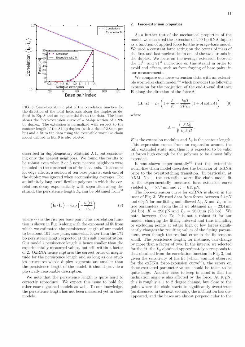
0 20 40 60 80 100 120Base pair index
10−1
100C
orre
latio
n⟨ l 0
·l n
⟩Correlations
Exponential fit
0.6 0.7 0.8 0.9 1.0 1.1 1.2 1.3Relative extension
01020304050607080
Forc
e[p
N]
Simulation
Extensible WLC fit
FIG. 3. Semi-logarithmic plot of the correlation function forthe direction of the local helix axis along the duplex as de-fined in Eq. 8 and an exponential fit to the data. The insetshows the force-extension curve of a 81-bp section of a 99-bp duplex. The extension is normalized with respect to thecontour length of the 81-bp duplex (with a rise of 2.8 nm perbp) and a fit to the data using the extensible wormlike chainmodel defined in Eq. 9 is also plotted.
described in Supplementary Material A 1, but consider-ing only the nearest neighbors. We found the results tobe robust even when 2 or 3 next nearest neighbors wereincluded in the construction of the local axis. To accountfor edge effects, a section of ten base pairs at each end ofthe duplex was ignored when accumulating averages. Foran infinitely long, semi-flexible polymer in which the cor-relations decay exponentially with separation along thestrand, the persistence length Lp can be obtained from83
⟨l0 · ln
⟩= exp
(−n 〈r〉
Lp
)(8)
where 〈r〉 is the rise per base pair. This correlation func-tion is shown in Fig. 3 along with the exponential fit fromwhich we estimated the persistence length of our modelto be about 101 base pairs, somewhat lower than the 171bp persistence length expected at this salt concentration.Our model’s persistence length is hence smaller than theexperimentally measured values, but still within a factorof 2. OxRNA hence captures the correct order of magni-tude for the persistence length and as long as one stud-ies structures whose duplex segments are smaller thanthe persistence length of the model, it should provide aphysically reasonable description.
We note that the persistence length is quite hard tocorrectly reproduce. We expect this issue to hold forother coarse-grained models as well. To our knowledge,the persistence length has not been measured yet in thesemodels.
2. Force-extension properties
As a further test of the mechanical properties of themodel, we measured the extension of a 99-bp RNA duplexas a function of applied force for the average-base model.We used a constant force acting on the center of mass ofthe first and last nucleotides in one of the two strands inthe duplex. We focus on the average extension betweenthe 11th and 91st nucleotide on this strand in order toavoid end effects, such as from fraying of base pairs, inour measurements.We compare our force-extension data with an extensi-
ble worm-like chain model,84 which provides the followingexpression for the projection of the end-to-end distanceR along the direction of the force z:
〈R · z〉 = L0
(1 +
F
K−
kBT
2FL0
(1 +A cothA)
)(9)
where
A =
√FL2
0
LpkBT,
K is the extension modulus and L0 is the contour length.This expression comes from an expansion around thefully extended state, and thus it is expected to be validat forces high enough for the polymer to be almost fullyextended.It was shown experimentally82 that this extensible
worm-like chain model describes the behavior of dsRNAprior to the overstretching transition. In particular, at0.5M [Na+], the extensible worm-like chain model fitto the experimentally measured force-extension curveyielded Lp = 57.7 nm and K = 615pN.The force-extension curve for oxRNA is shown in the
inset of Fig. 3. We used data from forces between 2.4 pNand 69 pN for our fitting and allowed L0, K and Lp to befree parameters. From the fit we obtained L0 = 23.4 nm(84 bp), K = 296 pN and Lp = 26.0 nm (93 bp). Wenote, however, that Eq. 9 is not a robust fit for ourmodel: changing the fitting interval and thus includingor excluding points at either high or low forces signifi-cantly changes the resulting values of the fitting param-eters, even though the residual error in the fit remainssmall. The persistence length, for instance, can changeby more than a factor of two. In the interval we selectedfor the fit, the Lp obtained approximately corresponds tothat obtained from the correlation function in Fig. 3, butgiven the sensitivity of the fit (which was not observedfor the oxDNA force-extension curve53), the errors onthese extracted parameter values should be taken to bequite large. Another issue to keep in mind is that theinclination angle is also affected by the force. At 10 pN,this is roughly a 1 to 2 degree change, but close to thepoint where the chain starts to significantly overstretch(as discussed in the next section), the inclination has dis-appeared, and the bases are almost perpendicular to the

12
65 66 67 68 69 70 71Number of base pairs
−1.5
−1.0
−0.5
0.0
0.5
1.0
1.5Fr
eeen
ergy
[kBT
](a)
81.3 pN83.8 pN86.3 pN88.7 pN93.7 pN
FIG. 4. (a) Free energy as a function of the number of basepairs in the duplex for different forces, where we set the freeenergy to be 0 for 68 bp for each force considered. At the over-stretching force, the slope of the free-energy profile is 0. (b)Snapshot from a VMMC simulation at F = 86.3 pN, showingunpeeling from the ends.
axis. It is likely that this deformation is not entirely phys-ical. However, the physical structure of stretched RNAis not experimentally known. In DNA, the structure ofthe extended state is a very active topic of research.
3. Overstretching
Both DNA and RNA duplexes are known to undergoan overstretching transition at high enough force. Re-cent experiments82 for different salt concentrations find63.6 (2.0) pN for RNA overstretching at 0.15M [Na+]up to 65.9 (3.3) pN at 0.5M [Na+]. Following the ap-proach taken in the study of DNA overstretching withthe oxDNA model,61 we used the average-base oxRNAmodel to run VMMC simulations of a 99-bp RNA du-plex with equal and opposite forces applied to the firstand last nucleotide of one strand. In our simulations,only native base pairs were allowed to form to aid equi-libration, i.e. no misbonds in the duplexes or intrastrandbase pairs in the unpeeled strand. The simulations werestarted from a partially unpeeled state to sample stateswhich have between 65 and 71 bp. The obtained free-energy profiles as a function of the number of base pairsare shown in Fig. 4(a). As the force increases, the slopeof the free energy profiles changes from negative (stateswith more base pairs are favored) to positive (it is fa-vorable for duplexes to unpeel). By estimating the forceat which the slope becomes zero, we obtained 86.2 pN asthe overstretching force. We note that our model was
G
G
C
C
C
C
U G
G
C
C
C
A
G
G
G
U
A
A
A
A
A
C
C
U
U
A
G
G
G
C
G
C
A
1
34
FIG. 5. A snapshot of the MMTV pseudoknot as representedby oxRNA. Stem 1 (shown in blue) has 6 base pairs whereasstem 2 (shown in red) has 5 base pairs. A schematic represen-tation of the secondary structure with the sequence is shownon the right.
parametrized for 1M [Na+], whereas the overstretchingexperiment was done at 0.5M. Furthermore, by not al-lowing any formation of secondary structure in the un-peeled strands, we overestimate the overstretching forcein the model, because these intramolecular base pairsstabilize the unpeeled state. For the oxDNA model, itwas shown that allowing secondary structure decreasesthe overstretching force by about 3 pN.61 We would ex-pect the stabilization to be slightly higher for RNA, asRNA base pairs are more stable. Our model hence over-estimates the value of the overstretching force by about16-20 pN. This overestimation is partly due to the higherextensibility of the duplex in oxRNA, which is aided bythe loss of inclination in the duplex when higher forcesare applied, as we already discussed in the previous sec-tion.
IV. EXAMPLE APPLICATIONS
A. Pseudoknots
Pseudoknots are a common structural motif in RNA. Ifa strand is represented as a circle and base pair contactsare represented as chords, then its structure is pseudo-knotted if the chords cross. Most secondary structureprediction tools do not include pseudoknotted structuresin their computations and thus cannot be used to studysystems where they are relevant, although some progresshas been made in developing efficient algorithms for thistask.85,86
Since oxRNA provides an explicitly three-dimensionalrepresentation of the RNA strands, it can be used to sim-ulate the folding and thermodynamics of RNA structuresthat involve pseudoknots. In this section, we use ourmodel to study the well known MMTV pseudoknot.87
The sequence and the three-dimensional representation

13
50 60 70 80 90 10055 65 75 85 95T [ C]
0.0
0.2
0.4
0.6
0.8
1.0Y
ield
(a)
Single strandHairpin 1Hairpin 2Pseudoknot
50 60 70 80 90 10055 65 75 85 95T [ C]
0
1
2
3
4
5
6
CV
[kca
lmol
−1
K−1 ]
(b)
FIG. 6. (a) Equilibrium yields and (b) CV as a function oftemperature for the MMTV pseudoknot. In (a) the pseudo-knot and hairpins are defined as having at least 1 native basepair in the relevant stems, whereas the unstructured single-stranded state has no native base pairs. In (b) the error barsare the standard deviations derived from 5 independent sim-ulations. The red vertical lines indicate the temperatures atwhich we observe equal yields of pseudoknot and hairpin 2(67.7 C) and hairpin 2 and the unstructured single strand(84.8 C).
of the MMTV pseudoknot by oxRNA are shown inFig. 5. The MMTV pseudoknot’s thermodynamic prop-erties were previously studied in experiment,87 as wellas with another coarse-grained RNA model.48 Moreover,the MMTV pseudoknot’s structure has also been investi-gated by NMR experiments88 and two stems were iden-tified in the folded structure: stem 1 with 6 base pairsand stem 2 with 5 base pairs, as schematically shown inFigure 5.To study the thermodynamics of the system, we ran
VMMC simulations of oxRNA for 3.4 × 10¹¹ steps at75 C. Umbrella sampling, using the number of base pairsin each of the pseudoknot stems as order parameters, was
0 2 4 6 8 101 3 5 7 9 11Base pairs in stem 1 + stem 2
0
2
4
6
8
10
12
14
16
18
Free
ener
gy[k
BT
]
(b) Stem 1 → Stem 2 pathway
Stem 2 → Stem 1 pathway
FIG. 7. (a) A free-energy landscape for pseudoknot formationat 48 C. White lines denote minimum free-energy pathways.(b) Free-energy profiles along the paths indicated in the free-energy landscape of (a). Dashed and solid lines correspond inboth pictures. Only native base pairs contribute to the orderparameters.
employed to enhance thermodynamic sampling. We alsoused histogram reweighting to extrapolate our results toother temperatures. The occupation probabilities of theunfolded state, a single hairpin with stem 1 or stem 2(denoted as hairpin 1 and hairpin 2), and the pseudoknotare shown in Fig. 6(a). Our simulations also allow us toextract the heat capacity CV from the expression
CV =∂ 〈U〉
∂T(10)
where we use a cubic interpolation to our simulation datafor 〈U〉 in order to compute the derivative with respectto T . The results are shown in Fig. 6(b).The experimentally measured CV at 1M [Na+] has
two peaks, one at 73.5 C and the other at 95.0 C.87 Itwas hypothesized that the two peaks correspond to the

14
transition from an unstructured strand to a hairpin struc-ture and a second transition from a hairpin structure tothe full pseudoknot. Analysis of our yields supports thisclaim, showing a pseudoknot to hairpin 2 transition at67.7 C and transition from hairpin 2 to a single strandwith no bonds in stem 1 or stem 2 at 84.8 C. The highertemperature transition coincides with a peak in the heatcapacity, whilst the lower temperature transition givesrise to a shoulder. While our simulations reproduce qual-itatively the behavior of the experimental system, theposition of the transitions is not exactly the same as theones measured experimentally. This is not surprising, aswe have shown in Section III B that the model generallyunderestimates the melting temperatures of hairpins.
It is of further interest to analyze the free-energy land-scape of the system (Fig. 7). Perhaps unsurprisingly, ourresults suggest that the minimum free-energy pathwayfor folding this pseudoknot from a single strand involvesfirst forming one of the stems (forming stem 2 first ismore likely as it is more stable and has a higher yield atthe considered temperature) and then closing the secondstem, instead of simultaneously forming both of them.We have previously seen similar pathways for a DNApseudoknot.66
One subtlety concerns the formation of the GU basepair between the seventh and thirty-fourth nucleotide.The NMR study88 did not observe the presence of thisGU base pair in the pseudoknot structure. However, inour simulations, we find some structures where this basepair forms (thus extending the size of stem 1 from sixto seven base pairs), although it has a 5 kBT free energypenalty at 48 C with respect to a pseudoknot state whichhad only six bases in stem 1. Including this additionalbase-pair within the definition of stem 1 had only a smalleffect on the calculated yields (the positions of the equalyields points changed by less than 0.3 C) and we sawat most 0.5 kBT free-energy change for the folding path-ways. We thus did not include this extra base pair in thedefinition of stem 1.
The experimental NMR study88 of the structure of theMMTV pseudoknot found that the two stems of the pseu-doknot are bent with respect to each other at about 112,and the AA mismatch between the sixth and the four-teenth nucleotides to be not stacked. As can be seenin Fig. 5, in oxRNA, this mismatch is typically stackedleading to an angle closer to 160. We think that thisstacking of the stems reflects the overestimation of thestability of mismatches in simpler motifs (see Table II).
In summary, our model is able to describe the thermo-dynamics of the pseudoknot folding and predict the sta-bilities of the two stems, supporting the hypothesis thatthe peak in heat capacity at higher temperature corre-sponds to the pseudoknot to hairpin 2 transition. Theoverall secondary structure of the pseudoknot is correctin our model, which also helps to understand the ter-tiary structure even though we found the angle betweenthe two stems to be larger than the one reported fromexperiment.
0 1 2 3 4 5 6 7Native base pairs
−10
−5
0
5
10
15
Free
ener
gy[k
BT
]
(b)
Duplex (average-base model)
Kissing complex (average-base model)
Duplex (sequence-dependent model)Kissing complex (sequence-dependent model)
FIG. 8. (a) A typical configuration of a kissing complex be-tween two hairpins that have a complementary 7-base loops.(b) Free-energy profiles at 45 C for forming the kissing com-plex and a 7-bp duplex with the same sequence as the hair-pin loops. Results are shown for the average-base and thesequence-dependent parametrization of oxRNA.
B. Kissing hairpin complex
A kissing complex is a naturally occurring motif inRNA structures1 and consists of two hairpins that havecomplementary loops and can thus bind to each other.An example of such a complex as represented by oxRNAis shown in Fig. 8. The kinetics and thermodynamicsof forming an RNA kissing complex with 7 bases in theloops was experimentally studied in Ref. 90 at varyingsalt concentrations, including 1M [Na+], the concentra-tion at which our model was parametrized.To examine the capability of oxRNA to describe kissing
complexes, we studied the melting of this kissing complexusing both the average-base and the sequence-dependentparametrization of oxRNA and found the transition ata point which is approximately consistent with the ob-served experimental behavior. The 7-base loops in thetwo hairpins have fully complementary sequences (5′-GGAAAUG-3′ and its Watson-Crick complementary se-quence). All melting simulations were run in a vol-ume corresponding to an equal strand concentration of3.36× 10−4M.For the average-base representation we found the melt-
ing temperature of the kissing hairpins to be 62.7 C

15
FIG. 9. The hexagonal RNA nanoring of Ref. 89, as repre-sented by oxRNA. The structure is composed of six strands,with a total of 264 nucleotides, connected by kissing loops.
which compared to 53.6 C for a 7-bp duplex with thesame sequences as the loops. For the sequence-dependentmodel, we found the melting temperature of the kissingcomplex to be 44.8 C, similar to 45.2 C for this 7-bp du-plex. The free-energy profiles for both average-base andsequence-dependent models at 45 C are shown in Fig. 8.For most sequences, we find that the kissing hairpin
loop is more stable than the separate duplexes with re-spect to the unbound state. We find that with increasingtemperature, the kissing complex loses less stability withrespect to the unbound state than a duplex at the sametemperature and strand concentration. This trend canbe rationalized in terms of the fact that a constrainedloop loses less configurational entropy upon binding thanan unconstrained single strand does. Furthermore, thekissing complex also gets an extra enthalpic stabiliza-tion from a coaxial stacking interaction between the loopand the stem nucleotides. These two effects help explainwhy, on average, the kissing hairpins are more stable,especially at higher temperatures. However, the kissinghairpins do not satisfy the enthalpic contributions as wellas a perfectly formed duplex does. Thus, at low temper-ature, the duplex can be more stable. Which of theseeffects dominates depends on the sequence, and if themelting happens before the switch of which motif is morestable, then the duplex will have a higher melting tem-perature, which we find for a minority of sequences at ourstrand concentration. For the sequence above, the melt-ing temperatures are very close. Note that the hairpinloops are sufficiently short that kissing complex forma-tion is not inhibited by the topological requirement thatthe linking number between the loops must remain zero.This contrasts with previous simulations of DNA kissingcomplexes between hairpins that have 20-base comple-mentary loops.58
The thermodynamics of this kissing complexes wasstudied in Ref. 90 using isothermal titration calorime-try (ITC) at 1M [Na+]. Up to 35 C the authors found
evidence of a transition to a kissing complex after theinjection of the reactants, but did not observe a transi-tion at 45 C. The authors claim to measure only a 0.6kcal/mol change in the ∆G for forming the kissing com-plex between 10 C and 35 C while observing a signifi-cant increase (19.5 kcal/mol) in ∆H along with a com-pensating increase in ∆S. Such behavior is not observedin our simulations, where we see a classic quasi-two-statetransition in which ∆H and ∆S change slowly with tem-perature, similarly to a duplex association. If the yieldsof the kissing complexes in our simulations were extrap-olated to the concentrations used in the experiment, wewould predict a yield of 36% at 35 C and 5% at 45 C.We note that the thermodynamic parameters in Ref. 90were derived with the assumption that the transition wasfully saturated after the injection of the reactants in theITC experiment, which is incompatible with the experi-mentally inferred value of ∆G. If the transitions were infact not fully saturated, then it is possible that the exper-iments are consistent with a more typical quasi-two-statetransition as observed in our model with a melting tem-perature approximately consistent with that found by us.
It is also interesting to use oxRNA to probe the struc-ture of this kissing complex because it is a motif that hasbeen used in RNA nanotechnology. Molecular dynam-ics simulations of the kissing complex using an all-atomrepresentation (Amber) measured the angle between thehelical stems at 300K and 0.5M monovalent salt to beapproximately 120.89 Based on this finding, a hexag-onal ring nanostructure that can be assembled from sixRNA building blocks was proposed. Each of the proposedbuilding blocks is an RNA strand that in the folded statehas a stem and two hairpin loops. The sequences in theloops are designed to bind to the complementary blockto allow the assembly of a hexagonal “nanoring” via thekissing complex interaction. This computationally pro-posed RNA nanoring design was later experimentally re-alized by self-assembly.91 The nanoring can be function-alized by including siRNA sequences either in the hair-pin stems or by appending siRNA sequences onto thestems. Experiments in blood serum have shown that thenanoring protects the loop regions of the assembly blocksfrom single-strand RNA endonucleolytic cleavage, mak-ing the nanoring a promising tool for in vivo nanotech-nology applications.91
Simulations of oxRNA at 25 C allowed us to measurethe angle between the helical axes of the hairpin stems.We found this angle to fluctuate around an average valueof 133.9 with a standard deviation of 14.8. Hence, anoctagon would probably be the most relaxed nanoring foroxRNA, and therefore favored by enthalpy. Smaller ringswould be favored by translational entropy.
To illustrate the capabilities of our oxRNA model,we constructed the hexagonal RNA nanoring of Ref. 89(Fig. 9) by starting a simulation with six folded hairpinsblocks and introducing a harmonic potential between thecomplementary loop regions, which helped the kissing in-teractions to form. Once the nanoring was completed,

16
the harmonic traps were removed and the assembledstructure was relaxed in a molecular dynamics simula-tion. We found the angle between the stems of the kissinghairpins in the nanoring to fluctuate around a mean of124.9. The thermal fluctuations around the mean valuehad a standard deviation of 14.4, which is similar to thatof the single kissing complex. It would also be possibleto use our model to study the mechanical properties ofthe nanoring, as well as the thermodynamics and kineticsof its self-assembly from the building blocks, but such astudy is beyond the scope of this article.A typical relaxation time for the angle between adja-
cent kissing complexes or the energy self-correlation func-tion of the assembled nanoring structure corresponds toabout a minute of CPU time or less on a standard 2.2GHz processor. This example shows that structural in-vestigations of systems of the order of hundreds of basesare well within the capabilities of the oxRNA model us-ing a single CPU, and if multiple CPUs are used, or aGPU chip is used,92 then structural properties and fluc-tuations around equilibrium can be studied for systemson the order of thousands of base pairs, as can be donefor oxDNA.66
C. Hairpin unzipping
RNA hairpin unzipping has been used in experimentto study the thermodynamics of base pairing and themechanical properties of RNA, with the kinetics of theprocess also being of interest.94–96 Unzipping the samesequence under different salt and temperature conditionscan provide systematic data on the physical propertiesof RNA that, for example, could be used to improve theparametrization of thermodynamic models of RNA.Here we consider the unzipping of the CD4 hairpin
(shown schematically in Fig. 10(a)), which has a 20-bpstem and 4 bases in the loop. It was studied experi-mentally by pulling at different rates and measuring theunzipping force94–96 for each trajectory. While it is pos-sible to simulate pulling the hairpins ends at a given ratein the oxRNA model, direct comparisons with experi-mentally observed unzipping forces are difficult becausefor a coarse-grained model there is not a straightforwardway to map the simulation time to the experimental one.Furthermore, to obtain an estimate of the average unzip-ping force for a given pulling rate, one needs to averageover multiple trajectories, and generating such trajecto-ries can be quite time consuming, especially for very slowpulling rates.A more suitable experimental setup for comparison
with a coarse-grained simulation comes from Ref. 96,where the authors performed force-clamp experiments atdifferent temperatures and salt concentrations. In theexperimental setup, they kept the forces applied to thefirst and last base of the hairpin constant and measuredthe folding and unfolding rate of the hairpin, from whichthey inferred the free-energy difference between the open
20 25 30 35 40 45T [ C]
17
18
19
20
21
22
Unz
ippi
ngfo
rce
[pN
]
(b)oxRNA modelFit to experimental data
FIG. 10. (a) A snapshot from an oxRNA simulation of un-zipping of the CD4 hairpin and a schematic representationof the CD4 hairpin sequence created with the PseudoViewersoftware.93 (b) The unzipping forces for the CD4 hairpin asa function of temperature for the oxRNA model (full circles)along with a linear fit to the data. For comparison, the fit tothe experimentally measured unzipping force at 1M [Na+] attemperatures 22, 27, 32, 37 and 42 C is also shown.
and closed state. By interpolating results for a range ofapplied forces, they were able to obtain the unzippingforce, which was defined as the force for which the free-energy difference was zero.
To compare to the experimental results, we ran VMMCsimulations of the CD4 hairpin with a constant force of19.7 pN applied to the first and last nucleotide at 23 C.We then extrapolated the free energies of the open andclosed state by the histogram reweighting method toforces ranging from 16.2 to 20.7 pN and to the tempera-tures at which the experiments were carried out (22, 27,32, 37 and 42 C). We only allowed bonds between thenative base pairs to avoid sampling of metastable sec-ondary structures that would slow down our simulations.We considered the hairpin to be closed if at least one ofthe bonds in the stem was present. For each tempera-ture considered, we performed a linear interpolation ofthe free-energy difference between closed and open stateas a function of force to obtain the unzipping force for

17
which the difference is 0. The unzipping forces we obtainare shown in Fig. 10, along with a fit. We also show forcomparison the fit to the experimentally observed unzip-ping forces96 at 1M [Na+], expressed in the form
Funzip(T ) = a− cT, (11)
where Funzip is the unzipping force at temperature T .The values obtained in the experiment96 were a =69.1 pN and c = −0.164 pN/K. Fitting Eq. 11 to oursimulation data, we obtained the same value for c and68.2 pN for a. The values of the fitting parameters variedby at most 6% between the fits to unzipping forces ob-tained from three independent sets of generated states.Thus, oxRNA is able to reproduce the unzipping forcewith 5% (1 pN) accuracy and fully captures the trendwith temperature.It would be of further interest to study the force-
extension properties of a hairpin which contains vari-ous secondary structure motifs such as bulges and in-ternal loops or which has regions with variable sequencestrengths (such as AU rich and GC rich regions). How-ever, such a study is beyond the scope of this article.
V. SUMMARY AND CONCLUSIONS
We have presented a new off-lattice coarse-grainedmodel for RNA, oxRNA, which aims to capture basicthermodynamic, structural and mechanical properties ofRNA structures with a minimal representation and pair-wise interaction potentials. OxRNA is specifically devel-oped to allow for efficient simulations of large structures(up to hundreds of nucleotides on a single CPU), andfor reactions involving multiple strands of RNA, whichare important for applications in RNA nanotechnology.Our coarse-graining strategy is closely linked to our pre-vious successful coarse-graining of DNA with oxDNA.53
Rather than focusing mainly on reproducing structure,as many other previous models have done, here we triedto systematically compare to a whole suite of differentproperties.We employed a “top-down” coarse-graining approach,
where we aim to reproduce free-energy differences be-tween different states (such as opened and closed stateof a hairpin) as measured in experiment. OxRNA repre-sents each nucleotide (i.e., sugar, phosphate and base) asa single rigid body with multiple interaction sites. Themodel is capable of representing RNA structures suchas hairpins and duplexes and is designed to reproducethe A-helical form of duplex RNA. We used a histogramreweighting method52 to parametrize the model to repro-duce the thermodynamics of short duplexes and hairpins.Currently, the oxRNA model captures Watson-Crick andwobble base-pairing interactions as well as various typesof stacking interaction. However it was not designed tocapture non-canonical interactions such as Hoogsteen orsugar-edge hydrogen-bonded base pairs, or ribose zip-pers. Nevertheless, it can reproduce some important ter-
tiary interaction motifs, in particular coaxial stacking ofhelices, kissing loop interactions, and pseudoknots.
The model is currently parametrized for a salt concen-tration of 1M, as this corresponds to the conditions forthe melting experiments used for the nearest-neighbormodel to which our model was parametrized. Explicitelectrostatic interactions are not included, because theyare very short-ranged at high salt and thus can be in-corporated into the short-ranged excluded volume termsin the potential. This excluded volume also preventsa strand from crossing itself or other strands, forbid-ding topologically impossible trajectories in kinetic sim-ulations. It is possible to use the same coarse-grainingtechniques to parametrize the model at lower salt con-centrations. However, as the screening lengths becomelonger, different longer-ranged forms for the interactionsmay need to be used to capture the correct physics. Wenote however that nanotechnology experiments in vitro
are usually carried out in high salt conditions.
To test our model, we investigated the thermodynam-ics of short duplexes and hairpins with different sequencecontent, as well as various other secondary structuressuch as bulges, internal and terminal mismatches. Wefound that in comparison with our previous model forDNA, parametrizing RNA thermodynamics is a morechallenging task. For example, experimental meltingtemperatures of a duplex of a given length can differ by asmuch as 70 C between weak and strong sequences withWatson-Crick base pairs, as opposed to 50 C for DNA.52
So sequence effects are stronger in RNA. Including wob-ble base pairs presents further challenges, as some basepair steps that include two or more neighboring wobblebase pairs have a positive contribution to the free energyof duplex. Although it is not possible to capture this ef-fect with the current representation of our model, addingthe structural effects of wobble base pairs on the doublehelix may provide means to improve this aspect of themodel. Finally, we found that oxRNA overestimates thestability of duplexes with mismatches in internal loops.This could lead to an overestimation of the stability ofmisfolded structures and complicate the study of the fold-ing of RNA strands that have multiple metastable stateswith mismatches. Nevertheless, even though oxRNAdoes not reproduce the exact melting temperatures forstructures with internal mismatches and bulges, we doobserve, as expected, a decrease in melting temperaturesof a duplex with internal mismatches or bulges with re-spect to the fully complementary strands. The observedchanges in melting temperatures are within the orders ofmagnitude as predicted by the NN-model for the samemotifs and capture correctly the direction of the change.
We have tested the mechanical properties of the RNAduplex as represented by the model and found its persis-tence length to be about half of the reported persistencelength of RNA molecules at high salt conditions. Themodel hence has the correct order of magnitude for theduplex persistence length and provides sufficiently accu-rate mechanical behavior for most applications, where

18
individual double helical sections are likely to be muchshorter than the persistence length.
In Section IV, we provided some applications of themodel that illustrate its strength and potential utility.In particular, we investigated the thermodynamics ofpseudoknot folding and the thermodynamics and struc-ture of a kissing hairpin complex. We also showed thatoxRNA can be used to model large nanostructures like anRNA nanoring composed of 264 nucleotides on a singleCPU. The computational cost of oxRNA is very simi-lar to oxDNA where simulations of a DNA origami motifwith 12 391 nucleotides have been achieved.66 Finally, ourmodel is able to reproduce experimental results for themechanical unzipping of a hairpin quite closely, to withinan accuracy of 1 pN, illustrating oxRNA’s potential tostudy mechanical properties of RNA constructs.
Although we did not describe applications with de-tailed dynamics (the simulations are typically quite in-volved and so beyond the scope of this study), we want toemphasize that oxRNA is particularly well-suited for suchtasks. For example, oxDNA has been used to study thedetailed dynamics of hybridization,62 toehold-mediatedstrand displacement59 and hairpin formation.66 Study-ing similar processes would be very feasible for oxRNA.For example, it should be possible to use the model toobtain estimates of the rates of a strand displacement re-actions as a function of length of the toehold as well astemperature. OxRNA can further be used to study theself-assembly of nanostructures such as RNA nanoringsand to investigate both the thermodynamics and the ki-netics of such systems. Although the model is currentlyonly parametrized at high salt concentration, oxRNA canbe also used to qualitatively study processes of biologi-cal relevance, for instance, the folding pathways of RNAstrands or in vivo applications of nanotechnology.
At this point it is interesting to reflect on some sim-ilarities and differences between the coarse-graining ofoxDNA and oxRNA. Although oxRNA can clearly re-produce a good number of properties of RNA, quanti-tative differences with experiment for the melting tem-peratures of certain motifs are larger than they are foroxDNA. Moreover, it was more difficult to simultaneouslyreproduce the thermodynamics and the correct persis-tence length or the force-extension curves. One reasonfor these differences may be that RNA itself exhibitsmore complex behavior than DNA, and so is harder tocoarse-grain. It is intuitively obvious that the compro-mises made to increase tractability mean that not allproperties of the underlying system can be simultane-ously captured by a more simplified description, a gen-eral phenomenon that has been called “representabil-ity problems”.97,98 One consequence of representabilityproblems is that in general, fitting too strongly to one setof input data (say structure, as is often done for otherRNA models) will often lead to larger errors in otherquantities, say thermodynamics. We tried to compro-mise between different requirements for oxRNA. How-ever, in order to make further progress, more detailed
fitting may not be enough. Instead a more complex andmost likely less tractable representation of the full inter-actions may need to be chosen. For example, for RNAit remains to be seen whether our single nucleotide-levelmodel can be easily extended to generate a better rep-resentation of both structure and thermodynamics, orwhether, say, a more complex model is needed to achievethe next level of accuracy. Clearly, including electrostaticeffects for lower salt-concentrations, or implementing ter-tiary structure contacts, for example non-canonical basepairing interaction (such as Hoogsteen base pairs) andhydrogen bonding between a sugar group and a base willalso need an extension of the current representation.Given the challenges and complexity of RNA mod-
elling, it is unsurprising that oxRNA performs less wellthan oxDNA for the whole ensemble of motifs we consid-ered. However, we believe that it is a non-trivial achieve-ment to create a model that can semi-quantitatively re-produce such a wide range of the thermodynamic data.The properties of our model have been comprehensivelytested on a variety of systems, ranging from secondarystructure motifs to systems such as kissing complexes andhairpin unzipping. Our model is also currently the onlyRNA model with reported mechanical properties, whichwere tested by measuring its persistence length, force-extension curve and duplex overstretching.Finally, the simulation code implementing molecular
dynamics and Monte Carlo algorithms for our oxRNAmodel is available for download at dna.physics.ox.ac.uk.
ACKNOWLEDGMENTS
The authors thank Agnes Noy for helpful discussionsand Lorenzo Rovigatti for his help with the simulationcode development. The authors acknowledge the En-gineering and Physical Sciences Research Council andUniversity College (Oxford) for financial support andthe Advanced Research Computing, University of Ox-ford for computer time. P. S. is grateful for the award ofa Scatcherd European Scholarship.
Supplementary Material A
1. Fitting of the helical axis of a duplex
We discussed oxRNA’s description of the structure ofthe A-helix in Section IIIA. For the considered 13-bplong duplex, we fitted the helical axis in the followingway. For each base in the first strand, we took the vectorpointing from its backbone site to the backbone site ofits 3′-neighbor and for each base in the second strand,we considered the vectors pointing to the 5′-neighbor’sbackbone site. For a perfect A-helical structure, the end-points of all the vectors would all lie in the same planeif the origins of the vectors were all placed at the samepoint. The structure of the duplex is subject to thermal
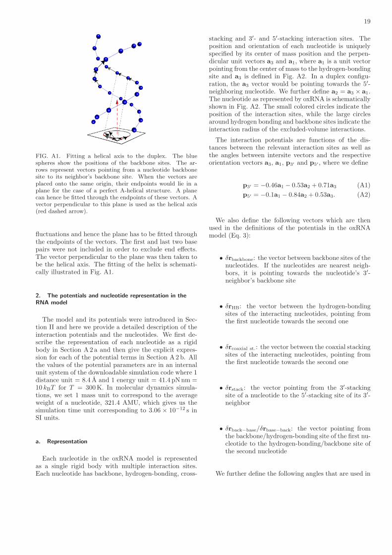
19
FIG. A1. Fitting a helical axis to the duplex. The bluespheres show the positions of the backbone sites. The ar-rows represent vectors pointing from a nucleotide backbonesite to its neighbor’s backbone site. When the vectors areplaced onto the same origin, their endpoints would lie in aplane for the case of a perfect A-helical structure. A planecan hence be fitted through the endpoints of these vectors. Avector perpendicular to this plane is used as the helical axis(red dashed arrow).
fluctuations and hence the plane has to be fitted throughthe endpoints of the vectors. The first and last two basepairs were not included in order to exclude end effects.The vector perpendicular to the plane was then taken tobe the helical axis. The fitting of the helix is schemati-cally illustrated in Fig. A1.
2. The potentials and nucleotide representation in the
RNA model
The model and its potentials were introduced in Sec-tion II and here we provide a detailed description of theinteraction potentials and the nucleotides. We first de-scribe the representation of each nucleotide as a rigidbody in Section A2 a and then give the explicit expres-sion for each of the potential terms in Section A2 b. Allthe values of the potential parameters are in an internalunit system of the downloadable simulation code where 1distance unit = 8.4 A and 1 energy unit = 41.4 pNnm =10 kBT for T = 300K. In molecular dynamics simula-tions, we set 1 mass unit to correspond to the averageweight of a nucleotide, 321.4 AMU, which gives us thesimulation time unit corresponding to 3.06 × 10⁻¹² s inSI units.
a. Representation
Each nucleotide in the oxRNA model is representedas a single rigid body with multiple interaction sites.Each nucleotide has backbone, hydrogen-bonding, cross-
stacking and 3′- and 5′-stacking interaction sites. Theposition and orientation of each nucleotide is uniquelyspecified by its center of mass position and the perpen-dicular unit vectors a3 and a1, where a1 is a unit vectorpointing from the center of mass to the hydrogen-bondingsite and a3 is defined in Fig. A2. In a duplex configu-ration, the a3 vector would be pointing towards the 5′-neighboring nucleotide. We further define a2 = a3 × a1.The nucleotide as represented by oxRNA is schematicallyshown in Fig. A2. The small colored circles indicate theposition of the interaction sites, while the large circlesaround hydrogen bonding and backbone sites indicate theinteraction radius of the excluded-volume interactions.
The interaction potentials are functions of the dis-tances between the relevant interaction sites as well asthe angles between intersite vectors and the respectiveorientation vectors a3, a1, p3′ and p5′ , where we define
p3′ = −0.46a1 − 0.53a2 + 0.71a3 (A1)
p5′ = −0.1a1 − 0.84a2 + 0.53a3. (A2)
We also define the following vectors which are thenused in the definitions of the potentials in the oxRNAmodel (Eq. 3):
• δrbackbone: the vector between backbone sites of thenucleotides. If the nucleotides are nearest neigh-bors, it is pointing towards the nucleotide’s 3′-neighbor’s backbone site
• δrHB: the vector between the hydrogen-bondingsites of the interacting nucleotides, pointing fromthe first nucleotide towards the second one
• δrcoaxial st.: the vector between the coaxial stackingsites of the interacting nucleotides, pointing fromthe first nucleotide towards the second one
• δrstack: the vector pointing from the 3′-stackingsite of a nucleotide to the 5′-stacking site of its 3′-neighbor
• δrback−base/δrbase−back: the vector pointing fromthe backbone/hydrogen-bonding site of the first nu-cleotide to the hydrogen-bonding/backbone site ofthe second nucleotide
We further define the following angles that are used in

20
the potential functions:
θ1 = arccos (a1 · b1) (A3)
θ2 = arccos(−b1 · δrHB
)(A4)
θ3 = arccos(a1 · δrHB
)(A5)
θ4 = arccos (a3 · b3) (A6)
θ5 = arccos(a3 · δrcoaxial st.
)(A7)
θ6 = arccos(−b3 · δrcoaxial st.
)(A8)
θ5′ = arccos(a1 · δrstack
)(A9)
θ6′ = arccos(−b3 · δrstack
)(A10)
θ7 = arccos(−b3 · δrHB
)(A11)
θ8 = arccos(a3 · δrHB
)(A12)
θ9 = arccos(−p3′ · δrbackbone
)(A13)
θ10 = arccos(−q5′ · δrbackbone
)(A14)
cos (φ1) = δrbackbone · a2 (A15)
cos (φ2) = δrbackbone · b2 (A16)
cos (φ3) = δrcoaxial st. ·(δrbackbone × a1
)(A17)
cos (φ4) = δrcoaxial st. ·(δrbackbone × b1
), (A18)
where we use the notation b1, b2, and b3 to define theorientation vectors of the second nucleotide participatingin the interaction (the orientation vectors of the first nu-cleotide are denoted as a1, a2, and a3). The vector q5′
corresponds to the p5′ vector of the 3′-neighbor of theinteracting nucleotide, i.e. using the same definition as inEq. A2, but substituting b for a.
b. Potentials
The oxRNA potential consists of a sum of potentialfunctions designed to represent different physical inter-actions, with some of the potentials being products ofmultiple potential functions. The functions that are usedin the potentials are:
• FENE spring (used in Vbackbone):
VFENE(r, ǫ, r0,∆) = −
ǫ
2ln
(1−
(r − r0)2
∆2
). (A19)
• Morse potential (used in Vstack and VH.B.):
VMorse(r, ǫ, r0, d) = ǫ
(1− exp (−d(r − r0))
)2. (A20)
• Harmonic potential (used in Vcross st. andVcoaxial st.):
Vharm(r, k, r0) =
k
2
(r − r0
)2. (A21)
• Lennard-Jones potential (used in excluded volume
potentials V′
exc and Vexc):
VLJ(r, ǫ, σ) = 4ǫ
[(σr
)12
−(σr
)6]. (A22)
• Quadratic modulation terms (used for angu-lar modulation of the anisotropic potentialsVH.B., Vcross st., Vstack and Vcoaxial st.):
Vmod(θ, a, θ0) = 1− a(θ − θ0)2. (A23)
• Quadratic smoothing terms for truncation (used inall potentials with the exception of Vbackbone) in or-der to make the potentials differentiable functionsthat are equal to 0 beyond some specific cutoff dis-tance:
Vsmooth(x, b, xc) = b(xc − x)2. (A24)
The smoothed functions used in the potentials have the following form:
• The radial part of the stacking and hydrogen-bonding potentials:
f1(r, ǫ, d, r0, rc, rlow, rhigh) =
VMorse(r, ǫ, r0, d)− VMorse(r
c, ǫ, r0, d)) if rlow < r < rhigh,
ǫVsmooth(r, blow, rc,low) if rc,low < r < rlow ,
ǫVsmooth(r, bhigh, rc,high) if rhigh < r < rc,high,
0 otherwise.
(A25)

21
FIG. A2. A schematic representation of the nucleotides as represented by the oxRNA model. The red circle indicates thebackbone site, the blue circle is the hydrogen-bonding site and the green circle is the coaxial stacking site. The yellow circleis the 3′-stacking site, and the black circle is the 5′ stacking site. The unfilled circle from which the a3, a2 and a1 vectorsoriginate is the center of mass. All distances are given in a unit system where 1 distance unit = 8.4 A. The left image showsthe projection of a single nucleotide where the a2 vector is pointing towards the reader, and the image on the right showsa projection where the a3 vector is pointing towards the reader. For comparison, we also show the schematic representationof the nucleotide that is used in producing pictures of oxRNA configurations. The backbone site is represented by a spherebecause of the isotropic nature of the its interactions, whereas the base is represented by an ellipsoid whose principal axes areparallel to a1, a2 and a3 respectively.
• The radial part of the cross-stacking and coaxial stacking potentials:
f2(r, k, r0, rc, rlow , rhigh) =
Vharm(r, k, r0)− Vharm(r
c, k, r0) if rlow < r < rhigh,
kVsmooth(r, blow, rc,low) if rc,low < r < rlow,
kVsmooth(r, bhigh, rc,high) if rhigh < r < rc,high,
0 otherwise.
(A26)
• The radial part of the excluded volume potential:
f3(r, ǫ, σ, r⋆) =
VLJ(r, ǫ, σ) if r < r⋆,
ǫVsmooth(r, b, rc) if r⋆ < r < rc,
0 otherwise.
(A27)
• The angular modulation factor used in stacking, hydrogen-bonding, cross-stacking and coaxial stacking:
f4(θ, a, θ0,∆θ⋆) =
Vmod(θ, a, θ0) if θ0 −∆θ⋆ < θ < θ0 +∆θ⋆,
Vsmooth(θ, b, θ0 −∆θc) if θ0 −∆θc < θ < θ0 −∆θ⋆,
Vsmooth(θ, b, θ0 +∆θc) if θ0 +∆θ⋆ < θ < θ0 +∆θc,
0 otherwise.
(A28)

22
• Another modulating term which is used to impose right-handedness:
f5(x, a, x⋆) =
1 if x > 0,
Vmod(x, a, 0) if x⋆ < x < 0,
Vsmooth(x, b, xc) if xc < x < x⋆,
0 otherwise.
(A29)
We note that for given parameters of the main part of the smoothed functions (for example, ǫ, r0, d and rc forthe VMorse part of f1), the parameters of the smoothed cutoff regions (blow, bhigh, rc,low, rc,high for f1) are uniquelydetermined by ensuring differentiability of the function at the boundaries (rlow and rhigh for f1) and thus they arenot explicitly listed in the function arguments and are not provided in the tables of values of model parameters.The potentials are then
Vbackbone = VFENE(δrbackbone, ǫbackbone, δr0backbone,∆backbone). (A30)
Vstack (i, j) = η(i, j)(1 + κ kBT )f1(δrstack, ǫstack, dstack, δr0stack, δr
cstack, δr
lowstack, δr
highstack)
× f4(θ5′ , astack,5, θ0stack,5,∆θ⋆stack,5) f4(θ6′ , astack,6, θ
0stack,6,∆θ⋆stack,6)
× f4(θ9, astack,9, θ0stack,9,∆θ⋆stack,9) f4(θ10, astack,10, θ
0stack,10,∆θ⋆stack,10)
× f5(cos(φ1), astack,1, cos(φ1)⋆stack) f5(cos(φ2), astack,2, cos(φ2)
⋆stack).
(A31)
VH.B.(i, j) = α(i, j)f1(δrHB, ǫHB, dHB, δr0HB, δr
cHB, δr
lowHB , δrhighHB )
× f4(θ1, aHB,1, θ0HB,1,∆θ⋆HB,1) f4(θ2, aHB,2, θ
0HB,2,∆θ⋆HB,2)
× f4(θ3, aHB,3, θ0HB,3,∆θ⋆HB,3) f4(θ4, aHB,4, θ
0HB,4,∆θ⋆HB,4)
× f4(θ7, aHB,7, θ0HB,7,∆θ⋆HB,7) f4(θ8, aHB,8, θ
0HB,8,∆θ⋆HB,8).
(A32)
Vcross st. = γf2(δrHB, kcross, δr0cross, δr
ccross, δr
lowcross, δr
highcross)f4(θ1, across,1, θ
0cross,1,∆θ⋆cross,1)
× f4(θ2, across,2, θ0cross,2,∆θ⋆cross,2) f4(θ3, across,3, θ
0cross,3,∆θ⋆cross,3)
×(f4(θ7, across,7, θ
0cross,7,∆θ⋆cross,7) + f4(π − θ7, across,7, θ
0cross,7,∆θ⋆cross,7)
)
×(f4(θ8, across,8, θ
0cross,8,∆θ⋆cross,8) + f4(π − θ8, across,8, θ
0cross,8,∆θ⋆cross,8)
).
(A33)
Vcoaxial st. = µf2(δrcoaxial st., kcoax, δr0coax, δr
ccoax, δr
lowcoax, δr
highcoax ) f4(θ4, acoax,4, θ
0coax,4,∆θ⋆coax,4)
×(f4(θ1, acoax,1, θ
0coax,1,∆θ⋆coax,1) + f4(2π − θ1, acoax,1, θ
0coax,1,∆θ⋆coax,1)
)
×(f4(θ5, acoax,5, θ
0coax,5,∆θ⋆coax,5) + f4(π − θ5, acoax,5, θ
0coax,5,∆θ⋆coax,5)
)
×(f4(θ6, acoax,6, θ
0coax,6,∆θ⋆coax,6) + f4(π − θ6, acoax,6, θ
0coax,6,∆θ⋆coax,6)
)
× f5(cos(φ3), acoax,3′ , cos(φ3)⋆coax) f5(cos(φ4), acoax,4′ , cos(φ4)
⋆coax).
(A34)
Vexc = f3(δrbackbone, ǫexc, σbackbone, δr⋆backbone) + f3(δrHB, ǫexc, σbase, δr
⋆base)
+ f3(δrback−base, ǫback−base, σback−base, δr⋆back−base)
+ f3(δrbase−back, ǫback−base, σback−base, δr⋆back−base)
(A35)
The excluded volume interaction between bonded neighbors, V ′exc, is the same as Vexc with the exception that it
does not include the first term which depends on δrbackbone, because the neighbors already interact with the FENEpotential through the Vbackbone interaction that ensures that the backbone sites do not come too close.The parameters of the interaction potentials are specified in tables A-I and A-II.
1D. Elliott and M. Ladomery, Molecular Biology of RNA, OxfordUniversity Press, 2011.
2W. Saenger, Principles of Nucleic Acid Structure, Springer-Verlag, New York, 1984.
3T. Kin et al., Nucleic Acids Res. 35, D145 (2007).4N. C. Seeman, Annu. Rev. Biochem. 79, 65 (2010).5J. Bath, S. J. Green, and A. J. Turberfield, Angew. Chem. Int.Ed. 117, 4432 (2005).

23
Interaction Parameters
backbone spring: Vbackbone
VFENE(δrbackbone) ǫbackbone = 2 ∆backbone = 0.25 δr0backbone = 0.76
hydrogen bonding: VH.B.
f1(δrHB) ǫHB = 1.0 dHB = 8 δr0HB = 0.4
αavg = 0.87 α(A,U) = 0.82 α(G,U) = 0.51 α(G,C) = 1.06
δrcHB = 0.75 δrlowHB = 0.34 δrhighHB = 0.70
f4(θ1) aHB,1 = 1.50 θ0HB,1 = 0 ∆θ⋆HB,1 = 0.70
f4(θ2) aHB,2 = 1.50 θ0HB,2 = 0 ∆θ⋆HB,2 = 0.70
f4(θ3) aHB,3 = 1.50 θ0HB,3 = 0 ∆θ⋆HB,3 = 0.70
f4(θ4) aHB,4 = 0.46 θ0HB,4 = π ∆θ⋆HB,4 = 0.70
f4(θ7) aHB,7 = 4.00 θ0HB,7 = π/2 ∆θ⋆HB,7 = 0.45
f4(θ8) aHB,8 = 4.00 θ0HB,8 = π/2 ∆θ⋆HB,8 = 0.45
stacking: Vstack
f1(δrstack) ǫstack = 1.0 dstack = 6 δr0stack = 0.43
δrcstack = 0.93 δrlowstack = 0.35 δrhighstack = 0.78
ηavg = 1.402 κ = 1.9756
η(G,C) = 1.276 η(C,G) = 1.603 η(G,G) = 1.494 η(C,C) = 1.473
η(G,A) = 1.621 η(U,C) = 1.167 η(A,G) = 1.394 η(C,U) = 1.471
η(U,G) = 1.286 η(C,A) = 1.583 η(G,U) = 1.571 η(A,C) = 1.210
η(A,U) = 1.385 η(U,A) = 1.246 η(A,A) = 1.316 η(U,U) = 1.175
f4(θ5′) astack,5 = 0.90 θ0stack,5 = 0 ∆θ⋆stack,5 = 0.95
f4(θ6′) astack,6 = 0.90 θ0stack,6 = 0 ∆θ⋆stack,6 = 0.95
f4(θ9) astack,9 = 1.3 θ0stack,9 = 0 ∆θ⋆stack,9 = 0.8
f4(θ10) astack,10 = 1.3 θ0stack,10 = 0 ∆θ⋆stack,10 = 0.8
f5(cos(φ1)) astack,1 = 2.00 cos(φ1)⋆stack = 0.65
f5(cos(φ2)) astack,2 = 2.00 cos(φ2)⋆stack = 0.65
excluded volume: Vexc
f3(δrbackbone) ǫexc = 2.00 σbackbone = 0.70 δr⋆backbone = 0.675
f3(δrHB) ǫexc = 2.00 σbase = 0.33 δr⋆base = 0.32
f3(δrback−base) ǫexc = 2.00 σback−base = 0.515 δr⋆back−base = 0.50
f3(δrbase−back) ǫexc = 2.00 σback−base = 0.515 δr⋆back−base = 0.50
TABLE A-I. Parameter values in the model. In this table, all energies and lengths are in terms of the simulation units of energyand distance. When more than one function is listed for an interaction, the total interaction is a product of all the terms withthe exception of Vexc, which is a sum of the respective terms.
6J. Bath, S. J. Green, K. E. Allan, and A. J. Turberfield, Small5, 1513 (2009).
7P. W. K. Rothemund, Nature 440, 297 (2006).8L. Adleman, Science 266, 1021 (1994).9P. Guo, Nature Nanotech. 5, 833 (2010).
10Y. Benenson, Curr. Opin. Biotechnol. 20, 471 (2009).11K. A. Afonin et al., Nature Nanotech. 5, 676 (2010).12A. Chworos et al., Science 306, 2068 (2004).13L. M. Hochrein, M. Schwarzkopf, M. Shahgholi, P. Yin, and N. A.Pierce, J. Am. Chem. Soc. 135, 17322 (2013).
14C. Laing and T. Schlick, J. Phys.: Condens. Matter 22, 283101(2010).
15C. Laing and T. Schlick, Curr. Opin. Struct. Biol. 21, 306 (2011).16M. J. Serra and D. H. Turner, Method. Enzymol. 259, 242(1995).
17T. Xia et al., Biochemistry 37, 14719 (1998).18D. H. Mathews et al., Proc. Natl. Acad. Sci. U.S.A. 101, 7287(2004).
19D. H. Mathews, J. Sabina, M. Zuker, and D. H. Turner, J. Mol.Biol. 288, 911 (1999).
20A. E. Walter and D. H. Turner, Biochemistry 33, 12715 (1994).

24
Interaction Parameters
cross-stacking: Vcross. st.
f2(δrHB) kcross = 1.0 r0cross = 0.5 δrccross = 0.6
γ = 59.96 δrlowcross = 0.42 δrhighcross = 0.58
f4(θ1) across,1 = 2.25 θ0cross,1 = 0.505 ∆θ⋆cross,1 = 0.58
f4(θ2) across,2 = 1.70 θ0cross,2 = 1.266 ∆θ⋆cross,2 = 0.68
f4(θ3) across,3 = 1.70 θ0cross,3 = 1.266 ∆θ⋆cross,3 = 0.68
f4(θ7) + f4(π − θ7) across,7 = 1.70 θ0cross,7 = 0.309 ∆θ⋆cross,7 = 0.68
f4(θ8) + f4(π − θ8) across,8 = 1.70 θ0cross,8 = 0.309 ∆θ⋆cross,8 = 0.68
coaxial stacking: Vcoaxial st.
f2(δrcoax) kcoax = 1.0 δr0coax = 0.5 δrccoax = 0.6
µ = 80.0 δrlowcoax = 0.42 δrhighcoax = 0.58
f4(θ1) + f4(2π − θ1) acoax,1 = 2.00 θ0coax,1 = 2.592 ∆θ⋆coax,1 = 0.65
f4(θ4) acoax,4 = 1.30 θ0coax,4 = 0.151 ∆θ⋆coax,4 = 0.8
f4(θ5) + f4(π − θ5) acoax,5 = 0.90 θ0coax,5 = 0.685 ∆θ⋆coax,5 = 0.95
f4(θ6) + f4(π − θ6) acoax,6 = 0.90 θ0coax,6 = 2.523 ∆θ⋆coax,6 = 0.95
f5(cos(φ3)) acoax,3′ = 2.00 cos(φ3)⋆coax = −0.65
f5(cos(φ4)) acoax,4′ = 2.00 cos(φ4)⋆coax = −0.65
TABLE A-II. Further model parameters.
21A. E. Walter et al., Proc. Natl. Acad. Sci. U.S.A. 91, 9218 (1994).22Z. J. Lu, D. H. Turner, and D. H. Mathews, Nucleic Acids Res.34, 4912 (2006).
23J. N. Zadeh et al., J. Comput. Chem. 32, 170 (2011).24I. L. Hofacker et al., Monatsh. Chem. 125, 167 (1994).25J. S. Reuter and D. H. Mathews, BMC Bioinformatics 11, 129(2010).
26C. Flamm, W. Fontana, I. L. Hofacker, and P. Schuster, RNA 6,325 (2000).
27A. Xayaphoummine, T. Bucher, and H. Isambert, Nucleic AcidsRes. 33, W605 (2005).
28J. Sponer, P. Jurecka, and P. Hobza, J. Am. Chem. Soc. 126,10142 (2004).
29D. Svozil, P. Hobza, and J. Sponer, J. Phys. Chem. B 114, 1191(2010).
30W. D. Cornell et al., J. Am. Chem. Soc. 117, 5179 (1995).31B. R. Brooks et al., J. Comput. Chem. 4, 187 (1983).32G. R. Bowman et al., J. Am. Chem. Soc. 130, 9676 (2008).33P. Kuhrova, B. Pavel, R. B. Best, J. Sponer, and M. Otyepka, J.Chem. Theory Comput. 9, 2115 (2013).
34M. Krepl et al., J. Chem. Theory Comput. 8, 2506 (2012).35J. Yoo and A. Aksimentiev, Proc. Natl. Acad. Sci. U.S.A. (2013).36A. T. Guy, T. J. Piggot, and S. Khalid, Biophys. J. 103, 1028(2012).
37P. Minary, M. E. Tuckerman, and G. J. Martyna, SIAM J. Sci.Comput. 30, 2055 (2008).
38A. Y. Sim, M. Levitt, and P. Minary, Proc. Natl. Acad. Sci.U.S.A. 109, 2890 (2012).
39M. A. Jonikas et al., RNA 15, 189 (2009).40Z. Xia, D. R. Bell, Y. Shi, and P. Ren, J. Phys. Chem. B 117,3135 (2013).
41M. Parisien and F. Major, Nature 452, 51 (2008).42R. Das, J. Karanicolas, and D. Baker, Nat. Methods 7, 291(2010).
43M. Paliy, R. Melnik, and B. A. Shapiro, PB 7, 036001 (2010).
44S. Pasquali and P. Derreumaux, J. Phys. Chem. B 114, 11957(2010).
45T. Cragnolini, P. Derreumaux, and S. Pasquali, J. Phys. Chem.B 117 (2013).
46F. Ding et al., RNA 14, 1164 (2008).47C. Hyeon and D. Thirumalai, Proc. Natl. Acad. Sci. U.S.A. 102,6789 (2005).
48N. A. Denesyuk and D. Thirumalai, J. Phys. Chem. B 117, 4901(2013).
49C. Hyeon, R. I. Dima, and D. Thirumalai, Structure 14, 1633(2006).
50S. Cao and S.-J. Chen, RNA 11, 1884 (2005).51D. Jost and R. Everaers, J. Chem. Phys. 132, 095101 (2010).52P. Sulc et al., J. Chem. Phys. 137, 135101 (2012).53T. E. Ouldridge, A. A. Louis, and J. P. K. Doye, J. Chem. Phys.134, 085101 (2011).
54T. E. Ouldridge, Coarse-grained modelling of DNA and DNA
nanotechnology, PhD thesis, Oxford University, 2011.55T. E. Ouldridge et al., ACS Nano 7, 2479 (2013).56P. Sulc, T. E. Ouldridge, F. Romano, J. P. K. Doye, and A. A.Louis, Nat. Comput. (2013), arXiv preprint arXiv:1212.4536 .
57T. E. Ouldridge, A. A. Louis, and J. P. K. Doye, Phys. Rev.Lett. 104, 178101 (2010).
58F. Romano, A. Hudson, J. P. K. Doye, T. E. Ouldridge, andA. A. Louis, J. Chem. Phys. 136, 215102 (2012).
59N. Srinivas et al., Nucleic Acids Res. 41, 10641 (2013).60C. Matek, T. E. Ouldridge, A. Levy, J. P. K. Doye, and A. A.Louis, J. Phys. Chem. B 116, 11616 (2012).
61F. Romano, D. Chakraborty, J. P. K. Doye, T. E. Ouldridge, andA. A. Louis, J. Chem. Phys. 138, 085101 (2013).
62T. E. Ouldridge, P. Sulc, F. Romano, J. P. K. Doye, and A. A.Louis, Nucleic Acids Res. 41, 8886 (2013).
63N. Markham and M. Zuker, Unafold, in Bioinformatics, HumanaPress, 2008.
64I. L. Hofacker, Nucleic Acids Res. 31, 3429 (2003).

25
65S. Bellaousov, J. S. Reuter, M. G. Seetin, and D. H. Mathews,Nucleic Acids Res. 41, W471 (2013).
66J. P. K. Doye et al., Phys. Chem. Chem. Phys. 15, 20395 (2013).67S. Whitelam, E. H. Feng, M. F. Hagan, and P. L. Geissler, SoftMatter 5, 1521 (2009).
68T. Schlick, Molecular modeling and simulation: An interdisci-
plinary guide, Springer, 2010.69J. Russo, P. Tartaglia, and F. Sciortino, J. Chem. Phys. 131,014504 (2009).
70G. Torrie and J. P. Valleau, J. Comp. Phys. 23, 187 (1977).71T. E. Ouldridge, A. A. Louis, and J. P. K. Doye, J. Phys.:Condens. Matter 22, 104102 (2010).
72T. E. Ouldridge, J. Chem. Phys. 137, 144105 (2012).73D. H. De Jong et al., J. Comput. Chem. 32, 1919 (2011).74J. SantaLucia, Jr. and D. Hicks, Annu. Rev. Biophys. Biomol.Struct. 33, 415 (2004).
75D. V. Pyshnyi and E. M. Ivanova, Nucleosides Nucleotides Nu-cleic Acids 23, 1057 (2004).
76S. Neidle, Principles of nucleic acid structure, Elsevier, 2010.77S. Neidle, Oxford handbook of nucleic acid structure, OxfordUniversity Press, 1999.
78P. Kebbekus, D. E. Draper, and P. Hagerman, Biochemistry 34,4354 (1995).
79R. Lavery, M. Moakher, J. H. Maddocks, D. Petkeviciute, andK. Zakrzewska, Nucleic Acids Res. 37, 5917 (2009).
80J. Abels, F. Moreno-Herrero, T. Van der Heijden, C. Dekker, andN. Dekker, Biophys. J. 88, 2737 (2005).
81P. J. Hagerman, Annu. Rev. Biophys. Biomol. Struct. 26, 139(1997).
82E. Herrero-Galan et al., J. Am. Chem. Soc. 135, 122 (2012).83S. F. Edwards and M. Doi, The theory of polymer dynamics,Oxford University Press, 1986.
84T. Odijk, Macromolecules 28, 7016 (1995).85E. Rivas and S. R. Eddy, J. Mol. Biol. 285, 2053 (1999).86M. Bon and H. Orland, Nucleic Acids Res. 39, e93 (2011).87C. A. Theimer and D. P. Giedroc, RNA 6, 409 (2000).88L. X. Shen and I. Tinoco Jr, J. Mol. Biol. 247, 963 (1995).89Y. G. Yingling and B. A. Shapiro, Nano Lett. 7, 2328 (2007).90N. Salim et al., Biophys. J. 102, 1097 (2012).91W. W. Grabow et al., Nano Lett. 11, 878 (2011).92L. Rovigatti, P. Sulc, I. Z. Reguly, and F. Romano, arXiv preprintarXiv:1401.4350 (2014).
93Y. Byun and K. Han, Nucleic Acids Res. 34, W416 (2006).94M. Manosas, D. Collin, and F. Ritort, Phys. Rev. Lett. 96,218301 (2006).
95C. Bizarro, A. Alemany, and F. Ritort, Nucleic Acids Res. 40,6922 (2012).
96W. Stephenson et al., Phys. Chem. Chem. Phys. 16, 906 (2014).97A. A. Louis, J. Phys.: Condens. Matter 14, 9187 (2002).98A. A. Louis, arXiv preprint arXiv:1001.1097 (2010).