A New PII Protein Structure Identifies the 2-Oxoglutarate Binding Site

BioMed CentralBMC Biochemistry
BMC Biochemistry 2002, 3 xResearch articleMutational analysis of human profilin I reveals a second PI(4,5)-P2 binding site neighbouring the poly(L-proline) binding siteAnja Lambrechts*, Veronique Jonckheere, Daisy Dewitte, Joel Vandekerckhove and Christophe Ampe
Address: Department of Medical Protein Research (VIB09), Flanders Interuniversity Institute of Biotechnology and Department of Biochemistry, Faculty of Medicine and Health Sciences, Ghent University, Ghent, Belgium
E-mail: Anja Lambrechts* - [email protected]; Veronique Jonckheere - [email protected]; Daisy Dewitte - [email protected]; Joel Vandekerckhove - [email protected]; Christophe Ampe - [email protected]
*Corresponding author
AbstractBackground: Profilin is a small cytoskeletal protein which interacts with actin, proline-richproteins and phosphatidylinositol 4,5-bisphosphate (PI(4,5)-P2). Crystallography, NMR andmutagenesis of vertebrate profilins have revealed the amino acid residues that are responsible forthe interactions with actin and poly(L-proline) peptides. Although Arg88 of human profilin I wasshown to be involved in PI(4,5)-P2-binding, it was suggested that carboxy terminal basic residuesmay be involved as well.
Results : Using site directed mutagenesis we have refined the PI(4,5)-P2 binding site of humanprofilin I. For each mutant we assessed the stability and studied the interactions with actin, aproline-rich peptide and PI(4,5)-P2 micelles. We identified at least two PI(4,5)-P2-binding regions inhuman profilin I. As expected, one region comprises Arg88 and overlaps with the actin binding site.The second region involves Arg136 in the carboxy terminal helix and neighbours the poly(L-proline) binding site. In addition, we show that adding a small protein tag to the carboxy terminusof profilin strongly reduces binding to poly(L-proline), suggesting local conformational changes ofthe carboxy terminal α-helix may have dramatic effects on ligand binding.
Conclusions : The involvement of the two terminal α-helices of profilin in ligand binding imposesimportant structural constraints upon the functions of this region. Our data suggest a model inwhich the competitive interactions between PI(4,5)-P2 and actin and PI(4,5)-P2 and poly(L-proline)regulate profilin functions.
BackgroundThe small actin binding protein profilin has multiplebinding partners and is thought to play a key-role in theregulation of actin dynamics [1–5]. Originally, profilinwas identified as an actin sequestering protein but recent-
ly more complex effects on actin polymerization havebeen proposed because actin-profilin complexes can addto free barbed ends thereby stimulating actin polymeriza-tion [6,7].
Published: 28 May 2002
BMC Biochemistry 2002, 3:12
Received: 13 February 2002Accepted: 28 May 2002
This article is available from: http://www.biomedcentral.com/1471-2091/3/12
© 2002 Lambrechts et al; licensee BioMed Central Ltd. Verbatim copying and redistribution of this article are permitted in any medium for any purpose, provided this notice is preserved along with the article's original URL.
Page 1 of 12(page number not for citation purposes)

BMC Biochemistry 2002, 3 http://www.biomedcentral.com/1471-2091/3/12
Profilins bind poly(L-proline) sequences and many pro-teins containing proline-rich stretches have been identi-fied as profilin ligands. Of these the interaction with theenabled/vasodilator stimulated phosphoprotein (Ena/VASP) family is best documented [8–10]. For several pro-line-rich proteins a direct link with signal transductionpathways has been described [11–13], thus positioningprofilins at crossroads of multiple pathways that lead toactin remodeling [5]. With the elucidation of the profilin-β-actin crystal structure, the residues at the interface ofboth proteins were identified [1]. Additionally, crystalo-graphic, mutagenesis and spectroscopic studies have ad-dressed the poly(L-proline) binding site and showed thata hydrophobic pocket between the amino and carboxyterminal α-helices forms the binding site for poly(L-pro-line) sequences [2,14–19].
The interaction of profilin with phosphatidylinositol lip-ids has been functionally studied. In vitro, PI(4,5)-P2 dis-sociates actin:profilin complexes [3] and these and otherauthors also demonstrated the specificity of the interac-tion between profilin I and PI(4,5)-P2 in both micellarform as well as in lipid vesicles [20,21]. More recently itwas shown that phosphatidylinositol (3,4)-bisphosphateand phosphatidylinositol (3,4,5)-triphosphate bind toprofilin with even higher affinity than PI(4,5)P2 and thatphosphatidylinositol (3,4,5)-triphosphate inhibits profi-lin sequestering activity much better than PI(4,5)P2[22].In addition, PI(4,5)-P2, bound to profilin, can only be hy-drolyzed by phospholipase Cγ1 (PLCγ1), when this lipaseis phosphorylated and activated, which occurs in responseto transmembrane signaling [21,23]. This leads to two,not mutually exclusive scenarios that profilins are in-volved in phosphoinositide metabolism or that PI(4,5)-P2 hydrolysis causes translocation of profilin from themembrane to the cytosol where it can interact with actinor other ligands. This suggests an important role for profi-lin-phosphoinositide interaction in vivo[24,25]. The struc-tural basis for this interaction is, however, only partlyresolved (see below).
The interaction of actin binding proteins with PI(4,5)-P2is usually assigned to the binding of the negativelycharged headgroup of the phoshoinositide to basic aminoacids. In agreement with this is that the more positivilycharged Acanthamoeba profilin II isoform has highest af-finity for PI(4,5)-P2[26]. Similarly, the more basic humanprofilin I isoform interacts better with PI(4,5)-P2 thandoes profilin IIa [27,28]. The identity of the amino acidsresponsible for binding of profilins to PI(4,5)-P2 is a mat-ter of debate, because there are discrepancies betweenstudies on profilins from lower eukaryotes and from ver-tebrates [29,30].
Based on comparison of the crystal structure of the twoAcanthamoeba profilin isoforms, Fedorov and co-workers[31] proposed that a surface with positive electrostatic po-tential, formed by residues 71, 80, 81 and 115 (corre-sponding to residues 74, 88, 90 and 125 in humanprofilin), was the main PI(4,5)-P2 binding site in Acan-thamoeba profilin. This surface largely overlaps with theactin binding surface and hence this model explained theobserved competition between actin and PI(4,5)-P2 forbinding to profilin [3]. Mutagenesis of the yeast homo-logue partially confirmed this model as residue 71, butnot residue 80, is implicated in phosphoinositide binding[14]. Based on the structural model, we previously sug-gested that Glu56 in mammalian profilin IIa would be re-sponsible for the weaker interaction of this isoformbecause the negative charge of this residue reduces thelarge, positively charged surface around the hypotheticalPI(4,5)-P2-binding site [27]. In profilin I, which has a ser-ine at position 56 this is less the case. In human profilin,however, only Arg88 and not Arg74, was argued to be in-volved in PI(4,5)-P2-binding since only the mutant inArg88 showed decreased inhibition of PI(4,5)-P2 hydrol-ysis by PLCγ [32]. We and others have speculated that ba-sic residues in the carboxy terminal α-helix of vertebrateprofilins may be involved in PI(4,5)-P2-binding. First, Yuand coworkers [33] postulated that the residues 126 to136 (KCYEMSHLRR) of human profilin I are a modifiedversion of the PI(4,5)-P2-binding motif in gelsolin (KS-GLKYKK). Second, using photoactivatable homologues ofPI(4,5)-P2, it was hypothesized that carboxy terminal ba-sic residues in human profilin I are involved in contactingthe negative headgroups of PI(4,5)-P2[34]. Third, the ob-served competition between poly(L-proline) and PI(4,5)-P2 for binding to profilin [27] is consistent with the pro-posal that the carboxy terminus of profilin is involved inPI(4,5)-P2-binding [35]. Fourth, we have shown thatmammalian profilins I and IIa have clearly different affin-ities for PI(4,5)-P2[27,28], even though their actin bind-ing surface including Arg74 and Arg88, are wellconserved. This suggests that still other residues must beinvolved in PI(4,5)-P2-binding.
In this study we experimentally investigated this hypothe-sis using site directed mutagenesis of human profilin I.Our data clearly show that, in addition to Arg88, alsoArg136 in the carboxy terminal helix has a major contri-bution to PI(4,5)-P2-binding. Given that mutant R136D,but not R88A, displays wild type actin binding activity, wepropose that the PI(4,5)-P2 and actin binding sites onlypartly overlap. Our data also suggest a connection be-tween PI(4,5)-P2-binding and the interaction with pro-line-rich ligands, since the profilin IIa mutant W3A,defective in poly(L-proline) binding shows increasedPI(4,5)-P2-binding. Given the observed conformationalchanges upon poly(L-proline) and PI(4,5)-P2-binding
Page 2 of 12(page number not for citation purposes)

BMC Biochemistry 2002, 3 http://www.biomedcentral.com/1471-2091/3/12
[27] we propose that correct orientation of the terminal α-helices is important for ligand binding. This is strength-ened by the fact that the addition of a myc tag to the car-boxy terminal helix of profilin IIa abolishes poly(L-proline) binding completely.
Results and discussionMutational analysis of human profilin IThe goal of this study was to get a better insight into thestructural basis of the interaction of vertebrate profilinswith PI(4,5)-P2. To investigate the possible role of theabove mentioned residues (see Background) in PI(4,5)-P2-binding and to obtain profilins that have reducedPI(4,5)-P2-binding capacity, we created a set of single anddouble mutants in the residues Ser56, Arg74, Arg88,Arg135 and Arg136 of human profilin I (Figure 1 and Ta-ble 1) and a mutant W3A defective in poly(L-proline)binding.
Wild type human profilin I as well as the mutants listed inTable 1 were expressed in E. coli and all could be purifiedby poly(L-proline) affinity chromatography, except forthe W3A mutant which does not bind poly(L-proline)(see below). We initially included the R88E mutant, butdue to its instability, we were unable to purify this proteinin sufficient amounts for biochemical analysis.
Mutants have a similar fold and stability as wild type pro-filin IWe first probed whether the introduced mutations did notaffect the conformation and stability by analyzing theconformational integrity of the mutants using circular di-chroism (CD) spectra. We measured and compared spec-tra for wild type and mutant profilins between 184 and260 nm (Figure 2). All mutants adopt a very similar foldas wild type profilin I. The wavelengths at which maximaland minimal peak values are observed do not or onlyslightly change. The small shoulders at lower wavelength,observed for the double mutants with R136D, suggeststhat mutation of this residue to aspartic acid affects insome way the stability or the position of the carboxy ter-minal α-helix. The differences are however too small to beinterpreted quantitatively.
To further test the stability of the mutants, especially theones that show greatly altered binding to PI(4,5)-P2 (seebelow), we measured urea denaturation curves (Figure 3).For R136D and R88A/R136D we observed a very smallshift of the transition to lower urea concentration whencompared to wild type profilin I. On the contrary, R88E/R136D, which has the most pronounced phenotype (seebelow) displays a denaturation curve very similar to thatof wild type profilin I. Together, these data show that themutants are stable and correctly folded under the condi-tions used in the assays described below.
Poly(L-proline) bindingTo sensor more subtle effects on the poly(L-proline) bind-ing of the mutants, we used surface plasmon resonancetechnology to monitor the binding of the mutants to the(GP5)3 peptide derived from VASP. The measured reaso-nance units (RU) for each mutant at three different con-centrations are given in Table 1. Although it is notpossible to calculate a Kd for profilin I by this method[36], from the obtained RU-values we can deduce relativeaffinities for the mutants as compared to wild type profi-lin I (Table 1). The most severe effects are observed forR135D, R136D and double mutants containing one ofthese mutations. This is logical because Arg135 andArg136 are located in the carboxy terminal α-helix, whichis involved in poly(L-proline) binding. These residues do,however, not directly contact the proline-rich peptide nordo they stabilize any of the crucial poly(L-proline) bind-ing residues [17,18]. Instead they are oriented outward,away from the poly(L-proline) moiety in the co-crystal.Therefore, the mutations may induce a conformationalchange in the carboxy terminal helix, which distorts cor-rect orientation of the poly(L-proline) binding residues.But as judged from the CD-spectra and modeling experi-ments, this structural change is probably very subtle (Fig-ure 2). In addition, mutations may inhibit or facilitate thepreviously observed conformational changes that occur in
Table 1: Interaction of wild type and mutant profilins with a pro-line-rich peptide
R.U. (200 µM) R.U. (140 µM) R.U. (100 µM)
Human profilin I
433 318 241
R74A N.T. 240 180R74E 280 194 146R88A 280 212 163S56E 258 211 167R135D N.T. 80 55R136D 168 131 93S56E/R74E N.T. N.T. N.T.S56E/R74A 224 177 137S56E/R88E 167 141 105R74E/R88E N.T. 157 80R135A/R136A 100 65 43R88A/R136D 132 121 86R88E/R136D 152 134 94W3A N.T. N.T. N.T.
rat profilin IIa 2041 1935 1890W3A N.T. 43 24
(GP5)3 peptide binding was determined using Biacore technology. The reasonance units (R.U.) for three different concentrations of profilin were measured. N.T. is not tested.
Page 3 of 12(page number not for citation purposes)

BMC Biochemistry 2002, 3 http://www.biomedcentral.com/1471-2091/3/12
profilin upon binding of poly(L-proline) [27]. Eventhough mutations at positions 56, 74 or 88 and combina-tions thereof are distant from the poly(L-proline) bindingsite, they also result in lowered poly(L-proline) binding.Remarkably, mutations in this region in yeast profilincaused a similar phenotype [14]. Apparently these muta-tions cause allosteric conformational changes, resulting inless efficient binding of the proline-rich peptide.
Interaction of mutants with actinWe determined the dissociation constants of our mutantsfor α-skeletal muscle actin using capped filaments (Table2). Under these conditions, profilin displays only G-actinsequestering activity. In addition, we studied the effect ofeach mutant on non-steady state actin polymerization(Figure 4). To analyze the obtained curves we determinedthe amount of F-actin formed at a time point (indicated inFigure 4 as T1/2) where the amount of F-actin in the ab-sence of profilin is 50% of the amount formed after 1500sec. In the presence of WT profilin I, only 12% of F-actinis formed at this time point. The values for the mutantprofilins are given in Table 2.
We could not calculate a Kd value for R74A, R74E, S56E/R74E, S56E/R74A and R74E/R88E because the concentra-tion of the actin-profilin complex was nearly zero, leadingto very high Kd estimates. This is consistent with the ob-servation that these mutants have no activity in the timecourse polymerization assay. As determined from the crys-tal structure of the actin-profilin complex [1] and muta-genesis studies [37], Arg74 is a crucial residue for actinbinding, since it forms a salt bridge with the carboxylgroup of Phe375. Consequently, changing the arginine toan alanine or glutamic acid abolishes this interactioncompletely. Arg88 is also part of the actin-profilin inter-face, but changing it to alanine decreases the affinity onlythree-fold, indicating that the binding is less stringentthan for Arg74. Mutating Arg88 to leucine [32] or toglutamic acid in combination with S56E (which on itsown has no effect), however, abolished actin bindingcompletely. Arg135 and Arg136 locate in the carboxy ter-minal helix on the opposite side of the molecule (Figure1) and do not participate in actin binding. As a conse-quence, mutations in these residues do not affect the af-finity for actin to a significant extent (Figure 4 and Table2).
Figure 1Three dimensional structure of human platelet profilin I (PDBentry, 1 fik). Helices are shown in red, β-strands in blue, β-turns in green and loops in grey. Residues mutated in thisstudy are indicated with space filling: Trp3 in yellow (poly(L-proline) binding), Ser56 and Arg135 in pink, Arg 74 in green(actin binding), Arg88 and Arg136 in blue (PI(4,5)-P2 binding).
Figure 2Circular dichroism spectra show that the mutants have asimilar fold as wild type profilin I. The molar ellipticity perresidue weight is shown. The spectra of several singlemutants (A) and of double mutants with altered PI(4,5)-P2binding (B) are compared with that of wild type profilin I.
A
B WT prof I
R135D/R136D
R88A/R136D
R88E/R136D
WT prof I
R88A
R74E
S56E
R136D
R135D
60
50
40
30
20
10
0
-10
-20
60
50
40
30
20
10
0
-10
-20
194 204 214 224 234 244 254
194 204 214 224 234 244 254
Wavelength (nm)
mDeg.cm
-1. M
-1mDeg.cm
-1. M
-1
Page 4 of 12(page number not for citation purposes)

BMC Biochemistry 2002, 3 http://www.biomedcentral.com/1471-2091/3/12
PI(4,5)-P2 binds to two distinct regions in human profilin IWe used microfiltration and gel filtration to assay the abil-ity of the mutants to bind PI(4,5)-P2 (Figure 5, Table 3).The results of both assays were comparable. Based onanalogy with invertebrate profilins (see background) andcombined with sequence comparison of profilin I and IIa,we expected S56E to contribute negatively to PI(4,5)-P2-binding. This is, however, not the case and thus this ami-no acid difference between profilin I and IIa cannot ex-plain the different affinities of the two profilin isoformsfor PI(4,5)-P2. A further difference with invertebrate pro-filins is the observation that mutating Arg74 to leucine,glutamic acid or alanine (this study and [32]) does notsignificantly affect PI(4,5)-P2-binding. Since substitutionto an acidic residue at this position results in only a slighteffect, we consider the contribution of Arg74 in PI(4,5)-P2-binding to be of minor importance. Consequently,also the double mutants S56E/R74A and S56E/R74E shownearly wild type PI(4,5)-P2-binding. Previously, it wasshown that Arg88 is involved in PI(4,5)-P2-binding of hu-man profilin I [32], in agreement with several crystal
structures showing a phosphate or sulfate anion associat-ed with Arg88 and surrounding residues [38,39]. In ourassays, R88A has a small effect on PI(4,5)-P2-binding (Fig-ure 5C). Unfortunately we were unable to purify mutantR88E for which we expected a more pronounced pheno-type. The effect of the latter mutation can, however, be in-ferred from the double mutants R74E/R88E and S56E/R88E. Both mutants show reduced PI(4,5)-P2-binding,compared to S56E, R74E and S56E/R74E which displaynearly wild type binding capacity (Table 3).
Interestingly, mutant R136D has a more pronounced ef-fect than R88A (Figure 5C and Table 3). In contrast, mu-tating the neighboring residue Arg135 has only a smalleffect on PI(4,5)-P2-binding. Combining mutations inArg88 and Arg136 has an additive effect : R88A/R136Dand R88E/R136D show a much larger reduction inPI(4,5)-P2-binding than the single mutants (Figure 5C).This suggests that the reduced PI(4,5)-P2-binding seen forR136D is due to a direct loss of an interaction. Althoughwe cannot exclude contribution from allosteric effects,modeling experiments substituting R136 with an asparticacid (data not shown) show no significant change in po-sition of the side-chain or of the carboxy terminal α-helix.We conducted gel filtration experiments at high profilin toPI(4,5)-P2 ratio's for wild type profilin I and the R136Dmutant to assess if the mutation affects overall saturablebinding ability. This seems, however, not to be the case
Table 2: The interaction of wild type and mutant profilins with α-actin.
Kd(µM) % F-actin at T1/2
Actin - 50Human profilin I 0.35 12R74A N.B. 35R74E N.B. 69R88A 1.4 23S56E 0.36 19R135D 0.4 18R136D 0.23 16S56E/R74E N.B. 56S56E/R74A N.B. 41S56E/R88E N.B. 43R74E/R88E N.B. 47R135A/R136A 0.41 14R88A/R136D 7.9 57R88E/R136D 6.6 76W3A 0.33 N.T.
Rat profilin IIa 0.38 N.T.W3A 0.18 N.T.Profilin IIa-myc 0.13 N.T.
The Kd-values were determined using capped filament ends, 5% pyrene labeled actin and 1.5 µM profilin. % F-actin at T1/2 is repre-sentative for the activity of the profilin mutants during actin polymeri-zation and is derived from curves as in Figure 4. T1/2 is the time point where actin alone reaches 50% polymerization. The values in this table are averages of three to five different measurements. N.B. indi-cates no binding and N.T. is not tested.
Figure 3Urea denaturation curves for human profilin I and the threemutants that have strongly reduced PI(4,5)-P2 binding. Foreach profilin the ratio of the intrinsic fluorescence (F) at twodifferent wavelengths F(352 nm)/F(332 nm) is plotted versusthe urea concentration. Wild type profilin I (closed squares),R136D (open circles), R88A/R136D (open triangles), R88E/R136D (closed circles). The inserted table lists the urea con-centration at the midpoint of the fluorescence transition.These values are a measure for the stability of the proteins.
0.75
1
1.25
1.5
0 1 2 3 4 5 6 7 8
urea concentration (M)
rela
tive f
luo
re
scen
ce r
ati
o
profilin [urea] (M)
Hum. profilin I 3.5R136D 3.2R88A/R136D 2.8R88E/R136D 3.5
Page 5 of 12(page number not for citation purposes)

BMC Biochemistry 2002, 3 http://www.biomedcentral.com/1471-2091/3/12
(data not shown), since we found for both wild type andmutant a ratio of ten profilin molecules per PI(4,5)-P2 mi-celle, suggesting a stoichiometry of 1:8 profilin : PI(4,5)-P2 molecules, consistent with a previous report [21]. De-pending on the assay conditions used, variable values forthe stoichiometry of the profilin : PI(4,5)-P2 complexwere found, varying between 1:4 and 1:10[3,21,22,25,26]. Given this 1:8 stoichiometry, it is diffi-cult to observe the loss of one interaction using PI(4,5)-P2micelles. We note, however, that in case of the mutanthigher concentrations of profilin and PI(4,5)-P2 than forwild type profilin were required to obtain saturation, inagreement with the lower affinity of this R136D mutant
Lassing and Lindberg [3] showed that the inhibition onactin polymerization of wild type human profilin I de-creases in the presence of PI(4,5)-P2. If Arg136 is involvedin PI(4,5)-P2-binding, then this mutant should be less af-fected in its inhibitory activity in the presence of PI(4,5)-P2. This is indeed what we observe (Figure 6). R136D be-haves similar to wild type profilin I in the absence ofPI(4,5)-P2 (Figure 2 and 6). In the presence of a 9-foldmolar excess of PI(4,5)-P2 we observe, however, a signifi-cant difference. For R136D we measure only a small re-duction in sequestering activity compared to an almost
complete inhibition of the sequestering activity of wildtype profilin I. In the presence of a 25-fold molar excess ofPI(4,5)-P2, however, R136D loses its sequestering activitycompletely (data not shown), indicating that the muta-tion did not entirely abolish PI(4,5)-P2-binding. This isconsistent with the results from the gel filtration experi-ment (Figure 3C) and implicates a role for other residuessuch as Arg88.
Recently we demonstrated that profilin IIa has a lower af-finity for PI(4,5)-P2 than profilin I [28]. This can be ex-plained with the data presented in this paper. In profilinI, Arg136 is important for PI(4,5)-P2-binding. In profilinIIa, there is an aspartic acid at this position (Asp136) andthe profilin I R136D mutant thus mimics the profilin IIaisoform with respect to PI(4,5)-P2-binding.
An indirect role of tryptophan 3 in PI(4,5)-P2-bindingBased on experiments with photoactivatable PI(4,5)-P2analogues, Chaudhary and coworkers (1998) [34] sug-gested that hydrophobic residues in the amino terminalhelix are involved in the interaction with PI(4,5)-P2. Trp3,the fluorescence of which is quenched in the presence ofPI(4,5)-P2[40], is spatially close to Arg136 (see Figure 1).Therefore we mutated the former residue to alanine,thereby reducing the hydrophobic moiety. Trp3 is a cru-cial residue for the interaction of profilin with poly(L-pro-
Figure 4Time course of α-actin polymerization in the absence orpresence of several mutant profilins. 10 µM actin and 5 µMprofilin are pre-incubated prior to addition of KCl and MgCl2to a final concentration of 100 mM and 2 mM, respectively.Curves for actin alone (closed triangles), or in the presenceof either wild type profilin I (closed circles), R74E (opensquares), R136D (open circles) are shown. T1/2 is the timepoint where the F-actin amount in the actin alone samplereaches 50% of the total F-actin formed after 1500 sec. Foreach profilin I mutant the percentage of F-actin at T1/2 isdetermined and given in Table 2.
0
50
100
150
200
250
300
350
0 300 600 900 1200 1500
Time (sec)
Rel
ativ
eFl
uore
scen
ce
T1/2
Table 3: PI(4,5)-P2-binding of mutants assayed by gel filtration
[PI(4,5)-P2]50%
Human profilin I 29R74A 39R74E 64R88A 62R135D 45R136D 159S56E/R74A 44S56E/R74E 50S56E/R88E 82R74E/R88E 85R135A/R136A 52R88A/R136D 260R88E/R136D 679W3A 30
rat profilin IIa 155W3A 27
PI(4,5)-P2-binding of mutants assayed by gel filtration (see Figure 5). Listed are the PI(4,5)-P2 concentrations where 50% of profilin is bound to the PI(4,5)-P2 micelles. In case of R88E/R136D, C50% was not reached and the listed value is an extrapolated value
Page 6 of 12(page number not for citation purposes)

BMC Biochemistry 2002, 3 http://www.biomedcentral.com/1471-2091/3/12
Figure 5PI(4,5)-P2-binding of profilin mutants. A. Microfiltration of profilin-PI(4,5)-P2 complexes. 4 µM profilin is incubated with increas-ing concentrations of PI(4,5)-P2 as indicated and applied to a filter with MWCO of 30.000. Non-bound profilin passes throughthe filter upon centrifugation. The flowthrough is analyzed by SDS-PAGE and is shown here for wild type profilin, R135D,R136D and R135A/R136A. B. Examples of gel filtration experiments. Profilin (10 µM) was pre-incubated with increasing con-centrations of PI(4,5)-P2 and run over a SMART Superdex75 gel filtration column. Free profilin elutes at 1.62 ml, while the pro-filin-PI(4,5)-P2 complex elutes in the void (0.96 ml). The profilin peak shifts to the void fraction upon binding to PI(4,5)-P2.Elution pattern of wild type profilin alone (black line), profilin with 40 µM PI(4,5)-P2 (dark grey line) and profilin with 150 µMPI(4,5)-P2 (light grey line) are shown. We calculated the peak surface of free profilin to determine the percentage of boundprofilin for different PI(4,5)-P2 concentrations. These data were then plotted in curves as shown in C. C. Percentage of boundprofilin in function of PI(4,5)-P2 concentration as determined from the gel filtration curves. Wild type profilin (closed circle),R136D (open circle), R88A (closed triangle), R88A/R136D (open triangle) and R88E/R136D (closed square) in the gel filtrationexperiment. The concentration of PI(4,5)-P2 where 50% of profilin is bound to the micelles was derived from these curves andis given in Table 3 for the different mutants.
Human Profilin I
R135D
R135A-R136A
R136D
A
B
C
0
0.01
0.02
0.03
0.04
0.05
0.06
0 0.5 1 1.5 2
Elution volume (ml)
Ab
sorb
ance
(280
nm
)
0 10 25 50 100 250 350 500
concentration PI(4,5)-P2 (µM)
0
20
40
60
80
100
120
0 100 200 300 400 500
%b
ou
nd
pro
filin
WTR136DR88AR88A/R136DR88E/R136D
concentration PI(4,5)-P2 (µM)
Page 7 of 12(page number not for citation purposes)

BMC Biochemistry 2002, 3 http://www.biomedcentral.com/1471-2091/3/12
line) [2,14–17,41] and as expected the W3A mutants ofprofilin I and IIa lack poly(L-proline) binding and werethus purified using alternative methods (see Materials andMethods). The dissociation constant for the actin-profilinI W3A-complex was similar to that of wild type profilin I(Table 2). The profilin I W3A mutant did not show a sig-nificant decrease in PI(4,5)-P2-binding, suggesting thisresidue does not directly contribute to the interaction. In-terestingly, the profilin IIa W3A mutant shows increasedaffinity for PI(4,5)-P2 and the affinity is comparable withthat of wild type profilin I (Figure 7). Given the profilin IW3A data presented here and in view of the conforma-tional changes observed upon ligand binding [27,40], wepropose that mutating Trp3 in profilin IIa promotes/in-duces a conformation which is more competent forPI(4,5)-P2-binding (see below).
Model for regulation of profilin-ligand interactionsThe data presented here show that in addition to Arg88,Arg136 is involved in PI(4,5)-P2-binding of mammalianprofilin I. Based on our quantitative gel filtration assay,the contribution of Arg136 is in fact more important thanthat of Arg88 and the double mutant hardly bindsPI(4,5)-P2 micelles. We conclude that the PI(4,5)-P2 bind-ing sites of profilin are located in two distinct regions ofthe molecule that are approximately 31 Å apart (see Figure1). It is remarkable that there are no corresponding posi-tively charged residue(s) in the carboxy terminus of yeastand Acanthamoeba profilins that could account for a simi-
lar interaction as found here for human profilin I. Thismay indicate that the structural basis for the interaction ofPI(4,5)-P2 with profilins from lower and higher eukaryo-tes is partially different. We also note that Acanthamoebaprofilin II has a ten fold lower affinity for PI(4,5)-P2 thanhuman profilin I [26].
Both PI(4,5)-P2-binding regions in vertebrate profilins areimplicated in the interaction with another profilin ligand.Arg136 is close to several poly(L-proline) binding resi-dues. Not surprisingly, mutations in Arg136 have alsostrongly decreased poly(L-proline) affinity, althoughArg136 itself is not directly contacting proline-rich lig-ands. On the other hand, Arg88, involved in PI(4,5)-P2-binding is also part of the actin binding site [1]. A partialoverlap of actin- and PI(4,5)-P2-binding sites was also ob-served for actophorin [42] and gelsolin [33,43,44], sug-gesting this is the basis for a general regulatorymechanism for several actin binding proteins, whosefunction is inhibited by PI(4,5)-P2. Our data thus offer anexplanation for the previously observed competition be-tween PI(4,5)-P2 and the two other profilin ligands : actin[3] and poly(L-proline) [27]. This offers a nice model forthe regulation of profilin with its different ligands. SincePI(4,5)-P2 inhibits both actin and poly(L-proline) bind-ing [3,27], it is conceivable that PI(4,5)-P2 may have amaster regulatory function in the cell. When PI(4,5)-P2 ishydrolyzed after cell stimulation, profilin may be set freeto interact both with proteins containing proline-rich re-gions and with actin to regulate actin dynamics. The con-
Figure 6PI(4,5)-P2 inefficiently competes with actin for binding toR136D profilin I. The curves shown are : 8 µM Mg2+-ATP-G-α-actin (5% pyrene labeled) alone (closed triangles) or with 4µM wild type profilin I (closed circles), 4 µM R136D (opencircles), 4 µM wild type profilin I and 36 µM PI(4,5)-P2(closed squares), 4 µM R136D and 36 µM PI(4,5)-P2 (opensquares).
0
100
200
300
400
500
0 300 600 900
Time (sec)
Rel
ativ
eF
luo
resc
ence
Figure 7Profilin IIa W3A mutant has increased affinity for PI(4,5)-P2.Percentage of bound profilin in function of PI(4,5)-P2 concen-tration as determined from gel filtration experimentsdescribed in Figure 5B. The concentration of PI(4,5)-P2where 50% of profilin is bound to the micelles was derivedfrom these curves and is given in Table 3.
0
20
40
60
80
100
120
0 100 200 300 400 500
% b
ou
nd
pro
fili
n
prof I
prof I W3A
prof IIa
prof IIa W3A
concentration PI(4,5)-P2 (µM)
Page 8 of 12(page number not for citation purposes)

BMC Biochemistry 2002, 3 http://www.biomedcentral.com/1471-2091/3/12
certed action in vivo of profilin-actin complexes withseveral proline-rich proteins such as Ena/VASP proteins,N-WASP and formins for the promotion of actin polymer-ization was suggested previously [9,11,45].
Several of our mutants suggest allosteric communicationwithin vertebrate profilins. Arg88 mutants have reducedpoly(L-proline) binding, although this residue is not partof the poly(L-proline) binding pocket. Conversely, W3A(in profilin IIa) influences PI(4,5)-P2-binding, but ap-pears not to be directly involved in PI(4,5)-P2-binding assuggested by the data on profilin I W3A, although a W3N
mutation in profilin I results in a higher affinity forPI(4,5)-P2[46]. These results suggest that the interactionof profilin with PI(4,5)-P2 and poly(L-proline) involveconformational changes, which have been experimentallyobserved before [27,40]. The interaction of profilin withPI(4,5)-P2 induces an increase in α-helical content[22,40]. We propose that the local structure of the neigh-boring binding sites may change upon binding of PI(4,5)-P2 and poly(L-proline). The fact that W3A of profilin Ibinds PI(4,5)-P2 similar to wild type, suggests that profi-lin I already has the correct conformation for optimalbinding of PI(4,5)-P2 and that mutating Trp3 to alaninedoes not ameliorate this conformation further (see alsobelow), while an asparagine at position 3 does have a pos-itive effect [46]. In contrast, the W3A mutation in profilinIIa increases the affinity for PI(4,5)-P2, suggesting that thismutation induces a conformational change which opti-mizes the interaction with PI(4,5)-P2 despite the presenceof an aspartic acid at the nearby position 136. The profilinIIa structure is, however, optimal for strong poly(L-pro-line) binding. In modeled and energy minimized profilinIIa structures we observed that the terminal α-helices arefurther apart from each other suggesting better access tothe poly(L-proline) binding cleft [27]. From this point ofview, it is logical to assume that changing the position ofthese terminal α-helices has dramatic effects on ligandbinding. This idea is consistent with our observation thatthe addition of a myc-tag to the carboxy terminal end ofprofilin IIa results in the dramatic loss of poly(L-proline)binding despite the fact that all known proline interactingresidues are present (Figure 8). The suggested conforma-tional change in profilin IIa-myc does, however, not sig-nificantly influence the affinity for actin (Table 2).Similarly, mouse profilin IIb, which has six additionalamino acids at its carboxy terminus, does not bind poly(L-proline) [47]. In addition, it has been reported that bothamino- and carboxy terminal GFP fusion proteins ofmammalian profilins display a dramatic loss in po-ly(Lproline) binding [48,49]. Some fusion proteins evenlack complete poly(L-proline) binding. Therefore we be-lieve that the correct positioning of the terminal α-helicesof profilin is a primary requirement for ligand interaction.It is clear that any distortion of the α-helices will reducethe interaction with poly(L-proline).
ConclusionsWe have identified Arg136, besides the previously identi-fied Arg88, of human profilin I as an important residuefor the interaction with PI(4,5)-P2. Since Arg136 is part ofthe poly(L-proline) binding helix and Arg88 is located inthe actin binding surface, we suggest that the interactionof profilin with its different ligands is regulated by com-petitive interactions, which may be partly allosteric. Ourresults also indicate that the position of the two large ter-minal α-helices is crucial for optimal ligand binding. The
Figure 8Addition of carboxy terminal myc-tag to profilin IIa dramati-cally reduces poly(L-proline) binding. A. Biacore bindingcurves for 100 µM wild type profilin IIa (blue), 1 µM wild typeprofilin IIa (green), or 100 µM profilin IIa-myc (red) to the(GP5)3 peptide derived from VASP. Resonance units (R.U.)are a measure for the number of profilin molecules retainedby the peptide on the sensor chip and this is also concentra-tion dependent (see B.). Even at a 100 times higher concen-tration, profilin IIa-myc (100 µM) binds less efficient to thepeptide than wild type profilin IIa (1 µM). B. R.U. valuesobtained with different concentrations of wild type profilinIIa and profilin IIa-myc. Note that the value for 100 µM wildtype profilin IIa is different from the one in Table 1, due to adifferent amount of peptide coupled to the sensor chip.
50 100 150 200 250 300
Time (sec)
200
400
600
800
1000
1200
1400
R.U.
R.U.
(100 µM)
1321
225
profilin IIa
profilin IIa - myc
R.U.
(50 µM)
1200
127
R.U.
(1 µM)
830
0
A
B
pIIa 100 µM
pIIa 1 µM
pIIa-myc 100 µM
Page 9 of 12(page number not for citation purposes)
BMC Biochemistry 2002, 3 http://www.biomedcentral.com/1471-2091/3/12
addition of (protein or peptide) tags to the carboxy termi-nus results in dramatic decreased affinity for poly(L-pro-line) ligands. Conceivably, this will result in alteredinteractions in cells and in vivo data obtained with taggedprofilin isoforms should be carefully (re)interpreted.
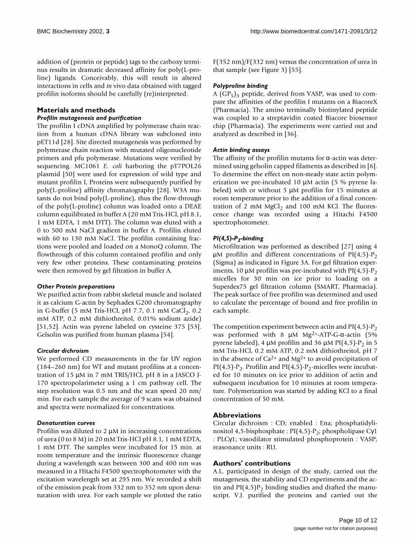
Materials and methodsProfilin mutagenesis and purificationThe profilin I cDNA amplified by polymerase chain reac-tion from a human cDNA library was subcloned intopET11d [28]. Site directed mutagenesis was performed bypolymerase chain reaction with mutated oligonucleotideprimers and pfu polymerase. Mutations were verified bysequencing. MC1061 E. coli harboring the pT7POL26plasmid [50] were used for expression of wild type andmutant profilin I, Proteins were subsequently purified bypoly(L-proline) affinity chromatography [28]. W3A mu-tants do not bind poly(L-proline), thus the flow-throughof the poly(L-proline) column was loaded onto a DEAEcolumn equilibrated in buffer A (20 mM Tris-HCl, pH 8.1,1 mM EDTA, 1 mM DTT). The column was eluted with a0 to 500 mM NaCl gradient in buffer A. Profilin elutedwith 60 to 130 mM NaCl. The profilin containing frac-tions were pooled and loaded on a MonoQ column. Theflowthrough of this column contained profilin and onlyvery few other proteins. These contaminating proteinswere then removed by gel filtration in buffer A.
Other Protein preparationsWe purified actin from rabbit skeletal muscle and isolatedit as calcium G-actin by Sephadex G200 chromatographyin G-buffer (5 mM Tris-HCl, pH 7.7, 0.1 mM CaCl2, 0.2mM ATP, 0.2 mM dithiothreitol, 0.01% sodium azide)[51,52]. Actin was pyrene labeled on cysteine 375 [53].Gelsolin was purified from human plasma [54].
Circular dichroismWe performed CD measurements in the far UV region(184–260 nm) for WT and mutant profilins at a concen-tration of 15 µM in 7 mM TRIS/HCl, pH 8 in a JASCO J-170 spectropolarimeter using a 1 cm pathway cell. Thestep resolution was 0.5 nm and the scan speed 20 nm/min. For each sample the average of 9 scans was obtainedand spectra were normalized for concentrations.
Denaturation curvesProfilin was diluted to 2 µM in increasing concentrationsof urea (0 to 8 M) in 20 mM Tris-HCl pH 8.1, 1 mM EDTA,1 mM DTT. The samples were incubated for 15 min. atroom temperature and the intrinsic fluorescence changeduring a wavelength scan between 300 and 400 nm wasmeasured in a Hitachi F4500 spectrophotometer with theexcitation wavelength set at 295 nm. We recorded a shiftof the emission peak from 332 nm to 352 nm upon dena-turation with urea. For each sample we plotted the ratio
F(352 nm)/F(332 nm) versus the concentration of urea inthat sample (see Figure 3) [55].
Polyproline bindingA (GP5)3 peptide, derived from VASP, was used to com-pare the affinities of the profilin I mutants on a BiacoreX(Pharmacia). The amino terminally biotinylated peptidewas coupled to a streptavidin coated Biacore biosensorchip (Pharmacia). The experiments were carried out andanalyzed as described in [36].
Actin binding assaysThe affinity of the profilin mutants for α-actin was deter-mined using gelsolin capped filaments as described in [6].To determine the effect on non-steady state actin polym-erization we pre-incubated 10 µM actin (5 % pyrene la-beled) with or without 5 µM profilin for 15 minutes atroom temperature prior to the addition of a final concen-tration of 2 mM MgCl2 and 100 mM KCl. The fluores-cence change was recorded using a Hitachi F4500spectrophotometer.
PI(4,5)-P2-bindingMicrofiltration was performed as described [27] using 4µM profilin and different concentrations of PI(4,5)-P2(Sigma) as indicated in Figure 3A. For gel filtration exper-iments, 10 µM profilin was pre-incubated with PI(4,5)-P2micelles for 30 min on ice prior to loading on aSuperdex75 gel filtration column (SMART, Pharmacia).The peak surface of free profilin was determined and usedto calculate the percentage of bound and free profilin ineach sample.
The competition experiment between actin and PI(4,5)-P2was performed with 8 µM Mg2+-ATP-G-α-actin (5%pyrene labeled), 4 µM profilin and 36 µM PI(4,5)-P2 in 5mM Tris-HCl, 0.2 mM ATP, 0.2 mM dithiothreitol, pH 7in the absence of Ca2+ and Mg2+ to avoid precipitation ofPI(4,5)-P2. Profilin and PI(4,5)-P2-micelles were incubat-ed for 10 minutes on ice prior to addition of actin andsubsequent incubation for 10 minutes at room tempera-ture. Polymerization was started by adding KCl to a finalconcentration of 50 mM.
AbbreviationsCircular dichroism : CD; enabled : Ena; phosphatidyli-nositol 4,5-bisphosphate : PI(4,5)-P2; phospholipase Cγ1: PLCγ1; vasodilator stimulated phosphoprotein : VASP;reasonance units : RU.
Authors' contributionsA.L. participated in design of the study, carried out themutagenesis, the stability and CD experiments and the ac-tin and PI(4,5)P2 binding studies and drafted the manu-script. V.J. purified the proteins and carried out the
Page 10 of 12(page number not for citation purposes)

BMC Biochemistry 2002, 3 http://www.biomedcentral.com/1471-2091/3/12
Biacore and microfiltration experiments. D.D. helpedwith the mutagenesis. J.V. participated in the design of thestudy. C.A. conceived the study, participated in the coor-dination and in the design of the study.
AcknowledgementsWe thank Frank Peelman for the modeling experiment and Lorene Lanier for critically reading the manuscript. A.L. is recipient of a post-doctoral fel-lowship of F.W.O.-Vlaanderen. This work was supported by F.W.O.-grant G022598 and BOF-GOA project 2051401 to J.V. and C.A. and F.W.O.-grant G004497 and a grant from the 'Geneeskundige stichting Koningin Elis-abeth' to C.A.
References1. Schutt CE, Myslik JC, Rozycki MD, Goonesekere NC, Lindberg U:
The structure of crystalline profilin-beta-actin. Nature 1993,365:810-816
2. Bjorkegren C, Rozycki M, Schutt CE, Lindberg U, Karlsson R: Muta-genesis of human profilin locates its poly(L-proline)-bindingsite to a hydrophobic patch of aromatic amino acids. FEBS Lett1993, 333:123-126
3. Lassing I, Lindberg U: Specific interaction between phosphati-dylinositol 4,5-bisphosphate and profilactin. Nature 1985,314:472-474
4. Theriot JA, Mitchison TJ: The three faces of profilin. Cell 1993,75:835-838
5. Sohn RH, Goldschmidt-Clermont PJ: Profilin: at the crossroads ofsignal transduction and the actin cytoskeleton. Bioessays 1994,16:465-472
6. Pantaloni D, Carlier MF: How profilin promotes actin filamentassembly in the presence of thymosin beta 4. Cell 1993,75:1007-1014
7. Kang F, Purich DL, Southwick FS: Profilin promotes barbed-endactin filament assembly without lowering the critical con-centration. J Biol Chem 1999, 274:36963-36972
8. Gertler FB, Niebuhr K, Reinhard M, Wehland J, Soriano P: Mena, arelative of VASP and Drosophila Enabled, is implicated inthe control of microfilament dynamics. Cell 1996, 87:227-239
9. Lambrechts AI, Kwiatkowski A, Lanier LM, Bear JE, VandekerckhoveJ, Ampe C, Gertler FB: PKA phosphorylation of EVL, a Mena/VASP relative, regulates its interaction with actin and SH3-domains. J Biol Chem 2000, 275:36143-36151
10. Reinhard M, Giehl K, Abel K, Haffner C, Jarchau T, Hoppe V, JockuschBM., Walter U.: The proline-rich focal adhesion and microfila-ment protein VASP is a ligand for profilins. EMBO J 1995,14:1583-1589
11. Yang C, Huang M, DeBiasio J, Pring M, Joyce M, Miki H, Takenawa T.,Zigmond SH.: Profilin enhances Cdc42-induced nucleation ofactin polymerization. J Cell Biol 2000, 150:1001-1012
12. Watanabe N, Madaule P, Reid T, Ishizaki T, Watanabe G, Kakizuka A,Saito Y., Nakao K., Jockusch BM., Narumiya S.: p140mDia, a mam-malian homolog of Drosophila diaphanous, is a target pro-tein for Rho small GTPase and is a ligand for profilin. EMBO J1997, 16:3044-3056
13. Krause M, Sechi AS, Konradt M, Monner D, Gertler FB, Wehland J:Fyn-binding protein (Fyb)/SLP-76-associated protein(SLAP), Ena/vasodilator-stimulated phosphoprotein (VASP)proteins and the Arp2/3 complex link T cell receptor (TCR)signaling to the actin cytoskeleton. J Cell Biol 2000, 149:181-194
14. Haarer BK, Petzold AS, Brown SS: Mutational analysis of yeastprofilin. Mol Cell Biol 1993, 13:7864-7873
15. Archer SJ, Vinson VK, Pollard TD, Torchia DA: Elucidation of thepoly-L-proline binding site in Acanthamoeba profilin I byNMR spectroscopy. FEBS Lett 1994, 337:145-151
16. Metzler WJ, Bell AJ, Ernst E, Lavoie TB, Mueller L: Identification ofthe poly-L-proline-binding site on human profilin. J Biol Chem1994, 269:4620-4625
17. Mahoney NM, Janmey PA, Almo SC: Structure of the profilin-poly-L-proline complex involved in morphogenesis and cy-toskeletal regulation. Nat Struct Biol 1997, 4:953-960
18. Mahoney NM, Rozwarski DA, Fedorov E, Fedorov AA, Almo SC:Profilin binds proline-rich ligands in two distinct amide back-bone orientations. Nat Struct Biol 1999, 6:666-671
19. Ostrander DB, Ernst EG, Lavoie TB, Gorman JA: Polyproline bind-ing is an essential function of human profilin in yeast. Eur J Bi-ochem 1999, 262:26-35
20. Lassing I, Lindberg U: Specificity of the interaction betweenphosphatidylinositol 4,5-bisphosphate and the profilin:actincomplex. J Cell Biochem 1988, 37:255-267
21. Goldschmidt-Clermont PJ, Machesky LM, Baldassare JJ, Pollard TD:The actin-binding protein profilin binds to PIP2 and inhibitsits hydrolysis by phospholipase C. Science 1990, 247:1575-1578
22. Lu PJ, Shieh WR, Rhee SG, Yin HL, Chen CS: Lipid products ofphosphoinositide 3-kinase bind human profilin with high af-finity. Biochemistry 1996, 35:14027-14034
23. Goldschmidt-Clermont PJ, Kim JW, Machesky LM, Rhee SG, PollardTD: Regulation of phospholipase C-gamma 1 by profilin andtyrosine phosphorylation. Science 1991, 251:1231-1233
24. Ostrander DB, Gorman JA, Carman GM: Regulation of profilin lo-calization in Saccharomyces cerevisiae by phosphoinositidemetabolism. J Biol Chem 1995, 270:27045-27050
25. Janmey PA: Protein regulation by phosphatidylinositol lipids.Chem Biol 1995, 2:61-65
26. Machesky LM, Goldschmidt-Clermont PJ, Pollard TD: The affinitiesof human platelet and Acanthamoeba profilin isoforms forpolyphosphoinositides account for their relative abilities toinhibit phospholipase C. Cell Regul 1990, 1:937-950
27. Lambrechts A, Verschelde JL, Jonckheere V, Goethals M, Vandeker-ckhove J, Ampe C: The mammalian profilin isoforms displaycomplementary affinities for PIP2 and proline-rich sequenc-es. EMBO J 1997, 16:484-494
28. Lambrechts A, Braun A, Jonckheere V, Aszodi A, Lanier LM, RobbensJ, Van Colen I, Vandekerckhove J., Fassler R, Ampe C: Profilin II isalternatively spliced, resulting in profilin isoforms that aredifferentially expressed and have distinct biochemical prop-erties. Mol Cell Biol 2000, 20:8209-8219
29. Gibbon B, Staiger C: Profilin. In Actin : a dynamic framework for multi-ple plant cell functions. (Edited by: Staiger C, Baluska F, Volkmann D, Bar-low P) Dordrecht: Kluwer Academic Publishers 2000, 45-65
30. Schluter K, Jockusch BM, Rothkegel M: Profilins as regulators ofactin dynamics. Biochim Biophys Acta 1997, 1359:97-109
31. Fedorov AA, Magnus KA, Graupe MH, Lattman EE, Pollard TD, AlmoSC: X-ray structures of isoforms of the actin-binding proteinprofilin that differ in their affinity for phosphatidylinositolphosphates. Proc Natl Acad Sci U S A 1994, 91:8636-8640
32. Sohn RH, Chen J, Koblan KS, Bray PF, Goldschmidt-Clermont PJ: Lo-calization of a binding site for phosphatidylinositol 4,5-bi-sphosphate on human profilin. J Biol Chem 1995, 270:21114-21120
33. Yu FX, Sun HQ, Janmey PA, Yin HL: Identification of a polyphos-phoinositide-binding sequence in an actin monomer-bindingdomain of gelsolin. J Biol Chem 1992, 267:14616-14621
34. Chaudhary A, Chen J, Gu QM, Witke W, Kwiatkowski DJ, PrestwichGD: Probing the phosphoinositide 4,5-bisphosphate bindingsite of human profilin I. Chem Biol 1998, 5:273-281
35. Cedergren-Zeppezauer ES, Goonesekere NC, Rozycki MD, MyslikJC, Dauter Z, Lindberg U, Schutt CE: Crystallization and struc-ture determination of bovine profilin at 2.0 A resolution. JMol Biol 1994, 240:459-475
36. Jonckheere V, Lambrechts A, Vandekerckhove J, Ampe C: Dimeri-zation of profilin II upon binding the (GP5)3 peptide fromVASP overcomes the inhibition of actin nucleation by profi-lin II and thymosin beta4. FEBS Lett 1999, 447:257-263
37. Korenbaum E, Nordberg P, Bjorkegren-Sjogren C, Schutt CE, Lind-berg U, Karlsson R: The role of profilin in actin polymerizationand nucleotide exchange. Biochemistry 1998, 37:9274-9283
38. Fedorov AA, Pollard TD, Almo SC: Purification, characterizationand crystallization of human platelet profilin expressed in Es-cherichia coli. J Mol Biol 1994, 241:480-482
39. Nodelman IM, Bowman GD, Lindberg U, Schutt CE: X-ray struc-ture determination of human profilin II: A comparativestructural analysis of human profilins. J Mol Biol 1999, 294:1271-1285
40. Raghunathan V, Mowery P, Rozycki M, Lindberg U, Schutt C: Struc-tural changes in profilin accompany its binding to phosphati-dylinositol, 4,5-bisphosphate. FEBS Lett 1992, 297:46-50
41. Kaiser DA, Pollard TD: Characterization of actin and poly-L-proline binding sites of Acanthamoeba profilin with mono-
Page 11 of 12(page number not for citation purposes)

BMC Biochemistry 2002, 3 http://www.biomedcentral.com/1471-2091/3/12
clonal antibodies and by mutagenesis. J Mol Biol 1996, 256:89-107
42. Van Troys M, Dewitte D, Verschelde JL, Goethals M, Vandekerck-hove J, Ampe C: The Competitive Interaction of Actin andPIP(2) with Actophorin Is Based on Overlapping TargetSites: Design of a Gain-of-Function Mutant. Biochemistry 2000,39:12181-12189
43. Yin HL, Iida K, Janmey PA: Identification of a polyphosphoi-nositide-modulated domain in gelsolin which binds to thesides of actin filaments. J Cell Biol 1988, 106:805-812
44. Sun HQ, Wooten DC, Janmey PA, Yin HL: The actin side-bindingdomain of gelsolin also caps actin filaments. Implications foractin filament severing. J Biol Chem 1994, 269:9473-9479
45. Evangelista M, Blundell K, Longtine MS, Chow CJ, Adames N, PringleJR, Peter M, Boone C: Bni1p, a yeast formin linking cdc42p andthe actin cytoskeleton during polarized morphogenesis. Sci-ence 1997, 276:118-122
46. Bjorkegren-Sjogren C, Korenbaum E, Nordberg P, Lindberg U, Karls-son R: Isolation and characterization of two mutants of hu-man profilin I that do not bind poly(L-proline). FEBS Lett 1997,418:258-264
47. Di Nardo A, Gareus R, Kwiatkowski D, Witke W: Alternativesplicing of the mouse profilin II gene generates functionallydifferent profilin isoforms. J Cell Sci 2000, 113(Pt 21):3795-3803
48. Wittenmayer N, Rothkegel M, Jockusch BM, Schluter K: Functionalcharacterization of green fluorescent protein-profilin fusionproteins. Eur J Biochem 2000, 267:5247-5256
49. Geese M, Schluter K, Rothkegel M, Jockusch BM, Wehland J, Sechi AS:Accumulation of profilin II at the surface of listeria is con-comitant with the onset of motility and correlates with bac-terial speed. J Cell Sci 2000, 113(Pt 8):1415-1426
50. Mertens N, Remaut E, Fiers W: Tight transcriptional controlmechanism ensures stable high-level expression from T7promoter-based expression plasmids. Biotechnology (N Y) 1995,13:175-179
51. Spudich JA, Watt S: The regulation of rabbit skeletal musclecontraction. I. Biochemical studies of the interaction of thetropomyosin-troponin complex with actin and the proteo-lytic fragments of myosin. J Biol Chem 1971, 246:4866-4871
52. Pardee JD, Spudich JA: Purification of muscle actin. Methods CellBiol 1982, 24:271-289
53. Brenner SL, Korn ED: On the mechanism of actin monomer-polymer subunit exchange at steady state. J Biol Chem 1983,258:5013-5020
54. Bryan J: Gelsolin has three actin-binding sites. J Cell Biol 1988,106:1553-1562
55. Lu J, Pollard TD: Profilin binding to poly-L-proline and actinmonomers along with ability to catalyze actin nucleotide ex-change is required for viability of fission yeast. Mol Biol Cell2001, 12:1161-1175
Publish with BioMed Central and every scientist can read your work free of charge
"BioMedcentral will be the most significant development for disseminating the results of biomedical research in our lifetime."
Paul Nurse, Director-General, Imperial Cancer Research Fund
Publish with BMC and your research papers will be:
available free of charge to the entire biomedical community
peer reviewed and published immediately upon acceptance
cited in PubMed and archived on PubMed Central
yours - you keep the copyright
[email protected] your manuscript here:http://www.biomedcentral.com/manuscript/
BioMedcentral.com
Page 12 of 12(page number not for citation purposes)
Copyright © 2022 FDOKUMEN