Extracellular Enzyme Stoichiometry Reveals Soil Microbial ...
Upload
independentCategory
view
3download
0
doi:10.1006/jmbi.2000.3858 available online at http://www.idealibrary.com on J. Mol. Biol. (2000) 300, 363±375
The Crystal Structure of Tetrameric MethionineAdenosyltransferase from Rat Liver Reveals theMethionine-binding Site
Beatriz Gonza lez1, MarõÂa A. Pajares2, Juan A. Hermoso1, Luis Alvarez2
Francisco Garrido2, Janice R. Sufrin3 and Julia Sanz-Aparicio1*
1Grupo de CristalografõÂaMacromolecular y BiologõÂaEstructural, Instituto deQuõÂmica-FõÂsica ``Rocasolano''CSIC, Serrano 11928006 Madrid, Spain2Instituto de InvestigacionesBiomeÂdicas ``Alberto Sols''CSIC-UAM, Arturo Duperier4, 28029 Madrid, Spain3Grace Cancer Drug CenterRoswell Park Cancer InstituteBuffalo, NY 14263, USA
E-mail address of the [email protected]
Abbreviations used: MAT I, methadenosyltransferase; c-MAT, methioadenosyltransferase from E. coli; SAadenosylmethionine; L-cisAMB, L-2-but-3-enoic acid; PPPi, tripolyphosporthophosphate; PPi, pyrophosphateethlymaleimide; DTT, dithiothreitol.
0022-2836/00/020363±13 $35.00/0
Most of the transmethylation reactions use the same methyl donor, S-ade-nosylmethionine (SAM), that is synthesised from methionine and ATP bymethionine adenosyltransferase (MAT). In mammals, two MAT enzymeshave been detected, one ubiquitous and another liver speci®c. The liverenzyme exists in two oligomeric forms, a tetramer (MAT I) and a dimer(MAT III), MAT I being the one that shows a higher level of af®nity formethionine but a lower SAM synthesis capacity. We have solvedthe crystal structure of rat liver MAT I at 2.7 AÊ resolution, complexedwith a methionine analogue: L-2-amino-4-methoxy-cis-but-3-enoic acid(L-cisAMB). The enzyme consists of four identical subunits arranged intwo tight dimers that are related by crystallographic 2-fold symmetry.The crystal structure shows the positions of the relevant cysteine residuesin the chain, and that Cys35 and Cys61 are perfectly oriented for forminga disulphide link. This result leads us to propose a hypothesis to explainthe control of MAT I/III exchange and hence, the effects observed onactivity. We have identi®ed the methionine-binding site into the active-site cavity, for the ®rst time. The L-cisAMB inhibitor is stacked againstPhe251 aromatic ring in a rather planar conformation, and its carboxylategroup coordinates a Mg2�, which, in turn, is linked to Asp180. The essen-tial role of the involved residues in MAT activity has been con®rmed bysite-directed mutagenesis. Phe251 is exposed to solvent and is located inthe beginning of the ¯exible loop Phe251-Ala260 that is connecting theN-terminal domain to the central domain. We postulate that a confor-mational change may take place during the enzymatic reaction and thisis possibly the reason of the unusual two-step mechanism involving tri-polyphosphate hydrolysis. Other important mechanistic implications arediscussed on the light of the results. Moreover, the critical role that cer-tain residues identi®ed in this study may have in methionine recognitionopens further possibilities for rational drug design.
# 2000 Academic Press
Keywords: methionine adenosyltransferase; disulphide bridges;methionine binding-site; X-ray structure
*Corresponding authorng author:
ioninenineM, S-amino-4-methoxy-cis-hate; Pi,; NEM, N-
Introduction
Methionine adenosyltransferases (MAT) (EC2.5.1.6) are cytosolic proteins exceptionally wellconserved during evolution (Kotb & Geller, 1993).MAT enzymes use methionine and ATP in a singu-lar reaction that leads to SAM (S-adenosylmethio-nine), and as side products, pyrophosphate andinorganic phosphate (Cantoni, 1953). All share sev-eral features: (i) they appear as oligomeric proteins,mainly tetramers, in all the tissues and organismsstudied; (ii) Mg2� are needed for activity; (iii)
# 2000 Academic Press
364 MAT I-Methionine Analogous Complex
monovalent cations (K�) are activators of theseenzymes; (iv) most enzymes show sigmoidal kin-etics for their substrates; and (v) they exhibit tripo-lyphosphatase activity (Mato et al., 1997). Theimportance of these enzymes is derived from thekey role that their product, SAM, has as the mainmethyl donor for transmethylation reactions(Cantoni, 1975). These reactions are involved in thesynthesis and modi®cation of an enormous varietyof compounds, among others, phospholipids, DNAand neurotransmitters. Moreover, after being dec-arboxylated, SAM can serve also as the propyla-mine donor for the synthesis of polyamines (Tabor& Tabor, 1984). Its role in such an enormous var-iety of processes makes its synthesis and the con-trol of its levels crucial for the cell and the wholeorganism. In mammals, two isoenzymes have beendetected, one ubiquitous (MAT II) and anotherliver speci®c (Mato et al., 1997). The fact that 85 %of the transmethylation reactions take place in theliver (Cantoni, 1975), and hence, about a 48 % ofthe ingested methionine is metabolised by thisorgan to produce 6-8 g of SAM daily in humans,may explain the need of a liver-speci®c enzyme(Aguilar et al., 1974; Mudd & Poole, 1975).
Rat liver MAT cDNA has been cloned and itsproduct found to be composed by 396 residues(Horikawa et al., 1989; Alvarez et al., 1991). Thissingle amino acid chain assembles in two func-tional oligomers, a tetramer (MAT I) and a dimer(MAT III), that differ in their level of af®nity formethionine (Alvarez et al., 1994; Cabrero et al.,1987). MAT I is tenfold more active under physio-logical concentrations of methionine (60 mM), whileMAT III presents a lower level of af®nity for meth-ionine, but a higher SAM synthesis ef®ciency, afact that may be useful in adapting to the changesin the levels of this essential amino acid upon feed-ing (Cabrero et al., 1987). Changes in the ratioMAT I/III may affect SAM production and indeed,it has been observed that in liver diseases, such asalcohol liver cirrhosis, the decreases observed inactivity and SAM levels correlate with a prevalenceof the dimer form (Cabrero et al., 1988). The studyof the regulatory mechanisms that control the oli-gomeric state and hence, the activity, has been car-ried out both in vivo and in vitro. Decreases inMAT activity in vivo, have been observed in sev-eral animal models where reductions in the GSHconcentration and hence changes in the redox statewere produced (Corrales et al., 1991, 1992). In thesestudies, a direct correlation between the reductionin GSH and SAM levels was detected. Moreover,drastic alterations in the morphology, structureand function in the hepatocyte followed thechanges in these key metabolites for the cell.In vitro, changes in the ratio between oligomericforms have been detected in several experiments(Cabrero & Alemani, 1988; Corrales et al., 1990;Pajares et al. 1994), and furthermore, Escherichia colioverexpression renders rat liver MAT oligomericforms susceptible of undergoing self-association
phenomena upon concentration (Mingorance et al.,1997).
All the results obtained in vitro led to the identi-®cation of residues that have an important role forthe activity and/or the association between sub-units. First, chemical modi®cation of SH groupsshow the role of cysteine residues on MAT activityand oligomerization; these last data have beencon®rmed by site-directed mutagenesis studies(Pajares et al., 1991; Mingorance et al., 1996).Second, the ATP-binding site was identi®ed byphotoaf®nity labelling using azido-ATP (Deigneret al., 1995). By using this technique a peptide (267-286) containing a P-loop consensus sequence wasisolated. Third, E. coli Arg244 has been shown tobe involved in the tripolyphosphate (PPPi) binding(Reczkowski et al., 1998). It is expected that resi-dues important for the function and structureshould be conserved, especially when the degree ofhomology among MAT enzymes is high. This isconsistent with the identi®cation of the corre-sponding P-loop containing peptide at the activesite of the only known crystal structure of thisfamily, that of E. coli MAT (Takusagawa et al.,1996a,b). However, neither the number of cysteineresidues (5-10 residues per monomer) nor theirlocalisation in the sequence is constant(Mingorance et al., 1996). In fact, only Cys35, Cys57and Cys105 (numbered according to the rat liversequence) are present in most of the sequencesfrom E. coli to mammals, while Cys9, Cys61,Cys69, Cys121 and Cys377 seem to be liverspeci®c. These differences between liver and non-liver MATs, the importance that Cys69 seems tohave in the oligomerization (Mingorance et al.,1996) and the presence of a disulphide bond(MartõÂnez-Chantar et al., 2000), may be related tothe fact that only liver speci®c MAT appears asboth a tetramer and a dimer.
Here, we describe the structure of the recombi-nant rat liver MAT upon refolding under reducingconditions. This form of the enzyme has been cho-sen, since its kinetic behaviour and overall second-ary structure is identical to the wild-type MAT,and because attempts to crystallise the puri®edwild-type protein have been so far unsuccessful.The structure we present now includes a complexwith a methionine analogue. Altogether, ourresults allow the identi®cation of the methionine-binding site, as well as the de®nition of key struc-tural and functional positions, such as those ofcysteine residues.
Results and Discussion
Overall structure and the architecture of theMAT subunit
The crystal structure of the tetrameric form ofmethionine adenosyltransferase (MAT I) from ratliver has been solved at 2.7 AÊ resolution. The crys-tals belong to the space group P4122 with 60 %solvent content in the cell, the asymmetric unit

MAT I-Methionine Analogous Complex 365
containing a tight dimer with two subunits relatedby local 2-fold symmetry. Two active sites arelocated per dimer with residues from each subunitcontributing to them. The tetramer structure(Figure 1) is generated by crystallographic 2-foldsymmetry and is essentially the same as that of theE. coli enzyme (c-MAT), which was crystallised inboth, hexagonal (Takusagawa et al., 1996a,b) andtetragonal (Fu et al., 1996) space groups. Each MATI subunit consists of 396 residues that are includedin the model except for two regions that are notvisible in the electron density maps. The ®rst ofthese regions is the N-terminal Met1-Glu16, whose
Figure 1. Structure of the tetrameric methionine ade-nosyltransferase from rat liver. The asymmetric unitcontains a tight dimer with two active sites in the inter-face. The tetramer is generated by crystallographic 2-fold symmetry. Each subunit is in a different colour andlabelled at the amino-terminal. Metal ions are includedas spheres: K�, magenta, Mg2�, orange.
presence was con®rmed by protein sequencing.The second region corresponds to the loop Ala118-Glu128, which was also found to be a ¯exible loopin the c-MAT that can seal the active centre bymeans of an open or closed conformation. Fu et al.(1996) concluded that if a substrate binds at theactive site, the enzyme crystallises in the hexagonalform, in which the ¯exible loop is disordered andnot visible in the electron density map. However,when the temperature decreases below 20 �C, theactive site is sealed by a ®xed conformation of the¯exible loop, preventing the binding of substrates,and thus the active-site is empty, even in the pre-sence of its substrate. In these conditions theenzyme crystallises in the tetragonal form.
Here, we co-crystallised the enzyme withL-cisAMB, an inhibitor analogous to one of thesubstrates, methionine, both at 0 and 20 �C. Theenzyme-ligand complex was obtained in both casesin a tetragonal crystal form. In agreement with theresults previously found in the enzyme-substratecomplexes from c-MAT, the ¯exible loop is disor-dered and hence supposed to be in the open con-formation. Nevertheless, our study reveals that thisconformation is not temperature dependent. Themobility of this loop might be a consequence of itsdirect involvement in catalysis, but this questionremains unclear at the moment.
The model was re®ned imposing non-crystallo-graphic symmetry restraints between the two mol-ecules of the asymmetric unit, resulting in a r.m.sdifference of 0.17 AÊ and, thus, the following dis-cussion will be based on a protomer. The overallfold of the monomer can be observed in Figure 2(a),and consists of three domains related by pseudo 3-fold symmetry: the N-terminal domain (residues17-28 and 146-254), the central domain (residues29-116 and 255-289), the C-terminal domain (resi-dues 129-145 and 290-396) and the mobile, non-vis-ible loop, linking the central domain to the C-terminal domain. A least-squares superposition(Kabsch, 1976) of the MAT I subunit Ca atoms tothe corresponding monomer from c-MAT yieldsr.m.s. deviation of 1.21 AÊ between both structures,but reveals a slightly different orientation of theirdomains. As depicted in Figure 2(b), the N- andthe central domains are rather conserved, but theC-terminal domain is twisted by 5.5 � with respectto c-MAT, the central loop acting as the hinge. Inaddition, two other differences between bothenzymes are detectable: ®rstly, MAT I presents afour-residue insertion (Asp192-Ala195) in a loopconnecting two b-strands in the N-terminaldomain, which is protruding to the C-terminaldomain. Consequently, the two domains interact toa greater extent. Secondly, and more remarkablyare the differences encountered in the extendedloop Phe251-Ala260, which connects the N-term-inal domain to the central domain (in yellow inFigure 2(a)). This loop, well conserved insequence, presents some ¯exibility as deducedfrom the larger thermal motions of this segment(The Boverall in this loop is 44.3 AÊ 2 for molecule

Figure 2. The protomer of MAT I from rat liver. (a)The overall fold consists of three domains related bypseudo 3-fold symmetry: the N-terminal domain (cyan),the central domain (red) and the C-terminal domain(green). The ¯exible, non-visible loop (Ala118-Glu128)links the central to the C-terminal domain. The looplinking the N-terminal to the central domain (Phe251-Ala260) is yellow and presents also some ¯exibility. TheL-cisAMB inhibitor and two sulphate ions are includedas ball-and-sticks model. Two K� (magenta) and twoMg2� (orange) are included as spheres. (b) Least-squaressuperposition of the Ca-trace from MAT I subunit (pink)to the corresponding monomer from c-MAT (green).The C-terminal domain of MAT is twisted by 5.5 � withrespect to c-MAT, the core loop acting as the hinge.Two major local differences are pointed with an arrow:a four-residue insertion in MAT (Asp192-Ala195), andloop (Phe251-Ala260) yellow in (a).
Table 1. Intersubunit polar interactions in the MATtetramer (<3.5 AÊ )
A. In the dimerMolecule A-Molecule BMet20 (O)-Gln318 (NE2)Glu58 (OE1)-Arg265 (NH1)Glu58 (OE1)-Arg265 (NH2)Glu58 (OE2)-Gly258 (O)Glu58 (OE2)-Ala260 (O)Gln136 (O)-Gln184 (NE2)Thr263 (O)-Arg265 (NH1)
B. Between dimersMolecule A-Molecule CGln81 (OE1)-Glu112 (OE2)Gln81 (NE2)-Leu111 (O)Arg85 (NH2)-Glu112 (OE2)Molecule A-Molecule DLys62 (O)-Asn106 (OD1)Thr63 (O)-Asn106 (OD1)
The listed interactions are found twice in the tetramer due tobinary symmetry (i.e. from molecule A to B and from moleculeB to A). See Figure 1 for subunits labelling.
366 MAT I-Methionine Analogous Complex
A and 48.9 AÊ 2 for molecule B; The Boverall for thewhole molecule is 31.7 AÊ 2). As discussed below,
the loop is directly involved in proper positioningof the methionine residue analogous upon binding.
The subunits interactions in the dimer andthe tetramer
An asymmetric unit contains a tight dimerrelated by a local 2-fold axis. Crystallographic diadaxis generates the active tetramer that, in turn, pre-sents pseudo 222 symmetry. The overall structureof the tetramer is the same as that found in c-MAT,but the arrangement of the subunits is slightlydifferent, as seen in Figure 3(a). In fact, this is aconsequence of a different orientation of the sub-units in the tight dimer of MAT I with respect tothe E. coli enzyme. After superposition of one sub-unit from each enzyme, a screw movement of theother subunit of MAT I is needed to superimposeoptimally to the c-MAT dimer. This screw move-ment involves a rotation of 7.4 � and an approachof 1.3 AÊ along an axis, which is almost perpendicu-lar to the local 2-fold axis (Figure 3(a)). Conversely,the relative orientation of the two dimers in the tet-ramer is very similar in both enzymes.
The pattern of interaction between subunits inthe dimer can be described as a contact surface,being mostly ¯at and hydrophobic betweenb-sheets from each subunit. In addition, the centralregion of the interface is marked by polar inter-actions between the equivalent core loops fromboth molecules, which are given in Table 1 anddepicted in Figure 3(b). On the other hand, Gly264and Arg265 carbonyl groups, and the symmetricalGly264* and Arg265*, are coordinated to the struc-tural K� atom, which is situated almost in thepseudo 2-fold axis relating both subunits, and con-fers a rigid geometry to the active sites. Finally,interactions between tight dimers are limited to afew polar interactions between subunits, which are

Figure 3. Subunits interaction in MAT I. (a) The Mat Itetramer (left), drawn as Ca-trace, presents a local binaryaxis relating both subunits of the tight dimer. Thescheme (right) relates the MAT tetramer (black) to the c-MAT tetramer (blue): after superimposing subunit Afrom both enzymes, a screw movement (7.4 � rotationand 1.4 AÊ displacement) about an axis almost perpen-dicular to the binary axis and passing through O isneeded, to superimpose subunit B from both enzymes.(b) Polar interactions between subunits at the core loopin the dimer interface. Residues from molecule A and B(labelled with asterisk) from the central domain of bothsubunits are shown. A K� is located at the local 2-foldaxis playing a structural role.
Figure 4. The cysteine residues of MAT I and possibledisulphide bridges. Ca-trace of the MAT subunit show-ing the location of eight cysteine residues. Cys9, in themissing N-terminal, and Cys121, in the ¯exible loop, arenot visible in the electron density maps. Cys9, Cys61,Cys69, Cys121 and Cys377 are liver-speci®c (labelled inblue). The ®ve cysteine residues located in the centraldomain have been found to be involved in association/dissociation processes of MAT by site-directed mutagen-esis.
MAT I-Methionine Analogous Complex 367
also given in Table 1. These interactions involvechain segments that are slightly different fromthose found in the c-MAT tetramer, but they areequivalent in strength and relevance (see Table 1).
The role of the cysteine residues and possibledisulphide bridges
Evidence underlining the importance of cysteineresidues in the activity and oligomerisation of rat
liver MAT have been obtained from several exper-iments, where regulation by GSH/GSSG (Pajareset al., 1992; MartõÂnez-Chantar & Pajares, 1996) andmodi®cation by nitric oxide donors (Ruiz et al.,1998) were investigated. It is believed that residuesthat show to be essential for activity are conservedalong the same family of proteins. However, ®veover the ten cysteine residues in MAT sequence areliver speci®c. This reveals some special feature ofthe hepatic enzyme that may be related to thein vivo control of the dimer/tetramer equilibrium,in response to changes in the substrate levels.
Figure 4 shows the location of these residues inthe monomer subunit of rat liver MAT, except forCys9 and Cys121 that are located in ¯exible areaswhich are not seen in the electron density map.These two non-visible residues, together withCys61, Cys69 and Cys377 (labelled in blue), areonly found in the liver enzyme. The crystal struc-ture reveals two interesting aspects. First of all,the ®ve cysteine residues alter the dimer/tetramerratio by Cys! Ser site-directed mutagenesis(Mingorance et al., 1996), i.e. Cys35 to Cys105, areall located in the central domain of the subunit.Taking into account that the interface between twodimers in the tetramer is only made up by thisdomain (see Figure 1), these ®ve cysteine residuesmust play a role in the stabilisation of the tetramerstructure. Secondly, the folding of the chain pos-itions only a pair of cysteine residues in a proper

368 MAT I-Methionine Analogous Complex
orientation to make a disulphide bridge, Cys35-Cys61, and this is consistent with the intrasubunitdisulphide group identi®ed in the puri®ed rat liverMAT (MartõÂnez-Chantar & Pajares, 2000). On thecontrary, the sulphydryl groups of Cys57 andCys69 are pointing in an opposite direction and alocal reorganisation of the main-chain would beneeded in order to make a bridge feasible.
From the reported experiments (Mingoranceet al., 1996) in which the Cys69Ser mutant only ledto the production of dimers, one would expect acrucial role for Cys69 in tetramer formation, poss-ibly by its involvement in an essential bridge. Thefact that the isolated tetramer form presents nosuch disulphide bridge suggests that the criticalrole of Cys69 in the oligomeric equilibriumtowards the tetramer seems to be related to pre-vious steps in the folding event. The hypothesismay be that non-native disulphide links areformed between different pairs of the involvedcysteine residues (Cys35 to Cys105) in an inter-mediate state/s, which can be rearranged in latersteps of the folding mechanism. Accordingly, theexistence of the disulphide Cys35-Cys61 in puri®edrat liver MAT could preclude association-dis-sociation processes. The presence of such disul-phide close to the contact area between dimerscould give greater stability to the b-sheet andhence to the whole oligomer. On the other hand,this bridge would restrict the chain ¯exibilityrequired in the intermediate state to constitute themixed S-S links that may determine the existenceof one form over the other. Disappearance of thisbond as upon folding under reducing conditions,may destabilise this part of the structure allowingeasier aggregation or dissociation, as observed byE. coli overexpression of rat liver MAT, where tenfree -SH groups can be detected (data not shown).
The implication of intramolecular disulphidelinks in protein oligomerisation is not new and hasbeen observed in some extra-cellular proteins(Marchand et al., 1996), but such a role has notbeen reported for cytosolic enzymes, up to now.Even when the cytosol is in a strongly reducingenvironment and disulphide links are scarce, someof these bonds have been detected in proteins suchas the cytosolic branched-chain amino transferaseand the human glucocorticoid receptor (Davoodiet al., 1998; Silva & Cidlowski, 1989). We havedemonstrated the presence of such a disulphidein puri®ed rat liver MAT, after isolation underreducing conditions and chemical modi®cation offree sulphhydryl groups (MartõÂnez-Chantar andPajares, 2000). However, a full description of theoligomerization of rat liver MAT under physiologi-cal conditions is not easy in the light of currentknowledge. Further work is needed to make pro-gress in this sense, but it is possible to envisage aregulation of the dimer/tetramer equilibrium ofMAT under oxidative stress, which would involvethe constitution of either mixed disulphide groups(SaÂnchez-GoÂngora et al., 1997), or non-native disul-phide bridges. The intensity of the oxidative stress,
mild to severe, would lead to a different ratiobetween native and oxidised or non-native formsthat would render, as a ®nal effect, the reductionof activity that is observed in pathologies such asliver cirrhosis.
The active site
The unusual synthetic reaction catalysed byMAT occurs in a sequential two-step process com-prising SAM formation and a subsequent tripoly-phosphate hydrolysis, prior to product release(Mudd, 1973), as shown below:
L -methionine�ATP�H2O
! �SAM� PPPi� �H2O! SAM� PPi� Pi
In a ®rst step, SAM is formed by direct attack ofthe sulphur atom of methionine to C50 position ofATP in a SN2 reaction, which cleaves the polypho-sphate chain from ATP. In the second step, the tri-polyphosphate moiety is further hydrolysed topyrophosphate (PPi) and inorganic orthophosphate(Pi) coming from the g position of ATP molecule.Divalent cations (Mg2�, Mn2�) are needed foractivity, and certain monovalent cations (K�, Tl�)activate or stimulate the reaction (Parry & Minta,1982; Markham et al., 1987). However, the couplingbetween both steps has made it dif®cult to studyeach in detail and, as such, the reason for tripoly-phosphate hydrolysis or its relevance for the over-all reaction remains unclear. A possibleexplanation for this activity is to prevent the rever-sal of the catalytic reaction and yet another is toconvert the PPPi moiety in more ef®cient leavinggroups (Reczkowski et al., 1998). A more appealingpossibility to be considered is that the tripolypho-sphatase activity is the source of the energyrequired by the enzyme to suffer a conformationalchange needed to accomplish the catalytic cycle(Reczkowski et al., 1998). Nonetheless, more exper-imental information concerning the enzymaticmechanism is needed to clarify this aspect. Indeed,deeper insight into the enzyme-substrates bindinginteractions is needed to make progress in thissense.
The possible ATP-binding site has been reportedby Takusagawa et al. (1996b) as determined by X-ray crystallography of c-MAT complexed withADP and BrADP (PDB codes 1MXB and 1MXC).This study showed that ATP was hydrolysed bythe enzyme to ADP and PPi, revealing an unex-pected ATPase activity. In these complexes, thediphosphate group interacts extensively with theenzyme while the ribose and adenine rings seem tobe more loosely bound and, in fact, the ribose wasfound to be disordered. Some differences in thepositions of Mg2� and K� were also obtained alongthe different reported forms with PPi and Pi(Takusagawa et al., 1996a,b). Moreover, the enzymepresents no substantial changes from the uncom-plexed form. However, nothing is known about

MAT I-Methionine Analogous Complex 369
the methionine-binding site. An attempt to predictthe location of the SAM product has been reported(Schalk-Hihi & Markham, 1999; Taylor &Markham, 1999), by docking and NMR, based onthe above mentioned X-ray complexes.
Owing to the metabolic transcendence of SAM,many attempts to obtain potent inhibitors of MATactivity have been carried out, although they havenot been analysed from a structural point of view.Several imido and methylene analogues of ATPhave been investigated as both, substrates andinhibitors (Qi-Feng et al., 1990; Reczkowsky &Markham, 1999). On the other hand, amino acidanalogues (Sufrin & Lombardini, 1982) and manycyclic compounds structurally and conformation-ally similar to L-methionine (Coulter et al., 1974)have been tested for inhibitory activity. Inaddition, the distinct features of the tumour-derived enzyme forms have been investigated todesign inhibitors of SAM synthesis (Sufrin &Lombardini, 1982; Sufrin et al., 1993). Followingthis strategy, Sufrin et al. (1982) identi®ed L-2-amino-4-methoxy-cis-but-3-enoic acid (L-cisAMB)as a competitive inhibitor of SAM synthesis withrespect to methionine (Ki 50 mm). A comparativestudy of the cis and trans analogues con®rms thehigh level of speci®city of MAT for the cis-isomersand therefore, their assumption was that theenzyme-bound conformations of L-cisAMB andL-methionine correlate closely (Sufrin et al., 1993).
We have carried out the crystallisation of MAT Iin the presence of L-cisAMB, and the electron den-sity map allows the identi®cation of the methion-ine analogue at the active-site cavity (see Figure 5).As shown in Figures 5 and 6, the inhibitor mol-ecule is stacked against Phe251 aromatic ring with
Figure 5. Electron density at the methionine-analogue binweighted 2Fobs ÿ Fcalc electron-density map calculated with0.9s. The L-cisAMB molecule is labelled. Two Mg2� are repre
the enol-ether side-chain in a rather planar confor-mation. On the other hand, the carboxylate moietyis coordinating a Mg2� which, in turn, is linked toAsp180, the other oxygen atom of the carboxylgroup being hydrogen bonded to His30. A secondMg2� has been found linked to Asp135* of theother subunit in the dimer, and is further coordi-nated to a sulphate that is supposed to mimic apossible phosphate anion. This role is supportedby the fact that this sulphate anion is linked toLys182, described to coordinate the b-phosphate inthe c-MAT-ADP complex. On the other hand,another sulphate ion has been found at the sameposition described for the phosphate g of c-MATcomplexes, both with ADP and PPi (Takusagawaet al., 1996b). Finally, the two K� of the c-MATcrystals are also found in equivalent positions ofthe MAT active site. Asp180, Lys182 and Gln184are located in a strand of the N-terminal b-sheetand therefore are in a rather ®xed frame. His30 ishydrogen bonded to Asp32, which participates inthe polar interactions network at the core loop (seeFigure 3(b)) and therefore is also rather ®xed. Onthe other hand, Phe251 whose aromatic side-chainis exposed to the solvent, is located in the begin-ning of the ¯exible loop Phe251-Ala260 which isconnecting the N-terminal to the central domain(yellow in Figure 2(a)). Mobility seems to be a fea-ture of this loop and, in fact, in one of the twoactive sites of the dimer it presents some kind ofdisorder and, consequently, no inhibitor has beenmodelled into the electronic density map of thissecond site. The sequence of this loop is very wellconserved and, indeed, the motif GGPXG mustconfer a high degree of ¯exibility to the peptidicchain.
ding site. Stereoview of the active-site region in the sA-phases derived from the ®nal model and contoured atsented as spheres.
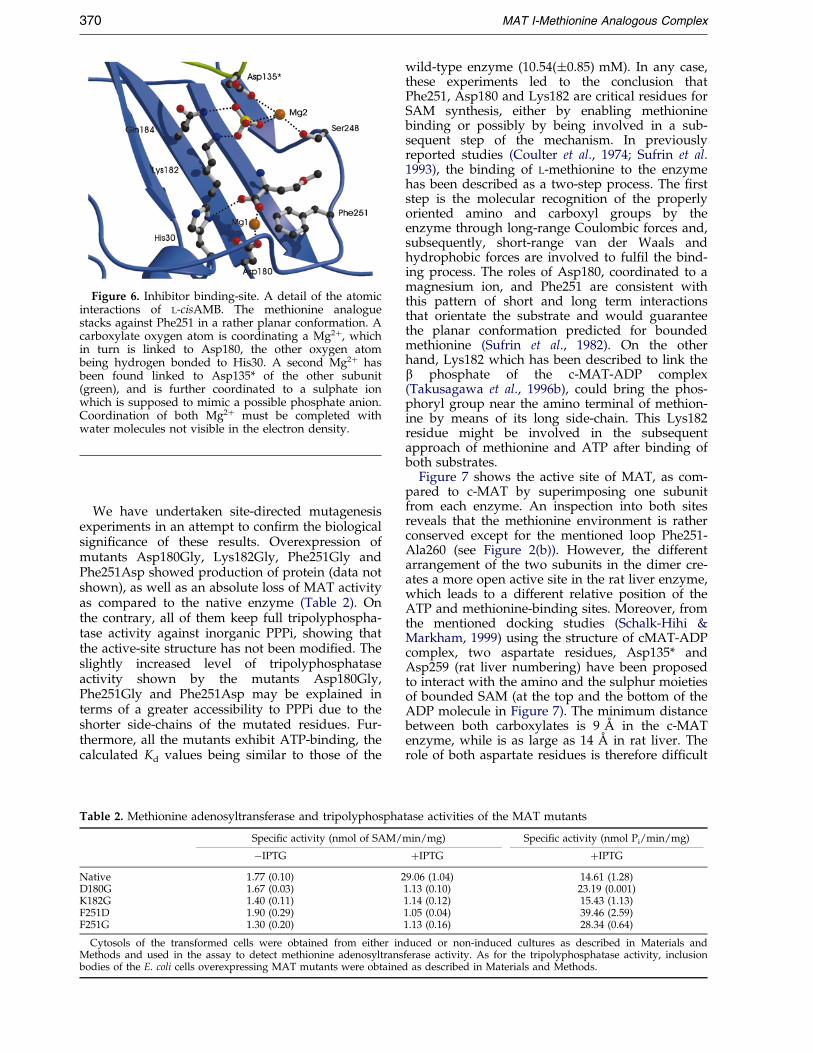
Figure 6. Inhibitor binding-site. A detail of the atomicinteractions of L-cisAMB. The methionine analoguestacks against Phe251 in a rather planar conformation. Acarboxylate oxygen atom is coordinating a Mg2�, whichin turn is linked to Asp180, the other oxygen atombeing hydrogen bonded to His30. A second Mg2� hasbeen found linked to Asp135* of the other subunit(green), and is further coordinated to a sulphate ionwhich is supposed to mimic a possible phosphate anion.Coordination of both Mg2� must be completed withwater molecules not visible in the electron density.
370 MAT I-Methionine Analogous Complex
We have undertaken site-directed mutagenesisexperiments in an attempt to con®rm the biologicalsigni®cance of these results. Overexpression ofmutants Asp180Gly, Lys182Gly, Phe251Gly andPhe251Asp showed production of protein (data notshown), as well as an absolute loss of MAT activityas compared to the native enzyme (Table 2). Onthe contrary, all of them keep full tripolyphospha-tase activity against inorganic PPPi, showing thatthe active-site structure has not been modi®ed. Theslightly increased level of tripolyphosphataseactivity shown by the mutants Asp180Gly,Phe251Gly and Phe251Asp may be explained interms of a greater accessibility to PPPi due to theshorter side-chains of the mutated residues. Fur-thermore, all the mutants exhibit ATP-binding, thecalculated Kd values being similar to those of the
Table 2. Methionine adenosyltransferase and tripolyphospha
Specific activity (nmol of SAM/
ÿIPTG
Native 1.77 (0.10) 2D180G 1.67 (0.03)K182G 1.40 (0.11)F251D 1.90 (0.29)F251G 1.30 (0.20)
Cytosols of the transformed cells were obtained from either inMethods and used in the assay to detect methionine adenosyltransbodies of the E. coli cells overexpressing MAT mutants were obtaine
wild-type enzyme (10.54(�0.85) mM). In any case,these experiments led to the conclusion thatPhe251, Asp180 and Lys182 are critical residues forSAM synthesis, either by enabling methioninebinding or possibly by being involved in a sub-sequent step of the mechanism. In previouslyreported studies (Coulter et al., 1974; Sufrin et al.1993), the binding of L-methionine to the enzymehas been described as a two-step process. The ®rststep is the molecular recognition of the properlyoriented amino and carboxyl groups by theenzyme through long-range Coulombic forces and,subsequently, short-range van der Waals andhydrophobic forces are involved to ful®l the bind-ing process. The roles of Asp180, coordinated to amagnesium ion, and Phe251 are consistent withthis pattern of short and long term interactionsthat orientate the substrate and would guaranteethe planar conformation predicted for boundedmethionine (Sufrin et al., 1982). On the otherhand, Lys182 which has been described to link theb phosphate of the c-MAT-ADP complex(Takusagawa et al., 1996b), could bring the phos-phoryl group near the amino terminal of methion-ine by means of its long side-chain. This Lys182residue might be involved in the subsequentapproach of methionine and ATP after binding ofboth substrates.
Figure 7 shows the active site of MAT, as com-pared to c-MAT by superimposing one subunitfrom each enzyme. An inspection into both sitesreveals that the methionine environment is ratherconserved except for the mentioned loop Phe251-Ala260 (see Figure 2(b)). However, the differentarrangement of the two subunits in the dimer cre-ates a more open active site in the rat liver enzyme,which leads to a different relative position of theATP and methionine-binding sites. Moreover, fromthe mentioned docking studies (Schalk-Hihi &Markham, 1999) using the structure of cMAT-ADPcomplex, two aspartate residues, Asp135* andAsp259 (rat liver numbering) have been proposedto interact with the amino and the sulphur moietiesof bounded SAM (at the top and the bottom of theADP molecule in Figure 7). The minimum distancebetween both carboxylates is 9 AÊ in the c-MATenzyme, while is as large as 14 AÊ in rat liver. Therole of both aspartate residues is therefore dif®cult
tase activities of the MAT mutants
min/mg) Specific activity (nmol Pi/min/mg)
�IPTG �IPTG
9.06 (1.04) 14.61 (1.28)1.13 (0.10) 23.19 (0.001)1.14 (0.12) 15.43 (1.13)1.05 (0.04) 39.46 (2.59)1.13 (0.16) 28.34 (0.64)
duced or non-induced cultures as described in Materials andferase activity. As for the tripolyphosphatase activity, inclusiond as described in Materials and Methods.

Figure 7. Stereo-view of the active site. Ca-trace of rat liver MAT (yellow) compared to c-MAT (cyan) by superim-posing one subunit from each dimer. The inhibitor molecule is highlighted in pink and relevant residues in the meth-ionine-binding site are represented as sticks; the two Mg2� are shown as spheres. The ADP and Pi of the c-MATcomplex are represented as sticks in cyan.
MAT I-Methionine Analogous Complex 371
to envisage in the light of the structure presentedhere. Consequently, although the sequence of MATenzymes is very well conserved along evolution, itis possible that some subtle differences at the activesite arise to account for the environmental particu-larities of each species.
A complete description of the enzymatic mech-anism is not trivial in the light of the two com-plexes reported until now. In fact, the positions ofATP and methionine are found to be unexpectedlyfar away for proper interaction to yield the pro-duct. However, two aspects need to be taken intoaccount. First of all, the unknown structure of theextended loop Asp117-Glu128, which is supposedto present a high level of ¯exibility, as previouslyreported, and could play an important role in thecatalysis that is still unknown. Secondly, the loopPhe251-Ala260 next to the postulated methionine-binding site also presents some degree of ¯exi-bility. We think that, apart from providing thecritical Phe251, it might further interact with themethionine moiety, possibly by coordinating thecarboxylate-linked Mg2� by means of the COgroups of the glycine residues in this loop. Conse-quently, a conformational change may take placeduring the enzymatic reaction and this is possiblythe reason of the unusual two-step mechanisminvolving PPPi hydrolysis. Further work is neededto clarify these aspects, mainly through structuralcharacterisation of intermediate states of the enzy-matic reaction, which is underway. Nonetheless,the critical role of certain residues in methioninerecognition presented here opens further possibili-ties for rational drug design.
Materials and Methods
Expression of rat liver MAT in E. coli and isolationof inclusion bodies
Competent E. coli BL21(DE3) cells were transformedwith the plasmid pSSRL-T7N as described (Alvarez et al.,1994). Overnight cultures prepared from single colonieswere used to inoculate 3 l of LB medium plus ampicillin(100 mg/ml). The cells were grown to A595 0.3-0.4 andIPTG (AMBION, Austin, TX, USA) was added to a ®nalconcentration of 0.5 mM. Incubations were continued forthree hours more, and the cells were harvested by cen-trifugation, washed with water and stored frozen untiluse.
Cell pellets were dissolved in 200 ml of 50 mM Tris-HCl (pH 8.0) containing 10 mM MgSO4, 1 mM EDTA,0.1 % (v/v) 2-mercaptoethanol and protease inhibitors(2 mg/ml aprotinin, 1 mg/ml pepstatin A, 0.5 mg/ml leu-peptin, 2.5 mg/ml antipain, 0.1 mM benzamidine,0.1 mM PMSF) (Sigma Chemical Co, St. Louis, MO,USA). Disruption of the cells was carried out by soni-cation at 4 �C (15 30 second pulses at 30 second intervals,output power level 20 in a Branson 250 soni®er). Solubleand insoluble fractions were separated by centrifugationfor 30 minutes at 48,000 g. All the extraction and thepuri®cation procedures were carried out at 4 �C. Pelletsof the insoluble fraction (7 g) were dissolved in 250 mlof 10 mM Hepes/Na (pH 7.5) containing 10 mM MgSO4
and 1 mM EDTA (buffer A), and disrupted by sonicationon ice (six pulses of ten seconds with ten second inter-vals, output power 20 in a Branson 250 soni®er). The sol-ution was centrifuged for 30 minutes at 48,000 g and thesupernatant discarded. The pellet was washed twice byresuspension in 250 ml of 100 mM Tris-HCl (pH 7.0)containing 5 mM EDTA, 5 mM DTT, 4 M urea (Merck,Germany), 5 % (v/v) Triton X-100 (Merck, Germany) and

372 MAT I-Methionine Analogous Complex
0.1 mM benzamidine and centrifuged for 30 minutes at48,000 g. A last wash with the same buffer devoided ofurea and Triton X-100 (250 ml) was performed. Thispreparation was stored at ÿ20 �C until use.
Refolding and purification of rat liver MAT
The washed pellet was solubilised in 250 ml of 50 mMTris-HCl (pH 8.0) (buffer B) containing 8 M urea and75 mM MgSO4 and incubated for two hours at 10 �C.Refolding was then performed by dilution to 2 M ureausing buffer B, and dialysis against buffer B containing10 mM DTT (15 l). The dialysed sample (950 ml) wasclari®ed by centrifugation at 4 �C for 30 minutes at48,000 g before puri®cation, and loaded on a DEAE-Sephacel column (2.5 � 18 cm) (Pharmacia LKB, Swe-den) equilibrated in buffer B containing 10 mM MgSO4
and 1 mM EDTA (buffer C). The column was washedusing 180 ml of buffer C and elution performed by a lin-ear gradient of 0 mM to 500 mM KCl in buffer C. The¯ow rate was 150 ml/h and 10 ml fractions were col-lected. MAT was detected measuring activity of the frac-tions, and the peak of activity was pooled andconcentrated by ultra®ltration using YM-30 membranes(AMICON Corp.,Beverly, MA, USA). The concentrate(4 ml) was then loaded on a Biogel A 1.5 m column(1.5 cm � 95 cm) (Bio-Rad,Richmond, CA, USA) equili-brated in buffer C containing 50 mM KCl. Elution wasthen carried out at a ¯ow rate of 15 ml/h using the samebuffer and 4 ml fractions were collected. The MAT con-taining peak was then concentrated by ultra®ltration to3 ml to achieve a ®nal protein concentration of 30 mg/ml as determined using the Bio-Rad protein assay kitand bovine serum albumin as the standard. MAT puritywas assessed by SDS-PAGE (Laemmli, 1970). For N-terminal sequencing, proteins (10-20 mg) resolved onPAGE-10 % SDS gels were electrotransferred to Inmoby-lon-P membranes using 10 mM CAPS (3-(cyclohexylani-no)-1-propane sulphonic acid; pH 11) containing 10 %(v/v) methanol, stained using Ponceau S and the bandexcised. Sequencing was performed on an Applied Bio-systems Procise Sequencer.
Production of active site mutants
Site-directed mutagenesis was performed by using theQuikChange Site-directed mutagenesis kit (Stratagene,La Jolla, CA, USA), following manufacturer's instruc-tions. Mutants were constructed in the vector pSSRL-T7N (Alvarez et al., 1994). Mutagenic oligonucleotideswere as follows (sense strand shown; the substitutedoligonucleotides are in bold): D180G, 50-GGCTGA-GACCTGGTTCTAAGACTCAGG-30; K182G, 50-GGCTGAGACCTGATT CTGGGACTCAGGTAACAG-30; F251G, 50-CCAAGTGGGCGGGGTGTCATCGG AGGTCCC-30; F251D, 50-CCAAGTGGGCGGGATGTCATCGGAGGTCCC-30. Mutations were veri®ed by sequencing theentire cDNA, using the dideoxy termination method(Sanger et al., 1977).
Determination of methionine adenosyltransferaseand tripolyphosphatase activities
MAT activity was measured as described by Gil et al.(1997). As for the tripolyphosphatase activity, refoldednative MAT and mutants were used in the assay. The®nal reaction volume was 400 ml containing 25 ml of theenzyme preparation and 50 mM Tris-HCl (pH 7.8),
100 mM KCl, 7 mM MgCl2, 1 mM DTT and 2 mM PPP.The reaction was carried out for 30 minutes at 30 �C andstopped by the addition of 1 ml of the stop solution(0.5 % ammonium molibdate (w/v), 2 % H2SO4 (v/v),0.5 % SDS (w/v)) and 10 ml 10 % (w/v) ascorbate, after®ve minutes, absorption at 750 nm was measured.
ATP-binding assays
ATP binding was determined using a gel ®ltrationtechnique described (Penefsky et al., 1977). Brie¯y, thereaction mixture was prepared as for MAT activityassays, except that the ®nal volume was 80 ml containing75 mg of protein, and various ATP concentrations(90 mM-15 mM). The protein was separated form the restof the mixture by gel ®ltration on Biogel P2 (Bio-Rad)columns (1 ml).
Crystallisation and data collection
Only very fresh puri®ed samples were suitable forcrystallisation settings by the hanging-drop method. Theprotein aggregated and precipitated very easy and onlya few drops per experiment yielded crystals. Equalamounts of protein solution (30 mg/ml protein in50 mM Tris-HCl (pH 8), 10 mM L-cisAMB, 10 mM DTT,50 mM KCl, 10 mM MgSO4 and 1 mM EDTA) and preci-pitant buffer (16 % (w/v) PEG 10000 in 100 mM Hepes(pH 7.5)) were mixed and vapour equilibrated againstthe latter solution. If crystals did not form within threeto four days, the drops became completely precipitated.Crystallisation trials were repeated at 4 and 20 �C withthe same results, crystals of bipyramidal habit growingfor a week up to a maximum size of about 0.3 mm. Theybelong to the P4122 space group with two molecules inthe asymmetric unit and 60 % solvent content. For cryo-protection, crystals of the protein-inhibitor complex weretransferred from the crystallisation drop to a solutioncontaining 18 % PEG 10000, 30 % glycerol and 100 mMHepes (pH 7.5). After soaking for one minute, they were¯ash-frozen at 120 K in nitrogen Oxford cryostream. Afull data set to 2.7 AÊ was collected from a very freshsingle crystal grown at 4 �C on an in-house 345 MARResearch Imaging Plate detector, using an Enraf-Noniusrotating-anode generator (l � 1.54 AÊ ). The data wereprocessed using Mos¯m (Leslie, 1987) and merged withthe CCP4 package (CCP4, 1994) to yield integrated inten-sities. A summary of data collection and data reductionstatistics is shown on Table 3.
Structure solution and refinement
Molecular replacement procedure was carried outwith the program AMoRe (Navaza, 1994), using data upto 3.5 AÊ . The search model was the monomer of MATfrom E. coli (PDB codes 1XRA, 1MXA), with the non-conserved residues truncated to Ala. The rotation searchshowed two peaks, which were tested in the translationsearch. Best results were obtained for the space groupP4122, which after rigid-body ®tting produced an R-fac-tor of 0.44 and a correlation coef®cient of 0.55. Thesedata correspond to a dimer in which both subunits wererelated by a local 2-fold symmetry axis. Crystallographicre®nement was performed by using X-PLOR (BruÈ nger,1993) with ¯at bulk-solvent correction and includinglow-resolution data to 24 AÊ . A subset of 7 % randomlyselected structure-factor amplitudes were excluded fromautomated re®nement and used to compute a free

Table 3. Structure and re®nement statistics
A. Data collectionTemperature (K) 120Source rotating-anodeSpace group P4122Unit cell dimensions (AÊ ) 115.20, 115.20, 159.98Unique reflections 28,742Rmerge
a 0.08Completeness (%)
Overall (44.7-2.66 AÊ ) 94.5Outer shell (2.8-2.66 AÊ ) 85.8
B. Final refinement parametersResolution range (AÊ ) 10-2.66Protein atoms 5692Solvent atoms 198Heterogen atoms 29Sigma cut-off 2R-factor 0.23Rfree
b 0.29r.m.s. deviation from ideal geometry
Bond lenghts (AÊ ) 0.017Bond angles (deg.) 2.1Dihedral angles (deg.) 26.5Improper angles (deg.) 2.03
Averaged B-factors (AÊ 2)Main-chain 30.7Side-chain 32.2Solvent 37.6
a Rmerge � � (I ÿ hIi)/� (I).b Rfree is equivalent to R-factor for a randomly selected 7 %
subset of re¯ections not used in structure re®nement.
MAT I-Methionine Analogous Complex 373
R-factor throughout the re®nement. Initial rounds ofSA-re®nement were alternated with rebuilding in theprogram O (Jones et al., 1991) and inspecting the modelin electron-density maps that had been averaged withthe RAVE package (Kleiwegt & Jones, 1994). Strict non-crystallographic symmetry (NCS) between both mol-ecules in the asymmetric unit was maintained until itbecame obvious that the loops surrounding the twoactive sites in the dimer were different, and conse-quently, NCS restraints strategy was followed thereafter.Implicated loops (61-65, 93-98 and 248-261) wereexcluded from the NCS-related groups and the modelwas subjected to the slow-cool simulated-annealing pro-tocol, using weak harmonic restraints for all atomsexcept the selected loops: a force constant of 20 kcalmolÿ1 AÊ ÿ2 was used for main-chain atoms, 10 kcal molÿ1
AÊ ÿ2 for other atoms, but only 1 kcal molÿ1 AÊ ÿ2 for theloops. However, neither this protocol nor other attempts(torsion-angle molecular dynamics re®nement, SA-omitmaps) succeeded in showing good quality electron den-sity for loop 248-261 of molecule B, which was assumedto be disordered, possibly due to unconstrained crystalpacking contacts. In connection to this fact, no densitywas clearly evident for the inhibitor moiety or the associ-ated Mg2�, and therefore, this active site was consideredto be uncomplexed. On the contrary, difference maps cal-culated with (Fo ÿ Fc) coef®cients, and calculated phasesshowed several signi®cant density peaks in the regioncorresponding to the other active site. They were con-®rmed by computing SA-omit maps around the sub-strate regions, which allowed model building of the L-cisAMB inhibitor, two putative Mg2� and two sulphateions. No restraints were used for the metal-ligand dis-tances during re®nement. The thermal parameters ofthese substrates were relatively large in comparison withthose of the residues constituting the active-site region,
suggesting partial occupancy, although this effect wasnot modelled. At the last stage water molecules wereincluded combined with more rounds of positional andindividual restrained B-factor re®nement, which led to a®nal R-factor of 0.23 (Rfree � 0.29) for all data withF > 2s(F) between 24.0 and 2.7 AÊ resolution. Final re®ne-ment parameters are reported in Table 3. Stereochemis-try of the model was checked using PROCHECK(Laskowsky et al., 1993). The Figures were generatedwith O (Jones et al., 1991), MOLSCRIPT (Kraulis, 1991),and Raster 3D (Merritt & Murphy, 1994).
Accession number
The atomic coordinates have been deposited in theRCSB Protein Data Bank (accession code 1QM4).
Acknowledgements
This work was supported by grants of the DireccioÂnGeneral de InvestigacioÂn Cientõ®ca y TeÂcnica (BIO97/0642 and PM 97/0064) and with support of BoehringerIngelheim EspanÄa.
References
Aguilar, T., Benevenga, W. & Harper, J. (1974). Effect ofdietary methionine level on its metabolism in rats.J. Nutr. 104, 761-771.
Alvarez, L., AsuncioÂn, M., Corrales, F., Pajares, M. A. &Mato, J. M. (1991). Analysis of the 50 non-codingregion of rat liver S-adenosylmethionine synthetasemRNA and comparison of the Mr deduced fromthe cDNA sequence and the puri®ed enzyme. FEBSLetters, 290, 142-146.
Alvarez, L., Mingorance, J., Pajares, M. A. & Mato, J. M.(1994). Expression of rat liver S-adenosylmethioninesynthetase in Escherichia coli results in two activeoligomeric forms. Biochem. J. 301, 557-561.
BruÈ nger, A. T. (1993). X-PLOR: A System for X-ray Crys-tallography and NMR, Yale University Press, NewHaven, CT.
Cabrero, C. & Alemany, S. (1988). Conversion of ratliver S-adenosyl-L-methionine synthetase from high-Mr form to low-Mr form by LiBr. Biochim. Biophys.Acta, 952, 277-281.
Cabrero, C., Puerta, J. L. & Alemany, S. (1987). Puri®-cation and comparison of two forms of S-adenosyl-L-methionine synthetase from rat liver. Eur. J. Bio-chem. 170, 299-304.
Cabrero, C., MartõÂn-Duce, A., OrtõÂz, P., Alemany, S. &Mato, J. M. (1988). Speci®c loss of the high-molecu-lar weight form of S-adenosylmethionine synthetasein human liver cirrhosis. Hepatology, 8, 1530-1534.
Cantoni, G. L. (1953). S-adenosylmethionine: a newintermediate formed enzymatically from L-methion-ine and adenosinetriphosphate. J. Biol. Chem. 204,403-416.
Cantoni, G. L. (1975). Biochemical methylations: selectedaspects. Annu. Rev. Biochem. 44, 435-451.
Collaborative Computational Project Number 4 (1994).The CCP4 suite: programs for protein crystallogra-phy. Acta Crystallog. sect. D, 50, 760-763.
Corrales, F., Cabrero, C., Pajares, M. A., Ortiz, P.,MartõÂn-Duce, A. & Mato, J. M. (1990). Inactivationand dissociation of S-adenosylmethionine synthe-

374 MAT I-Methionine Analogous Complex
tase by modi®cation of sulfhydryl groups and itspossible occurence in liver cirrhosis. Hepatology, 11,216-222.
Corrales, F., Ochoa, P., Rivas, C., MartõÂn-Lomas, M.,Mato, J. M. & Pajares, M. A. (1991). Inhibition ofglutathione synthesis in the liver leads to S-adeno-syl-L-methionine synthetase reduction. Hepatology,14, 528-533.
Corrales, F., GimeÂnez, A., Alvarez, L., CaballerõÂa, J.,Pajares, M. A., Andreu, H., PareÂs, A., Mato, J. M. &RodeÂs, J. (1992). S-adenosylmethionine treatmentprevents CCl4-induced S-adenosylmethioninesynthetase inactivation and attenuates liver injury.Hepatology, 16, 1022-1027.
Coulter, A. W., Lombardini, J. B., Sufrin, J. & Talalay, P.(1974). Structural and conformational analogues ofL-methionine as inhibitors of the enzymatic syn-thesis of S-adenosyl-L-methionine. Mol. Pharmacol.10, 319-334.
Davoodi, J., Drown, P. M., Bledsoe, R. K., Wallin,R., Reinhart, G. D. & Hutson, S. M. (1998). Over-expression and characterization of the humanmitochondrial and cytosolic branched-chain amino-transferases. J. Biol. Chem. 273, 4982-4989.
Deigner, H. P., Mato, J. M. & Pajares, M. A. (1995).Study of the rat liver S-adenosylmethionine synthe-tase active site with 8-azido ATP. Biochem. J. 308,565-571.
Fu, Z., Hu, Y., Markham, G. D. & Tukusawa, F. (1996).Flexible loop in the structure of S-adenosylmethio-nine synthetase crystallised in the tetragonal modi®-cation. J. Biomol. Struct. Dynam. 13, 727-739.
Gil, B., Pajares, M. A., Mato, J. M. & Alvarez, L. (1997).Glucocorticoid regulation of hepatic S-adenosyl-methionine synthetase gene expression. Endocrin-ology, 138, 1251-1258.
Horikawa, S., Ishikawa, M., Ozasa, H. & Tsukada, K.(1989). Isolation of a cDNA encoding the rat liverS-adenosylmethionine synthetase. Eur. J. Biochem.184, 497-501.
Jones, T. A., Zou, J. Y., Cowan, S. W. & Kjeldgaard, M.(1991). Improved methods for building proteinmodels in electron density maps and the location oferrors in these maps. Acta Crystallog. sect. A, 47,110-119.
Kabsch, W. (1976). A solution for the best rotation torelation two sets of vectors. Acta Crystallog. sect. A,32, 922-923.
Kleiwegt, G. J. & Jones, T. A. (1994). Halloween . . .masks and bones. In From Firsts Map to Final Model(Bailey, S., Hubbard, R. & Waller, R., eds), pp. 59-66, SERC Daresbury Laboratory, Warrington, UK.
Kotb, M. & Geller, A. M. (1993). Methionine adenosyl-transferase: structure and function. Pharmacol. Ther.59, 125-143.
Kraulis, P. J. (1991). MOLSCRIPT - A program to pro-duce both detailed and schematic plots of proteinstructures. J. Appl. Crystallog. 24, 946-950.
Laemmli, U. K. (1970). Cleavage of structural proteinsduring the assembly of the head of bacteriophageT4. Nature, 227, 680-685.
Laskowsky, R. A., MacArthur, M. W., Moss, D. S. &Thornton, J. M. (1993). PROCHECK: a program tocheck the stereochemical quality of protein struc-tures. J. Appl. Crystallog. 26, 283-291.
Leslie, A. G. W. (1987). Pro®le ®tting. In Proceedings ofthe CCP4 Study Weekend (Helliwell, J. R., Machin,P. A. & Papiz, M. Z., eds), vol. 39-50, SERCDaresbury Laboratory, Warrington, UK.
Marchand, P., Volkmann, M. & Bond, J. S. (1996).Cysteine mutations in the MAM domain result inmonomeric meprin and alter stability and activityof the proteinase. J. Biol. Chem. 271, 24236-24241.
Markham, G. D., Parkin, D. W., Mentch, F. & Schramm,V. L. (1987). A kinetic isotope effect study and tran-sition state analysis of the S-adenosylmethioninesynthetase reaction. J. Biol. Chem. 262, 5609-5615.
MartõÂnez-Chantar, M. L. & Pajares, M. A. (1996). Role ofthioltranferases on the modulation of rat liver S-adenosylmethionine synthetase activity by gluta-thione. FEBS Letters, 397, 293-297.
MartõÂnez-Chantar, M. L. & Pajares, M. A. (2000). Assign-ment of a single disulphide bridge in rat livermethionine adenosyltransferase. Eur. J. Biochem. 267,132-137.
Mato, J. M., Alvarez, L., Ortiz, P. & Pajares, M. A.(1997). S-adenosylmethionine synthesis: molecularmechanisms and clinical implications. Pharmacol.Ther. 73, 265-280.
Merritt, E. A. & Murphy, M. E. P. (1994). RASTER 3DVersion-2.0 - a program for photorealistic moleculargraphics. Acta Crystallog. sect. D, 50, 869-873.
Mingorance, J., Alvarez, L., SaÂnchez-GoÂngora, E., Mato,J. M. & Pajares, M. A. (1996). Site-directed mutagen-esis of rat liver S-adenosylmethionine synthetase.Identi®cation of a cysteine residue critical for theoligomeric state. Biochem. J. 315, 761-766.
Mingorance, J., Alvarez, L., Pajares, M. A. & Mato, J. M.(1997). Recombinant rat liver S-adenosyl-L-methion-ine synthetase tetramers and dimers are in equili-brium. Int. J. Biochem. Cell. Biol. 29, 485-491.
Mudd, S. H. (1973). The Enzymes, 3rd edit., vol. 8, pp.121-154, Academic Press, NY.
Mudd, S. H. & Poole, J. R. (1975). Labile methylbalances for normal humans on various dietaryregimens. Metabolism, 24, 721-735.
Navaza, J. (1994). AMoRe: an automated package formolecular replacement. Acta Crystallog. sect. A, 50,157-163.
Pajares, M. A., Corrales, F. J., Ochoa, P. & Mato, J. M.(1991). The role of cysteine 150 in the structure andactivity of rat liver S-adenosyl-L-methionine synthe-tase. Biochem. J. 274, 225-229.
Pajares, M. A., DuraÂn, C., Corrales, F. & Mato, J. M.(1994). Phosphorylation by PKC of rat liverS-adenosylmethionine synthetase. Dissociation andproduction of a monomer. Biochem. J. 303, 949-955.
Pajares, M. A., DuraÂn, C., Corrales, F., Pliego, M. M. &Mato, J. M. (1992). Modulation of rat liver S-adeno-sylmethionine synthetase activity by glutathione.J. Biol. Chem. 267, 17598-17605.
Parry, R. & Minta, A. (1982). Studies of stereochemistry.Elucidation of the stereochemistry of S-adenosyl-methionine formation by yeast methionine adeno-syltransferase. J. Am. Chem. Soc. 104, 871-872.
Penefsky, H. S. (1977). Reversible binding of Pi by beefheart mitochondrial adenosine triphosphatase.J. Biol. Chem. 252, 2891-2899.
Qi-Feng, M., Kenyon, G. L. & Markham, G. D. (1990).Speci®city of S-adenosylmethionine synthetase forATP analogues mono- and disubstituted in bridgingpositions of the polyphosphate chain. Biochemistry,29, 1412-1416.
Reczkowski, R. S., Taylor, J. C. & Markham, G. D.(1998). The active-site arginine of S-adenosylmethio-nine synthetase orients the reaction intermediate.Biochemistry, 37, 13499-13506.

MAT I-Methionine Analogous Complex 375
Reczkowsky, R. S. & Markham, G. D. (1999). Slow bind-ing inhibition of S-adenosylmethionine synthetaseby imidophosphate analogues of an intermediateand product. Biochemistry, 38, 9063-9068.
Ruiz, F., Corrales, F. J., Miqueo, C. & Mato, J. M. (1998).Nitric oxide inactivates rat hepatic methionine ade-nosyltransferase in vivo by S-nitrosylation. Hepatol-ogy, 28, 1051-1057.
SaÂnchez-GoÂngora, E., Ruiz, F., Mingorance, J., An, W.,Corrales, F. J. & Mato, J. M. (1997). Interaction ofliver methionine adenosyltransferase with hydroxylradical. FASEB J. 11, 1013-1019.
Sanger, F., Nicklen, S. & Coulson, A. R. (1977). Title.Proc. Natl Acad. Sci. USA, 74, 5463-5467.
Schalk-Hihi, C. & Markham, G. D. (1999). The confor-mation of a substrate and a product bound to theactive site of S-adenosylmethionine synthetase. Bio-chemistry, 38, 2542-2550.
Silva, C. M. & Cidlowski, J. A. (1989). Direct evidencefor intra- and intermolecular disulphide bondformation in the human glucocorticoid receptor.Inhibition of DNA binding and identi®cation of anew receptor-associated protein. J. Biol. Chem. 264,6638-6647.
Sufrin, J. R. & Lombardini, J. B. (1982). Differences inthe active-site region of tumour versus normal iso-
zymes of mammalian ATP:L-methionine S-adenosyl-transferase. Mol. Pharmacol. 22, 752-759.
Sufrin, J., Lombardini, J. B. & Keith, D. D. (1982).L-2-amino-4-methoxy-cis-but-3-enoic-acid, a potentinhibitor of the enzymatic synthesis of S-adenosyl-methionine. Biochem. Bioph. Res. Comm. 106, 251-255.
Sufrin, J. R., Lombardini, J. B. & Alks, V. (1993). Differ-ential kinetic properties of L-2-amino-4-methylthio-cis-but-3-enoic acid, a methionine analogue inhibitorof S-adenosylmethionine synthetase. Biochim. Bio-phys. Acta, 1202, 87-91.
Tabor, C. W. & Tabor, H. (1984). Methionine adenosyl-transferase (S-adenosylmethionine synthetase) andS-adenosylmethionine decarboxylase. Advan. Enzy-mol. 56, 251-282.
Takusagawa, F., Kamitori, S. & Markham, G. D. (1996a).Structure and function of S-adenosylmethioninesynthetase: crystal structures of S-adenosylmethio-nine synthetase with ADP, BrADP, and PPi at 2.8 AÊ
resolution. Biochemistry, 35, 2586-2596.Takusagawa, F., Kamitori, S., Misaki, S. & Markham,
G. D. (1996b). Crystal structure of S-adenosyl-methionine synthetase. J. Biol. Chem. 271, 136-147.
Taylor, J. C. & Markham, G. D. (1999). The bifunctionalactive site of S-adenosylmethionine synthetase.J. Biol. Chem. 274, 32909-32914.
Edited by R. Huber
(Received 16 May 2000; received in revised form 8 May 2000; accepted 8 May 2000)
Copyright © 2022 FDOKUMEN


![Accuracy of distinguishing between dysembryoplastic neuroepithelial tumors and other epileptogenic brain neoplasms with [11C]methionine PET](https://static.fdokumen.com/doc/165x107/63360da5cd4bf2402c0b568c/accuracy-of-distinguishing-between-dysembryoplastic-neuroepithelial-tumors-and-other.jpg)