The Membrane Protein LeuT in Micellar Systems: Aggregation Dynamics and Detergent Binding to the S2...
19
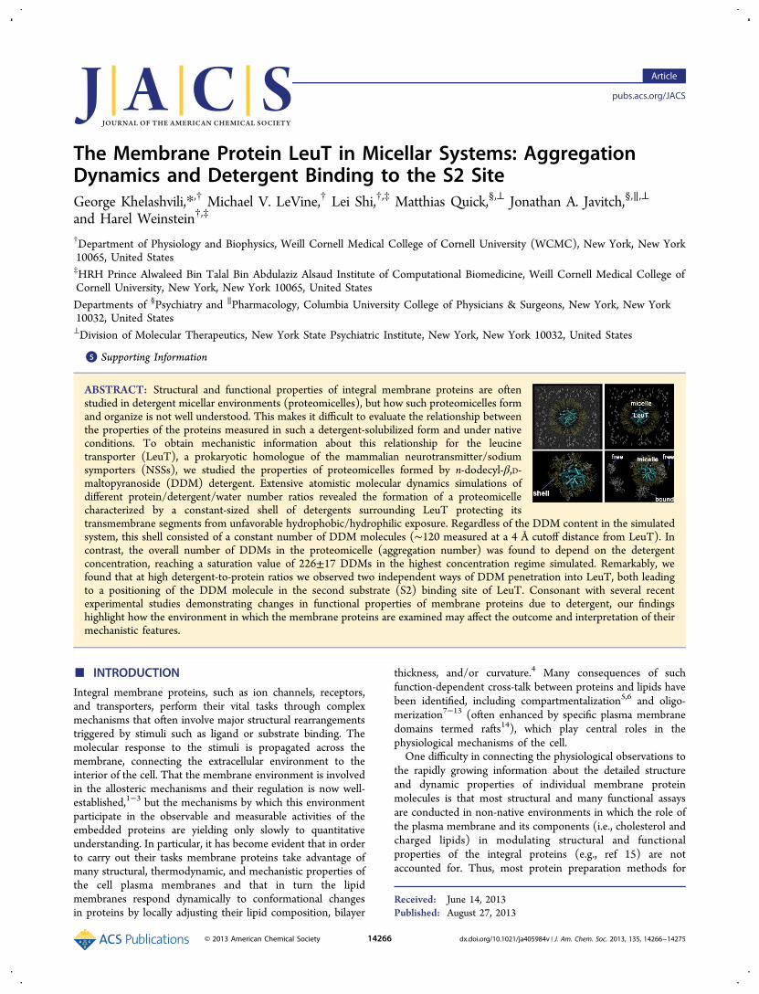
The Membrane Protein LeuT in Micellar Systems: Aggregation Dynamics and Detergent Binding to the S2 Site George Khelashvili,* ,† Michael V. LeVine, † Lei Shi, †,‡ Matthias Quick, §,⊥ Jonathan A. Javitch, §,∥,⊥ and Harel Weinstein †,‡ † Department of Physiology and Biophysics, Weill Cornell Medical College of Cornell University (WCMC), New York, New York 10065, United States ‡ HRH Prince Alwaleed Bin Talal Bin Abdulaziz Alsaud Institute of Computational Biomedicine, Weill Cornell Medical College of Cornell University, New York, New York 10065, United States Departments of § Psychiatry and ∥ Pharmacology, Columbia University College of Physicians & Surgeons, New York, New York 10032, United States ⊥ Division of Molecular Therapeutics, New York State Psychiatric Institute, New York, New York 10032, United States * S Supporting Information ABSTRACT: Structural and functional properties of integral membrane proteins are often studied in detergent micellar environments (proteomicelles), but how such proteomicelles form and organize is not well understood. This makes it difficult to evaluate the relationship between the properties of the proteins measured in such a detergent-solubilized form and under native conditions. To obtain mechanistic information about this relationship for the leucine transporter (LeuT), a prokaryotic homologue of the mammalian neurotransmitter/sodium symporters (NSSs), we studied the properties of proteomicelles formed by n-dodecyl-β,D- maltopyranoside (DDM) detergent. Extensive atomistic molecular dynamics simulations of different protein/detergent/water number ratios revealed the formation of a proteomicelle characterized by a constant-sized shell of detergents surrounding LeuT protecting its transmembrane segments from unfavorable hydrophobic/hydrophilic exposure. Regardless of the DDM content in the simulated system, this shell consisted of a constant number of DDM molecules (∼120 measured at a 4 Å cutoff distance from LeuT). In contrast, the overall number of DDMs in the proteomicelle (aggregation number) was found to depend on the detergent concentration, reaching a saturation value of 226±17 DDMs in the highest concentration regime simulated. Remarkably, we found that at high detergent-to-protein ratios we observed two independent ways of DDM penetration into LeuT, both leading to a positioning of the DDM molecule in the second substrate (S2) binding site of LeuT. Consonant with several recent experimental studies demonstrating changes in functional properties of membrane proteins due to detergent, our findings highlight how the environment in which the membrane proteins are examined may affect the outcome and interpretation of their mechanistic features. ■ INTRODUCTION Integral membrane proteins, such as ion channels, receptors, and transporters, perform their vital tasks through complex mechanisms that often involve major structural rearrangements triggered by stimuli such as ligand or substrate binding. The molecular response to the stimuli is propagated across the membrane, connecting the extracellular environment to the interior of the cell. That the membrane environment is involved in the allosteric mechanisms and their regulation is now well- established, 1−3 but the mechanisms by which this environment participate in the observable and measurable activities of the embedded proteins are yielding only slowly to quantitative understanding. In particular, it has become evident that in order to carry out their tasks membrane proteins take advantage of many structural, thermodynamic, and mechanistic properties of the cell plasma membranes and that in turn the lipid membranes respond dynamically to conformational changes in proteins by locally adjusting their lipid composition, bilayer thickness, and/or curvature. 4 Many consequences of such function-dependent cross-talk between proteins and lipids have been identified, including compartmentalization 5,6 and oligo- merization 7−13 (often enhanced by specific plasma membrane domains termed rafts 14 ), which play central roles in the physiological mechanisms of the cell. One difficulty in connecting the physiological observations to the rapidly growing information about the detailed structure and dynamic properties of individual membrane protein molecules is that most structural and many functional assays are conducted in non-native environments in which the role of the plasma membrane and its components (i.e., cholesterol and charged lipids) in modulating structural and functional properties of the integral proteins (e.g., ref 15) are not accounted for. Thus, most protein preparation methods for Received: June 14, 2013 Published: August 27, 2013 Article pubs.acs.org/JACS © 2013 American Chemical Society 14266 dx.doi.org/10.1021/ja405984v | J. Am. Chem. Soc. 2013, 135, 14266−14275
-
Upload
sacklerinstitute -
Category
Documents
-
view
1 -
download
0
Transcript of The Membrane Protein LeuT in Micellar Systems: Aggregation Dynamics and Detergent Binding to the S2...
The Membrane Protein LeuT in Micellar Systems: AggregationDynamics and Detergent Binding to the S2 SiteGeorge Khelashvili,*,† Michael V. LeVine,† Lei Shi,†,‡ Matthias Quick,§,⊥ Jonathan A. Javitch,§,∥,⊥
and Harel Weinstein†,‡
†Department of Physiology and Biophysics, Weill Cornell Medical College of Cornell University (WCMC), New York, New York10065, United States‡HRH Prince Alwaleed Bin Talal Bin Abdulaziz Alsaud Institute of Computational Biomedicine, Weill Cornell Medical College ofCornell University, New York, New York 10065, United States
Departments of §Psychiatry and ∥Pharmacology, Columbia University College of Physicians & Surgeons, New York, New York10032, United States⊥Division of Molecular Therapeutics, New York State Psychiatric Institute, New York, New York 10032, United States
*S Supporting Information
ABSTRACT: Structural and functional properties of integral membrane proteins are oftenstudied in detergent micellar environments (proteomicelles), but how such proteomicelles formand organize is not well understood. This makes it difficult to evaluate the relationship betweenthe properties of the proteins measured in such a detergent-solubilized form and under nativeconditions. To obtain mechanistic information about this relationship for the leucinetransporter (LeuT), a prokaryotic homologue of the mammalian neurotransmitter/sodiumsymporters (NSSs), we studied the properties of proteomicelles formed by n-dodecyl-β,D-maltopyranoside (DDM) detergent. Extensive atomistic molecular dynamics simulations ofdifferent protein/detergent/water number ratios revealed the formation of a proteomicellecharacterized by a constant-sized shell of detergents surrounding LeuT protecting itstransmembrane segments from unfavorable hydrophobic/hydrophilic exposure. Regardless of the DDM content in the simulatedsystem, this shell consisted of a constant number of DDM molecules (∼120 measured at a 4 Å cutoff distance from LeuT). Incontrast, the overall number of DDMs in the proteomicelle (aggregation number) was found to depend on the detergentconcentration, reaching a saturation value of 226±17 DDMs in the highest concentration regime simulated. Remarkably, wefound that at high detergent-to-protein ratios we observed two independent ways of DDM penetration into LeuT, both leadingto a positioning of the DDM molecule in the second substrate (S2) binding site of LeuT. Consonant with several recentexperimental studies demonstrating changes in functional properties of membrane proteins due to detergent, our findingshighlight how the environment in which the membrane proteins are examined may affect the outcome and interpretation of theirmechanistic features.
■ INTRODUCTION
Integral membrane proteins, such as ion channels, receptors,and transporters, perform their vital tasks through complexmechanisms that often involve major structural rearrangementstriggered by stimuli such as ligand or substrate binding. Themolecular response to the stimuli is propagated across themembrane, connecting the extracellular environment to theinterior of the cell. That the membrane environment is involvedin the allosteric mechanisms and their regulation is now well-established,1−3 but the mechanisms by which this environmentparticipate in the observable and measurable activities of theembedded proteins are yielding only slowly to quantitativeunderstanding. In particular, it has become evident that in orderto carry out their tasks membrane proteins take advantage ofmany structural, thermodynamic, and mechanistic properties ofthe cell plasma membranes and that in turn the lipidmembranes respond dynamically to conformational changesin proteins by locally adjusting their lipid composition, bilayer
thickness, and/or curvature.4 Many consequences of suchfunction-dependent cross-talk between proteins and lipids havebeen identified, including compartmentalization5,6 and oligo-merization7−13 (often enhanced by specific plasma membranedomains termed rafts14), which play central roles in thephysiological mechanisms of the cell.One difficulty in connecting the physiological observations to
the rapidly growing information about the detailed structureand dynamic properties of individual membrane proteinmolecules is that most structural and many functional assaysare conducted in non-native environments in which the role ofthe plasma membrane and its components (i.e., cholesterol andcharged lipids) in modulating structural and functionalproperties of the integral proteins (e.g., ref 15) are notaccounted for. Thus, most protein preparation methods for
Received: June 14, 2013Published: August 27, 2013
Article
pubs.acs.org/JACS
© 2013 American Chemical Society 14266 dx.doi.org/10.1021/ja405984v | J. Am. Chem. Soc. 2013, 135, 14266−14275
studies of integral membrane proteins involve their over-expression, followed by their detergent-mediated extractionfrom the cell and purification and reconstitution intoproteoliposomes.16 When present in water above certain criticalconcentrations, the detergents, which have been selected toreplace the native environment of these proteins (i.e. themembrane) and possess a relatively large polar headgroup and ashort hydrophobic tail, self-assemble in aggregates of positivecurvature, termed micelles.17 In such micelles, the polarheadgroups of the detergent are exposed to the aqueoussolvent whereas hydrophobic tails face each other and thusmimic the membrane hydrophobic environment for anencapsulated protein while excluding aqueous solvent fromthe micelle interior. Although micellar aggregates effectivelyshield hydrophobic trans-membrane (TM) segments of integralproteins from unfavorable polar exposure while bringing polarloop regions into contact with an aqueous phase, they cannotcapture all of the complexities of the native plasmamembranes.16 Indeed, several recent studies have provideddramatic demonstrations of changes in functional properties ofmembrane proteins due to detergent (e.g., refs 18 and 19).One such class of membrane proteins studied in detergent
environments is the family of neurotransmitter/sodiumsymporters (NSS) that is responsible for the removal ofextracellular solutes from the synaptic cleft into the presynapticnerve terminal.20 The uptake mechanism is energized by thecoupling the transmembrane Na+ gradient to the uphilltransport of the respective substrate. Several X-ray structuresof a prokaryotic homologue of NSS, the leucine transporter
(LeuT), have identified the centrally located high-affinitysubstrate binding site, termed the S1 site,21−28 and two sodiumbinding sites termed Na1 and Na2. Computational andexperimental studies29 have identified a second high-affinitysubstrate binding site, the S2 site, located in an extracellularvestibule ∼11 Å above the S1 site that is essential for thetransport mechanisms of LeuT. In particular, an allostericmechanistic model of a Na+-coupled symport was proposed29
in which intracellular release of the S1-bound substrate istriggered by the binding of a second substrate molecule in theS2 site.Remarkably, follow-up studies19 suggested that experimental
conditions and, in particular, a high concentration of n-dodecyl-β,D-maltopyranoside (DDM) detergent, can obscure thefunctionally relevant S2 site and result in reduced substratebinding. But the reconstitution of LeuT, previously preincu-bated with high concentrations of DDM detergent, into E. colimembranes, showed a full recovery of functionality for the S2site.19 Interestingly, the loss of the S2 site upon detergenttreatment appeared to have a concentration threshold in therange of 0.15−0.175% DDM. These experimental resultsillustrate that the function of integral membrane proteins canbe modulated by the experimental preparation and theenvironment in which the protein is studied. To extrapolatethe knowledge gained from the experimental explorationsconducted under non-native conditions to mechanisms in vivo,it is necessary to understand on the molecular level howdetergent micelles form around proteins and what fundamentalchanges occur when the protein is surrounded by a detergent
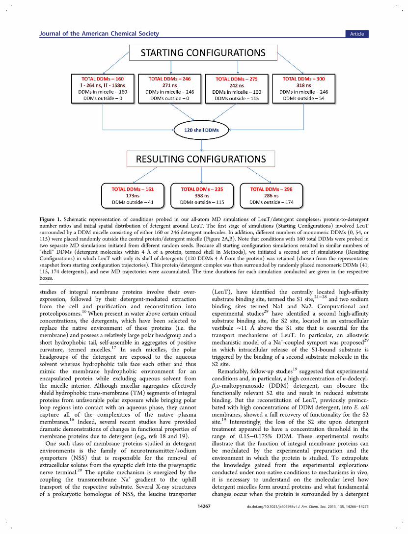
Figure 1. Schematic representation of conditions probed in our all-atom MD simulations of LeuT/detergent complexes: protein-to-detergentnumber ratios and initial spatial distribution of detergent around LeuT. The first stage of simulations (Starting Configurations) involved LeuTsurrounded by a DDM micelle consisting of either 160 or 246 detergent molecules. In addition, different numbers of monomeric DDMs (0, 54, or115) were placed randomly outside the central protein/detergent micelle (Figure 2A,B). Note that conditions with 160 total DDMs were probed intwo separate MD simulations initiated from different random seeds. Because all starting configuration simulations resulted in similar numbers of“shell” DDMs (detergent molecules within 4 Å of a protein, termed shell in Methods), we initiated a second set of simulations (ResultingConfigurations) in which LeuT with only its shell of detergents (120 DDMs 4 Å from the protein) was retained (chosen from the representativesnapshot from starting configuration trajectories). This protein/detergent complex was then surrounded by randomly placed monomeric DDMs (41,115, 174 detergents), and new MD trajectories were accumulated. The time durations for each simulation conducted are given in the respectiveboxes.
Journal of the American Chemical Society Article
dx.doi.org/10.1021/ja405984v | J. Am. Chem. Soc. 2013, 135, 14266−1427514267
micelle. The literature discussing molecular models ofdetergent/protein interactions (e.g., refs 30−39 and citationstherein) has not addressed these fundamental questions in asystematic way.To point out the shortcomings associated with the
interpretation of membrane protein structure and function inexperimental environments, we provide here, to our knowledgefor the first time, a detailed molecular view of the LeuT proteinembedded in DDM detergent micelles formed at differentdetergent/water/protein ratios. This view is offered fromextensive atomistic molecular dynamics (MD) simulationscarried out in order to (1) establish the aggregation number ofDDM micelles surrounding LeuT, (2) explore the overallorganization of the detergent micelle containing the trans-porter, and (3) obtain molecular-level insight into the natureand consequences of interactions between LeuT and DDM.Analyzing various protein-to-detergent (P/D) number ratios
(i.e., from 1:160 to 1:300), we show that the aggregationnumber of DDM in the micelle that surrounds the transporteris strongly dependent on the P/D ratio. Moreover, the MDsimulations of the system at various P/D ratios suggest amechanism for the dependence of LeuT substrate bindingstoichiometry on detergent concentration. Thus, we found thatthe detergent can penetrate LeuT through two alternativepathways. As a consequence of such penetration, DDMmolecules establish long-lasting contacts with several function-ally critical residues located in the S2 site of LeuT. Remarkably,we find that the detergent penetration phenotype is determinedby the aggregation number of DDM around LeuT so thatnontransient DDM insertion is observed only in the high-detergent-concentration regime. These results, discussed herein the light of recent experimental findings suggesting themodulation of LeuT activity by detergent, can explain
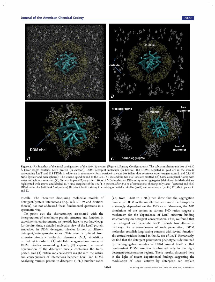
Figure 2. (A) Snapshot of the initial configuration of the 160/115 system (Figure 1, Starting Configurations). The cubic simulation unit box of ∼180Å linear length contains LeuT protein (in cartoon), DDM detergent molecules (in licorice, 160 DDMs depicted in gold are in the micellesurrounding LeuT and 115 DDMs in white are in monomeric form outside), a water box (silver dots represent water oxygen atoms), and 0.15 MNaCl (yellow and cyan spheres). The leucine ligand bound to the LeuT S1 site and the two Na+ ions are omitted. (B) Same as in panel A only withwater and salt ions removed. (C) Same as in panel B, only after 140 ns of MD simulations. Different types of aggregates (definitions in Methods) arehighlighted with arrows and labeled. (D) Final snapshot of the 160/115 system, after 242 ns of simulations, showing only LeuT (cartoon) and shellDDM molecules (within 4 Å of protein) (licorice). Notice strong intermixing of initially micellar (gold) and monomeric (white) DDMs in panels Cand D.
Journal of the American Chemical Society Article
dx.doi.org/10.1021/ja405984v | J. Am. Chem. Soc. 2013, 135, 14266−1427514268
experimentally observed phenotypes caused by the occlusion ofthe S2 site in LeuT at high detergent concentration.
■ METHODSMolecular Constructs. For atomistic molecular dynamics (MD)
simulations, we used the X-ray structure of LeuT with the PDBaccession code 3GJD.21 The transporter in this structure is in theoccluded state with leucine (Leu) at the S1 primary binding site andthe two Na+ ions bound at Na1 and Na2 sites, respectively. Thus, thestructure also contains detergent n-octyl-β,D-glucopyranoside (OG) atthe S2 binding site. The detergent molecule was removed prior to thesimulations, leaving the S2 site empty at the beginning of the MD runs.The LeuT residues that were missing from the 3GJD structure (firstfour residues on N terminus, last eight residues on C-terminus, and theP132−N133−A134 stretch in the EL2 loop) were added withModeler.40 All crystallographic waters were retained in the simulations,and Glu112, Glu287, and Glu419 were treated as protonated.41,42
The LeuT model was immersed in a box containing water andDDM detergent molecules. As described in Figure 1 (StartingConfigurations) and illustrated in Figure 2, some DDM moleculeswere initially placed in a spherical micelle formation around LeuT (seebelow) whereas others were placed randomly as monomers outsidethis central micelle. The starting conditions were varied with respect tothe number of DDMs in the initial micelle and in the monomers.Thus, for the initial set of simulations, the micelle was composed ofeither 160 or 246 DDM molecules and was surrounded by differentnumbers of monomeric DDMs (Figure 1). Accordingly, throughoutthis work, various simulated constructs are given the designation A/B,where A denotes the initial number of DDMs in the central micellesurrounding LeuT (Figure 2) and B is the starting number ofmonomeric detergent molecules outside this micelle.To build a micelle containing a number A of detergent molecules
around LeuT, we used a multistep algorithm described in ref 37.According to this procedure, in step 1 N pseudoparticles wererandomly placed on an imaginary sphere surrounding the protein,excluding areas around intracellular and extracellular parts of LeuT(Figure 2); in step 2, the pseudoparticles were replaced with explicitDDM molecules, oriented with their hydrophobic tails facing thecenter of LeuT; and in step 3, the imaginary sphere (containing LeuTand all of the DDM molecules) was incrementally shrunk subject toconcomitant energy minimization to a final radius of 51 Å. With that,we ensured that in the starting configuration DDM tails wereappropriately placed to cover the hydrophobic core of LeuT whileleaving hydrophilic regions of the protein exposed to the solvent.To the box containing the LeuT-detergent micelle complex
(proteomicelle) obtained with the procedure described above, weadded the desired number of monomeric DDMs (Figures 1 and 2)positioned randomly outside the central micelle, and the system wasthen solvated with TIP3 waters and ionized with Na+ and Cl− toachieve an ionic concentration of 0.15 M. The final simulation box hadnearly cubic geometry with a linear dimension of ∼180 Å andcontained ∼500 000 atoms. Correspondingly, the detergent concen-tration was kept above the established critical micelle concentration(cmc) of 0.17 mM for DDM43,44 in all of the constructs (Table S1 inthe Supporting Information).Molecular Dynamics Simulations. The all-atom MD simulations
were done with the NAMD 2.7 package,45 and the all-atomCHARMM27 force field, with CMAP corrections for proteins46 anda CHARMM-compatible force-field parameter set for detergents.47
Molecular constructs were initially equilibrated using a two-phaseprotocol: (i) short energy minimization was carried out during whichprotein, water, and ion atoms were fixed and the coordinates of onlyDDM molecules were allowed to evolve freely and (ii) 1.5-ns-long MDsimulations were conducted with the protein backbone harmonicallyconstrained. The constraints were released gradually, in 0.5 ns steps,with decreasing force constants of 1, 0.5, and 0.01 kcal/(mol·Å2). Theequilibration procedure was similar to that implemented by others forMD simulations of protein/micelle complexes (e.g., ref 38).
After the equilibration phase, unbiased MD simulations were carriedout. (See Figure 1 for a listing of simulation durations.) Integrationsteps were 1 fs for the equilibration stage and 2 fs thereafter. Thesimulations implemented PME for electrostatics interactions48 andwere carried out in an NPT ensemble under isotropic pressurecoupling conditions and at 310 K temperature. The Nose-HooverLangevin piston algorithm45 was used to control the target P = 1 atmpressure with the Langevin piston period set to 100 fs and theLangevin piston decay set to 50 fs.
Definition of Detergent Aggregates Formed during MDSimulations. As detailed in the Results, the MD simulations led tothe spontaneous formation of aggregates of different types (Figure2C). Accordingly, we distinguished DDM detergents in the followingtypes of aggregates:
Detergent micelles (DMs) − DDMs that are in the centralmicelle around LeuT;Bound aggregates (BAs) − DDMs in aggregates that bind to thecentral micelle;Bound monomers (BMs) − DDMs that are bound to the centralmicelle as monomers;Free aggregates (FAs) − DDMs that are part of aggregates in thesolution;Free monomers (FM) − DDMs that are monomeric in thesolution.DDM shell (shell) − DDMs within 4 Å from LeuT. (See FigureS1 in the Supporting Information for a comparison with theresults using alternative definitions of the shell.)
To identify the number of constituent detergent molecules in eachtype of aggregate defined above, we used an expansion algorithm.Thus, to calculate the number of DDMs in DM, we first identifieddetergent molecules within 4 Å of LeuT (DDM shell, Figure 2D).Next, we found all DDM molecules within 4 Å of this detergent shell,and such an expansion was repeated until the search failed to identifyany new DDM molecules. In this procedure, only the atoms in thehydrophobic tails of detergent molecules were used to differentiatebetween DDMs in the protein-surrounding micelle from those that areinteracting with this micelle via headgroup atoms.
After DM was defined, DDMs in BAs and BMs were counted byselecting detergent molecules whose headgroup atoms were within 5 Åof the DM and building complexes using the expansion algorithmdescribed above. After all bound aggregates were located, thealgorithm identified the DDMs in FAs and FMs by building theremaining aggregates in solution. Note that all types of aggregatesdefined above can, in principle, contain DDM molecules that wereinitially either part of the central micelle or were monomeric outsidethe micelle (Figure 2B−D).
The micellar aggregation number is a description of the number ofmolecules present in a micelle once the CMC has been reached and isdefined as the ratio of micelle concentration over the concentration ofmonomeric detergent.49 Here, the aggregation number of the LeuT/DDM proteomicelle was calculated as the DM + BA + BM sum. (Seeabove.)
Quantification of Micelle Shape. To quantify the shape of themicelle around LeuT, we calculated the eccentricity E of the DMfollowing the procedure implemented in ref 34. To this end, wedetermined the three principal moments of inertia (I) of an ellipsoidthat encloses the non-hydrogen tail atoms of those DDM moleculesthat were identified as being part of the DM. Using the magnitudes ofthe smallest principal moment (Imin) and of the average of all threemoments (Iav), we then obtained the eccentricity of the micelle asE = 1 − (Iav/Imin). Note that according to this definition, E = 0 for aperfectly spherical micelle.
Quantification of Detergent Penetration into LeuT from MDSimulations. To quantify the extent of detergent penetration intoLeuT in our MD simulations, we monitored the time evolution ofselected distance measures from different trajectories. Specifically,because we observed that DDM is inserted into the transporter andbinds in the S2 site (see Results), we first screened the residues thathave been established from experimental and computational
Journal of the American Chemical Society Article
dx.doi.org/10.1021/ja405984v | J. Am. Chem. Soc. 2013, 135, 14266−1427514269
studies19,21,24,25,29 to comprise the S2 site in LeuT (see alsoDiscussion): Leu25, Gly26, Leu29, Arg30, Gln34, Tyr107, Tyr108,Ile111, W114, Ala319, Phe320, Phe324, Leu400, and Asp404. We thentracked the minimal distance from these residues to the nearestdetergent molecule in different simulations. The penetration of thedetergent molecule was observed in different simulations to follow twodistinct pathways (see Results), one resulting ultimately in interactionswith Arg30 and Gln34 in transmembrane helix 1 (TMH1) of LeuTand the other resulting in interactions with Phe320 in extracellularloop 4 (ECL4) and Leu400 in TMH10. Therefore, we chose toquantify DDM insertion by monitoring the time evolution of theminimal distance (dmin) of these four key residuesArg30, Gln34,Phe320, and Leu400to the nearest DDM molecule.In a certain MD trajectory, therefore, a detergent molecule was
considered to be fully inserted (complete insertion) into the LeuT S2site if dmin between the detergent and any of the above-mentioned fourresidues was 3 Å or shorter during at least the last third of thetrajectory. If the detergent molecule interacted with either Arg30,Gln34, Phe320, or Leu400 (dmin < 3 Å) for a shorter period of time, itwas considered to be transiently inserted into the S2 site. In thisscenario, the interactions between the detergent and S2 site residueswere forming and breaking dynamically (e.g., Figure 5). Finally, ifdmin > 5 Å at all times during the trajectory, then the LeuT moleculewas not considered to be penetrated by DDM.
■ RESULTS
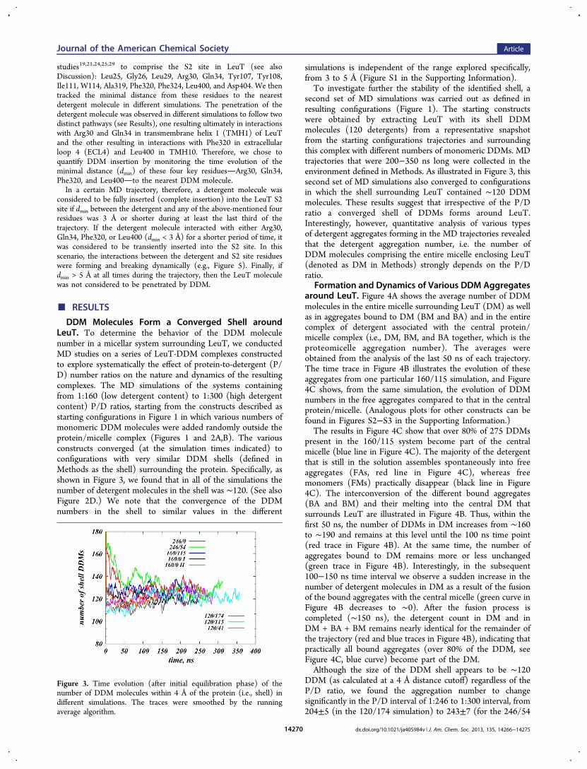
DDM Molecules Form a Converged Shell aroundLeuT. To determine the behavior of the DDM moleculenumber in a micellar system surrounding LeuT, we conductedMD studies on a series of LeuT-DDM complexes constructedto explore systematically the effect of protein-to-detergent (P/D) number ratios on the nature and dynamics of the resultingcomplexes. The MD simulations of the systems containingfrom 1:160 (low detergent content) to 1:300 (high detergentcontent) P/D ratios, starting from the constructs described asstarting configurations in Figure 1 in which various numbers ofmonomeric DDM molecules were added randomly outside theprotein/micelle complex (Figures 1 and 2A,B). The variousconstructs converged (at the simulation times indicated) toconfigurations with very similar DDM shells (defined inMethods as the shell) surrounding the protein. Specifically, asshown in Figure 3, we found that in all of the simulations thenumber of detergent molecules in the shell was ∼120. (See alsoFigure 2D.) We note that the convergence of the DDMnumbers in the shell to similar values in the different
simulations is independent of the range explored specifically,from 3 to 5 Å (Figure S1 in the Supporting Information).To investigate further the stability of the identified shell, a
second set of MD simulations was carried out as defined inresulting configurations (Figure 1). The starting constructswere obtained by extracting LeuT with its shell DDMmolecules (120 detergents) from a representative snapshotfrom the starting configurations trajectories and surroundingthis complex with different numbers of monomeric DDMs. MDtrajectories that were 200−350 ns long were collected in theenvironment defined in Methods. As illustrated in Figure 3, thissecond set of MD simulations also converged to configurationsin which the shell surrounding LeuT contained ∼120 DDMmolecules. These results suggest that irrespective of the P/Dratio a converged shell of DDMs forms around LeuT.Interestingly, however, quantitative analysis of various typesof detergent aggregates forming in the MD trajectories revealedthat the detergent aggregation number, i.e. the number ofDDM molecules comprising the entire micelle enclosing LeuT(denoted as DM in Methods) strongly depends on the P/Dratio.
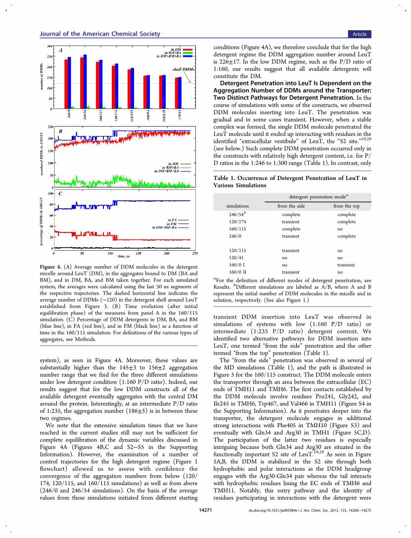
Formation and Dynamics of Various DDM Aggregatesaround LeuT. Figure 4A shows the average number of DDMmolecules in the entire micelle surrounding LeuT (DM) as wellas in aggregates bound to DM (BM and BA) and in the entirecomplex of detergent associated with the central protein/micelle complex (i.e., DM, BM, and BA together, which is theproteomicelle aggregation number). The averages wereobtained from the analysis of the last 50 ns of each trajectory.The time trace in Figure 4B illustrates the evolution of theseaggregates from one particular 160/115 simulation, and Figure4C shows, from the same simulation, the evolution of DDMnumbers in the free aggregates compared to that in the centralprotein/micelle. (Analogous plots for other constructs can befound in Figures S2−S3 in the Supporting Information.)The results in Figure 4C show that over 80% of 275 DDMs
present in the 160/115 system become part of the centralmicelle (blue line in Figure 4C). The majority of the detergentthat is still in the solution assembles spontaneously into freeaggregates (FAs, red line in Figure 4C), whereas freemonomers (FMs) practically disappear (black line in Figure4C). The interconversion of the different bound aggregates(BA and BM) and their melting into the central DM thatsurrounds LeuT are illustrated in Figure 4B. Thus, within thefirst 50 ns, the number of DDMs in DM increases from ∼160to ∼190 and remains at this level until the 100 ns time point(red trace in Figure 4B). At the same time, the number ofaggregates bound to DM remains more or less unchanged(green trace in Figure 4B). Interestingly, in the subsequent100−150 ns time interval we observe a sudden increase in thenumber of detergent molecules in DM as a result of the fusionof the bound aggregates with the central micelle (green curve inFigure 4B decreases to ∼0). After the fusion process iscompleted (∼150 ns), the detergent count in DM and inDM + BA + BM remains nearly identical for the remainder ofthe trajectory (red and blue traces in Figure 4B), indicating thatpractically all bound aggregates (over 80% of the DDM, seeFigure 4C, blue curve) become part of the DM.Although the size of the DDM shell appears to be ∼120
DDM (as calculated at a 4 Å distance cutoff) regardless of theP/D ratio, we found the aggregation number to changesignificantly in the P/D interval of 1:246 to 1:300 interval, from204±5 (in the 120/174 simulation) to 243±7 (for the 246/54
Figure 3. Time evolution (after initial equilibration phase) of thenumber of DDM molecules within 4 Å of the protein (i.e., shell) indifferent simulations. The traces were smoothed by the runningaverage algorithm.
Journal of the American Chemical Society Article
dx.doi.org/10.1021/ja405984v | J. Am. Chem. Soc. 2013, 135, 14266−1427514270
system), as seen in Figure 4A. Moreover, these values aresubstantially higher than the 145±3 to 156±2 aggregationnumber range that we find for the three different simulationsunder low detergent condition (1:160 P/D ratio). Indeed, ourresults suggest that for the low DDM constructs all of theavailable detergent eventually aggregates with the central DMaround the protein. Interestingly, at an intermediate P/D ratioof 1:235, the aggregation number (186±5) is in between thesetwo regimes.We note that the extensive simulation times that we have
reached in the current studies still may not be sufficient forcomplete equilibration of the dynamic variables discussed inFigure 4A (Figures 4B,C and S2−S5 in the SupportingInformation). However, the examination of a number ofcontrol trajectories for the high detergent regime (Figure 1flowchart) allowed us to assess with confidence theconvergence of the aggregation numbers from below (120/174, 120/115, and 160/115 simulations) as well as from above(246/0 and 246/54 simulations). On the basis of the averagevalues from these simulations initiated from different starting
conditions (Figure 4A), we therefore conclude that for the highdetergent regime the DDM aggregation number around LeuTis 226±17. In the low DDM regime, such as the P/D ratio of1:160, our results suggest that all available detergents willconstitute the DM.
Detergent Penetration into LeuT Is Dependent on theAggregation Number of DDMs around the Transporter:Two Distinct Pathways for Detergent Penetration. In thecourse of simulations with some of the constructs, we observedDDM molecules inserting into LeuT. The penetration wasgradual and in some cases transient. However, when a stablecomplex was formed, the single DDM molecule penetrated theLeuT molecule until it ended up interacting with residues in theidentified “extracellular vestibule” of LeuT, the “S2 site.”19,29
(see below.) Such complete DDM penetration occurred only inthe constructs with relatively high detergent content, i.e. for P/D ratios in the 1:246 to 1:300 range (Table 1). In contrast, only
transient DDM insertion into LeuT was observed insimulations of systems with low (1:160 P/D ratio) orintermediate (1:235 P/D ratio) detergent content. Weidentified two alternative pathways for DDM insertion intoLeuT, one termed “from the side” penetration and the othertermed “from the top” penetration (Table 1).The “from the side” penetration was observed in several of
the MD simulations (Table 1), and the path is illustrated inFigure 5 for the 160/115 construct. The DDM molecule entersthe transporter through an area between the extracellular (EC)ends of TMH11 and TMH6. The first contacts established bythe DDM molecule involve residues Pro241, Gly242, andIle245 in TMH6, Trp467, and Val466 in TMH11 (Figure S4 inthe Supporting Information). As it penetrates deeper into thetransporter, the detergent molecule engages in additionalstrong interactions with Phe405 in TMH10 (Figure S3) andeventually with Gln34 and Arg30 in TMH1 (Figure 5C,D).The participation of the latter two residues is especiallyintriguing because both Gln34 and Arg30 are situated in thefunctionally important S2 site of LeuT.19,29 As seen in Figure5A,B, the DDM is stabilized in the S2 site through bothhydrophobic and polar interactions as the DDM headgroupengages with the Arg30-Gln34 pair whereas the tail interactswith hydrophobic residues lining the EC ends of TMH6 andTMH11. Notably, this entry pathway and the identity ofresidues participating in interactions with the detergent were
Figure 4. (A) Average number of DDM molecules in the detergentmicelle around LeuT (DM), in the aggregates bound to DM (BA andBM), and in DM, BA, and BM taken together. For each simulatedsystem, the averages were calculated using the last 50 ns segments ofthe respective trajectories. The dashed horizontal line indicates theaverage number of DDMs (∼120) in the detergent shell around LeuTestablished from Figure 3. (B) Time evolution (after initialequilibration phase) of the measures from panel A in the 160/115simulation. (C) Percentage of DDM detergents in DM, BA, and BM(blue line), in FA (red line), and in FM (black line) as a function oftime in the 160/115 simulation. For definitions of the various types ofaggregates, see Methods.
Table 1. Occurrence of Detergent Penetration of LeuT inVarious Simulations
detergent penetration modea
simulations from the side from the top
246/54b complete complete120/174 transient complete160/115 complete no246/0 transient complete
120/115 transient no120/41 no no160/0 I no transient160/0 II transient no
aFor the definition of different modes of detergent penetration, seeResults. bDifferent simulations are labeled as A/B, where A and Brepresent the initial number of DDM molecules in the micelle and insolution, respectively. (See also Figure 1.)
Journal of the American Chemical Society Article
dx.doi.org/10.1021/ja405984v | J. Am. Chem. Soc. 2013, 135, 14266−1427514271
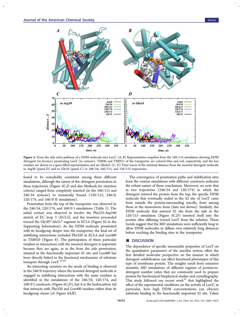
found to be remarkably consistent among these differentsimulations, although the extent of the detergent penetration inthese trajectories (Figure 5C,D and also Methods for insertioncriteria) ranged from completely inserted (in the 160/115 and246/54 systems) to transiently bound (120/115, 246/0,120/174, and 160/0 II simulations).Penetration from the top of the transporter was observed in
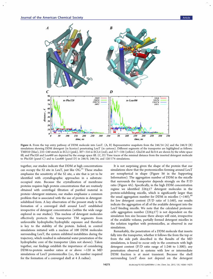
the 246/54, 120/174, and 160/0 I simulations (Table 1). Theinitial contact was observed to involve the Phe235-Asp240stretch of EC loop 3 (ECL3), and the insertion proceededtoward the Gly307-Ala317 segment in ECL4 (Figure S5 in theSupporting Information). As the DDM molecule penetratedwith its headgroup deeper into the transporter, the final set ofstabilizing interactions included Phe320 in ECL4 and Leu400in TMH10 (Figure 6). The participation of these particularresidues in interactions with the inserted detergent is importantbecause they are again, as in the from the side penetration,situated in the functionally important S2 site and Leu400 hasbeen directly linked to the functional mechanisms of substratetransport through LeuT.19,29
An interesting variation on the mode of binding is observedin the 246/0 trajectory where the inserted detergent molecule isengaged in stabilizing interactions with the same residues asidentified in the simulations of the 246/54, 120/174, and160/0 I constructs (Figure 6C,D), but it is the hydrocarbon tailthat interacts with Phe320 and Leu400 residues rather than itsheadgroup atoms (cf. Figure 6A,B).
The convergence of penetration paths and stabilization sitesfrom the various simulations with different constructs indicatesthe robust nature of these conclusions. Moreover, we note thatin two trajectories (246/54 and 120/174) in which thedetergent entered the protein from the top, the specific DDMmolecule that eventually ended in the S2 site of LeuT camefrom outside the protein-surrounding micelle, from amongthose in the monomeric form (data not shown). Similarly, theDDM molecule that entered S2 site from the side in the120/115 simulation (Figure 5C,D) inserted itself into theprotein after diffusing toward LeuT from the solution. Thesetrends suggest that the MD simulations were sufficiently long toallow DDM molecules to diffuse over relatively long distancesbefore reaching the binding sites in the transporter.
■ DISCUSSIONThe dependence of specific measurable properties of LeuT onthe quantitative parameters of the micellar system offers thefirst detailed molecular perspective on the manner in whichdetergent solubilization can affect functional phenotypes of thistype of membrane protein. The insights result from extensiveatomistic MD simulations of different regimes of protein-to-detergent number ratios that are commonly used to prepareprotein for biochemical/biophysical studies and crystallography.This study followed our recent work19 that highlighted theeffect of the experimental conditions on the activity of LeuT, inparticular, how high DDM concentrations can obscuresubstrate binding to the functionally important S2 site. Taken
Figure 5. From the side entry pathway of a DDM molecule into LeuT. (A, B) Representative snapshot from the 160/115 simulation showing DDMdetergent (in licorice) penetrating LeuT (in cartoon). TMH6 and TMH11 of the transporter are colored blue and red, respectively, and the keyresidues are shown in a space-filled representation and are labeled. (C, D) Time traces of the minimal distance from the inserted detergent moleculeto Arg30 (panel D) and to Gln34 (panel C) in 246/54, 160/115, and 120/115 trajectories.
Journal of the American Chemical Society Article
dx.doi.org/10.1021/ja405984v | J. Am. Chem. Soc. 2013, 135, 14266−1427514272
together, our studies indicate that DDM at high concentrationscan occupy the S2 site in LeuT, just like OG.21 These studiesemphasize the sensitivity of the S2 site, a site that is yet to beidentified with crystallographic approaches in a substrate-occupied state. Because the crystallization of membraneproteins requires high protein concentrations that are routinelyobtained with centrifugal filtration of purified material inprotein−detergent mixtures, our studies emphasize a commonproblem that is associated with the use of protein in detergent-solubilized form. A key observation of the present study is theformation of a converged shell around LeuT establishedirrespective of detergent concentration (within the wide rangeexplored in our studies). This nucleus of detergent moleculeseffectively protects the transporter TM segments fromunfavorable hydrophobic/hydrophilic exposure and thereforeis key to the stability of the protein. Indeed, in controlsimulations initiated with a nucleus of 100 DDM moleculessurrounding LeuT, the system exhibited instabilities during thetrajectory, which resulted in substantial water penetration of thehydrophobic core of the transporter (data not shown). Takentogether, our findings establish the importance of consideringDDM-to-protein number ratios at or above ∼120 in MDsimulations of LeuT proteomicelles (i.e., the number requiredfor the formation of a converged shell at 4 Å radius).
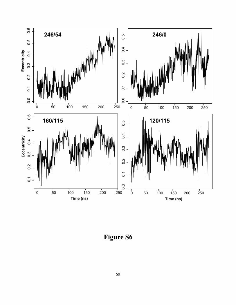
It is not surprising given the shape of the protein that oursimulations show that the proteomicelles forming around LeuTare nonspherical in shape (Figure S6 in the SupportingInformation). The aggregation number of DDM in the micellethat surrounds the transporter depends strongly on the P/Dratio (Figure 4A). Specifically, in the high DDM concentrationregime we identified 226±17 detergent molecules in theprotein-solubilizing micelle, which is significantly larger thanthe usual aggregation number for DDM in micelles (∼140);49for low detergent content (P/D ratio of 1:160), our resultsindicate the aggregation of all of the available detergent into theLeuT-binding micelle. We note that the calculated proteomi-celle aggregation number (226±17) is not dependent on thesimulation box size because there always will exist, irrespectiveof the available volume, partially formed detergent micelles inthe solution together with protemicelles, as observed in oursimulations.Remarkably, the penetration of a DDM molecule that inserts
fully into the transporter, whether it follows the from the top orfrom the side path described in the Results of the MDsimulations, is found to occur only in the constructs with highdetergent content (P/D ratio range of 1:246 to 1:300); anypenetration observed in systems with low or intermediateDDM fraction is at most transient. Because the shellsurrounding LeuT does not depend on the detergent
Figure 6. From the top entry pathway of DDM molecule into LeuT. (A, B) Representative snapshots from the 246/54 (A) and the 246/0 (B)simulations showing DDM detergent (in licorice) penetrating LeuT (in cartoon). Different segments of the transporter are highlighted as follows:TMH10 (blue), 235−240 stretch in ECL3 (pink), 307−316 in ECL4 (red), and 317−336 (yellow). Glu236 and Ile314 are shown by the white spacefill, and Phe320 and Leu400 are depicted by the orange space fill. (C, D) Time traces of the minimal distance from the inserted detergent moleculeto Phe320 (panel C) and to Leu400 (panel D) in 246/0, 246/54, and 120/174 simulations.
Journal of the American Chemical Society Article
dx.doi.org/10.1021/ja405984v | J. Am. Chem. Soc. 2013, 135, 14266−1427514273
concentration and it is the DDM aggregation number for thesystem (and thus the protein-to-detergent number ratio) thatdetermines penetration, it becomes clear that DDM insertioninto LeuT is not simply related to the interaction with thedetergent immediately surrounding the transporter. Rather, itreflects the dynamic properties of the detergent in the system.Specifically, we reason that at low detergent concentrations (ator below 226±17 aggregation number established by ourstudies) all of the DDM molecules in the system are expectedto participate in the stabilization of the proteomicelle. But athigher concentrations, excess DDMs, not associated with theproteomicelle, will be present in the solution and will diffusefreely in monomeric and/or in aggregate forms. Such dynamicsof free detergent increases the probability of randomencounters with the protein regions exposed to the solventand specifically with the large extracellular vestibule of LeuT,which eventually leads to the observed detergent penetration.The final position of any completely penetrating DDM was
found to be the S2 site in the extracellular vestibule of LeuTand to involve Arg30, Gln34, Phe320, and Leu400 in stronginteractions with the inserted DDM. These residues have beenidentified to have functional importance in the transportmechanism. Specifically, in crystal structures of LeuT theseresidues are among those implicated in stabilizing interactionswith tricyclic antidepressants (TCAs) in the extracellularvestibule25 as well as with OG detergent21 and to bindinhibitors (such as Trp).24 Furthermore, highly conserved ionicinteractions between Arg30 and Asp404 in TMH10 have beenestablished as one of the structural/functional hallmarks in NSSfamily proteins that regulate the access of the substrate from theEC vestibule down to the S1 site during the transportcycle.20,29,50 In addition, the importance of Leu400 residue inthe LeuT function is highlighted by the phenotypes of Leu400-to-Ser or Leu400-to-Cys mutations that impair Leu substratebinding in the S2 site.19
Notably, detergent penetration events observed in thesimulations occur at P/D ratios that are expected to be realizedin the local environments of the solubilized LeuT proteinsduring in vitro experiments. Thus, the results from thesimulations presented here correspond to the experimentalobservation19 that the binding of the Leu substrate at the S2site is impaired if LeuT is preincubated in the presence of highDDM (0.3%), yielding a protein-to-detergent ratio of ∼1:225,but this does not occur at low DDM concentrations (0.1%, witha protein-to-detergent ratio of ∼1:170). A detailed scanthrough the detergent concentration range revealed that theloss of Leu binding occurs abruptly in the interval between 0.15and 0.175% DDM concentration.19 In qualitative agreementwith these experimental observations, our data indicate theexistence of two distinct regimes for DDM concentration,separated by a narrow range of P/D ratios (from 1:235 to1:246), that determine detergent penetration. This suggests amechanistic explanation for the experimentally observedimpairment of the S2 site under high detergent conditionsthat involves the steric hindrance of the S2 site by a penetratingDDM molecule, much like the proposed effect of OG on LeuTwhen bound in the S2 site.21 In the low detergentconcentration regime, our results suggest that DDM moleculespenetrate LeuT only transiently, therefore leaving the S2 sitemore accessible for substrate binding. Importantly, we stressthat because even at the lowest P/D ratios studied here (1:160)the detergent can still penetrate LeuT (albeit transiently) andgiven the narrow P/D ratio range that determines the extent of
detergent insertion, the data and hypothesis presented aboveprovide a plausible explanation for the findings from the recentfunctional experiments on LeuT28 that were interpreted tosuggest the existence of only a single high-affinity S1 site evenat low detergent concentrations. Furthermore, the resultspresented here illustrate, on the molecular level, how even smallvariations in protein preparation with detergent can lead to thedifferential behavior of the proteomicelle and to differences inprocesses such as the penetration of detergent into thetransporter. Such differences have potentially quite differentoutcomes in measurements of protein function.We note that to establish unambiguously the conditions
concerning DDM penetration behavior and protection by lipidsit becomes essential to expand the current MD simulations tomore complex environments that involve mixed phospholipid-detergent micelles. This is because the treatment of the proteinwith detergent generally retains a relatively small annulus oflipid molecules that are being extracted together with theprotein during the solubilization process. This annulus of lipidscan be expected to create a new set of local interactions withthe protein and with the detergent, and these could affect thedetergent penetration (especially from the side) of LeuT. Toestablish the specific conditions regarding the lipid core, we arecurrently extending the studies of LeuT in mixed micelles byprobing different simulation conditions, and the results will bepresented in a subsequent publication.
■ ASSOCIATED CONTENT*S Supporting InformationNumber of detergent molecules, volumes, and molarconcentrations of the detergent in the simulated systems.DMM molecule penetration. Time evolution of the number ofDDM molecules under various conditions. Characterization ofmicelle shape. This material is available free of charge via theInternet at http://pubs.acs.org.
■ AUTHOR INFORMATIONCorresponding [email protected] authors declare no competing financial interest.
■ ACKNOWLEDGMENTSWe acknowledge stimulating discussions with Sayan Mondal.G.K. is grateful to Helgi Ingolfsson for advice on setting updetergent micelle constructs around LeuT. This work wassupported by National Institutes of Health grantsP01DA012408, R01DA17293, K05DA022413, andU54GM087519. M.L. was supported by the National Institutesof Health under Ruth L. Kirschstein National Research ServiceAward F31DA035533. Computational resources were providedby Teragrid allocation MCB120008 on the Ranger andStampede machines, by the NERSC allocation m1710 on theCarver cluster, and by the Institute for ComputationalBiomedicine at Weill Cornell Medical College of CornellUniversity.
■ REFERENCES(1) Andersen, O. S.; Koeppe, R. E. Annu. Rev. Biophys. Biomol. Struct.2007, 36, 107−130.(2) Dart, C. J. Physiol. 2010, 588, 3169−3178.(3) McIntosh, T. J.; Simon, S. A. Annu. Rev. Biophys. Biomol. Struct.2006, 35, 177−198.
Journal of the American Chemical Society Article
dx.doi.org/10.1021/ja405984v | J. Am. Chem. Soc. 2013, 135, 14266−1427514274
(4) Marsh, D. Biochim. Biophys. Acta 2008, 1778, 1545−1575.(5) Lingwood, D.; Simons, K. Science 2010, 327, 46−50.(6) Staubach, S.; Hanisch, F. G. Exp. Rev. Proteomics 2011, 8, 263−277.(7) Han, Y.; Moreira, I. S.; Urizar, E.; Weinstein, H.; Javitch, J. A. Nat.Chem. Biol. 2009, 5, 688−695.(8) Huang, P.; Xu, W.; Yoon, S. I.; Chen, C.; Chong, P. L.; Liu-Chen,L. Y. Biochem. Pharmacol. 2007, 73, 534−549.(9) Xu, W.; Yoon, S. I.; Huang, P.; Wang, Y.; Chen, C.; Chong, P. L.;Liu-Chen, L. Y. J. Pharmacol. Exp. Ther. 2006, 317, 1295−1306.(10) Pontier, S. M.; Percherancier, Y.; Galandrin, S.; Breit, A.; Gales,C.; Bouvier, M. J. Biol. Chem. 2008, 283, 24659−24672.(11) Boesze-Battaglia, K.; Hennessey, T.; Albert, A. D. J. Biol. Chem.1989, 264, 8151−8155.(12) Mondal, S.; Khelashvili, G.; Shan, J.; Andersen, O. S.; Weinstein,H. Biophys. J. 2011, 101, 2092−2101.(13) Shan, J.; Khelashvili, G.; Mondal, S.; Mehler, E. L.; Weinstein,H. PLoS Comput. Biol. 2012, 8, e1002473.(14) Simons, K.; Ikonen, E. Nature 1997, 387, 569−572.(15) Cremona, M. L.; Matthies, H. J.; Pau, K.; Bowton, E.; Speed, N.;Lute, B. J.; Anderson, M.; Sen, N.; Robertson, S. D.; Vaughan, R. A.;Rothman, J. E.; Galli, A.; Javitch, J. A.; Yamamoto, A. Nat. Neurosci.2011, 14, 469−477.(16) Seddon, A. M.; Curnow, P.; Booth, P. J. Biochim. Biophys. Acta2004, 1666, 105−117.(17) Lichtenberg, D.; Opatowski, E.; Kozlov, M. M. Biochim. Biophys.Acta 2000, 1508, 1−19.(18) Dodes Traian, M. M.; Cattoni, D. I.; Levi, V.; Gonzalez Flecha,F. L. PLoS One 2012, 7, e39255.(19) Quick, M.; Shi, L.; Zehnpfennig, B.; Weinstein, H.; Javitch, J. A.Nat. Struct. Mol. Biol. 2012, 19, 207−211.(20) Gether, U.; Andersen, P. H.; Larsson, O. M.; Schousboe, A.Trends Pharmacol. Sci. 2006, 27, 375−383.(21) Quick, M.; Winther, A. M.; Shi, L.; Nissen, P.; Weinstein, H.;Javitch, J. A. Proc. Natl. Acad. Sci. U.S.A 2009, 106, 5563−5568.(22) Krishnamurthy, H.; Gouaux, E. Nature 2012, 481, 469−474.(23) Piscitelli, C. L.; Gouaux, E. EMBO J. 2012, 31, 228−35.(24) Singh, S. K.; Piscitelli, C. L.; Yamashita, A.; Gouaux, E. Science2008, 322, 1655−61.(25) Singh, S. K.; Yamashita, A.; Gouaux, E. Nature 2007, 448, 952−956.(26) Wang, H.; Elferich, J.; Gouaux, E. Nat. Struct. Mol. Biol. 2012,19, 212−219.(27) Yamashita, A.; Singh, S. K.; Kawate, T.; Jin, Y.; Gouaux, E.Nature 2005, 437, 215−223.(28) Wang, H.; Gouaux, E. EMBO Rep. 2012, 13, 861−866.(29) Shi, L.; Quick, M.; Zhao, Y.; Weinstein, H.; Javitch, J. A. Mol.Cell 2008, 30, 667−677.(30) Kasimova, A. O.; Pavan, G. M.; Danani, A.; Mondon, K.;Cristiani, A.; Scapozza, L.; Gurny, R.; Moller, M. J. Phys. Chem. B2012, 116, 4338−4345.(31) Bond, P. J.; Cuthbertson, J.; Sansom, M. S. Biochem. Soc. Trans.2005, 33, 910−912.(32) Bond, P. J.; Cuthbertson, J. M.; Deol, S. S.; Sansom, M. S. J. Am.Chem. Soc. 2004, 126, 15948−15949.(33) Bond, P. J.; Holyoake, J.; Ivetac, A.; Khalid, S.; Sansom, M. S. JStruct Biol 2007, 157, 593−605.(34) Bond, P. J.; Sansom, M. S. J. Mol. Biol. 2003, 329, 1035−1053.(35) Khalid, S.; Bond, P. J.; Carpenter, T.; Sansom, M. S. Biochim.Biophys. Acta 2008, 1778, 1871−1880.(36) Psachoulia, E.; Bond, P. J.; Sansom, M. S. Biochemistry 2006, 45,9053−9058.(37) Ingolfsson, H. I.; Li, Y.; Vostrikov, V. V.; Gu, H.; Hinton, J. F.;Koeppe, R. E., II; Roux, B.; Andersen, O. S. J. Phys. Chem. B 2011, 115,7417−7426.(38) Patargias, G.; Bond, P. J.; Deol, S. S.; Sansom, M. S. J. Phys.Chem. B 2005, 109, 575−582.(39) Friemann, R.; Larsson, D. S.; Wang, Y.; van der Spoel, D. J. Am.Chem. Soc. 2009, 131, 16606−16607.
(40) Sali, A.; Blundell, T. L. J. Mol. Biol. 1993, 234, 779−815.(41) Shaikh, S. A.; Tajkhorshid, E. PLoS Comput. Biol. 2010, 6.(42) Zhao, C.; Stolzenberg, S.; Gracia, L.; Weinstein, H.; Noskov, S.;Shi, L. Biophys. J. 2012, 103, 878−88.(43) le Maire, M.; Champeil, P.; Moller, J. V. Biochim. Biophys. Acta2000, 1508, 86−111.(44) Kaufmann, T. C.; Engel, A.; Remigy, H. W. Biophys. J. 2006, 90,310−317.(45) Phillips, J. C.; Braun, R.; Wang, W.; Gumbart, J.; Tajkhorshid,E.; Villa, E.; Chipot, C.; Skeel, R. D.; Kale, L.; Schulten, K. J. Comput.Chem. 2005, 26, 1781−1802.(46) Brooks, B. R.; Brooks, C. L., III; Mackerell, A. D., Jr.; Nilsson,L.; Petrella, R. J.; Roux, B.; Won, Y.; Archontis, G.; Bartels, C.;Boresch, S.; Caflisch, A.; Caves, L.; Cui, Q.; Dinner, A. R.; Feig, M.;Fischer, S.; Gao, J.; Hodoscek, M.; Im, W.; Kuczera, K.; Lazaridis, T.;Ma, J.; Ovchinnikov, V.; Paci, E.; Pastor, R. W.; Post, C. B.; Pu, J. Z.;Schaefer, M.; Tidor, B.; Venable, R. M.; Woodcock, H. L.; Wu, X.;Yang, W.; York, D. M.; Karplus, M. J. Comput. Chem. 2009, 30, 1545−1614.(47) Abel, S.; Dupradeau, F. Y.; Raman, E. P.; MacKerell, A. D., Jr.;Marchi, M. J Phys Chem B 2011, 115, 487−499.(48) Essmann, U.; Perera, L.; Berkowitz, M. L.; Darden, T.; Lee, H.;Pedersen, L. G. J. Chem. Phys. 1995, 103, 8577−8593.(49) Tummino, P. J.; Gafni, A. Biophys. J. 1993, 64, 1580−1587.(50) Shan, J.; Javitch, J. A.; Shi, L.; Weinstein, H. PLoS One 2011, 6,e16350.
Journal of the American Chemical Society Article
dx.doi.org/10.1021/ja405984v | J. Am. Chem. Soc. 2013, 135, 14266−1427514275
S1
SUPPORTING INFORMATION
“The membrane protein LeuT in micellar systems: Aggregation dynamics
and detergent binding to the S2 site”
George Khelashvili1, Michael V. LeVine1, Lei Shi1,5, Matthias Quick2,4,
Jonathan A. Javitch2,3,4, Harel Weinstein1,5
1 Department of Physiology and Biophysics, Weill Cornell Medical College of Cornell
University (WCMC), New York, NY, 10065, USA
2 Departments of Psychiatry and 3Pharmacology, Columbia University College of Physicians &
Surgeons, New York, NY, 10032, USA
4 Division of Molecular Therapeutics, New York State Psychiatric Institute, New York, NY, 10032,
USA
5 HRH Prince Alwaleed Bin Talal Bin Abdulaziz Alsaud Institute of Computational
Biomedicine, Weill Cornell Medical College of Cornell University, New York, NY, 10065,
USA
S2
SUPPLEMENTAL TABLES:
Table S1:
Number of detergent molecules (NDDM), volumes, and molar concentrations a
of the detergent in the simulated systems
SIMULATIONS NDDM Volume, Å3 Molarity, M
246/54 300 5 � 106 0.10
120/174 294 4 � 106 0.12
160/115 275 5 � 106 0.10
246/0 246 3 � 106 0.14
120/115 235 4.7 � 106 0.09
120/41 161 3.5 � 106 0.08
160/0 I 160 3 � 106 0.09
160/0 II 160 3 � 106 0.09
a – Note that general caution needs to be taken to compare the concentration numbers determined from
the simulations and those obtained for the experimental constructs as the simulated and experimental
system volumes usually differ substantially.
S3
SUPPLEMENTAL FIGURE CAPTIONS:
Figure S1: Time evolution (after initial equilibration phase) of the number of DDM molecules within
3 Å (upper panel) and 5 Å (lower panel) of LeuT in different simulations (compare to Figure 3 in the
main text).
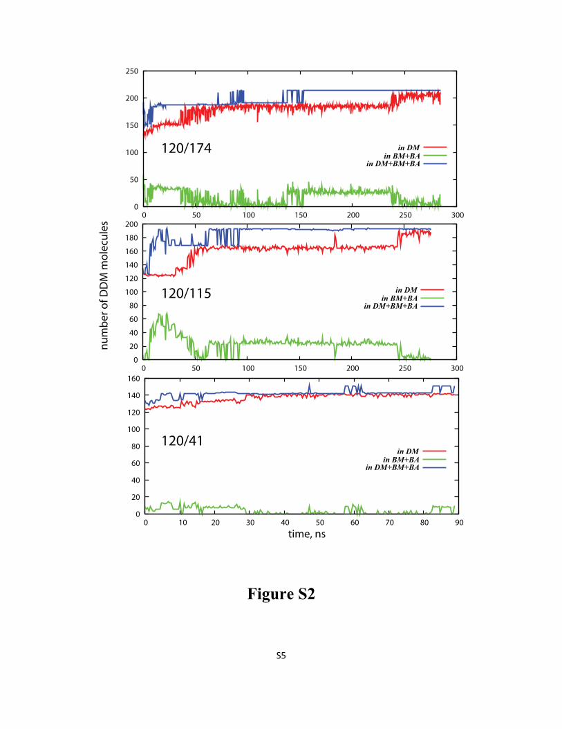
Figure S2: Time evolution (after initial equilibration phase) of the average number of DDM
molecules in the detergent micelle around LeuT (DM), in the aggregates bound to DM (BA and BM), and
together in DM, BA, and BM from 120/174 (top), 120/115 (middle), and 120/41 (bottom) simulations.
For definitions of the various types of aggregates please see Methods in the main text.
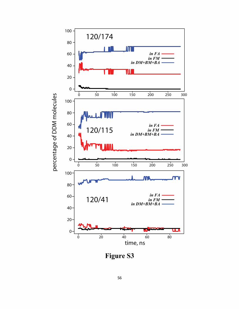
Figure S3: Percentage of DDM detergents in DM, BA, and BM (blue line), in FA (red line), and in FM
(black line) as a function of time in 120/174 (top), 120/115 (middle), and 120/41 (bottom) simulations.
For definitions of the various types of aggregates please see Methods in the main text.
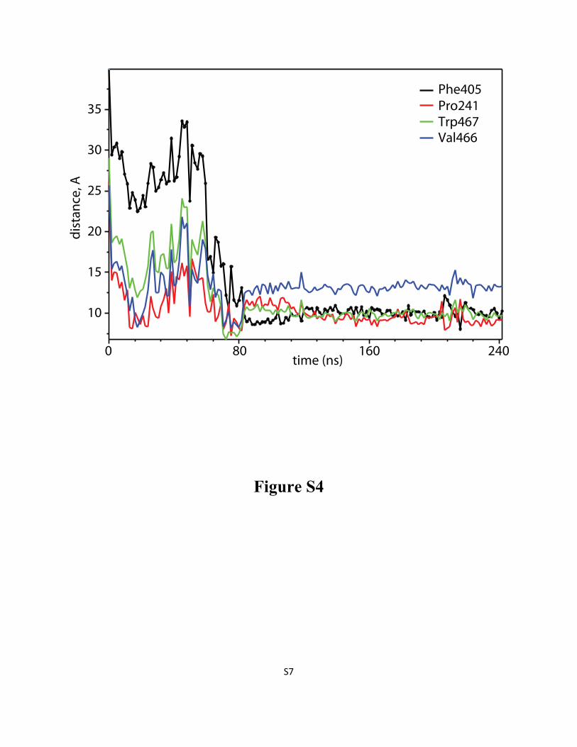
Figure S4: When DDM molecule penetrates LeuT “from the side”, it establishes first contacts with
the transporter through residues located at the extracellular (EC) side of TMH6, and TMH11, before
inserting deeper into the S2 site of LeuT. Time evolution (after initial equilibration phase) of the
distance between Cα atoms of Phe405 (black), Pro241 (red), Trp467 (green), Val466 (blue) residues on
LeuT and the center-of-mass of the head group of the penetrating detergent molecule. The profiles were
obtained from the 160/115 simulation.
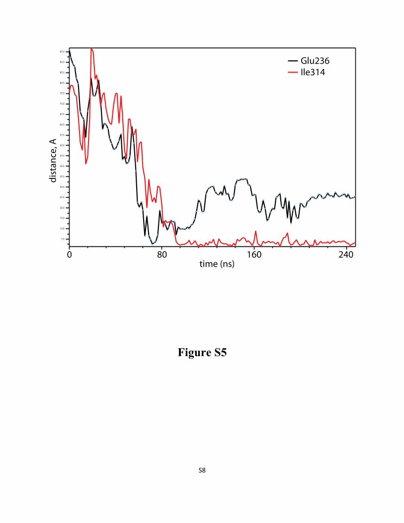
Figure S5: DDM penetration into LeuT “from the top” involves encounters with residues in EC loops
3 and 4 before the detergent takes its final position in the S2 site of the transporter. Time evolution (after
initial equilibration phase) of the distance between Cα atom of Glu236 in ECL3 and the center-of-mass
of DDM detergent head-group (black line), and between Cα atom of Ile314 in ECL4 and DDM
hydrocarbon tail (red line). The profiles were plotted from the 246/54 simulation.
Figure S6: Characterization of micelle shape by the eccentricity parameter, as a function of time for
selected simulations (see Methods in the main text for the eccentricity definition).
S4
Figure S1
S5
0
50
100
150
200
250
0 50 100 150 200 250 300
120/174
0
20
40
60
80
100
120
140
160
180
200
0 50 100 150 200 250 300
0
20
40
60
80
100
120
140
160
0 10 20 30 40 50 60 70 80 90
time, ns
nu
mb
er
of
DD
M m
ole
cule
s
120/115
120/41
in DM
in BM+BA
in DM+BM+BA
in DM
in BM+BA
in DM+BM+BA
in DM
in BM+BA
in DM+BM+BA
Figure S2
S6
0 50 100 150 200 250 300
0 50 100 150 200 250 300
0 20 40 60 80
0
20
40
60
80
100
0
20
40
60
80
100
0
20
40
60
80
100
120/174
time, ns
pe
rce
nta
ge
of
DD
M m
ole
cule
s
120/115
120/41in FA
in FM
in DM+BM+BA
in FA
in FM
in DM+BM+BA
in FA
in FM
in DM+BM+BA
Figure S3
S7
time (ns)
dis
tan
ce, A
0 80 160 240
10
15
20
25
30
35
Phe405
Pro241
Trp467
Val466
Figure S4
S8
7.5
12.5
17.5
22.5
27.5
32.5
37.5
42.5
47.5
52.5
57.5
62.5
67.5
72.5
77.5
82.5
87.5
92.5
97.5
time (ns)
dis
tan
ce, A
0 80 160 240
Glu236
Ile314
Figure S5
S9
0 50 100 150 200 250
0.0
0.1
0.2
0.3
0.4
0.5
0.6
246/54
Ec
ce
ntr
icit
y
0 50 100 150 200 250
0.0
0.1
0.2
0.3
0.4
0.5 246/0
0 50 100 150 200 250
0.1
0.2
0.3
0.4
0.5
0.6
Time (ns)
Ec
ce
ntr
icit
y
160/115
0 50 100 150 200 250
0.0
0.1
0.2
0.3
0.4
0.5
Time (ns)
120/115
Figure S6