Detergent Resistant Membrane Domains in Broccoli Plasma ...
21
International Journal of Molecular Sciences Article Detergent Resistant Membrane Domains in Broccoli Plasma Membrane Associated to the Response to Salinity Stress Lucía Yepes-Molina 1 , Micaela Carvajal 1 and Maria Carmen Martínez-Ballesta 2, * 1 Group of Aquaporins, Department of Plant Nutrition, Centro de Edafología y Biología Aplicada del Segura (CEBAS-CSIC), Campus de Espinardo, E-30100 Murcia, Spain; [email protected] (L.Y.-M.); [email protected] (M.C.) 2 Department of Agronomy Engineering Universidad Politécnica de Cartagena, Paseo Alfonso XIII, 48, 30203 Cartagena (Murcia), Spain * Correspondence: [email protected] Received: 25 September 2020; Accepted: 13 October 2020; Published: 17 October 2020 Abstract: Detergent-resistant membranes (DRMs) microdomains, or “raft lipids”, are key components of the plasma membrane (PM), being involved in membrane trafficking, signal transduction, cell wall metabolism or endocytosis. Proteins imbibed in these domains play important roles in these cellular functions, but there are few studies concerning DRMs under abiotic stress. In this work, we determine DRMs from the PM of broccoli roots, the lipid and protein content, the vesicles structure, their water osmotic permeability and a proteomic characterization focused mainly in aquaporin isoforms under salinity (80 mM NaCl). Based on biochemical lipid composition, higher fatty acid saturation and enriched sterol content under stress resulted in membranes, which decreased osmotic water permeability with regard to other PM vesicles, but this permeability was maintained under control and saline conditions; this maintenance may be related to a lower amount of total PIP1 and PIP2. Selective aquaporin isoforms related to the stress response such as PIP1;2 and PIP2;7 were found in DRMs and this protein partitioning may act as a mechanism to regulate aquaporins involved in the response to salt stress. Other proteins related to protein synthesis, metabolism and energy were identified in DRMs independently of the treatment, indicating their preference to organize in DMRs. Keywords: aquaporins; broccoli; detergent-resistant membrane; microdomains; plasma membrane; salt stress 1. Introduction Plant plasma membrane (PM) is a selective barrier between cells and the environment, playing a crucial role in the reception and transduction of environmental signals and the regulation of cell-environment communication [1]. Therefore, PM is important in essential functions such as cellular nutrition, endocytosis and response to environment stresses [2]. Salt stress is one of the most common abiotic stresses; it is widespread throughout the world and causes losses in the production of the most common crops. PM is important in the response and defence of plants against salinity because it is one of the cell parts that salt reaches first. Membrane lipids and transport proteins have an important function in regulating the permeability of PM, which triggers responses to salinity [3]. Since the control of water and nutrient distribution in the whole plant is an important factor in the acclimation of plants to a saline environment, the role of membrane proteins, such as aquaporins, is crucial, which has been widely reported in different species, such as Arabidopsis [4], maize [5] and broccoli [6]. Aquaporins work out mainly as channels to facilitate and regulate the permeability of water across cell membranes [7] and are representative markers of Int. J. Mol. Sci. 2020, 21, 7694; doi:10.3390/ijms21207694 www.mdpi.com/journal/ijms
-
Upload
khangminh22 -
Category
Documents
-
view
3 -
download
0
Transcript of Detergent Resistant Membrane Domains in Broccoli Plasma ...
International Journal of
Molecular Sciences
Article
Detergent Resistant Membrane Domains in BroccoliPlasma Membrane Associated to the Response toSalinity Stress
Lucía Yepes-Molina 1 , Micaela Carvajal 1 and Maria Carmen Martínez-Ballesta 2,*1 Group of Aquaporins, Department of Plant Nutrition, Centro de Edafología y Biología Aplicada del
Segura (CEBAS-CSIC), Campus de Espinardo, E-30100 Murcia, Spain; [email protected] (L.Y.-M.);[email protected] (M.C.)
2 Department of Agronomy Engineering Universidad Politécnica de Cartagena, Paseo Alfonso XIII, 48,30203 Cartagena (Murcia), Spain
* Correspondence: [email protected]
Received: 25 September 2020; Accepted: 13 October 2020; Published: 17 October 2020�����������������
Abstract: Detergent-resistant membranes (DRMs) microdomains, or “raft lipids”, are key componentsof the plasma membrane (PM), being involved in membrane trafficking, signal transduction, cellwall metabolism or endocytosis. Proteins imbibed in these domains play important roles in thesecellular functions, but there are few studies concerning DRMs under abiotic stress. In this work,we determine DRMs from the PM of broccoli roots, the lipid and protein content, the vesicles structure,their water osmotic permeability and a proteomic characterization focused mainly in aquaporinisoforms under salinity (80 mM NaCl). Based on biochemical lipid composition, higher fatty acidsaturation and enriched sterol content under stress resulted in membranes, which decreased osmoticwater permeability with regard to other PM vesicles, but this permeability was maintained undercontrol and saline conditions; this maintenance may be related to a lower amount of total PIP1 andPIP2. Selective aquaporin isoforms related to the stress response such as PIP1;2 and PIP2;7 werefound in DRMs and this protein partitioning may act as a mechanism to regulate aquaporins involvedin the response to salt stress. Other proteins related to protein synthesis, metabolism and energy wereidentified in DRMs independently of the treatment, indicating their preference to organize in DMRs.
Keywords: aquaporins; broccoli; detergent-resistant membrane; microdomains; plasma membrane;salt stress
1. Introduction
Plant plasma membrane (PM) is a selective barrier between cells and the environment, playinga crucial role in the reception and transduction of environmental signals and the regulation ofcell-environment communication [1]. Therefore, PM is important in essential functions such as cellularnutrition, endocytosis and response to environment stresses [2].
Salt stress is one of the most common abiotic stresses; it is widespread throughout the world andcauses losses in the production of the most common crops. PM is important in the response and defenceof plants against salinity because it is one of the cell parts that salt reaches first. Membrane lipids andtransport proteins have an important function in regulating the permeability of PM, which triggersresponses to salinity [3]. Since the control of water and nutrient distribution in the whole plantis an important factor in the acclimation of plants to a saline environment, the role of membraneproteins, such as aquaporins, is crucial, which has been widely reported in different species, suchas Arabidopsis [4], maize [5] and broccoli [6]. Aquaporins work out mainly as channels to facilitateand regulate the permeability of water across cell membranes [7] and are representative markers of
Int. J. Mol. Sci. 2020, 21, 7694; doi:10.3390/ijms21207694 www.mdpi.com/journal/ijms

Int. J. Mol. Sci. 2020, 21, 7694 2 of 21
water uptake and whole plant water status [8]. Moreover, salinity modifies aquaporin expression andprotein abundance, which indicates that these proteins must be involved in the tolerance response tosalt stress [9].
Despite the multiple studies concerning PM, there are still many unknown and pending detailsto study, especially related to the distribution of proteins and lipids in the membrane. In this sense,several aspects with regard to membrane proteins and salinity stress response or other abiotic stressescannot be elucidated with the classic “fluid mosaic” model for PM, which consists of a homogeneouslipid bilayer and randomly embedded proteins [10,11]. Regarding the PM structure, new approacheshave appeared and enhanced recent models, revealing unknown aspects about the function of PM andthe response to abiotic stress. The models consist of the existence of nanodomains (10–200 nm) [12],which are regions rich in sterols and sphingolipids [13]; this confers to PM a heterogeneous system.These domains are membrane-resident protein clusters in higher-order structures and are actuallyknown as membrane nanodomains or “raft lipids” [14,15]. One of the most effective ways to determinespecific functions of raft lipids in PM is through the isolation of detergent-resistant membranes (DRMs),that is, Triton X-100 (TX100) insoluble membrane fractions [16,17]. This methodology constitutes abiochemical approach in which the study of changes in DRMs in order to determine modification in thecomposition of raft domains in vivo was standard for a long time and is still a technique that is usedtoday [18]. Moreover, it is possible to prepare membrane microdomains with detergent-free methods.These methods have certain advantages in that they involve fewer negative effects for lipid-lipid andprotein-lipid interactions. On the other hand, detergent-free methods involve more contamination byother low-density membranes [19,20].
As it occurs in animal cells, proteomic analysis showed that DRMs in plants are involvedin membrane trafficking, signal transduction, cell wall metabolism occurring on the PM andendocytosis [21–23]. Furthermore, numerous studies establish a relation between DRMs and aresponse to biotic stress [24] but the function of DRMs with regard to abiotic stress in plants is notentirely understood [25]. Only a few studies have been developed along these lines; for example,Minami et al. [17] showed alterations in proteomic profiles of DRMs during cold acclimation inArabidopsis plants. However, there is evidence that elucidates an important relation between anabiotic stress response and proteins located in DRM fractions [18,25] such as aquaporins, heat shockproteins, actins and clathrins [2,22,25]. Li et al. [26] determined the partitioning in PM and traffickingof aquaporin PIP2:1 of Arabidopsis in controlled conditions and under salt stress (100 mM NaCl for twodays). They showed that membrane rafts have an important role in this respect, since under salinitystress as well as clathrin-dependent endocytosis, membrane raft-associated endocytosis is involved.
Broccoli (Brassica oleracea L. var Italica) is a vegetable known to have a health-promoting effect,due to its high content of beneficial compounds, such as glucosinolates and isothiocyanates [27]; so thiscrop has an important economic and agronomic interest. Moreover, new ways to leverage broccolibyproducts are being investigated, for example, the use of vesicles from the plasma membrane ofleaves and roots in biotechnological applications [28,29]. Interestingly, in a previous work, it wasdetermined that these vesicles are more stable over time when they are obtained from plants that hadbeen cultured under high salinity [29]. This stability is an important factor to consider when usingthese vesicles in biotechnological applications, such as for carriers of bioactive compounds in cosmetics,agriculture or pharmaceuticals [30,31].
Thus, the objective of this work was the investigation of the appearance of plasma membranerafts through detergent-resistant membranes isolated from broccoli roots in order to determine lipidsand protein characteristics. The changes in membrane rafts under saline stress were related to salinitytolerance. Different approaches were used, in general terms, to perform this characterization: Analysisof protein and lipid content, ultrastructural analysis and study of the size of the vesicles and measuresof water-osmotic permeability. In addition, part of this study was focused on aquaporins, since theseproteins play an important role in the response to saline stress. In this sense, western-blot analysis of

Int. J. Mol. Sci. 2020, 21, 7694 3 of 21
PIP1 and PIP2 aquaporins and a proteomic analysis by means of LC−MS/MS were carried out in orderto determine the distribution of different isoforms concerning detergent-resistant membranes.
2. Results
2.1. Quantitative Analysis of Protein Amounts in DRM Isolation
The protein amount in each membrane fraction from the microsomal fraction to the DRMs wasdetermined and expressed by the weight of fresh tissue (Figure 1). The amount of protein was higherin membrane fractions obtained from NaCl plants. In control samples, the protein yield in DRMs was5.56 ± 2.02% of that of the total PM and in NaCl samples this value was 12.51 ± 0.85%.
Int. J. Mol. Sci. 2020, 21, 7694 3 of 22
analysis of PIP1 and PIP2 aquaporins and a proteomic analysis by means of LC−MS/MS were carried out in order to determine the distribution of different isoforms concerning detergent-resistant membranes.
2. Results
2.1. Quantitative Analysis of Protein Amounts in DRM Isolation
The protein amount in each membrane fraction from the microsomal fraction to the DRMs was determined and expressed by the weight of fresh tissue (Figure 1). The amount of protein was higher in membrane fractions obtained from NaCl plants. In control samples, the protein yield in DRMs was 5.56 ± 2.02% of that of the total PM and in NaCl samples this value was 12.51 ± 0.85%.
Figure 1. Protein concentration (mg g-1 FW) in microsomal fraction (MF), plasma membrane (PM) and detergent-resistant membranes (DRMs) from broccoli roots treated with 0 (control) and 80 mM NaCl. Data are means ± SE (n = 5). Analyses were done with three biological samples. Asterisks (*) represent significant differences according to t-test for each fraction (p < 0.05).
2.2. Lipid Composition of Broccoli Root DRMs Compared to PM
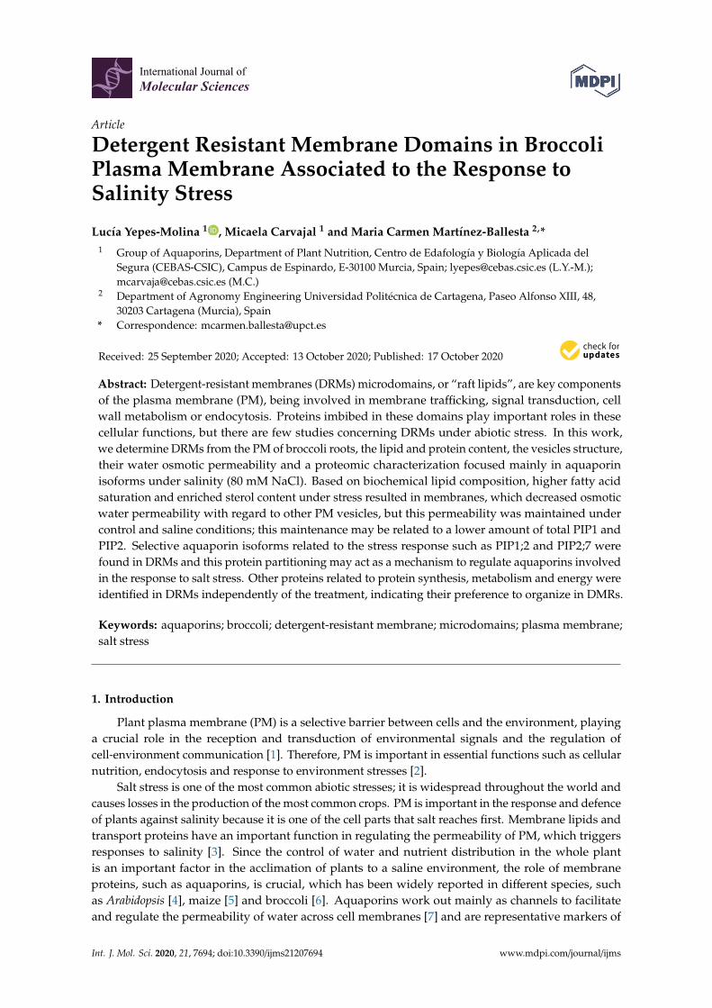
The lipid composition of root PM and DRMs extracted from control and salt-treated plant was analysed. The sterol analyses of the PM and DRMs appear summarized in Figure 2. In PM fractions, exposure to NaCl (80 mM) for 15 days triggers a decrease in sitosterol content. On the contrary, regarding free sterols in DRMs, no statistically significant changes were observed comparing control and salinity. Furthermore, an increase in the content of campesterol in the DRM fraction compared to PM under control conditions was shown (Figure 2A). Regarding the total content of sterols, there is a statistically significant increase in DRMs with respect to PM in both control and NaCl conditions (Figure 2B). The ratio of sitosterol to stigmasterol increases in PM in salt-treated plants and does not change in DRMs if comparing control and NaCl conditions (Figure 2C).
Figure 1. Protein concentration (mg g-1 FW) in microsomal fraction (MF), plasma membrane (PM) anddetergent-resistant membranes (DRMs) from broccoli roots treated with 0 (control) and 80 mM NaCl.Data are means ± SE (n = 5). Analyses were done with three biological samples. Asterisks (*) representsignificant differences according to t-test for each fraction (p < 0.05).
2.2. Lipid Composition of Broccoli Root DRMs Compared to PM
The lipid composition of root PM and DRMs extracted from control and salt-treated plant wasanalysed. The sterol analyses of the PM and DRMs appear summarized in Figure 2. In PM fractions,exposure to NaCl (80 mM) for 15 days triggers a decrease in sitosterol content. On the contrary,regarding free sterols in DRMs, no statistically significant changes were observed comparing controland salinity. Furthermore, an increase in the content of campesterol in the DRM fraction compared toPM under control conditions was shown (Figure 2A). Regarding the total content of sterols, there isa statistically significant increase in DRMs with respect to PM in both control and NaCl conditions(Figure 2B). The ratio of sitosterol to stigmasterol increases in PM in salt-treated plants and does notchange in DRMs if comparing control and NaCl conditions (Figure 2C).
On the other hand, in Table 1 fatty acid composition as a percentage is shown. This analysisrevealed that in PM palmitoleic acid (C16:1) was the predominant fatty acid both in control and salttreated plants, but no significant differences appeared between treatments. At this salinity level therewas a statistically significant increase in linoleic acid (C18:3). The unsaturation grade of the membranefractions was determined by the RUFA (ratio of unsaturated fatty acids) and DBI (double bond index)indices. In this case, the RUFA increased significantly in PM isolated from roots of salt-treated plantsrelative to control plants. Regarding DRMs, palmitoleic acid (C16:1) is also the predominant fatty acidand an increase in oleic acid (C18:1) instead of in linoleic acid (C18:3) was produced under salinity.Besides, in DRMs, a decrease in RUFA was produced in salinity conditions. As for the differencesbetween PM and DRM fractions, this analysis showed an increase in oleic acid (C18:1) in DRMs
Int. J. Mol. Sci. 2020, 21, 7694 4 of 21
under salinity. In control, RUFA increased in DRM fractions, but under salinity this ratio decreasedsignificantly in DRMs compared to PM.Int. J. Mol. Sci. 2020, 21, 7694 4 of 22
Figure 2. Sterol analysis. Campesterol, stigmasterol and sitosterol (µg mg-1 protein) (A), total sterols (µg mg-1 protein) (B) and stigmasterol/sitosterol ratio (C) in plasma membrane (PM) and detergent-resistant membranes (DRMs) isolated from broccoli roots control and treated with NaCl. Data are means ± SE (n = 3). Analyses were done with three biological samples. Columns with different letters (a, b, c) for each variable differ significantly according to Tukey’s test (p < 0.05).
On the other hand, in Table 1 fatty acid composition as a percentage is shown. This analysis revealed that in PM palmitoleic acid (C16:1) was the predominant fatty acid both in control and salt treated plants, but no significant differences appeared between treatments. At this salinity level there was a statistically significant increase in linoleic acid (C18:3). The unsaturation grade of the membrane fractions was determined by the RUFA (ratio of unsaturated fatty acids) and DBI (double bond index) indices. In this case, the RUFA increased significantly in PM isolated from roots of salt-treated plants relative to control plants. Regarding DRMs, palmitoleic acid (C16:1) is also the predominant fatty acid and an increase in oleic acid (C18:1) instead of in linoleic acid (C18:3) was produced under salinity. Besides, in DRMs, a decrease in RUFA was produced in salinity conditions. As for the differences between PM and DRM fractions, this analysis showed an increase in oleic acid (C18:1) in DRMs under salinity. In control, RUFA increased in DRM fractions, but under salinity this ratio decreased significantly in DRMs compared to PM.
Figure 2. Sterol analysis. Campesterol, stigmasterol and sitosterol (µg mg−1 protein) (A), totalsterols (µg mg−1 protein) (B) and stigmasterol/sitosterol ratio (C) in plasma membrane (PM) anddetergent-resistant membranes (DRMs) isolated from broccoli roots control and treated with NaCl.Data are means ± SE (n = 3). Analyses were done with three biological samples. Columns with differentletters (a, b, c) for each variable differ significantly according to Tukey’s test (p < 0.05).
Table 1. The fatty acid percentage (%), double bond index (DBI = (unsaturated fatty acid × number ofdouble bonds)) and ratio of unsaturated fatty acids (RUFA = (18:2 + 18:3/18:1)) in the plasma membrane(PM) and detergent-resistant membranes (DRMs) of roots from control and salt-treated broccoli plants.
Fraction Treatment% Fatty Acids
Palmitoleic(C16:1) Oleic (C18:1) Linoleic
(C18:2)Linolenic
(C18:3) DBI RUFA
ControlPM 56.14 ± 8.18 a 10.99 ± 0.04 ab 29.02 ± 1.64 a 1.96 ± 0.36 b 129.40 ± 2.96 a 1.77 ± 0.50 b
DRMs 68.62 ± 10.46 a 5.23 ± 1.92 b 14.61 ± 4.46 ab 11.53 ± 4.08 a 136.28 ± 13.61 a 5.01 ± 0.14 a
NaClPM 60.91 ± 6.43 a 7.91 ± 3.24 b 20.46 ± 5.54 a 11.87 ± 2.67 a 137.66 ± 2.22 a 4.94 ± 1.27 a
DRMs 53.62 ± 10.07 a 34.77 ± 9.57 a 5.77 ± 0.30 b 7.21 ± 1.13 a 117.4 ± 0.70 a 0.38 ± 0.09 c
Data are means ± SE (n = 3). Analyses were done with three biological samples. Values followed by different letters(a, b, c) in each column differ significantly according to Tukey’s test (p < 0.05).

Int. J. Mol. Sci. 2020, 21, 7694 5 of 21
2.3. Ultrastructural Analysis And Size of Broccoli Root DRMs Compared to PM
Membrane fractions (PM and DRMs) were processed by chemical fixation. Electron microscopyimages show that control PM (Figure 3A,B), NaCl PM (Figure 3E,F), control DRM (Figure 3C,D) andNaCl DRM (Figure 3G,H) fractions contained mostly membrane vesicles. It has to be noted that nochange was observed between PM and DRMs obtained from control plants and plants grown undersalinity conditions as far as structure is concerned.
Int. J. Mol. Sci. 2020, 21, 7694 5 of 22
Table 1. The fatty acid percentage (%), double bond index (DBI = (unsaturated fatty acid × number of double bonds)) and ratio of unsaturated fatty acids (RUFA = (18:2 + 18:3/18:1)) in the plasma membrane (PM) and detergent-resistant membranes (DRMs) of roots from control and salt-treated broccoli plants.
Fraction Treatment % Fatty Acids
Palmitoleic (C16:1)
Oleic (C18:1)
Linoleic (C18:2)
Linolenic (C18:3) DBI RUFA
Control PM 56.14 ± 8.18 a
10.99 ± 0.04 ab
29.02 ± 1.64 a
1.96 ± 0.36 b
129.40 ± 2.96 a
1.77 ± 0.50 b
DRMs 68.62 ± 10.46
a 5.23 ± 1.92 b
14.61 ± 4.46 ab
11.53 ± 4.08 a
136.28 ± 13.61 a
5.01 ± 0.14 a
NaCl PM 60.91 ± 6.43 a
7.91 ± 3.24 b
20.46 ± 5.54 a
11.87 ± 2.67 a
137.66 ± 2.22 a
4.94 ± 1.27 a
DRMs 53.62 ± 10.07
a 34.77 ± 9.57 a
5.77 ± 0.30 b
7.21 ± 1.13 a
117.4 ± 0.70 a
0.38 ± 0.09 c
Data are means ± SE (n = 3). Analyses were done with three biological samples. Values followed by different letters (a, b, c) in each column differ significantly according to Tukey’s test (p < 0.05).
2.3. Ultrastructural Analysis And Size of Broccoli Root DRMs Compared to PM
Membrane fractions (PM and DRMs) were processed by chemical fixation. Electron microscopy images show that control PM (Figure 3A,B), NaCl PM (Figure 3E,F), control DRM (Figure 3C,D) and NaCl DRM (Figure 3G,H) fractions contained mostly membrane vesicles. It has to be noted that no change was observed between PM and DRMs obtained from control plants and plants grown under salinity conditions as far as structure is concerned.
Figure 3. Transmission electronic microscopy images of control plasma membrane (PM) (1 µm) (A) and 500 nm (B), control detergent-resistant membranes (DRMs) (1 µm) (C) and 500 nm (D), NaCl PM (1 µm) (E) and 500 nm (F), and NaCl DRMs (1 µm) (G) and 500 nm (H).
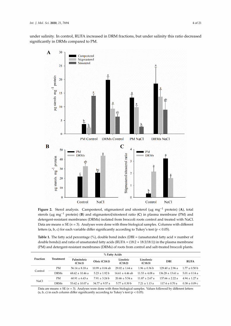
The determinations of vesicle diameter appear in Figure 4 and reveal a significant difference between the PM and DRM vesicles. Dynamic light scattering of the PM and DRM vesicles under control and NaCl conditions showed the size distribution of the different vesicles (Figure 4A). PM had a larger diameter, but all populations are distributed in the range of 100 to 1000 nm (Figure 4A). Control PM vesicles had a diameter of 401.18 nm and NaCl PM vesicles 335.78 nm, but no significant
Figure 3. Transmission electronic microscopy images of control plasma membrane (PM) (1 µm) (A)and 500 nm (B), control detergent-resistant membranes (DRMs) (1 µm) (C) and 500 nm (D), NaCl PM(1 µm) (E) and 500 nm (F), and NaCl DRMs (1 µm) (G) and 500 nm (H).
The determinations of vesicle diameter appear in Figure 4 and reveal a significant differencebetween the PM and DRM vesicles. Dynamic light scattering of the PM and DRM vesicles undercontrol and NaCl conditions showed the size distribution of the different vesicles (Figure 4A). PM hada larger diameter, but all populations are distributed in the range of 100 to 1000 nm (Figure 4A).Control PM vesicles had a diameter of 401.18 nm and NaCl PM vesicles 335.78 nm, but no significantstatistical differences were found between the two populations of vesicles; similarly, there were nodifferences between control and NaCl DRM vesicles, but these vesicles had lower diameter than PMvesicles: In control conditions, DRMs vesicles were 210.38 nm smaller (190.82 nm) and under salt stress177.22 nm (158.59 nm). Regarding the polydispersity index, in all samples the value ranged from 0.26to 0.56 and no significant statistical differences were found (Figure 4B).
Int. J. Mol. Sci. 2020, 21, 7694 6 of 21
Int. J. Mol. Sci. 2020, 21, 7694 6 of 22
statistical differences were found between the two populations of vesicles; similarly, there were no differences between control and NaCl DRM vesicles, but these vesicles had lower diameter than PM vesicles: In control conditions, DRMs vesicles were 210.38 nm smaller (190.82 nm) and under salt stress 177.22 nm (158.59 nm). Regarding the polydispersity index, in all samples the value ranged from 0.26 to 0.56 and no significant statistical differences were found (Figure 4B).
Figure 4. Size distribution (A) and average size and polydispersity index (B) of plasma membrane (PM) and detergent-resistant membranes (DRMs) from broccoli roots treated with 0 (control) and 80 mM NaCl. Data are means ± SE (n = 3). Analyses were done with three biological samples. Different letters (a, b) represent significant differences according to t-test for each fraction (p < 0.05).
2.4. Vesicle Integrity and Functionality
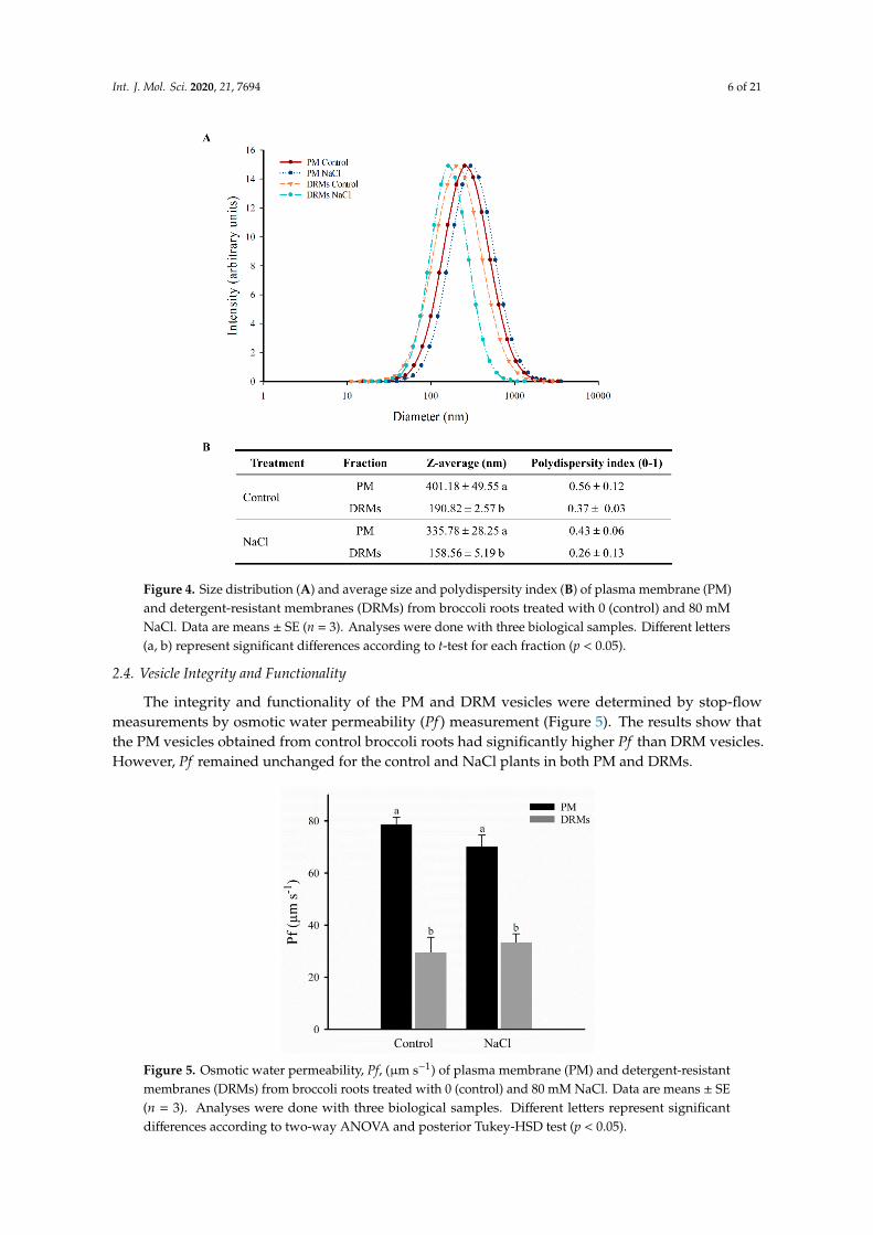
The integrity and functionality of the PM and DRM vesicles were determined by stop-flow measurements by osmotic water permeability (Pf) measurement (Figure 5). The results show that the PM vesicles obtained from control broccoli roots had significantly higher Pf than DRM vesicles. However, Pf remained unchanged for the control and NaCl plants in both PM and DRMs.
Figure 4. Size distribution (A) and average size and polydispersity index (B) of plasma membrane (PM)and detergent-resistant membranes (DRMs) from broccoli roots treated with 0 (control) and 80 mMNaCl. Data are means ± SE (n = 3). Analyses were done with three biological samples. Different letters(a, b) represent significant differences according to t-test for each fraction (p < 0.05).
2.4. Vesicle Integrity and Functionality
The integrity and functionality of the PM and DRM vesicles were determined by stop-flowmeasurements by osmotic water permeability (Pf ) measurement (Figure 5). The results show thatthe PM vesicles obtained from control broccoli roots had significantly higher Pf than DRM vesicles.However, Pf remained unchanged for the control and NaCl plants in both PM and DRMs.Int. J. Mol. Sci. 2020, 21, 7694 7 of 22
Figure 5. Osmotic water permeability, Pf, (µm s−1) of plasma membrane (PM) and detergent-resistant membranes (DRMs) from broccoli roots treated with 0 (control) and 80 mM NaCl. Data are means ± SE (n = 3). Analyses were done with three biological samples. Different letters represent significant differences according to two-way ANOVA and posterior Tukey-HSD test (p < 0.05).
2.5. PIP1 And PIP2 Aquaporin Quantification
The abundance of PIP1 and PIP2 aquaporins was carried out using western blot analysis. Two bands corresponding to 29 KDa (aquaporin monomer) and 58 KDa (aquaporin dimer) were detected with PIP1 and PIP2 antibodies in PM and DRM (control and NaCl) (Figure 6). The immunostaining intensity differed among treatments and membrane fractions; this, moreover, depends on the PIP subfamily. The content of PIP1 increased both in PM and DRMs under saline stress, while a decrease in the total amount of PIP1 in DRM fractions with respect to PM was observed in control and NaCl (Figure 6A). Regarding the PIP2 subfamily (Figure 6B), we found that in PM there was an increase in the content of PIP2 when the broccoli was grown under saline stress, although this fact did not occur in DRMs, since in this fraction a decrease in PIP2 appeared under stress conditions. In DRM fractions there appeared a decrease in the content of PIP2 aquaporins, in the same way that occurred with the PIP1 subfamily.
Figure 6. Immunodetection of PIP1 (A) and PIP2 (B) homologues in the root plasma membrane (PM) and detergent-resistant membranes (DRMs) of control broccoli plants and plants treated with 80 mM
Figure 5. Osmotic water permeability, Pf, (µm s−1) of plasma membrane (PM) and detergent-resistantmembranes (DRMs) from broccoli roots treated with 0 (control) and 80 mM NaCl. Data are means ± SE(n = 3). Analyses were done with three biological samples. Different letters represent significantdifferences according to two-way ANOVA and posterior Tukey-HSD test (p < 0.05).

Int. J. Mol. Sci. 2020, 21, 7694 7 of 21
2.5. PIP1 And PIP2 Aquaporin Quantification
The abundance of PIP1 and PIP2 aquaporins was carried out using western blot analysis. Two bandscorresponding to 29 KDa (aquaporin monomer) and 58 KDa (aquaporin dimer) were detected withPIP1 and PIP2 antibodies in PM and DRM (control and NaCl) (Figure 6). The immunostaining intensitydiffered among treatments and membrane fractions; this, moreover, depends on the PIP subfamily.The content of PIP1 increased both in PM and DRMs under saline stress, while a decrease in the totalamount of PIP1 in DRM fractions with respect to PM was observed in control and NaCl (Figure 6A).Regarding the PIP2 subfamily (Figure 6B), we found that in PM there was an increase in the content ofPIP2 when the broccoli was grown under saline stress, although this fact did not occur in DRMs, sincein this fraction a decrease in PIP2 appeared under stress conditions. In DRM fractions there appeared adecrease in the content of PIP2 aquaporins, in the same way that occurred with the PIP1 subfamily.
Int. J. Mol. Sci. 2020, 21, 7694 7 of 22
Figure 5. Osmotic water permeability, Pf, (µm s−1) of plasma membrane (PM) and detergent-resistant membranes (DRMs) from broccoli roots treated with 0 (control) and 80 mM NaCl. Data are means ± SE (n = 3). Analyses were done with three biological samples. Different letters represent significant differences according to two-way ANOVA and posterior Tukey-HSD test (p < 0.05).
2.5. PIP1 And PIP2 Aquaporin Quantification
The abundance of PIP1 and PIP2 aquaporins was carried out using western blot analysis. Two bands corresponding to 29 KDa (aquaporin monomer) and 58 KDa (aquaporin dimer) were detected with PIP1 and PIP2 antibodies in PM and DRM (control and NaCl) (Figure 6). The immunostaining intensity differed among treatments and membrane fractions; this, moreover, depends on the PIP subfamily. The content of PIP1 increased both in PM and DRMs under saline stress, while a decrease in the total amount of PIP1 in DRM fractions with respect to PM was observed in control and NaCl (Figure 6A). Regarding the PIP2 subfamily (Figure 6B), we found that in PM there was an increase in the content of PIP2 when the broccoli was grown under saline stress, although this fact did not occur in DRMs, since in this fraction a decrease in PIP2 appeared under stress conditions. In DRM fractions there appeared a decrease in the content of PIP2 aquaporins, in the same way that occurred with the PIP1 subfamily.
Figure 6. Immunodetection of PIP1 (A) and PIP2 (B) homologues in the root plasma membrane (PM) and detergent-resistant membranes (DRMs) of control broccoli plants and plants treated with 80 mM
Figure 6. Immunodetection of PIP1 (A) and PIP2 (B) homologues in the root plasma membrane (PM)and detergent-resistant membranes (DRMs) of control broccoli plants and plants treated with 80 mMNaCl. Data are means ± SE (n = 3). Analyses were done with three biological samples. Different lettersrepresent significant differences according to Tukey-HSD test (p < 0.05).
2.6. Proteomic Analysis of DRMs from B. oleracea Root
A Venn diagram was made according to the accession numbers of the identified protein in theproteomic analysis in both samples (DRM control and NaCl) after the specific selection of proteins waslocated in the PM in order to show proteins that were identified only in the control or in salt treatment inDRMs and proteins that were identified in both treatments (Figure 7A). The results obtained show that224 proteins were shared between control and NaCl DRMs, and 175 and 137 proteins were categorizedas unique to the control and NaCl DRMs, respectively.
Identified proteins were classified into nine functional categories according to [32]: Metabolismand energy, gene expression, protein synthesis, protein destination and storage, transport, cell structure,signal transduction, disease/defence and secondary metabolism. In Figure 7B, normalized distributionsof functional categories in each sample are shown, and the results indicate that an increase in theproportion of proteins involved in transport and a decrease in proteins associated with proteindestination and storage were detected in the saline treatment, although salt stress did not greatly alterthe proportion of different types of proteins.
Int. J. Mol. Sci. 2020, 21, 7694 8 of 21
Int. J. Mol. Sci. 2020, 21, 7694 8 of 22
NaCl. Data are means ± SE (n = 3). Analyses were done with three biological samples. Different letters represent significant differences according to Tukey-HSD test (p < 0.05).
2.6. Proteomic Analysis of DRMs from B. oleracea Root
A Venn diagram was made according to the accession numbers of the identified protein in the proteomic analysis in both samples (DRM control and NaCl) after the specific selection of proteins was located in the PM in order to show proteins that were identified only in the control or in salt treatment in DRMs and proteins that were identified in both treatments (Figure 7A). The results obtained show that 224 proteins were shared between control and NaCl DRMs, and 175 and 137 proteins were categorized as unique to the control and NaCl DRMs, respectively.
Figure 7. Venn diagram of control DRMs and NaCl DRM proteins identified in broccoli roots (A) and functional classification of identified proteins in both samples as percentages (B).
Identified proteins were classified into nine functional categories according to [32]: Metabolism and energy, gene expression, protein synthesis, protein destination and storage, transport, cell structure, signal transduction, disease/defence and secondary metabolism. In Figure 7B, normalized distributions of functional categories in each sample are shown, and the results indicate that an increase in the proportion of proteins involved in transport and a decrease in proteins associated with protein destination and storage were detected in the saline treatment, although salt stress did not greatly alter the proportion of different types of proteins.
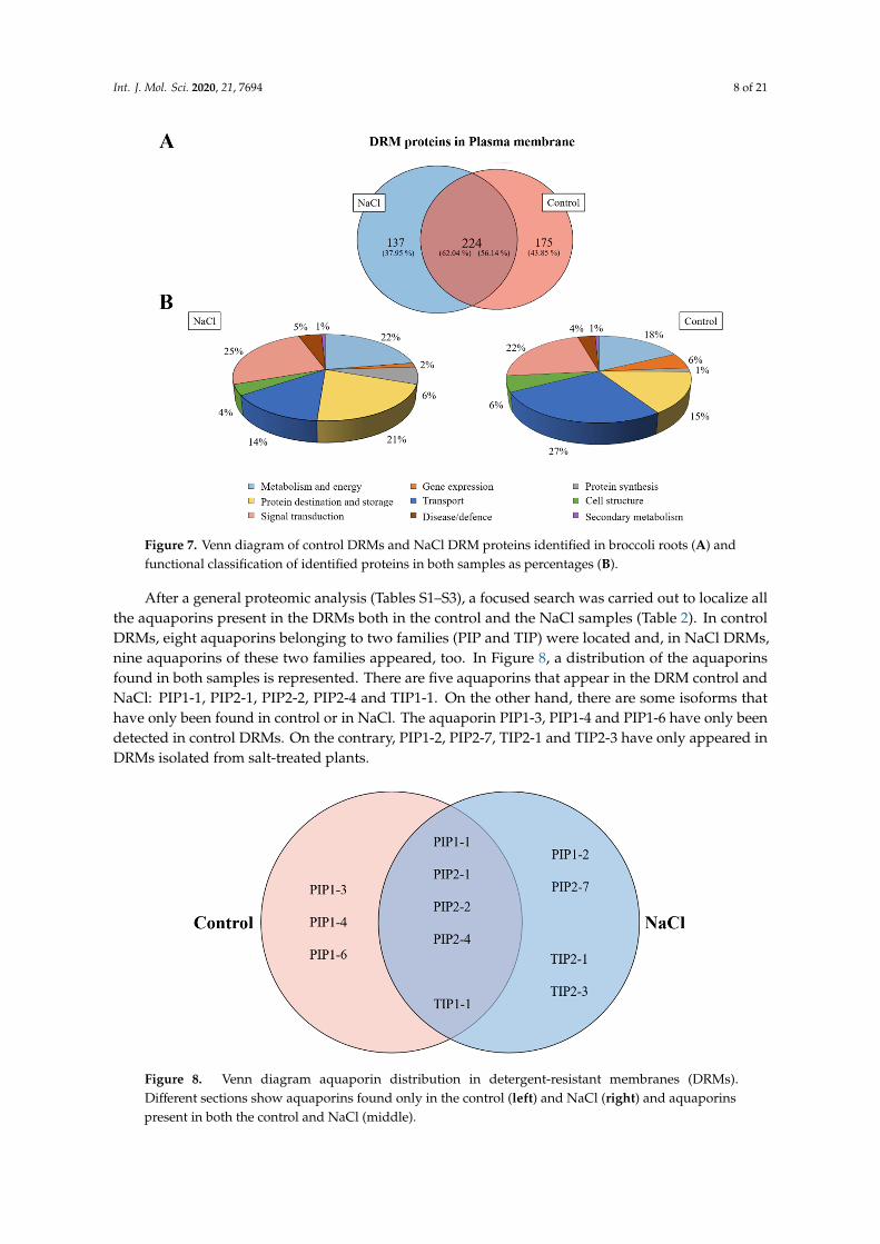
After a general proteomic analysis (Tables S1–S3), a focused search was carried out to localize all the aquaporins present in the DRMs both in the control and the NaCl samples (Table 2). In control DRMs, eight aquaporins belonging to two families (PIP and TIP) were located and, in NaCl DRMs, nine aquaporins of these two families appeared, too. In Figure 8, a distribution of the aquaporins found in both samples is represented. There are five aquaporins that appear in the DRM control and NaCl: PIP1-1, PIP2-1, PIP2-2, PIP2-4 and TIP1-1. On the other hand, there are some isoforms that have only been found in control or in NaCl. The aquaporin PIP1-3, PIP1-4 and PIP1-6 have only been detected in control DRMs. On the contrary, PIP1-2, PIP2-7, TIP2-1 and TIP2-3 have only appeared in DRMs isolated from salt-treated plants.
Figure 7. Venn diagram of control DRMs and NaCl DRM proteins identified in broccoli roots (A) andfunctional classification of identified proteins in both samples as percentages (B).
After a general proteomic analysis (Tables S1–S3), a focused search was carried out to localize allthe aquaporins present in the DRMs both in the control and the NaCl samples (Table 2). In controlDRMs, eight aquaporins belonging to two families (PIP and TIP) were located and, in NaCl DRMs,nine aquaporins of these two families appeared, too. In Figure 8, a distribution of the aquaporinsfound in both samples is represented. There are five aquaporins that appear in the DRM control andNaCl: PIP1-1, PIP2-1, PIP2-2, PIP2-4 and TIP1-1. On the other hand, there are some isoforms thathave only been found in control or in NaCl. The aquaporin PIP1-3, PIP1-4 and PIP1-6 have only beendetected in control DRMs. On the contrary, PIP1-2, PIP2-7, TIP2-1 and TIP2-3 have only appeared inDRMs isolated from salt-treated plants.
Int. J. Mol. Sci. 2020, 21, 7694 9 of 22
Table 2. Aquaporins identified in the control and NaCl detergent-resistant membranes (DRMs) after a database search. MW is molecular weight (in dalton), PSMs is the number of fragmentation spectra acquired, peptides is the number of distinct peptide sequences in the protein group and coverage is the percentage of the protein sequence covered by identified peptides. UniProt Accession Description MW (Da) Score PSMs Peptides Coverage
Control A0A178VIZ0 PIP1-1 30,897 72 2 1 21 A0A078IDF7 PIP1-1 30,852 206 4 3 29.7 A0A0D3C6T1 PIP1-3 30,825 135 2 2 29.7
A0A0D3DZM3 PIP1-4 30,769 194 2 2 29.7 F2X5K3 PIP1-6 16,451 198 2 2 21.3
A0A0D3B5C8 PIP2-1 30,208 736 18 9 39.6 O80369 PIP2-1 30,670 380 8 6 39.7
A0A0D3BRA6 PIP2-2 30,512 137 3 2 39.3 A0A0D3BRA5 PIP2-2 30,495 108 2 2 39.3 A0A0D3B2E7 PIP2-4 30,293 466 7 6 37.9 A0A078F7W0 PIP2-4 27,697 151 2 2 35.1 A0A0D3C4M6 TIP1-1 25,635 59 3 2 6.8 A0A078JDN1 TIP2-2 25,148 106 2 2 7.2
NaCl A0A178VIZ0 PIP1-1 30,897 61 2 1 21
R0HDM0 PIP1-1 30,779 273 4 4 29 A0A078G0R2 PIP1-2 30,792 961 19 13 35 A0A078HJQ7 PIP1-2 30,736 177 2 2 35 A0A0D3BRA6 PIP2-1 30,512 74 2 1 34
O80369 PIP2-1 30,670 293 6 4 29 M4CLX8 PIP2-2 25,497 59 2 2 27
A0A0D3B2E7 PIP2-4 30,293 472 7 7 38 A0A078IUF9 PIP2-7 29,949 91 3 2 30.2
A0A0D3C4M6 TIP1-1 25,635 130 3 3 7 A0A078GXB0 TIP2-1 24,983 205 3 3 7.3 A0A078J7S3 TIP2-3 25,348 165 6 2 7.2
Figure 8. Venn diagram aquaporin distribution in detergent-resistant membranes (DRMs). Different sections show aquaporins found only in the control (left) and NaCl (right) and aquaporins present in both the control and NaCl (middle).
Figure 8. Venn diagram aquaporin distribution in detergent-resistant membranes (DRMs).Different sections show aquaporins found only in the control (left) and NaCl (right) and aquaporinspresent in both the control and NaCl (middle).

Int. J. Mol. Sci. 2020, 21, 7694 9 of 21
Table 2. Aquaporins identified in the control and NaCl detergent-resistant membranes (DRMs) after adatabase search. MW is molecular weight (in dalton), PSMs is the number of fragmentation spectraacquired, peptides is the number of distinct peptide sequences in the protein group and coverage is thepercentage of the protein sequence covered by identified peptides.
UniProt Accession Description MW (Da) Score PSMs Peptides Coverage
Control
A0A178VIZ0 PIP1-1 30,897 72 2 1 21A0A078IDF7 PIP1-1 30,852 206 4 3 29.7A0A0D3C6T1 PIP1-3 30,825 135 2 2 29.7
A0A0D3DZM3 PIP1-4 30,769 194 2 2 29.7F2X5K3 PIP1-6 16,451 198 2 2 21.3
A0A0D3B5C8 PIP2-1 30,208 736 18 9 39.6O80369 PIP2-1 30,670 380 8 6 39.7
A0A0D3BRA6 PIP2-2 30,512 137 3 2 39.3A0A0D3BRA5 PIP2-2 30,495 108 2 2 39.3A0A0D3B2E7 PIP2-4 30,293 466 7 6 37.9A0A078F7W0 PIP2-4 27,697 151 2 2 35.1A0A0D3C4M6 TIP1-1 25,635 59 3 2 6.8A0A078JDN1 TIP2-2 25,148 106 2 2 7.2
NaCl
A0A178VIZ0 PIP1-1 30,897 61 2 1 21R0HDM0 PIP1-1 30,779 273 4 4 29
A0A078G0R2 PIP1-2 30,792 961 19 13 35A0A078HJQ7 PIP1-2 30,736 177 2 2 35A0A0D3BRA6 PIP2-1 30,512 74 2 1 34
O80369 PIP2-1 30,670 293 6 4 29M4CLX8 PIP2-2 25,497 59 2 2 27
A0A0D3B2E7 PIP2-4 30,293 472 7 7 38A0A078IUF9 PIP2-7 29,949 91 3 2 30.2
A0A0D3C4M6 TIP1-1 25,635 130 3 3 7A0A078GXB0 TIP2-1 24,983 205 3 3 7.3A0A078J7S3 TIP2-3 25,348 165 6 2 7.2
3. Discussion
One important modification in broccoli under salt stress was the chemical composition PM ofboth lipids [33] and proteins [29] that has been reported to relate transport and salt tolerance. In recentyears, the role of proteins associated with lipid DRMs in plants has been the focus of many studiesdue to these domains being commonly reported as key systems in many biological processes, such asstress response [34]. Based on the idea that sterols are required for the formation of a liquid-orderedlipid phase in plants and that this fact will condition protein membrane location [35], in the presentstudy we determined the DRMs isolated from the root PM of broccoli plants, describing the changesinduced by salinity. Regarding the percentage of protein from the total PM recovery in DRMs, ourresults (5.56 2.02% in control and 12.51 0.85% in salinity) were in accordance with those obtained inprevious reports in other plants as Medicago truncatula [23], Arabidopsis thaliana [17], Avena sativa andOryza sativa [34], pointing out the general characteristics according to the protein/lipid ratio in DRMs.However, in our work, the protein level was higher in DRMs isolated from the PM of plants grownunder NaCl than in the control. This result differed from the data obtained in A. thaliana plants undercold acclimation, in which the total protein recovered from DRMs was lower in plants grown understress [17]. This could be due to the different stress tolerance response in Arabidopsis, very sensitive toabiotic stress [36], and Brassica, tolerant to abiotic stress [37], which could affect PM protein partitioning,resulting in a higher concentration of proteins in DRMs when plants are stress tolerant.
In addition to protein content, lipid composition of PM was previously reported to be largelyaffected by salinity [33,37]. Previously, the sterol analysis of PM from broccoli plants in control and salt
Int. J. Mol. Sci. 2020, 21, 7694 10 of 21
conditions revealed results similar to this work. Thus, an increase of stigmasterol and a decrease ofsitosterol were shown. It has been described that sitosterol plays a significant role in the ordering offatty acid chains in the membrane, decreasing membrane water and ion permeability, and the activityof membrane proteins [38]. Thus, sitosterol decrease in broccoli PM roots could result in a mechanismto cope with salt stress. DRM lipid composition analysis evidenced an enrichment in total sterols withrespect to PM in both, control and NaCl samples, which is an important characteristic that makes outlipid raft domains from the rest of the PM [13]. In other reports working with artificial proteoliposomes,cholesterol was found to induce higher resistance to detergent solubilization on a broader range oftemperatures (from 4 to 15 ◦C) [39]. In our work, both control- and NaCl-treated DRMs differ mainlyby increasing campesterol and decreasing sitosterol content. This has not been observed previously,but the campesterol has been related to membrane stabilization, tightly packing the phospholipidbilayer [40]. The authors described a strong ordering ability for campesterol, in the same range as thecholesterol one, and a less efficient sitosterol.
Moreover, our results showed an increase in oleic acid in DRMs under salt stress instead oflinolenic acid as occurred in PM. Similarly, in the PM of safflower and broccoli plants an increase inlinoleic acids under salt stress and a decrease in oleic acid have been observed [37]. However, fattyacid modifications in DRMs isolated from plant PM have not been previously described under abioticstress conditions. In control conditions, however, the results are similar to PM except for an increasein linolenic acid increasing the unsaturation. An increase in oleic acid in NaCl-DRMs could reflect amembrane remodelling in order to maintain membrane environment integrity preservation after NaClapplication. It has been observed that glycerolipids of tobacco plant DRMs contained more saturatedfatty acyl chains, which would contribute to the rigidity of the liquid-ordered phase of membranerafts [22]. However, this only occurs in our broccoli DRMs under salinity stress conditions, pointing toa stress response.
It has been reported that stability in DRM lipid content could be due to the fact that in thesedomains the lipids form a dense packing, increasing membrane viscosity to provide a conservedenvironment for proteins with specific biological functions [41]. Therefore, for raft functionality it hasbeen stated that specific PM lipids, mainly sterols and fatty acids from phospholipids, could interactwith proteins providing signalling and trafficking platforms in the PM [42]. From the results of thiswork, DRMs from broccoli plants are distinctive, but also the fact that NaCl plants provided higherlevels of sterols and higher saturation of fatty acids could be related to the key for stress tolerance.
Transmission electron microscopy was used to characterize PM and DRM morphology from rootsof broccoli plants. As has been already reported, isolated PM forms vesicular structures in vitro [43],which was also observed in both the control and NaCl PM vesicles from broccoli roots. Similarly, isolatedDRMs from PM were likewise vesicular-shaped, although these vesicles were smaller. Vesicles ofDRMs have also been described in DRMs isolated from mammals, such as from kidney cells [16],demyelinated membranes of rat brain [44] or human erythrocytes [45], and, in addition to vesicles,shaped membrane sheets may also appear in DRMs isolated from sucrose gradients. Moreover, in plantDRMs, similar morphology was observed and lined/shaped membrane fragments appeared togetherwith vesicles [23]. Differences in the display of DRM ultrastructure could be due to the differentdetergent applied to PM, since detergent isolation may define membrane subdomains and specificlipid ensemble and structure [46].
The diameter of the vesicles was measured through light-scattering technique. PM vesicles inboth control and NaCl, as we showed in several previous works [28,29], have a diameter between 300and 400 nm. On the other hand, DRM vesicles were smaller than PM vesicles. In this case, the diameterof the control and NaCl DRM vesicles was less than 200 nm, which was in accordance with previousresults [45], where DRM vesicles from human erythrocytes had a diameter around 100 nm. These typesof vesicles could also be used as nanocarriers and, in addition, due to their smaller size, DRM vesiclescould be suitable for a finer application, specifically as nanomedical devices [47]. The size of DRMvesicles obtained is difficult to relate with the size of the lipids raft domains in vivo because DRMs are

Int. J. Mol. Sci. 2020, 21, 7694 11 of 21
not lipid raft in the same way that these domains are located in the plasma membrane in vivo [48].The real size of the lipids raft in cells is still unclear and is still a matter of debate. Different studiesreveal that these domains must be quite small; roughly, they set values between 50 and 200 nm [49].
The osmotic water permeability coefficient (Pf ) was measured in PM and DRM vesicles usingstopped-flow light scattering. In DRMs isolated from the PM of broccoli roots, this measure determinedthe vesicles’ integrity and functionality, which is of interest for their potential biotechnological use.Our results reported similar Pf in vesicles isolated from broccoli plants grown under salt stressregarding the control, in both PM and DRM membrane fractions. As occurred for PM, where salt stressinduced changes in lipid composition without altering water transport properties [33], a similar effectwas described in DRMs from broccoli plants.
Moreover, Pf was lower in DRM vesicles than in PM vesicles; this could be due to the smalleraquaporin content in DRMs compared with PM vesicles, as was shown in the western analysis. However,it has been postulated that water permeability of AQP4 depends on the bilayer composition [50] andthis fact must also be further considered in our DMRs, as well as differences in aquaporin isoformpartitioning in DRMs and PM [26]. Belugin et al. [51] observed that osmotic water conductivity wassignificantly lower in the bilayers containing raft components. The authors proposed that, in DMRs,aquaporins had a high affinity to sterol molecules, being in close contact with them. This may causechanges in the water permeability of the protein, leading to the channel “closure” form upon suchcontact or to an “opening” state when this contact is disrupted. Similarly, the conductivity of NtAQP1for CO2 and of NtPIP2;1 for water resulted in a lower sterol environment [52].
The amount of PIP1 and PIP2 present in the different types of vesicles were determined bySDS-PAGE electrophoresis and western blots. A base-containing Triton X-114 kit was used formembrane protein extraction, followed by a urea/NaOH clean-up of proteins [53–55] in order toremove peripheral proteins and concentrate membrane proteins. However, while Triton X-114 phasepartitioning has been previously used for vesicle membrane protein isolation, demonstrating that thedetergent phase fraction contained more transmembrane proteins than other fractions [56], difficultiesfor complete transmembrane protein isolation cannot be ruled out, but the protein yield and purity arein accordance with our previous work [57].
Regarding PM vesicles, similar results to others already published [33,58,59] were observed; thatis, PIP1 aquaporins scarcely changed due to saline stress but PIP2 aquaporins increased significantlyunder this stress condition. Aquaporins belonging to the PIP subfamily have been described as playingan important role in salt stress, since this stress involves both water and ionic stress. This increasein the amount of PIP2 under conditions of excess salt has been proposed as a cellular mechanism ofroots to promote the transport of water within the cells and thus dilute NaCl to avoid the toxicitycaused by excess NaCl in the whole plant [58]. Thereby, aquaporins and specially PIP2 aquaporins,should be a type of stress-response protein [58]. As far as PIP aquaporins in DRMs are concerned,some studies carried out in four-week-old Arabidopsis seedlings and seven-day etiolated pea (Pisumsativum L.) seedlings have shown that in DRMs isolated from PM there was a higher amount of theseproteins than in PM fractions [17,60]. However, this did not occur in our work, where a lower amountof PIP1 and PIP2 isoforms were found in DRMs compared to PM, in both control and salt conditions.These differences regarding previous results, in addition to the genotype, could be due to the factthat DMR isolations were carried out from the roots of one-month broccoli plants and at this stageplant physiology differs from that of the seedling stage. In any case, in our broccoli plants, PIP1 andPIP2 isoforms were positioned in DRMs in a greater proportion under salinity and control conditions,respectively. While partitioning of PIP2;1 was associated with membrane rafts after 10 min of 100 mMNaCl exposition in a PM and DRM-dynamic partitioning [26], the long-term effect of salinity seems tooperate in a different way in broccoli plants when compared to control and NaCl DRMs. However,western analysis did not consider all PIP1 and PIP2 isoforms and there are few works in which theaquaporins in DRMs have been investigated under different stress conditions. Further research toexplain in detail the presence of aquaporins in DRMs and PM at the isoform level is needed.

Int. J. Mol. Sci. 2020, 21, 7694 12 of 21
A proteomic study was performed in order to characterize the proteomic profile in DRMs isolatedfrom control and NaCl roots of broccoli plants. In previous works, proteomic analyses have beenrealised to find differences between the proteomic profile in PM and DRMs [1] or to characterise theproteome of DRMs in different plants [17,22,23], but there is no information about protein identificationin DRMs isolated from the root of salt-treated broccoli plants compared to control DRMs. In thisproteomic study, some possible contaminating proteins were identified. Soluble proteins are highlyabundant and could appear as contaminates, but others such as ribosomal proteins have been identifiedin these samples due to cytoskeletal-bound polysomes [61,62]. In addition, a search in the Uniprotdatabase was carried out against a very broad taxonomic group in order to provide more extensiveprotein identification, but discrepancies and overlaps may have appeared when the same proteinappeared in several organisms.
In our study, a similar protein distribution by categories was found in control and NaCl DRMs.In both cases, we found that the majority of proteins present in DRMs were proteins involved indestination and storage, metabolism and energy, transport and signal transduction. In these categories,some proteins have been reported to be associated and to play an important role in DRMs such asH+-ATPase, aquaporin, tubulin and clathrin [2,17]. Pang et al. [63] showed in a proteomic studyof Arabidopsis and Thellungiella spp plants that these proteins involved in energy, metabolism andprotein synthesis play an important role in the response to salinity. Under low salt conditions(50 mM) Arabidopsis plants enhanced their energy metabolism to deal with the stress due to water andosmotic homeostasis perturbation [64], where plants have to regulate different energetically dependentprocesses. In the same study, some proteins with a role in the protein synthesis in Arabidopsis wereup-regulated under salt stress, such as the elongation factor 1-β-alpha-subunit 2 protein, which isnecessary to ensure the normal process of protein synthesis [63]. This fact was also revealed whenthe overexpression of the sugar beet translation initiation factor improved salt tolerance and proteinsynthesis under salt stress in Arabidopsis [65].
It is widely known that aquaporins belonging to the PIP subfamily were associated with DRMsand several isoforms have been identified in these domains under control conditions in leaves fromNicotiana tabacum cv. Xanthi (PIP1-5), N. tabacum BY-2 cells (PIP1-5, PIP2-1 and PIP2-4) [2,22], cotyledonsfrom A. thaliana (PIP1-2, PIP1-3, PIP1-5, PIP2-1, PIP2-6 and PIP2-8) [66], A. thaliana callus (PIP1-2 andPIP2-8) [67] and A. thaliana seedlings (PIP1-2, PIP1-4, PIP1-5, PIP2-1, PIP2-2, PIP2-3, PIP2-5, PIP2-6 andPIP2-7) [17].
In our study PIP1-5, PIP2-3, PIP2-5, PIP2-6 and PIP2-8 have not been detected in DRMs, butother PIP isoforms that had not been previously described were identified, such as PIP1-1 and PIP1-6.In addition to the PIPs, three TIPs (TIP1-1, TIP2-1 and TIP2-3) were identified in DRMs. The presenceof TIP aquaporins in DRMs has not been not widely described, although Krügel et al. [68] showeda TIP aquaporin associated to the DMR fraction from potato leaves. Under our analysis conditions,PIP1-2 and PIP2-7 were only detected in DRMs from NaCl-treated plants, these isoforms have beenassociated to a possible role in saline stress. In other studies, carried out under saline stress, increasedPIP1-2 and PIP2-7 expression was observed, which reveals a role of these aquaporins in the responseto salt stress. The authors suggest a possible function in the maintenance of water uptake and in thebalance in the plant [69]. Similarly and also in Arabidopsis plants, it has been suggested that salinestress triggers a removal of PIP2-7 from the PM [70] and this fact could be carried out through raftdomain-dependent endocytosis, although this assumption requires further research. Thus, these twoisoforms are in higher amounts in DRMs allowing their identification in broccoli plants and NaClmay participate as a signal to its reorganization in DRMs. DRMs have been proposed as sterol- andsphingolipid-enriched domains, where specific proteins are associated for their translocation [41]; inour plants, PIP1-2 and PIP2-7 were aquaporin isoforms partitioned into the PM.
Moreover, there were two TIPs (TIP2-1 and TIP2-3) that were only detected in DRMs isolatedfrom a plant under salt stress. In a similar way as occurs with PIP2-7, TIP2-1 could be recruited in raftdomains, since it seems that this protein increases sensitivity to saline stress and its membrane location

Int. J. Mol. Sci. 2020, 21, 7694 13 of 21
may be important for enhancing stress tolerance [71]. Whether the ability to temporally organizeindividual aquaporin isoforms, while excluding others in DRMs, is related to a distinct aquaporincycling rate from/to the PM or different aquaporin sensibility to salinity with implications in the stressresponse, needs further investigation.
4. Materials and Methods
4.1. Plant Growth
Seeds of broccoli (Brassica oleracea L. var. Italica cv. Parthenon) were germinated and cultivatedas previously reported in [9] with some modifications. The pre-hydration with deionized water incontinuous aeration was carried out for 24 h. The seeds were germinated in vermiculite in the dark at28 ◦C for two days and the sprouts were then transferred and cultivated in hydroponic solution in acontrolled-environment chamber. After two weeks of growth, a saline treatment (80 mM NaCl) wasapplied to half of the plants. The roots from control and treatment plants were harvested for plasmamembrane and detergent-resistant membrane isolation after another two weeks of growth.
4.2. Plasma Membrane Isolation and Enzyme Assay
A pooling of samples with five different plants of each treatment was used for 10 membraneextractions in order to get large enough amounts of proteins for analysis. Root PMs were purifiedfrom microsomal fractions using the two-phase aqueous polymer technique first described by [72] andmodified by [57]. Roots were cut before vacuum filtering with 0.5 g of PVP and 160 mL of extractionbuffer (0.5 M sucrose, 1 mM DTT, 50 mM HEPES and 1.37 mM ascorbic acid, pH 7.5). The sampleswere homogenized using a blender and filtered through a nylon mesh (pore diameter of 100 µm).The homogenate was centrifuged at 10,000× g for 30 min at 4 ◦C. The supernatant was centrifugedat 100,000× g for 35 min at 4 ◦C. Then, the pellet was suspended in a FAB buffer (5 mM PBS and0.25 sucrose, pH 6.5). Afterwards, 2 mL of microsomal fractions were introduced into the two-phasesystem (PEG-3350/Dextran-T500 6.3% (w/w)) in the presence of 5 mM KCl, 330 mM sucrose, 2.5 mMNaF and 5 mM potassium phosphate pH 7.8. The system was centrifuged at 4000× g for 5 min and theupper phase was diluted with a solution containing 9 mM KCl, 0.2 M EDTA, 0.2 M EGTA, 0.5 M NaFand 10 mM Tris-borate, pH 8.3. The mixture was centrifuged at 100,000× g for 35 min. The final pelletcontaining the PM fraction was suspended in the FAB buffer.
The purity of the PM was estimated after measuring the enzymatic activity characteristics of theplasma membrane and other organelles (Table S4). The activity of the PM-associated, vanadate-sensitiveATPase was assayed [73]. The activities of nitrate sensitive ATPase [74], latent inosine diphosphatase [75]and cytochrome C oxidase [76] were used as enzymatic markers of tonoplast, Golgi apparatus andmitochondria, respectively. Enzymatic activities were determined in a thermostated Thermo-Spectronicspectrophotometer at 25 ◦C. The final extracted PMs were washed in a buffer containing 2 mM EDTA,2 mM EGTA, 100 mM NaCl, 5 mM Tris-HCl, pH 8, and centrifuged at 100,000× g for 30 min and finallyresuspended in 9 mM KCl, 300 mM sucrose, 5 mM Na2EDTA, 5 mM Na2EGTA, 50 mM NaF, 5 mMdithiothreitol, 2 µg/mL leupeptin, 10 mM Tris-borate, pH 8.3.
4.3. Detergent-Resistant Membrane (DRM) Isolation
Plasma membranes were resuspended in a Tris-buffered saline (TBS) buffer (140 mM NaCl,3 mM KCl, 25 mM Tris-HCl, pH 7.5) and treated with 1% Triton X-100 (w/v) for 30 min on ice withshaking every 10 min as previously described [2] and [23]. Solubilized membranes were placed at theultracentrifuge tube and mixed with 60% sucrose in TBS buffer (w/w) to reach a concentration of 48%and overlaid carefully with successive 3 mL steps of 40%, 35% and 30% sucrose in TBS buffer (w/w)to generate a gradient. Then, the sucrose gradient was centrifuged for 20 h at 100,000× g in a SW28rotor (Beckman). After centrifugation, DRMs were recovered at 30–35% interface (opaque band) anddetergent-soluble fractions (DSF) were recovered at the bottom. DRMs and DSF were diluted in TBS

Int. J. Mol. Sci. 2020, 21, 7694 14 of 21
buffer, and centrifuged for 4 h at 100,000× g. DRMs obtained by sucrose gradient were used for totalprotein quantification, lipid analysis, transmission electron microscopy (TEM), size of membranes andstopped flow light scattering.
4.4. Isolation of Proteins Associated with Membrane Rafts and Plasma Membrane
Proteins associated with membrane rafts and plasma membrane were isolated from control andsalt-treated (80 mM) broccoli roots by two different kits from Bio-Rad (Bio-Rad Laboratories, Inc.,Hercules, CA, USA): (1) The ReadyPrep™ Protein Extraction Kit-Signal for proteins associated withraft lipid, which employs differential solubilization at 4 ◦C in the presence of Triton X-100 to isolateproteins associated with membrane rafts and (2) the ReadyPrep™ Protein Extraction Kit-Membrane Ifor proteins associated with membranes, both following the recommendations of the manufacturer.The samples obtained by the protein extraction kits were used for western-blot and proteomic analyses.
4.5. Total Protein Quantification
The protein concentrations from microsomal fraction (MF), plasma membrane (PM) anddetergent-resistant membranes (DRMs) were determined with an RC DC Protein Assay kit (BioRad),using BSA as standard.
4.6. Lipid Analysis
Sterol and fatty acids were determined as described by [77]. A mixture of chloroform-methanol(1:2, 0.75 mL) was added in an Eppendorf tube to different membrane fractions obtained with method I(0.5 mL), along with β-cholestanol (20 µL, 0.1 mg mL−1) used here as an internal standard for sterolanalysis. Chloroform (CHCl3; 0.25 mL) was added and the mixture was shaken and centrifuged at10,000× g for 6 min. The CHCl3 layer was retained, evaporated to dryness under N2 and made upto 100 µL with CHCl3. For sterol analysis, 20 µL of the CHCl3 extract was placed in a glass vial(2 mL), evaporated to dryness under N2 and acetylated using pyridine (50 µL) and Ac2O (100 µL).After 2 h, the solvents were evaporated under N2, ethyl acetate (20 µL) was added and the sterolanalysed by GC using an HP5-bonded capillary column (30 m–0.25 mm–0.25 lm) coupled to a flameionisation detector (FID), with H2 as carrier (1 mL min−1) and a temperature programme of 120–260 ◦Cat 5 ◦C min−1, then 260–280 ◦C at 2 ◦C min−1 and finally 280–300 ◦C at 6 ◦C min−1. The injector anddetector temperatures were 150 and 320 ◦C, respectively. Bound fatty acids were determined by using20-ll portions of the CHCl3 extract; evaporating them to dryness under N2, transmethylating withsodium methoxide (0.5 N) in methanol (0.5 mL) and heating at 30 ◦C for 7 min. The resultant fattyacids methyl esters were extracted with hexane (1 mL), evaporated under N2, dissolved in ethyl acetate(20 µL) and analysed by GC using an HP5-bonded capillary column (30 m × 0.25 mm × 0.25 µm), withFID, He as carrier (1 mL min−1) and a temperature programme of 150–195 ◦C at 3 ◦C min−1, then195–220 ◦C at 2 ◦C min−1 and finally 220–300 ◦C at 6 ◦C min−1. The injector and detector temperatureswere 280 and 300 ◦C, respectively.
4.7. Transmission Electron Microscopy
PM and DRMs from broccoli roots were pelleted at 100,000 g. For chemical fixation, pelletedvesicles were sequentially fixed with glutaraldehyde (2.5% in 100 mM phosphate buffer, 2 h at 4 ◦C),osmium tetroxide (1% buffered, 2 h at 4 ◦C), and tannic acid (1% in deionized water, 30 min at 22 ◦C).The pellets were then thoroughly rinsed with water and covered with 2% low melting point agarose,then dehydrated with ethanol and epoxypropane at 22 ◦C and embedded in Epon. Blocks weresectioned on a Leica EM UC6 ultramicrotome, collected on Formvar-coated copper grids and stainedwith uranyl acetate followed by lead citrate. Sections were examined using a JEOL 1011 transmissionelectron microscope with digital camera GATAN ORIUS SC200. For each treatment, an average of5–10 ultrathin sections were examined.

Int. J. Mol. Sci. 2020, 21, 7694 15 of 21
4.8. Size of Membranes Vesicles
The average size of the membranes vesicles, PM and DRMs, was checked using light-scatteringtechnology; through intensity measurements with a Malvern ZetaSizer Nano XL machine (MalvernInstruments Ltd., Orsay, France), as previously described in [78]. This allowed the analysis of particleswith a size range from 1 nm to 3 µm.
4.9. Stopped-Flow Light Scattering
The osmotic water permeability (Pf ) was measured by the velocity of the volume adjustment ofthe membrane vesicles after changing the osmotic potential of the surrounding media. The volumeof the vesicles (PM and DRMs) was followed by 90◦ light scattering at λex = 515 nm. Measurementswere carried out at 20 ◦C in a PiStar-180 Spectrometer (Applied Photophysics, Leatherhead, UK),as described previously [79].
4.10. Gel Electrophoresis and Immunoblotting
Protein (10 µg per lane) was loaded for 12% sodium dodecyl sulfate-polyacrylamide gelelectrophoresis (SDS-PAGE) [9]. The antibodies used were against the 42 N-terminous residuesof PIP1:1 from A. thaliana (kindly provided by Prof. Dr Schäffner) and against a 17-amino-acidC-terminal peptide of A. thaliana PIP2:2 (kindly provided by Dr. Santoni). The first antibody (PIP1;1)used in broccoli plants recognise four different PIP1 (PIP1;1, PIP1;2, PIP1;3 and PIP1;4) and the secondantibody (PIP2;2) recognise different PIP2 aquaporins isoforms. Goat anti-rabbit IgG coupled tohorseradish peroxidase was used as the secondary antibody. A chemiluminescent signal was developedusing the West-Pico Super Signal substrate (Pierce, Rockford, IL, USA). The intensity of each band wasdetermined by ImageJ software [80].
4.11. Proteomic Analysis
4.11.1. In-Gel Protein Digestion (Stacking Gel)
Each sample was diluted with loading sample buffer and then applied onto 1.2-cm wide wells ofa conventional SDS-PAGE gel (1mm-thick, 4% stacking, and 12% resolving) (Figure S1). Then run wasstopped as soon as the front entered 1 cm into the resolving gel, so that the whole proteome becameconcentrated in the stacking/resolving gel interface. The unseparated protein bands were visualizedby Coomassie staining, excised, cut into cubes (1 mm2), deposited in 96-well plates and processedautomatically in a Proteineer DP (Bruker Daltonics, Bremen, Germany). The digestion protocol used wasbased on [81] with minor variations: gel plugs were washed firstly with 50 mM ammonium bicarbonateand secondly with ACN prior to reduction with 10 mM DTT in 25 mM ammonium bicarbonate solution,and alkylation was carried out with 55 mM IAA in 50 mM ammonium bicarbonate solution. Gel pieceswere then rinsed firstly with 50 mM ammonium bicarbonate and secondly with ACN, and then weredried under a stream of nitrogen. Proteomics Grade Trypsin (Sigma Aldrich) at a final concentrationof 16 ng/µL in 25% ACN/50 mM ammonium bicarbonate solution was added and the digestion tookplace at 37 ◦C for 4h. The reaction was stopped by adding 50%ACN/0.5%TFA for peptide extraction.The tryptic eluted peptides were dried by speed-vacuum centrifugation.
4.11.2. Liquid Chromatography and Mass Spectrometer Analysis (LC-ESI-MS/MS)
A 1 µg aliquot of each digested sample was subjected to 1D-nano LC ESI-MSMS analysis using anano-liquid chromatography system (Eksigent Technologies nanoLC Ultra 1D plus, SCIEX, Foster City,CA, USA) coupled to a high speed Triple TOF 5600 mass spectrometer (SCIEX, Foster City, CA, USA)with a Nanospray III source. The analytical column used was a silica-based reversed phase AcquityUPLC® M-Class Peptide BEH C18 Column, 75 µm × 150 mm, 1.7 µm particle size and 130 Å pore size(Waters). The trap column was a C18 Acclaim PepMapTM 100 (Thermo Scientific), 100 µm × 2 cm,
Int. J. Mol. Sci. 2020, 21, 7694 16 of 21
5 µm particle diameter, 100 Å pore size, switched on-line with the analytical column. The loadingpump delivered a solution of 0.1% formic acid in water at 2 µL/min. The nano-pump provided aflow-rate of 250 nL/min and was operated under gradient elution conditions. Peptides were separatedusing a 150 min gradient ranging from 2% to 90% mobile phase B (mobile phase A: 2% acetonitrile,0.1% formic acid; mobile phase B: 100% acetonitrile, 0.1% formic acid). The injection volume was 5 µL.
Data acquisition was performed with a TripleTOF 5600 System (SCIEX, Foster City, CA, USA).Data was acquired using an ionspray voltage floating (ISVF) 2300 V, curtain gas (CUR) 35, interfaceheater temperature (IHT) 150, ion source gas 1 (GS1) 25, declustering potential (DP) 100 V. All data wereacquired using the information-dependent acquisition (IDA) mode with Analyst TF 1.7 software (SCIEX,USA). For IDA parameters, a 0.25 s MS survey scan in the mass range of 350–1250 Da was followed by35 MS/MS scans of 100 ms in the mass range of 100–1800 (total cycle time: 4 s). Switching criteria wereset to ions greater than mass to charge ratio (m/z) 350 and smaller than m/z 1250 with a charge state of2–5 and an abundance threshold of more than 90 counts (cps). Former target ions were excluded for15 s. The IDA rolling collision energy (CE) parameter script was used for automatically controllingthe CE.
4.11.3. Proteomics Data Analysis and Sequence Search
Mass spectrometry data obtained were processed using PeakView v2.2 Software (SCIEX) andexported as mgf files which were found using Mascot Server v2.6.0 (Matrix Science, London, UK) againstthe Brassicaceae protein database from Uniprot (last update: 20181026, 483.295 sequences), togetherwith commonly occurring contaminants. Search parameters were set as follows: Enzyme, trypsin;allowed missed cleavages, 2; carbamidomethyl (C) as fixed modification and acetyl (Protein N-term),pyrrolidone from E, pyrrolidone from Q and oxidation (M) as variable modifications. Peptide masstolerance was set to ±25 ppm for precursors and 0.05 Da for fragment masses. The confidence intervalfor protein identification was set to≥95% (p < 0.05) and only peptides with an individual ion score above20 were considered correctly identified. Predictions of transmembrane helices (TMH) (Table S5) wereperformed with TMHMM Server v. 2.0 [82], available at http://www.cbs.dtu.dk/services/TMHMM/.Predictions of protein location were performed with DeepLoc-1.0 [83] and UniProtKB [84].
4.12. Data Analysis
The statistical analyses were carried out using R software [85]. t-Test and Tukey’s HSD test andp < 0.05 was chosen to determine significant differences between treatments. Small letters or asteriskspoint to the significant differences between different samples.
5. Conclusions
In summary, as the biophysical and chemical characterization is fundamental for studyinglipid/protein interactions in order to better understand the molecular relationships, the basis of domainsegregation, dynamics, signaling and function are important features. In our results, the fact that thechemical composition of these domains was related to stress response in broccoli plants should beconsidered. In DRMs, typical high content of sterols appeared. However, salinity stress under oursalt stress conditions increased fatty acid saturation, providing a higher liquid-ordered phase thatturned out to be relevant for protein localization. This attribute could be related with stress tolerancethat induced changes in lipid composition without altering membrane water transport propertiesregarding control. A minor but selective aquaporin content in sterol enriched domains comparedwith the rest of PM indicated that specific isoforms involved in salt stress response such as PIP1;2 andPIP2;7 preferred an enriched sterol or liquid-ordered environment. DRMs broccoli proteome indicatedincreased protein content with the main category, transport, and decreased protein destination andstorage. Whether there is a protein-protein interaction between proteins or a narrow relation withaquaporin isoforms needs further investigation, but in any case a clear partitioning of these proteinsinto the membrane was associated with the broccoli response to salt stress. Regarding the next studies
Int. J. Mol. Sci. 2020, 21, 7694 17 of 21
focusing on DRMs and protein partitioning, different efforts must be focused on the mechanisms thatcontrol the spatial and temporal plasma membrane lateral subdomains and protein remobilizationunder salt stress.
Supplementary Materials: The following are available online at http://www.mdpi.com/1422-0067/21/20/7694/s1,Figure S1: 12% SDS-PAGE stained with Coomassie Brilliant Blue of purified DRMs proteins isolated from roots ofcontrol and NaCl-treated broccoli plants. Table S1: Proteins identified in detergent resistant membranes (DRMs)isolated from control broccoli roots, after database search. Where MW is molecular weight (in dalton), PSMs isthe number of fragmentation spectra acquired, peptides; the number of distinct peptide sequences in the proteingroup, emPAI is Exponentially Modified Protein Abundance Index and coverage; the percentage of the proteinsequence covered by identified peptides. Table S2: Proteins identified in detergent resistant membranes (DRMs)isolated from NaCl (80 mM) broccoli roots, after database search. Where MW is molecular weight (in dalton), PSMsis the number of fragmentation spectra acquired, peptides; the number of distinct peptide sequences in the proteingroup, emPAI is Exponentially Modified Protein Abundance Index and coverage; the percentage of the proteinsequence covered by identified peptides. Table S3: Proteins identified in detergent resistant membranes (DRMs)isolated from control and NaCl (80 mM) broccoli roots, after database search. Where MW is molecular weight (indalton), PSMs is the number of fragmentation spectra acquired, peptides; the number of distinct peptide sequencesin the protein group, emPAI is Exponentially Modified Protein Abundance Index and coverage; the percentage ofthe protein sequence covered by identified peptides. Table S4: Enzymatic activities (nmol min−1 mg−1 Protein)of control PM and microsomal fractions measured in the supernatant of the purification after aqueous polymertwo-phase partitioning method.
Author Contributions: Conceptualization, M.C.M.-B. and M.C.; methodology, L.Y.-M. and M.C.M.-B.; software,L.Y.-M.; validation, M.C., and M.C.M.-B.; formal analysis, L.Y.-M.; investigation, L.Y.-M. and M.C.M.-B.; datacuration, L.Y.-M.; writing—original draft preparation, L.Y.-M.; writing—review and editing, M.C. and M.C.M.-B.;visualization, M.C. and M.C.M.-B.; supervision, M.C. and M.C.M.-B.; project administration, M.C.; fundingacquisition, M.C. All authors have read and agreed to the published version of the manuscript.
Funding: This research was funded by the Spanish Ministerio de Economía, Industria y Competitividad(AGL2016-80247-C2-1-R) and with a grant for L Yepes-Molina (FPU17/02261).
Acknowledgments: The authors thank Sakata Seed Iberica S.L.U. for providing the broccoli seeds and to DavidWalker for the English correction.
Conflicts of Interest: The authors declare no conflict of interest.
References
1. Gronnier, J.; Gerbeau-Pissot, P.; Germain, V.; Mongrand, S.; Simon-Plas, F. Divide and Rule: Plant PlasmaMembrane Organization. Trends Plant Sci. 2018, 23, 899–917. [CrossRef]
2. Morel, J.; Claverol, S.; Mongrand, S.; Furt, F.; Fromentin, J.; Bessoule, J.-J.; Blein, J.-P.; Simon-Plas, F. Proteomicsof Plant Detergent-resistant Membranes. Mol. Cell. Proteom. 2006. [CrossRef] [PubMed]
3. Guo, Q.; Liu, L.; Barkla, B.J. Membrane Lipid Remodeling in Response to Salinity. Int. J. Mol. Sci. 2019, 20,4264. [CrossRef] [PubMed]
4. Boursiac, Y.; Chen, S.; Luu, D.T.; Sorieul, M.; Van Den Dries, N.; Maurel, C. Early effects of salinity on watertransport in Arabidopsis roots. Molecular and cellular features of aquaporin expression. Plant Physiol. 2005,139, 790–805. [CrossRef]
5. Zhu, C.; Schraut, D.; Hartung, W.; Schäffner, A.R. Differential responses of maize MIP genes to salt stress andABA. J. Exp. Bot. 2005, 56, 2971–2981. [CrossRef]
6. Martínez-Ballesta, M.C.; Cabañero, F.J.; Maurel, C.; Olmos, E.; Carvajal, M. Nutritional Calcium as theregulator of the aquaporin activity in plants grown under salinity. Planta 2008, 228, 15–25. [CrossRef][PubMed]
7. Maurel, C.; Verdoucq, L.; Luu, D.-T.; Santoni, V. Plant aquaporins: Membrane channels with multipleintegrated functions. Annu. Rev. Plant Biol. 2008, 59, 595–624. [CrossRef] [PubMed]
8. Martinez Ballesta, M.C.; Carvajal, M. New challenges in plant aquaporin biotechnology. Plant Sci. 2014, 217,71–77. [CrossRef]
9. Muries, B.; Mohamed, F.; Carvajal, M.; Martínez-Ballesta, M.C. Identification and differential induction ofthe expression of aquaporins by salinity in broccoli plants. Mol. Biosyst. 2011, 7, 1322–1335. [CrossRef]
10. Singer, S.J.; Nicolson, G.L. The fluid mosaic model of the structure of cell membranes. Science 1972, 175,720–731. [CrossRef]

Int. J. Mol. Sci. 2020, 21, 7694 18 of 21
11. Yu, M.; Cui, Y.; Zhang, X.; Li, R.; Lin, J. Organization and dynamics of functional plant membranemicrodomains. Cell. Mol. Life Sci. 2020, 77, 75–287. [CrossRef] [PubMed]
12. Anderson, R.G.W.; Jacobson, K. A role for lipid shells in targeting proteins to caveolae, rafts, and other lipiddomains. Science 2002, 296, 1821–1825. [CrossRef]
13. Ferrer, A.; Altabella, T.; Arró, M.; Boronat, A. Emerging roles for conjugated sterols in plants. Prog. Lipid Res.2017, 67, 27–37. [CrossRef] [PubMed]
14. Nicolson, G.L. The Fluid—Mosaic Model of Membrane Structure: Still relevant to understanding thestructure, function and dynamics of biological membranes after more than 40 years. Biochim. Biophys.Acta Biomembr. 2014, 1838, 1451–1466. [CrossRef] [PubMed]
15. Jaillais, Y.; Ott, T. The nanoscale organization of the plasma membrane and its importance in signaling:A proteolipid perspective. Plant Physiol. 2020, 182, 1682–1696. [CrossRef] [PubMed]
16. Brown, D.A.; Rose, J.K. Sorting of GPI-anchored proteins to glycolipid-enriched membrane subdomainsduring transport to the apical cell surface. Cell 1992, 68, 533–544. [CrossRef]
17. Minami, A.; Fujiwara, M.; Furuto, A.; Fukao, Y.; Yamashita, T.; Kamo, M.; Kawamura, Y.; Uemura, M.Alterations in detergent-resistant plasma membrane microdomains in Arabidopsis thaliana during coldacclimation. Plant Cell Physiol. 2009, 50, 341–359. [CrossRef]
18. Gutierrez-Carbonell, E.; Takahashi, D.; Lüthje, S.; González-Reyes, J.A.; Mongrand, S.; Contreras-Moreira, B.;Abadía, A.; Uemura, M.; Abadía, J.; López-Millán, A.F. A shotgun proteomic approach reveals that fedeficiency causes marked changes in the protein profiles of plasma membrane and detergent-resistantmicrodomain preparations from beta vulgaris roots. J. Proteome Res. 2016, 15, 2510–2524. [CrossRef]
19. Macdonald, J.L.; Pike, L.J. A simplified method for the preparation of detergent-free lipid rafts. J. Lipid Res.2005, 46, 1061–1067. [CrossRef]
20. Smart, E.J.; Ying, Y.S.; Mineo, C.; Anderson, R.G.W. A detergent-free method for purifying caveolae membranefrom tissue culture cells. Proc. Natl. Acad. Sci. USA 1995, 92, 10104–10108. [CrossRef]
21. Peskan, T.; Westermann, M.; Oelmüller, R. Identification of low-density Triton X-100-insoluble plasmamembrane microdomains in higher plants. Eur. J. Biochem. 2000, 267, 6989–6995. [CrossRef] [PubMed]
22. Mongrand, S.; Morel, J.; Laroche, J.; Claverol, S.; Carde, J.P.; Hartmann, M.A.; Bonneu, M.; Simon-Plas, F.;Lessire, R.; Bessoule, J.J. Lipid rafts in higher plant cells: Purification and characterization of tritonX-100-insoluble microdomains from tobacco plasma membrane. J. Biol. Chem. 2004, 279, 36277–36286.[CrossRef] [PubMed]
23. Lefebvre, B.; Furt, F.; Hartmann, M.-A.; Michaelson, L.V.; Carde, J.-P.; Sargueil-Boiron, F.; Rossignol, M.;Napier, J.A.; Cullimore, J.; Bessoule, J.-J.; et al. Characterization of Lipid Rafts from Medicago truncatulaRoot Plasma Membranes: A Proteomic Study Reveals the Presence of a Raft-Associated Redox System.Plant Physiol. 2007, 144, 402–418. [CrossRef] [PubMed]
24. Fujiwara, M.; Hamada, S.; Hiratsuka, M.; Fukao, Y.; Kawasaki, T.; Shimamoto, K. Proteome analysisof detergent-resistant membranes (DRMs) associated with osrac1-mediated innate immunity in rice.Plant Cell Physiol. 2009, 50, 1191–1200. [CrossRef] [PubMed]
25. Takahashi, D.; Kawamura, Y.; Uemura, M. Detergent-resistant plasma membrane proteome to elucidatemicrodomain functions in plant cells. Front. Plant Sci. 2013, 4. [CrossRef]
26. Li, X.; Wang, X.; Yang, Y.; Li, R.; He, Q.; Fang, X.; Luu, D.-T.; Maurel, C.; Lin, J. Single-Molecule Analysis ofPIP2;1 Dynamics and Partitioning Reveals Multiple Modes of Arabidopsis Plasma Membrane AquaporinRegulation. Plant Cell 2011, 23, 3780–3797. [CrossRef]
27. Ravikumar, C. Therapeutic potential of Brassica oleracea (broccoli)—A review. Int. J. Drug Dev. Res. 2015, 7,9–10.
28. Martínez Ballesta, M.C.; Pérez-Sánchez, H.; Moreno, D.A.; Carvajal, M. Plant plasma membrane aquaporinsin natural vesicles as potential stabilizers and carriers of glucosinolates. Colloids Surfaces B Biointerfaces 2016,143, 318–326. [CrossRef]
29. Martínez Ballesta, M.C.; García-Gomez, P.; Yepes-Molina, L.; Guarnizo, A.L.; Teruel, J.A.; Carvajal, M. Plasmamembrane aquaporins mediates vesicle stability in broccoli. PLoS ONE 2018, 13, e0192422. [CrossRef]
30. Rios, J.J.; Garcia-Ibañez, P.; Carvajal, M. The use of biovesicles to improve the efficiency of Zn foliarfertilization. Colloids Surfaces B Biointerfaces 2019, 173, 899–905. [CrossRef]
31. Yepes-Molina, L.; Martínez-Ballesta, M.C.; Carvajal, M. Plant plasma membrane vesicles interaction withkeratinocytes reveals their potential as carriers. J. Adv. Res. 2020, 23, 101–111. [CrossRef] [PubMed]

Int. J. Mol. Sci. 2020, 21, 7694 19 of 21
32. Bevan, M.; Bancroft, I.; Bent, E.; Love, K.; Goodman, H.; Dean, C.; Bergkamp, R.; Dirkse, W.; Van Staveren, M.;Stiekema, W.; et al. Analysis of 1.9 Mb of contiguous sequence from chromosome 4 of Arabidopsis thaliana.Nature 1998, 391, 485–488. [CrossRef]
33. Krogh, A.; Larsson, B.; Von Heijne, G.; Sonnhammer, E.L.L. Predicting transmembrane protein topology witha hidden Markov model: Application to complete genomes. J. Mol. Biol. 2001, 305, 567–580. [CrossRef][PubMed]
34. López-Pérez, L.; Martínez-Ballesta, M.C.; Maurel, C.; Carvajal, M. Changes in plasma membrane lipids,aquaporins and proton pump of broccoli roots, as an adaptation mechanism to salinity. Phytochemistry 2009,70, 492–500. [CrossRef] [PubMed]
35. Takahashi, D.; Kawamura, Y.; Yamashita, T.; Uemura, M. Detergent-resistant plasma membrane proteome inoat and rye: Similarities and dissimilarities between two monocotyledonous plants. J. Proteome Res. 2012, 11,1654–1665. [CrossRef]
36. Huang, D.; Sun, Y.; Ma, Z.; Ke, M.; Cui, Y.; Chen, Z.; Chen, C.; Ji, C.; Tran, T.M.; Yang, L.; et al. Salicylicacid-mediated plasmodesmal closure via Remorin-dependent lipid organization. Proc. Natl. Acad. Sci. USA2019, 116, 21274–21284. [CrossRef]
37. Amtmann, A.; Bohnert, H.J.; Bressan, R.A. Abiotic stress and plant genome evolution. Search for new models.Plant Physiol. 2005, 138, 127–130. [CrossRef]
38. Chalbi, N.; Martínez-Ballesta, M.C.; Youssef, N.B.; Carvajal, M. Intrinsic stability of Brassicaceae plasmamembrane in relation to changes in proteins and lipids as a response to salinity. J. Plant Physiol. 2015, 175,148–156. [CrossRef]
39. Valitova, J.N.; Sulkarnayeva, A.G.; Minibayeva, F.V. Plant sterols: Diversity, biosynthesis, and physiologicalfunctions. Biochemistry 2016, 81, 819–834. [CrossRef]
40. Staneva, G.; Seigneuret, M.; Koumanov, K.; Trugnan, G.; Angelova, M.I. Detergents induce raft-like domainsbudding and fission from giant unilamellar heterogeneous vesicles: A direct microscopy observation.Chem. Phys. Lipids 2005, 136, 55–66. [CrossRef]
41. Grosjean, K.; Der, C.; Robert, F.; Thomas, D.; Mongrand, S.; Simon-Plas, F.; Gerbeau-Pissot, P. Interactionsbetween lipids and proteins are critical for organization of plasma membrane-ordered domains in tobaccoBY-2 cells. J. Exp. Bot. 2018, 69, 3545–3557. [CrossRef] [PubMed]
42. Bieberich, E. Sphingolipids and lipid rafts: Novel concepts and methods of analysis. Chem. Phys. Lipids 2018,216, 114–131. [CrossRef]
43. Sezgin, E.; Levental, I.; Mayor, S.; Eggeling, C. The mystery of membrane organization: Composition,regulation and roles of lipid rafts. Nat. Rev. Mol. Cell Biol. 2017, 18, 361–374. [CrossRef]
44. Sandelius, A.S.; Penel, C.; Auderset, G.; Brightman, A.; Millard, M.; Morré, D.J. Isolation of Highly PurifiedFractions of Plasma Membrane and Tonoplast from the Same Homogenate of Soybean Hypocotyls byFree-Flow Electrophoresis. Plant Physiol. 1986, 81, 177–185. [CrossRef]
45. Chen, X.; Jen, A.; Warley, A.; Lawrence, M.J.; Quinn, P.J.; Morris, R.J. Isolation at physiological temperature ofdetergent-resistant membranes with properties expected of lipid rafts: The influence of buffer composition.Biochem. J. 2009, 417, 525–533. [CrossRef]
46. Dubois, L.; Löf, L.; Larsson, A.; Hultenby, K.; Waldenstrom, A.; Hamali-Moghaddam, M.; Ronquist, G.;Ronquist, K.G. Human erythrocyte-derived nanovesicles can readily be loaded with doxorubicin and act asanticancer agents. Cancer Res. Front. 2018, 4, 13–26. [CrossRef]
47. van Gestel, R.A.; Brouwers, J.F.; Ultee, A.; Helms, J.B.; Gadella, B.M. Ultrastructure and lipid compositionof detergent-resistant membranes derived from mammalian sperm and two types of epithelial cells.Cell Tissue Res. 2016, 363, 129–145. [CrossRef] [PubMed]
48. Bozzuto, G.; Molinari, A. Liposomes as nanomedical devices. Int. J. Nanomed. 2015, 10, 975–999. [CrossRef]49. Shogomori, H.; Brown, D.A. Use of detergents to study membrane rafts: The good, the bad, and the ugly.
Biol. Chem. 2003, 384, 1259–1263. [CrossRef] [PubMed]50. Murase, K.; Fujiwara, T.; Umemura, Y.; Suzuki, K.; Iino, R.; Yamashita, H.; Saito, M.; Murakoshi, H.;
Ritchie, K.; Kusumi, A. Ultrafine membrane compartments for molecular diffusion as revealed by singlemolecule techniques. Biophys. J. 2004, 88, 4075–4093. [CrossRef] [PubMed]
51. Tong, J.; Briggs, M.M.; McIntosh, T.J. Water permeability of aquaporin-4 channel depends on bilayercomposition, thickness, and elasticity. Biophys. J. 2012, 103, 1899–1908. [CrossRef]

Int. J. Mol. Sci. 2020, 21, 7694 20 of 21
52. Belugin, B.V.; Zhestkova, I.M.; Piotrovskii, M.S.; Lapshin, N.K.; Trofimova, M.S. PIP1 aquaporins, sterols,and osmotic water permeability of plasma membranes from etiolated pea seedlings. Biochem. Suppl. Ser. AMembr. Cell Biol. 2017, 11, 168–176. [CrossRef]
53. Kai, L.; Kaldenhoff, R. A refined model of water and CO2 membrane diffusion: Effects and contribution ofsterols and proteins. Sci. Rep. 2014, 4, 6665. [CrossRef]
54. Santoni, V.; Vinh, J.; Pflieger, D.; Sommerer, N.; Maurel, C. A proteomic study reveals novel insights intothe diversity of aquaporin forms expressed in the plasma membrane of plant roots. Biochem. J 2003, 373,289–296. [CrossRef] [PubMed]
55. Santoni, V.; Kieffer, S.; Desclaux, D.; Masson, F.; Rabilloud, T. Membrane proteomics: Use of additivemain effects with multiplicative interaction model to classify plasma membrane proteins according to theirsolubility and electrophoretic properties. Electrophoresis 2000, 21, 3329–3344. [CrossRef]
56. Santoni, V.; Molloy, M.; Rabilloud, T. Membrane proteins and proteomics: Un amour impossible?Electrophoresis 2000, 21, 1054–1070. [CrossRef]
57. Hu, S.; Musante, L.; Tataruch, D.; Xu, X.; Kretz, O.; Henry, M.; Meleady, P.; Luo, H.; Zou, H.; Jiang, Y.; et al.Purification and Identification of Membrane Proteins from Urinary Extracellular Vesicles using Triton X-114Phase Partitioning. J. Proteome Res. 2018, 17, 86–96. [CrossRef] [PubMed]
58. Casado-Vela, J.; Muries, B.; Carvajal, M.; Iloro, I.; Elortza, F.; Martínez-Ballesta, M.C. Analysis of root plasmamembrane aquaporins from brassica oleracea: Post-translational modifications, de novo sequencing anddetection of isoforms by high resolution mass spectrometry. J. Proteome Res. 2010, 9, 3479–3494. [CrossRef][PubMed]
59. Suga, S.; Komatsu, S.; Maeshima, M. Aquaporin isoforms responsive to salt and water stresses andphytohormones in radish seedlings. Plant Cell Physiol. 2002, 43, 1229–1237. [CrossRef]
60. Martínez Ballesta, M.C.; Muries, B.; Moreno, D.Á.; Dominguez-Perles, R.; García-Viguera, C.; Carvajal, M.Involvement of a glucosinolate (sinigrin) in the regulation of water transport in Brassica oleracea grownunder salt stress. Physiol. Plant. 2014, 150, 145–160. [CrossRef]
61. Belugin, B.V.; Zhestkova, I.M.; Trofimova, M.S. Affinity of PIP-aquaporins to sterol-enriched domains inplasma membrane of the cells of etiolated pea seedlings. Biochem. Suppl. Ser. A Membr. Cell Biol. 2011, 5,56–63. [CrossRef]
62. Alexandersson, E.; Saalbach, G.; Larsson, C.; Kjellbom, P. Arabidopsis plasma membrane proteomicsidentifies components of transport, signal transduction and membrane trafficking. Plant Cell Physiol. 2004,45, 1543–1556. [CrossRef]
63. Davies, E.; Fillingham, B.D.; Oto, Y.; Abe, S. Evidence for the existence of cytoskeleton-bound polysomes inplants. Cell Biol. Int. Rep. 1991, 15, 973–981. [CrossRef]
64. Pang, Q.; Chen, S.; Dai, S.; Chen, Y.; Wang, Y.; Yan, X. Comparative proteomics of salt tolerance in arabidopsisthaliana and thellungiella halophila. J. Proteome Res. 2010, 9, 2584–2599. [CrossRef] [PubMed]
65. Hossain, M.S.; Dietz, K.J. Tuning of redox regulatory mechanisms, reactive oxygen species and redoxhomeostasis under salinity stress. Front. Plant Sci. 2016, 7, 548. [CrossRef] [PubMed]
66. Rausell, A.; Kanhonou, R.; Yenush, L.; Serrano, R.; Ros, R. The translation initiation factor elF1A is animportant determinant in the tolerance to NaCl stress in yeast and plants. Plant J. 2003, 34. [CrossRef]
67. Shahollari, B.; Peskan-Berghöfer, T.; Oelmüller, R. Receptor kinases with leucine-rich repeats are enrichedin Triton X-100 insoluble plasma membrane microdomains from plants. Physiol. Plant. 2004, 122, 397–403.[CrossRef]
68. Borner, G.H.H.; Sherrier, D.J.; Weimar, T.; Michaelson, L.V.; Hawkins, N.D.; MacAskill, A.; Napier, J.A.;Beale, M.H.; Lilley, K.S.; Dupree, P. Analysis of detergent-resistant membranes in arabidopsis. Evidence forplasma membrane lipid rafts. Plant Physiol. 2005, 137, 104–116. [CrossRef]
69. Krügel, U.; He, H.X.; Gier, K.; Reins, J.; Chincinska, I.; Grimm, B.; Schulze, W.X.; Kühn, C. The potato sucrosetransporter StSUT1 interacts with a DRM-associated protein disulfide isomerase. Mol. Plant 2012, 5, 43–62.[CrossRef]
70. Jang, J.Y.; Kim, D.G.; Kim, Y.O.; Kim, J.S.; Kang, H. An expression analysis of a gene family encoding plasmamembrane aquaporins in response to abiotic stresses in Arabidopsis thaliana. Plant Mol. Biol. 2004, 54,713–725. [CrossRef]

Int. J. Mol. Sci. 2020, 21, 7694 21 of 21
71. Pou, A.; Jeanguenin, L.; Milhiet, T.; Batoko, H.; Chaumont, F.; Hachez, C. Salinity-mediated transcriptionaland post-translational regulation of the Arabidopsis aquaporin PIP2;7. Plant Mol. Biol. 2016, 92, 731–744.[CrossRef]
72. Wang, X.; Li, Y.; Ji, W.; Bai, X.; Cai, H.; Zhu, D.; Sun, X.L.; Chen, L.J.; Zhu, Y.M. A novel Glycine soja tonoplastintrinsic protein gene responds to abiotic stress and depresses salt and dehydration tolerance in transgenicArabidopsis thaliana. J. Plant Physiol. 2011, 168, 1241–1248. [CrossRef] [PubMed]
73. Larsson, C.; Widell, S.; Kjellbom, P. Preparation of high-purity plasma membranes. Methods Enzymol. 1987,148, 558–568. [CrossRef]
74. Widell, S.; Larsson, C. A Critical Evaluation of Markers Used in Plasma Membrane Purification. In The PlantPlasma Membrane; Springer: Berlin, Heidelberg/Germany, 1990; pp. 16–43.
75. O’Neill, S.; Bennett, A.; Spanswick, R. Characterization of a NO3-Sensitive H+-ATPase from Corn Roots.Plant Physiol. 1983, 72, 837–846. [CrossRef] [PubMed]
76. Lundborg, T.; Widell, S.; Larsson, C. Distribution of ATPases in wheat root membranes separated by phasepartition. Physiol. Plant. 1981, 52, 89–95. [CrossRef]
77. Hodges, T.K.; Leonard, R. Purification of a Plasma Membrane-Bound Adenosine Triphosphatase from PlantRoots. Methods Enzym. 1974, 36, 392–406.
78. Mas, A.; Navarro-Pedreño, J.; Cooke, D.T.; James, C.S. Characterization and lipid composition of the plasmamembrane in grape leaves. Phytochemistry 1994, 35, 1249–1253. [CrossRef]
79. Barrajon-Catalan, E.; Menendez-Gutierrez, M.P.; Falco, A.; Carrato, A.; Saceda, M.; Micol, V. Selective deathof human breast cancer cells by lytic immunoliposomes: Correlation with their HER2 expression level.Cancer Lett. 2010, 290, 192–203. [CrossRef]
80. Maurel, C.; Tacnet, F.; Güclü, J.; Guern, J.; Ripoche, P. Purified vesicles of tobacco cell vacuolar and plasmamembranes exhibit dramatically different water permeability and water channel activity. Proc. Natl. Acad.Sci. USA 1997, 94, 7103–7108. [CrossRef]
81. Schindelin, J.; Arganda-Carreras, I.; Frise, E.; Kaynig, V.; Longair, M.; Pietzsch, T.; Preibisch, S.; Rueden, C.;Saalfeld, S.; Schmid, B.; et al. Fiji: An open-source platform for biological-image analysis. Nat. Methods 2012,9, 676–682. [CrossRef]
82. Shevchenko, A.; Wilm, M.; Vorm, O.; Mann, M. Mass spectrometric sequencing of proteins from silver-stainedpolyacrylamide gels. Anal. Chem. 1996, 68, 850–858. [CrossRef] [PubMed]
83. Almagro Armenteros, J.J.; Sønderby, C.K.; Sønderby, S.K.; Nielsen, H.; Winther, O. DeepLoc: Prediction ofprotein subcellular localization using deep learning. Bioinformatics 2017, 33, 3387–3395. [CrossRef] [PubMed]
84. Bateman, A. UniProt: A worldwide hub of protein knowledge. Nucleic Acids Res. 2019, 47, 506–515.[CrossRef]
85. R Core Team. R: A language and Environment for Statistical Computing; R Foundation for Statistical Computing:Vienna, Austria, 2018.
Publisher’s Note: MDPI stays neutral with regard to jurisdictional claims in published maps and institutionalaffiliations.
© 2020 by the authors. Licensee MDPI, Basel, Switzerland. This article is an open accessarticle distributed under the terms and conditions of the Creative Commons Attribution(CC BY) license (http://creativecommons.org/licenses/by/4.0/).