Detergent-like actions of linear amphipathic cationic antimicrobial peptides
11
Review Detergent-like actions of linear amphipathic cationic antimicrobial peptides Burkhard Bechinger a, ⁎ , Karl Lohner b, ⁎ a Université Louis Pasteur/CNRS LC3-UMR7177, Institut/Faculté de Chimie, 4, rue Blaise Pascal, F-67070 Strasbourg, France b Institute of Biophysics and X-ray Structure Research, Austrian Academy of Sciences, Schmiedlstrasse 6, A-8010 Graz, Austria Received 9 June 2006; received in revised form 30 June 2006; accepted 6 July 2006 Available online 13 July 2006 Abstract Antimicrobial peptides have raised much interest as pathogens become resistant against conventional antibiotics. We review biophysical studies that have been performed to better understand the interactions of linear amphipathic cationic peptides such as magainins, cecropins, dermaseptin, δ-lysin or melittin. The amphipathic character of these peptides and their interactions with membranes resemble the properties of detergent molecules and analogies between membrane-active peptide and detergents are presented. Several models have been suggested to explain the pore-forming, membrane-lytic and antibiotic activities of these peptides. Here we suggest that these might be ‘special cases’ within complicated phase diagrams describing the morphological plasticity of peptide/lipid supramolecular assemblies. © 2006 Elsevier B.V. All rights reserved. Keywords: Polypeptide lipid interaction; Peptide pore formation; Phospholipid membrane; Bilayer; Regulation; Selectivity Contents 1. Models for membrane disruption by antimicrobial peptides ..................................... 1530 1.1. The wormhole- or torroidal pore model ............................................ 1531 1.2. Carpet model ......................................................... 1531 1.3. The ‘detergent-like’ model .................................................. 1531 Acknowledgements ............................................................ 1535 References ................................................................. 1535 The increasing resistance of pathogens against many commonly used antibiotics requires urgent actions and the continuous research and development of new bactericidal and fungicidal compounds [1,2]. An interesting approach consists in searching for naturally occurring antibiotic molecules and both the plant and animal kingdom are rich sources. Antibiotic peptides have been isolated from amphibians or insects as early as 1962 [3], but this field of research has attracted considerably more attention during the last two decades. Both plants and animals produce, store and secrete antibiotic peptides in exposed tissues, or synthesize such compounds upon induction. The availability of these molecules establishes a defense system that can be set into action immediately when infections occur [4–8]. Meanwhile the number of antimicrobial peptides identified in nature is continuously increasing and compiled in the Antimicrobial Sequences Database (http://www.bbcm. univ.trieste.it/~tossi/antimic.html ). By now nearly 1000 Biochimica et Biophysica Acta 1758 (2006) 1529 – 1539 www.elsevier.com/locate/bbamem Abbreviations: DMPC,1,2-dimyristoyl-sn-glycero-3-phosphocholine;DOPA, 1,2-dioleoyl- sn-glycero-3-phosphatidic acid; DPPC, 1,2-dipalmitoyl-sn-glycero- 3-phosphocholine; NMR, nuclear magnetic resonance; PC, 1-2-diacyl-sn-glycero- 3-phosphocholine; PE, 1,2-diacyl-sn-glycerol-3-phosphoethanolamine; PG, 1,2- diacyl-sn-glycerol-3-phosphoglycerol; POPC, 1-palmitoyl-2-oleoyl-sn-glycero-3- phosphocholine; POPG, 1-palmitoyl-2-oleoyl-sn-glycero-3-phosphoglycerol ⁎ Corresponding authors. B. Bechinger is to be contacted at Tel.: +33 3 90 24 51 50; fax: +33 3 90 24 51 51. K. Lohner, Tel.: +43 316 4120 323; fax: +43 316 4120 390. E-mail addresses: [email protected] (B. Bechinger), [email protected] (K. Lohner). 0005-2736/$ - see front matter © 2006 Elsevier B.V. All rights reserved. doi:10.1016/j.bbamem.2006.07.001
-
Upload
independent -
Category
Documents
-
view
1 -
download
0
Transcript of Detergent-like actions of linear amphipathic cationic antimicrobial peptides
Biochimica et Biophysica Acta 1758 (2006) 1529–1539www.elsevier.com/locate/bbamem
Review
Detergent-like actions of linear amphipathic cationic antimicrobial peptides
Burkhard Bechinger a,⁎, Karl Lohner b,⁎
a Université Louis Pasteur/CNRS LC3-UMR7177, Institut/Faculté de Chimie, 4, rue Blaise Pascal, F-67070 Strasbourg, Franceb Institute of Biophysics and X-ray Structure Research, Austrian Academy of Sciences, Schmiedlstrasse 6, A-8010 Graz, Austria
Received 9 June 2006; received in revised form 30 June 2006; accepted 6 July 2006Available online 13 July 2006
Abstract
Antimicrobial peptides have raised much interest as pathogens become resistant against conventional antibiotics. We review biophysicalstudies that have been performed to better understand the interactions of linear amphipathic cationic peptides such as magainins, cecropins,dermaseptin, δ-lysin or melittin. The amphipathic character of these peptides and their interactions with membranes resemble the properties ofdetergent molecules and analogies between membrane-active peptide and detergents are presented. Several models have been suggested to explainthe pore-forming, membrane-lytic and antibiotic activities of these peptides. Here we suggest that these might be ‘special cases’ withincomplicated phase diagrams describing the morphological plasticity of peptide/lipid supramolecular assemblies.© 2006 Elsevier B.V. All rights reserved.
Keywords: Polypeptide lipid interaction; Peptide pore formation; Phospholipid membrane; Bilayer; Regulation; Selectivity
Contents
1. Models for membrane disruption by antimicrobial peptides . . . . . . . . . . . . . . . . . . . . . . . . . . . . . . . . . . . . . 15301.1. The wormhole- or torroidal pore model . . . . . . . . . . . . . . . . . . . . . . . . . . . . . . . . . . . . . . . . . . . . 15311.2. Carpet model . . . . . . . . . . . . . . . . . . . . . . . . . . . . . . . . . . . . . . . . . . . . . . . . . . . . . . . . . 15311.3. The ‘detergent-like’ model . . . . . . . . . . . . . . . . . . . . . . . . . . . . . . . . . . . . . . . . . . . . . . . . . . 1531
Acknowledgements . . . . . . . . . . . . . . . . . . . . . . . . . . . . . . . . . . . . . . . . . . . . . . . . . . . . . . . . . . . . 1535References . . . . . . . . . . . . . . . . . . . . . . . . . . . . . . . . . . . . . . . . . . . . . . . . . . . . . . . . . . . . . . . . . 1535
The increasing resistance of pathogens against manycommonly used antibiotics requires urgent actions and thecontinuous research and development of new bactericidal and
Abbreviations:DMPC,1,2-dimyristoyl-sn-glycero-3-phosphocholine;DOPA,1,2-dioleoyl- sn-glycero-3-phosphatidic acid; DPPC, 1,2-dipalmitoyl-sn-glycero-3-phosphocholine; NMR, nuclear magnetic resonance; PC, 1-2-diacyl-sn-glycero-3-phosphocholine; PE, 1,2-diacyl-sn-glycerol-3-phosphoethanolamine; PG, 1,2-diacyl-sn-glycerol-3-phosphoglycerol; POPC, 1-palmitoyl-2-oleoyl-sn-glycero-3-phosphocholine; POPG,1-palmitoyl-2-oleoyl-sn-glycero-3-phosphoglycerol⁎ Corresponding authors. B. Bechinger is to be contacted at Tel.: +33 3 90 24
51 50; fax: +33 3 90 24 51 51. K. Lohner, Tel.: +43 316 4120 323; fax: +43 3164120 390.
E-mail addresses: [email protected] (B. Bechinger),[email protected] (K. Lohner).
0005-2736/$ - see front matter © 2006 Elsevier B.V. All rights reserved.doi:10.1016/j.bbamem.2006.07.001
fungicidal compounds [1,2]. An interesting approach consists insearching for naturally occurring antibiotic molecules and boththe plant and animal kingdom are rich sources. Antibioticpeptides have been isolated from amphibians or insects as earlyas 1962 [3], but this field of research has attracted considerablymore attention during the last two decades. Both plants andanimals produce, store and secrete antibiotic peptides inexposed tissues, or synthesize such compounds upon induction.The availability of these molecules establishes a defense systemthat can be set into action immediately when infections occur[4–8]. Meanwhile the number of antimicrobial peptidesidentified in nature is continuously increasing and compiledin the Antimicrobial Sequences Database (http://www.bbcm.univ.trieste.it/~tossi/antimic.html). By now nearly 1000
1530 B. Bechinger, K. Lohner / Biochimica et Biophysica Acta 1758 (2006) 1529–1539
peptides have been identified and many more have been createdby design using also synthetic combinatorial library approaches[9]. Understanding the structure–function relationship of thesepeptides allows for the design of cheaper and more efficientanalogues [10,11]. In general the peptides exhibit broadbandantimicrobial activities, when at the same time the detailedspectrum of antibiotic and antifungal activities varies with theiramino acid sequence. Antimicrobial peptides can also exhibitvirucidal and tumoricidal activities [12,13]. The peptidecomposition and structure thereby reflect the adaptation of thespecies to their particular microenvironments.
The focus of this review will be on linear amphipathicpeptides which are found in many species including plants,amphibians, insects or humans [14–16]. Some of the beststudied linear peptide antibiotics are the members of themagainin family first found in amphibians [17,18], as well ascecropins, which were isolated from the pupae of the cecropiamoths [19,20]. Whereas magainins are stored in granula in theskin of frogs, cecropins are induced upon infection.
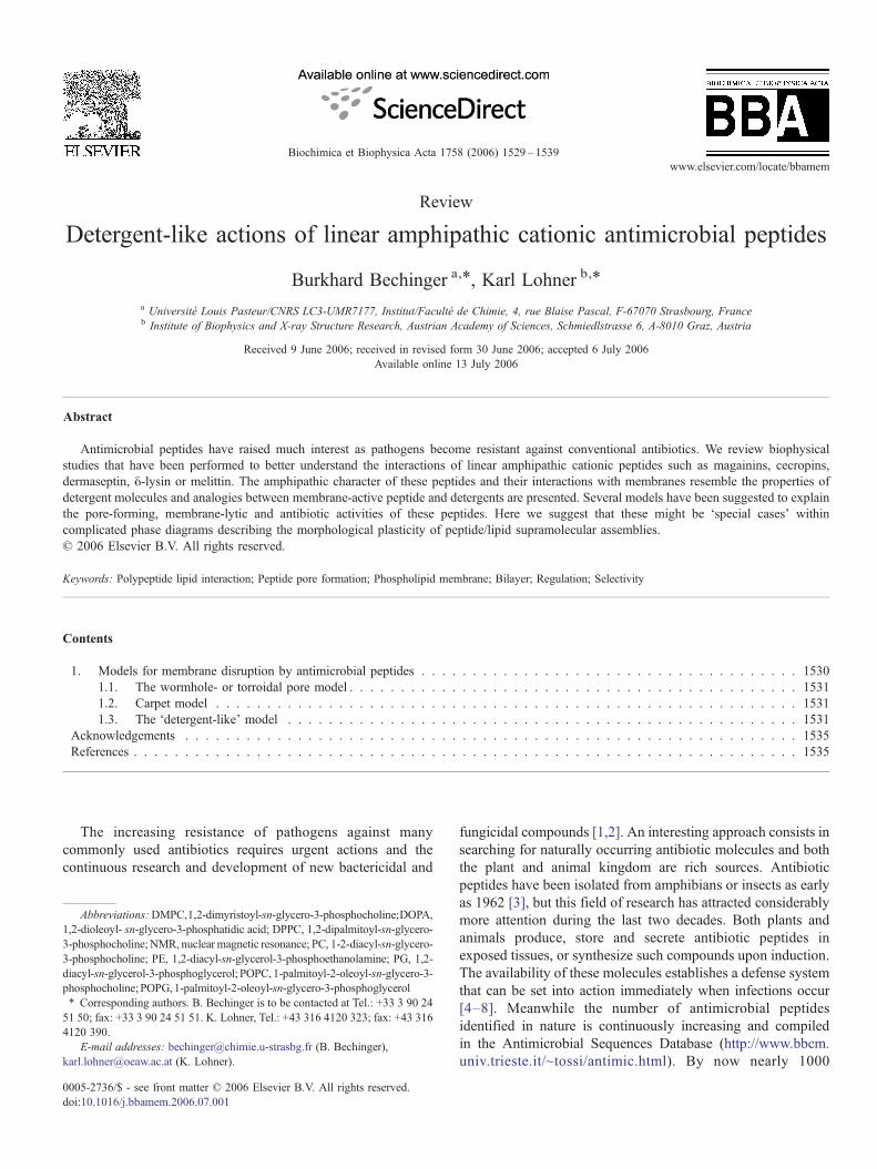
The amphipathic distribution of polar and hydrophobicresidues can result in pronounced interactions of the peptideswith phospholipid membranes. Therefore, a common character-istic observed for membrane-active peptides is their capabilityto disturb bilayer integrity, either by creation of defects,disruption or pore formation (Fig. 1). The resulting openingsin the lipid bilayer lead to the collapse of the transmembraneelectrochemical gradients and, therefore, provide an explanationof the cell killing activities of these peptides (reviewed e.g.
Fig. 1. Models describing the interactions of linear cationic amphipathicpeptides with membranes. (A) At low concentrations three effects of detergent-like molecules are shown. A peptide micelle ‘hits’ the membrane, therebyadsorbing a few lipid molecules concomitant with pore formation (top). Themicelle can also ‘insert’ in the membrane resulting in a ‘micellar aggregatechannel’ (right side). Furthermore, as in the presence of detergents in-planeoriented amphipathic peptides destabilize the membrane within a diameter ofseveral nanometers (circles). Lateral diffusion of the peptides along themembrane surface results in transient changes in local peptide density and thetemporary collapse of the membrane ohmic resistance when peptides approacheach other. (B) At higher concentrations the membrane disintegrates asdescribed by the carpet-, torroidal pore- or wormhole models.
[21,22]). The formation of pores affects cellular respiration [23],deprives sensitive organisms of their source of energy bydisrupting the electrochemical gradient across free-energytransducing membranes [24,25] and results in increased waterand ion flow across the membrane concomitant with cellswelling and osmolysis [26,27].
Interestingly, all-D-magainins, all-D-cecropins, cecropinswith inverted sequences (retro) or inversed D-cecropins (retro-enantio), all possess the high antibiotic and the pore-formingactivities of the parent L-enantiomer [28–30]. Moreover, boththe L- and D-enantiomer of protegrin-1 showed the same effecton membrane model systems composed of neutral or chargedphospholipids [31]. Furthermore β-amino acid oligomers [32]or helical pseudo-peptides show antibiotic activities in the sameconcentration range as observed for most active antimicrobialpeptides [33]. These observations indicate that the cell killingactivities of such amphipathic peptides are not mediated throughspecific, chiral receptor interactions, but are mostly due to directpeptide–lipid interactions. This is in agreement with initialobservations that permeabilization of microbial membranes is anearly and necessary step in killing microbes as demonstrated formagainin [24] and human defensin [34]. Further support comesfrom electron microscopy [35] and atomic force microscopy[36] studies on E. coli cells exposed to magainin or to PGLa,another member of the magainin family, which show that thelethal event is rupture of the inner membrane. Indeed systematicinvestigations demonstrate that the interplay between overallcharge, hydrophobicity and hydrophobic moment is importantfor selectivity and antibiotic activity of amphipathic helicalmodel peptides [37–41]. However, it should be noted that morerecent investigations give evidence that the antibiotic activitymay also develop due to interactions of these peptides withintracellular targets (for a review see [12,42]). Nevertheless, alsoin these cases the membrane has to be crossed to allowpenetration into the cell interior. Importantly many linearpeptides, such as those of the magainin family, adoptamphipathic structures only when in contact with membranes,membrane-mimicking environments [43,44] or when aggre-gated [45]. As a matter of fact this transition is important foractivity as it provides about 50% of the energy of membraneassociation [46,47]. It is therefore important to study theirstructures in their appropriate environments.
1. Models for membrane disruption by antimicrobialpeptides
Regarding membrane permeation and disruption two distinctmechanisms have particularly been presented and discussed.One model proposes that antimicrobial peptides act byperturbing the barrier function of membranes through trans-membrane pore formation [48], while the secondmodel suggestsmembrane disruption via the so-called “carpet”mechanism [49].As these modes of action of antimicrobial peptides will bediscussed in more detail elsewhere in this special issue we shallonly briefly describe these models (Fig. 1).
The formation of discrete multi-level openings and repro-ducible channel characteristics observed with predominantly

1531B. Bechinger, K. Lohner / Biochimica et Biophysica Acta 1758 (2006) 1529–1539
hydrophobic peptides such as the fungal peptide alamethicinhave been taken as an indication for the formation oftransmembrane helical bundles [50]. Similar models havebeen initially suggested also for charged amphipathic peptideantibiotics such as magainins, cecropins or pardaxin, however,it has soon been realized that these peptides exhibit differencesin functional and structural properties. Moreover, it was shownfor synthetic pardaxin P1a and Pa4 that membrane perturbationalso depends on the membrane composition [51,52] as has beenemphasized earlier for the action of antimicrobial peptides[53,54]. Linear cationic peptide antibiotics exhibit erraticcurrents in electrophysiological experiments, a strong tendencyof the membrane to rupture and an ion specificity that isstrongly dependent on the lipid composition [55–57]. Theformation of other forms of supramolecular structures in themembrane has, therefore, been suggested for these peptides.
1.1. The wormhole- or torroidal pore model
This model is an extension of the transmembrane helicalbundle in which the pores are lined with peptides and lipids(Fig. 1B, front). Such a pore structure was first proposed byMatsuzaki et al. [58] based on the observation that magainin 2induces rapid lipid flip-flop coupled with pore formation. Thepresence of negatively charged phospholipids in the pore-liningreduces the repulsive interactions due to the high positivecharge-density of the peptides [21] and could explain modula-tions in ion selectivity due to changes in the membrane lipidcomposition [56]. Indeed at high magainin concentrations(>3.3mol% corresponding to >10wt.%) neutron in-planescattering on mechanically aligned membrane systems indicatesthe presence of water filled cavities, which resemble ‘worm-holes’ [47,59]. The model thus postulates that peptides and lipidform together well-defined pores [60]. The arguments in favouror contradicting such a mechanism have been discussed inconsiderable detail previously [21,61].
1.2. Carpet model
Based on experimental results obtained with the antibioticpeptide dermaseptin the carpet model has been proposed[19,22,62]. This 34-residue antibiotic peptide is rich in lysinesand adopts an amphipathic α-helix (residues 1–27). It therebyresembles cecropins and magainins for which an in-planealignment had been demonstrated [63,64]. The peptide parti-tions into acidic and zwitterionic membranes [62]. In thepresence of negatively charged lipids and at high peptideconcentrations dermaseptin is located at the membrane surfaceand it has been suggested to self-associate in a ‘carpet-like’manner (Fig. 1B, back). The model further suggests that themembrane breaks into pieces when a threshold concentration ofpeptide is reached [65]. The presence of negatively chargedlipids helps in the formation in a dense peptide carpet, as theyhelp to reduce the repulsive electrostatic forces betweenpositively charged peptides.
Whereas the in-plane orientation of these peptides reflectsthe detergent-like properties of these amphipathic peptides, the
carpet model describes the lysis of the membranes at high localpeptide concentrations and reflects the morphological changesof the membrane after a threshold concentration at themembrane surface is reached (Fig. 2). Although high peptidedensities have been observed at the surface of bacterial cells, theconcentrations at the membrane itself remain a matter ofspeculation [66]. Furthermore, membrane-activities have beendemonstrated at reduced peptide-to-lipid ratios. Indeed com-paratively low peptide-to-lipid ratios are needed to dissipate theionic gradient across cell membranes or to develop antibioticactivity [25,37,67]. Notably, it has been speculated that smallopenings are sufficient for ion release concomitantly withinhibition of respiration [25] when at the same time bigger poresand higher peptide-to-lipid ratios are required for osmolysis ofred blood cells. In a similar manner single-channel recordings ofcecropin or magainin require small amounts of peptide withinthe membrane as otherwise lysis occurs [55,56,68]. Wetherefore prefer to describe the activities of these peptides bya more general model which is based on their detergent-likeproperties [69–71].
1.3. The ‘detergent-like’ model
Represents the most generally applicable explanation for themembrane-activities of amphiphiles. The model is based on theintercalation of these molecules into the bilayer (Fig. 1A).Thereby, the nature of the molecules such as charge andhydrophobic volume strongly contribute to the variety ofactions observed and the lipid polymorphism induced [72]. Theinteraction of detergents with lipid membranes in particularabove the critical micelle concentration (cmc), where themolecules exist in their aggregated, micellar state is rathercomplex. However, it has to be noted that antimicrobial peptidescan also exist as oligomers (Fig. 1A, top) and thus may exhibitdifferent behaviour as compared to the monomeric peptides (seebelow). Let us consider those general features of detergent–lipid interactions, which are more related to the action ofantimicrobial peptides. Whereas detergents at very lowdetergent-to-lipid ratios can have a neutral effect on modelmembranes, or can even result in their stabilization [73,74],openings might form temporarily at intermediate concentra-tions, and membrane disintegration becomes apparent at higherpeptide-to-lipid ratios (Figs. 1B and 2). Disintegration of thebilayer takes into account the loss of the membrane barrier,dissipation of the transmembrane electrochemical gradient, lossof cytoplasmic constituents and concomitantly interference withthe energy metabolism of living cells, all being observed alsowith antimicrobial peptides. However, it remains possible that,depending on the peptide and membrane composition, thepeptides merely form small and transient openings (Figs. 1Aand 2) which suffice for the diffusion of smaller molecules,including the peptides themselves in and out of the cell interior.The mechanism by which antimicrobial and cytolytic peptidesfinally damage the membrane will be determined by the peptideand lipid structure [42].
The step-wise changes in conductivity that have beenobserved for magainin 2 [55–57] or for cecropins [75] are
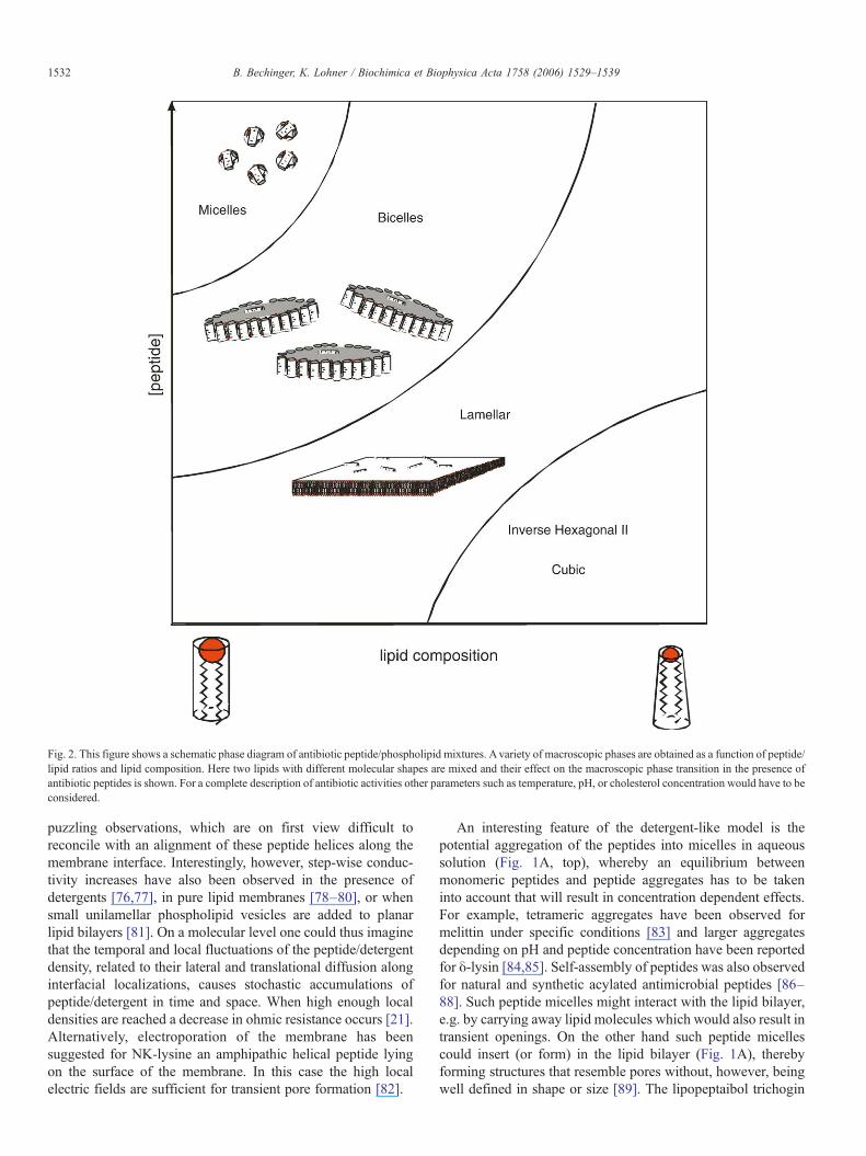
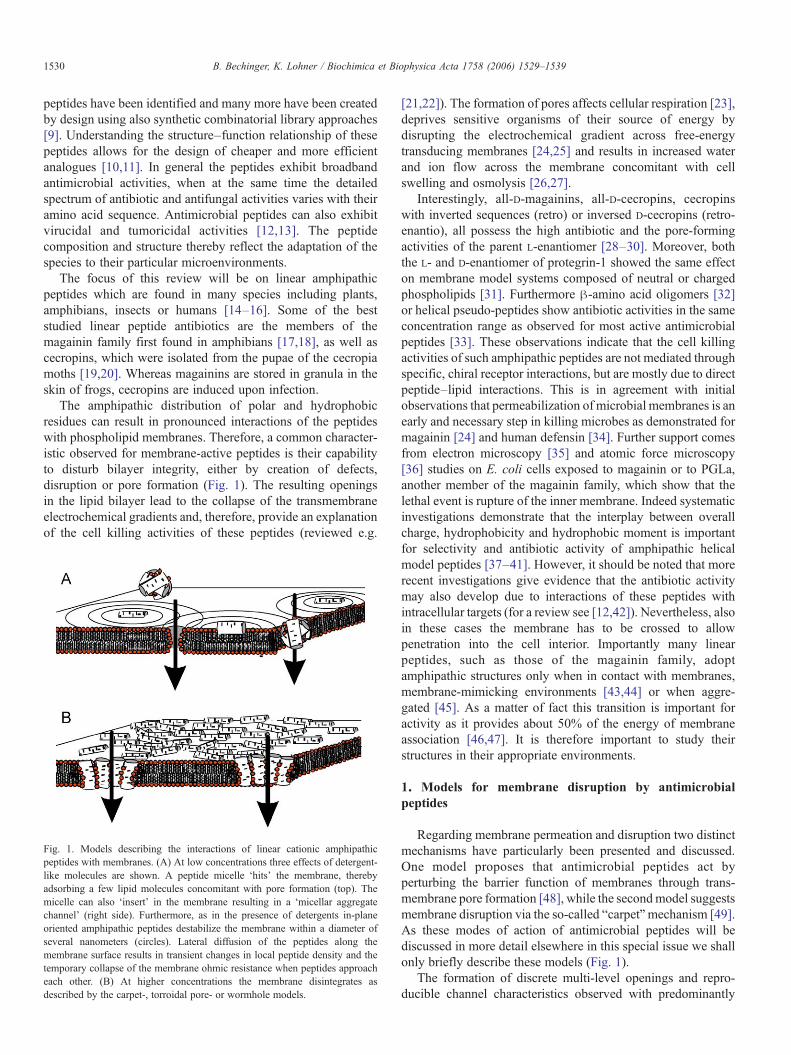
Fig. 2. This figure shows a schematic phase diagram of antibiotic peptide/phospholipid mixtures. Avariety of macroscopic phases are obtained as a function of peptide/lipid ratios and lipid composition. Here two lipids with different molecular shapes are mixed and their effect on the macroscopic phase transition in the presence ofantibiotic peptides is shown. For a complete description of antibiotic activities other parameters such as temperature, pH, or cholesterol concentration would have to beconsidered.
1532 B. Bechinger, K. Lohner / Biochimica et Biophysica Acta 1758 (2006) 1529–1539
puzzling observations, which are on first view difficult toreconcile with an alignment of these peptide helices along themembrane interface. Interestingly, however, step-wise conduc-tivity increases have also been observed in the presence ofdetergents [76,77], in pure lipid membranes [78–80], or whensmall unilamellar phospholipid vesicles are added to planarlipid bilayers [81]. On a molecular level one could thus imaginethat the temporal and local fluctuations of the peptide/detergentdensity, related to their lateral and translational diffusion alonginterfacial localizations, causes stochastic accumulations ofpeptide/detergent in time and space. When high enough localdensities are reached a decrease in ohmic resistance occurs [21].Alternatively, electroporation of the membrane has beensuggested for NK-lysine an amphipathic helical peptide lyingon the surface of the membrane. In this case the high localelectric fields are sufficient for transient pore formation [82].
An interesting feature of the detergent-like model is thepotential aggregation of the peptides into micelles in aqueoussolution (Fig. 1A, top), whereby an equilibrium betweenmonomeric peptides and peptide aggregates has to be takeninto account that will result in concentration dependent effects.For example, tetrameric aggregates have been observed formelittin under specific conditions [83] and larger aggregatesdepending on pH and peptide concentration have been reportedfor δ-lysin [84,85]. Self-assembly of peptides was also observedfor natural and synthetic acylated antimicrobial peptides [86–88]. Such peptide micelles might interact with the lipid bilayer,e.g. by carrying away lipid molecules which would also result intransient openings. On the other hand such peptide micellescould insert (or form) in the lipid bilayer (Fig. 1A), therebyforming structures that resemble pores without, however, beingwell defined in shape or size [89]. The lipopeptaibol trichogin
Fig. 3. The molecular shape of lipids and in-plane oriented amphipathic peptidesis illustrated. Whereas lipids such as POPC are considered to exhibit acylindrical overall molecular shape, the decreased size of the PE head groupresults in an inverted cone structure. On the other hand monoacyl-phospholipids,detergents or in-plane oriented amphipathic peptides predominantly fill thespace at the head group region/lipid interface thereby resembling a cone. Thismodel should be considered only as a first order conceptual approach to the realsituation. Notably, the ‘molecular shape’ of the amphipathic molecule reflectsthe geometrical as well as the interaction space of these molecules. Thereforecharge and hydrogen interactions between the lipids and the peptide have to beconsidered. Furthermore, in-plane oriented peptides can reside as monomers oras side-by-side oligomers in the membrane (e.g. [125]).
1533B. Bechinger, K. Lohner / Biochimica et Biophysica Acta 1758 (2006) 1529–1539
GA IV isolated from the soil fungus Trichoderma long-ibrachiatum [90] may be an example for such a mechanism.Leakage and light scattering experiments suggest that within theexplored concentration range membrane perturbation of thispeptide is due to pore formation of the peptide aggregates andnot to micellization [88]. Furthermore, the ability to self-associate in aqueous medium may be important for target cellselectivity [86,91]. The role of pre-assembly was tested usingmonomeric diastereomeric cationic peptides and their cova-lently linked pentameric bundles [92]. In contrast to the peptideaggregates that expressed similar potent antifungal and highantimicrobial activity as well as haemolytic activity indepen-dent on peptide length, the monomeric peptides showed lengthdependent antimicrobial activity and were devoid of haemolyticactivity.
A complete description of the peptide–membrane interac-tions and the resulting membrane morphologies would need totake into account a wide variety of parameters and conditions.These include the peptide-to-lipid ratio, the detailed membranecomposition, temperature, hydration and buffer composition.As with detergents this can be done by establishing phasediagrams (Fig. 2). Notably, the detergent-like properties ofamphipathic peptides are not in contradiction but include theabove mentioned wormhole and carpet models, in fact theselatter models should be considered ‘special cases’ where theconditions are such that these kind of supramolecular structuresare observed within a much more complex phase diagram. Ittherefore seems tedious to argue about the ‘correct model’ as allof them might occur depending on the detailed conditions.Notably, the extensive plasticity of peptide/detergent–lipidcomplexes opens up the possibility that a peptide induces acertain macromolecular structure when interacting with themembranes of one organism, but a different one wheninteracting with another species. On the other hand themutagenesis of the peptide sequence causes shifts in the phasediagram and therefore also affects the biological mechanisms.
In the following we have assembled a number of experi-mental results which illustrate the detergent-like properties ofamphipathic peptides. When the permeability changes ofmembranes are investigated the leakage of fluorescence dyestakes place at magainin concentrations of approximately 3 mol% which is equivalent to 81g/mole lipid. This latter value isvery similar to those observed for the permeability increases inthe presence of the detergents Triton-X100 and octyl glucoside[76,93].
Furthermore, the macroscopic phase properties as monitoredby proton-decoupled 31P solid-state NMR spectroscopy exhi-bits interesting parallels when amphipathic peptides, short chainlipids, lysolipids and detergents are compared to each other[70,71]. Pore formation due to the increased membrane tensionhas also been suggested for other amphipathic helical peptides[94] and cecropin B activity on bacterial cells was compared tothat of quartenary detergents [95].
When the molecular shape of amphipathic helices anddetergents are compared to each other it becomes evident that,when intercalated into a lipid bilayer, neither of them fills thevolume at the level of the fatty acyl chains and in the lipid head
group region equally well (Fig. 3). Whereas the peptide acts as aspacer at the level of the lipid headgroup it does not occupy thecorresponding volume at the level of the fatty acyl chainscreating a void in the hydrophobic region of the membranebilayer [21,42,70]. This parallels the behaviour described forcharged alkyl compounds, which are one of the simplest, amongthe variety of detergent molecules being also anchored at thelipid–water interface with their polar headgroup (for a reviewsee [72]). Systematic studies using PC model membranesrevealed that molecules with short alkyl chains (C6–12) arelocated in the more ordered plateau region of the acyl chainregion of the phospholipids inducing voids below their terminalmethyl groups. Since the formation of such free volume isenergetically unfavourable [96], the hydrocarbon chains musteliminate these voids by chain bends, increased trans-gaucheisomerization [97] or chain interdigitation as found for veryshort chain amphiphiles like alcohols (e.g. [98]) thus affectingthe phospholipids' acyl chain packing. Similar effects wereobserved also for non-ionic surfactants such as N-alkyl-N,N-dimethylamine-N-oxides [99] which were shown to exhibitantimicrobial activity [100,101]. Increasing the size of the polarheadgroup would lead to a molecular shape of the compoundthat can be described by a cone which would further promotethe formation of voids.
Amphipathic peptides thus exhibit effects similar to thoseobserved with cone- or wedge-shaped molecules. Intercalationof peptides into the membrane interface therefore creates voids
1534 B. Bechinger, K. Lohner / Biochimica et Biophysica Acta 1758 (2006) 1529–1539
in the hydrophobic membrane region and in addition imposescurvature strain on the lipid bilayer [102]. On the other hand thehead group of phosphatidylethanolamine occupies a smallerarea in the interfacial region when compared to the hydrophobicmembrane interior. Therefore, this lipid represents an invertedcone structure. Due to their shape, mixing cone and inverted-cone molecules release the strain and lipid bilayers that exhibit atendency to form hexagonal phases e.g. at increased tempera-tures are stabilized by the insertion of amphipathic peptides[37,103]. Stabilization of phosphatidylethanolamine bilayerswas also observed for a number of detergents such as Triton X-100, deoxycholate or octylglucoside which exhibit a coneshaped molecular geometry [73].
Considering the large variety of conditions it seemsimpossible to establish a complete phase diagram for peptide–lipid interactions. However, over the years a considerableamount of data has been collected for melittin. Its overallamphipathic character composed of a cluster of cationic aminoacids at the C-terminus and a stretch of hydrophobic aminoacids, resembles the features of many detergents beingcharacterized by a polar/charged headgroup and a hydrophobicmoiety. This peptide is too lytic to be used as an antibiotic but itcan nevertheless illustrate the complexity of such interactions.There is experimental evidence that melittin inserts into themembrane interface [104]. The possibility remains that a smallproportion also adopts a transmembrane alignment in thepresence of POPC but not POPG [105,106]. Melittin was foundto exhibit pronounced effects on the phase behaviour of DPPCalready at very low peptide concentrations (lipid-to-peptidemolar ratio of 1000/1) [107,108]. A similar concentrationdependent behaviour was reported for the detergent cetyltri-methylammonium chloride [74]. Since these effects cannot beaccounted for only by local perturbations around the sites ofinteraction, long range effects beyond the immediate neigh-bourhood of the incorporated peptide must be involved.Therefore, it was suggested that the peptide-affected domainscreate line defects in an ordered lipid lattice [104]. Such adefect-like action at low peptide concentrations may beexplained by shifting the percolation balance of coexistinggel- and fluid-like states [109]. However, at high melittinconcentrations (lipid-to-peptide molar ratio of 15/1), disk-shaped particles were found for the DMPC/melittin mixture[110,111], suggesting a detergent-like solubilization of themembrane under these experimental conditions. Below the gelto fluid phase transition of the pure lipid the presence ofintermediate amounts of melittin (<5mol%) results in thereversible disintegration of the bilayer into disks (cf. Fig. 2)[110,112,113]. It should be noted that some of the mixedpeptide/phospholipid phases exhibit meta-stable properties.Therefore in the presence of melittin or magainins thetransitions between one phase to another might take manyhours even months [70,114, A. Ramamoorthy, personalcommunication].
The idea of gradual membrane disintegration is alsosupported by data gained on δ-lysin/PC multilamellar vesicles[115,116]. This peptide from S. aureus is, like melittin, 26residues in size and surface-active. It lyses erythrocytes and
many other types of mammalian cells, as well as intracellularorganelles and bacterial protoplasts [117]. Similarly to theDPPC/melittin system, the addition of small quantities (lipid-to-peptide molar ratio ≥1000/1) of peptide had only minor butsignificant effects on the phase behaviour of DPPC or DMPCbilayers. Increasing the peptide concentration to molar lipid-to-peptide ratios lower than 125/1 promoted the formation of twopopulations of lipid particles, which could be separated bycentrifugation. Thereby, small-angle X-ray scattering measure-ments confirmed that the pellet consists of multilamellarvesicles, while disk-shaped lipid–peptide aggregates werefound in the supernatant, the predominant aggregate form athigh peptide concentration [116]. Interestingly, the structuralparameters of the DMPC/δ-lysin discoidal aggregates (diameterof about 14nm and a bilayer thickness of 5.2nm) correspondwell with the hydrodynamic radius of 6.9nm estimated for thedisk-shape particles of DMPC/melittin from gel-filtrationexperiments [110]. Modelling of the X-ray data suggested apeptide ring of about 1 nm thickness surrounding the discoidallipid bilayer (illustrated in Fig. 2). These data support the ideathat lytic peptides like detergents may have concentration-dependent effects on the membrane structure inducing bilayerperturbations of long-range order at low peptide concentrationsand exhibiting a detergent-like solubilization of the membraneat high peptide concentrations. Pore formation, which has alsobeen reported for δ-lysin [118], is not necessarily in contra-diction to this proposal, but may either reflect a transient state oroccur at distinct environmental conditions (Fig. 2). Poreformation might be particularly sensitive to the lipid classcomposing the membrane. Whereas melittin and δ-lysin disruptPC bilayers into small structures, the peptides exhibit bilayer-stabilizing effects when mixed with phosphatidylethanolaminemembranes, which in the absence of peptide show a tendencyfor the inverse hexagonal phase (HII) [119].
A different behaviour is observed when melittin is insertedinto charged phospholipids such as cardiolipin, DOPA or egg-PG. In these cases a preference for inverted macroscopic phasesis observed (HII or cubic) [120,121]. Fluorescence spectroscopyindicates that the macroscopic phase properties depend not onlyon the molecular shape of the lipid but also on the insertiondepth of the peptide [122]. Interestingly, the presence ofnegative surface charge densities stabilizes PC lipid bilayers inthe presence of melittin [106,111,123,124]. Thus the position-ing and topology of melittin, and concomitantly its effect onmembrane structure, are all strongly dependent on a widevariety of parameters that determine the local environment.
Much less data are available for the peptide–lipid interactionsinvolving antibiotic peptides. However, comparable correlationsbetween the molecular shape of the phospholipids andmacroscopic phase transitions have been observed for magaininor its analogue MSI-78 [70,71]. The peptides have been shownto be oriented parallel to the membrane surface using solid-stateNMR spectroscopy [64] andMSI-78 to form antiparallel dimers,when solubilized in dodecylphosphocholine (DPC) micelles[125]. The latter observation suggests that magainin activity maybe also modulated by oligomerization. Fluorescence energytransfer measurements indicate that the hydrophobic regions of
1535B. Bechinger, K. Lohner / Biochimica et Biophysica Acta 1758 (2006) 1529–1539
magainin 2 are located approximately 10 Å from the bilayercenter [126]. The peptide or its analogue MSI-78 thus acts like awedge, which augments the curvature of the membrane. Byinserting in-plane peptides into the headgroup region the bilayerpacking is disturbed at an estimated radius around the peptide ofapproximately 50Å resulting in a reduction in the averagebilayer thickness [127–130]. A current atomic force microscopystudy on DMPC bilayers and the magainin analogue MSI-78also revealed that the membrane thickness is not reduceduniformly over the entire bilayer area [130]. Deuterium or 13Csolid-state NMR measurements indicate a decrease in orderparameter at the lipid bilayer interior for both magainin andMSI-78 [131,132]. More recent investigations indicate that in mixedPC/PS lipid membranes cationic amphipathic peptides preferen-tially interact with the acidic phosphatidylserine [133,134]. Apreferential interaction with the negatively charged lipidcomponent in mixed model membranes was also observedearlier for human neutrophil peptide, HNP-2 [135], PGLa [53] ornisin [136] resulting in charge segregation. Such a behaviour canbe expected because of the cationic nature of the peptides, and istypical for a large number of antimicrobial peptides, such as e.g.the α-helical peptides belonging to the magainin-family[35,137] and buforin II [138], the β-sheet peptides tachyplesin[139] and protegrin-1 [54,140], the cyclic peptides gramicidin S[141] and rhesus theta defensin, RTD-1 [142] as well as nisin Z[143]. There is also evidence that other biologically activeamphipathic molecules, such as cardiotoxin [144] and syntheticpeptides [145] interact with model membranes inducing lateralseparation of phospholipids into co-existing domains.
Additional support for the detergent-like model arises fromthe observation that antibiotic or model peptides too short tospan the membrane exhibit channel-like activities as well [146–153]. In particular the model is in perfect agreement with theexperimentally observed in-plane orientation of amphipathicpeptides [45,64,126].
The membrane lipid composition can thus have pronouncedeffects on the membrane-disrupting activities of amphipathicpeptides. This is reflected by shifts in the borders within thepeptide–membrane phase diagram or by the occurrence ofdifferent macroscopic phases when one diagram is compared toanother. The lipid composition thereby modulates the sensitivityof the membrane to a given peptide [42,53,54,154]. Forexample, the alignment and the dynamics of the C-terminalhelical domain of pardaxin are a function of the phosphati-dylcholine fatty acyl chain composition [51,52]. Furthermore,cholesterol which is only present in eukaryotic cells changes theinteractions of cationic peptides with the membrane [68,155].Cholesterol affects the fluidity and the dipole potential ofphospholipid membranes. Furthermore, its capacity to formhydrogen bonds with the peptide has also been suggested toreduce antibiotic activity [156]. However, recent experimentson δ-lysin showed that in ternary lipid systems mimickingmammalian cell membranes the peptide binds preferentially tothe liquid-disordered domain suggesting that there is no specificinteraction between δ-lysin and cholesterol and sphingomyelin,respectively, emphasizing the importance of membrane proper-ties in lipid–peptide interactions [157]. As a result of peptide
accumulation peptide aggregates may form that becomeorientationally ordered, which in turn may have an impact onseveral processes such as peptide translocation across mem-branes [158]. Moreover, peptide enriched lipid domains willform especially in the presence of anionic lipids that differsignificantly in their local properties from the membrane bulkphase [42]. This may result in packing defects giving rise toincreased membrane permeability. In addition, exclusion ofcertain lipids, e.g. segregation of anionic lipids, from areas ofthe cell membrane due to their preferential interaction with thecationic peptides may affect membrane structure and integrity[133,134]. Thus, it is evident from the discussion above that themolecular mechanism of membrane permeation and disruptionobviously depends on a number of parameters such as the natureof the peptides and membrane lipids, peptide concentration andenvironmental conditions.
Acknowledgements
We are grateful to the members of our laboratories for theirimportant contributions to this subject as well as to many friendsand colleagues for ongoing discussions on this issue. We areindebted to Sabine Danner for her kind and competent helpduring the preparation of the manuscript.
References
[1] K. Lohner (Ed.), Development of Novel Antimicrobial Agents: EmergingStrategies, Horizon Scientific Press, Wymondham, Norfolk, UK, 2001.
[2] R. Novak, B. Henriques, E. Charpentier, S. Normark, E. Tuomanen,Emergence of vancomycin tolerance in Streptococcus pneumoniae,Nature 399 (1999) 590–593.
[3] G. Kiss, H. Michl, Über das Giftsekret der Gelbbauchunke Bombinavariegata L, Toxicon (Oxford) 1 (1962) 33–39.
[4] E.S. Kristyanne, K.S. Kim, J.M. Steward, Magainin 2 effects on theultrastructure of five plant pathogens, Mycologia 89 (1997) 353–360.
[5] L. Cavallarin, D. Andreu, B. San Segundo, Cecropin A-derived peptidesare potent inhibitors of fungal plant pathogens, Mol. Plant-Microb.Interact. 11 (1998) 218–227.
[6] M. Zasloff, Antimicrobial peptides in health and disease, N. Engl. J. Med.347 (2002) 1199–1200.
[7] A.R. Alan, E.D. Earle, Sensitivity of bacterial and fungal plant pathogensto the lytic peptides, MSI-99, magainin II, and cecropin B, Mol. Plant-Microb. Interact. 15 (2002) 701–708.
[8] H.G. Boman, Antibacterial peptides: basic facts and emerging concepts,J. Intern. Med. 254 (2003) 197–215.
[9] S.E. Blondelle, K. Lohner, Combinatorial libraries: a tool to designantimicrobial and antifungal peptide analogues having lytic specificitiesfor structure–activity relationship studies, Biopolymers 55 (2000) 74–87.
[10] B. Bechinger, Structure and functions of channel-forming polypeptides:magainins, cecropins, melittin and alamethicin, J. Membr. Biol. 156(1997) 197–211.
[11] P.M. Hwang, H.J. Vogel, Structure–function relationships of antimicro-bial peptides, Biochem. Cell. Biol. 76 (1998) 235–246.
[12] J.P. Powers, R.E. Hancock, The relationship between peptide structureand antibacterial activity, Peptides 24 (2003) 1681–1691.
[13] R.I. Lehrer, Primate defensins, Nat. Rev., Microbiol. 2 (2004) 727–738.[14] C.L. Bevins, M. Zasloff, Peptides from frog skin, Annu. Rev. Biochem.
59 (1990) 395–414.[15] H.G. Boman, Antibacterial peptides: key components needed in
immunity, Cell 65 (1991) 205–207.[16] F. Garcia-Olmedo, A. Molina, J.M. Alamillo, P. Rodriguez-Palenzuela,
Plant defense peptides, Biopolymers 47 (1999) 479–491.
1536 B. Bechinger, K. Lohner / Biochimica et Biophysica Acta 1758 (2006) 1529–1539
[17] A. Csordas, H. Michl, Isolierung und Strukturaufklärung eineshämolytisch wirkenden Polypeptides aus dem Abwehrsekret euro-päischer Unken, Monatsh. Chem. 101 (1970) 182–189.
[18] W. Hoffmann, K. Richter, G. Kreil, A novel peptide designated PYLa andits precursor as predicted from cloned mRNA of Xenopus laevis skin,EMBO J. 2 (1983) 711–714.
[19] H. Steiner, D. Hultmark, A. Engstrom, H. Bennich, H.G. Boman,Sequence and specificity of two antibacterial proteins involved in insectimmunity, Nature 292 (1981) 246–248.
[20] D. Hultmark, A. Engstrom, H. Bennich, R. Kapur, H.G. Boman, Insectimmunity: isolation and structure of cecropin D and four minorantibacterial components from Cecropia pupae, Eur. J. Biochem. 127(1982) 207–217.
[21] B. Bechinger, The structure, dynamics and orientation of antimicrobialpeptides in membranes by multidimensional solid-state NMR spectro-scopy, Biochim. Biophys. Acta 1462 (1999) 157–183.
[22] Y. Shai, Mechanism of the binding, insertion and destabilization ofphospholipid bilayer membranes by alpha-helical antimicrobial and cellnon-selective membrane-lytic peptides, Biochim. Biophys. Acta 1462(1999) 55–70.
[23] M. Hugosson, D. Andreu, H.G. Boman, E. Glaser, Antibacterialpeptides and mitochondrial presequences affect mitochondrial coupling,respiration and protein import, Eur. J. Biochem. 223 (1994)1027–1033.
[24] H.V. Westerhoff, D. Juretic, R.W. Hendler, M. Zasloff, Magainins and thedisruption of membrane-linked free-energy transduction, Proc. Natl.Acad. Sci. U. S. A. 86 (1989) 6597–6601.
[25] L. Silvestro, K. Gupta, J.N. Weiser, P.H. Axelsen, The concentration-dependent membrane activity of cecropin A, Biochemistry 36 (1997)11452–11460.
[26] W.F. DeGrado, G.F. Musso, M. Lieber, E.T. Kaiser, F.J. Kezdy, Kineticsand mechanism of hemolysis induced by melittin and by a syntheticmelittin analogue, Biophys. J. 37 (1982) 329–338.
[27] M.T. Tosteson, S.J. Holmes, M. Razin, D.C. Tosteson, Melittin lysis ofred cells, J. Membr. Biol. 87 (1985) 35–44.
[28] S. Vunnam, P. Juvvadi, K.S. Rotondi, R.B. Merrifield, Synthesis andstudy of normal, enantio, retro, and retroenantio isomers of cecropinA-melittin hybrids, their end group effects and selective enzymeinactivation, J. Pept. Res. 51 (1998) 38–44.
[29] R.B. Merrifield, E.L. Merrifield, P. Juvvadi, D. Andreu, H.G. Boman,Design and synthesis of antimicrobial peptides, Ciba Found. Symp. 186(1994) 5–20.
[30] D. Wade, A. Boman, B. Wahlin, C.M. Drain, D. Andreu, H.G. Boman,R.B. Merrifield, All-D amino acid-containing channel-forming antibioticpeptides, Proc. Natl. Acad. Sci. U. S. A. 87 (1990) 4761–4765.
[31] A. Latal, R.I. Lehrer, S. Harwig, K. Lohner, Interaction of enantiomericprotegrins with liposomes, Prog. Biophys. Mol. Biol. 65 (1996) 121.
[32] R.P. Cheng, S.H. Gellman, W.F. DeGrado, beta-Peptides: from structureto function, Chem. Rev. 101 (2001) 3219–3232.
[33] J.E. Oh, S.Y. Hong, K.H. Lee, Design, synthesis and characterization ofantimicrobial pseudopeptides corresponding to membrane-active peptide,J. Pept. Res. 54 (1999) 129–136.
[34] R.I. Lehrer, A. Barton, K.A. Daher, S.S. Harwig, T. Ganz, M.E. Selsted,Interaction of human defensins with Escherichia coli. Mechanism ofbactericidal activity, J. Clin. Invest. 84 (1989) 553–561.
[35] K. Matsuzaki, K. Sugishita, M. Harada, N. Fujii, K. Miyajima,Interactions of an antimicrobial peptide, magainin 2, with outer andinner membranes of Gram-negative bacteria, Biochim. Biophys. Acta1327 (1997) 119–130.
[36] A. da Silva Jr., O. Teschke, Effects of the antimicrobial peptide PGLa onlive Escherichia coli, Biochim. Biophys. Acta 1643 (2003) 95–103.
[37] T. Wieprecht, M. Dathe, R.M. Epand, M. Beyermann, E. Krause, W.L.Maloy, D.L. MacDonald, M. Bienert, Influence of the angle subtended bythe positively charged helix face on themembrane activity of amphipathic,antibacterial peptides, Biochemistry 36 (1997) 12869–12880.
[38] M. Dathe, M. Schumann, T. Wieprecht, A. Winkler, M. Beyermann,E. Krause, K. Matsuzaki, O. Murase, M. Bienert, Peptide helicity andmembrane surface charge modulate the balance of electrostatic and
hydrophobic interactions with lipid bilayers and biological membranes,Biochemistry 35 (1996) 12612–12622.
[39] M. Sharon, Z. Oren, Y. Shai, J. Anglister, 2D-NMR and ATR-FTIR studyof the structure of a cell-selective diastereomer of melittin and itsorientation in phospholipids, Biochemistry 38 (1999) 15305–15316.
[40] M. Dathe, H. Nikolenko, J. Meyer, M. Beyermann, M. Bienert,Optimization of the antimicrobial activity of magainin peptides bymodification of charge, FEBS Lett. 501 (2001) 146–150.
[41] T. Tachi, R.F. Epand, R.M. Epand, K. Matsuzaki, Position-dependenthydrophobicity of the antimicrobial magainin peptide affects the mode ofpeptide–lipid interactions and selective toxicity, Biochemistry 41 (2002)10723–10731.
[42] K. Lohner, S.E. Blondelle, Molecular mechanisms of membraneperturbation by antimicrobial peptides and the use of biophysical studiesin the design of novel peptide antibiotics, Comb. Chem. High ThroughoutScreen. 8 (2005) 241–256.
[43] D. Marion, M. Zasloff, A. Bax, A two-dimensional NMR study of theantimicrobial peptide magainin 2, FEBS Lett. 227 (1988) 21–26.
[44] H.C. Chen, J.H. Brown, J.L. Morell, C.M. Huang, Synthetic magaininanalogues with improved antimicrobial activity, FEBS Lett. 236 (1988)462–466.
[45] T.C. Vogt, B. Bechinger, The interactions of histidine-containingamphipathic helical peptide antibiotics with lipid bilayers. The effectsof charges and pH, J. Biol. Chem. 274 (1999) 29115–29121.
[46] T. Wieprecht, M. Beyermann, J. Seelig, Binding of antibacterial magaininpeptides to electrically neutral membranes: thermodynamics andstructure, Biochemistry 38 (1999) 10377–10387.
[47] H.W. Huang, Action of antimicrobial peptides: two-state model,Biochemistry 39 (2000) 8347–8352.
[48] K. Matsuzaki, Molecular mechanisms of membrane perturbation byantimicrobial peptides, in: K. Lohner (Ed.), Development of NovelAntimicrobial Agents: Emerging Strategies, Horizon Scientific Press,Wymondham, Norfolk, UK, 2001, pp. 167–181.
[49] Z. Oren Y. Shai, Molecular mechanism of cell selectivity by linearamphipathic-helical and diasteriomeric antimicrobial peptides, in:K. Lohner (Ed.), Development of Novel Antimicrobial Agents:Emerging Strategies, Horizon Scientific Press, Wymondham, Norfolk,UK, 2001, pp. 183–204.
[50] M.S. Sansom, Alamethicin and related peptaibols–model ion channels,Eur. Biophys. J. 22 (1993) 105–124.
[51] K.J. Hallock, D.K. Lee, J. Omnaas, H.I. Mosberg, A. Ramamoorthy,Membrane composition determines pardaxin's mechanism of lipidbilayer disruption, Biophys. J. 83 (2002) 1004–1013.
[52] F. Porcelli, B. Buck, D.K. Lee, K.J. Hallock, A. Ramamoorthy, G. Veglia,Structure and orientation of pardaxin determined by NMR experiments inmodel membranes, J. Biol. Chem. 279 (2004) 45815–45823.
[53] K. Lohner, E.J. Prenner, Differential scanning calorimetry and X-raydiffraction studies of the specificity of the interaction of antimicrobialpeptides with membrane-mimetic systems, Biochim. Biophys. Acta 1462(1999) 141–156.
[54] K. Lohner, The role of membrane lipid composition in cell targeting ofantimicrobial peptides, in: K. Lohner (Ed.), Development of NovelAntimicrobial Agents: Emerging Strategies, Horizon Scientific Press,Wymondham, Norfolk, UK, 2001, pp. 149–165.
[55] H. Duclohier, G. Molle, G. Spach, Antimicrobial peptide magainin I fromXenopus skin forms anion-permeable channels in planar lipid bilayers,Biophys. J. 56 (1989) 1017–1021.
[56] R.A. Cruciani, J.L. Barker, S.R. Durell, G. Raghunathan, H.R. Guy,M. Zasloff, E.F. Stanley, Magainin 2, a natural antibiotic from frog skin,forms ion channels in lipid bilayer membranes, Eur. J. Pharmacol. 226(1992) 287–296.
[57] B. Haimovich, J.C. Tanaka, Magainin-induced cytotoxicity in eukaryoticcells: kinetics, dose-response and channel characteristics, Biochim.Biophys. Acta 1240 (1995) 149–158.
[58] K. Matsuzaki, O. Murase, N. Fujii, K. Miyajima, An antimicrobalpeptide, magainin 2, induced rapid flip-flop of phospholipids coupledwith pore formation and peptide translocation, Biochemistry 35 (1996)11361–11368.

1537B. Bechinger, K. Lohner / Biochimica et Biophysica Acta 1758 (2006) 1529–1539
[59] S.J. Ludtke, K. He, W.T. Heller, T.A. Harroun, L. Yang, H.W. Huang,Membrane pores induced by magainin, Biochemistry 35 (1996)13723–13728.
[60] L. Yang, T.A. Harroun, T.M. Weiss, L. Ding, H.W. Huang, Barrel-stavemodel or toroidal model? A case study on melittin pores, Biophys. J. 81(2001) 1475–1485.
[61] B. Bechinger, Structure and function of membrane-lytic peptides, Crit.Rev. Plant Sci. 23 (2004) 271–292.
[62] Y. Pouny, D. Rapaport, A. Mor, P. Nicolas, Y. Shai, Interaction ofantimicrobial dermaseptin and its fluorescently labeled analogues withphospholipid membranes, Biochemistry 31 (1992) 12416–12423.
[63] B. Bechinger, K. Shon, H. Eck, M. Zasloff, S.J. Opella, NMR studies ofmagainin peptide antibiotics in membranes, Hoppe-Seyler Z. Physiol.Chem. 371 (1990) 758.
[64] B. Bechinger, M. Zasloff, S.J. Opella, Structure and orientation of theantibiotic peptide magainin in membranes by solid-state nuclear magneticresonance spectroscopy, Protein Sci. 2 (1993) 2077–2084.
[65] Z. Oren, Y. Shai, Mode of action of linear amphipathic alpha-helicalantimicrobial peptides, Biopolymers 47 (1998) 451–463.
[66] H. Steiner, D. Andreu, R.B. Merrifield, Binding and action of cecropinand cecropin analogues: antibacterial peptides from insects, Biochim.Biophys. Acta 939 (1988) 260–266.
[67] D. Juretic, R.W. Hendler, F. Kamp, W.S. Caughey, M. Zasloff, H.V.Westerhoff, Magainin oligomers reversibly dissipate delta microH+ incytochrome oxidase liposomes, Biochemistry 33 (1994) 4562–4570.
[68] B. Christensen, J. Fink, R.B. Merrifield, D. Mauzerall, Channel-formingproperties of cecropins and related model compounds incorporated intoplanar lipid membranes, Proc. Natl. Acad. Sci. U. S. A. 85 (1988)5072–5076.
[69] B. Bechinger, The structure, dynamics and orientation of antimicrobialpeptides by multidimensional solid-state NMR spectroscopy, Biochim.Biophys. Acta 1462 (1999) 157–183.
[70] B. Bechinger, Detergent-like properties of magainin antibiotic peptides: a31P solid-state NMR spectroscopy study, Biochim. Biophys. Acta 1712(2005) 101–108.
[71] K.J. Hallock, D.K. Lee, A. Ramamoorthy, MSI-78, an analogue of themagainin antimicrobial peptides, disrupts lipid bilayer structure viapositive curvature strain, Biophys. J. 84 (2003) 3052–3060.
[72] K. Lohner, Effects of small organic molecules on phospholipid phasetransitions, Chem. Phys. Lipids 57 (1991) 341–362.
[73] T.D. Madden, P.R. Cullis, Stabilization of bilayer structure forunsaturated phosphatidylethanolamines by detergents, Biochim. Bio-phys. Acta 684 (1982) 149–153.
[74] K. Müller, G. LIPKA, K. Lohner, P. Laggner, The effect of the detergentcetyltrimethaylammoniumchloride on the phase behavior of Dipalmi-toylphosphatidylcholine. A high precision calorimetric studies, Thermo-chim. Acta 94 (1985) 187–197.
[75] A.M. Christensen, J. Schaefer, Solid-state NMR determination of intra-and intermolecular 31P-13C distances for shikimate 3-phosphate and[1–13C]glyphosateboundtoenolpyruvylshikimate-3-phosphatesynthase,Biochemistry 32 (1993) 2868–2873.
[76] P. Schlieper, E. De Robertis, Triton X-100 as a channel-forming substancein artificial lipid bilayer membranes, Arch. Biochem. Biophys. 184(1977) 204–208.
[77] G.M. Alder, W.M. Arnold, C.L. Bashford, A.F. Drake, C.A. Pasternak,U. Zimmermann, Divalent cation-sensitive pores formed by natural andsynthetic melittin and by Triton X-100, Biochim. Biophys. Acta 1061(1991) 111–120.
[78] V.F. Antonov, V.V. Petrov, A.A. Molnar, D.A. Predvoditelev, A.S. Ivanov,The appearance of single-ion channels in unmodified lipid bilayermembranes at the phase transition temperature, Nature 283 (1980)585–586.
[79] K. Kaufmann, I. Silman, The induction by protons of ion channelsthrough lipid bilayer membranes, Biophys. Chem. 18 (1983) 89–99.
[80] K. Yoshikawa, T. Fujimoto, T. Shimooka, H. Terada, N. Kumazawa,T. Ishii, Electrical oscillation and fluctuation in phospholipid membranes.Phospholipids can form a channel without protein, Biophys. Chem. 29(1988) 293–299.
[81] D.J. Woodbury, Pure lipid vesicles can induce channel-like conductancesin planar bilayers, J. Membr. Biol. 109 (1989) 145–150.
[82] M. Miteva, M. Andersson, A. Karshikoff, G. Otting, Molecularelectroporation: a unifying concept for the description of membranepore formation by antibacterial peptides, exemplified with NK-lysin,FEBS Lett. 462 (1999) 155–158.
[83] T.C. Terwilliger, D. Eisenberg, The structure of melittin: I. Structuredetermination and partial refinement, J. Biol. Chem. 257 (1982)6010–6015.
[84] J.E. Fitton, Physicochemical studies on delta Haemolysin, a Staphylo-coccal cytolytic polypeptide, FEBS Lett. 130 (1981) 257–260.
[85] E. Thiaudiere, O. Siffert, J.-C. Talbot, J. Bolard, J.E. Alouf, J. Dufourcq,The amphiphilic a-helix concept. Consequences on the structure ofstaphylococcal d-toxin in solution and bound to lipids, Eur. J. Biochem.195 (1991) 203–213.
[86] Z. Oren, Y. Shai, Cyclization of a cytolytic amphipathic alpha-helicalpeptide and its diastereomer: effect on structure, interaction with modelmembranes, and biological function, Biochemistry 39 (2000) 6103–6114.
[87] I. Kustanovich, D.E. Shalev, M. Mikhlin, L. Gaidukov, A. Mor, Structuralrequirements for potent versus selective cytotoxicity for antimicrobialdermaseptin S4 derivatives, J. Biol. Chem. 277 (2002) 16941–16951.
[88] L. Stella, C. Mazzuca, M. Venanzi, A. Palleschi, M. Didone, F.Formaggio, C. Toniolo, B. Pispisa, Aggregation and water–membranepartition as major determinants of the activity of the antibiotic peptidetrichogin GA IV, Biophys. J. 86 (2004) 936–945.
[89] R.E. Hancock, D.S. Chapple, Peptide antibiotics, Antimicrob. AgentsChemother. 43 (1999) 1317–1323.
[90] C. Auvin-Guette, S. Rebufatt, Y. Prigent, B. Bodo, Trichogin A IV, an11-residue lipopeptaibol from Trichoderma longibrachiatum, J. Am.Chem. Soc. 114 (1992) 2170–2174.
[91] I.S. Radzishevsky, S. Rotem, F. Zaknoon, L. Gaidukov, A. Dagan,A. Mor, Effects of acyl versus aminoacyl conjugation on the properties ofantimicrobial peptides, Antimicrob. Agents Chemother. 49 (2005)2412–2420.
[92] N. Sal-Man, Z. Oren, Y. Shai, Preassembly of membrane-active peptidesis an important factor in their selectivity toward target cells, Biochemistry41 (2002) 11921–11930.
[93] E. Grant Jr., T.J. Beeler, K.M. Taylor, K. Gable, M.A. Roseman,Mechanism of magainin 2a induced permeabilization of phospholipidvesicles, Biochemistry 31 (1992) 9912–9918.
[94] I.V. Polozov, G.M. Anantharamaiah, J.P. Segrest, R.M. Epand,Osmotically induced membrane tension modulates membrane permea-bilization by class L amphipathic helical peptides: nucleation model ofdefect formation, Biophys. J. 81 (2001) 949–959.
[95] M. Vaara, T. Vaara, Ability of cecropin B to penetrate the enterobacterialouter membrane, Antimicrob. Agents Chemother. 38 (1994) 2498–2501.
[96] J.N. Israelachvili, S. Marcelja, R.G. Horn, Physical principles ofmembrane organization, Q. Rev. Biophys. 13 (1980) 121–200.
[97] J. Gallova, F. Devinsky, P. Balgavy, Interaction of surfactants with modelandbiologicalmembranes: II.Effect ofN-alkyl-N,N,N-trimethylammoniumions on phosphatidylcholine bilayers as studied by spin probe ESR, Chem.Phys. Lipids 53 (1990) 231–241.
[98] S.A. Simon, T.J. McIntosh, Interdigitated hydrocarbon chain packingcauses the biphasic transition behavior in lipid/alcohol suspensions,Biochim. Biophys. Acta 773 (1984) 169–172.
[99] J. Karlovska, K. Lohner, G. Degovics, I. Lacko, F. Devinsky, P. Balgavy,Effects of non-ionic surfactants N-alkyl-N,N-dimethylamine-N-oxides onthe structure of a phospholipid bilayer: small-angle X-ray diffractionstudy, Chem. Phys. Lipids 129 (2004) 31–41.
[100] P. Balgavy, F. Devinsky, Cut-off effects in biological activities ofsurfactants, Adv. Colloid Interface Sci. 66 (1996) 23–63.
[101] R.E. Glover, R.R. Smith, M.V. Jones, S.K. Jackson, C.C. Rowlands, AnEPR investigation of surfactant action on bacterial membranes, FEMSMicrobiol. Lett. 177 (1999) 57–62.
[102] P.R. Cullis, B. de Kruijff, Lipid polymorphism and the functional roles oflipids in biological membranes, Biochim. Biophys. Acta 559 (1979)399–420.
[103] K. Matsuzaki, K. Sugishita, N. Ishibe, M. Ueha, S. Nakata, K. Miyajima,
1538 B. Bechinger, K. Lohner / Biochimica et Biophysica Acta 1758 (2006) 1529–1539
R.M. Epand, Relationship of membrane curvature to the formation ofpores by magainin 2, Biochemistry 37 (1998) 11856–11863.
[104] A. Colotto, D.P. Kharakoz, K. Lohner, P. Laggner, Ultrasonic study ofmelittin effects on phospholipid model membranes, Biophys. J. 65 (1993)2360–2367.
[105] R. Smith, F. Separovic, T.J. Milne, A. Whittaker, F.M. Bennett, B.A.Cornell, A. Makriyannis, Structure and orientation of the pore-formingpeptide, melittin, in lipid bilayers, J. Mol. Biol. 241 (1994) 456–466.
[106] A.S. Ladokhin, S.H. White, ‘Detergent-like’ permeabilization ofanionic lipid vesicles by melittin, Biochim. Biophys. Acta 1514(2001) 253–260.
[107] M. Posch, U. Rakusch, C. Mollay, P. Laggner, Cooperative effects in theinteraction between melittin and phosphatidylcholine model membranes.Studies by temperature scanning densitometry, J. Biol. Chem. 258 (1983)1761–1766.
[108] A. Collotto, K. Lohner, P. Laggner, Low-dose effects of melittin onphospholipid structure. Differences between diester and diether phos-pholipids, Prog. Colloid Polym. Sci. 89 (1992) 334.
[109] P.F. Almeida, W.L. Vaz, T.E. Thompson, Percolation and diffusion inthree-component lipid bilayers: effect of cholesterol on an equimolarmixture of two phosphatidylcholines, Biophys. J. 64 (1993) 399–412.
[110] J. Dufourcq, J.F. Faucon, G. Fourche, J.L. Dasseux, M. le Maire,T. Gulik-Krzywicki, Morphological changes of phosphatidylcholinebilayers induced by melittin: vesicularization, fusion, discoidal particles,Biochim. Biophys. Acta 859 (1986) 33–48.
[111] M. Monette, M. Lafleur, Modulation of melittin-induced lysis by surfacecharge density of membranes, Biophys. J. 68 (1995) 187–195.
[112] E.J. Dufourc, I.C. Smith, J. Dufourcq, Molecular details of melittin-induced lysis of phospholipid membranes as revealed by deuterium andphosphorus NMR, Biochemistry 25 (1986) 6448–6455.
[113] C.E. Dempsey, A. Watts, A deuterium and phosphorus-31 nuclearmagnetic resonance study of the interaction of melittin with dimyr-istoylphosphatidylcholine bilayers and the effects of contaminatingphospholipase A2, Biochemistry 26 (1987) 5803–5811.
[114] J.F. Faucon, J.M. Bonmatin, J. Dufourcq, E.J. Dufourc, Acyl chain lengthdependence in the stability of melittin-phosphatidylcholine complexes. Alight scattering and 31P-NMR study, Biochim. Biophys. Acta 1234 (1995)235–243.
[115] K. Lohner, P. Laggner, J.H. Freer, Dilatometric and calorimetric studies ofthe effect of Staphylococcus aureus delta-lysin on the phospholipid phasetransition, J. Solution Chem. 15 (1986) 189–198.
[116] K. Lohner, E. Staudegger, E.J. Prenner, R.N. Lewis, M. Kriechbaum,G. Degovics, R.N. McElhaney, Effect of staphylococcal delta-lysin on thethermotropic phase behavior and vesicle morphology of dimyristoylpho-sphatidylcholine lipid bilayer model membranes. Differential scanningcalorimetric, 31P nuclear magnetic resonance and Fourier transforminfrared spectroscopic, and X-ray diffraction studies, Biochemistry 38(1999) 16514–16528.
[117] M. Thelestam, in: C.S.F. Easman, C.A. Easman (Eds.), Staphylococciand Staphylococcal Diseases, Academic Press, London, 1983, pp.705–744.
[118] I.R. Mellor, D.H. Thomas, M.S. Sansom, Properties of ion channelsformed by Staphylococcus aureus delta-toxin, Biochim. Biophys. Acta942 (1988) 280–294.
[119] A.M. Batenburg, J.H. van Esch, B. de Kruijff, Melittin-induced changesof the macroscopic structure of phosphatidylethanolamines, Biochemistry27 (1988) 2324–2331.
[120] A.M. Batenburg, J.C. Hibbeln, A.J. Verkleij, B. de Kruijff, Melittininduces HII phase formation in cardiolipin model membranes, Biochim.Biophys. Acta 903 (1987) 142–154.
[121] A.M. Batenburg, J.H. van Esch, J. Leunissen-Bijvelt, A.J. Verkleij, B.de Kruijff, Interaction of melittin with negatively charged phospholi-pids: consequences for lipid organization, FEBS Lett. 223 (1987)148–154.
[122] A.M. Batenburg, J.C. Hibbeln, B. de Kruijff, Lipid specific penetration ofmelittin into phospholipid model membranes, Biochim. Biophys. Acta903 (1987) 155–165.
[123] C. Dempsey, M. Bitbol, A. Watts, Interaction of melittin with mixed
phospholipid membranes composed of dimyristoylphosphatidylcholineand dimyristoylphosphatidylserine studied by deuterium NMR, Bio-chemistry 28 (1989) 6590–6596.
[124] S. Ohki, E. Marcus, D.K. Sukumaran, K. Arnold, Interaction of melittinwith lipid membranes, Biochim. Biophys. Acta 1194 (1994) 223–232.
[125] F. Porcelli, B.A. Buck-Koehntop, S. Thennarasu, A. Ramamoorthy,G. Veglia, Structures of the dimeric and monomeric variants of magaininantimicrobial peptides (MSI-78 and MSI-594) in micelles and bilayers,determined by NMR spectroscopy, Biochemistry 45 (2006) 5793–5799.
[126] K. Matsuzaki, O. Murase, H. Tokuda, S. Funakoshi, N. Fujii, K.Miyajima, Orientational and aggregational states of magainin 2 inphospholipid bilayers, Biochemistry 33 (1994) 3342–3349.
[127] S. Ludtke, K. He, H. Huang, Membrane thinning caused by magainin 2,Biochemistry 34 (1995) 16764–16769.
[128] C. Münster, Y. Lu, S. Schinzel, B. Bechinger, T. Salditt, Grazingincidence X-ray scattering of highly aligned phospholipid membranescontaining antimicrobial peptides magainin 2, Eur. Biophys. J. 28 (1999)683–688.
[129] F.Y. Chen, M.T. Lee, H.W. Huang, Evidence for membrane thinningeffect as the mechanism for peptide-induced pore formation, Biophys. J.84 (2003) 3751–3758.
[130] A. Mecke, D.K. Lee, A. Ramamoorthy, B.G. Orr, M.M. Banaszak Holl,Membrane thinning due to antimicrobial peptide binding: an atomic forcemicroscopy study of MSI-78 in lipid bilayers, Biophys. J. 89 (2005)4043–4050.
[131] B. Bechinger, M. Zasloff, S.J. Opella, Structure and interactions ofmagainin antibiotic peptides in lipid bilayers: a solid-state nuclearmagnetic resonance investigation, Biophys. J. 62 (1992) 12–14.
[132] S. Dvinskikh, U. Durr, K. Yamamoto, A. Ramamoorthy, A high-resolution solid-state NMR approach for the structural studies of bicelles,J. Am. Chem. Soc. 128 (2006) 6326–6327.
[133] A.J. Mason, A. Matinez, C. Glaubitz, O. Danos, A. Kichler, B. Bechinger,The antibiotic and DNA-transfecting peptide LAH4 selectively associateswith, and disorders, anionic lipids in mixed membranes, FASEB J. 20(2006) 320–322.
[134] A.J. Mason, I.N. Husnal-Chotimah, P. Bertani, B. Bechinger, Aspectroscopic study of the membrane interaction of the antimicrobialpeptide pleurocidin, Mol. Membr. Biol. 23 (2006) 185–194.
[135] K. Lohner, A. Latal, R.I. Lehrer, T. Ganz, Differential scanningmicrocalorimetry indicates that human defensin, HNP-2, interactsspecifically with biomembrane mimetic systems, Biochemistry 36(1997) 1525–1531.
[136] B.B. Bonev, W.C. Chan, B.W. Bycroft, G.C. Roberts, A. Watts,Interaction of the lantibiotic nisin with mixed lipid bilayers: a 31P and2H NMR study, Biochemistry 39 (2000) 11425–11433.
[137] A. Latal, G. Degovics, R.F. Epand, R.M. Epand, K. Lohner, Structuralaspects of the interaction of peptidyl–glycylleucine–carboxyamide, ahighly potent antimicrobial peptide from frog skin, with lipids, Eur. J.Biochem. 248 (1997) 938–946.
[138] S. Kobayashi, K. Takeshima, C.B. Park, S.C. Kim, K. Matsuzaki,Interactions of the novel antimicrobial peptide buforin 2 with lipidbilayers: proline as a translocation promoting factor, Biochemistry 39(2000) 8648–8654.
[139] T. Nakamura, H. Furunaka, T. Miyata, F. Tokunaga, T. Muta, S. Iwanaga,M. Niwa, T. Takao, Y. Shimonishi, Tachyplesin, a class of antimicrobialpeptide from the hemocytes of the horseshoe crab (Tachypleustridentatus), isolation and chemical structure, J. Biol. Chem. 263(1988) 16709–16713.
[140] D. Gidalevitz, Y. Ishitsuka, A.S. Muresan, O. Konovalov, A.J. Waring, R.I. Lehrer, K.Y. Lee, Interaction of antimicrobial peptide protegrin withbiomembranes, Proc. Natl. Acad. Sci. U. S. A. 100 (2003) 6302–6307.
[141] E.J. Prenner, R.N. Lewis, L.H. Kondejewski, R.S. Hodges, R.N.McElhaney, Differential scanning calorimetric study of the effect of theantimicrobial peptide gramicidin S on the thermotropic phase behavior ofphosphatidylcholine, phosphatidylethanolamine and phosphatidylglycerollipid bilayer membranes, Biochim. Biophys. Acta 1417 (1999) 211–223.
[142] P.M. Abuja, A. Zenz, M. Trabi, D.J. Craik, K. Lohner, The cyclicantimicrobial peptide RTD-1 induces stabilized lipid–peptide domains
1539B. Bechinger, K. Lohner / Biochimica et Biophysica Acta 1758 (2006) 1529–1539
more efficiently than its open-chain analogue, FEBS Lett. 566 (2004)301–306.
[143] R.A. Demel, T. Peelen, R.J. Siezen, B. de Kruijff, O.P. Kuipers, Nisin Z,mutant nisin Z and lacticin 481 interactions with anionic lipids correlatewith antimicrobial activity. A monolayer study, Eur. J. Biochem. 235(1996) 267–274.
[144] M.A. Carbone, P.M. Macdonald, Cardiotoxin II segregates phosphati-dylglycerol from mixtures with phosphatidylcholine: (31)P and (2)HNMR spectroscopic evidence, Biochemistry 35 (1996) 3368–3378.
[145] R.M. Epand, Lipid polymorphism and protein–lipid interactions,Biochim. Biophys. Acta 1376 (1998) 353–368.
[146] I.R. Mellor, M.S. Sansom, Ion-channel properties of mastoparan, a 14-residue peptide from wasp venom, and of MP3, a 12-residue analogue,Proc. R. Soc. London, B Biol. Sci. 239 (1990) 383–400.
[147] E.L. Merrifield, S.A. Mitchell, J. Ubach, H.G. Boman, D. Andreu, R.B.Merrifield, D-enantiomers of 15-residue cecropin A-melittin hybrids, Int.J. Pept. Protein Res. 46 (1995) 214–220.
[148] J.D. Lear, Z.R. Wasserman,W.F. DeGrado, Synthetic amphiphilic peptidemodels for protein ion channels, Science 240 (1988) 1177–1181.
[149] K. Anzai, M. Hamasuna, H. Kadono, S. Lee, H. Aoyagi, Y. Kirino,Formation of ion channels in planar lipid bilayer membranes by syntheticbasic peptides, Biochim. Biophys. Acta 1064 (1991) 256–266.
[150] Y. Higashimoto, H. Kodama, M. Jelokhani-Niaraki, F. Kato, M. Kondo,Structure–function relationship of model Aib-containing peptides as iontransfer intermembrane templates, J. Biochem. (Tokyo) 125 (1999)705–712.
[151] S.E. Blondelle, R.A. Houghten, Design of model amphipathicpeptides having potent antimicrobial activities, Biochemistry 31 (1992)12688–12694.
[152] K.H. Lee, Development of short antimicrobial peptides derived from hostdefense peptides or by combinatorial libraries, Curr. Pharm. Des. 8 (2002)795–813.
[153] L. Beven, S. Castano, J. Dufourcq, A. Wieslander, H. Wroblewski, Theantibiotic activity of cationic linear amphipathic peptides: lessons fromthe action of leucine/lysine copolymers on bacteria of the classMollicutes, Eur. J. Biochem. 270 (2003) 2207–2217.
[154] H. Schroder-Borm, R. Willumeit, K. Brandenburg, J. Andra, Molecularbasis for membrane selectivity of NK-2, a potent peptide antibioticderived from NK-lysin, Biochim. Biophys. Acta 1612 (2003)164–171.
[155] Y. Nakajima, X.M. Qu, S. Natori, Interaction between liposomes andsarcotoxin IA, a potent antibacterial protein of Sarcophaga peregrina(flesh fly), J. Biol. Chem. 262 (1987) 1665–1669.
[156] E.M. Tytler, G.M. Anantharamaiah, D.E. Walker, V.K. Mishra, M.N.Palgunachari, J.P. Segrest, Molecular basis for prokaryotic specificity ofmagainin-induced lysis, Biochemistry 34 (1995) 4393–4401.
[157] A. Pokorny, P.F. Almeida, Permeabilization of raft-containing lipidvesicles by delta-lysin: a mechanism for cell sensitivity to cytotoxicpeptides, Biochemistry 44 (2005) 9538–9544.
[158] P.F. Almeida, F.W. Wiegel, A simple theory of peptide interactions on amembrane surface: excluded volume and entropic order, J. Theor. Biol.238 (2006) 269–278.