Influence of Transglutaminase Crosslinking on Casein Protein ...
Upload
khangminh22Category
view
4download
0
�����������������
Citation: Fuentes-Lemus, E.;
Hägglund, P.; López-Alarcón, C.;
Davies, M.J. Oxidative Crosslinking
of Peptides and Proteins:
Mechanisms of Formation, Detection,
Characterization and Quantification.
Molecules 2022, 27, 15. https://
doi.org/10.3390/molecules27010015
Academic Editor: Chryssostomos
Chatgilialoglu
Received: 29 November 2021
Accepted: 18 December 2021
Published: 21 December 2021
Publisher’s Note: MDPI stays neutral
with regard to jurisdictional claims in
published maps and institutional affil-
iations.
Copyright: © 2021 by the authors.
Licensee MDPI, Basel, Switzerland.
This article is an open access article
distributed under the terms and
conditions of the Creative Commons
Attribution (CC BY) license (https://
creativecommons.org/licenses/by/
4.0/).
molecules
Review
Oxidative Crosslinking of Peptides and Proteins: Mechanismsof Formation, Detection, Characterization and Quantification
Eduardo Fuentes-Lemus 1 , Per Hägglund 1 , Camilo López-Alarcón 2 and Michael J. Davies 1,*1 Department of Biomedical Sciences, Panum Institute, University of Copenhagen, 2200 Copenhagen, Denmark;
[email protected] (E.F.-L.); [email protected] (P.H.)2 Departamento de Química Física, Facultad de Química y de Farmacia, Pontificia Universidad Catolica de
Chile, Santiago 7820436, Chile; [email protected]* Correspondence: [email protected]; Tel.: +45-2364-9445
Abstract: Covalent crosslinks within or between proteins play a key role in determining the structureand function of proteins. Some of these are formed intentionally by either enzymatic or molecularreactions and are critical to normal physiological function. Others are generated as a consequenceof exposure to oxidants (radicals, excited states or two-electron species) and other endogenous orexternal stimuli, or as a result of the actions of a number of enzymes (e.g., oxidases and peroxidases).Increasing evidence indicates that the accumulation of unwanted crosslinks, as is seen in ageingand multiple pathologies, has adverse effects on biological function. In this article, we review thespectrum of crosslinks, both reducible and non-reducible, currently known to be formed on proteins;the mechanisms of their formation; and experimental approaches to the detection, identification andcharacterization of these species.
Keywords: crosslink; dimerization; protein oxidation; radicals; di-tyrosine; di-tryptophan; disulfides;thiols; aggregation; proteomics; mass spectrometry
1. Introduction
The formation of covalently linked peptides and proteins plays a key role in manybiological processes, both physiologically and pathologically. These can be formed inten-tionally, such as in the oxidative folding of nascent proteins within mammalian cells inthe endoplasmic reticulum or Golgi involving the generation of disulfide bonds from twocysteine (Cys) residues and in the assembly of insect exoskeletons via the crosslinking oftwo tyrosine (Tyr) residues, or as a result of accidental exposure to oxidizing species (low-molecular mass or enzymes) that chemically link two protein sites. These crosslinks canbe formed between different sites within the same molecule (intramolecular or intrachaincrosslinks), between two different chains in a single molecule (e.g., the interchain crosslinksin mammalian insulins), or between two separate species (intermolecular crosslinks). Someof these crosslinks play a key role in stabilizing or maintaining proteins structures and canbe essential to functional activity [1], whereas others have negative effects of biologicalfunction (e.g., altered turnover, lifetime or activity) [2]. Whilst some crosslinks appear tobe benign and devoid of adverse effects and end up as targets of catabolic processes (e.g.,degradation by proteasomes, lysosomes, other proteases), others are strongly associatedwith adverse effects and are implicated (in some cases, causally) in the development ofpathologies (e.g., [3,4]).
Whilst it is well established that crosslink formation can have major effects on bio-logical systems—either positively or negatively—our knowledge of the full complementof crosslinks formed in biological systems (the ‘crosslink-ome’, ‘X-link-ome’) is far fromcomplete [2]. The number of disulfide-containing proteins is large and relatively welldefined, with these being particularly abundant in proteins found in biological fluids (e.g.,human serum albumin in plasma contains 17 disulfides), in structural proteins such asreceptors (e.g., the low-density lipoprotein receptor, LDLR, contains 30 unique intradomain
Molecules 2022, 27, 15. https://doi.org/10.3390/molecules27010015 https://www.mdpi.com/journal/molecules
Molecules 2022, 27, 15 2 of 31
disulfides [5]) and extracellular matrix proteins (e.g., laminin contains ~200 disulfides).Disulfides are also present at more modest levels in intracellular proteins, where they canfunction as structural elements, allosteric effectors, or be involved in catalytic cycles [1,6].For proteins containing many disulfides, ensuring correct pairing of Cys residues intodisulfides during synthesis and assembly is a complex problem [7].
Unlike the relatively well-characterized complement of disulfide-containing proteins,the number, occurrence and sites of other potential crosslinks is poorly understood, butthe subject of active research. Recent developments in our understanding of the chemistryof crosslink formation, and the development of new and more sensitive methods for theirdetection is driving advances in this field. This article summarizes recent developments,with a focus on crosslinks formed by oxidation (either enzyme-mediated or via the reactionsof low-molecular-mass oxidants). Crosslinks formed by deliberately added reagents (e.g.,dicarbonyl and related crosslinking agents, such as glutaraldehyde) or those arising fromglycation/glycoxidation reactions of sugars are not discussed for reasons of space. Thesehave been discussed elsewhere [8–10].
2. Enzymatic Protein Crosslinking
Multiple enzymes can mediate the crosslinking of proteins, with a few key examplesbriefly summarized below. Enzyme-generated crosslinks are critical to the formation ofmany three-dimensional structures as these provide strength and rigidity, if biologicallyrequired. Examples include crosslinks formed within the extracellular matrix (ECM) ofmost, if not all, tissues, such as those formed between matrix proteins, and particularlycollagens by the copper-containing lysyl oxidase (LOX) and LOX-like (LOXL) enzymes(reviewed in [11]). LOX oxidizes specific lysine (Lys) and hydroxylysine residues to car-bonyls that undergo subsequent reactions to crosslink collagens (e.g., types I and III) andelastin [11–14]. In contrast, the LOXL family of enzymes acts on collagen type IV and drivesthe assembly of basement membranes [11,15]. Other enzymes also contribute to collagencrosslinking in the ECM with peroxidasin, a member of the heme peroxidase superfamily,mediating the formation of highly specific methionine (Met) to Lys crosslinks within theNC1 domains on collagen via generation of the oxidant hypobromous acid (HOBr). Thisspecies reacts rapidly with the Met residue to form an intermediate that then reacts witha suitably positioned Lys residue [16,17] (see also below). This type of crosslinking hasbeen reported across many species [18]. Other members of the peroxidase superfamilies(e.g., horseradish peroxidase, myeloperoxidase, laccase) can also generate crosslinks viaenzyme-mediated oxidation of substrates to radicals which then undergo radical–radicalcoupling. A classic example is oxidative coupling of Tyr and a wide range of other phenolsvia phenoxyl radical generation [19–21].
Isopeptide crosslinks involving reactions of the Lys side-chain amine with the car-boxylic acid of aspartic acid (Asp), or the amides of asparagine (Asn) and glutamine (Gln)residues can be formed spontaneously or enzymatically [22–24]. These can be formed intra-cellularly, or within the ECM, with their function being associated in the latter case not onlywith enhanced ECM rigidity, but also in the attachment of bacterial pathogens to host tissuecollagens and fibrinogens [25]. Enzymatic isopeptide bonds, formed by factor XIIIa (FXIIIa)between Lys and Gln residues, are critical to the formation of fibrin assemblies in bloodclotting. Related crosslinks are generated by tissue transglutaminase enzymes [26]. Thesecrosslinks are used to attach targeting moieties to specific proteins, including ubiquitinto proteins destined for proteosomal degradation [27], and small-ubiquitin-like modifierproteins (SUMOs, via the C-terminal glycine, Gly, residue) in the case of protein transport,targeting and regulation. These crosslinks involve the initial activation of the ubiqui-tin/SUMO, and subsequent transfer to the target, involving multiple activating (e.g., E1),conjugating (E2) and ligation (E3) enzymes [28]. Multiple glutamic, Glu and Gly residueshave also been detected attached to Glu residues in the C-terminal regions of microtubulin,with this reported to be involved in microtubule function [29].

Molecules 2022, 27, 15 3 of 31
Covalent bonds are also widely and deliberately generated between proteins andco-factors to produce functional enzymes, with examples including the crosslinking ofheme, flavin, pyridoxal, biotin, thiamine, molybdopterin and lipoic acid to proteins (see,for example, [30]). Whilst of considerable interest and importance, further discussion ofthese reactions lies outside the scope of this review, and subsequent sections are focused onoxidant-mediated reactions, though some of these enzyme-mediated crosslinks involve theformation and reactions of oxidants (e.g., by peroxidases and peroxidasin).
An overview of the crosslinks that are discussed further in this review is presented inFigure 1.
Figure 1. Overview of crosslinks formed on proteins, their nature and mechanisms of formation.
3. One-Electron (Radical–Radical) Reactions
Dimerization of two radicals to form a new covalent bond is typically a very fastprocess due to the low energy barriers for such reactions. Therefore, they are a majorsource of crosslinks in peptides and proteins when the radical flux is high and there arelimited competing reactions. Most carbon-centered protein radicals (P•) formed fromaliphatic side-chains by hydrogen–atom abstraction reactions react rapidly with O2 atdiffusion-controlled rates (k ~ 109 M−1 s−1) to give peptide or protein peroxyl radicals(P-OO•) [31]. The rapidity of these reactions limits direct reactions of two P•, except incircumstances where the O2 concentration is low. This is of biological relevance, as hypoxiais a common phenomenon, with endogenous levels of O2 being typically in the range3–70 µM [32]. However, lower concentrations are present in situations where demandis great (e.g., high metabolic rates) or perfusion is poor (e.g., in the core of many solidtumors), thereby limiting P-OO• formation and allowing (P-P) dimer formation [33]. Forthe limited number of P•, where reaction with O2 is slow or modest, as is the case forCys-derived thiyl radicals (RS•, k < 107 M−1 s−1 [34]), tryptophan (Trp) indolyl radicals(Trp•, k < 4 × 10⁶ M−1 s−1 [35,36]) and Tyr phenoxyl radicals (Tyr•, k < 103 M−1 s−1 [37]),formation of disulfides (cystine) from two RS•, di-tyrosine from two Tyr•, di-tryptophanfrom two Trp•, and crossed dimers between these (e.g., Tyr–Trp) can be generated. Thestructure and mechanisms of the formation of these species are discussed below.
Light, particularly of wavelengths >~280 nm, which are not absorbed by the ozonelayer, can penetrate significantly into biological structures and be absorbed either directlyby protein residues, particularly Trp, Tyr and cystine [38], or by other species with highextinction coefficients in the long wavelength UV or visible regions. Energy absorption
Molecules 2022, 27, 15 4 of 31
by non-protein species can give rise to indirect protein oxidation via the formation ofexcited states (e.g., singlet oxygen, 1O2 and reactive triplets) and/or radicals [38]. DirectUV absorption by proteins can form RS• from homolysis of the –S–S– bond of cystine (withC-S cleavage being an alternative pathway), and Tyr and Trp radicals by photo-ionizationof these side-chains. These species can then give rise to crosslinks.
4. Radical–Molecule Reactions
Radical–molecule reactions appear to be a limited pathway for the formation of proteincrosslinks, due to the absence of double bonds to which radicals might add in proteins,and limited stability of adducts to aromatic rings. Notable exceptions are the rare aminoacids dehydroalanine (DHA; 2-aminoacrylic acid) and dehydroaminobutyric acid (DHB; 2-aminocrotonic acid). These contain a double bond between the α- and β-carbons of the side-chain and are non-proteinogenic species [39], with these being generated via eliminationreactions of serine residues (Ser), phospho-Ser and selenocysteine (Sec) residues (in thecase of DHA) [39], and from threonine (Thr) and phospho-Thr (in the case of DHB) [40].DHA can also be formed via cleavage of the carbon–sulfur bonds of the disulfide cystine,via mechanisms involving RS• or nucleophilic elimination reactions [39].
Although radical addition to double bonds is typically rapid and energetically favor-able due to low energy barriers, these reactions are rare as the concentrations of both DHAand DHB (with the former more abundant) and the radicals that might undergo additionwith them are very low. Nevertheless, some examples are known for radicals that haverelatively long lifetimes and modest rates of reaction with O2 (i.e., Cys thiyl, Tyr phenoxyl,Trp indolyl) [41].
5. Two-Electron (Molecule–Molecule) Reactions
Reactions between two molecules are typically much slower than between two radicalsor radical–molecule reactions. However, the concentration of the reactants is often muchhigher than for reactive intermediates, and consequently, the overall rates of these reactionsmay be significant—and the yield of products greater—than for the processes outlinedabove. These reactions are therefore major sources of protein crosslinks. The rate constantsfor these reactions would be expected to vary enormously—though quantitative data islacking for most systems—with some reactions involving unstable species (e.g., sulfenicacids (RSOH), S-nitrosothiols (RSNO), unsaturated aldehydes/ketones, quinones) beingrelatively rapid (i.e., occurring over seconds/minutes).
6. Types of Crosslinks Detected within and between Proteins and Peptides
The following sections and Table 1 summarize various types of crosslinks that havebeen detected within and between peptides and proteins, the nature of these species, theirreversibility, mechanisms of formation and, subsequently, methods available to detect,identify, characterize and quantify these species.
6.1. Sulfur-Containing Crosslinks6.1.1. Sulfur–Sulfur Crosslinks (Disulfides)
Sulfur–sulfur crosslinks are the most common form of crosslinks present in biologicalsystems, generated by multiple enzymatic and non-enzymatic pathways. Many endoge-nous (native) disulfides are formed during or shortly after protein synthesis in the endo-plasmic reticulum or Golgi, via enzyme-guided reactions. During this process, erroneous(incorrect) disulfides appear to be formed to a significant extent, as judged by the presenceof multiple enzymes/repair systems that allow reshuffling of incorrect linkages (e.g., [7,88]).These processes have been widely researched and are not discussed further here.
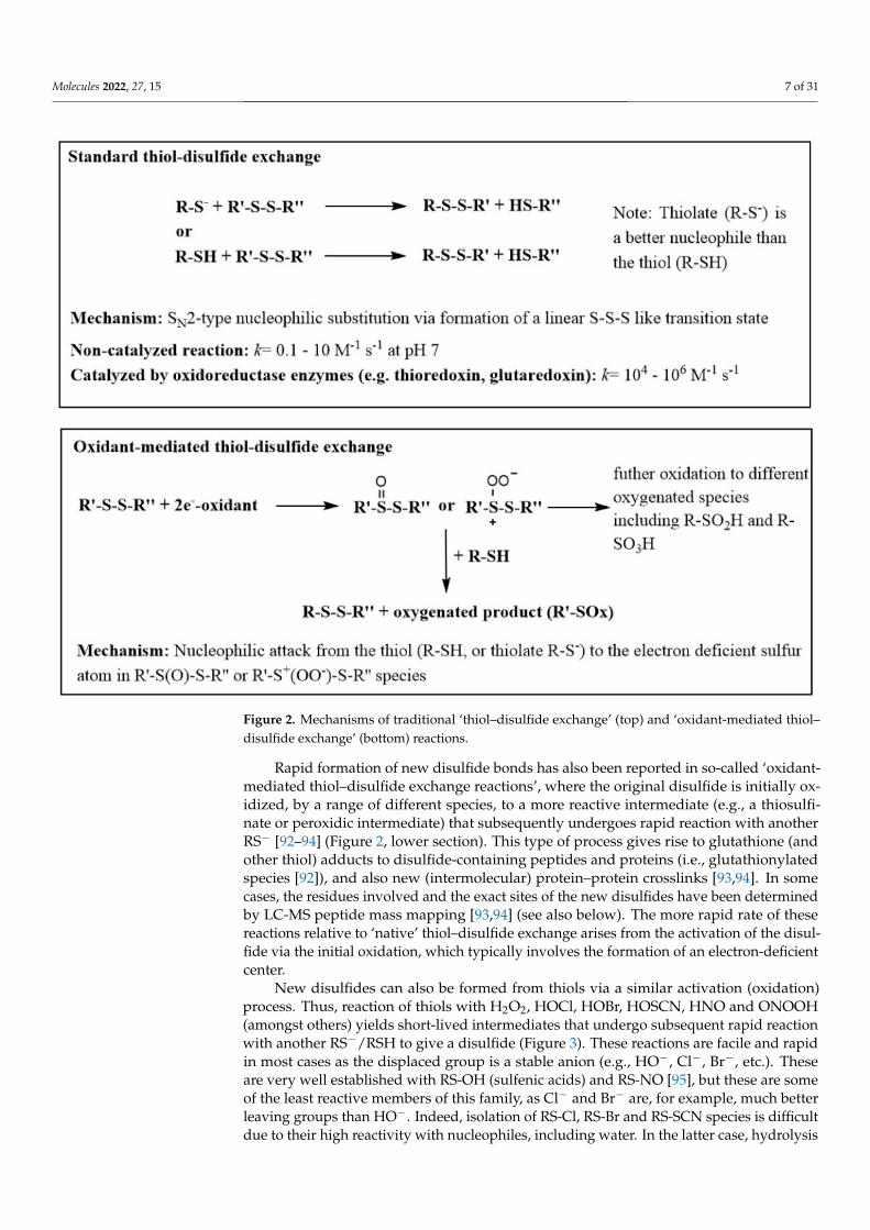
Multiple other pathways can also generate non-intended disulfides, including thiol–disulfide exchange reactions in which a thiolate anion (RS−, the ionized and more reactiveform of a thiol) reacts with a disulfide, with the formation of a new disulfide and releaseof a thiol [89] (Figure 2, top section). These reactions are typically slow, as there is little
Molecules 2022, 27, 15 5 of 31
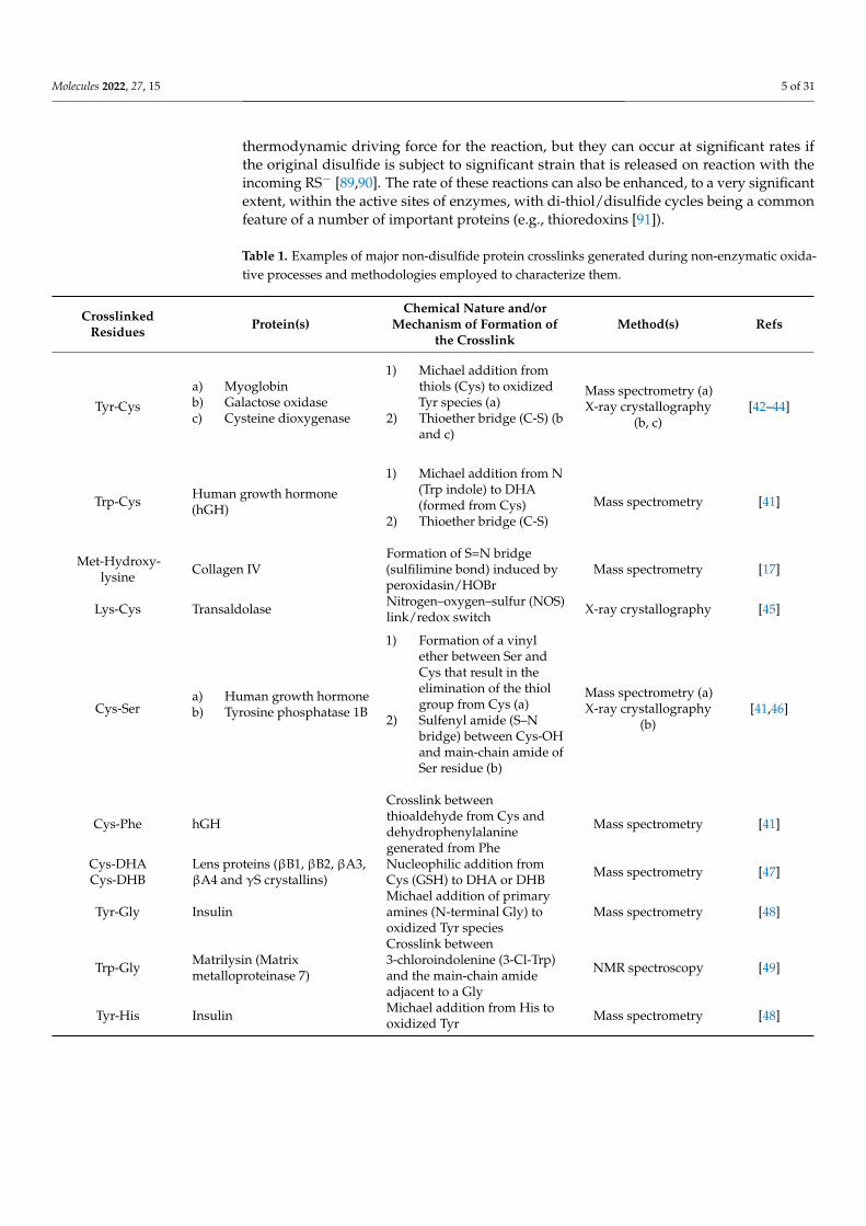
thermodynamic driving force for the reaction, but they can occur at significant rates ifthe original disulfide is subject to significant strain that is released on reaction with theincoming RS− [89,90]. The rate of these reactions can also be enhanced, to a very significantextent, within the active sites of enzymes, with di-thiol/disulfide cycles being a commonfeature of a number of important proteins (e.g., thioredoxins [91]).
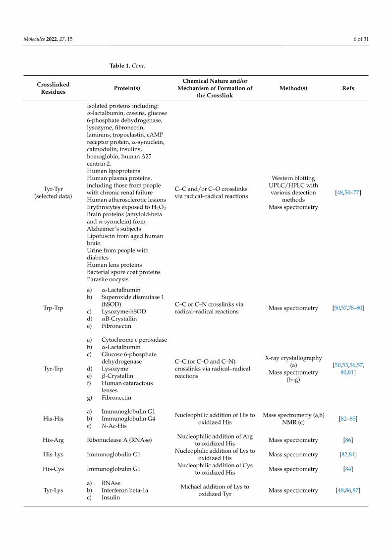
Table 1. Examples of major non-disulfide protein crosslinks generated during non-enzymatic oxida-tive processes and methodologies employed to characterize them.
CrosslinkedResidues Protein(s)
Chemical Nature and/orMechanism of Formation of
the CrosslinkMethod(s) Refs
Tyr-Cys
a) Myoglobinb) Galactose oxidasec) Cysteine dioxygenase
1) Michael addition fromthiols (Cys) to oxidizedTyr species (a)
2) Thioether bridge (C-S) (band c)
Mass spectrometry (a)X-ray crystallography
(b, c)[42–44]
Trp-Cys Human growth hormone(hGH)
1) Michael addition from N(Trp indole) to DHA(formed from Cys)
2) Thioether bridge (C-S)Mass spectrometry [41]
Met-Hydroxy-lysine Collagen IV
Formation of S=N bridge(sulfilimine bond) induced byperoxidasin/HOBr
Mass spectrometry [17]
Lys-Cys Transaldolase Nitrogen–oxygen–sulfur (NOS)link/redox switch X-ray crystallography [45]
Cys-Sera) Human growth hormoneb) Tyrosine phosphatase 1B
1) Formation of a vinylether between Ser andCys that result in theelimination of the thiolgroup from Cys (a)
2) Sulfenyl amide (S–Nbridge) between Cys-OHand main-chain amide ofSer residue (b)
Mass spectrometry (a)X-ray crystallography
(b)[41,46]
Cys-Phe hGH
Crosslink betweenthioaldehyde from Cys anddehydrophenylalaninegenerated from Phe
Mass spectrometry [41]
Cys-DHACys-DHB
Lens proteins (βB1, βB2, βA3,βA4 and γS crystallins)
Nucleophilic addition fromCys (GSH) to DHA or DHB Mass spectrometry [47]
Tyr-Gly InsulinMichael addition of primaryamines (N-terminal Gly) tooxidized Tyr species
Mass spectrometry [48]
Trp-Gly Matrilysin (Matrixmetalloproteinase 7)
Crosslink between3-chloroindolenine (3-Cl-Trp)and the main-chain amideadjacent to a Gly
NMR spectroscopy [49]
Tyr-His Insulin Michael addition from His tooxidized Tyr Mass spectrometry [48]
Molecules 2022, 27, 15 6 of 31
Table 1. Cont.
CrosslinkedResidues Protein(s)
Chemical Nature and/orMechanism of Formation of
the CrosslinkMethod(s) Refs
Tyr-Tyr(selected data)
Isolated proteins including:α-lactalbumin, caseins, glucose6-phosphate dehydrogenase,lysozyme, fibronectin,laminins, tropoelastin, cAMPreceptor protein, α-synuclein,calmodulin, insulins,hemoglobin, human ∆25centrin 2.Human lipoproteinsHuman plasma proteins,including those from peoplewith chronic renal failureHuman atherosclerotic lesionsErythrocytes exposed to H2O2Brain proteins (amyloid-betaand α-synuclein) fromAlzheimer’s subjectsLipofuscin from aged humanbrainUrine from people withdiabetesHuman lens proteinsBacterial spore coat proteinsParasite oocysts
C–C and/or C–O crosslinksvia radical–radical reactions
Western blottingUPLC/HPLC withvarious detection
methodsMass spectrometry
[48,50–77]
Trp-Trp
a) α-Lactalbuminb) Superoxide dismutase 1
(hSOD)c) Lysozyme-hSODd) αB-Crystalline) Fibronectin
C–C or C–N crosslinks viaradical–radical reactions Mass spectrometry [50,57,78–80]
Tyr-Trp
a) Cytochrome c peroxidaseb) α-Lactalbuminc) Glucose 6-phosphate
dehydrogenased) Lysozymee) β-Crystallinf) Human cataractous
lensesg) Fibronectin
C–C (or C–O and C–N)crosslinks via radical–radicalreactions
X-ray crystallography(a)
Mass spectrometry(b–g)
[50,53,56,57,80,81]
His-Hisa) Immunoglobulin G1b) Immunoglobulin G4c) N-Ac-His
Nucleophilic addition of His tooxidized His
Mass spectrometry (a,b)NMR (c) [82–85]
His-Arg Ribonuclease A (RNAse) Nucleophilic addition of Argto oxidized His Mass spectrometry [86]
His-Lys Immunoglobulin G1 Nucleophilic addition of Lys tooxidized His Mass spectrometry [82,84]
His-Cys Immunoglobulin G1 Nucleophilic addition of Cysto oxidized His Mass spectrometry [84]
Tyr-Lysa) RNAseb) Interferon beta-1ac) Insulin
Michael addition of Lys tooxidized Tyr Mass spectrometry [48,86,87]
Molecules 2022, 27, 15 7 of 31
Figure 2. Mechanisms of traditional ‘thiol–disulfide exchange’ (top) and ‘oxidant-mediated thiol–disulfide exchange’ (bottom) reactions.
Rapid formation of new disulfide bonds has also been reported in so-called ‘oxidant-mediated thiol–disulfide exchange reactions’, where the original disulfide is initially ox-idized, by a range of different species, to a more reactive intermediate (e.g., a thiosulfi-nate or peroxidic intermediate) that subsequently undergoes rapid reaction with anotherRS− [92–94] (Figure 2, lower section). This type of process gives rise to glutathione (andother thiol) adducts to disulfide-containing peptides and proteins (i.e., glutathionylatedspecies [92]), and also new (intermolecular) protein–protein crosslinks [93,94]. In somecases, the residues involved and the exact sites of the new disulfides have been determinedby LC-MS peptide mass mapping [93,94] (see also below). The more rapid rate of thesereactions relative to ‘native’ thiol–disulfide exchange arises from the activation of the disul-fide via the initial oxidation, which typically involves the formation of an electron-deficientcenter.
New disulfides can also be formed from thiols via a similar activation (oxidation)process. Thus, reaction of thiols with H2O2, HOCl, HOBr, HOSCN, HNO and ONOOH(amongst others) yields short-lived intermediates that undergo subsequent rapid reactionwith another RS−/RSH to give a disulfide (Figure 3). These reactions are facile and rapidin most cases as the displaced group is a stable anion (e.g., HO−, Cl−, Br−, etc.). Theseare very well established with RS-OH (sulfenic acids) and RS-NO [95], but these are someof the least reactive members of this family, as Cl− and Br− are, for example, much betterleaving groups than HO−. Indeed, isolation of RS-Cl, RS-Br and RS-SCN species is difficultdue to their high reactivity with nucleophiles, including water. In the latter case, hydrolysis

Molecules 2022, 27, 15 8 of 31
gives RS-OH, which undergoes similar reactions. Some RS-OH and RS-NO species aresufficiently long-lived to be detectable on proteins, though their lifetimes are structure- andenvironment-dependent [96].
Figure 3. Generation of crosslinks via oxidized thiol residues. Similar reactions of the ‘activated’thiols (RS–OH, RS–Cl, RS–Br, RS–SCN, RS–NO) can occur with nitrogen nucleophiles (e.g., RNH2) togive new S–N bonded species (see text for further details).
Disulfides can also be formed from rapid termination reactions of two RS•. How-ever, reaction of RS• with another R’S− to give the corresponding disulfide radical anion(RSS’R•−) is also very rapid, and as the concentration of R’S− (either on a protein, or alow molecular mass species such as glutathione) is typically much higher than that of RS•,the radical anion pathway usually predominates. The resulting RSS’R•− undergoes rapidelectron transfer with electron acceptors, including O2, resulting in the formation of a newdisulfide (which may be either a protein–protein species, or a glutathionylated protein) andthe superoxide radical anion, O2
•−.
6.1.2. Sulfur–Nitrogen (S–N) Crosslinks (Sulfimines, Sulfenamides, Sulfinamidesand Sulfonamides)
A wide range of S–N crosslinks are known, with these arising predominantly frommolecule reactions of activated sulfur centers with nitrogen nucleophiles, and particularlythe side-chain amine of Lys (or hydroxy-Lys) residues (see also Figure 3). Reactions withother nitrogen nucleophiles are known, including the more reactive (when compared to theLys side-chain) N-terminal amine of peptides or proteins. Reactions at (backbone) amideand guanidine (Arg side-chain) functions have also been reported (see below), but suchexamples are limited, as these are weaker nucleophiles.
Crosslinking can occur via the thioether group of methionine in the enzymatic(peroxidasin)-induced crosslinking of the NC1 domains of collagens. Reaction of a specificMet residue (Met93) with HOBr formed by peroxidasin generates a transient bromosulfo-nium ion [–S(Br)+–] that reacts with the amine group of a suitably positioned hydroxy-Lys
Molecules 2022, 27, 15 9 of 31
residue (Hyl211) to give an interchain sulfilimine (–S=N–) crosslink. These crosslinks arecritical for the generation of functional extracellular matrices in many organisms, withan absence of this enzymatic activity causing tissue dysfunction that is embryonicallylethal in some species [16,17]. Reaction with the amine function occurs in competitionwith the reaction with water (acting as a nucleophile), with consequent methionine sulfox-ide formation. This type of crosslink is not unique to peroxidasin-generated HOBr, withother Met-containing peptides undergoing similar sulfilimine crosslink formation with Lysresidues, either intra- or intermolecularly [97]. These reactions are also not limited to HOBr,with HOCl, bromamines and chloramines (RNHBr and RNHCl species, respectively) alsogenerating these species in competition with the sulfoxide [97]. In the case of HOCl, thesespecies are usually formed in low yield. For the chloramines, initial oxidation of the sulfurcenter of the Met to give the chlorosulfonium ion [–S(Cl)+–] appears to be followed byrapid reaction with the nitrogen atom of the original chloramine [97], indicating that thesereactions can be highly specific, and that the [–S(Cl)+–] species is short lived.
Related reactions of sulfenyl halides (RS–X) and related species formed from reaction ofthe –SH group of Cys or GSH with an oxidant (e.g., HOCl, HOBr, chloramines, bromamines,ONOOH) with amine nucleophiles have been reported to give rise to a large family ofsulfenamides (RS–NHR’), sulfinamides [RS(O)–NHR’] and sulfonamides [RS(O)2–NHR’].These have been detected as both intra- and intermolecular crosslinks in both peptides andproteins [98–100]. A well-established example is glutathione sulfonamide (GSA) formation,which has been used as a biomarker of oxidative damage in both cells (e.g., [101,102]) andhuman tissues and fluids [103,104]. In this case, the linkage is formed intramolecularlywith the amide nitrogen of the terminal Gly residue, resulting in an eight-membered ringspecies [105]. The formation of GSA occurs in competition with reaction of the RS-X withanother RSH to give the disulfide (i.e., GSSG in the case of oxidation of GSH) with the ratioof GSA:GSSG dependent on the oxidant [98]. The disulfide is usually the major product,but the GSA yield can be high in some cases [98,105], including in some cells [101,102,106].The formation of GSA requires three equivalents of oxidant, and the exact sequence ofoxidation events is unclear (i.e., whether oxygenation at the sulfur occurs prior to, or afterthe formation of the S–N bond) [105]. The detection of sulfenamides and sulfinamidesin other systems (e.g., S100A8 proteins [99] and peptides [100,107]) suggests that S–Nbond formation may be the initial event, with oxidation at the sulfur occurring on reactionwith the second and third oxidizing equivalents. These species are poorly reducible whencompared to disulfides [99], and only ~50% of the GSH oxidized by HOCl is recovered(presumably from GSSG) on reduction [108]. Evidence has also been reported for modelpeptides for the involvement of the guanidine group of Arg residues in the formation ofS–N crosslinks [100].
There is considerable evidence of the formation of an intramolecular sulfenamide(sulfenyl amide, S–N species) in the enzyme protein tyrosine phosphatase 1B (PTP1B), withthe linkage formed between the sulfenic acid (RS–OH) form of the catalytic Cys residue,and the adjacent main chain amide of a Ser residue [46]. This modification appears toprotect the Cys residue from irreversible oxidation to sulfinic (RSO2H) or sulfonic (RSO3H)acids and loss of enzyme activity. This species appears to be the stable, inactive ‘restingstate’ form of the enzyme, with the sulfenyl amide being readily reversed by cellular thiolssuch as GSH, with conversion back to its catalytically active Cys form [46].
6.1.3. Sulfur–Carbon (S–C) Crosslinks
Sulfur–carbon crosslinks can be generated via the addition of RS• (radical addition)or RS− (Michael adduction) to the double bonds of DHA and DHB. These are likely to beminor pathways in biological samples due to the low concentrations of DHA/DHB and theoccurrence of other alternative reactions of RS• (see above), though these reactions havebeen detected with some isolated proteins [109] and also in the nucleus of the lens [63].
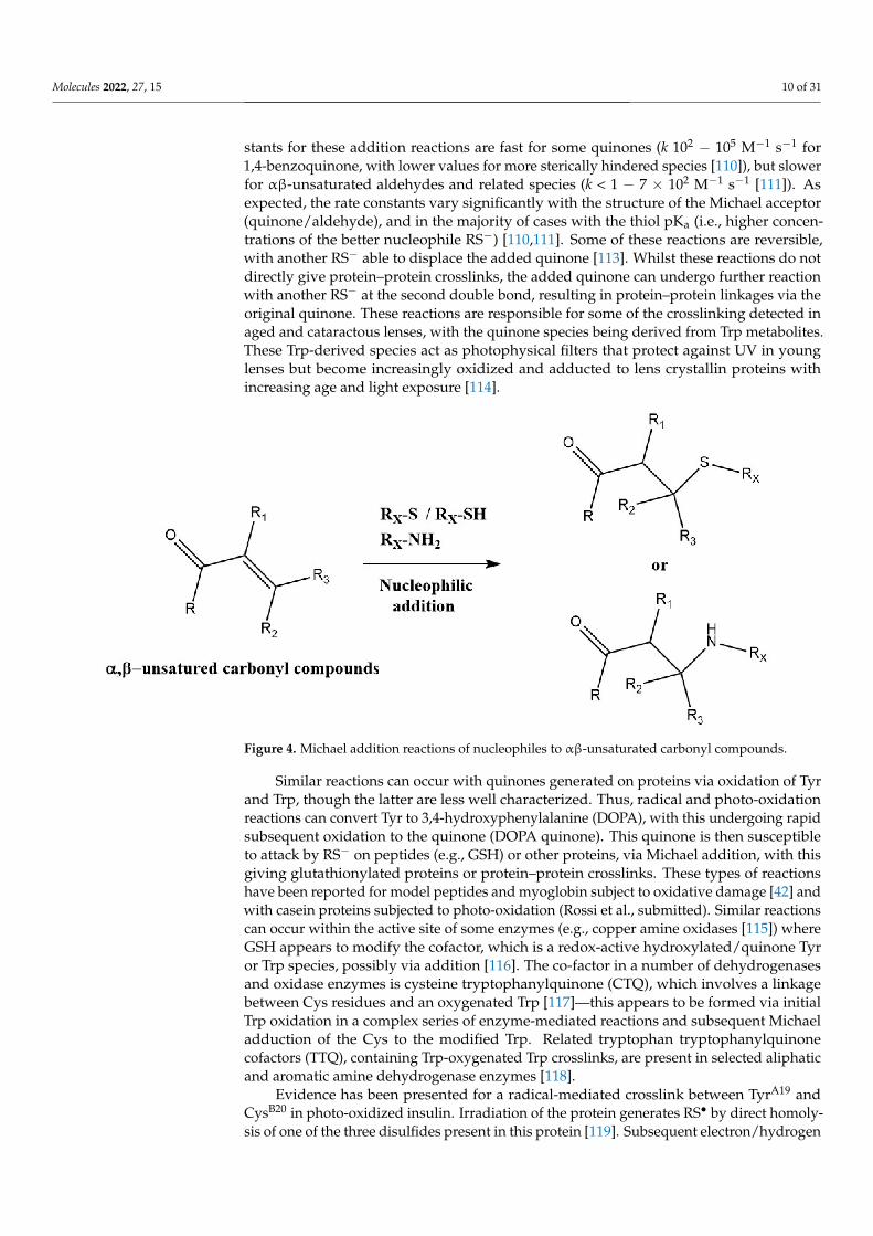
Related Michael addition reactions of RS− with quinones, or αβ-unsaturated alde-hydes and related species yield S-C bonds (e.g., [110–112] (Figure 4). The rate con-
Molecules 2022, 27, 15 10 of 31
stants for these addition reactions are fast for some quinones (k 102 − 105 M−1 s−1 for1,4-benzoquinone, with lower values for more sterically hindered species [110]), but slowerfor αβ-unsaturated aldehydes and related species (k < 1 − 7 × 10² M−1 s−1 [111]). Asexpected, the rate constants vary significantly with the structure of the Michael acceptor(quinone/aldehyde), and in the majority of cases with the thiol pKa (i.e., higher concen-trations of the better nucleophile RS−) [110,111]. Some of these reactions are reversible,with another RS− able to displace the added quinone [113]. Whilst these reactions do notdirectly give protein–protein crosslinks, the added quinone can undergo further reactionwith another RS− at the second double bond, resulting in protein–protein linkages via theoriginal quinone. These reactions are responsible for some of the crosslinking detected inaged and cataractous lenses, with the quinone species being derived from Trp metabolites.These Trp-derived species act as photophysical filters that protect against UV in younglenses but become increasingly oxidized and adducted to lens crystallin proteins withincreasing age and light exposure [114].
Figure 4. Michael addition reactions of nucleophiles to αβ-unsaturated carbonyl compounds.
Similar reactions can occur with quinones generated on proteins via oxidation of Tyrand Trp, though the latter are less well characterized. Thus, radical and photo-oxidationreactions can convert Tyr to 3,4-hydroxyphenylalanine (DOPA), with this undergoing rapidsubsequent oxidation to the quinone (DOPA quinone). This quinone is then susceptibleto attack by RS− on peptides (e.g., GSH) or other proteins, via Michael addition, with thisgiving glutathionylated proteins or protein–protein crosslinks. These types of reactionshave been reported for model peptides and myoglobin subject to oxidative damage [42] andwith casein proteins subjected to photo-oxidation (Rossi et al., submitted). Similar reactionscan occur within the active site of some enzymes (e.g., copper amine oxidases [115]) whereGSH appears to modify the cofactor, which is a redox-active hydroxylated/quinone Tyror Trp species, possibly via addition [116]. The co-factor in a number of dehydrogenasesand oxidase enzymes is cysteine tryptophanylquinone (CTQ), which involves a linkagebetween Cys residues and an oxygenated Trp [117]—this appears to be formed via initialTrp oxidation in a complex series of enzyme-mediated reactions and subsequent Michaeladduction of the Cys to the modified Trp. Related tryptophan tryptophanylquinonecofactors (TTQ), containing Trp-oxygenated Trp crosslinks, are present in selected aliphaticand aromatic amine dehydrogenase enzymes [118].
Evidence has been presented for a radical-mediated crosslink between TyrA19 andCysB20 in photo-oxidized insulin. Irradiation of the protein generates RS• by direct homoly-sis of one of the three disulfides present in this protein [119]. Subsequent electron/hydrogen

Molecules 2022, 27, 15 11 of 31
transfer from Tyr to one of the RS• would then give the required radical pair for formationof the dimer. Alternatively, photo-induced electron transfer from a photo-excited Tyr toa disulfide could occur, with this yielding Tyr• and a single RS•, with the crosslink beingformed by subsequent reaction between these two species. A third potential pathway mayinvolve addition of RS• formed from CysB20 radical to the intact TyrA19 residue followedby oxidation of the radical adduct [119,120]. The exact structure of the adduct species (i.e.,whether this involves an S–O or S–C bond) remains to be resolved. In addition to thisTyr–Cys crosslink, a dithiohemiacetal [–C(SH)–S–C–] crosslink was also detected, involvingCysA20 and CysB19, on photolysis of human insulin in the solid state [121]. The proposedmechanism involves formation of two RS• from the disulfide, followed by disproportiona-tion of the two radicals to give a thiol (RSH) and a thioaldehyde (–C=S), and then molecularaddition of the thiol to the thioaldehyde. Similar species have been detected with otherphoto-oxidized peptides/proteins, including mouse and human growth hormones andsome monoclonal antibodies (reviewed in [120]). A variant on the above reaction hasbeen detected when Ser residues are located in the vicinity of the thioaldehyde, with thehydroxyl group of the Ser reacting with the thioaldehyde to give a thiohemiacetal [–C(SH)–O–C–] [41]. Subsequent cleavage of the C–S bond under continued irradiation, and lossof the sulfur center via homolytic or heterolytic reactions, results in a vinylether crosslink(–C=C–O–C) [41]. It is likely that similar reactions occur with Thr residues, and analogousreactions have been detected with a Tyr in place of the Ser (reviewed in [120]).
A complex crosslink involving a nitrogen–oxygen–sulfur (N–O–S) linkage has beenreported between a Lys and Cys in a transaldolase reductase from Neisseria gonorrhoeae, thepathogen that causes gonorrhea [45]. Interestingly, this crosslink regulates enzyme activityvia an allosteric switch, suggesting that its formation may be of biological importance.Whilst the structure of the crosslink has been determined in detail (by X-ray crystallographyand others), the mechanism of its formation is unresolved. Multiple mechanisms havebeen proposed, with each involving initial oxidation at the sulfur to give oxygenatedproducts [45].
6.1.4. Sulfur–Selenium Crosslinks
As sulfur and selenium have considerable similarities in their chemistry, there isevidence for intra- and intermolecular selenium–sulfur and selenium–nitrogen species.Examples of both types are found in two of the major families of enzymes that protect cellsagainst oxidative stress: the glutathione peroxidases (GPx’s), which remove H2O2 (and lipidhydroperoxides in the case of GPx4), and the thioredoxin reductase (TrxR)/thioredoxin(Trx) redox systems, that maintains thiols in their reduced form. In order to rationalizethe resistance of GPx enzymes to deselenation (loss of selenium to give DHA), it has beenproposed that internal selenenyl amide (Se–N) linkages with neighboring amides involvingfive- or eight-membered rings [122] are formed during the catalytic cycle [123]. Theseselenenyl amides subsequently react with GSH to give a selenenyl GSH (i.e., Se–S) species,which is then repaired at the expense of GSH.
Similar Se–S species are generated during the catalytic cycle of the TrxR/Trx redoxsystem. The resting state of TxrR contains an intramolecular Se–S bond between a Cys andSec residue, which is reduced by NADPH via an FAD cofactor. The resulting selenate anion(RSe−) then reacts with the intramolecular disulfide bond of oxidized thioredoxin to givean intermolecular Se–S linkage between the TrxR and Trx. This linkage is then repaired byanother (resolving) Cys present in TrxR to regenerate the resting state Se–S species [124].
6.2. Carbon–Carbon6.2.1. Tyrosine–Tyrosine (Di-Tyrosine, Di-Tyr) Crosslinks
Di-Tyr crosslinks are formed deliberately by some enzymes, post-translationally, togive stability and elasticity to structural proteins (e.g., [125]). Nevertheless, protein expo-sure to oxidative environments, both biologically [126,127] and in food systems [128,129],can produce additional di-Tyr crosslinks. Thus, exposure of proteins to hydroxyl (HO•) and

Molecules 2022, 27, 15 12 of 31
peroxyl (ROO•), peroxynitrous acid/peroxynitrite (ONOOH/ONOO−), nitrogen dioxide(NO2
•), nitrosoperoxycarbonate (ONOOCO2−), carbonate (CO3
•−), lipid hydroperoxides(LOOH) and photoinduced reactions can yield di-Tyr crosslinks [130–132]. These can beformed inter- or intramolecularly between two proteins, from two free Tyr, or between freeTyr and proteins, by radical–radical reactions involving two Tyr• (Figure 5) [130,133]. Elec-tron delocalization over the benzene ring and the phenolic oxygen results in the formationof two regio-isomers, one involving a carbon–oxygen bond (C3–O, iso-dityrosine, vide infra)and another with a carbon–carbon (C3–C3, o,o’) bond. The latter is thermodynamically pre-ferred (Figure 5) [19,134,135]. The production of di-Tyr crosslinks is limited by alternativereactions, such as that with O2 (though this is slow, k < 1× 10³ M−1 s−1, [130]), and also viafast reaction with other radicals such as O2
•− (which gives short-lived peroxides [37,136])and nitric oxide (NO•) [137], and also with reducing agents such as ascorbate, thiols andpolyphenols [138–140]. The extent of dimerization is also likely to be strongly modulatedby steric and electronic interactions.
Figure 5. Formation and reactions of Tyr phenoxyl radicals (Tyr•). Tyr• self-react to produce di-Tyr(o,o’-di-Tyr, red; iso-di-Tyr, black) or react with O2 to generate oxygenated products. Kinetic datafrom [130].
In pathophysiological contexts, di-Tyr crosslinking has been reported in low-densitylipoproteins, arising from oxidative damage within human atherosclerotic lesions [61];in erythrocytes exposed to a continuous flux of H2O2 [62]; in lens proteins, where itappears to originate from photo-oxidation of amino acids [63]; in tissues from a number ofneurodegenerative conditions (e.g., amyloid-beta dimers in Alzheimer’s disease [64] and inα-synuclein [65,66], which may contribute to Parkinson’s disease); in lipofuscin pigmentsin the aged human brain [67]; in plasma of patients with chronic renal failure; and in urineof people with diabetes [68,69].
Di-Tyr crosslinks have also been detected on synthetic peptides exposed to perox-idase activity (e.g., hemoglobin and H2O2 [141]), and a large number of isolated pro-teins including (amongst others): lipoproteins [61,70,71], glucose 6-phosphate dehydro-genase (G6PDH) [53], ribonuclease A (RNAse A) [86], multiple extracellular matrix pro-teins [57,58,76], calmodulin [72], lysozyme [54,79], myoglobin [142,143], insulin [48,73,74]and human ∆25 centrin 2 [75].
The large body of evidence for di-Tyr crosslinks in oxidized proteins, fluids and tissues;the stability of this species (which is immune to reduction or repair); the association of thiscrosslink with the onset and development of human pathologies; and advances in analyticalmethods for the detection and quantification of di-Tyr (see below) have supported its useas a biomarker of protein oxidation in clinical studies [144,145].
Di-Tyr formation has also been reported in food systems, with both positive andnegative effects. Protein crosslinking can give desirable textures and stability to processedfoods, while negative effects have been reported with regard to poor protein digestibility

Molecules 2022, 27, 15 13 of 31
and other undesirable properties [128,129]. Di-Tyr crosslinks have been detected in milkpowders [146,147], during breadmaking [148], processing of myofibrillar proteins [149], inisolated caseins exposed to riboflavin-mediated photoreactions [51], and lactalbumin ex-posed to a laccase enzyme system [150] and UV-B light [50]. The presence of di-Tyr in foods,and consequent dietary intake, has resulted in studies on its toxicological properties. Intra-gastric administration of pure di-Tyr has been associated with metabolic alterations [151],including disrupted glucose metabolism [152], and renal alterations [153]. Interestingly,and of potential biological relevance, processed milks containing di-Tyr (and other Tyroxidation products) can induce oxidative damage in plasma, liver and brain tissues, as wellas spatial learning and memory impairments [154].
Di-Tyr crosslinks have also been detected in a wide variety of other organisms, includ-ing in insect cuticles, where di-Tyr crosslinks connect resilin proteins in a three-dimensionalnetwork [155]. Peroxidase-catalyzed di-Tyr crosslinks have also been detected in highconcentrations in the oocyst walls of the apicomplexan parasite, Eimeria maxima, and insome bacterial spore proteins (e.g., those of Bacillus subtilis), with these providing a rigidframework that protects the oocyst or spores from harsh environmental conditions [60,77].On the basis of these data, novel biomaterials have been developed that contain di-Tyrcrosslinks that have excellent mechanical properties. Different strategies to induce efficientdi-Tyr crosslink formation have been developed, including photoreactions mediated byruthenium complexes and riboflavin [132].
6.2.2. Tryptophan–Tryptophan (Di-Tryptophan, di-Trp) Crosslinks
Trp is a major target for many oxidants, and a large number of oxidation productshave been characterized [156]. Many of these involve the initial formation of (nitrogen-centered) indolyl radicals (Trp•) which, depending on the properties of the protein andenvironment, can self-react to generate di-Trp crosslinks (Figure 6), though less informationis available on these species than di-Tyr. Di-Trp crosslinks have been reported on humansuperoxide dismutase 1 (hSOD1) upon exposure to H2O2 in the presence of carbonateions [78], with the peroxidase activity of hSOD1 being responsible for the generation ofCO3
•− that induce one-electron oxidation of the single, solvent-exposed, Trp32 residue inthis protein and consequent intermolecular di-Trp crosslinking. This results in irreversibledimerization and is associated with non-amyloid protein aggregation and the occurrence ofamyotrophic lateral sclerosis (ALS) [157]. CO3
•−-mediated di-Trp crosslinks have also beenreported for both free Trp [158] and lysozyme [79]. In the latter case, oxidation mediatedby hSOD1 resulted in the detection of both hetero-dimers of lysozyme with hSOD1 andhSOD1 dimers [79]. These reports indicate a high propensity of CO3
•− to generate di-Trp crosslinks [79]. Di-Trp crosslinks have also been reported on lysozyme exposed toONOOH/ONOO− [159] and photo-oxidation mediated by kynurenic acid [160], as wellas in solutions of free Trp and N-acetyl-Trp exposed to photo-reactions of riboflavin andkynurenic acid, respectively [161,162]. Di-Trp crosslinks have been also detected in lensproteins of patients with nuclear cataracts [80] and in α-crystallin proteins exposed to UVradiation and a photosensitizer [163].
Di-Trp crosslinks have also been detected in aggregates of α-lactalbumin (α-LA) exposedto UV-B light [50], and in FtsZ proteins of M. jannaschii, a thermophilic microorganism, exposedto peroxyl radicals [164]. The dimerization of Trp• is fast (k ~ 2− 6× 10⁸ M−1 s−1 [159]),but dependent on the residue environment in peptides and proteins. Due to electrondelocalization across the pyrrole and benzene rings, and the presence of chiral carbons, acomplex mixture of regio- and stereo-isomers can be formed. This heterogeneity is likely tocontribute to the limited number of reports on this type of crosslink, due to the difficulty inidentifying and quantifying all of these species. This is exacerbated by a current absence ofantibodies against this linkage (unlike di-Tyr, see below). Nonetheless, di-Trp crosslinksappear to be generated in low yields, probably due to the large number of alternative fatesof Trp• (Figure 6) and the low abundance of Trp in proteins (~1–2%). Despite the modestrate constant for addition of O2 to Trp• (k ≤ 4 × 10⁶ M−1 s−1 [35,36]), oxygenated products

Molecules 2022, 27, 15 14 of 31
and alternative crosslinks (e.g., Trp-Tyr, see below) are commonly detected, and at higheryields than di-Trp. Thus, only a small extent of the aggregation of α-LA induced by UV-Blight was associated with di-Trp crosslinks, in contrast to high yields of disulfide bonds [50].Nonetheless, under anaerobic conditions, or environments with low O2 levels (e.g., humanlens [161]), the formation of di-Trp crosslinks appears to be favored.
Figure 6. Formation and reactions of Trp indolyl radicals (Trp•). Self-reactions of Trp• produce carbon–carbon (C3–C3) and carbon–nitrogen (C3–N1) di-Trp crosslinks. It should be noted that multiplestereoisomers are potentially formed for both di-Trp dimers. Kinetic constants for self-reactions ofTrp• and their reaction with O2 are from [159] and [36], respectively.
The production of di-Trp crosslinks is also modulated by protein conformations,as reported for the extracellular matrix (ECM) protein fibronectin, when exposed toONOO−/ONOOH [57]. Fibronectin is polymerized into elastic fibrils (fibrillation) atthe cell surface, with this then allowing binding of other ECM components. This pro-cess is controlled (amongst others) by alterations to the protein conformation, with thecompact structure present in plasma being resistant to fibrillation. However, an extendedconformation generated by cell-generated forces (as well as by altered pH and salt con-centrations) is more prone to fibrillation. MS analysis of tryptic peptides from fibronectinexposed to ONOO−/ONOOH showed that two of the four crosslinks detected correspondto di-Trp crosslinks [57]. One di-Trp crosslink (Trp445–Trp2264) was detected in both thecompact and extended conformations, while another (Trp177–Trp2250) was only detectedin the compact state. Interestingly, Trp2250 was oxidized to a high extent in the extendedconformation, indicating that compact conformation of fibronectin favors radical–radicalreactions (involving Trp177 and Trp2250), while the extended state favors other fates,including formation of 6-nitro-Trp [57].
Trp–Trp bonds have also been detected in the active sites of some enzymes, includingamine dehydrogenases (e.g., methylamine dehydrogenase and aromatic amine dehydroge-nases), where a tryptophan tryptophanylquinone crosslink is generated between C2 of an

Molecules 2022, 27, 15 15 of 31
unmodified Trp residue, and C5 (on the benzene ring) of a Trp quinone (with the carbonylgroups present at C7 and C8) [165].
6.2.3. Tyrosine–Tryptophan (Tyr–Trp) Crosslinks
Cross-reaction between Tyr• and Trp• can lead to Tyr–Trp crosslinks. These speciesare less well characterized than those described above, and the precise structure of thesespecies remains to be elucidated (i.e., the nature of the crosslink bond and sites on the tworings that are joined). MS analyses of solutions containing free Tyr and Trp incubated withCO3
•− have provided evidence for three different, isomeric, Tyr–Trp crosslinks, with theselikely to involve carbon–carbon bonds, and probably between the ortho (C3) position onthe Tyr ring, and C3 on the indole ring of Trp [158].
Tyr–Trp crosslinks have been detected in peptides and isolated proteins exposedto multiple oxidants, including a cytochrome c peroxidase mutant exposed to a hemeiron-peroxide reaction [81]; in glucose-6-phosphate dehydrogenase (G6PDH) exposedto ROO• [53]; model peptides exposed to pulsed UV light [166]; in lysozyme treatedwith ROO• and exposed to photooxidation reactions mediated by riboflavin and roseBengal [54–56]; in fibronectin exposed to ONOOH/ONOO− [57]; in protein extracts of theGram-positive bacterium Lactococcus lactis exposed to photooxidation [167]; and proteinsextracted from human cataractous lenses [80].
6.2.4. Other Carbon–Carbon Crosslinks
As most protein carbon radicals, P•, react rapidly with O2 (see above), terminationreactions between two P• are limited under most biological conditions. The formation ofsome P–P crosslinks has, however, been reported in model peptide systems exposed toradiation-induced radicals [33], and similar species may be formed in biological situationswhere the pO2 value is low (e.g., in solid tumors, tissues subject to hypoxia). This requiresfurther study.
6.3. Carbon–Nitrogen (C–N) Crosslinks
A significant number of carbon–nitrogen crosslinks have been identified, with themajority of these arising from molecule–molecule reactions, with the nitrogen atom acting asa nucleophile, though a small number of radical–radical crosslinks have been characterized.
Delocalization of the unpaired electron over the aromatic rings of Tyr• and Trp•,followed by radical–radical reactions has been proposed, on the basis of MS data, to givedi-Trp species with carbon–nitrogen (C3-N1) bonds (Figure 6) [158]. The exact structures ofthese species are not completely clarified, though it is clear that multiple isobaric speciesare formed [159,161]. Some of these crosslinks appear to be of pathological relevance, asthey have been detected on lens crystallin proteins subjected to photo-oxidation [161] andin human cataractous lenses [80].
The much larger group of molecule–molecule reactions that generate C–N crosslinksprimarily involve nucleophilic reactions of the nitrogen centers on Lys and hydroxy-Lys(Nε-amine), Arg (guanidine nitrogen) and His (imidazole nitrogen) with either the carbonatom of carbonyl functions (Schiff base reactions) or other electron-deficient carbon cen-ters (via Michael addition). For the former type of reaction, the carbonyl can be formedenzymatically (e.g., by LOX and LOXL enzymes, see earlier) or as a result of oxidation ofalcohols (Ser/Thr) or C–H bonds in the presence of O2 (e.g., via ROO• and ROOH [31,168]).
In the case of the Cu2+-dependent LOX/LOXL reactions and collagen crosslinking, theinitial Schiff base adducts can undergo multiple further condensation reactions that allowseveral chains to be linked together via a single site [11]. Thus, there is abundant evidencefor lysyl pyrrole, hydroxylysyl pyrrole, lysyl pyridinoline and hydroxylysyl pyridinolinespecies involving three or four collagen chains [169]. The mechanism of formation of thesespecies involves the initial formation of a two-chain crosslink and then further condensationwith a Lys/hydroxy-Lys on third and fourth chains. These species are critical to the correctassembly of collagen-containing extracellular matrices in tissues [11].

Molecules 2022, 27, 15 16 of 31
Similar reactions occur with (Ca2+-dependent) transglutaminases that form isopep-tide bonds between the Lys side-chain amine and amide (Gln, Asn) or carboxylate (Asp)residues, with the amide/carboxylate ‘activated’ via reaction with the Cys residue of a Cys–His–Asp catalytic triad on the enzyme. The reaction of the carbonyl function with the Cysresidue generates a thioester that is then susceptible to reaction with the Lys amine [22–24].Such isopeptide bonds are highly resistant to most proteases and are widely encountered inthe formation of insoluble protein polymers/aggregates, such as those found in brain tissuelesions of people with Alzheimer’s disease [24,170], in some bacterial spore proteins [60]and in the food industry, where these reactions are used as glues to alter food texture andproperties [171,172].
Reactions also occur with α,β-unsaturated aldehyde and ketones and related speciesvia Michael addition reactions (Figure 7). Thus, intra- and interchain crosslinks can begenerated by the reaction of Lys/Arg/His residues with an oxidized imidazole group ofthe amino acid His, which acts as a Michael acceptor. These Lys–His, Arg–His and His–Hisproducts have been detected on multiple proteins, and particularly those subjected to photo-oxidation, where His residues are a major initial target of damage (e.g., [82,84–86,173]).They have also been detected in antibodies exposed to (photo)oxidation [82,84]. His-containing crosslinks have also been reported in some collagens, with these probablyformed by nucleophilic attack of an imidazole nitrogen on an α,β-unsaturated structureformed via other crosslinking reactions [174].
Figure 7. Michael addition reactions of amino acid side-chains to oxidized His and Tyr residues.
Related Michael reactions occur with Tyr-derived quinones formed on oxidation of Tyrresidues, with nitrogen nucleophiles, such as the Nε-amine of Lys side-chains undergoingadduction reactions with the oxidized ring (Figure 7). These reactions occur with lower

Molecules 2022, 27, 15 17 of 31
rate constants than the corresponding reaction with the RS− from Cys [110,175] (i.e., C–Scrosslinks predominate over C–N linkages, through this is dependent on the availability andabundance of Cys versus Lys residues, with the latter usually predominating numerically).These reactions are important in the coupling of proteins, including via N-terminal aminegroups, by tyrosinase and polyphenol oxidases (e.g., [176]) and in the formation of insectexoskeletons/cuticles of many insects [177], as well as the ‘glues’ that attach mussels torocks [178,179].
6.4. Carbon–Oxygen
As alcohols (ROH) are poorer nucleophiles when compared to RS− and neutralamines (RNH2), there are limited numbers of known carbon–oxygen crosslinks formedvia molecular processes such as Michael reactions. However, such coupling can occurvia radical–radical reactions, with the most well-established example being the formationof iso-di-tyrosine (iso-di-Tyr), where two Tyr• dimerize via the phenolate oxygen of onering, and C3 on the second ring (Figure 5); this appears to be a minor reaction comparedto carbon–carbon coupling (see above) [19]. Iso-di-Tyr crosslinks have been reported inthe extensin proteins that contribute to the architecture of wall plant cells [180], and insystems exposed to the myeloperoxidase–H2O2 system of human phagocytes, suggestingthat these species may be present in tissues subject to acute or chronic inflammation, suchas atherosclerosis [19].
7. Secondary Reactions of Crosslinks
In most biological systems, protein crosslinks, and particularly the formation of ir-reversible covalent crosslinks such as di-Tyr, di-Trp and Tyr–Trp, are considered as ‘final’oxidation products [181]. However, over-oxidation of these species is possible, particularlyunder conditions of extensive oxidative damage, or under environments with long-termprotein exposure to oxidants, where secondary one-electron oxidation with formationof radicals such as di-Tyr•, di-Trp•, or Tyr–Trp• may occur. Such radicals can mediatesimilar reactions to those described above for Tyr• and Trp•, including reaction with O2to produce oxygenated products (e.g., alcohols and hydroperoxides) and self-reactions togenerate trimers and oligomers. Thus, formation of tri-Tyr and pulcherosine crosslinks havebeen detected in human phagocytes [19], while di-, tri- and tetra-Tyr have been reportedin structural proteins of plant parasitic nematodes [182]. In addition, oligomers of Tyr(n = 2–8) have been reported in α-lactalbumin exposed to a horseradish peroxidase–H2O2system [183]. Tri-Trp has been reported in trimers of hSOD1 triggered by CO3
•− [78], whiletri-Trp and a di-Trp hydroperoxide (di-Trp-OOH) were reported in solutions of free Trpand riboflavin illuminated with a high-intensity 365 nm light-emitting diode [162].
In contrast, photo-oxidation (at 320 nm) of di-Tyr, in the presence of O2, has beenreported to occur via processes involving O2
•−, singlet oxygen (1O2) and H2O2 [184].These observations were ascribed to the action of di-Tyr crosslinks as photosensitizers thatcould induce photo-damage to other biomolecules [184]. These findings suggest that di-Tyrcrosslinks may be able to extend oxidation processes, opening new pathways of reactions,though these are only likely to be of major impact in systems with very extensive extentsof oxidation. The scope and role of these pathways is unexplored, as well as the abilityof peroxides such as di-Trp-OOH to extend protein oxidation (in line with the capacity ofother hydroperoxides [31]).
8. Detection of Crosslinks, including Advantages and Disadvantages ofDifferent Methods
Despite considerable methodological advancements, analysis of intra- or intermolec-ular protein crosslinks remains a challenging task, and the mechanisms underlying theformation of some known crosslinks remain to be established. There are also, undoubt-edly, more types of crosslinks that remain to be discovered. This lack of knowledge arisespartly due to the complexity of some structures, and the relatively poor current ‘tool-box’

Molecules 2022, 27, 15 18 of 31
to detect and predict the chemistry of the species and the sites involved. The followingsection therefore provides an overview of current approaches to the identification andcharacterization of oxidation-induced crosslinks, with a focus on both mass spectrometry(MS) and techniques (e.g., UPLC, immunological methods) that provide complementaryinformation. It is clearly advisable to use combinations of methods and to investigate bothgross modifications (e.g., changes in the molecular mass) and structural/conformationalchanges in crosslinked vs. native proteins with methods that allow the chemical nature andthe residues involved in the formation of these species to be determined (Figure 8). Each ofthese methods has individual advantages and disadvantages.
Figure 8. Overview of methods to detect and characterize crosslinked proteins and the sites/types ofmodifications. Abbreviations used: CD: circular dichroism, SANS: small angle neutron scattering,SAXS: small angle X-ray scattering, H–D: hydrogen–deuterium exchange mass spectrometry.
8.1. Analysis of Changes in Molecular Mass by Electrophoresis and Size ExclusionChromatography (SEC)
Protein electrophoresis (e.g., SDS-PAGE) and SEC-derived methodologies are excellenttools to assess the presence of crosslinked proteins in samples. Separation by electrophoresisis typically achieved through the use of polyacrylamide gels with different pore sizes.Diverse strategies can be utilized with this approach, such as running gels under native,denaturing and/or reducing conditions. This allows the investigation and differentiationof the contributions of reducible intermolecular bonds (e.g., disulfides between two proteinchains) from non-reducible bonds (e.g., carbon–carbon bonds). Similarly, SEC can separateand fractionate soluble proteins based on their hydrodynamic radius and molecular mass,and depending on the mobile phase used, it can also provide information about the natureof the crosslinks (i.e., reducible or non-reducible intermolecular bonds).
Whilst these techniques can provide information on the presence of intermolecularbonds, they rarely provide information about the presence of intramolecular species. More-over, data analysis needs to be carried out with care, as multiple proteins may be presentin each band/fraction from complex matrices. This complexity can be overcome by usingtwo-dimensional gel electrophoresis, with proteins separated firstly on the basis of theirisoelectric point, and subsequently by molecular mass. Both methodologies have beensuccessfully employed in combination with MS-based strategies [54,56,80,84,93,94,141,185],

Molecules 2022, 27, 15 19 of 31
with isolation and enrichment of fractions containing dimers (and species with higherdegrees of oligomerization) from non-crosslinked monomers before MS analysis. Thisapproach has been used successfully to characterize crosslinked forms of IgG, α-synucleinand lysozyme [56,84,141]. Two-dimensional gel electrophoresis can also be used for specificdetection of proteins connected by intermolecular disulfides [185]. In this approach, theproteins are first separated by non-reducing SDS-PAGE in the first dimension, and thenseparated by SDS-PAGE under reducing conditions in the second dimension. Proteinslacking intermolecular disulfide bonds line up on the diagonal, whereas proteins connectedby intermolecular disulfide bonds dissociate from each other in the second dimension andmigrate below the diagonal.
8.2. Analysis of Protein Crosslinks by Western (Immuno-) Blotting and ELISA Assays
The use of antibodies to investigate the formation of oxidation products, includingdi-Tyr, is a widely used strategy. These are typically examined using immunoblotting orELISA assays, with the former providing (limited) information on the nature and identityof the proteins on which the crosslinks are present, and whether these are intramolecular(in a monomer) or interchain species. However, there are few well-characterized antibodiesagainst crosslinked species, and these vary significantly in their specificity and selectivity,with some having significant cross-reactivity with other materials. Furthermore, crosslinksburied within highly aggregated species may be poorly, or not, recognized by (large)antibodies. Thus, appropriate control experiments are critical, and both positive andnegative data should be validated by alternative methods.
Despite these caveats, immunoblotting has been widely used to detect di-Tyr presentin α-synuclein in Lewy Bodies in Parkinson’s disease [186], in atherosclerotic plaques [187],and on multiple isolated proteins, including tropoelastin, α-synuclein, caseins, glucose-6-phosphate dehydrogenase, laminins and fibronectin [51,53,58,65,76,188]. Relative yields(i.e., versus the parent) can be achieved by ELISA, and this can serve as a very usefulscreening tool.
8.3. Direct Detection by Spectrophotometric and Fluorometric Assays
Some crosslinked species, as well as heavily aggregated proteins, can be monitoredby spectrophotometry and fluorescence spectroscopy. For example, turbidity changes insolutions can be used (in an approximate manner) to monitor the time-course of proteincrosslinking and aggregation [189,190]. However, this approach does not provide informa-tion on the type and nature of the crosslinked proteins and is useful only when there isextensive formation of heavily aggregated species, as soluble dimers and oligomers do notcontribute significantly to the turbidity of solutions.
In contrast, fluorescence experiments can provide relatively specific data on crosslinkssuch as di-Tyr (excitation and emission maximum at ~280 and ~410 nm, respectively) [191].Such data need to be interpreted with care, particularly with intact proteins or complexsystems, as other fluorescent or optically absorbing species (e.g., Tyr, Trp, Trp-derivedproducts, co-factors) may be present that distort excitation or emission processes. Thus,depending on the sample complexity, di-Tyr detection by fluorescence can provide usefulinformation, but requires further confirmation using other methodologies. In this context,coupling fluorescence detection methods with HPLC, UPLC or GC separation (see below)has proven to be a valuable strategy to detect and quantify di-Tyr in mixtures of aminoacids and oxidation products arising from acid hydrolysis of complex protein and tissuesamples [191,192].
8.4. HPLC/UPLC Methodologies
These techniques can provide important quantitative data on both the consumption ofthe parent amino acid residues, and product formation, including Trp- and Tyr-derivedcrosslinks [158,191,193,194]. Before analysis, proteins are often isolated by precipitation(e.g., from homogenates or cell lysates) using acids (e.g., trichloroacetic acid) or organic

Molecules 2022, 27, 15 20 of 31
solvents (e.g., ice-cold acetone or ethanol), followed by sample clean-up (e.g., delipidation),and then hydrolysis (with acid, base or enzymes) to give free amino acids and products. Theadvantages and disadvantages of this approach have been recently reviewed in [192], andtherefore, the following is focused solely on the detection and quantification of crosslinkedspecies.
The free amino acids and crosslinked products obtained from hydrolysis are separatedby HPLC/UPLC and can be quantified by MS [158,159,193], by fluorescence detection(directly for di-Tyr [51], or by pre-column tagging for parent amino acids using sensitivefluorescent tags such as o-phthaldialdehyde [191]), UV absorption, or, for some species,electrochemical oxidation [191,195]. For HPLC/UPLC methodologies coupled to MSdetection, heavy atom labeling (usually 2H, 13C, or 15N) can be utilized for accurate quan-tification [193]. Unfortunately, these approaches do not usually provide information on thesites of modification within proteins, or information on which proteins they were locatedon, if multiple species were present. This drawback, along with the limited number ofcrosslinked species that can be detected and quantified using these methods (di-Tyr, di-Trp,Trp-Tyr and cystine), makes it sensible to combine this approach with other methods (e.g.,peptide mass mapping or immunoblotting).
8.5. Detection and Characterization of Crosslinked Proteins Using Other Biophysical Approaches
Biophysical techniques including circular dichroism (CD), light scattering, small angleneutron scattering (SANS), small angle X-ray scattering (SAXS), X-ray crystallography,NMR spectroscopy, and electron microscopy can provide useful information on proteinstructure. These methods are sensitive to modified structures, supplying valuable in-formation on changes on morphology (i.e., mass, size and shape), secondary structureand solubility [45,196,197]. Some of these can also yield data on increased electron den-sity between residues, thus supporting the presence of both intra- and intermolecularcrosslinked species. This approach has been used in X-ray crystallographic studies, todetermine the exact sites of crosslinks in oxidized peroxiredoxin 5, thioredoxin 2 andγS-crystallin, and to elucidate a covalent crosslink between Cys and Lys containing anN–O–S bridge [45,197–199]. However, most of these methods (with the exception of X-raycrystallography, NMR spectroscopy and cryogenic electron microscopy) cannot provide astructure of sufficiently high-resolution to provide definitive identifications and they musttherefore be combined with other methodologies. Moreover, these methods are currentlylimited to homogeneous (single protein) samples that are available in large quantities (mgamounts).
8.6. Mass Spectrometry (MS)-Based Detection and Structural Characterization ofCrosslinked Proteins
MS is a highly versatile technique for analysis of protein crosslinks that can be appliedto (i) detect crosslinks and quantify their abundance, (ii) localize the specific crosslinkingsites within polypeptides and (iii) reveal the identity of the crosslinked proteins. All ofthese questions cannot, however, be readily answered in a single experiment, and carefulconsideration must be given to appropriate workflows for specific applications.
8.6.1. MS Analysis of Crosslinked Amino Acids
Crosslinked amino acids can be detected and quantified by GC-MS or LC-MS, withthe latter being most commonly used as it limits the need for derivatization and high-temperature conditions. Free crosslinked amino acids (i.e., not protein-bound) such asdi-Tyr can be detected in body fluids such as urine, plasma and cerebrospinal fluid [200,201].As outlined above, crosslinked amino acids can also be released from proteins eitherthrough acidic or alkaline hydrolysis, or through the use of unspecific proteases (e.g.,pronase that hydrolyses proteins to single amino acids) [61,66,186]. The choice of hydrolysismethod depends on the chemical stability of the investigated crosslink. For example, di-Trp

Molecules 2022, 27, 15 21 of 31
crosslinks are unstable to acid hydrolysis (as the indole ring undergoes acid-catalyzedcleavage) and therefore, alkaline hydrolysis or pronase digestion is preferred [159].
8.6.2. MS Analysis of Crosslinked Peptides
Degradation of proteins using specific proteases such as trypsin releases peptidesthat can be analyzed by MS. This is the principle behind protein identification in standard‘bottom-up’ proteomics, where the experimental mass-to-charge (m/z) values for the ex-perimentally generated peptides are compared to theoretical values based on the knownspecificity of the enzyme. This type of workflow can also be applied for the identification ofcrosslinked peptides, since the mass of the crosslinked peptides can be predicted (for exam-ple, di-Tyr yields a mass shift of −2 Da relative to the sum of the two crosslinked peptides).The analysis of crosslinked peptides is, however, often more challenging, since the massspectra of crosslinked peptides are more complex than those from regular (non-crosslinked)peptides. This is mainly due to the fact that the MS/MS spectra of crosslinked peptides maycontain fragments from both of the linked individual peptides. Furthermore, the numberof potential combinations of crosslinked peptides to take into consideration may be verylarge due to the complex chemistry of oxidation-induced crosslinking. This is particularlyproblematic if complex (cell/tissue) samples are analyzed and if intermolecular crosslinksare considered in database searches against large proteins databases (e.g., the entire humanproteome). Due to these issues, there is a significant risk of false positive identifications, andit is therefore important to carefully validate the data, for example, using an 18O-labelingapproach. This strategy relies on the ability of trypsin (and other related proteases) toincorporate two 18O atoms from isotope-labeled water molecules into each C-termini ofproteolytic peptides [202]. Since crosslinked peptides contain two C-termini, a total of four18O atoms will be incorporated instead of two 18O for regular (non-crosslinked peptides).Thus, it is possible to distinguish crosslinked peptides from non-crosslinked peptides basedon their isotope labeling pattern [167,203]. This strategy has been successfully appliedto validate oxidation-induced crosslinks in a wide range of proteins, including glucose-6-phosphate dehydrogenase, lysozyme, RNase A, elastin, fibronectin, cyclic AMP receptorprotein, C-reactive protein and β-2-microglobulin (B2M) [53–57,59,86,93,94,203].
Identification of crosslinked peptides can also be facilitated by MS-based fragmen-tation of the crosslink bond, yielding individual mass spectra of the two formerly linkedpeptides, which can be analyzed separately. For stable crosslinks such as di-Tyr, this has re-cently been accomplished by ultraviolet photodissociation (UVPD) fragmentation [141]. Formore labile crosslinks, such as disulfides, in-source fragmentation can be utilized [204,205].Disulfides can also be probed indirectly through MS analysis of alkylated Cys thiol groupsreleased after in vitro disulfide reduction using chemical (e.g., dithiothreitol) or enzymatic(e.g., thioredoxin [206]) treatment.
9. Crosslink Quantification
At present, there are few methods that allow absolute quantification of crosslinkconcentrations, with a major limitation being the non-availability of pure standards, partic-ularly from commercial sources. Thus, there is a pressing need for further pure crosslinkstandards for quantitative analyses. Disulfides are a major exception, together with di-Tyr(which is commercially available) and a few species generated via glycation reactions (e.g.,pentosidine). Di-Tyr can therefore be quantified in absolute terms using some of the meth-ods outlined above, using the purified material to construct standard curves (e.g., for MSor fluorescence detection, and UPLC/LC separation). Relative concentrations can also beobtained from ELISA assays using an anti-di-Tyr antibody, and approximate concentrationsfrom MS analyses. In nearly all cases, even with isolated proteins where protein turnoveris not a confounding factor, the absolute concentrations determined by these approachesgive low values, suggesting that there are many other important crosslinks which are eitherundetected, or which we cannot quantify.
Molecules 2022, 27, 15 22 of 31
10. Future Perspectives
Irreversible protein crosslinks have been associated with the onset and developmentof pathological conditions and human diseases. Similarly, reversible crosslinks, such asdisulfides, appear to play a key role in cell signaling events, primarily as a result of re-versible thiol–disulfide switches or related species (e.g., in the case of protein tyrosinephosphatase-1B, PTP-1B). The growing realization of the importance of these events hasincreased interest in the detection and quantification of these species together with irre-versible crosslinks and other protein oxidation products (such as 3-nitroTyr, 3-chloroTyr,etc.) as biomarkers of biological events. This includes both physiological signaling and theassessment of the extent of oxidative damage for clinical purposes, such as diagnosis, prog-nosis, prevention or decisions on therapies/treatments. Di-Tyr crosslinks are recognized asreliable biomarkers of several pathologies, particularly neurodegenerative disorders [144].However, this species only represents a fraction of the total pool of covalent crosslinksgenerated when proteins are exposed to oxidative conditions. As a consequence, newinvestigations aimed at understanding the role that this and other crosslinks (e.g., di-Trp orTyr–Trp) play in controlling biological activity and contributing to human disease, and theiruse as clinical biomarkers, are necessary. In particular, it will be of interest and importanceto determine how individual crosslinks modulate protein lifetime and turnover (e.g., byproteasomes). Current data do not typically allow such an assessment, as the methods usedto generate crosslinks also induce multiple other alterations on the proteins, prohibitingthe identification of cause and effect relationships. Studies focused on the mechanismsunderlying crosslink formation in biological systems as well as the analytical strategies fortheir detection and quantification should also be considered. Such investigations are alsolikely to be valuable in other contexts where oxidation is relevant, such as in the processestriggered by physical exercise [207]; in food processing, formulation and degradation; in de-termining the shelf-life of protein-based medicines and therapeutics; and the developmentof new biomaterials.
Author Contributions: All the authors contributed to the drafting, editing and final review of themanuscript. All authors have read and agreed to the published version of the manuscript.
Funding: This project received funding from the European Union’s Horizon 2020 Research andInnovation Programme under the Marie Skłodowska-Curie grant agreement no. 890681 (to E.F.L.),the Novo Nordisk Foundation (NNF13OC0004294, NNF19OC0058493, and NNF20SA0064214 toM.J.D.), the Carlsberg Foundation (CF19-0451 to P.H.) and Fondecyt (grant no. 1180642 to C.L.A.).
Conflicts of Interest: M.J.D. declares consultancy contracts with Novo Nordisk A/S. This funder hadno role in the design of the study; in the collection, analyses or interpretation of data; in the writingof the manuscript, or in the decision to publish the results. The other authors declare no conflictof interest.
Abbreviations
ALS amyotrophic lateral sclerosisα-LA α-lactalbuminCD circular dichroismCTQ cysteine tryptophanylquinoneDHA dehydroalanineDHB dehydrobutyric acidDi-Trp di-tryptophanDi-Trp-OOH di-tryptophan hydroperoxidesDi-Tyr di-tyrosineDOPA 3,4-hydroxyphenylalanineELISA enzyme-linked immunoassay;ECM extracellular matrixFtsZ filamenting temperature-sensitive mutant ZGC-MS gas chromatography mass spectrometry

Molecules 2022, 27, 15 23 of 31
G6PDH glucose 6-phosphate dehydrogenaseGPx’s glutathione peroxidasesGSA glutathione sulfonamideGSH glutathioneGSSG glutathione disulfideH2O2 hydrogen peroxideHOBr the physiological mixture of hypobromous acid and its anion, hypobromiteHOCl the physiological mixture of hypochlorous acid and its anion, hypochloriteHPLC high performance liquid chromatographyhSOD1 human superoxide dismutase 1Iso-di-Tyr iso-di-tyrosineLC-MS liquid chromatography mass spectrometryLDLR low-density-lipoprotein receptorLOOH lipid hydroperoxideLOX lysyl oxidaseLOXL lysyl oxidase-like enzymeMS mass spectrometryNMR nuclear magnetic resonance1O2 singlet oxygenONOO−/ONOOH the physiological mixture of peroxynitrite anion and peroxynitrous acidP• protein carbon-centered radicalPOO• protein peroxyl radicalP-P protein dimersPTP1B protein tyrosine phosphatase 1BRS• thiyl radical RSRS− thiolate anionR-NHBr bromaminesR-NHCl chloraminesROH alcoholROOH alkyl hydroperoxideRNAse A ribonuclease AR-NH2 neutral amineRS-NHR′ sulfenamideRSNO S-nitroso thiolRSOH sulfenic acidRSO2H sulfinic acidRSO3H sulfonic acidRS-(O)-NHR′ sulfinamideRS-(O)2-NHR′ sulfonamideRS-X sulfenyl halideSANS small angle neutron scatteringSAXS small angle X-ray scatteringSDS-PAGE sodium dodecyl sulphate–polyacrylamide gel electrophoresisSEC size exclusion chromatographySUMOs small-ubiquitin-like modifier proteinsTri-Trp tri-tryptophanTrp• tryptophan indolyl radicalTrx thioredoxinTrxR thioredoxin reductaseTTQ tryptophan-tryptophanylquinoneTyr• tyrosine phenoxyl radicalUPLC ultra-performance liquid chromatographyUVPD ultraviolet photodissociation
References1. Hogg, P.J. Disulfide bonds as switches for protein function. Trends Biochem. Sci. 2003, 28, 210–214. [CrossRef]2. Hägglund, P.; Mariotti, M.; Davies, M.J. Identification and characterization of protein cross-links induced by oxidative reactions.
Expert Rev. Proteom. 2018, 18, 665–681. [CrossRef] [PubMed]
Molecules 2022, 27, 15 24 of 31
3. Adam, O.; Theobald, K.; Lavall, D.; Grube, M.; Kroemer, H.K.; Ameling, S.; Schafers, H.J.; Bohm, M.; Laufs, U. Increased lysyloxidase expression and collagen cross-linking during atrial fibrillation. J. Mol. Cell Cardiol. 2011, 50, 678–685. [CrossRef] [PubMed]
4. Andringa, G.; Lam, K.Y.; Chegary, M.; Wang, X.; Chase, T.N.; Bennett, M.C. Tissue transglutaminase catalyzes the formation ofalpha-synuclein crosslinks in Parkinson’s disease. FASEB J. 2004, 18, 932–934. [CrossRef] [PubMed]
5. Martinez-Olivan, J.; Fraga, H.; Arias-Moreno, X.; Ventura, S.; Sancho, J. Intradomain Confinement of Disulfides in the Folding ofTwo Consecutive Modules of the LDL Receptor. PLoS ONE 2015, 10, e0132141. [CrossRef] [PubMed]
6. Azimi, I.; Wong, J.W.; Hogg, P.J. Control of mature protein function by allosteric disulfide bonds. Antioxid. Redox Signal. 2011, 14,113–126. [CrossRef]
7. Depuydt, M.; Messens, J.; Collet, J.F. How proteins form disulfide bonds. Antioxid. Redox Signal. 2011, 15, 49–66. [CrossRef]8. Sinz, A.; Arlt, C.; Chorev, D.; Sharon, M. Chemical cross-linking and native mass spectrometry: A fruitful combination for
structural biology. Protein Sci. 2015, 24, 1193–1209. [CrossRef]9. Chellan, P.; Nagaraj, R.H. Protein crosslinking by the Maillard reaction: Dicarbonyl-derived imidazolium crosslinks in aging and
diabetes. Arch. Biochem. Biophys. 1999, 368, 98–104. [CrossRef] [PubMed]10. Nandi, S.K.; Nahomi, R.B.; Rankenberg, J.; Glomb, M.A.; Nagaraj, R.H. Glycation-mediated inter-protein cross-linking is promoted
by chaperone-client complexes of alpha-crystallin: Implications for lens aging and presbyopia. J. Biol. Chem. 2020, 295, 5701–5716.[CrossRef] [PubMed]
11. Pehrsson, M.; Mortensen, J.H.; Manon-Jensen, T.; Bay-Jensen, A.C.; Karsdal, M.A.; Davies, M.J. Enzymatic cross-linking ofcollagens in organ fibrosis—Resolution and assessment. Expert Rev. Mol. Diagn. 2021, 21, 1049–1064. [CrossRef] [PubMed]
12. Kagan, H.M. Lysyl oxidase: Mechanism, regulation and relationship to liver fibrosis. Pathol. Res. Pract. 1994, 190, 910–919.[CrossRef]
13. López, B.; González, A.; Hermida, N.; Valencia, F.; de Teresa, E.; Díez, J. Role of lysyl oxidase in myocardial fibrosis: From basicscience to clinical aspects. Am. J. Physiol. Heart Circ. Physiol. 2010, 299, H1–H9. [CrossRef] [PubMed]
14. Trackman, P.C. Lysyl Oxidase isoforms and potential therapeutic opportunities for fibrosis and cancer. Expert Opin. Ther. Targets2016, 20, 935–945. [CrossRef]
15. Bignon, M.; Pichol-Thievend, C.; Hardouin, J.; Malbouyres, M.; Brechot, N.; Nasciutti, L.; Barret, A.; Teillon, J.; Guillon, E.; Etienne,E.; et al. Lysyl oxidase-like protein-2 regulates sprouting angiogenesis and type IV collagen assembly in the endothelial basementmembrane. Blood 2011, 118, 3979–3989. [CrossRef]
16. McCall, A.S.; Cummings, C.F.; Bhave, G.; Vanacore, R.; Page-McCaw, A.; Hudson, B.G. Bromine is an essential trace element forassembly of collagen IV scaffolds in tissue development and architecture. Cell 2014, 157, 1380–1392. [CrossRef] [PubMed]
17. Bhave, G.; Cummings, C.F.; Vanacore, R.M.; Kumagai-Cresse, C.; Ero-Tolliver, I.A.; Rafi, M.; Kang, J.-S.; Pedchenko, V.; Fessler,L.I.; Fessler, J.H.; et al. Peroxidasin forms sulfilimine chemical bonds using hypohalous acids in tissue genesis. Nat. Chem. Biol.2012, 8, 784–790. [CrossRef] [PubMed]
18. Peterfi, Z.; Geiszt, M. Peroxidasins: Novel players in tissue genesis. Trends Biochem. Sci. 2014, 39, 305–307. [CrossRef] [PubMed]19. Jacob, J.S.; Cistola, D.P.; Hsu, F.F.; Muzaffar, S.; Mueller, D.M.; Hazen, S.L.; Heinecke, J.W. Human phagocytes employ the
myeloperoxidase-hydrogen peroxide system to synthesize dityrosine, trityrosine, pulcherosine, and isodityrosine by a tyrosylradical-dependent pathway. J. Biol. Chem. 1996, 271, 19950–19956. [CrossRef] [PubMed]
20. Selinheimo, E.; Autio, K.; Kruus, K.; Buchert, J. Elucidating the mechanism of laccase and tyrosinase in wheat bread making. J.Agric. Food Chem. 2007, 55, 6357–6365. [CrossRef] [PubMed]
21. Dunford, H.B. Peroxidases. In Advances in Inorganic Biochemistry; Elsevier: Amsterdam, The Netherlands, 1982; pp. 41–68.22. Lorand, L.; Graham, R.M. Transglutaminases: Crosslinking enzymes with pleiotropic functions. Nat. Rev. Mol. Cell Biol. 2003, 4,
140–156. [CrossRef] [PubMed]23. Heck, T.; Faccio, G.; Richter, M.; Thony-Meyer, L. Enzyme-catalyzed protein crosslinking. Appl. MicroBiol. Biotechnol. 2013, 97,
461–475. [CrossRef]24. Wang, D.-S.; Dickson, D.W.; Malter, J.S. Tissue transglutaminase, protein cross-linking and Alzheimer’s disease: Review and
views. Int. J. Clin. Experim. Pathol. 2008, 1, 5–18.25. Sridharan, U.; Ponnuraj, K. Isopeptide bond in collagen- and fibrinogen-binding MSCRAMMs. Biophys. Rev. 2016, 8, 75–83.
[CrossRef] [PubMed]26. Siebenlist, K.R.; Mosesson, M.W. Evidence of intramolecular cross-linked A alpha.gamma chain heterodimers in plasma fibrinogen.
Biochemistry 1996, 35, 5817–5821. [CrossRef] [PubMed]27. Kerscher, O.; Felberbaum, R.; Hochstrasser, M. Modification of proteins by ubiquitin and ubiquitin-like proteins. Ann. Rev. Cell
Dev. Biol. 2006, 22, 159–180. [CrossRef] [PubMed]28. Scheffner, M.; Nuber, U.; Huibregtse, J.M. Protein ubiquitination involving an E1-E2-E3 enzyme ubiquitin thioester cascade.
Nature 1995, 373, 81–83. [CrossRef]29. Westermann, S.; Weber, K. Post-translational modifications regulate microtubule function. Nat. Rev. Mol. Cell Biol. 2003, 4,
938–947. [CrossRef]30. Cao, Y.; Li, H. Dynamics of protein folding and cofactor binding monitored by single-molecule force spectroscopy. Biophys. J.
2011, 101, 2009–2017. [CrossRef]31. Davies, M.J. Protein oxidation and peroxidation. Biochem. J. 2016, 473, 805–825. [CrossRef] [PubMed]

Molecules 2022, 27, 15 25 of 31
32. Carreau, A.; El Hafny-Rahbi, B.; Matejuk, A.; Grillon, C.; Kieda, C. Why is the partial oxygen pressure of human tissues a crucialparameter? Small molecules and hypoxia. J. Cell Mol. Med. 2011, 15, 1239–1253. [CrossRef] [PubMed]
33. Dizdaroglu, M.; Simic, M.G. Isolation and characterization of radiation-induced aliphatic peptide dimers. Int. J. Radiat. Biol. 1983,44, 231–239. [CrossRef] [PubMed]
34. Schoneich, C. Thiyl radicals and induction of protein degradation. Free Radic. Res. 2016, 50, 143–149. [CrossRef]35. Fang, X.; Jin, F.; Jin, H.; von Sonntag, C. Reaction of the superoxide radical with the N-centred radical derived from N-
acetyltryptophan methyl ester. J. Chem. Soc. Perkin. Trans. 1998, 259–263. [CrossRef]36. Candeias, L.P.; Wardman, P.; Mason, R.P. The reaction of oxygen with radicals from oxidation of tryptophan and indole-3-acetic
acid. Biophys. J. 1997, 67, 229–237. [CrossRef]37. Hunter, E.P.; Desrosiers, M.F.; Simic, M.G. The effect of oxygen, antioxidants, and superoxide radical on tyrosine phenoxyl radical
dimerization. Free Radic. Biol. Med. 1989, 6, 581–585. [CrossRef]38. Pattison, D.I.; Rahmanto, A.S.; Davies, M.J. Photo-oxidation of proteins. PhotoChem. PhotoBiol. Sci. 2012, 11, 38–53. [CrossRef]39. Siodlak, D. α,β-Dehydroamino acids in naturally occurring peptides. Amino Acids 2015, 47, 1–17. [CrossRef] [PubMed]40. Friedman, M. Chemistry, biochemistry, nutrition, and microbiology of lysinoalanine, lanthionine, and histidinoalanine in food
and other proteins. J. Agric. Food Chem. 1999, 47, 1295–1319. [CrossRef] [PubMed]41. Steinmann, D.; Mozziconacci, O.; Bommana, R.; Stobaugh, J.F.; Wang, Y.J.; Schoneich, C. Photodegradation pathways of protein
disulfides: Human growth hormone. Pharm. Res. 2017, 34, 2756–2778. [CrossRef] [PubMed]42. Nagy, P.; Lechte, T.P.; Das, A.B.; Winterbourn, C.C. Conjugation of glutathione to oxidized tyrosine residues in peptides and
proteins. J. Biol. Chem. 2012, 287, 26068–26076. [CrossRef] [PubMed]43. Rokhsana, D.; Howells, A.E.; Dooley, D.M.; Szilagyi, R.K. Role of the Tyr-Cys cross-link to the active site properties of galactose
oxidase. Inorg. Chem. 2012, 51, 3513–3524. [CrossRef]44. Davies, C.G.; Fellner, M.; Tchesnokov, E.P.; Wilbanks, S.M.; Jameson, G.N. The Cys-Tyr cross-link of cysteine dioxygenase changes
the optimal pH of the reaction without a structural change. Biochemistry 2014, 53, 7961–7968. [CrossRef]45. Wensien, M.; von Pappenheim, F.R.; Funk, L.M.; Kloskowski, P.; Curth, U.; Diederichsen, U.; Uranga, J.; Ye, J.; Fang, P.; Pan,
K.T.; et al. A lysine-cysteine redox switch with an NOS bridge regulates enzyme function. Nature 2021, 593, 460–464. [CrossRef][PubMed]
46. Van Montfort, R.L.; Congreve, M.; Tisi, D.; Carr, R.; Jhoti, H. Oxidation state of the active-site cysteine in protein tyrosinephosphatase 1B. Nature 2003, 423, 773–777. [CrossRef] [PubMed]
47. Wang, Z.; Lyons, B.; Truscott, R.J.; Schey, K.L. Human protein aging: Modification and crosslinking through dehydroalanine anddehydrobutyrine intermediates. Aging Cell 2014, 13, 226–234. [CrossRef] [PubMed]
48. Torosantucci, R.; Mozziconacci, O.; Sharov, V.; Schöneich, C.; Jiskoot, W. Chemical modifications in aggregates of recombinanthuman insulin induced by metal-catalyzed oxidation: Covalent cross-linking via michael addition to tyrosine oxidation products.Pharm. Res. 2012, 29, 2276–2293. [CrossRef]
49. Fu, X.; Kao, J.L.; Bergt, C.; Kassim, S.Y.; Huq, N.P.; d’Avignon, A.; Parks, W.C.; Mecham, R.P.; Heinecke, J.W. Oxidative cross-linking of tryptophan to glycine restrains matrix metalloproteinase activity: Specific structural motifs control protein oxidation. J.Biol. Chem. 2004, 279, 6209–6212. [CrossRef]
50. Zhao, Z.; Engholm-Keller, K.; Poojary, M.M.; Boelt, S.G.; Rogowska-Wrzesinska, A.; Skibsted, L.H.; Davies, M.J.; Lund, M.N.Generation of aggregates of alpha-lactalbumin by UV-B light exposure. J. Agric. Food Chem. 2020, 68, 6701–6714. [CrossRef][PubMed]
51. Fuentes-Lemus, E.; Silva, E.; Leinisch, F.; Dorta, E.; Lorentzen, L.G.; Davies, M.J.; López-Alarcón, C. alpha- and beta-caseinaggregation induced by riboflavin-sensitized photo-oxidation occurs via di-tyrosine cross-links and is oxygen concentrationdependent. Food Chem. 2018, 256, 119–128. [CrossRef] [PubMed]
52. Colombo, G.; Clerici, M.; Altomare, A.; Rusconi, F.; Giustarini, D.; Portinaro, N.; Garavaglia, M.L.; Rossi, R.; Dalle-Donne, I.;Milzani, A. Thiol oxidation and di-tyrosine formation in human plasma proteins induced by inflammatory concentrations ofhypochlorous acid. J. Proteom. 2017, 152, 22–32. [CrossRef]
53. Leinisch, F.; Mariotti, M.; Rykaer, M.; López-Alarcón, C.; Hägglund, P.; Davies, M.J. Peroxyl radical- and photo-oxidation ofglucose 6-phosphate dehydrogenase generates cross-links and functional changes via oxidation of tyrosine and tryptophanresidues. Free Radic. Biol. Med. 2017, 112, 240–252. [CrossRef] [PubMed]
54. Fuentes-Lemus, E.; Mariotti, M.; Hägglund, P.; Leinisch, F.; Fierro, A.; Silva, E.; Davies, M.J.; López-Alarcón, C. Oxidation oflysozyme induced by peroxyl radicals involves amino acid modifications, loss of activity, and formation of specific crosslinks.Free Radic. Biol. Med. 2021, 167, 258–270. [CrossRef] [PubMed]
55. Fuentes-Lemus, E.; Mariotti, M.; Hägglund, P.; Leinisch, F.; Fierro, A.; Silva, E.; López-Alarcón, C.; Davies, M.J. Binding of rosebengal to lysozyme modulates photooxidation and cross-linking reactions involving tyrosine and tryptophan. Free Radic. Biol.Med. 2019, 143, 375–386. [CrossRef] [PubMed]
56. Fuentes-Lemus, E.; Mariotti, M.; Reyes, J.; Leinisch, F.; Hägglund, P.; Silva, E.; Davies, M.J.; López-Alarcón, C. Photo-oxidation oflysozyme triggered by riboflavin is O2-dependent, occurs via mixed type 1 and type 2 pathways, and results in inactivation,site-specific damage and intra- and inter-molecular crosslinks. Free Radic. Biol. Med. 2020, 152, 61–73. [CrossRef] [PubMed]
Molecules 2022, 27, 15 26 of 31
57. Mariotti, M.; Rogowska-Wrzesinska, A.; Hägglund, P.; Davies, M.J. Cross-linking and modification of fibronectin by peroxynitrousacid: Mapping and quantification of damage provides a new model for domain interactions. J. Biol. Chem. 2021, 296, 100360.[CrossRef]
58. Degendorfer, G.; Chuang, C.Y.; Hammer, A.; Malle, E.; Davies, M.J. Peroxynitrous acid induces structural and functionalmodifications to basement membranes and its key component, laminin. Free Radic. Biol. Med. 2015, 89, 721–733. [CrossRef][PubMed]
59. Leinisch, F.; Mariotti, M.; Andersen, S.H.; Lindemose, S.; Hägglund, P.; Mollegaard, N.E.; Davies, M.J. UV oxidation of cyclicAMP receptor protein, a global bacterial gene regulator, decreases DNA binding and cleaves DNA at specific sites. Sci. Rep. 2020,10, 3106. [CrossRef]
60. Ursem, R.; Swarge, B.; Abhyankar, W.R.; Buncherd, H.; de Koning, L.J.; Setlow, P.; Brul, S.; Kramer, G. Identification of nativecross-links in Bacillus subtilis spore coat proteins. J. Proteome Res. 2021, 20, 1809–1816. [CrossRef]
61. Leeuwenburgh, C.; Rasmussen, J.E.; Hsu, F.F.; Mueller, D.M.; Pennathur, S.; Heinecke, J.W. Mass spectrometric quantification ofmarkers for protein oxidation by tyrosyl radical, copper, and hydroxyl radical in low density lipoprotein isolated from humanatherosclerotic plaques. J. Biol. Chem. 1997, 272, 3520–3526. [CrossRef]
62. Giulivi, C.; Davies, K.J. Dityrosine and tyrosine oxidation products are endogenous markers for the selective proteolysis ofoxidatively modified red blood cell hemoglobin by (the 19 S) proteasome. J. Biol. Chem. 1993, 268, 8752–8759. [CrossRef]
63. Truscott, R.J.W.; Friedrich, M.G. The etiology of human age-related cataract. Proteins don’t last forever. Biochim. Biophys. Acta2016, 1860, 192–198. [CrossRef] [PubMed]
64. Serpell, L.C.; Williams, T.L.; Stewart-Parker, M.; Ford, L.; Skaria, E.; Cole, M.; Bucher, W.G.r.; Morris, K.L.; Sada, A.A.; Thorpe, J.R.A central role for dityrosine crosslinking of Amyloid-β in Alzheimer’s disease. Acta Neuropath Commun. 2013, 1, 83. [CrossRef]
65. Tiwari, M.K.; Leinisch, F.; Sahin, C.; Møller, I.M.; Otzen, D.E.; Davies, M.J.; Bjerrum, M.J. Early events in copper-ion catalyzedoxidation of α-synuclein. Free Radic. Biol. Med. 2018, 121, 38–50. [CrossRef] [PubMed]
66. Pennathur, S.; Jackson-Lewis, V.; Przedborski, S.; Heinecke, J.W. Mass spectrometric quantification of 3-nitrotyrosine, ortho-tyrosine, and o,o′-dityrosine in brain tissue of 1-methyl-4-phenyl-1,2,3,6- tetrahydropyridine-treated mice, a model of oxidativestress in Parkinson’s disease. J. Biol. Chem. 1999, 274, 34621–34628. [CrossRef] [PubMed]
67. Kato, Y.; Maruyama, W.; Naoi, M.; Hashizume, Y.; Osawa, T. Immunohistochemical detection of dityrosine in lipofuscin pigmentsin the aged human brain. FEBS Lett. 1998, 439, 231–234. [CrossRef]
68. Kato, Y.; Dozaki, N.; Nakamura, T.; Kitamoto, N.; Yoshida, A.; Naito, M.; Kitamura, M.; Osawa, T. Quantification of modifiedtyrosines in healthy and diabetic human urine using liquid chromatography/tandem mass spectrometry. J. Clin. Biochem. Nutr.2009, 44, 67–78. [CrossRef]
69. Wu, G.R.; Cheserek, M.; Shi, Y.H.; Shen, L.Y.; Yu, J.; Le, G.W. Elevated plasma dityrosine in patients with hyperlipidemiacompared to healthy individuals. Ann. Nutr. Metab. 2014, 66, 44–50. [CrossRef] [PubMed]
70. Ziouzenkova, O.; Asatryan, L.; Akmal, M.; Tetta, C.; Wratten, M.L.; Loseto-Wich, G.; Jurgens, G.; Heinecke, J.; Sevanian, A.Oxidative cross-linking of ApoB100 and hemoglobin results in low density lipoprotein modification in blood. Relevance toatherogenesis caused by hemodialysis. J. Biol. Chem. 1999, 274, 18916–18924. [CrossRef]
71. Francis, G.A.; Mendez, A.J.; Bierman, E.L.; Heinecke, J.W. Oxidative tyrosylation of high density lipoprotein by peroxidaseenhances cholesterol removal from cultured fibroblasts and macrophage foam cells. Proc. Natl. Acad. Sci. USA 1993, 90, 6631–6635.[CrossRef]
72. Malencik, D.A.; Anderson, S.R. Dityrosine formation in calmodulin: Cross-linking and polymerization catalyzed by Arthromycesperoxidase. Biochemistry 1996, 35, 4375–4386. [CrossRef] [PubMed]
73. Aeschbach, R.; Amado, R.; Neukom, H. Formation of dityrosine cross-links in proteins by oxidation of tyrosine residues. Biochim.Biophys. Acta 1976, 439, 292–301. [CrossRef]
74. Das, A.B.; Nauser, T.; Koppenol, W.H.; Kettle, A.J.; Winterbourn, C.C.; Nagy, P. Rapid reaction of superoxide with insulin-tyrosylradicals to generate a hydroperoxide with subsequent glutathione addition. Free Radic. Biol. Med. 2014, 70, 86–95. [CrossRef]
75. Gatin, A.; Billault, I.; Duchambon, P.; Van der Rest, G.; Sicard-Roselli, C. Oxidative radicals (HO. or N3.) induce several di-tyrosine
bridge isomers at the protein scale. Free Radic. Biol. Med. 2021, 162, 461–470. [CrossRef]76. Degendorfer, G.; Chuang, C.Y.; Kawasaki, H.; Hammer, A.; Malle, E.; Yamakura, F.; Davies, M.J. Peroxynitrite-mediated oxidation
of plasma fibronectin. Free Radic. Biol. Med. 2016, 97, 602–615. [CrossRef]77. Mai, K.; Smith, N.C.; Feng, Z.P.; Katrib, M.; Šlapeta, J.; Šlapetova, I.; Wallach, M.G.; Luxford, C.; Davies, M.J.; Zhang, X.;
et al. Peroxidase catalysed cross-linking of an intrinsically unstructured protein via dityrosine bonds in the oocyst wall of theapicomplexan parasite, Eimeria maxima. Int. J. Parasitol. 2011, 41, 1157–1164. [CrossRef] [PubMed]
78. Medinas, D.B.; Gozzo, F.C.; Santos, L.F.A.; Iglesias, A.H.; Augusto, O. A ditryptophan cross-link is responsible for the covalentdimerization of human superoxide dismutase 1 during its bicarbonate-dependent peroxidase activity. Free Radic. Biol. Med. 2010,49, 1046–1053. [CrossRef]
79. Paviani, V.; Queiroz, R.F.; Marques, E.F.; Di Mascio, P.; Augusto, O. Production of lysozyme and lysozyme-superoxide dismutasedimers bound by a ditryptophan cross-link in carbonate radical-treated lysozyme. Free Radic. Biol. Med. 2015, 89, 72–82. [CrossRef][PubMed]

Molecules 2022, 27, 15 27 of 31
80. Paviani, V.; Junqueira de Melo, P.; Avakin, A.; Di Mascio, P.; Ronsein, G.E.; Augusto, O. Human cataractous lenses containcross-links produced by crystallin-derived tryptophanyl and tyrosyl radicals. Free Radic. Biol. Med. 2020, 160, 356–367. [CrossRef][PubMed]
81. Bhaskar, B.; Immoos, C.E.; Shimizu, H.; Sulc, F.; Farmer, P.J.; Poulos, T.L. A novel heme and peroxide-dependent tryptophan-tyrosine cross-link in a mutant of cytochrome c peroxidase. J. Mol. Biol. 2003, 328, 157–166. [CrossRef]
82. Liu, M.; Zhang, Z.; Cheetham, J.; Ren, D.; Zhou, Z.S. Discovery and characterization of a photo-oxidative histidine-histidinecross-link in IgG1 antibody utilizing 18O-labeling and mass spectrometry. Anal. Chem. 2014, 86, 4940–4948. [CrossRef]
83. Powell, T.; Knight, M.J.; O’Hara, J.; Burkitt, W. Discovery of a photoinduced histidine-histidine cross-link in an IgG4 antibody. J.Am. Soc. Mass Spectrom. 2020, 31, 1233–1240. [CrossRef] [PubMed]
84. Xu, C.F.; Chen, Y.; Yi, L.; Brantley, T.; Stanley, B.; Sosic, Z.; Zang, L. Discovery and characterization of histidine oxidation initiatedcross-links in an IgG1 monoclonal antibody. Anal. Chem. 2017, 89, 7915–7923. [CrossRef]
85. Agon, V.V.; Bubb, W.A.; Wright, A.; Hawkins, C.L.; Davies, M.J. Sensitizer-mediated photooxidation of histidine residues:Evidence for the formation of reactive side-chain peroxides. Free Radic Biol. Med. 2006, 40, 698–710. [CrossRef] [PubMed]
86. Leinisch, F.; Mariotti, M.; Hägglund, P.; Davies, M.J. Structural and functional changes in RNAse A originating from tyrosine andhistidine cross-linking and oxidation. Free Radic. Biol. Med. 2018, 126, 73–86. [CrossRef] [PubMed]
87. Torosantucci, R.; Sharov, V.S.; Van Beers, M.; Brinks, V.; Schöneich, C.; Jiskoot, W. Identification of oxidation sites and covalentcross-links in metal catalyzed oxidized interferon beta-1a: Potential implications for protein aggregation and immunogenicity.Mol. Pharm. 2013, 10, 2311–2322. [CrossRef]
88. Oka, O.B.V.; Bulleid, N.J. Forming disulfides in the endoplasmic reticulum. Biochim Biophys. Acta Mol. Cell Res. 2013, 1833,2425–2429. [CrossRef]
89. Nagy, P. Kinetics and mechanisms of thiol–disulfide exchange covering direct substitution and thiol oxidation-mediated pathways.Antioxid. Redox Signal. 2013, 18, 1623–1641. [CrossRef] [PubMed]
90. Singh, R.; Whitesides, G.M. Degenerate intermolecular thiolate-disulfide Interchange involving cyclic five-membered disulfidesis faster by ~103 than that involving six- or seven-membered disulfides. J. Am. Chem. Soc. 1990, 112, 6304–6309. [CrossRef]
91. Berndt, C.; Lillig, C.H.; Holmgren, A. Thiol-based mechanisms of the thioredoxin and glutaredoxin systems: Implications fordiseases in the cardiovascular system. Am. J. Physiol. Heart Circ. Physiol. 2007, 292, H1227–H1236. [CrossRef]
92. Carroll, L.; Jiang, S.; Irnstorfer, J.; Beneyto, S.; Ignasiak, M.T.; Rasmussen, L.M.; Rogowska-Wrzesinska, A.; Davies, M.J. Oxidant-induced glutathionylation at protein disulfide bonds. Free Radic. Biol. Med. 2020, 160, 513–525. [CrossRef]
93. Jiang, S.; Carroll, L.; Mariotti, M.; Hägglund, P.; Davies, M.J. Formation of protein cross-links by singlet oxygen-mediated disulfideoxidation. Redox Biol. 2021, 41, 101874. [CrossRef]
94. Jiang, S.; Hägglund, P.; Carroll, L.; Rasmussen, L.M.; Davies, M.J. Crosslinking of human plasma C-reactive protein to humanserum albumin via disulfide bond oxidation. Redox Biol. 2021, 41, 101925. [CrossRef]
95. Yang, J.; Carroll, K.S.; Liebler, D.C. The expanding landscape of the thiol redox proteome. Mol. Cell Proteom. 2016, 15, 1–11.[CrossRef]
96. Gupta, V.; Paritala, H.; Carroll, K.S. Reactivity, selectivity, and stability in sulfenic acid detection: A comparative study ofnucleophilic and electrophilic probes. Bioconjug. Chem. 2016, 27, 1411–1418. [CrossRef]
97. Ronsein, G.E.; Winterbourn, C.C.; Di Mascio, P.; Kettle, A.J. Cross-linking methionine and amine residues with reactive halogenspecies. Free Radic. Biol. Med. 2014, 70, 278–287. [CrossRef]
98. Harwood, D.T.; Kettle, A.J.; Winterbourn, C.C. Production of glutathione sulfonamide and dehydroglutathione from GSH bymyeloperoxidase-derived oxidants and detection using a novel LC-MS/MS method. Biochem. J. 2006, 399, 161–168. [CrossRef]
99. Raftery, M.J.; Yang, Z.; Valenzuela, S.M.; Geczy, C.L. Novel intra- and inter-molecular sulfinamide bonds in S100A8 produced byhypochlorite oxidation. J. Biol. Chem. 2001, 276, 33393–33401. [CrossRef]
100. Fu, X.; Mueller, D.M.; Heinecke, J.W. Generation of intramolecular and intermolecular sulfenamides, sulfinamides, and sulfon-amides by hypochlorous acid: A potential pathway for oxidative cross-linking of low-density lipoprotein by myeloperoxidase.Biochemistry 2002, 41, 1293–1301. [CrossRef]
101. Pullar, J.M.; Vissers, M.C.; Winterbourn, C.C. Glutathione oxidation by hypochlorous acid in endothelial cells produces glutathionesulfonamide as a major product but not glutathione disulfide. J. Biol. Chem. 2001, 276, 22120–22125. [CrossRef]
102. Carr, A.C.; Winterbourn, C.C. Oxidation of neutrophil glutathione and protein thiols by myeloperoxidase-derived hypochlorousacid. Biochem. J. 1997, 327, 275–281. [CrossRef]
103. Harwood, D.T.; Kettle, A.J.; Brennan, S.; Winterbourn, C.C. Simultaneous determination of reduced glutathione, glutathionedisulphide and glutathione sulphonamide in cells and physiological fluids by isotope dilution liquid chromatography-tandemmass spectrometry. J. Chromatogr. B Analyt. Technol. Biomed. Life Sci. 2009, 877, 3393–3399. [CrossRef] [PubMed]
104. Kettle, A.J.; Turner, R.; Gangell, C.L.; Harwood, D.T.; Khalilova, I.S.; Chapman, A.L.; Winterbourn, C.C.; Sly, P.D. Oxidationcontributes to low glutathione in the airways of children with cystic fibrosis. Eur. Respir. J. 2014, 44, 122–129. [CrossRef]
105. Winterbourn, C.C.; Brennan, S.O. Characterization of the oxidation products of the reaction between reduced glutathione andhypochlorous acid. Biochem. J. 1997, 326, 87–92. [CrossRef]
106. Erve, J.C.; Svensson, M.A.; von Euler-Chelpin, H.; Klasson-Wehler, E. Characterization of glutathione conjugates of the remox-ipride hydroquinone metabolite NCQ-344 formed in vitro and detection following oxidation by human neutrophils. Chem. Res.Toxicol. 2004, 17, 564–571. [CrossRef] [PubMed]

Molecules 2022, 27, 15 28 of 31
107. Shetty, V.; Spellman, D.S.; Neubert, T.A. Characterization by tandem mass spectrometry of stable cysteine sulfenic acid in acysteine switch peptide of matrix metalloproteinases. J. Am. Soc. Mass Spectrom 2007, 18, 1544–1551. [CrossRef]
108. Winterbourn, C.C. Comparative reactivities of various biological compounds with myeloperoxidase-hydrogen peroxide-chloride,and similarity of the oxidant to hypochlorite. Biochim. Biophys. Acta 1985, 840, 204–210. [CrossRef]
109. Mozziconacci, O.; Kerwin, B.A.; Schoneich, C. Reversible hydrogen transfer reactions of cysteine thiyl radicals in peptides:The conversion of cysteine into dehydroalanine and alanine, and of alanine into dehydroalanine. J. Phys. Chem. B 2011, 115,12287–12305. [CrossRef]
110. Shu, N.; Lorentzen, L.G.; Davies, M.J. Reaction of quinones with proteins: Kinetics of adduct formation, effects on enzymaticactivity and protein structure, and potential reversibility of modifications. Free Radic. Biol. Med. 2019, 137, 169–180. [CrossRef][PubMed]
111. Sauerland, M.; Mertes, R.; Morozzi, C.; Eggler, A.L.; Gamon, L.F.; Davies, M.J. Kinetic assessment of Michael addition reactions ofalpha, beta-unsaturated carbonyl compounds to amino acid and protein thiols. Free Radic. Biol. Med. 2021, 169, 1–11. [CrossRef][PubMed]
112. Li, W.W.; Heinze, J.; Haehnel, W. Site-specific binding of quinones to proteins through thiol addition and addition-eliminationreactions. J. Am. Chem. Soc. 2005, 127, 6140–6141. [CrossRef]
113. Miura, T.; Kakehashi, H.; Shinkai, Y.; Egara, Y.; Hirose, R.; Cho, A.K.; Kumagai, Y. GSH-mediated S-transarylation of a quinoneglyceraldehyde-3-phosphate dehydrogenase conjugate. Chem. Res. Toxicol. 2011, 24, 1836–1844. [CrossRef]
114. Truscott, R.J. Age-related nuclear cataract-oxidation is the key. Exp. Eye Res. 2005, 80, 709–725. [CrossRef] [PubMed]115. McIntire, W.S. Quinoproteins. FASEB J. 1994, 8, 513–521. [CrossRef]116. Nappi, A.J.; Vass, E. The effects of glutathione and ascorbic acid on the oxidations of 6-hydroxydopa and 6-hydroxydopamine.
Biochim. Biophys. Acta 1994, 1201, 498–504. [CrossRef]117. Datta, S.; Mori, Y.; Takagi, K.; Kawaguchi, K.; Chen, Z.W.; Okajima, T.; Kuroda, S.; Ikeda, T.; Kano, K.; Tanizawa, K.; et al. Structure
of a quinohemoprotein amine dehydrogenase with an uncommon redox cofactor and highly unusual crosslinking. Proc. Natl.Acad. Sci. USA 2001, 98, 14268–14273. [CrossRef]
118. Davidson, V.L.; Wilmot, C.M. Posttranslational biosynthesis of the protein-derived cofactor tryptophan tryptophylquinone. Ann.Rev. Biochem. 2013, 82, 531–550. [CrossRef]
119. Mozziconacci, O.; Williams, T.D.; Kerwin, B.A.; Schoneich, C. Reversible intramolecular hydrogen transfer between proteincysteine thiyl radicals and alpha C-H bonds in insulin: Control of selectivity by secondary structure. J. Phys. Chem. B 2008, 112,15921–15932. [CrossRef] [PubMed]
120. Schoneich, C. Thiyl radical reactions in the chemical degradation of pharmaceutical proteins. Molecules 2019, 24, 4357. [CrossRef]121. Mozziconacci, O.; Haywood, J.; Gorman, E.M.; Munson, E.; Schoneich, C. Photolysis of recombinant human insulin in the solid
state: Formation of a dithiohemiacetal product at the C-terminal disulfide bond. Pharm. Res. 2012, 29, 121–133. [CrossRef][PubMed]
122. Orian, L.; Mauri, P.; Roveri, A.; Toppo, S.; Benazzi, L.; Bosello-Travain, V.; De Palma, A.; Maiorino, M.; Miotto, G.; Zaccarin, M.;et al. Selenocysteine oxidation in glutathione peroxidase catalysis: An MS-supported quantum mechanics study. Free Radic. Biol.Med. 2015, 87, 1–14. [CrossRef]
123. Mauri, P.; Benazzi, L.; Flohe, L.; Maiorino, M.; Pietta, P.G.; Pilawa, S.; Roveri, A.; Ursini, F. Versatility of selenium catalysis inPHGPx unraveled by LC/ESI-MS/MS. Biol. Chem. 2003, 384, 575–588. [CrossRef] [PubMed]
124. Zhong, L.; Arner, E.S.; Holmgren, A. Structure and mechanism of mammalian thioredoxin reductase: The active site is a redox-active selenolthiol/selenenylsulfide formed from the conserved cysteine-selenocysteine sequence. Proc. Natl. Acad. Sci. USA 2000,97, 5854–5859. [CrossRef] [PubMed]
125. Heinecke, J.W.; Shapiro, B.M. Respiratory burst oxidase of fertilization. Proc. Natl. Acad. Sci. USA 1989, 86, 1259–1263. [CrossRef]126. Sies, H.; Berndt, C.; Jones, D.P. Oxidative Stress. Ann. Rev. Biochem. 2017, 86, 715–748. [CrossRef]127. Sies, H. Oxidative stress: Concept and some practical aspects. Antioxidants 2020, 9, 852. [CrossRef]128. Hellwig, M. The chemistry of protein oxidation in food. Angew. Chem. Int. Ed. Engl. 2019, 58, 16742–16763. [CrossRef]129. Xiong, Y.L.; Guo, A. Animal and plant protein oxidation: Chemical and functional property significance. Foods 2020, 10, 40.
[CrossRef] [PubMed]130. Houée-Lévin, C.; Bobrowski, K.; Horakova, L.; Karademir, B.; Schöneich, C.; Davies, M.J.; Spickett, C.M. Exploring oxidative
modifications of tyrosine: An update on mechanisms of formation, advances in analysis and biological consequences. Free Radic.Res. 2015, 49, 347–373. [CrossRef] [PubMed]
131. Gross, A.J.; Sizer, I.W. The oxidation of tyramine, tyrosine, and related compounds by peroxidase. J. Biol. Chem. 1959, 234,1611–1614. [CrossRef]
132. Liu, C.; Hua, J.; Ng, P.F.; Fei, B. Photochemistry of bioinspired dityrosine crosslinking. J. Mater. Sci. Technol. 2021, 63, 182–191.[CrossRef]
133. Heinecke, J.W.; Li, W.; Francis, G.A.; Goldstein, J.A. Tyrosyl radical generated by myeloperoxidase catalyzes the oxidativecross-linking of proteins. J. Clin. Investig. 1993, 91, 2866–2872. [CrossRef]
134. Giulivi, C.; Traaseth, N.J.; Davies, K.J. Tyrosine oxidation products: Analysis and biological relevance. Amino Acids 2003, 25,227–232. [CrossRef] [PubMed]
Molecules 2022, 27, 15 29 of 31
135. Karmakar, S.; Datta, A. Understanding the reactivity of CO3.- and NO2
. radicals toward S-containing and aromatic amino acids. J.Phys. Chem. B 2017, 121, 7621–7632. [CrossRef] [PubMed]
136. Winterbourn, C.C.; Parsons-Mair, H.N.; Gebicki, S.; Gebicki, J.M.; Davies, M.J. Requirements for superoxide-dependent tyrosinehydroperoxide formation in peptides. Biochem. J. 2004, 381, 241–248. [CrossRef]
137. Folkes, L.K.; Bartesaghi, S.; Trujillo, M.; Wardman, P.; Radi, R. The effects of nitric oxide or oxygen on the stable products formedfrom the tyrosine phenoxyl radical. Free Radic. Res. 2021, 55, 141–153. [CrossRef]
138. Gebicki, J.M.; Nauser, T.; Domazou, A.; Steinmann, D.; Bounds, P.L.; Koppenol, W.H. Reduction of protein radicals by GSH andascorbate: Potential biological significance. Amino Acids 2010, 39, 1131–1137. [CrossRef] [PubMed]
139. Nauser, T.; Koppenol, W.H.; Gebicki, J.M. The kinetics of oxidation of GSH by protein radicals. Biochem. J. 2005, 392, 693–701.[CrossRef] [PubMed]
140. Nauser, T.; Gebicki, J.M. Antioxidants and radical damage in a hydrophilic environment: Chemical reactions and concepts. EssaysBiochem. 2020, 64, 67–74. [CrossRef]
141. Mukherjee, S.; Kapp, E.A.; Lothian, A.; Roberts, A.M.; Vasil’Ev, Y.V.; Boughton, B.A.; Barnham, K.J.; Kok, W.M.; Hutton, C.A.;Masters, C.L.; et al. Characterization and identification of dityrosine cross-linked peptides using tandem mass spectrometry. Anal.Chem. 2017, 89, 6136–6145. [CrossRef] [PubMed]
142. Tew, D.; Ortiz de Montellano, P.R. The myoglobin protein radical. Coupling of Tyr-103 to Tyr-151 in the H2O2-mediatedcross-linking of sperm whale myoglobin. J. Biol. Chem. 1988, 263, 17880–17886. [CrossRef]
143. Das, A.B.; Nagy, P.; Abbott, H.F.; Winterbourn, C.C.; Kettle, A.J. Reactions of superoxide with the myoglobin tyrosyl radical. Free.Radic. Biol. Med. 2010, 48, 1540–1547. [CrossRef]
144. Kehm, R.; Baldensperger, T.; Raupbach, J.; Hohn, A. Protein oxidation—Formation mechanisms, detection and relevance asbiomarkers in human diseases. Redox Biol. 2021, 42, 101901. [CrossRef]
145. DiMarco, T.; Giulivi, C. Current analytical methods for the detection of dityrosine, a biomarker of oxidative stress, in biologicalsamples. Mass Spectrom. Rev. 2007, 26, 108–120. [CrossRef] [PubMed]
146. Chen, Z.; Leinisch, F.; Greco, I.; Zhang, W.; Shu, N.; Chuang, C.Y.; Lund, M.N.; Davies, M.J. Characterisation and quantification ofprotein oxidative modifications and amino acid racemisation in powdered infant milk formula. Free Radic. Res. 2019, 53, 68–81.[CrossRef] [PubMed]
147. Fenaille, F.; Parisod, V.; Vuichoud, J.; Tabet, J.C.; Guy, P.A. Quantitative determination of dityrosine in milk powders by liquidchromatography coupled to tandem mass spectrometry using isotope dilution. J. Chromatogr. A 2004, 1052, 77–84. [CrossRef][PubMed]
148. Rodriguez-Mateos, A.; Millar, S.J.; Bhandari, D.G.; Frazier, R.A. Formation of dityrosine cross-links during breadmaking. J. Agric.Food Chem. 2006, 54, 2761–2766. [CrossRef] [PubMed]
149. Ma, J.B.; Wang, X.Y.; Li, Q.; Zhang, L.; Wang, Z.; Han, L.; Yu, Q.L. Oxidation of myofibrillar protein and crosslinking behaviorduring processing of traditional air-dried yak (Bos grunniens) meat in relation to digestibility. LWT Food Sci. Technol. 2021, 142,110984. [CrossRef]
150. Ma, L.; Li, A.L.; Li, T.Q.; Li, M.; Wang, X.D.; Hussain, M.A.; Qayum, A.; Jiang, Z.M.; Hou, J.C. Structure and characterization oflaccase-crosslinked alpha-lactalbumin: Impacts of high pressure homogenization pretreatment. LWT Food Sci. Technol. 2020, 118,108843. [CrossRef]
151. Yang, Y.; Zhang, H.; Yan, B.; Zhang, T.; Gao, Y.; Shi, Y.; Le, G. Health effects of dietary oxidized tyrosine and dityrosineadministration in mice with nutrimetabolomic strategies. J. Agric. Food Chem. 2017, 65, 6957–6971. [CrossRef] [PubMed]
152. Ding, Y.Y.; Tang, X.; Cheng, X.R.; Wang, F.F.; Li, Z.Q.; Wu, S.J.; Kou, X.R.; Shi, Y.H.; Le, G.W. Effects of dietary oxidized tyrosineproducts on insulin secretion via the thyroid hormone T3-regulated TR beta 1-Akt-mTOR pathway in the pancreas. RSC Adv.2017, 7, 54610–54625. [CrossRef]
153. Li, Z.L.; Shi, Y.; Ding, Y.; Ran, Y.; Le, G. Dietary oxidized tyrosine (O-Tyr) stimulates TGF-beta1-induced extracellular matrixproduction via the JNK/p38 signaling pathway in rat kidneys. Amino Acids 2017, 49, 241–260. [CrossRef] [PubMed]
154. Li, B.W.; Mo, L.; Yang, Y.H.; Zhang, S.; Xu, J.B.; Ge, Y.T.; Xu, Y.C.; Shi, Y.H.; Le, G.W. Processing milk causes the formation ofprotein oxidation products which impair spatial learning and memory in rats. RSC Adv. 2019, 9, 22161–22175. [CrossRef]
155. Schaefer, J.; Kramer, K.J.; Garbow, J.R.; Jacob, G.S.; Stejskal, E.O.; Hopkins, T.L.; Speirs, R.D. Aromatic cross-links in insect cuticle:Detection by solid-state 13C and 15N NMR. Science 1987, 235, 1200–1204. [CrossRef]
156. Bellmaine, S.; Schnellbaecher, A.; Zimmer, A. Reactivity and degradation products of tryptophan in solution and proteins. FreeRadic. Biol. Med. 2020, 160, 696–718. [CrossRef]
157. Coelho, F.R.; Iqbal, A.; Linares, E.; Silva, D.F.; Lima, F.S.; Cuccovia, I.M.; Augusto, O. Oxidation of the tryptophan 32 residue ofhuman superoxide dismutase 1 caused by its bicarbonate-dependent peroxidase activity triggers the non-amyloid aggregation ofthe enzyme. J. Biol. Chem. 2014, 289, 30690–30701. [CrossRef]
158. Figueroa, J.D.; Zarate, A.M.; Fuentes-Lemus, E.; Davies, M.J.; López-Alarcón, C. Formation and characterization of crosslinks,including Tyr-Trp species, on one electron oxidation of free Tyr and Trp residues by carbonate radical anion. RSC Adv. 2020, 10,25786–25800. [CrossRef]
159. Carroll, L.; Pattison, D.I.; Davies, J.B.; Anderson, R.F.; López-Alarcón, C.; Davies, M.J. Formation and detection of oxidant-generated tryptophan dimers in peptides and proteins. Free Radic. Biol. Med. 2017, 113, 132–142. [CrossRef]

Molecules 2022, 27, 15 30 of 31
160. Zhuravleva, Y.S.; Sherin, P.S. Influence of pH on radical reactions between kynurenic acid and amino acids tryptophan andtyrosine. Part II. Amino acids within the protein globule of lysozyme. Free Radic. Biol. Med. 2021, 174, 211–224. [CrossRef]
161. Sormacheva, E.D.; Sherin, P.S.; Tsentalovich, Y.P. Dimerization and oxidation of tryptophan in UV-A photolysis sensitized bykynurenic acid. Free Radic. Biol. Med. 2017, 113, 372–384. [CrossRef]
162. Silva, E.; Barrias, P.; Fuentes-Lemus, E.; Tirapegui, C.; Aspee, A.; Carroll, L.; Davies, M.J.; López-Alarcón, C. Riboflavin-inducedType 1 photo-oxidation of tryptophan using a high intensity 365nm light emitting diode. Free Radic. Biol. Med. 2019, 131, 133–143.[CrossRef]
163. Sherin, P.S.; Zelentsova, E.A.; Sormacheva, E.D.; Yanshole, V.V.; Duzhak, T.G.; Tsentalovich, Y.P. Aggregation of α-crystallins inkynurenic acid-sensitized UVA photolysis under anaerobic conditions. Phys. Chem. Chem. Phys. 2016, 18, 8827–8839. [CrossRef][PubMed]
164. Reyes, J.S.; Fuentes-Lemus, E.; Aspee, A.; Davies, M.J.; Monasterio, O.; López-Alarcón, C.M. Jannaschii FtsZ, a key protein inbacterial cell division, is inactivated by peroxyl radical-mediated methionine oxidation. Free Radic. Biol. Med. 2021, 166, 53–66.[CrossRef]
165. Barik, S. The uniqueness of tryptophan in biology: Properties, metabolism, interactions and localization in proteins. Int. J. Mol.Sci. 2020, 21, 8776. [CrossRef] [PubMed]
166. Leo, G.; Altucci, C.; Bourgoin-Voillard, S.; Gravagnuolo, A.M.; Esposito, R.; Marino, G.; Costello, C.E.; Velotta, R.; Birolo, L.Ultraviolet laser-induced cross-linking in peptides. Rapid Commun. Mass Spectrom. 2013, 27, 1660–1668. [CrossRef]
167. Mariotti, M.; Leinisch, F.; Leeming, D.J.; Svensson, B.; Davies, M.J.; Hägglund, P. Mass-spectrometry-based identification of cross-links in proteins exposed to photo-oxidation and peroxyl radicals using 18O labeling and optimized tandem mass spectrometryfragmentation. J. Proteome Res. 2018, 17, 2017–2027. [CrossRef]
168. Gebicki, J.M. Protein hydroperoxides as new reactive oxygen species. Redox Rep. 1997, 3, 99–110. [CrossRef] [PubMed]169. Hanson, D.A.; Eyre, D.R. Molecular site specificity of pyridinoline and pyrrole cross-links in type I collagen of human bone. J.
Biol. Chem. 1996, 271, 26508–26516. [CrossRef]170. Wilhelmus, M.M.M.; Grunberg, S.C.S.; Bol, J.G.J.M.; Van Dam, A.M.; Hoozemans, J.J.M.; Rozemuller, A.J.M.; Drukarch, B.
Transglutaminases and transglutaminase-catalyzed cross-links colocalize with the pathological lesions in Alzheimer’s diseasebrain. Brain Pathol. 2009, 19, 612–622. [CrossRef]
171. De Jong, G.A.H.; Koppelman, S.J. Transglutaminase catalyzed reactions: Impact on food applications. J. Food Sci. 2002, 67,2798–2806. [CrossRef]
172. Griffin, M.; Casadio, R.; Bergamini, C.M. Transglutaminases: Nature’s biological glues. Biochem. J. 2002, 368, 377–396. [CrossRef][PubMed]
173. Shen, H.-R.; Spikes, J.D.; Smith, C.J.; Kopecek, J. Photodynamic cross-linking of proteins: IV. Nature of the His-His bond(s)formed in the rose bengal-photosensitized cross-linking of N-benzoyl-L-histidine. J. PhotoChem. PhotoBiol. A Chem. 2000, 130, 1–6.[CrossRef]
174. Tanzer, M.L.; Housley, T.; Berube, L.; Fairweather, R.; Franzblau, C.; Gallop, P.M. Structure of two histidine-containing crosslinksfrom collagen. J. Biol. Chem. 1973, 248, 393–402. [CrossRef]
175. Li, Y.; Jongberg, S.; Andersen, M.L.; Davies, M.J.; Lund, M.N. Quinone-induced protein modifications: Kinetic preference forreaction of 1,2-benzoquinones with thiol groups in proteins. Free Radic. Biol. Med. 2016, 97, 148–157. [CrossRef] [PubMed]
176. Marmelstein, A.M.; Lobba, M.J.; Mogilevsky, C.S.; Maza, J.C.; Brauer, D.D.; Francis, M.B. Tyrosinase-mediated oxidative couplingof tyrosine tags on peptides and proteins. J. Am. Chem. Soc. 2020, 142, 5078–5086. [CrossRef]
177. Rzepecki, L.M.; Nagafuchi, T.; Waite, J.H. α,β-Dehydro-3,4-dihydroxyphenylalanine derivatives: Potential schlerotizationintermediates in natural composite materials. Arch. Biochem. Biophys. 1991, 285, 17–26. [CrossRef]
178. Burzio, L.A.; Waite, J.H. Cross-linking in adhesive proteins: Studies with model decapeptides. Biochemistry 2000, 39, 11147–11153.[CrossRef]
179. Rzepecki, L.M.; Waite, J.H. Wresting the muscle from mussel beards: Research and applications. Mol. Mar. Biol. Biotechnol. 1995,4, 313–322.
180. Mishler-Elmore, J.W.; Zhou, Y.; Sukul, A.; Oblak, M.; Tan, L.; Faik, A.; Held, M.A. Extensins: Self-assembly, crosslinking, and therole of peroxidases. Front. Plant. Sci. 2021, 12, 664738. [CrossRef]
181. Breusing, N.; Grune, T. Biomarkers of protein oxidation from a chemical, biological and medical point of view. Exp. Gerontol.2010, 45, 733–737. [CrossRef] [PubMed]
182. Lopezllorca, L.V.; Fry, S.C. Dityrosine, trityrosine and tetratyrosine, potential cross-links in structural proteins of plant-parasiticnematodes. Nematologica 1989, 35, 165–179. [CrossRef]
183. Dhayal, S.K.; Sforza, S.; Wierenga, P.A.; Gruppen, H. Peroxidase induced oligo-tyrosine cross-links during polymerization ofalpha-lactalbumin. Biochim Biophys. Acta Proteins Proteom. 2015, 1854, 1898–1905. [CrossRef] [PubMed]
184. Reid, L.O.; Vignoni, M.; Martins-Froment, N.; Thomas, A.H.; Dantola, M.L. Photochemistry of tyrosine dimer: When an oxidativelesion of proteins is able to photoinduce further damage. PhotoChem. PhotoBiol. Sci. 2019, 18, 1732–1741. [CrossRef]
185. Sommer, A.; Traut, R.R. Diagonal polyacrylamide-dodecyl sulfate gel electrophoresis for the identification of ribosomal proteinscrosslinked with methyl-4-mercaptobutyrimidate. Proc. Natl. Acad. Sci. USA 1974, 71, 3946–3950. [CrossRef]

Molecules 2022, 27, 15 31 of 31
186. Al-Hilaly, Y.K.; Biasetti, L.; Blakeman, B.J.F.; Pollack, S.J.; Zibaee, S.; Abdul-Sada, A.; Thorpe, J.R.; Xue, W.-F.; Serpell, L.C.; Goedert,M.; et al. The involvement of dityrosine crosslinking in α-synuclein assembly and deposition in Lewy bodies in Parkinson’sdisease. Sci. Rep. 2016, 6, 39171. [CrossRef]
187. Kato, Y.; Wu, X.; Naito, M.; Nomura, H.; Kitamoto, N.; Osawa, T. Immunochemical detection of protein dityrosine in atheroscleroticlesion of apo-E-deficient mice using a novel monoclonal antibody. Biochem. Biophys. Res. Commun. 2000, 275, 11–15. [CrossRef]
188. Degendorfer, G.; Chuang, C.Y.; Mariotti, M.; Hammer, A.; Hoefler, G.; Hägglund, P.; Malle, E.; Wise, S.G.; Davies, M.J. Exposureof tropoelastin to peroxynitrous acid gives high yields of nitrated tyrosine residues, di-tyrosine cross-links and altered proteinstructure and function. Free Radic. Biol. Med. 2017, 115, 219–231. [CrossRef] [PubMed]
189. Huang, Y.R.; Hua, Y.F.; Qiu, A.Y. Soybean protein aggregation induced by lipoxygenase catalyzed linoleic acid oxidation. FoodRes. Int. 2006, 39, 240–249. [CrossRef]
190. Cui, X.H.; Xiong, Y.L.L.; Kong, B.H.; Zhao, X.H.; Liu, N. Hydroxyl radical-stressed whey protein isolate: Chemical and structuralproperties. Food Bioprocess. Technol. 2012, 5, 2454–2461. [CrossRef]
191. Hawkins, C.L.; Morgan, P.E.; Davies, M.J. Quantification of protein modification by oxidants. Free Radic. Biol. Med. 2009, 46,965–988. [CrossRef] [PubMed]
192. Hawkins, C.L.; Davies, M.J. Detection, identification and quantification of oxidative protein modifications. J. Biol. Chem. 2019,294, 19683–19708. [CrossRef] [PubMed]
193. Gamon, L.F.; Guo, C.; He, J.; Hägglund, P.; Hawkins, C.L.; Davies, M.J. Absolute quantitative analysis of intact and oxidizedamino acids by LC-MS without prior derivatization. Redox Biol. 2020, 36, 101586. [CrossRef]
194. Desmons, A.; Thioulouse, E.; Hautem, J.Y.; Saintier, A.; Baudin, B.; Lamaziere, A.; Netter, C.; Moussa, F. Direct liquid chromatog-raphy tandem mass spectrometry analysis of amino acids in human plasma. J. Chromatogr. A 2020, 1622, 461135. [CrossRef][PubMed]
195. Hensley, K.; Maidt, M.L.; Yu, Z.; Sang, H.; Markesbery, W.R.; Floyd, R.A. Electrochemical analysis of protein nitrotyrosine anddityrosine in the Alzheimer brain indicates region-specific accumulation. J. NeuroSci. 1998, 18, 8126–8132. [CrossRef]
196. Verzini, S.; Shah, M.; Theillet, F.X.; Belsom, A.; Bieschke, J.; Wanker, E.E.; Rappsilber, J.; Binolfi, A.; Selenko, P. Megadalton-sizeddityrosine aggregates of alpha-synuclein retain high degrees of structural disorder and internal dynamics. J. Mol. Biol. 2020, 432,166689. [CrossRef]
197. Thorn, D.C.; Grosas, A.B.; Mabbitt, P.D.; Ray, N.J.; Jackson, C.J.; Carver, J.A. The structure and stability of the disulfide-linkedgammaS-crystallin dimer provide insight into oxidation products associated with lens cataract formation. J. Mol. Biol. 2019, 431,483–497. [CrossRef]
198. Evrard, C.; Capron, A.; Marchand, C.; Clippe, A.; Wattiez, R.; Soumillion, P.; Knoops, B.; Declercq, J.P. Crystal structure of adimeric oxidized form of human peroxiredoxin 5. J. Mol. Biol. 2004, 337, 1079–1090. [CrossRef] [PubMed]
199. Smeets, A.; Evrard, C.; Landtmeters, M.; Marchand, C.; Knoops, B.; Declercq, J.P. Crystal structures of oxidized and reducedforms of human mitochondrial thioredoxin 2. Protein Sci. 2005, 14, 2610–2621. [CrossRef]
200. Marvin, L.F.; Delatour, T.; Tavazzi, I.; Fay, L.B.; Cupp, C.; Guy, P.A. Quantification of o,o’-dityrosine, o-nitrotyrosine, ando-tyrosine in cat urine samples by LC/ electrospray ionization-MS/MS using isotope dilution. Anal. Chem. 2003, 75, 261–267.[CrossRef]
201. Abdelrahim, M.; Morris, E.; Carver, J.; Facchina, S.; White, A.; Verma, A. Liquid chromatographic assay of dityrosine in humancerebrospinal fluid. J. Chromatogr. B Biomed. Sci. 1997, 696, 175–182. [CrossRef]
202. Rose, K.; Savoy, L.A.; Simona, M.G.; Offord, R.E.; Wingfield, P. C-terminal peptide identification by fast atom bombardment massspectrometry. Biochem. J. 1988, 250, 253–259. [CrossRef] [PubMed]
203. Liu, M.; Zhang, Z.; Zang, T.; Spahr, C.; Cheetham, J.; Ren, D.; Zhou, Z.S. Discovery of undefined protein cross-linking chemistry:A comprehensive methodology utilizing 18O-labeling and mass spectrometry. Anal. Chem. 2013, 85, 5900–5908. [CrossRef][PubMed]
204. Cramer, C.N.; Kelstrup, C.D.; Olsen, J.V.; Haselmann, K.F.; Nielsen, P.K. Generic workflow for mapping of complex disulfidebonds using in-source reduction and extracted ion chromatograms from data-dependent mass spectrometry. Anal. Chem. 2018,90, 8202–8210. [CrossRef]
205. Massonnet, P.; Haler, J.R.N.; Upert, G.; Smargiasso, N.; Mourier, G.; Gilles, N.; Quinton, L.; De Pauw, E. Disulfide connectivityanalysis of peptides bearing two intramolecular disulfide bonds using MALDI in-source decay. J. Am. Soc. Mass Spectrom. 2018,29, 1995–2002. [CrossRef]
206. Hägglund, P.; Bunkenborg, J.; Maeda, K.; Svensson, B. Identification of thioredoxin disulfide targets using a quantitativeproteomics approach based on isotope-coded affinity tags. J. Proteome Res. 2008, 7, 5270–5276. [CrossRef]
207. Gomez-Cabrera, M.C.; Carretero, A.; Millan-Domingo, F.; Garcia-Dominguez, E.; Correas, A.G.; Olaso-Gonzalez, G.; Vina, J.Redox-related biomarkers in physical exercise. Redox. Biol. 2021, 42, 101956. [CrossRef] [PubMed]
Copyright © 2022 FDOKUMEN