Natriuretic Peptides in Clinical Practice - Semantic Scholar
42
Address for correspondence: Yüksel Çavuşoğlu, MD, FESC, Department of Cardiology, Faculty of Medicine, Eskişehir Osmangazi University, 26480, Eskişehir-Turkey Phone: +90 222 239 29 79 E-mail: [email protected] ©Copyright 2018 by Turkish Society of Cardiology - Available online at www.anatoljcardiol.com DOI:10.14744/AnatolJCardiol.2019.55623 Review 1 Yüksel Çavuşoğlu, Ahmet Taha Alper 1 , Hakan Altay 2 , Ahmet Çelik 3 , Burcu Demirkan 4 , Tolga Sinan Güvenç 5 , Mehmet Serdar Küçükoğlu 6 , Sanem Nalbantgil 7 , Murat Özdemir 8 , Bülent Özin 9 , Tamer Sayın 10 , Özlem Yıldırımtürk 1 , Mehmet Birhan Yılmaz 11 , Cafer Zorkun 12 Department of Cardiology, Faculty of Medicine, Eskişehir Osmangazi University, Eskişehir-Turkey 1 Department of Cardiology, University of Health Sciences, Siyami Ersek Thoracic and Cardiovascular Surgery Training and Research Hospital, İstanbul-Turkey 2 Department of Cardiology, Faculty of Medicine, Baskent University, İstanbul-Turkey 3 Department of Cardiology, Faculty of Medicine, Mersin University, Mersin-Turkey 4 Turkey Training and Research Hospital, Cardiology Clinic, Ankara-Turkey 5 University of Alberta, Department of Medicine and Dentistry, Division of Cardiology, Alberta-Canada 6 Department of Cardiology, Istanbul University, Cardiology Institute, İstanbul-Turkey 7 Department of Cardiology, Faculty of Medicine, Ege University, İzmir-Turkey 8 Department of Cardiology, Faculty of Medicine, Gazi University, Ankara-Turkey 9 Department of Cardiology, Faculty of Medicine, Başkent University, Ankara-Turkey 10 Department of Cardiology, Faculty of Medicine, Ankara University, Ankara-Turkey 11 Department of Cardiology, Faculty of Medicine, Dokuz Eylül University, İzmir-Turkey 12 Department of Cardiology, Faculty of Medicine, Trakya University, Edirne-Turkey Natriuretic Peptides in Clinical Practice 1.0 Introduction – Yüksel Çavuşoğlu Natriuretic peptides (NP) are commonly used diagnostic biomarkers in heart failure (HF). While the American HF guide- line strongly recommends NPs as class I indication with the level of evidence A in the diagnosis of both acute and chronic HF (1), the European HF guideline recommends them as class I indica- tionwith the level of evidence A for acute HF and as class IIa in- dication with the level of evidence C for chronic HF (2). Similarly, NPs have been proven to be strong biomarkers for prognosis and risk assessment in HF. The American HF guideline recom- mends NPs as class I indication with the level of evidence A in the assessment of prognosis of both acute and chronic HF (1). Moreover, the American HF guideline recommends NP monitor- ization as class IIa indication with the level of evidence B for achieving optimal treatment target in chronic HF (1). However, it Natriuretic peptides have long been introduced into clinical practice. These biomarkers have certainly been shown to provide useful information in the diagnosis, prognosis and risk stratification in heart failure and also may have a role in the guidance of heart failure therapy. Although, there are some limitations in using of these markers such as lack of specificity, aging, renal dysfunction or obesity, among the huge number of candidates for heart failure biomarkers, only natriuretic peptides are currently widely used in daily clinical practice in heart failure. Recent heart failure guidelines recognize natriuretic peptides as an essential tool in the new diagnostic and therapeutic algorithms. Furthermore, natriuretic peptides are not only used in the diagnosis or prognosis of heart failure, but also these biomarkers are referred to have some potential role in primary prevention, cardio-oncology, advanced heart failure, assessment of response to cardiac resynchronization therapy, pulmonary arterial hypertension, acute coronary syndromes, atrial fibrillation and valvular heart disease. In this article, natriuretic peptides have been reviewed for their updated information and new recommendations in heart failure and also potential role of these biomarkers in the management of various clinical conditions have been addressed in the form of expert opinion based on the available data in the literature. (Anatol J Cardiol 2019: 21 Suppl 1; 1-40) Key words: natriuretic peptides, heart failure ABSTRACT
-
Upload
khangminh22 -
Category
Documents
-
view
0 -
download
0
Transcript of Natriuretic Peptides in Clinical Practice - Semantic Scholar
Address for correspondence: Yüksel Çavuşoğlu, MD, FESC, Department of Cardiology, Faculty of Medicine,Eskişehir Osmangazi University, 26480, Eskişehir-Turkey
Phone: +90 222 239 29 79 E-mail: [email protected]©Copyright 2018 by Turkish Society of Cardiology - Available online at www.anatoljcardiol.com
DOI:10.14744/AnatolJCardiol.2019.55623
Review 1
Yüksel Çavuşoğlu, Ahmet Taha Alper1, Hakan Altay2, Ahmet Çelik3, Burcu Demirkan4, Tolga Sinan Güvenç5, Mehmet Serdar Küçükoğlu6, Sanem Nalbantgil7, Murat Özdemir8,
Bülent Özin9, Tamer Sayın10, Özlem Yıldırımtürk1, Mehmet Birhan Yılmaz11, Cafer Zorkun12
Department of Cardiology, Faculty of Medicine, Eskişehir Osmangazi University, Eskişehir-Turkey1Department of Cardiology, University of Health Sciences, Siyami Ersek Thoracic and Cardiovascular Surgery
Training and Research Hospital, İstanbul-Turkey2Department of Cardiology, Faculty of Medicine, Baskent University, İstanbul-Turkey
3Department of Cardiology, Faculty of Medicine, Mersin University, Mersin-Turkey4Turkey Training and Research Hospital, Cardiology Clinic, Ankara-Turkey
5University of Alberta, Department of Medicine and Dentistry, Division of Cardiology, Alberta-Canada6Department of Cardiology, Istanbul University, Cardiology Institute, İstanbul-Turkey
7Department of Cardiology, Faculty of Medicine, Ege University, İzmir-Turkey8Department of Cardiology, Faculty of Medicine, Gazi University, Ankara-Turkey
9Department of Cardiology, Faculty of Medicine, Başkent University, Ankara-Turkey10Department of Cardiology, Faculty of Medicine, Ankara University, Ankara-Turkey
11Department of Cardiology, Faculty of Medicine, Dokuz Eylül University, İzmir-Turkey12Department of Cardiology, Faculty of Medicine, Trakya University, Edirne-Turkey
Natriuretic Peptides in Clinical Practice
1.0 Introduction – Yüksel Çavuşoğlu
Natriuretic peptides (NP) are commonly used diagnostic biomarkers in heart failure (HF). While the American HF guide-line strongly recommends NPs as class I indication with the level of evidence A in the diagnosis of both acute and chronic HF (1), the European HF guideline recommends them as class I indica-tionwith the level of evidence A for acute HF and as class IIa in-
dication with the level of evidence C for chronic HF (2). Similarly, NPs have been proven to be strong biomarkers for prognosis and risk assessment in HF. The American HF guideline recom-mends NPs as class I indication with the level of evidence A in the assessment of prognosis of both acute and chronic HF (1). Moreover, the American HF guideline recommends NP monitor-ization as class IIa indication with the level of evidence B for achieving optimal treatment target in chronic HF (1). However, it
Natriuretic peptides have long been introduced into clinical practice. These biomarkers have certainly been shown to provide useful information in the diagnosis, prognosis and risk stratification in heart failure and also may have a role in the guidance of heart failure therapy. Although, there are some limitations in using of these markers such as lack of specificity, aging, renal dysfunction or obesity, among the huge number of candidates for heart failure biomarkers, only natriuretic peptides are currently widely used in daily clinical practice in heart failure. Recent heart failure guidelines recognize natriuretic peptides as an essential tool in the new diagnostic and therapeutic algorithms. Furthermore, natriuretic peptides are not only used in the diagnosis or prognosis of heart failure, but also these biomarkers are referred to have some potential role in primary prevention, cardio-oncology, advanced heart failure, assessment of response to cardiac resynchronization therapy, pulmonary arterial hypertension, acute coronary syndromes, atrial fibrillation and valvular heart disease. In this article, natriuretic peptides have been reviewed for their updated information and new recommendations in heart failure and also potential role of these biomarkers in the management of various clinical conditions have been addressed in the form of expert opinion based on the available data in the literature. (Anatol J Cardiol 2019: 21 Suppl 1; 1-40)Key words: natriuretic peptides, heart failure
ABSTRACT

Çavuşoğlu et al.NPs in Clinical Practice
Anatol J Cardiol 2019; 21 Suppl 1: 1–40DOI:10.14744/AnatolJCardiol.2019.556232
is very difficult to make a clear recommendation on NP-guided HF therapy in the guidelines since there are both positive and negative results from the clinical trials.
In the last two decades, the use of NPs in daily practice has become gradually widespread and their indications and recom-mended usage expanded beyond diagnosis, prognosis and risk assessment in HF. Thus, it is striking that the use of NPs stands out in the latest European HF guideline (2). Even though class indication is not specified, some of the new recommendations in the European HF guideline include the measuring NP in pa-tients with a risk factor, symptom, signs or ECG abnormality for HF in the diagnosis algorithm of HF; making NP measurement a mandatory diagnostic criterion in the diagnosis of HF with pre-served ejection faction (HFpEF) and HF with mid-range ejection fraction (HFmEF); establishing NP level criteria for initiating sacubitril-valsartan and mineralocorticoid receptor antagonist (MRA); recommending NPs as prognostic indicators in follow-up and monitorization in HF; and highlighting the role of NP in diagnosis and monitorization of cardio-oncologic cases (2). The recommendation of using NPs as class IIa indication with the level of evidence B-R for screening to determine early-stage left ventricular dysfunction in primary prevention to prevent the development of evident clinical HF in the latest updates of the American HF guideline (3) and the recommendation of using NPs for primary prevention without any class indication in the Euro-pean guideline (2) are other attention-grabbing developments in the HF guidelines.
Increasing evidence suggest the use of NPs not only for di-agnosis, prognosis, risk assessment, treatment guidance and primary prevention in HF but also in the evaluation of advanced HF cases with ventricular assist device or who have undergone heart transplantation; determination of response to cardiac resynchronization therapy, and in the assessment of pulmonary arterial hypertension, acute coronary syndromes, atrial fibril-lation and valvular diseases. Therefore, their use in the clinical practice as a supplementary method that guides the decision of clinicians is on the agenda although clear guideline recom-mendations are not available in such cases. This document ad-dresses the potential roles of NPs in the management of various clinical conditions with new recommendations and updated in-formation on NPs in the clinical evaluation and treatment of HF as expert opinion based on the available literature data.
2.0 Natriuretic Peptides in Heart Failure – Hakan Altay
Natriuretic peptides (NPs) are peptide hormones that are a part of endocrine, autocrine and paracrine system which reg-ulate the vessel tonus, cardiac remodeling and intravascular hemostasis by affecting heart, vessels and kidneys. There are three genetically different but structurally associated NPs: atrial natriuretic peptide (ANP), B-type or brain natriuretic peptide (BNP) and C-type natriuretic peptide (CNP). ANP is the first de-
scribed NP. It consists of 28 amino-acids and is released from the atria secondary to atrial distension (4). ANP is available in the circulation much more than BNP in healthy individuals. This demonstrates that ANP acts as a ‘physiological’ hormone of NP system and controls the heart-kidney activity under normal con-ditions. BNP is mainly created and released by cardiac myocytes associated with cardiac wall stress due to volume overload (4). Myocardial wall stress increasing with volume or pressure over-load is the most significant signal that causes the activation of the BNP gene in the cardiomyocytes. Apart from myocardial wall stress, cardiomyocyte damage or hypoxia may also cause the activation of BNP gene. Natriuretic peptide B (NPPB), a precur-sor molecule, is produced in the cell as a consequence of BNP gene transcription. Later, a pre-propeptide of 134 amino-acids is produced and rapidly cleaved into a signal peptide of 26 amino-acids. This process creates proBNP108, an important interme-diate precursor molecule of 108 amino-acids. This intermediate molecule breaks down into various amounts with corin and furin and is cleaved into a biologically inactive molecule, NT-proBNP of 76 amino-acids, and a biologically active molecule, BNP of 32 amino-acids (BNP1-32). After being produced, NT-proBNP, BNP and non-fragmented proBNP108 are released into circulation within minutes so that proBNP108, NT-proBNP, bioactive BNP1-32 and other inactive BNP fragments are present in serum. Con-ventional tests that are used for measuring BNP or NT-proBNP have cross-reactivity with proBNP108, which shows that the measured NP value is essentially the sum of fragmented and non-fragmented peptides. Unlike ANP, BNP is present in very small amounts in the circulation of healthy individuals and its serum level dramatically increase in heart failure (HF). CNP is a 22 amino-acid peptide and released upon the stimulation of proinflammatory cytokines (interleukin-1 and tumor necrosis factor) and endothelium-dependent agonists (acetylcholine) by vascular endothelium (5). Like BNP, CNP’s serum level is low un-der normal conditions, and its serum level increase in case of pathological conditions (Table 1).
Natriuretic peptides interact with three different NP recep-tors: NPR-A, NPR-B and NPR-C. While ANP and BNP bind to NPR-A, CNP binds to NPR-B. As a consequence of binding of NPs to NPR-A or NPR-B, membrane-bound guanylate cyclase is ac-tivated leading to the stimulation of cyclic guanosine monophos-phate (cGMP) and the secondary messenger cascade, which is responsible for most of the physiological effects of NPs. NPR-C is essentially known as a clearance receptor and helps removal of NPs from circulation via binding and internalization of NPs. Another way for clearance of natriuretic peptides from the circu-lation is through enzymatic breakdown by neprilysin, which is a neutral endopeptidase and is mainly found as membrane-bound in the kidney. Neprilysin is also responsible for the breakdown of vasodilators such as substance P and bradykinin.
Natriuretic peptide system forms the key neurohormonal system together with RAAS and the sympathetic nervous sys-tem. Interaction of these three systems plays a significant role

Çavuşoğlu et al.NPs in Clinical Practice
Anatol J Cardiol 2019; 21 Suppl 1: 1–40DOI:10.14744/AnatolJCardiol.2019.55623 3
in the pathophysiology of HF. While biologically active BNP1-32 provides benefits such as natriuresis, diuresis and vasore-laxation by binding to NPR-A receptor, it also antagonizes the harmful effects of RAAS and the sympathetic nervous system. Moreover, it inhibits the development of fibrosis in kidney, heart and the vascular system. NPs play a significant role in the com-pensation process of HF. However, studies using sensitive mass spectrophotometry have shown that biologically active BNP1-32 level decreases even though the total BNP seems to increase as a consequence of the changes in the anabolism and catabolism process of BNP in HF (6). Again, an increase in myocardial neu-tral endopeptidase mRNA levels showing accelerated break-down of NPs in HF supports the fact that HF is somehow charac-terized by NP deficiency (7). It was demonstrated that increased activity of sympathetic nervous system in HF also decreases ANP release (8).
Among NPs, BNP and NT-proBNP are most frequently used, particularly for diagnostic and prognostic purposes in the clini-cal practice of HF. Although they are released in a ratio of 1:1, the level of NT-proBNP in circulation is higher than the BNP level. The reason for this is slower clearance of NT-proBNP from the circulation. (The half life of NT-proBNP is 120 minutes and the half life of BNP is 20 minutes). While NT-proBNP is eliminated from the circulation passively by the skeletal muscles, liver and kidneys, BNP is cleared through NPR-C receptors and the neu-tral endopeptidase system (neprilysin) (9). Elimination of both molecules is equally affected by renal functions and their lev-
els increase in case of renal failure (10). Decreased activity of neprilysin, reduced renal functions and volume overload might be among the causes of increased BNP and NT-proBNP levels in renal failure. Although circulating NP level increases in both HF types, BNP or NT-proBNP levels increase less in HF with preserved ejection fraction (HFpEF) than HF with reduced ejec-tion fraction (HFrEF) (11). Apart from these, right HF (associated with primary cardiac pathology or pulmonary embolism or pul-monary hypertension), valvular heart diseases, acute coronary syndrome, myocarditis and arrhythmias (such as atrial fibrilla-tion) might also cause elevation in BNP or NT-proBNP levels (12-14). In addition to cardiovascular factors that affects NP levels, advanced age and renal dysfunction, might also elevate BNP or NT-proBNP levels without overt HF, obesity, on the contrary, may cause unexpectedly lower levels of these NPs by potentially sup-pressing formation or secretion even in the presence of HF (15).
Even though information about NPs are obtained mainly from those related to BNP and NT-proBNP, the first described NP is ANP. Recently, the use of ANP as a biomarker in HF has drawn much attention. Although it is found in the circulation of healthy adults, its level further increases in HF. Although formation of BNP is stimulated by myocardial stress, ANP is already produced and stored in the myocardium, particularly in the atrium. The fact that it is analytically unstable and difficult to measure due to a very short half life (2-5 minutes) prevents common use of this peptide. However, the half life of the precursor protein proANP is longer and its measurement in serum is possible. Recently, mid-regional
Table 1. Natriuretic Peptides
NP ANP BNP NT-proBNP CNP
Structure 28-amino-acid peptide 32-amino-acid peptide 76-amino-acid peptide 22-amino-acid peptideSite of synthesis Atrium Ventricle Ventricle Vessel endotheliumSignal Atrial distension Ventricular wall Ventricular wall Cytokines (IL-1,TNF), stress (volume load) stress (volume load) endothelium-dependent agonist (Ach)Main physiological action Natriuresis Natriuresis Inactive Vasodilation? Vasodilation Vasodilation Antifibrotic? RAAS and SNS RAAS and Anti-hypertrophic? suppression SNS suppression Anti-inflammatory? Antifibrotic Antifibrotic Anti-thrombotic? Anti-hypertrophicReceptor NPR-A NPR-A - NPR-BSecondary messenger Guanylate Guanylate - Guanylate cyclase/cGMP cyclase/cGMP cyclase/cGMPClearance NPR-C NPR-C Passively by NPR-C NEP breakdown NEP breakdown skeletal muscle, NEP breakdown liver and kidneys
Ach: acetylcholine; ANP: atrial natriuretic peptide; BNP: Brain natriuretic peptide; CNP: C-type natriuretic peptide; cGMP: cyclic guanylate monophosphate; IL-1: interleukin-1; NEP: neutral endopeptidase; NP: natriuretic peptide; NPR: natriuretic peptide receptor; RAAS: renin-angiotensin-aldosterone system; SNS: sympathetic nervous system; TNF: tumor necrosis factor

Çavuşoğlu et al.NPs in Clinical Practice
Anatol J Cardiol 2019; 21 Suppl 1: 1–40DOI:10.14744/AnatolJCardiol.2019.556234
MR-proANP assay has been tested in a large prospective study (16). This study reported that MR-proANP could be used for the diagnosis of HF like BNP and NT-proBNP, and moreover this mol-ecule could be more useful in patients with obesity and renal failure in whom the use of BNP and NT-proBNP was less reli-able. However, information collected in the subsequent studies showed that factors influencing BNP or NT-proBNP levels (age, renal function, obesity) could also influence the MR-proANP level in the same way (17).
3.0 Natriuretic Peptides in the Diagnosis of Heart Failure and New Diagnostic Algorithms – Tolga Sinan Güvenç
Natriuretic peptides (NPs) are the name given to a group of peptide hormones that are primarily released from cardiac cham-bers. The structure and effects of natriuretic peptides will not be elaborated on here as they have been previously discussed. However, it is necessary to remember some points about NP physiology in order to better understand the role and diagnos-tic limitations of NPs. Both hormones increase in heart failure (HF) due to myocardial stretch that occurs as a result of pres-sure and volume load in the heart (18-20). Therefore, natriuretic peptides do not reflect structural/functional changes but shows increased myocardial wall stretch caused by these changes. NP blood levels can be normal or close to normal when intracardiac pressures are relatively low and the general scheme is domi-nated by low-flow symptoms. A study that included 558 patients with reduced ejection fraction found a BNP level of <100 pg/ml in 24% of patients (21). Common characteristics of these patients include being relatively young women, presenting with non-is-chemic cardiomyopathy and having normal kidney-liver func-tions (21). In addition, NP levels may vary due to demographic factors such as cardiovascular diseases, renal insufficiency and age and gender, where the primary etiology is not left ventricular cardiomyopathy (22-26). Moreover, as one of the mechanisms of action of recently introduced angiotensin receptor blocker/neprilysin inhibitors is reducing the breakdown of BNP, BNP lev-els increase in patients who are using these drugs (27). It was suggested that even though it is not expected for NT-proBNP lev-els to be affected by neprilysin inhibition, increased BNP levels could decrease proBNP and thus NT-proBNP through negative feedback mechanisms, and therefore, NT-proBNP might not be correlated with the severity of HF (28). While using natriuretic peptides for diagnostic purposes, all demographic and clinical factors should be taken into consideration.
As natriuretic peptides reflect myocardial stretch, elevated NP levels are observed both in patients with HFrEF and HF with preserved ejection fraction (HFpEF). However, as myocardial wall stretch is lower in HFpEF, the magnitude of increase in na-triuretic peptide levels is also lower as compared to HFrEF (25). As HFpEF and HF with mid-range ejection fraction (HFmEF) are discussed in the other articles featured in this issue, this article
mostly covers HFrEF. However, as studies including the general population do not make a distinction between HFrEF and HFpEF, and ejection fraction limit values defined for HFrEF may vary in the methodology of some studies, some topics to be mentioned also cover HFpEF and HFmEF to a degree.
3.1 Natriuretic Peptides in the Diagnosis of Chronic Heart FailureRecommendations in the international guidelines for NP
measurement for the diagnosis of chronic HF, and their place in diagnostic algorithms are provided in Table 2 and Figure 1. As na-triuretic peptides are very sensitive to myocardial stretch, their sensitivity in HF diagnosis is quite high; however, their specificity is low as they can be elevated secondary to cardiac or non-car-diac causes. NPs can be low in compensated chronic HF pa-tients or HF patients undergoing optimized treatment (29, 30). Despite such limitations, there are several studies showing that BNP or NT-proBNP measurement increases diagnostic accu-racy for HF when combined with past medical history and phys-ical examination findings (31-33). Both the updated 2017 AHA/ACCF/HFSA guideline for heart failure and 2016 ESC guideline for heart failure recommend measuring BNP or NT-proBNP lev-els in addition to routine clinical assessment for the diagnosis of chronic HF (2, 34). However, the diagnostic algorithm in the ESC guideline recommends NP measurement for chronic HF only in patients who have at least one sign and symptom consistent with HF (2). Additionally, there are clear differences between the guidelines in terms of cut-off values and diagnostic algorithms recommended for the diagnosis of chronic HF (Table 2, Fig. 1). The updated 2017 AHA/ACCF/HFSA guideline avoided specify-ing a cut-off value for NPs and did not make any distinction be-tween acute and chronic HF (34). On the other hand, the 2016 ESC guideline specified cut-off values for BNP and NT-proBNP as 35 pg/ml and 125 pg/ml, respectively, and stated that negative predictive value was high (0.94-0.98) but positive predictive value was low (0.44-0.57) for these aforementioned cut-off values (2). The ESC guideline does not refer to a single study for these cut-off values but instead cites several observational studies which do not include a prospective, multicenter, determinative study, such as one similar to the Breathing Not Properly (BNP) trial (31, 35-39). In the more recent STOP-HF screening study, BNP cut-off value was accepted as 50 pg/ml for the diagnosis of chronic HF and this study has demonstrated that outcomes could be improved with optimized medical treatment in participants who had a BNP value above this limit (40). Therefore, it should be noted that these cut-off values are not absolute but continuous, and that other parameters should also be taken into considera-tion in diagnostic decisions.
All relevant international guidelines recommend measure-ment of NPs in the initial assessment as a part of work up in pa-tients with suspected chronic HF and they emphasize that NPs should precede imaging studies. NP level on the first admission is useful for screening, diagnostic and prognostic purposes (41).

Çavuşoğlu et al.NPs in Clinical Practice
Anatol J Cardiol 2019; 21 Suppl 1: 1–40DOI:10.14744/AnatolJCardiol.2019.55623 5
Table 2. Recommendations, levels of evidence and recommendation, and recommended cut-off values in major guidelines for the use of natriuretic peptides for patients who present with symptoms and findings that are compatible with chronic heart failure
Guideline Recommendation Level of Evidence/ Recommended
Recommendation Cut-off Value(s)
2017 AHA/ACCF/HFSA To diagnose HF or rule it out in I/A No certain cut-off
HF Update (34) patients who present with value is specified.
acute or chronic dyspnea
2016 ESC HF To exclude HF diagnosis in 1. For diagnosis– not specified For Exclusion
Guideline (2) non-acute conditions 2. After diagnosis – IIa/C BNP: 35 pg/ml
NT-proBNP: 125 pg/ml
2010 NICE HF 1. All patients without Not specified For Exclusion
Guideline (55) previous MI and with BNP <100 pg/ml
suspected HF. NT-proBNP <400 pg/ml
2. Patients with normal High Suspicion/Emergency
EF on 2D echocardiography Assessment
BNP >400 pg/ml
NT-proBNP >2000 pg/ml
2017 Canadian HF In acute or ambulatory Strong Recommendation For Exclusion
Guideline (57) patients, particularly if /High-Quality Evidence BNP: 50 pg/ml
there is suspicion NT-proBNP: 125 pg/ml
in diagnosis
2010 HFSA HF All patients for whom Evidence level A No certain value is specified.
Guideline (58) HF diagnosis is considered
BNP, Type B natriuretic peptide; EF, ejection fraction; HF, heart failure; NT-proBNP, N-terminal pro Type B natriuretic peptide
Figure 1. Admission approaches in patients presenting with chronic (non-acute) symptoms, as mentioned in various guidelines. Cut-off values recommended in guidelines to exclude diagnosis or continue with the next line are provided in the text and tables. 2016 ESC and 2017 Canadian guidelines remark that the diagnostic approach can be maintained with echocardiography in case natriuretic peptide measurement cannot be performed (2, 57). The figure was created by compiling data from update of the 2017 AHA/ACCF/HFSA guideline for heart failure (34), 2016 ESC guideline for heart failure (2), 2010 NICE guideline for chronic heart failure (55) and 2017 Canadian guideline for heart failure (57)*The updated 2017 AHA/ACCF/HFSA guideline for heart failure emphasizes that natriuretic peptides can be used for diagnostic purposes only in those presenting with de novo symptoms among patients with suspected chronic heart failure (34)
Patient admitting with a possible diagnosis of chronic/non-acute heart failure*
AHA/ACC/HFSA 2017-Measurement of natriuretic peptides
(BNP/NT-proBNP) at the initial admission
Echocardiograph/other imaging modalities
Echocardiograph EchocardiographElective or emergent 2D
echocardiography, according to the initial BNP/NT-proBNP level
BNP/NT-proBNP if not already done at the initial admission
BNP/NT-proBNP in patients whose initial evaluation was done
with cardiac imaging
ESC 2016-BNP/NT-proBNP if there is at least one sign, symptom or ECG finding compatible
with heart failure at admission
NICE 2010-BNP/NT-proBNP at the initial
admission in patients without a histrory of myocardial infarction
Canada 2017-BNP/NT-proBNP in patients whom initial signs,
symptoms and ECG findigs are suggestive of heart failure on the initial evaluation

Çavuşoğlu et al.NPs in Clinical Practice
Anatol J Cardiol 2019; 21 Suppl 1: 1–40DOI:10.14744/AnatolJCardiol.2019.556236
As data from echocardiography and other non-invasive imaging methods are not sufficient for the diagnosis of diastolic heart failure, the 2016 ESC guideline for heart failure emphasized that non-invasive imaging results should be supported with NPs (2). Therefore, NP measurement is significant in terms of support-ing the diagnosis and providing prognostic information even in patients whose diagnostic assessment was initially made by an imaging method (42, 43).
3.2 Natriuretic Peptides in the Diagnosis of Acute Heart FailureThe recommendations in the international guidelines and
their place in the diagnostic algorithms for NPs in patients with acute dyspnea and suspected HF are presented in Table 2. Con-trary to chronic HF, recommendations and specified cut-off values for NPs in acute HF do not demonstrate heterogeneity between the guidelines. The Breathing Not Properly (BNP) trial, which was conducted in 2002, is the first large-scale study that demon-strated diagnostic significance of BNP in patients who presented to emergency services with acute dyspnea (44). Although a sim-ilar diagnostic accuracy was demonstrated for several cut-off values in this study, a cut-off value of 100 pg/ml was considered as the optimal value, giving a sensitivity of 90% and specificity of 76% (44). Similar results were also reported for NT-proBNP in pa-tients who presented with acute dyspnea - in the PRIDE study, the negative predictive value of NT-proBNP was reported as 99% for a 300 pg/ml cut-off value (45). As NT-proBNP increases with age, it was recommended that the specific cut-off value for the diagnosis of HF should be determined according to the patient’s age. In a ret-rospective study, the cut-off values for NT-proBNP were found as 450, 900 and 1800, respectively, for those aged <50, 50-75 and >75, with a sensitivity and specificity of 90% and 84% for the diagnosis of acute HF (46). Based on these findings, both American (34, 47) and European (2, 48) guidelines have recommended since the mid-2000s that NPs should be used in the first assessment in patients presenting with acute dyspnea. Although a clear cut-off value was not given in the updated 2017 ACCF/AHA/HFSA guideline for heart failure, the ESC 2016 guideline specifies cut-off values for BNP and NT-proBNP as 100 pg/ml and 300 pg/ml, respectively, for the diag-nosis of acute HF (2, 34).
Both international guidelines recommend measuring NP on admission and combine it with other findings. NP measure-ment is more sensitive than clinical assessment but has a lower specificity (49). However, NPs increase diagnostic accuracy for HF when combined with clinical assessment (49). The IMPROVE-HF trial demonstrated that using NT-proBNP along with clinical assessment was a cost-effective strategy that increased diag-nostic accuracy (50). Since measuring baseline NP value allows comparing this value with subsequent measurements and has a predictive value in acute HF, first NP measurement should be done on admission (25, 34, 41).
ANP is secreted secondary to atrial stretch and, similar to BNP, increases in acute HF (51). As blood levels are more sta-
ble, mid-regional proANP (MR-proANP) is preferred over ANP as a biomarker. The Biomarkers in Acute Congestive Heart Failure (BACH) trial showed that a cut-off value of 120 pmol/ml and above for MR-proANP had a diagnostic accuracy similar to BNP and a high MR-proANP value supported the diagnosis of HF in cases where BNP was within the diagnostic grey zone (16). The 2016 ESC guideline for heart failure recommended that a MR-proANP value of 120 pmol/ml and below could be used to exclude diagnosis in cases presenting with acute dys-pnea (2). On the other hand, the 2017 ACCF/AHA/HFSA update did not make any specific comments on the diagnostic useful-ness of MR-proANP and did not provide a diagnostic cut-off value (34).
Unlike the other NPs, C-type natriuretic peptide (CNP) is pri-marily released from the kidneys and, contrary to what its name suggests, its main effect is tissue repair and vasodilation rather than natriuresis (52). In patients with decompensated heart fail-ure, CNP is rather concentrated in urine and not in serum, thus making it a potential biomarker for the diagnosis and grading of cardiorenal syndromes (52-54). Unlike other NPs, there are no large-scale observational studies on the role of CNP as a biomarker in the diagnosis of acute or chronic HF; considering the current diagnostic performance of other NPs, it is not pos-sible to see such a study in the near future. Major international guidelines do not give specific recommendations for CNP at this time.
3.3 The Role of Natriuretic Peptides in Other Diagnostic AlgorithmsApart from the AHA/ACCF/HFSA and ESC guidelines, some
national guidelines with international relevance also supply recommendations for the role of NPs in the diagnosis of acute and non-acute (chronic) HF and provide diagnostic algorithms (Tables 2 and 3, Fig. 1). The 2010 NICE (the National Institute for Health and Care Excellence) guideline for chronic HF rec-ommends BNP or NT-proBNP measurement during the first admission in ambulatory patients with suspected HF but no previous MI, and suggests ruling out HF or referring patients to two-dimensional echocardiography using NP measurement (55). Cut-off values recommended to exclude HF diagnosis are 100 pg/ml and 400 pg/ml for BNP and NT-proBNP, respectively. According to the same guideline, if ejection fraction is normal in two-dimensional echocardiography in patients with possible HF whose NP levels were not measured initially, NP measurement should be considered for the diagnosis of HFpEF (55). The 2014 NICE guideline for acute HF recommends referring patients with possible new-onset HF to two-dimensional echocardiog-raphy depending on the results of BNP or NT-proBNP measure-ment (56). Cut-off values provided in this guideline are the same as those in the ESC guideline (BNP: 100 pg/ml, NT-proBNP: 300 pg/ml). The recently published 2017 Canadian HF guideline rec-ommended different cut-off values for acute and chronic HF with an approach similar to ESC. This guideline recommended

Çavuşoğlu et al.NPs in Clinical Practice
Anatol J Cardiol 2019; 21 Suppl 1: 1–40DOI:10.14744/AnatolJCardiol.2019.55623 7
NP measurement for HF diagnosis in both acute and non-acute (ambulatory) patients,and emphasized that NP measurements alone was not sufficient for the diagnosis and the primary aim of NPs was ruling out a diagnosis (57). Similar to the ESC diagno-sis algorithm, this guideline also recommends NP measurement for chronic HF not in the initial assessment but only in patients who still had suspected HF after the initial assessment (57). The cut-off values recommended for NPs in the 2017 Canadian HF guideline are provided in the tables.
3.4. ConclusionMeasuring NPs during the first assessment for the diagno-
sis of heart failure is strongly recommended in contemporary international guidelines. However, the same guidelines highlight that NPs have low specificity and diagnosis should be made not only based on NPs but also by taking other parameters into ac-count. All current diagnostic algorithms recommend measuring NPs preferably in the initial clinical assessment in patients who present with either acute dyspnea and chronic symptoms, and this measurement should be used to determine the necessity
of non-invasive imaging. However, NPs can be measured after imaging to exclude HFpEF in patients whose NPs were not mea-sured in the initial assessment and found to have a normal ejec-tion fraction on non-invasive imaging. Recommendations for NPs mostly cover BNP and NT-proBNP; however, if it can be mea-sured, MR-proANP can also be used for differential diagnosis of acute dyspnea. There are no sufficient data regarding the use of MR-proANP in chronic HF. This article only mentions the role of NPs in diagnostic algorithms, and the role of NPs in HF screening, and their effect on prognosis and optimization of HF treatment with NP targets will be addressed in other articles.
4.0 Natriuretic Peptides in the Diagnosis of Heart Failure with Preserved and Mid-Range Ejection Fraction – Özlem Yıldırımtürk
Patients with heart failure with preserved ejection fraction and mid-range ejection fraction constitute an ever-increasing medical and epidemiological problem. Like HF patients with re-duced EF, similar symptoms and findings are observed in patients
Table 3. Recommendations, levels of evidence and recommendation, and recommended cut-off values in major guidelines for the use of natriuretic peptides for patients who present with acute dyspnea and have suspected heart failure
Guideline Recommendation Level of Evidence Recommended /Recommendation Cut-off Value(s)
2017 AHA/ACCF/HFSA To diagnose HF or rule it out in I/A No certain cut-offHF Update (34) patients who present with value is specified. acute or chronic dyspnea2016 ESC HF To exclude HF diagnosis in For diagnosis – not specified To ExcludeGuideline (2) patients who present with After diagnosis – IIa/C BNP: 100 pg/ml acute dyspnea NT-proBNP: 300 pg/ml MR-proANP: 120 pmol/ml2014 NICE HF All patients who present Not specified BNP: 100 ng/LGuideline (56) with acute dyspnea and NT-proBNP: 300 ng/L have suspected HF2017 Canadian HF In acute or ambulatory Strong Recommendation/ To ExcludeGuideline (57) patients, particularly if there High-Quality Evidence BNP <100 pg/ml is suspicion in diagnosis NT-proBNP <300 pg/ml High HF Suspicion BNP >400 pg/ml <50 years of age: NT-proBNP >450 pg/ml 50-75 years of age: NT-proBNP >900 pg/ml >75 years of age: NT-proBNP >1800 pg/ml2010 HFSA HF All patients for whom Evidence level A No certain value is specified.Guideline (58) HF diagnosis is considered
BNP, Type B natriuretic peptide; HF, heart failure; MR-proANP, mid-regional pro-atrial natriuretic peptide; NT-proBNP, N-terminal pro Type B natriuretic peptide

Çavuşoğlu et al.NPs in Clinical Practice
Anatol J Cardiol 2019; 21 Suppl 1: 1–40DOI:10.14744/AnatolJCardiol.2019.556238
with HFpEF and HFmEF clinically. Previous publications about patients with preserved EF demonstrate that they have better prognosis than those with reduced EF; however, recent studies demonstrate similar mortality rates (59-61). In addition, although survival can be improved in patients with reduced EF through new approaches, such improvement cannot be provided in pa-tients with HF with preserved EF and mid-range HF. In the COACH study, the highest mortality was observed in HFmEF patients after a 18-month follow-up of patients with HF (62). In this respect, di-agnosing these patients is even more important.
Studies show that patients with HFpEF and HFmEF are gen-erally female and older and comorbidities such as hypertension and atrial fibrillation are more frequent in these patients while patients with HFrEF more frequently coexist with coronary artery disease (63, 64). Coexisting diseases may show similar symptoms with HF, which may lead to misdiagnosing patients and provid-ing unnecessary treatments to patients without HF. Therefore, it is seen that both the European Society of Cardiology (ESC) and American College of Cardiology HF guidelines included biomark-ers in addition to symptoms, findings and preserved EF for diag-nosing HF (2, 34).
Data obtained in the last 15 years have demonstrated that an increase in plasma concentrations of both BNP and NT-proBNP are clearly correlated with HF as well as New York Heart Associ-ation (NYHA) classification of the patients (65). Many studies put forth the benefit of these biomarkers in diagnosing acute HF. The Breathing Not Properly study, which is the most well-known study, assessed 1600 individuals and revealed that HF diagnosis could be ruled out with 90% sensitivity in patients with BNP plasma lev-els <100 pg/ml. In this study, negative predictive value was cal-culated at 96% when cut-off value was 50 pg/ml (44). In another similar multicenter study, this cut-off value for NT-pro BNP was <300 pg/ml (46). According to the results of the majority of the studies, a positive predictive level is very high for HF in cases of plasma BNP concentration >400 pg/ml and NT-pro BNP concen-tration >2000 pg/ml (66). However, in the recently published ESC
HF guideline, that value was determined as >35 pg/ml and >125 pg/ml, respectively, for BNP and NT-pro BNP. According to this guideline, increased NP levels in addition to HF-related symptoms and results as well as HF-related structural functional changes as well as echocardiographically determined preserved EF are nec-essary for the diagnosis of HFmEF and HFpEF (Table 4) (2).
An increase in plasma BNP levels could suggest diagnosis of HFpEF and HFmEF with similar precision as in HF with reduced EF; however BNP levels are generally lower in patients with HFpEF and HFmEF (38, 67). In the Breathing Not Properly study, when patients diagnosed with HF were evaluated echocardiographi-cally, EF was assessed as >45% in 452 of them and a significant difference was found between BNP levels of these patients and HFrEF patients. However, the trial showed that a clear differenti-ation cannot be made between HF diagnoses by only assessing BNP levels (68, 69).
Neither the ESC heart failure guideline nor ACC/AHA heart failure guideline made a distinction between HFpEF, HFmEF or HFrEF in terms of the use of biomarkers. It was recommended to use biomarkers in heart failure for prevention, diagnosis and risk stratification, and the determination of prognosis (2, 34) (Table 5).
Table 4. Identification of HF with preserved EF and mid-range EF (2)
Type of HF HFmEF HFpEF
1 Symptom±Findings Symptom±Findings
2 Left ventricular EF 40-49% Left ventricular EF ≥50%
3 1. Increased natriuretic peptide levels 1. Increased natriuretic peptide levels (BNP>35 pg/ml or NT-proBNP>125 pg/ml) (BNP>35 pg/ml or NT-proBNP>125 pg/ml) 2. Minimum one additional criterion: 2. Minimum one additional criterion: a. associated structural heart disease a. associated structural heart disease (LVH and/or left atrial enlargement) (LVH and/or left atrial enlargement)
CRIT
ERIA
b. diastolic dysfunction b. diastolic dysfunction
Prepared as amended from ESC guideline for heart failureAbbreviations: BNP: brain natriuretic peptide; EF: ejection fraction; HF: heart failure; HFpEF: heart failure with preserved ejection fraction; HFmEF: heart failure with mid-range ejection fraction; LVH: left ventricular hypertrophy
Table 5. Recommendations of ESC and ACC/AHA guidelines for heart failure regarding the use of biomarkers in patients with heart failure (2, 34)
Acute Heart Failure Chronic Heart Failure
DiagnosingDiagnosis, prognosis and determination of the severity of disease
Determination of prognosis Determination of prognosis after discharge
Determination of prognosis after discharge Stratification of additional risk

Çavuşoğlu et al.NPs in Clinical Practice
Anatol J Cardiol 2019; 21 Suppl 1: 1–40DOI:10.14744/AnatolJCardiol.2019.55623 9
5.0 Conditions Causing Natriuretic Peptide Elevation Other Than Heart Failure – Tamer Sayın
The use of natriuretic peptide levels released in response to overloaded conditions in the diagnostic processes for heart failure (HF) was introduced to clinical diagnostic processes and accepted and started to be commonly used in the 2000s (25, 70). Today, natriuretic peptides (BNP, NT-proBNP) have an important place in diagnosis and prognosis assessment in clinical practice. However, their guidance in HF treatment is still controversial and not yet widely accepted.
In up-to-date guidelines for heart failure, the use of natri-uretic peptides is defined as mandatory especially in the diag-nosis of heart failure with preserved ejection fraction (HFpEF) and HF with mid-range EF (2). On a chronic basis, values lower than predictive levels of 35 pg/mL for BNP and 125 pg/mL for NT-proBNP strongly rule out diagnosis; however, higher values sug-gest further examination, asserting the possibility of heart failure diagnosis but do not provide diagnosis alone.
Although BNP and NT-proBNP are very significant diag-nostic and prognostic tests in HF, several clinical presentations affect test values (age, gender, renal functions, acute coronary syndromes, left ventricular dysfunction, atrial fibrillation, etc.) (25, 28, 71-73). Therefore, these clinical presentations should be well-known and diagnostic and prognostic processes should be carefully considered in a coherent history including physical ex-amination, echocardiography and other well-known prognostic markers of HF. Cardiac and non-cardiac causes that elevate na-triuretic peptide levels are provided in Table 6.
5.1 Interpretation of BNP and NT-proBNP Levels and Daily PracticeIn the last two decades, natriuretic peptides gained an un-
deniable place in diagnostic and prognostic processes of HF. However, numerous limitations of this valuable examination are well known by many clinicians and it should be used as a tool that is part of the entire clinical picture. The current European HF guideline highlights the use of natriuretic peptides primarily for ex-cluding diagnosis (2). Three of these presentations that increase natriuretic peptide levels and cause interpretation difficulties dur-ing the diagnostic process have been highlighted in this guideline: advanced age, atrial fibrillation and renal dysfunction (2). However, the studies that report predictive value for exclusion in these fre-quent clinical presentations are limited. As the levels of natriuretic peptide elevate, the possibility of HF diagnosis increases. Some authors defined diagnostic (“rule-in”) values on the basis of acute heart failure: 450 pg/mL, 900 pg/mL and 1800 pg/mL for ages be-low 50, between 50-75 and above 75 years, respectively, for NT-proBNP; and 400 pg/mL without any age criterion for BNP (74).
It is known that natriuretic peptide levels are lower in HFpEF presentations than in HF with reduced ejection fraction (HFrEF). Similarly, it is known in daily practice and literature that natri-uretic peptide levels are low in obese individuals with unclear
mechanisms. I think that borderline natriuretic peptide levels (especially in the absence of atrial fibrillation and renal dys-function) are valuable for diagnosis in clinical presentations of elderly, hypertensive, obese women with suspected heart fail-ure (classical HFrEF demography) in my daily practice. Some authors advocate that borderline values might be diagnostic in obese patients for whom echocardiographic imaging windows can be insufficient (75).
A study conducted in patients who underwent angiography demonstrated that borderline NT-proBNP levels could predict stable ischemic heart disease in a group of selected patients without clinical heart failure, with normal left ventricular func-tions, and without cardiac and non-cardiac conditions which might increase natriuretic peptides (76). It would be appropriate to keep in mind that coronary artery disease could be an expla-nation for patients who do not correlate with heart failure and have borderline NT-proBNP.
Table 6. Conditions that Elevate Natriuretic Peptide Levels
Cardiac
Heart failureMyocarditisPericarditis / tamponadeLeft ventricular dysfunction (systolic / diastolic)Acute coronary syndromePulmonary embolismHypertrophic or restrictive cardiomyopathyLeft ventricular hypertrophyValvular heart diseaseCongenital heart diseaseCardiac traumaAtrial/ventricular tachyarrhythmiaPulmonary hypertensionCardioversion/ICD shocksCardiac surgeryNon-cardiacAdvanced ageRenal dysfunctionIschemic stroke/subarachnoid hemorrhageSevere hepatic dysfunction (liver cirrhosis generally presenting with ascites)Chronic obstructive pulmonary diseaseHigh cardiac rate settingAnemiaSevere burn settingSevere infectionsAnemiaSevere metabolic and hormonal abnormalitiesParaneoplastic syndrome

Çavuşoğlu et al.NPs in Clinical Practice
Anatol J Cardiol 2019; 21 Suppl 1: 1–40DOI:10.14744/AnatolJCardiol.2019.5562310
In a study, chronic severe BNP elevation was reported in eight patients with non-specific symptoms without a clear cause and heart failure was detected in detailed examinations (AxSYM Plus BNP assay, Abbott Diagnostics, IL, USA) (77). In patients whose results were normal based on BNP tests and NT-proBNP values of two different firms, the authors thought that the lab-oratory technique of microparticle enzyme immunoassay could be responsible and warned about false positivity regarding the method of examination for Abbott AxSYM Plus BNP assay.
In summary, strengths and weaknesses of natriuretic peptide measurements which broke new revolutionary grounds in the di-agnosis and prognosis of heart failure in the last two decades should be well-known. Natriuretic peptides should be used as an examination that supplements a thorough history as well as physical examinations, ECG, telecardiography and biochemical analyses. It should be kept in mind that further unnecessary ex-amination can be prevented with also diagnostic algorithms and natriuretic peptide measurements defined in the guidelines.
6.0 Natriuretic Peptides in the Identification of Heart Failure Prognosis and Risks – Mehmet Birhan Yılmaz
Even though it is not frequently used in clinical practice, car-diovascular risk assessment is among the leading topics that are taken into consideration in the modern world. At this point, the role of natriuretic peptides in HF is, in spite of its limitations, at the point of being the HbA1c of diabetes mellitus, or even beyond. In the literature, there are several studies conducted with natriuretic peptides evaluating the prognoses in both HFrEF and HFpEF pa-tients (78, 79). Interestingly, although absolute values are generally lower in HFpEF than HFrEF, prognosis can be poorer in HFpEF at a certain NP value (62). The ACC/AHA/HFSA HF guideline, which was updated in 2017, thoroughly summarizes the body of knowl-edge on the subject and makes suggestions in this regard (3).
With the exception of advanced HF, natriuretic peptides gen-erally have a good performance of identifying prognosis and risks in chronic HF and acute HF4 (Table 7). It has been demonstrated that even if it is among the diagnostic criteria for advanced stage HF and even if high values indicate adverse prognosis, a random value does not perform well in indicating mechanical support de-vice or transplantation list, etc (80).
The effect of change in natriuretic peptides in follow-ups on the prognosis in chronic HF was also evaluated. It does not seem
to have a great effect on optimally treated patients. However, an increase of >30% was associated with a poor prognosis (81). Prog-nosis assessment could be strengthened by integrating NP change with other biomarkers (82). However, biological variation is one of the significant issues in chronic follow-ups. The place of natri-uretic peptides in prognosis assessment for chronic HF is in the range of biological variation (83). Absolute and follow-up levels of NP in an early-stage HF patient might remain within biological vari-ation limits. In such cases, greater changes should be sought out.
Guidelines suggest that admission values of NP are more significant in AHF. On the other hand, there is still no answer to whether absolute value or percentage change is more significant (84). Admission value is significant; however, percentage change is generally more widely accepted in scientific research (84). By also taking natriuretic peptide values on discharge into account and observing the change, it is possible to obtain a significant discharge risk score in prognosis assessment of patients (85). Ad-ding NT-proBNP to other risk markers provides an improvement of 62% in risk assessment of AHF patients; and this fact reveals the effect and strength of NPs in prognosis assessment. There-fore, due to practical reasons, more than one result is needed in prognosis assessment. Moreover, using multiple biomarkers can be significant in assessing prognosis, particularly in AHF patients. However, it should be kept in mind that a biomarker per-forming well in prognosis assessment might not show the same performance in therapy guidance.
6.1 Natriuretic peptide-guided treatment in acute heart failureThere are several scientific studies in the literature on the
use of natriuretic peptides on hospital admission, discharge or between the two for diagnostic or prognostic purposes in pa-tients who are monitored in hospital due to acute heart failure; however, the use of NP guidance in the management of AHF is not a frequently studied topic even though it is encouraged in scientific literature (86). As a general concept, increased, de-creased or stable NPs (as absolute or percentage change) are useful and semi-quantitatively reflect ventricular wall stress at that time. As an absolute value, BNP <350 pg/ml or 30% decrease on discharge was found to be correlated with positive prognosis in AHF patients (87-89). It is known that this prognosis improve-ment is also associated with hemodynamic improvement (89). On the contrary, absolute BNP >700 pg/ml or a lack of decrease was correlated with an adverse prognosis and this can be more signif-
Table 7. Natriuretic peptides in prognosis assessment and therapy guidance in Acute and Chronic Heart Failure
Acute heart failure On admission In-hospital change prognosis On discharge In-hospital change therapy guidance
+++ ++ ++ -?
Chronic heart failure At baseline (dry) Change in follow-up, prognosis Change in follow-up, therapy guidance
+++ ++ +/-

Çavuşoğlu et al.NPs in Clinical Practice
Anatol J Cardiol 2019; 21 Suppl 1: 1–40DOI:10.14744/AnatolJCardiol.2019.55623 11
icant in terms of revealing patients at risk12. On the other hand, it is also known that a NT-proBNP level ≤1000 pg/ml in patients with chronic HF is correlated with a positive prognosis (88). The logic behind therapy guidance, which is one step ahead of this concept, is based on the expectation that NPs will decrease and the patient will improve through HF treatment; and this can be partially true in the event that certain conditions are provided. The initiation and/or up-titrating treatment with an ACE inhibitor, MRA, beta-blocker, and CRT treatment reduce NP levels in chronic HF, and it is known that these agents also improve life expectancy. This parallel rela-tionship in chronic HF has been partially confirmed through scien-tific studies; however, according to a meta-analysis, this relation-ship is not strong and contains uncertainties (88). In this group of patients, cardiovascular events may decrease and left ventricular reverse remodeling might be performed. However, it is also known that prognosis remains poor if NP levels do not change or increase after the start and/or up-titration of treatment. Therefore, therapy guidance is promising as a concept but suitability of NPs to this concept, especially to AHF, is doubtful.
On the other hand, it is also known that standard AHF treat-ment, i.e. diuretics and nitrates, alters NP levels although no therapy changing prognosis of AHF has been found yet. Changes in NP levels also have prognostic significance apart from HFpEF (90). The idea of using NP guidance in drug treatment that does not change prognosis (at least not currently known) is not at-tractive to investigators. Natriuretic peptides may not meet ex-pectations as a biomarker reflecting only a part of the problem, i.e., ventricular wall tension, in a multi-dimensional problem with hemodynamic and systemic effects such as acute HF. For instance, very high NP levels on admission are concerning; how-ever, they have not been found to be associated with congestion in proportion to elevation and do not lead to re-hospitalization due to HF (91). Moreover, it has been demonstrated that a single measurement cannot reflect the degree of congestion and thus, the real course can be established through repeated measurements (92). NP elevation is one of the distorted laboratory tests in AHF and correcting a single figure by ignoring other tests might not provide the expected outcome. The first large study confirming all these questions (PRIMA II) has recently been published. In the aforemen-tioned study, the investigators randomized AHF patients to standard treatment and treatment that targeted NT-proBNP decrease >30% from admission to discharge (93). In conclusion, NP-guided therapy did not decrease mortality or hospitalization rates in AHF patients. However, there is a striking problem at this point. Even though there was a statistical difference, a NT-proBNP decrease of >30% was achieved in 64% of patients in the standard treatment arm and this ratio was 80% in the active treatment arm. In summary, the fact that a standard treatment, whether or not it exactly reflects real life is not known, decreases NT-proBNP with a known effect of improving prognosis demonstrates the success of standard treatment, and al-though the absolute difference is 16%, the results are not reflected in endpoints as NP guidance does not constitute a sufficiently great difference compared to standard treatment. The available findings
might limit the efforts in terms of AHF therapy guidance until a drug that clearly changes prognosis is discovered. On the other hand, the fact that standard treatment is too good to represent real life might be associated with the “contamination” problem in such studies. When a contaminated standard treatment arm is treated better than usual, the difference can minimize the statistical significance of the treatment effect. There is an ongoing study about prognosis assessment after early discharge that takes this aspect into consid-eration (NCT03412201, STRONG-HF).
7.0 Natriuretic Peptide-Guided Treatment in Chronic Heart Failure – Yüksel Çavuşoğlu
Elevated natriuretic peptide (NP) levels in heart failure (HF) are accepted as an indicator of neurohormonal activation and hemodynamic abnormality. An increase in ventricular filling pres-sure or ventricular volume triggers NP release (94). There is a close relationship between the severity of the clinical picture and NP levels. As the NYHA class deteriorates, NP levels increase. On the other hand, NP levels decrease with effective treatment. This situation suggests the idea that decreasing NP levels with optimal therapy can positively affect clinical HF as well as prog-nosis. Therefore, it seems reasonable to increase and optimize treatment intensity according to NP level measurements (under NP guidance) during clinical follow-up.
When NP-guided studies on chronic HF are examined, it is seen that the rates of reaching target dose with maximum dose in ACEI/ARB, beta-blocker, MRA and diuretic treatments are much better in NP-guided treatment arms than symptom-guided treatment arms (95). Some of these studies aimed to lower NT-proBNP value below 1000 pg/mL. However, fast optimization and fast dose up-titration performed to achieve the target during treatment optimization in the groups treated with NP guidance might lead to the occurrence of more adverse outcomes.
According to some of the studies which compared NP-guided treatment approaches and standard treatment approaches in chronic HF so far, the NP-guided treatment approach to improve the prognosis tends to improve prognosis in some cases and has no benefits in others (Table 8).
One of the positive studies, in which the natriuretic peptide-guided treatment approach has been shown to improve progno-sis, is the PROTECT study (96). The study included 150 chronic HF cases with NYHA II-IV and <40% EF, monitored them for one year and compared NP-guided treatment and standard treatment approaches. The NP-guided treatment arm aimed to lower NT-proBNP levels below 1000 ng/L. At the end of the study, it was demonstrated that the NP-guided treatment strategy significantly decreased total cardiovascular events (HF worsening, HF hos-pitalization, ventricular arrhythmia, acute coronary syndrome, stroke, cardiac death) better than standard treatments (p=0.009). Moreover, HF worsening alone (p=0.001) and HF hospitalization (p=0.002) alone significantly decreased with the NP-guided treatment strategy. Additionally, a significant improvement in

Çavuşoğlu et al.NPs in Clinical Practice
Anatol J Cardiol 2019; 21 Suppl 1: 1–40DOI:10.14744/AnatolJCardiol.2019.5562312
echocardiographic EF (p=0.01) as well as a significant decrease in left ventricular end-systolic (p=0.001) and end-diastolic volume (p=0.008) indices were detected with the NP-guided treatment strategy. The lowest NT-proBNP levels obtained showed a cor-relation with low clinical event rates.
One of the studies suggesting that natriuretic peptide-guided treatment approach tends to improve prognosis is the TIME-CHF study (95). In the TIME-CHF study, which lasted for 18 months and included 499 chronic HF cases who were hospitalized in the last year, aged > 60 years, had NYHA ≥II, EF <45% and NT-proBNP ≥2 times, showed that there was no difference in terms of mortality despite a significant decrease in HF-related hospitalizations in the NP-guided treatment arm (HR: 0.60 (0.49-0.90), p<0.008). However, when the group of cases aged <75 were taken into consideration, it was reported that both mortality (p=0.02) and HF-related hos-pitalizations (p=0.002) significantly decreased in the NP-guided treatment arm but no difference was found in terms of both mor-tality and HF-related hospitalizations in ≥75 age arm. Similarly in the BATTLESCARRED study comparing intensive clinical care and standard clinical care with the guidance of NP in a three-year fol-low-up, it was observed that mortality rates significantly decreased in the group of cases aged <75 (15.5%, 30.9% and 31.3%, respec-tively, p=0.021); however, mortality benefit was lost in the ≥75 age group (97). These results indicate that NP-guided treatment can be beneficial for mortality and HF-related hospitalizations especially in cases aged <75. It is thought that, in the ≥75 age group, increased comorbid conditions limit the potential benefits of NP guidance.
Analyses of studies such as STARBRITE (98) and SIGNAL-HF (99) asserting that natriuretic peptide-guided treatment has no benefit indicate that no significant decrease in NP levels was seen in the NP-guided treatment arms compared to the control arm in these studies.
One of the latest studies that tested natriuretic-peptide guided treatment is the GUIDE-IT study (100). The study in-cluded high-risk systolic HF cases with an EF <40% and a his-
tory of previous HF hospitalization. The NP-guided treatment arm aimed to lower NT-proBNP levels below 1000 pg/ml. In the control group, treatment was optimized according to standard follow-up. It was planned to include 1050 cases in the study, but 894 cases were enrolled. The results of the study, which was terminated prematurely, demonstrated that there was no difference between the groups in terms of primary endpoints of the first HF hospitalization or cardiovascular death (37% and 37%, respectively, p=0.88). There was no difference in terms of cardiovascular deaths alone and the first hospitalization alone, either. It is noteworthy that the rate of patients with NT-proBNP <1000 pg/ml drops is similar in both groups, suggesting that this may have affected the results.
In a meta-analysis of studies on clinical benefits of natriuretic peptide-guided treatment, it was seen that NP-guided treatment provided a significant mortality advantage in chronic HF com-pared to a standard treatment strategy (HR: 0.69, 95% CI 0.55-0.86) (101). Other studies showing that NP-guided treatment provides significant improvement in the quality of life, significant decrease in the severity of mitral regurgitation and significant decrease in hospital costs support an NP-guided HF treatment strategy. How-ever, different results from the studies prevent making a clear recommendation regarding an NP-guided treatment strategy. Therefore, NP-guided treatment optimization is recommended with class IIa indication and the strategy of decreasing mortality and hospitalizations through serial NP measurements is recom-mended with class IIb indication in chronic HF (1).
7.1 Natriuretic peptide criteria required for initiating medi-cation in new treatment algorithmsHeart failure guidelines take inclusion criteria used in clinical
studies with the relevant drugs into account while determining drug indications that they recommend for HF treatment. Thus, it is thought that clinical benefit will occur in the patient group with the criteria for evidence of efficacy of the drug. For that reason, HF
Table 8. Randomized clinical studies investigating the efficacy of natriuretic peptide-guided treatment
PROTECT TIME-CHF BATTLESCARRED STARBRITE GUIDE-IT
Number of patients 151 499 364 137 894Age 63 77 76 61 65Randomized Randomized Randomized Randomized Randomized RandomizedEF 28 30% 37% 20% 24%NP NT-proBNP NT-proBNP NT-proBNP BNP NT-proBNPDuration of follow-up 10 months 18 months Minimum 12 months 3 months 15 monthsConclusion of study CV events Mortality Mortality No difference in No difference in (p=0.009) and CV (p=0.02) and CV (p=0.021) hospitalization and CV hospitalization hospitalizations hospitalizations decreased in survival. More and CV death (p=0.002) (p=0.002) cases aged <75 medical treatments (p=0.88). decreased decreased in cases were reached in aged <75 the NP arm

Çavuşoğlu et al.NPs in Clinical Practice
Anatol J Cardiol 2019; 21 Suppl 1: 1–40DOI:10.14744/AnatolJCardiol.2019.55623 13
guidelines, particularly the European HF guideline, recommend that if NP criteria have been used in the studies, this should be taken into account as a criterion of the indication for starting the drug.
By taking the criteria of the PARADIGM HF study into ac-count, the 2016 European HF guideline (2) requires BNP ≥150 pg/mL or NT-proBNP ≥600 pg/mL to start sacubitril/valsartan, or BNP ≥100 pg/mL or NT-proBNP ≥400 pg/mL if the patient was hospitalized in the last year (Table 9). 2016 ACC/AHA HF guideline (102) does not stipulate NP criteria for initiating sacubitril/valsar-tan. By taking the NP criteria in the MRA-related studies into ac-count, the 2016 European HF guideline requires BNP ≥250 pg/mL, or NT-proBNP ≥500 pg/mL in men and NT-proBNP ≥750 pg/mL in women to start MRA. On the other hand, the ACC/AHA guideline does not stipulate NP to start MRA. As the studies on ACEI/ARB, beta-blocker, digoxin and ivabradine do not have NP criteria, HF guidelines do not require a NP criterion to start these drugs.
7.2 Natriuretic peptides in the follow-up of patients who are taking sacubitril/valsartanNeprilysin is an enzyme that breaks down NPs. When this en-
zyme is blocked, NPs (ANP, CNP, BNP) cannot be broken down and blood levels increase (103, 104). NPs demonstrate their ben-eficial effects on HF by primarily causing vasodilation and diure-sis/natriuresis. Neprilysin is not responsible for the breakdown of only NPs. It also plays a role in the breakdown of vasoactive peptides such as angiotensin (AT) II, bradykinin, substance-p and adrenomedullin. Therefore, AT II levels also increase with the in-hibition of neprilysin (103).
Sacubitril causes an increase in NP levels by inhibiting neprilysin (104). This is a desirable effect. However, increased AT II levels resulting from the inhibition of neprilysin is an unfavor-able result. Therefore, ARB should accompany sacubitril to block the effects of AT II. The sacubitril / valsartan compound promotes a useful mechanism by causing neprilysin inhibition to increase NP levels, while it inhibits the effects of an abuse mechanism by blocking the effects of increased AT II with valsartan (103, 104).
Natriuretic peptides are peptides that are released as a result of an increase in myocardial wall stress (94). Ventricular filling pressure or volume increase triggers NP release. Pro-brain natri-uretic peptide, a prohormone, is divided into BNP and NT-proBNP. BNP is its active form and NT-proBNP is its inactive form. Inhi-bition of neprilysin by sacubitril decreases BNP breakdown and increases its levels (103, 104). However, as the elimination of NT-
proBNP is not mediated by neprilysin, it is not affected by the inhibition of neprilysin. Therefore, it is strongly recommended to use NT-proBNP while evaluating NP levels in patients who take sacubitril/valsartan (103, 104).
8.0 Natriuretic Peptides in the Management of Chemotherapy and Radiotherapy Patients – Cafer Zorkun
Even though classifications and definitions used in the prepa-ration of cardio-oncology guidelines or clinical studies might differ, all articles reported a decrease in LVEF value and a mild distortion in LV functions following the completion of anti-cancer therapy (105-109). Studies evaluating LV functions with biomarkers and echocardiographic parameters focused on patients who did not previously receive cardiotoxic treatment, required very high doses of tyrosine kinase inhibitor, HER2 (Human Epidermal growth factor Receptor 2) and anthracycline (>450 mg/m2 of cumulative dose) in treatment, and had more than two cardiovascular risk factors (110).
Considering the fact that heart failure associated with chemo-therapy and radiotherapy (with a significantly extended life span) might develop 28-30 years after the administration, it is easily un-derstood that patient admission criteria do not accord with real life and the research diverged from the patient population which should be really examined. Guidelines and recommendations pub-lished so far have been prepared without taking these limitations into account, and fall behind in classifying and assessing a signif-icant portion of patients with cancer and heart disease. Whereas, cardiovascular diseases which are not detected or diagnosed late in cancer patients play a more important role in mortality than the cancer. Therefore, there is a need for safe and easy biomarkers which can predict mortality and morbidity. Natriuretic peptides are tests that can satisfy this need and be studied in almost every laboratory meeting the standards. In order to gain a place in daily practice, these tests should be used in different patient groups, with other diagnostic methods in various treatment periods (clin-ical, imaging, other biomarkers such as cTnI, hsTnT, etc.), and be standardized and assessed on a patient-basis (Table 10) (110, 111).
As potential markers of cardiotoxicity, BNP and NT-proBNP are mentioned in the Common Terminology Criteria for Adverse Events (CTCAE) that is used in AHA/ACC and ESC guidelines as well as oncology studies. However, natriuretic peptides might in-crease in cancer patients without evidence of a cardiac disease
Table 9. The NP criteria in the ESC 2016 HF guideline for starting MRA and sacubitril/valsartan
BNP criterion NT-proBNP criterion
MRA ≥250 pg/mL ≥500 pg/mL in men, ≥750 pg/mL in women
Sacubitril/valsartan ≥150 pg/mL ≥600 pg/mL ≥100 pg/mL in case of hospitalization in the last 1 year ≥400 pg/mL in case of hospitalization in the last 1 year

Çavuşoğlu et al.NPs in Clinical Practice
Anatol J Cardiol 2019; 21 Suppl 1: 1–40DOI:10.14744/AnatolJCardiol.2019.5562314
which can be explained through available examinations and test methods. There are both; studies which assess this increase as false positive and publications which state that malign cells can secrete atrial natriuretic peptide (ANP) and Type B natriuretic peptide (BNP) in a manner by which the mechanism cannot be entirely explained (113-116).
Another study in patients who received anthracyclines re-ported that an elevation in BNP values was associated with E/A increase, which could indicate diastolic dysfunction (117, 118). The same study concluded that signs of heart failure occurred in the presence of permanently elevated BNP values during the use of anthracycline (119). Additionally, there are other studies reporting that there is no correlation between echocardiographic LV parameters and BNP levels unless a cumulative doxorubicin dose of >500mg/m2 is reached (120), that BNP levels increase as LVEF decreases in patients on chemotherapy (121), and that baseline BNP values and BNP levels that increased during the study are not associated with LVEF (122).
It is not possible to evaluate patients who receive anti-cancer therapy in all aspects with today’s heart failure classifications (such as HFpEF, HFmrEF and HFrEF). There is no possibility to eval-uate myocardium which might be damaged during or after each CT, IT or RT session and can partially lose its function, based on this classification. Although several studies assert that gradually elevated BNP values in patients receiving anthracycline treatment are associated with decreased LVEF values, there are also some studies which stated no significant correlation between these two parameters. Such studies tried to explain elevated BNP and NT-proBNP developing without clinical signs of heart failure with the presence of anemia and circulating cancer cells. These studies could not measure baseline (before treatment) NT-proBNP and perform echocardiography, and the presence of patient comorbidi-ties could not be addressed in a sufficient manner (122).
LV functions can be rapidly disrupted depending on the anti-cancer therapy applied. Coronary artery spasm, alterations of coro-nary blood flow, permanent myocardial damage due to myocardial infarction or myocarditis, and transient LV dysfunction might occur
starting form the first dose of medication (122). Recent studies have defined chronic heart disease as inter-correlated damage (myocar-dial infarction, cardiotoxic drugs), fatigue (advanced age, diabetes, chronic kidney diseases, microvascular dysfunction, myocardial fibrosis, arterial hypertension) and injury (generally transient clin-ical pictures such as Takotsubo or viral myocarditis), i.e. “damage, fatigue and injury concept”. However, natriuretic peptide values fit-ting these clinical pictures have not been defined (123).
When physiopathology of heart failure is considered, is-chemia has a significant place in the etiology of heart failure characterized by reduced LVEF (HFrEF), while fibrosis due to pro-inflammation, increased LV filling pressure, affected coronary vascular endothelium and decreased nitric oxide bioavailability are significant in the etiology of heart failure characterized by preserved LVEF (HFpEF) (Fig. 2).
According to the results of a prospective study with a follow-up period of 16-31 months (mean 25 months) involving 555 patients who did not receive previous cardiotoxic treatment (34% mortality throughout the study), NT-proBNP (N-Terminal pro–B-type Natri-uretic Peptide) and MR-proANP (Mid-Regional pro-atrial Natri-uretic Peptide) are significant markers of mortality. Additionally, as seen in the results of BIOSTAT-CHF (BIOlogy Study to TAilored Treatment in Chronic Heart Failure) study, elevation of NT-proBNP was associated with the patient’s LVEF level (Table 11). It was not
Table 11. Scottish cohort of BIOSTAT-CHF study. Correlation between NT-proBNP level, LVEF values and HF classification is significant (cited from Tromp J et al. J Am Col Cardiol 2018;72,(10):1081-90 as modified)
HFrEF (718 Pts) HFmrEF (395 Pts) HFpEF (431 Pts) P
LVEF% 30.1±7.1 43.7±2.8 57.3±6.0 <0.0001NT-proBNP (ng/l) 1672 (667-4615) 1209.5 (428-2942) 1062 (392-2820) <0.0001
Figure 2. Biomarkers in the physiopathology of heart failure (HFrEF vs. HFpEF). Modified from Tromp J et al. J Am Col Cardiol 2018;72,(10):1081-90
HFpEF
Biological Process• Cell adhesion• Leukocyte migration• Inflammatory response• Neutrophil degranulation• Integrin pathways• Extracellular matrix organizationSpecific Markers• Integrin Subunit Beta 2
HFrEF
Biological Process• Regulation of sequence-specific DNA transcription• Smooth muscle cell proliferation• Nitric Oxide biosynthesisSpecific Markers• Activating factor-2 transcription by AMP-dependent transcription factor• NT-proBNP
Table 10. Cardiovascular hormones at various stages of tumor (112)
Biomarker Stage 1 (96 Pts) Stage 2 (50 Pts) Stage 3 (108 Pts) Stage 4 (183 Pts) P
hsTnT (ng/ml) 0.005 (0.003-0.009) 0.004 (0.003-0.007) 0.006 (0.003-0.011) 0.007 (0.004-0.012) <0.001NT-proBNP (pg/ml) 103 (48-190) 105 (58-191) 118 (73-257) 168 (74-377) 0.002MR-proANP (pmol/l) 56.2 (45.3-83.8) 61.1 (51.4-71.2) 59.4 (41.6-99.9) 67.7 (47.4-113.5) 0.016
Çavuşoğlu et al.NPs in Clinical Practice
Anatol J Cardiol 2019; 21 Suppl 1: 1–40DOI:10.14744/AnatolJCardiol.2019.55623 15
noted whether patients who were diagnosed with malignancy were enrolled in this study or not. Study group characteristics and comorbidities were not different from any oncology cohort.
Our routine practice at Cardio-Oncology Clinic of Trakya University School of Medicine is investigating anemia and thyroid functions before CT, IT and RT as well as baseline NT-proBNP and cTnI in addition to echocardiographic assessment in patients with cancer.
At most (depends on patient referral from oncologists), we measure NT-proBNP and obtain ECG prior every CT, IT and RT sessions. In the presence of increased NT-proBNP (in patients without a significant decrease in LVEF value), anti-cancer ther-apy continues without interruption, more intensified diuretic treatment is applied in patients and they are recommended to restrict the use of salt.
Treatment and follow-up of all patients continue in the light of ECG, cTnI and NT-proBNP values that are checked before each session (depending on classification and guideline recommen-dations by also taking LVEF levels into account). Clinical findings obtained in the follow-up period that has been ongoing for more than two years in a very homogenous patient group demonstrate by 2D strain echo assessment that NT-proBNP levels are corre-lated and closely related.
Consequently, NT-proBNP and MR-proANP can be used as predictors of early cardiotoxicity diagnosis and mortality through myocardial strain imaging in patients to undergo CT, IT and RT or any combination of them for cancer therapy. Furthermore, despite publications asserting the contrary, natriuretic peptides have a guiding role in the treatment of heart failure (124, 125). It should be kept in mind that treatment should be customized in patient with cancer and heart disease who have many coexisting variables.
8.1 Points to Keep in Mind:1. We will see patients with cancer and heart disease much more
frequently than reported, with very different clinical vignettes. Medications that are used in such patients do not vary pro-vided that attention is paid to the drugs which may interact with cancer therapy (e.g. statins and oral anti-coagulants), and they offer a classical heart failure treatment as mentioned in the published guidelines. As each application of CT/IT and RT might lead to a rapid decompensation, controls and measure-ments should be performed at frequent intervals.
2. NT-proBNP should definitely be measured before anti-cancer therapy (at baseline).
3. Before the treatment, pathologies such as anemia and thyroid dysfunction should be investigated and treated as required.
4. Echocardiographic examination should be performed at the time of NT-proBNP measurement. Although superiority of echocardiographic GLS (Global Longitudinal Strain) imaging is advocated, measurement and registration of LVEF should be done meticulously.
5. It should be taken into consideration that NT-proBNP might not always correlate with HFrEF, HFmrEF and HFpEF, that car-
diac signs might be absent when it is elevated. Therefore, it cannot be used alone in determining such patients.
9.0 Natriuretic Peptides in Advanced Heart Failure: – Sanem Nalbantgil
The role of natriuretic peptides has been proven in determining the prognosis in the diagnosis of chronic heart failure, and it has been included in the guidelines with Class I recommendation (2, 25). Natriuretic peptides are guiding in determining prognosis in patients with advanced stage heart failure. A study by Logeart et al. demonstrated that discharge values were more determinant than in-hospital natriuretic peptide changes in terms of mortality and re-hospitalization in patients who were hospitalized due to congestive heart failure (87). A BNP value >700 ng/L in the follow-up of approx-imately 100 patients was associated with a risk increase of 31% and 93% for mortality and rehospitalization, respectively, at month 1 and 6. Risk started to increase at values of BNP above 350 ng/L. In another study with approximately 700 patients who had advanced heart failure and were hospitalized for decompensation, ADHF/NT-proBNP risk score assessment including NT-proBNP was closely associated with short-term prognosis (126). In this study, 90-day mortality exceeded 15% and heart transplantation or ventricular assist devices were used in 5% of the patients. Other prognostic indicators of this study were age, chronic obstructive pulmonary disease, systolic blood pressure, eGFR, serum sodium, Hb, left ven-tricular ejection fraction and tricuspid regurgitation, and two of the most powerful variables were repeated hospitalization and NT-proBNP values. Another study conducted by the same investiga-tors examined kidney functions and NT-proBNP values in a similar patient group and found that a NT-proBNP value > 5180ng/dL was correlated with poor prognosis regardless of kidney function (127). In a study conducted in Germany, the most powerful parameters for mortality were found as INTERMACS class, vasoactive drug use and high natriuretic peptide values in the follow-up of patients who were in a heart transplantation program (128). A similar study by Jasseron et al. in France revealed that high natriuretic peptide values were among the indicators of one-year mortality during a waiting period in more than 2000 patients who were listed for heart transplantation (129).
A position paper on advanced heart failure published this year by ESC-HFA, stated that NT-proBNP ≥1000 μg/mL was a fac-tor for referring patients with heart failure to centers which were more experienced in heart failure, so that advanced treatment options could be evaluated for those patients (80).
The strongest data about the use of natriuretic peptides in planning treatment for advanced heart failure patients is from the GUIDE-IT study (100). This study included around 450 high-risk patients and a group was treated according to NT-proBNP values. The primary endpoints of the study were hospitalization and cardiovascular mortality, and study results could not demon-strate the superiority of treatment which was planned according to natriuretic peptide values.
Çavuşoğlu et al.NPs in Clinical Practice
Anatol J Cardiol 2019; 21 Suppl 1: 1–40DOI:10.14744/AnatolJCardiol.2019.5562316
9.1 Natriuretic peptides in patients with mechanical sup-port devices:Ventricular assist devices help unloading by mechanically
supporting the left ventricle in advanced heart failure, and re-duce mechanical stress on the heart. Studies showed that these devices could restore myocardial function and morphology in a small group of patients. Reduced stress would also decrease neurohumoral activation which plays a significant role in hemo-dynamic restoration and pathophysiology of heart failure. A lim-ited number of studies showed that biomarkers of heart failure in patients in whom left ventricular assist devices were used did not reach normal values despite a significant decrease after implantation (130). It is emphasized that these biomarkers can be beneficial in the follow-up of recovery after an assist device. However, available data are insufficient (131).
A study by Ahmad et al. demonstrated that pre-implantation natriuretic peptide values correlated with the severity of the dis-ease and these values decreased after implantation, but were still higher than in those with chronic heart failure (132). It was determined that NT-proBNP values were more distinctive in the Caucasian race with normal body mass index, and post-implan-tation ACEI/ARB and beta-blocker treatment did not affect the values.
A study by Bruggink et al. involving a limited number of patients detected that BNP mRNA and protein expression decreased in the samples from myocardium. The study also demonstrated that stretch in cardiomyocytes and myocyte ‘cross-sectional’ areas decreased, which resulted in a significant decrease in BNP production. The same study determined that BNP was re-leased not only by myocytes but also heart-infiltrating T cells and macrophages as well as endothelial cells (133).
NT-proBNP values were also used in treatment planning after LVAD implantation. In one study therapy with inotropic drugs, diuretics and device's RPM settings was adjusted with frequent control of natriuretic peptides. Similar treatment was applied in the other group without checking natriuretic peptide levels. It was demonstrated that NT-proBNP-guided treatment significantly reduced the duration of hospitalization. However, more comprehensive studies are needed for a definite conclu-sion (134).
Right heart failure is a common complication that occurs af-ter device treatment. It was demonstrated that natriuretic pep-tide values measured before implantation could be predictive for postoperative right heart failure (135). Another complication is postoperative ventricular arrhythmia. High BNP values were found to be the most significant markers that reveal the develop-ment of this complication (136).
In summary, it can be said that natriuretic peptide values de-crease after the application of a cardiac assist device but do not completely return to normal, and high values can be associated with complications. Complications should be kept in mind espe-cially in the case of elevated values in the patient's follow-up. There is a need for studies to support this limited data.
9.2 Natriuretic peptides after heart transplantation:Natriuretic peptides are expected to return to normal levels
when hemodynamic values return to normal after heart trans-plantation. However, a study revealed that this biomarker value was still high in stable transplant patients (137). It was reported that post-transplantation natriuretic peptide levels decreased over the months after reaching peak level in the first couple of months but did not return to normal values (138, 139). It was found that high natriuretic peptide levels in transplant patients were not only affected by intracardiac pressure alterations but correlated with BNP gene expression and release throughout the ongoing inflammatory process of cytokines such as IL-1β, TNFα (140).
Acute rejection is an important cause of post-transplantation mortality and morbidity. The relationship between natriuretic peptides and acute rejection has been evaluated in some stud-ies. The results of the studies are controversial. A study by Her-vas et al. found a correlation between BNP values and Grade 2 and above acute rejection in the biopsies performed within the first three months (141). Another similar study highlighted that BNP values were only significant in predicting severe rejection such as Grade 3 rejection (142). A study by O’Neill et al. could not find any correlation between post-transplantation early-stage acute rejection and BNP values (143). Another study did not detect any correlation between acute rejection and natriuretic peptide levels; however, described a weak correlation between cardiac allograft functions and elevating values after the first six months (144). It is not possible to establish a ‘cut-off’ value for the diagnosis of rejection with natriuretic peptide based on the data from studies. It is recommended to consider acute rejec-tion in elevated values as repeated natriuretic value for each pa-tient is more prognostic (145). The specificity of the NT-proBNP value doubling for acute rejection 2R and above was 91%, and the specificity of a ten-fold increase was 99.5% (146). A recently completed study of more than 4000 biopsies from 205 patients reported that the specificity of >100 pg/ml increase in the BNP value (Δ BNP) for 3A and above rejection (severe rejection) was 93.3% and negative predictive value was 97.3% (147).
One of the important points that correlated with high natri-uretic peptide values in these patients was right ventricular sys-tolic and diastolic function. A study by Almenar et al. found that BNP values correlated with mean pulmonary artery pressure, right ventricular end-diastolic and systolic pressures as well as right ventricular size (148). In another study, they revealed that patients with higher BNP values (considered as ≥150 pg/mL in this study) had more clinical complaints such as fatigue and weakness, high right atrial/systolic pulmonary artery pressure/capillary wedge pressure, low cardiac index, and high left ven-tricular dysfunction and tricuspid regurgitation (149).
A study investigating post-transplantation survival with natri-uretic peptides found that NT-proBNP value ≤800 pg/ml was pre-dictive of survival (95% confidence interval 92-100) (150). How-ever, it should be kept in mind that the number of patients in this study was low and the observation period was short.

Çavuşoğlu et al.NPs in Clinical Practice
Anatol J Cardiol 2019; 21 Suppl 1: 1–40DOI:10.14744/AnatolJCardiol.2019.55623 17
In summary, it has been demonstrated that natriuretic peptide values do not return to normal after heart transplantation. High values were associated with allograft dysfunction as well as on-going inflammation. Rather than a single value, values elevating in serial measurements in a patient suggest complication and particularly acute rejection in the early period.
10.0 Natriuretic Peptides in Predicting Response to Cardiac Resynchronization Therapy and Evalu-ating its Efficacy – Bülent Özin
Systolic heart failure (HF) is a severe clinical issue manifesting with high mortality rates despite all treatments. Brain natriuretic peptide (BNP) is an indicator of volume, pressure load and stress in heart failure and plays an important role in diagnosis and prognosis determination. In recent years, cardiac resynchronization therapy (CRT) has emerged as a treatment method that provides significant improvements in mortality and morbidity in patients with systolic heart failure and asynchrony. By using this method, systolic func-tions can be restored by ensuring synchronous contraction of the left and right ventricles via the atria. In the guidelines of the European Society of Cardiology, published in 2013, CRT indication is accepted as a Class I indication, with evidence level A in patients with ejection fraction at or below 35% in sinus rhythm, QRS duration above 150 ms and left bundle branch block despite adequate medical therapy (151). In spite of very favorable outcomes in many patients, around 35-45% of patients do not respond to CRT (151). Therefore, many studies tried to predetermine the patients who would respond to CRT, through various methods. Although many promising studies were conducted to determine response to CRT primarily through echocardiography, it was understood in the PROSPECT study that this method was inef-fective in determining the response (152). Some other studies exam-ined the role of BNP levels in determining CRT response.
10.1 Efficacy of natriuretic peptide levels in predicting response to CRTDelgado et al. assessed 70 patients who underwent CRT (153).
They measured the BNP of all patients at the baseline and saw that the BNP values were not significantly different between the patients who responded and the patients who did not respond to CRT. However, another study evaluating 50 successive patients who underwent CRT showed that elevated BNP values both at the baseline and one month after CRT application could determine heart failure progression in both single- and multivariate analyses (154). Contrary to this study’s results, another study evaluating a total of 164 patients detected that pre-implantation values of BNP were clearly higher in patients who responded to CRT than those who did not respond (800±823 vs. 335±348 pg/ml p<0.0002) (155). In the multivariate analysis, BNP level was the only factor that could predict CRT response. It was found that a BNP level of 447 pg/ml or above could determine CRT response with 62% sensitivity and 79% specificity (155). In another multicenter study, Shalaby et al. evalu-ated the effect of baseline BNP and troponin values on prognosis
after CRT. The patients were evaluated in three groups depending on the presence of risk factors such as high troponin (at detectable level) and high BNP (>440 pg/ml) (156). At the end of a one-year follow-up, the rate of mortality or heart failure-related hospital-ization was clearly higher in the group with high troponin or high BNP values compared to the group without any risk factors. It was also found that the highest mortality was seen in the group with both risk factors. Another study enrolling some of the patients who were examined in this study found that high baseline BNP values were the only parameter that predicted both mortality and heart failure-related hospitalizations in a multivariate analysis (157).
After these studies with a relatively low number of patients, the results of the CARE-HF study, which examined the factors de-termining CRT response, were published (158). 813 patients were monitored for 29.4 months in this study. The outcome of a multivari-ate analysis by the Cox proportional hazard model, found that fac-tors such as ischemic heart failure, severe mitral regurgitation and elevated N-terminal pro BNP (NT-proBNP) value were indepen-dent factors in determining heart failure-related hospitalization and mortality. Again in another analysis of the CARE-HF study, Cleland et al. investigated predictive factors in response to CRT (159). This study found that, among all other parameters investigated, high NT-proBNP levels, as measured three months after implantation, were the most significant criterion in identifying endpoints such as mortality and heart failure-related hospitalizations. Another study evaluating the same data with different methods found that base-line NT-proBNP values were the most significant parameter in de-termining sudden death and death due to heart failure. At the end of the study, mortality rate was found to be 12% and 35% in patients with low and high NT-proBNP values, respectively (160).
All studies that have been summarized herein show that BNP plays an important role in terms of predicting CRT response. How-ever, it is not possible to predict the use of this measurement alone based on this data when selecting patients for CRT. The compli-cated pathophysiology of heart failure and asynchrony, and the complicated treatment mechanisms of CRT already show us that it is not possible to predict response to this treatment based on a single parameter. Therefore, even though BNP values play a signif-icant role in determining prognosis, it is still not deemed possible to use these values clinically in predicting CRT response.
10.2 Efficacy of natriuretic peptide levels in assessing response to CRTNatriuretic peptides play a pre-eminent role in assessing
the efficacy of treatment as well as diagnosing and identifying heart failure. Because of this property, they are commonly used in evaluating CRT treatment and identifying the response. How-ever, there is not a method that definitely makes a distinction between the patients who respond and do not respond to CRT. The patients are generally assessed according to their clinical or echocardiographic characteristics but clinical response and echocardiographic response do not usually show parallelism. Therefore, it is thought that natriuretic peptides yield more ob-

Çavuşoğlu et al.NPs in Clinical Practice
Anatol J Cardiol 2019; 21 Suppl 1: 1–40DOI:10.14744/AnatolJCardiol.2019.5562318
jective results in assessing response to CRT as they provide a quantitative outcome regarding the condition of the patient.
In the first study using natriuretic peptides in this regard, Braun et al. examined 124 patients with heart failure (161). 65 of these patients underwent CRT and 59 of them formed the control group. At one- and 12-month follow-ups plasma norepinephrine and BNP values showed a statistically significant decrease in CRT patients while these values remained unchanged in the control group. A sub-group analysis of the PATH-CHF study evaluated functional capacity, neurohumoral activation and cytokines in CRT patients (162). This study, evaluating a total of 22 patients, found that func-tional capacity and ejection fraction increased, while BNP and norepinephrine values as well as cytokine levels decreased with CRT. In another study, it was observed that heart failure progressed clinically in 13 of 43 patients who underwent CRT (163). It was found that baseline BNP values clearly decreased in responders; however, no change occurred in non-responders (BNP values at month 12: 227.1±308.8, 361.5±328.3, p<0.05). The largest data in this area also comes from the CARE-HF study (164). This analy-sis assessed pre-implantation, 3 month and 18 month NT-proBNP values of 813 patients. While baseline median NT-proBNP values were similar in CRT and medical therapy groups (1920 pg/mL vs. 1809 pg/mL p=AD), values in the CRT group decreased gradually throughout the study. While the median NT-proBNP difference between the treatment group and the control group was 537 pg/mL (P<0.0001) at month 3, the difference was found to be 567 pg/mL (P<0.0001) at month 18. The study by Delgado et al. also ob-served that BNP values significantly decreased in the follow-up in patients responding to CRT (from 758±611 pg/mL to 479±451 pg/mL, P=.044), while it significantly increased in patients who did not respond (from 1191±466 pg/mL to 1611±1583 pg/mL, P=.046) (153).
The relationship between natriuretic peptide levels and clini-cal and echocardiographic improvement in CRT patients was ex-amined in a series of studies involving 170 patients (165). It was found that 71% of patients who responded to echocardiography also had neurohumoral responses (NT-proBNP decrease >15%); however, this rate was only 58% in patients who showed clinical improvement. The investigators thought that natriuretic peptide levels may be more closely associated with echocardiographic responses rather than clinical responses.
Natriuretic peptides are closely associated with diastolic functions in patients with heart failure. A study conducted in Turkey to evaluate 54 patients who underwent CRT found that improved diastolic function was closely correlated with the de-crease in BNP levels (from 270.5 pg/mL to 47.2 pg/mL, p<0.05) and BNP levels remained unchanged in patients without any improvement (from 646 pg/mL to 387.7 pg/mL, p=NS) (166). The study also showed that NT-proBNP levels were correlated with the improvement in mitral regurgitation (167); pro-atrial natri-uretic peptides could also determine prognosis (168); natriuretic peptides gave information about prognosis in patients with mild heart failure (169); and natriuretic peptide levels were also asso-ciated with the improvement in right ventricular functions (170).
It is understood by these studies, all of which yielded paral-lel results, that natriuretic peptides can definitely demonstrate response to CRT. Moreover, the fact that many studies found a close correlation between the degree of treatment response and decrease in natriuretic peptide levels further increases the significance of this biomarker. Although not applied clinically in a routine manner, natriuretic peptides appear to be at least as significant as clinical and echocardiographic parameters in as-sessing CRT response.
Consequently, natriuretic peptides are examinations which provide highly clinical contribution in both predicting response to CRT and in the assessment of this response. By using these ex-aminations in the routine follow-up of patients, response to CRT can also be monitored continuously and early intervention can be applied to patients in case natriuretic peptides elevate during clinical course.
11.0 Natriuretic Peptides in Primary Prevention – Ahmet Çelik
It has been showed through a plethora of data that natri-uretic peptides are effective in the identification of cardiovas-cular (CV) disease risks, and it is indisputable that, when used alongside classical risk factors, they further contribute to such factors. In preclinical CV damages, NPs elevate early as an en-dogenic response and save time for treatment management. BNP and NT-proBNP have significant diagnostic and prognostic values in selected populations such as those with heart failure (HF), early-stage and asymptomatic CV diseases (171-173). In 2016, a large meta-analysis including 95,617 patients without a history of CV disease (coronary heart disease, stroke, transient ischemic attack, peripheral vascular disease, pulmonary heart disease, atrial fibrillation and HF) in 40 prospective studies showed that NT-proBNP was a predictor of HF, coronary heart disease and stroke which could develop in the future (174). Today, BNP and/or NT-proBNP measurements are used more commonly and they can already be accessed at many centers in our country. The fact that the use of these markers is afford-able, CV diseases are the most common causes of death in the world and place a great burden on the country’s economy, and that it is possible to prevent HF before it develops makes the use of NPs even more attractive for primary prevention, partic-ularly in individuals who are at risk.
Two randomized studies examined the role of NP in identi-fying high-risk patients for CV prevention. One of these studies, St. Vincent’s Screening to Prevent Heart Failure Study (STOP-HF) study, randomized 1374 asymptomatic patients who had at least one risk factor for CV disease and were over 40 years of age, to BNP-guided therapy (biomarker arm) and usual standard care (control arm) arms (175). CV risk factors accepted for the study are summarized in Table 12.
The BNP of the patients in the biomarker arm was measured every year, and cardiology consultation was requested for those
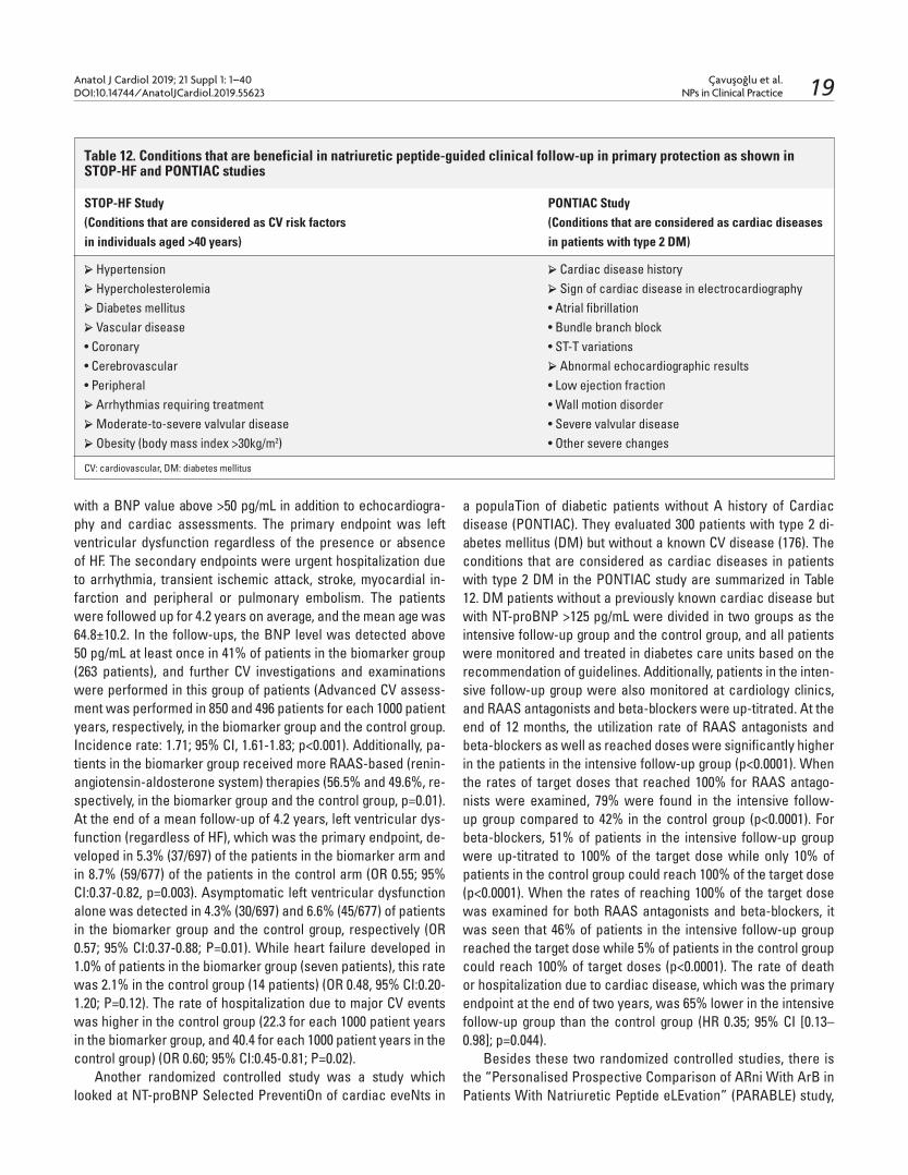
Çavuşoğlu et al.NPs in Clinical Practice
Anatol J Cardiol 2019; 21 Suppl 1: 1–40DOI:10.14744/AnatolJCardiol.2019.55623 19
with a BNP value above >50 pg/mL in addition to echocardiogra-phy and cardiac assessments. The primary endpoint was left ventricular dysfunction regardless of the presence or absence of HF. The secondary endpoints were urgent hospitalization due to arrhythmia, transient ischemic attack, stroke, myocardial in-farction and peripheral or pulmonary embolism. The patients were followed up for 4.2 years on average, and the mean age was 64.8±10.2. In the follow-ups, the BNP level was detected above 50 pg/mL at least once in 41% of patients in the biomarker group (263 patients), and further CV investigations and examinations were performed in this group of patients (Advanced CV assess-ment was performed in 850 and 496 patients for each 1000 patient years, respectively, in the biomarker group and the control group. Incidence rate: 1.71; 95% CI, 1.61-1.83; p<0.001). Additionally, pa-tients in the biomarker group received more RAAS-based (renin-angiotensin-aldosterone system) therapies (56.5% and 49.6%, re-spectively, in the biomarker group and the control group, p=0.01). At the end of a mean follow-up of 4.2 years, left ventricular dys-function (regardless of HF), which was the primary endpoint, de-veloped in 5.3% (37/697) of the patients in the biomarker arm and in 8.7% (59/677) of the patients in the control arm (OR 0.55; 95% CI:0.37-0.82, p=0.003). Asymptomatic left ventricular dysfunction alone was detected in 4.3% (30/697) and 6.6% (45/677) of patients in the biomarker group and the control group, respectively (OR 0.57; 95% CI:0.37-0.88; P=0.01). While heart failure developed in 1.0% of patients in the biomarker group (seven patients), this rate was 2.1% in the control group (14 patients) (OR 0.48, 95% CI:0.20-1.20; P=0.12). The rate of hospitalization due to major CV events was higher in the control group (22.3 for each 1000 patient years in the biomarker group, and 40.4 for each 1000 patient years in the control group) (OR 0.60; 95% CI:0.45-0.81; P=0.02).
Another randomized controlled study was a study which looked at NT-proBNP Selected PreventiOn of cardiac eveNts in
a populaTion of diabetic patients without A history of Cardiac disease (PONTIAC). They evaluated 300 patients with type 2 di-abetes mellitus (DM) but without a known CV disease (176). The conditions that are considered as cardiac diseases in patients with type 2 DM in the PONTIAC study are summarized in Table 12. DM patients without a previously known cardiac disease but with NT-proBNP >125 pg/mL were divided in two groups as the intensive follow-up group and the control group, and all patients were monitored and treated in diabetes care units based on the recommendation of guidelines. Additionally, patients in the inten-sive follow-up group were also monitored at cardiology clinics, and RAAS antagonists and beta-blockers were up-titrated. At the end of 12 months, the utilization rate of RAAS antagonists and beta-blockers as well as reached doses were significantly higher in the patients in the intensive follow-up group (p<0.0001). When the rates of target doses that reached 100% for RAAS antago-nists were examined, 79% were found in the intensive follow-up group compared to 42% in the control group (p<0.0001). For beta-blockers, 51% of patients in the intensive follow-up group were up-titrated to 100% of the target dose while only 10% of patients in the control group could reach 100% of the target dose (p<0.0001). When the rates of reaching 100% of the target dose was examined for both RAAS antagonists and beta-blockers, it was seen that 46% of patients in the intensive follow-up group reached the target dose while 5% of patients in the control group could reach 100% of target doses (p<0.0001). The rate of death or hospitalization due to cardiac disease, which was the primary endpoint at the end of two years, was 65% lower in the intensive follow-up group than the control group (HR 0.35; 95% CI [0.13–0.98]; p=0.044).
Besides these two randomized controlled studies, there is the “Personalised Prospective Comparison of ARni With ArB in Patients With Natriuretic Peptide eLEvation” (PARABLE) study,
Table 12. Conditions that are beneficial in natriuretic peptide-guided clinical follow-up in primary protection as shown in STOP-HF and PONTIAC studies
STOP-HF Study PONTIAC Study(Conditions that are considered as CV risk factors (Conditions that are considered as cardiac diseasesin individuals aged >40 years) in patients with type 2 DM)
➢ Hypertension ➢ Cardiac disease history➢ Hypercholesterolemia ➢ Sign of cardiac disease in electrocardiography➢ Diabetes mellitus • Atrial fibrillation➢ Vascular disease • Bundle branch block• Coronary • ST-T variations• Cerebrovascular ➢ Abnormal echocardiographic results• Peripheral • Low ejection fraction➢ Arrhythmias requiring treatment • Wall motion disorder➢ Moderate-to-severe valvular disease • Severe valvular disease➢ Obesity (body mass index >30kg/m2) • Other severe changes
CV: cardiovascular, DM: diabetes mellitus

Çavuşoğlu et al.NPs in Clinical Practice
Anatol J Cardiol 2019; 21 Suppl 1: 1–40DOI:10.14744/AnatolJCardiol.2019.5562320
the results of which are expected to be obtained in the future. PARABLE is an ongoing study designed to investigate the effect of sacubitril/valsartan on the change of left atrial volume index with cardiac MR (primary endpoint) at the end of 18 months in 250 patients who have hypertension and/or diabetes mellitus and high natriuretic peptide level (BNP>50 pg/mL, NT-proBNP> 150 pg/mL) and are over 40 years of age (177).
Recommendations of international guidelines regarding NPs in primary prevention are summarized in Table 13. Based on the STOP-HF and PONTIAC studies, 2014 the Canadian HF Guidelines recommended the use of NPs for preventing HF in individuals with risk factors playing a role in HF develop-ment (178). The Canadian guidelines recommended the use of BNP level of >100 pg/mL and NT-proBNP level of >300 pg/mL to prevent unnecessary screenings. Referring to the same studies, the 2017 American Guideline for the Management of Heart Failure recommended NP-based screening in patients with HF risk factors as the optimization of guideline-directed medical therapy by a cardiovascular disease specialist could prevent the development of left ventricular systolic or diastolic dysfunction or new-onset HF in individuals with risk factors for HF development (34).
There is a need for conducting studies to determine opti-mal levels in risk classification of natriuretic peptides and their concurrent use with other cardiac biomarkers such as troponin, ST-2, galectin-3, etc. It is clear that additional evidence is also necessary to determine the optimal medical treatment recom-mended for preventing the development of, particularly, HF and other CV diseases in patients with elevated NP and without a history of CV disease.
Consequently, it is very important to prevent HF and CV dis-eases before they develop when both mortality rates and the excessive burden on the country’s economy are taken into con-sideration. It is inevitable to obtain satisfactory results in order to prevent the development of CV diseases as a result of good definition of the individuals at risk in primary prevention, mea-surement of NP in these individuals, and evidence of optimal fol-low-up and treatment in high-risk individuals.
12.0 Natriuretic Peptides in the Pulmonary Arterial Hypertension – Mehmet Serdar Küçükoğlu
Although many biomarkers related to pulmonary arterial hypertension (PAH) and pulmonary vascular remodelling were investigated, only BNP and NT-proBNP were found to be asso-ciated with myocardial dysfunction and prognosis in the follow-up. Therefore, PH centers generally use these two biomarkers in routine practice and clinical studies. These two biomarkers are not specific to PH and their levels might increase in many structural and functional heart diseases. As they are released secondary to pulmonary vascular disease induced afterload increase on the right ventricle, they are indirect indicators of vasculopathy. Both of them are released in response to over-stretch of cardiomyocytes and are strongly correlated with right ventricular dysfunction which is the cause of death in PAH patients. The use of these two biomarkers are recommended by the up-to-date international guidelines for risk and progno-sis assessment of PAH patients at the time of diagnosis and in follow-up (22, 179).
There is no evidence showing the superiority of BNP use to the use of NT-proBNP. While BNP is less affected by kidney functions and more correlated with pulmonary hemodynamic parameters, NT-proBNP is a more potent indicator of prognosis (180).
BNP levels increase with many diseases that cause PH. Th-ese diseases primarily include idiopathic PAH (IPAH), PAH as-sociated with connective tissue disorders, PAH associated with congenital heart disease, PH associated with interstitial lung dis-ease and chronic obstructive lung disease, and chronic throm-boembolic PH (CTEPH). BNP levels are correlated with hemody-namic indicators, functional capacity and exercise capacity. BNP also has a prognostic significance in patients with IPAH, chronic lung disease and pulmonary embolism (181). Recently, the NT-proBNP molecule, which is an alternative to BNP and provides the same information but has a more stable structure, has be-come more popular. There are many studies that investigate the role of NT-proBNP in PAH patients. In these studies, NT-proBNP levels were found to be higher in PAH patients than in the con-
Table 13. Recommendations of the Canadian HF guideline and ACC/AHA HF guidelines for the use of NPs in primary protection
Recommendations of the 2014 Canadian HF Guideline Recommendations of the 2017 ACC/AHA HF Guideline
NPs can be used as a strategy for preventing HF in NP-guided screening that includes guideline-driven medical therapyindividuals with risk factors that have an effect optimized by a cardiovascular disease specialist in patientson the development of HF. who are at risk of developing HF can be used to prevent left ventricular systolic or diastolic dysfunction or new-onset HF. It is recommended to perform more frequent monitoring Class IIa, Evidence Level B-Rand intensify the current therapy in individualswith BNP >100 pg/mL and NT-proBNP >300 pg/mL.
HF: heart failure

Çavuşoğlu et al.NPs in Clinical Practice
Anatol J Cardiol 2019; 21 Suppl 1: 1–40DOI:10.14744/AnatolJCardiol.2019.55623 21
trol group and correlated with hemodynamic parameters and peak oxygen consumption. Again, many studies showed that high NT-proBNP levels correlated with high mortality and a decrease in NT-proBNP levels through treatment increased survival (182).
BNP and NT-proBNP are important biomarkers in detect-ing right ventricular dysfunction in PAH patients. A study com-paring patients with right ventricular pressure (CTEPH) and/or volume (atrial septal defect-ASD) overload with the control group found plasma BNP levels to be significantly higher in the patients with pressure overload than in the other two groups (183). BNP levels were also found to be significantly higher in patients with volume loading than the control group. In this pa-tient group, plasma BNP levels negatively correlated with RVEF and positively correlated with right ventricular end-diastolic volume index (RVEDVI).
Apart from right ventricular function, various studies showed BNP and NT-proBNP’s correlation with functional capacity, ex-ercise performance (six-minute walking distance, peak oxygen consumption), echocardiographic and hemodynamic indicators which had previously found to be associated to survival in PAH patients. In a study which investigated prognostic significance of plasma natriuretic peptides in PAH patients and demonstrated that baseline plasma BNP levels were an independent indicator of mortality, it was seen that baseline plasma BNP levels increased as the functional capacity of patients deteriorated. Another study showed that baseline BNP values were negatively correlated with exercise performance indicators (6MW distance and peak VO2), and based on hemodynamic indicators, BNP had a positive corre-lation with pulmonary vascular resistance (PVR), pulmonary artery pressure (PAP) and right atrial pressure (RAP) and a negative cor-relation with cardiac index (CI) in PAH patients (184).
The relationship of NT-proBNP with echocardiographic right ventricular function indicators, baseline functional status and long-term survival was also investigated in PAH patients. Accordingly, NT-proBNP was found to have a positive correlation with echocar-diographic RV/LV diastolic area ratio, right ventricular diameter, Tei index, diameter and respiratory collapsibility of inferior vena cava (IVC) and presence of pericardial effusion, and a negative correla-tion with RV acceleration time. No correlation was found between tricuspid regurgitation peak velocity and NT-proBNP.
Many studies used BNP and NT-proBNP to determine clini-cal severity of the disease in PAH patients. These two natriuretic peptides are attention-grabbing as simple, non-invasive and ob-server-independent parameters particularly in identifying PAH patients who develop right heart failure. They are correlated with parameters that show the severity of the disease like functional class, 6MW distance, peak VO2 and hemodynamic variables (RAP, PVR, CI). Many studies conducted in recent years showed that NT-proBNP also, correlated with many parameters (CI, RAP, PVR and mixed venous O2 saturation) showing the severity of PAH, and used as prognostic indicators. Investigating the relation-ship between high baseline NT-proBNP levels and prognosis in PAH patients, Mauritz et al. (185) found that serial NT-proBNP
measurements in 198 PAH patients predicted survival better than a single baseline NT-proBNP measurement. According to the results of this study, ≥1256 pg/ml serum NT-proBNP level at the time of diagnosis correlated with poor prognosis in PAH patients. Additionally, a decrease of >15%/year NT-proBNP was associated with increased survival. Nickel et al. showed in their study that changes in NT-proBNP lev-el closely correlated with hemodynamic changes in terms of prognosis. Prognosis of pa-tients whose baseline NT-proBNP level was >1800 pg/ml and decreased with treatment was similar to those with baseline NT-proBNP <1800 pg/ml (186).
In a study by Fijalkowska et al. (187), baseline NT-proBNP level >1400 pg/ml was associated with long-term poor prognosis both in all PH patients and in the IPAH group. This study also showed that survival was better in patients whose NT-proBNP levels re-duced below this value through treatment. In a study by Nagaya et al. (181), patients with a baseline plasma BNP level ≥150 pg/ml had more mortalities in the long-term follow-up. Patients whose BNP levels during PAH-specific treatment could be maintained below 180 pg/ml had better survival outcomes. Consequently, this study highlighted that baseline BNP levels were the only non-invasive marker that could be associated with mortality in a multivariate analysis, and follow-up BNP levels were signifi-cant in terms of prognosis and should be monitored regularly to predict the endpoint. It is also mentioned that mortality can be lower in patients whose BNP levels can be maintained within the targeted interval through treatment. A study monitoring patients who developed scleroderma-associated PAH concluded that increased NT-proBNP levels were directly associated with the severity of PAH, NT-proBNP >395 pg/ml levels meant high poten-tial of PH development in programs which screened scleroderma patients in terms of PH development, and baseline and serial NT-proBNP measurements directly correlated with survival. Ac-cordingly, it was emphasized that a ten-fold increase in follow-up NT-proBNP levels was associated with three times more mortal-ity and demonstrated treatment failure (188, 189).
BNP and NT-proBNP are also used to monitor response to treatment in PAH patients. A study monitoring PAH patients compared baseline and follow-up BNP levels of patients who survived and died under prostanoid therapy. The comparison re-vealed that while BNP levels tended to decrease in follow-ups in patients who survived the prostanoid therapy, they tended to increase in the patients who died despite therapy (190, 191). Another study showed that NT-proBNP levels decreased in a 12-month follow-up under IV epo-prostenol therapy in PAH pa-tients, and this decrease correlated with a decrease in PVR (192, 193). Hemodynamic improvement with postoperative IV epoprostenol therapy in CTEPH patients who have undergone endarterectomy can be monitored with BNP levels. Persistently elevated BNP in the postoperative period shows the presence of residual PH (194, 195).
The fact that natriuretic peptides are closely associated with the severity of the disease, the degree of right ventricular

Çavuşoğlu et al.NPs in Clinical Practice
Anatol J Cardiol 2019; 21 Suppl 1: 1–40DOI:10.14744/AnatolJCardiol.2019.5562322
dysfunction and prognosis in PAH, has led to these biomark-ers being included in the guidelines. BNP or NT-proBNP levels measured at the time of diagnosis of a PAH patient can identify one-year mortality risk of the disease when accompanied with other prognostic parameters (functional capacity, clinical right heart failure and its symptoms, six-minute walking distance, cardiopulmonary exercise test and hemodynamic parameters) (22). The Pulmonary Arterial Hypertension guideline published in 2015 determined low-risk (annually <5% mortality risk) values for BNP and NT-ProBNP as <50 ng/dl and <300 ng/dl (Table 14). BNP >300 ng/dl and NT-proBNP >1400 ng/dl indicate that an-nual mortality risk is >10%. Monitoring BNP/NT-proBNP levels with treatment in the follow-up and reducing these values to a low-risk level stand out among the most important findings in
terms of prognosis. Therefore, measuring BNP/NT-proBNP reg-ularly during the follow-up period is recommended with some other tests (Table 15) (22).
Some studies also questioned whether BNP and NT-proBNP could be used as a screening biomarker in the early diagnosis of disease in PAH patients. Although the use of natriuretic peptides in the diagnosis of right heart disease lacks specificity compared to their use in the diagnosis of left heart disease, they still have a potential for use for this purpose (196). However, both peptides should be examined in large studies to be used as diagnosis markers in diagnosing PAH and ruling out the disease. Their ef-ficacy and safety in diagnosing and ruling out in patient groups which constitute PAH risk should be examined. Moreover, using them with other biomarkers in current diagnosis algorithms will
Table 15. PAH follow-up recommendations (22)
At baseline Every 3-6 Every 6-12 3-6 months after In case of clinical months months changes in therapy worsening
Medical assessment and determination + + + + +of functional classECG + + + + +6MWT/Borg dyspnoea score + + + + +CPET + + +Echo + + + +Basic lab + + + + +Extended lab + + +Blood gas analysis + + + +Right heart catheterization + + + +
Basic Laboratory: Blood count, serum creatinine, sodium, potassium, AST, ALT, Bilirubins, BNP/NT-proBNPAdvanced Laboratory: TSH, Troponin, uric acid, blood iron levels and other necessary tests
Table 14. Risk Assessment in PAH (22)
Determinants of prognosisa (estimated I-year mortality) Low risk <5% Intermediate risk 5-10% High risk >10%
Clinical signs of right heart failure Absent Absent PresentProgression of symptoms No Slow RapidSyncope No Occasional syncopeb Repeated syncopec
WHO functional class I, II III IV6MWD >440 m 165-440 m <165 mCardiopulmonary exercise testing Peak VO2 >15 ml/min/kg Peak VO2 Peak VO2 <11 ml/min/kg (>65% pred.) 11-15 ml/min/kg (35-65% pred.) (<35% pred.) VE/VCO2 slope <36 VE/VCO2 slope 36-44.9 VE/VCO2 slope ≥45NT-proBNP plasma levels BNP <50 ng/l BNP 50-300 ng/l BNP >300 ng/l NT-proBNP <300 ng/l NT-proBNP1400 ng/l NT-proBNP >1400 ng/lImaging (echocardiography, CMR imaging) RA area <18 cm2 RA area 18-26 cm2 RA area >26 cm2
No pericardial effusion No or minimal, pericardial Pericardial effusionHaemodynamics RAP <8 mm Hg effusion RAP >14 mm Hg CI ≥2.5 l/min/m2 RAP 8-14 mm Hg CI <2.0 l/min/m2
SvO2 >65% CI 2.0-2.4 l/min/m2 SvO2 <60% SvO2 60-65%

Çavuşoğlu et al.NPs in Clinical Practice
Anatol J Cardiol 2019; 21 Suppl 1: 1–40DOI:10.14744/AnatolJCardiol.2019.55623 23
increase their screening sensitivity. A study including 49 sclero-derma patients (23 with PAH and 26 without PAH) detected NT-proBNP levels to be higher in scleroderma patients with PAH than those without PAH (3365 pg/ml vs. 347 pg/ml). The study found a positive correlation between NT-proBNP and PVR, mean PAB and right ventricular end-diastolic pressure (RVEDP). The area under curve (ROC) analysis showed that the upper normal limit of 395.34 pg/ml for NT-proBNP had 69% sensitivity and 100% specificity in diagnosing PAH in this patient group. Consequently, it was highlighted that NT-proBNP was a potentially beneficial biomarker with its high specificity and negative predictive value in diagnosing scleroderma-associated PAH and could play a significant role in risk assessment and treatment follow-up al-gorithms in the future (194). Another study investigating the role of NT-proBNP in scleroderma-associated PAH found NT-proBNP elevation directly correlated with the severity of PAH and empha-sized the safety of NT-proBNP in screening this patient group. DETECT (DETECTion of PAH in SSc) is a prospective study that questions the development of PAH in patients with scleroderma and includes 500 patients. Electrocardiography (ECG), echocar-diography (echo) and NT-proBNP were used in the screening. The values of these parameters in diagnosis and differential di-agnosis were compared to right heart catheterization (RHC), a gold standard in diagnosis. Scleroderma patients with and with-out PAH were identified and all three variables were examined in these patient groups. Consequently, it was emphasized that ECG, echo and natriuretic peptides as well as clinical assessment and physical examination were beneficial in terms of early detection of PAH development in the first assessment and follow-up of PAH patients (197).
In conclusion, natriuretic peptides are beneficial in the di-agnosis, risk stratification, follow-up and prognostic assess-ment of PAH patients. NT-proBNP stands out more as a more stable biomarker. Both natriuretic peptides can show early RV dysfunction and haemodynamic deterioration. Their use in prognostic assessment and treatment follow-up is beneficial. The increase in BNP levels in treated patients indicates wors-ening of PAH and inadequate target-oriented therapy. Our po-tential treatment targets are <50 ng/ml for BNP and <300 ng/ml for NT-proBNP. The use of natriuretic peptides for screening in the early diagnosis of disease should be examined in larger studies in terms of validity.
13.0 Natriuretic Peptides in the Assessment of Risk and Prognosis in Acute Coronary Syndromes – Murat Özdemir
Prognostic assessment by biomarkers in acute coronary syndromes (ACS) traditionally utilizes cardiac troponins as the biomarker. An increase of troponin in the presence of ischemia severe enough to cause myocardial damage is an absolute com-ponent in the contemporary definition of “myocardial infarction”, and it is known that prognosis is impaired as troponin levels in-
crease. However, troponins are associated with only one (my-ocardial ischemia) of many pathobiological processes that oc-cur in the course of ACS and do not quite reflect other potential events (such as hemodynamic stress, left ventricular dysfunction and strain). In recent years, various biomarkers were investi-gated in ACS cases for both short-term (in-hospital) and long-term prognosis. Natriuretic peptides (NPs) have a significant place among these biomarkers.
This review will try to summarize the current status of NPs in risk and prognosis assessment in ACS.
13.1 Natriuretic peptides (NPs):There are different types of NPs (A, B and C). As Type B na-
triuretic peptide (BNP) is the one whose prognostic significance has been investigated the most in ACSs, this article will focus on BNP’s prognostic significance in ACS.
Type B NP (BNP) and its precursor N-terminal proBNP (NT-proBNP) are released from atrial and ventricular myocytes as a result of myocardial stretch (due to pressure or volume load). Myocardial ischemia that occurs in ACS may create both di-astolic and systolic left ventricular dysfunction and therefore myocardial stretch and may lead to an increase in BNP levels. Thus, it should be expected that the greater the extent of is-chemia is, the greater the myocardial stretch and elevation in BNP levels will be. Based on this logic, BNP is rationally ex-pected to be a prognostic indicator (both short- and long-term) in ACS.
13.2 BNP in the assessment of risk and prognosis in ST-seg-ment elevation (STE) ACS:In cases of ST-segment elevation myocardial infarction
(STEMI) treated with thrombolytics, it was reported that BNP above 80 pg/mL on admission was associated with inadequate reperfusion and a 7.2 fold increase in 30-day mortality (198).
ASSENT-2 and ASSENT-PLUS sub-studies reported that NT-proBNP on admission was an independent determinant of one-year mortality in STEMI cases treated with thrombolytics (199).
In 142 STEMI cases treated with primary PCI, it was reported that left ventricular systolic function deteriorated more at follow-up in those with higher NT-proBNP at 72-96 hours of admission (200).
In an analysis of 589 STEMI cases treated with primary PCI, NT-proBNP >2300 pg/mL was found to predict in-hospital mor-tality (201).
In another analysis of 145 ACS cases (80 % with STEMI) com-plicated with cardiogenic shock, NT-proBNP>4500 ng/L at the 12th hour of admission was reported to contribute to the predic-tion of 30-day mortality (202).
In acute MI cases treated with PCI, BNP was shown to pre-dict one-year major adverse cardiac events (203).
In summary, it is possible to say that high NP levels at admis-sion or in-hospital denote poor short- and long-term prognosis in STE ACS cases treated with either thrombolytics or PCI.

Çavuşoğlu et al.NPs in Clinical Practice
Anatol J Cardiol 2019; 21 Suppl 1: 1–40DOI:10.14744/AnatolJCardiol.2019.5562324
13.3 BNP in the assessment of risk and prognosis in non-ST elevation (NSTE) ACS:The TACTICS-TIMI 18 study reported that BNP levels on ad-
mission determined six-month mortality independent of Troponin I and heart failure in 1676 NSTE ACS cases (204). Therefore, it should be considered that BNP provides an additional contribu-tion to troponin in terms of risk identification in NSTE ACS cases.
In NSTE ACS cases monitored in the GUSTO-IV trial, it was shown that NT-proBNP, sampled at a median of 9.5 hours from the onset of symptoms, was strongly correlated with one-year mortality (205). The analysis of diabetic patients in this same study reported that NT-proBNP was an independent determinant of one-year mortality (206).
In ACS cases with normal Troponin T value on admission, six-month mortality was found to be five to ten times higher (different ratios in two different cohorts) when NT-proBNP on admission was above 474 pg/mL (207). This interesting finding is significant as it draws attention to the role of NT-proBNP in medium-term prognosis assessment of cases who are considered to have un-stable angina pectoris (ACS+normal troponin).
A study examining a total of 390 ACS cases (a mixed group of unstable angina, STEMI and NSTEMI) found that proBNP per-formed better than Troponin T in predicting major adverse car-diac events both in-hospital and at six-month follow-up (208).
A follow-up of 132 ACS cases showed that heart failure and duration of hospitalization was longer, and in-hospital mortality and cardiogenic shock development trended to increase among those with NT-proBNP>474 pg/mL at hospitalization (209).
An analysis of 5174 NSTE ACS cases who were all revascu-larized in the PLATO study reported that NT-proBNP at random-ization played a role in predicting the occurrence of one-year death/spontaneous MI (210).
A single center registry analysis of 1324 NSTE ACS cases re-ported that NT-proBNP on admission played a role in predicting 30-day mortality (211).
A study on cardiovascular safety of an antidiabetic drug in 7226 cases with ACS and Type 2 Diabetes Mellitus (DM) found that NT-proBNP was among the independent total mortality de-terminants at a median two-year follow-up (212).
Another study on cardiovascular safety of an antidiabetic drug in 5225 Type 2 DM cases with recent ACS reported that, in a mean follow-up of 26 months, BNP and NT-proBNP were the most powerful predictors of all cardiovascular endpoints (death, cardiovascular death, heart failure, MI and stroke) independent of heart failure or any cardiovascular disease history (213).
In a report which investigated the “very long-term” prognos-tic value of NT-proBNP in 224 NSTE ACS cases, NT-proBNP on admission was an independent determinant of total mortality at a median follow-up of 9.34 years;a level > 100 pg/mL was associ-ated with a 6.24 fold increased risk (214).
In summary, on the basis of data from many studies, it is pos-sible to say that NP measurement plays a role in both short- and long-term prognosis assessment in NSTE ACS.
13.4 When to collect samples, and can temporal changes in NP levels be clinically significant?The time of drawing a serum sample for NP levels during the
course of ACS is a point that should be discussed. As seen above, most of the studies in this regard used the levels of samples taken on hospital admission. An analysis of NSTE ACS cases in the FRISC-II trial concluded that “NT-proBNP predicts two-year mortality regardless of the time of sampling (on admission, day 2, week 6, months 3 and 6)”. However, NT-proBNP sampled in the “acute phase” and NT-proBNP sampled in a “chronic and relatively stable period” can be a better indicator of “myocardial damage” and “mortality”, respectively (215).
The importance of NP level changes over time (temporal changes) after ACS has not been adequately evaluated. A study investigating this issue reported that in 276 cases admitted to the emergency service with chest pain, five successive measure-ments of NT-proBNP in the first 24 hours did not contribute to its 90-day prognostic value (216).
On the other hand, a large observational study including 4497 ACS (STEMI and NSTEMI) cases demonstrated that serial mea-surements of NT-proBNP in one year could provide valuable infor-mation. In fact, although BNP values above 80 pg/mL on admission, at month 4 and month 12 could predict two-year mortality or de-velopment of heart failure with different hazard ratios (hazard ra-tios=2.5, 3.9, 4.7, respectively), what is more impressive is that BNP values which were normal at baseline and increased at month 4 indicated a 4.7 fold increased risk and, on the contrary, BNP val-ues which were high at baseline and returned to normal at month 4 indicated only a 1.7 fold increased risk (217). Consequently, one might say that BNP levels which tend to rise at follow-up indicate a poor prognosis, and which tend to decrease indicate a good prognosis in cases with ACS. A similar finding was observed in a sub-study of the OPTIMAAL trial. In this analysis, while baseline NP levels did not have any prognostic significance in cases with acute MI complicated with clinical or radiographic heart failure, NT-proBNP which remained high (non-decreasing pattern) on day 30 pointed to a poor prognosis (218).
An analysis on NSTEMI cases monitored in the TRILOGY ACS study reported that each 40% increase in NT-proBNP level at the 6th month as compared to baseline was associated with a 14% increase in both cardiovascular and total mortality (219). Another study reported that, in 5450 Type 2 DM cases with recent ACS, a 3-4 times increase in NP levels measured at follow-up compared to baseline could point out to the emergence of a need for hospi-talization related to heart failure (220).
In summary, it seems reasonable that sampling for NP in the course of ACS be done at admission. There is evidence showing the potential benefits of measuring NP levels in a serial manner at follow-up; however, it is not possible to make a conclusive comment in this regard at the moment.
13.5 Conditions that might affect the prognostic value of NT-proBNP in ACS: An analysis investigating whether the prognostic value of
NT-proBNP in ACS was affected by age or not reported that dif-

Çavuşoğlu et al.NPs in Clinical Practice
Anatol J Cardiol 2019; 21 Suppl 1: 1–40DOI:10.14744/AnatolJCardiol.2019.55623 25
ferent NT-proBNP cut-off values were observed in different age groups for the prediction of total mortality and this cut-off value increased with age; however, NT-proBNP was an independent predictor of total mortality in all age groups (221).
Other factors potentially affecting the prognostic value of NT-proBNP in ACS might be body weight and composition. A study reported that NT-proBNP lost its prognostic significance in predicting total and CV mortality in ACS in cases with high body mass index, lean mass index and percent body fat (222).
13.6 Can natriuretic peptides contribute to other risk stratifi-cation methods (scoring systems)?NT-proBNP is one of the biomarkers used in the BETTER risk
scoring system which was established for one-year prognosis assessment in unstable angina pectoris and reported to perform well in this regard (223).
In STEMI cases treated with primary PCI, NT-proBNP was reported to provide additional contribution to predict in-hospi-tal mortality using the Canada Acute Coronary Syndrome Risk Score, which is another risk prediction score for ACS (201).
An analysis evaluating many biomarkers in NSTE ACS demonstrated that, when NT-proBNP was added to the GRACE risk score which is a risk scoring system frequently used in acute coronary syndromes, the predictive strength of the score increased for six-month death/MI (224). On the other hand, a registration report of 1324 cases showed that NT-proBNP mea-sured on admission for NSTE ACS was similar to the GRACE score and superior to the TIMI risk score in terms of predicting 30-day mortality; it did not contribute to the GRACE score (211). An analysis conducted in 1892 cases to investigate the contri-bution of various biomarkers (high-sensitivity troponin T, NT-proBNP and high-sensitivity CRP) to the GRACE score reported that NT-proBNP (at a threshold of 174 ng/L) could contribute to the GRACE score in predicting 30-day and one-year mortality and non-fatal MI recurrence (225).
The seven-component OASIS risk score that was created by using five classical risk markers (age, gender, history of MI or stroke, ST-segment deviation and Troponin T) and two additional biomarkers (NT-proBNP and HbA1C) was found to be superior to the TIMI and GRACE risk scores in predicting the one-year rate of the composite endpoint of CV death/MI/stroke in NSTE ACS cases. The NT-proBNP values used in this scoring system was evaluated in four quartiles and the scoring system looks quite complex and unpractical (226).
13.7 Can natriuretic peptides guide the treatment approach in ACS? Myocardial function after ACS is one of the most significant
factors that determine prognosis. Myocardial dysfunction ob-served in the early period of an event might be misguiding, and it is important to predict the possibility of improvement at follow-up. A study reported that, when NT-proBNP measured on day 3 of infarction in STEMI cases treated with primary PCI was above
1115 pg/mL, no improvement occurred in global and regional my-ocardial function at follow-up (227).
A meta-analysis of five studies investigating the role of BNP/NT-proBNP in the guidance for early invasive treatment strate-gies in acute coronary syndromes showed that such strategies could play a role, but its contribution to the existing approaches was controversial (228).
An analysis reported that NT-proBNP played a role in the identification of ACS cases that would benefit from ACE-i treat-ment in prognostic sense (death/MI/hospitalization for HF), and ACE-i treatment showed prognostic benefit only in cases with NT-proBNP levels in the highest quartile (229).
13.8 Is there a cut-off value that can be helpful in clinical use?It is seen that different cut-off values have been used for BNP
or NT-proBNP in studies conducted over time. The TACTICS-TIMI 18 study showed that if BNP on hospital-
ization was >80 pg/mL, seven-day and six-month mortality rates were higher (204).
The ENTIRE-TIMI 23 sub-study showed that reperfusion was insufficient and seven-day mortality was higher in STEMI cases treated with thrombolytics and had a BNP > 80 pg/mL (198).
A sub-group analysis of diabetic cases in GUSTO-IV trial recorded that NT-proBNP above 669 ng/mL in samples taken at a median 9.5 hours of NSTE ACS increased one-year mortality by 2.0 times (95% CI: 1.1-3.6) (206).
NT-proBNP above 474 pg/mL indicated a five to ten fold in-crease in six-month total mortality in ACS cases with normal Troponin T levels (207). When the same cut-off value was used by another group, NT-proBNP above 474 pg/mL indicated a poor in-hospital prognosis in ACS (209).
In acute MI cases treated with PCI, it was reported that a BNP value above 285.73 pg/mL predicted one-year major adverse cardiac events with 96% sensitivity and 77% specificity (203). Another study reported that the best threshold NT-proBNP value for predicting in-hospital mortality in STEMI cases treated with primary PCI was 2300 pg/mL (201).
In ACS complicated with cardiogenic shock, the best thresh-old NT-proBNP value for predicting 30-day mortality was 4500 ng/L in samples taken at the 12th hour (202).
It was reported that NT-proBNP of 100 pg/mL or above on admission for NSTE ACS was associated with 6.24 times in-creased total mortality at an approximately ten-years long fol-low-up (214).
Another very recently published study reported that a 174 ng/L cut-off value of NT-proBNP contributed to the GRACE score in predicting the 30-day and 1-year mortality and non-fatal MI recurrence rates (225).
As is seen, various NPs were measured in various patient populations in different studies, and most of them reported dif-ferent NP cut-off values for prognosis assessment.

Çavuşoğlu et al.NPs in Clinical Practice
Anatol J Cardiol 2019; 21 Suppl 1: 1–40DOI:10.14744/AnatolJCardiol.2019.5562326
13.9 What do the guidelines recommend?When the current North American and European guidelines
are examined, it is seen that STE ACS guidelines do not refer to natriuretic peptides in either risk stratification or prognosis as-sessment.
Among the guidelines for NSTE ACS, the ACC/AHA guideline published in 2014 (230) recommends BNP or NT-proBNPmeasure-ment at a Class IIb (Evidence level=B) indication for assessing risk in cases with suspected ACS. Similarly, NP measurement is rec-ommended as a Class IIB (Evidence level=B) indication in terms of providing prognostic data in addition to troponin, and the biomarker that is recommended for prognostic assessment in NSTE ACS as a Class I (Evidence level=B) indication is troponin (230).
The ESC NSTE ACS guideline published in 2015 (231) accepts that natriuretic peptides have been intensively evaluated and confirmed to provide prognostic data in addition to troponin. However, as such assessment does not have any demonstrated benefit in disease management yet, the benefit of these biomark-ers in addition to GRACE 2.0 risk calculation is considered mar-ginal and NP measurement for prognostic purposes is not rec-ommended.
13.10 Conclusion and summary:The role of natriuretic peptide levels in both in-hospital and
mid-to-long term prognosis assessment in acute coronary syn-dromes has been investigated comprehensively in registry stud-ies and post hoc analyses of prospective trials. There is a large body of evidence showing that high NP levels indicate high in-hospital risk and poor mid-to-long term prognosis. However, it is not quite clear how this information can contribute to disease management and whether or not there is a contribution of mea-suring NP levels in addition to troponins and/or the established risk scoring systems. There are still some questions that remain answered, such as on which phase/day of acute coronary syn-drome should sampling be performed and what, if any, the ideal cut-off value that will indicate the relevant risk is.
14.0 Natriuretic Peptides in the Management of Mitral and Aortic Valve Diseases – Burcu Demirkan
The timing of surgical interventions for valvular disease is generally determined based on the development of symptoms, left ventricular (LV) diameter and systolic function changes or elevation in systolic pulmonary artery pressure (232). However, advanced age or comorbidities might lead to incorrect evalua-tion of patients in terms of symptoms. Echocardiography is an examination mainly used in the assessment and follow-up of valvular diseases and it is a method which is dependent upon image quality and requires experienced operators. Therefore, the use of biomarkers can play a supplementary role in the management of valvular diseases, allow for easy and serial monitorization, reflect the severity of the disease, increase in parallel with the progression of the disease and help distin-
guish the patients who will become symptomatic in the near future. Natriuretic peptides are well-known examples to these biomarkers.
Natriuretic peptides (NPs) are endogenous cardiac hormones which have prognostic value in diagnosing heart failure and guid-ing the treatment. They are released as a result of myocyte stretch from the atrial and ventricular myocardium. The pressure load on aortic stenosis, mitral regurgitation and the volume load in aortic insufficiency lead to the increase of natriuretic peptides (233, 234).
14.1 Aortic StenosisAs society gets older, the prevalence of aortic stenosis (AS)
increases (232). In particular, elderly patients might describe atypical symptoms, or existing symptoms can be attributed to other concomitant diseases. Therefore, it might not always be possible to clearly evaluate when AS becomes symptomatic. The optimal treatment method in symptomatic patients is aortic valve replacement; however, the timing of interventions should be dis-cussed for asymptomatic patients (235). Even though a follow-up option is an optimal and safe approach for asymptomatic patients, the risk of sudden death cannot be eliminated completely (236). In addition to other non-invasive methods, NP can be added as a supplementary method.
Natriuretic peptides increase in relation to the severity of AS (237, 238). This correlation exists with both the transvalvular gradient and aortic valve area (239, 240). NPs were found to be higher in symptomatic AS patients than asymptomatic ones. A study revealed that >66 pg/ml serum BNP levels could determine symptomatic patients (241). Another study shows that the devel-opment of symptoms and the occurrence of the need for surgical intervention in the subsequent six to nine months is less in pa-tients with BNP <130 pg/ml and NT-proBNP <592 pg/ml (239). Both NT-proBNP and BNP levels increase with the severity of aortic stenosis, onset of symptoms and development of LV dysfunction. While BNP is very helpful, particularly in diagnosing patients in the New York Heart Association (NYHA) functional class III-IV, its benefits in distinguishing asymptomatic patients from mildly symp-tomatic patients are much less (242). However, it was observed that symptoms develop earlier in the follow-up in asymptomatic patients with high NP levels compared to those with low NP levels (243). Abnormal blood pressure response to exercise is higher in patients who are asymptomatic but have high BNP levels (244). On the contrary, as opposed to dyspnea, the presence of angina or syncope as a symptom does not appear to correlate with NP levels (243).
Increased NP levels is a significant marker of mortality in both symptomatic and asymptomatic AS (241). In a prospective study by Katz et al. that included patients who were diagnosed with severe AS and had normal LV systolic function, it was observed that patients with BNP >135 pg/ml and NT-proBNP >1150 pg/ml had a higher mortality risk in the long term (245). In another study assessing patients with normal left ventric-ular ejection fraction and AVA <1.2 cm2, the mortality rate was

Çavuşoğlu et al.NPs in Clinical Practice
Anatol J Cardiol 2019; 21 Suppl 1: 1–40DOI:10.14744/AnatolJCardiol.2019.55623 27
around 60% in patients with BNP > 819 pg/ml among patients who were monitored without surgical intervention during a one-year follow-up while no mortality occurred in those with BNP <100 pg/ml during the same period (246). In another study using the BNP ratio calculated with the adjusted BNP value ac-cording to age and gender, survival rates were around 44%, 25% and only around 15% in patients with a BNP ratio of 1-2, a BNP ratio of 2-3, and 3a BNP ratio of 3, respectively (247). Although BNP levels are not helpful in distinguishing true AS patients from pseudo-AS patients in low-flow, low-gradient AS, one-year survival rate is again quite low in patients with 3550 pg/ml BNP levels in a similar way (248). In this study, furthermore, high BNP levels predicted poor outcome independently of con-tractile reserve, defined by increase in stroke volume greater than 20% on dobutamine stress echocardiography (248). This evidence shows that NPs have prognostic value independent of symptoms and LV functions.
It was found that BNP levels showed postoperative all-cause mortalities in a more accurate manner compared to the logis-tic EuroSCORE risk classification, which is frequently used to identify operational risk in patients who will undergo aortic valve replacement surgery (249). On the other hand, AVR has benefits in terms of mortality in patients with high BNP similar to those with low BNP (247). It was found that BNP levels decreased in patients following aortic valve replacement surgery (250). If BNP elevation continues in the postoperative period, this can be an indicator of patient-valve mismatch or permanent ventricular dysfunction (251). BNP levels can help distinguish risky patients in the postoperative period and offer guidance with respect to patients who need follow-ups with closer and more intense treatment.
Among the patients who underwent transcatheter aortic valve implantation (TAVI), an alternative treatment approach for patients with severely symptomatic AS and high surgical risk, those with a baseline BNP > 428 pg/ml were found to have less than a 30-day survival rate (252). In another study monitoring longer-term results, BNP > 202 pg/ml on discharge after a TAVI procedure was found to be correlated with two-year all-cause mortalities and a repeated need of hospitalization (253). There is evidence again in the group of patients who underwent TAVI that NP levels can be helpful in both identifying operational risks and distinguishing patients who could be associated with adverse outcomes after the procedure.
In summary, NPs;• Reflect the severity of the disease in patients with aortic
stenosis• Provide guidance for safe follow-ups without intervention in
asymptomatic AS patients, and play a determinative role in the development of symptoms
• Help identify risks in terms of mortality• In patients who underwent intervention, evaluate the suc-
cess of the procedure can and differentiate patients who may experience negative outcomes after the procedure.
14.2 Aortic RegurgitationIn patients with severe aortic regurgitation (AR), the indica-
tion for surgery is mainly the presence of symptoms and a 50% LV ejection fraction (232). However, there is no intervention sug-gestion for the group of patients who have asymptomatic but hemodynamically severe AR and preserved LV systolic function. Although there is echocardiographic data suggesting a corre-lation between poor outcome and effective regurgitant orifice area, there is a need for a parameter that can determine the risky ones in this group of asymptomatic patients (254).
NT-proBNP is correlated with the degree of AR and func-tional capacity (255).
In a study by Pizzaro et al. involving 294 patients with asymp-tomatically severe AR and preserved LV systolic function, BNP >130 pg/ml was found to be correlated with the development of congestive HF, LV dysfunction and adverse events including death (256). This study found that echocardiographic LV end-systolic diameter, regurgitant orifice area and BNP levels were independent indicators of adverse outcomes and the prognostic value of BNP was higher than other parameters. The risk of ad-verse events is also increased in patients who show increased BNP elevations within one year in serial measurements (256).
Detection of AR after the TAVI procedure is a frequent compli-cation that determines mortality. In a study by Kaneko et al. which included 856 patients who underwent the TAVI procedure and de-velopment of more than mild AR was considered to be significant at the end of the procedure, and it was found that not every patient who developed significant AR was at the same risk and some base-line characteristics could influence the risk status. This study also revealed that the factors that identified patients with higher risk were baseline LVEF and NT-proBNP levels. In patients with 340% baseline LVEF and £5000 pg/ml NT-proBNP, it was found that the presence of significant AR did not increase one-year mortality at the end of the procedure. However, on the contrary, the presence of significant AR after the procedure in patients with LVEF <40% and NT-proBNP >5000 pg/ml, increases mortality in the patients (257).
In summary, NPs;• Reflect the severity and functional capacity of the disease in
aortic regurgitation.• Are prognostic markers that can predict the development of
adverse events in the follow-up.• Might play a potential role in determining patients with high
risk of poor outcome in case of AR developing after a TAVI procedure.
14.3 Mitral RegurgitationDegenerative mitral regurgitation (MR) is a common valvular
disease that is treated surgically. Surgical intervention indica-tions are determined depending upon the presence of symp-toms and/or LV ejection fraction <60%, widened LV end-systolic diameter and the development of pulmonary hypertension and/or atrial fibrillation. Criteria that define surgical indications are factors that also define the development of postoperative mor-

Çavuşoğlu et al.NPs in Clinical Practice
Anatol J Cardiol 2019; 21 Suppl 1: 1–40DOI:10.14744/AnatolJCardiol.2019.5562328
tality and heart failure (232). Mitral regurgitation is a commonly well-tolerated disease and patients with MR can live without symptoms for years. Preload and afterload, contractility and LV ejection fraction are normal in this period, and total stroke volume increases depending on the increased end-diastolic volume. How-ever, this adaptation will eventually deteriorate and the patient will go towards a process that leads to irreversible myocardial damage (258). If patients are not given surgical intervention within the ap-propriate time, HF, pulmonary hypertension and right HF develop-ment may potentially occur (258). While the success of procedures has been increasing over the years thanks to developments in the methods of mitral valve repair, a tendency has occurred towards applying early surgical intervention in patients in the asymptomatic period based on the evidence of decreased risk and better results of early surgery (259). On the contrary, there is data suggesting that early surgical intervention does not reduce all-cause mortalities compared to patients who are monitored without intervention and can even be associated with more fatal strokes and AF develop-ments (259). It is not possible even at the most experienced cen-ters to eliminate the risks of surgical interventions and eliminate the need of mitral valve replacement completely in the case of a failed repair. Therefore, there have been some studies suggesting that NP levels might be helpful in determining the time of surgical intervention more correctly in MR.
BNP increases depending on the severity and symptoms of MR (260). However, it is not possible to assess the degree of MR by taking only BNP values into account (261).
There is evidence of the prognostic value of BNP in asympto-matic MR patients. In a study evaluating patients with normal LV ejection fraction, asymptomatically severe organic MR and exer-cise capacity of 7 METs or above documented through exercise tests, it was seen that patients with a baseline BNP value >105 pg/ml reached the composite endpoint comprising congestive HF symptoms, LV dysfunction or death at the one-year follow-up sig-nificantly more than those with a BNP value <105 pg/ml (262).
A study by Magne et al. found in the follow-up that BNP levels increasing with exercise in asymptomatic patients with MR was an indicator of adverse cardiac events (263).
Another multicenter study that included 1331 patients with degenerative mild to moderate MR and normal LV ejection frac-tion revealed that BNP activation was an indicator of survival and that mortality reached 60% in eight years in patients in the medical follow-up group with a BNP ratio > 4. On the other hand, BNP activation was not an independent indicator of survival in the group who had surgery. Another significant finding from this study was that patients with non-elevated BNP activation (BNP £1) had better prognosis (264). If the patients with asymptomatic severe MR do not have the criteria for surgical intervention and the BNP ratio is low, conservative follow-up of these patients is a very safe approach. Moreover, if surgical intervention indication occurs in a patient and BNP activation also increases in patients, it is still possible to obtain better long-term results with surgical intervention in such patients (264).
In summary, NPs;• Reflect the severity and symptom status of MR• Help determine the patients for whom conservative follow-
ups are safe among those with asymptomatically severe MR• Determine pre- and post-surgery prognosis in patients who
had surgery.
14.4 Mitral StenosisMitral stenosis (MS) is a valvular disease that frequently de-
velops due to rheumatic fever, that carries the risk of pulmonary hypertension and AF development, and that develops slowly with symptoms occurring in adulthood. The indications for interven-tion are presence of mitral valve area (MVA) £1.5 cm2 and symp-tom development (232). However, the development of symptoms might not be noticed in the early stage if the patient changes their life style during the long-term asymptomatic course of the disease. A parameter which is easily accessible, repeatable and comparable in the follow-up of mitral stenosis might provide ad-ditional contribution to echocardiographic data.
Sharma et al. revealed that both ANP and BNP levels in-creased in parallel to the severity of disease, ANP and BNP levels correlated with one another, but for reduced exercise capacity, BNP was a more powerful indicator than ANP in MS patients. An interesting finding from this study is that when MS patients with NP levels within the normal range were compared to the control group, it was found that the left atrial area was wider, resting and exercise pulmonary artery pressures were higher and exercise capacity was reduced in patients with MS (265). Accordingly, cardiac remodeling and early dysfunc-tion might develop in MS patients without BNP increase as well. However, increased BNP levels (> 82 pg/ml) was found to be correlated with wider left atrial area index, higher pul-monary artery pressure and poorer exercise capacity (265). Another study investigated NT-proBNP levels in patients with MS and found that NT-proBNP levels were higher in patients with MS than the control group. NT-proBNP levels were found to be higher in patients with atrial fibrillation than those in si-nus rhythm. Also, it was seen in patients who were assessed through exercise tests that these levels correlated with left atrial diameter after resting and exercise, right ventricular end-diastolic diameter, exercise duration, heart rate and pulmonary artery pressure. Again in this study, NT-proBNP level >251 pg/ml was an indicator of pulmonary artery pressure increasement with exercise above 60 mmHg (266).
Percutaneous mitral balloon valvuloplasty is the method of choice for intervention for eligible patients in the treatment of severe symptomatic MS. It was demonstrated that BNP and NT-proBNP levels decreased after a successful procedure in pa-tients who underwent percutaneous mitral balloon valvuloplasty (267). In another study, again assessing the relationship between NPs and procedure success, NP levels decreased after the procedure in patients only in sinus rhythm while no effect was observed on NP levels in patients with AF. According to the in-

Çavuşoğlu et al.NPs in Clinical Practice
Anatol J Cardiol 2019; 21 Suppl 1: 1–40DOI:10.14744/AnatolJCardiol.2019.55623 29
terpretation of the investigators, procedures performed without development of AF might be more beneficial for patients (268).
In summary, NPs;• Reflect the severity of MS, cardiac remodeling and increased
pulmonary artery pressure.• Can reveal the relationship between the existing symptoms
and MS.• Can provide information about the success and outcomes of
the procedure.Consequently, NPs elevate depending on myocardial stretch
that develops as a result of increased pressure and volume load in valvular diseases. There is evidence showing that NPs can be useful in follow-ups with asymptomatic patients with severe valvular disease, confirming that the existing symptoms are associated with valvular disease in symptomatic patients, monitoring the progression of the disease, determining the prog-nosis and determining patients who need close follow-up. From this perspective, they might provide information in addition to echocardiographic findings. However, there are some limitations that should be taken into consideration while using NPs in valvu-lar diseases. First of all, unlike HF, defined diagnostic predictive values are not available. In addition, conditions such as age, gen-der, obesity and renal function have an effect on NP levels. Some studies preferred to use BNP ratio so that age and gender fac-tors did not affect the assessment adversely. Cardiac conditions such as coronary artery disease, pulmonary embolism and AF which affect NP levels can also coexist. On the other hand, the presence of multiple valvular diseases is not rare and it may not be possible to understand the extent to which the pathology of which valve was altered based on NP levels. It might be helpful for the clinicians to evaluate the changes in NP levels with se-quential measurements after baseline value, instead of making an assessment with only one NP value in the follow-up of pa-tients with valvular diseases. There is not sufficient evidence for the necessity of performing an intervention in case of elevated NP levels in patients with severe valvular disease without inter-vention indications defined in the guidelines, and comprehensive studies are needed to shed light on this issue.
15.0 Natriuretic Peptides in the Identification of Atrial Fibrillation Risks – Ahmet Taha Alper
15.1 IntroductionAtrial fibrillation (AF) is the most common continuous arrhyth-
mia with a prevalence of 2%, and it is diagnosed in one-third of hospitalizations due to cardiac arrhythmia1. Incidence of AF, which is seen in 5% of the population above the age of 65 and around 8-10% above the age of 80, increases with age. Beyond adversely affecting the quality of life, AF is associated with in-creased total mortality, cardiovascular mortality and cardio-vascular morbidity including heart failure, stroke and systemic embolism (269-271). As these risk factors emerging with atrial fibrillation do not occur equally in all patient groups, treatment
strategies are determined through clinical risk scoring systems such as CHADS2 or CHA2DS2VASc scores for risk assessments for stroke. These risk scoring systems exclude some biochemi-cal parameters which are seen to be associated with stroke, and do not provide clear information about risk assessments in other morbidities caused by AF. In our compilation, we tried to summa-rize the role of natriuretic peptides (NP) in risk identification in the case of different AF-related clinical conditions.
15.2 Identifying the occurrence, duration and progression of AFIn their study, Mandalenakis et al. enrolled 528 men and as-
sessed the occurrence of AF with the baseline ANP levels at the end of 16 years. This study demonstrated that ANP levels (a range of 192-253 pg/ml, HR: 3.14 95% CI: 1.59-6.20 p: 0.001 and above 254 pg/ml, HR: 3.36 95% CI: 1.72-6.54, p<0.001) independently pre-dicted the occurrence of AF (272). Additionally, ANP facilitates the occurrence of AF by shortening atrial transmission time and the effective refractory period even in completely healthy atri-ums (273). Mutations occurring in atrial natriuretic peptide en-coding gene (NPPA) cause the synthesis of mutant ANP (mANP) which has five or ten times higher plasma concentration. In the studies, a correlation between mANP and familial AF develop-ment was seen (274).
Today, AF classification is mainly based on the duration of AF and this classification is the main determinant in treatment strategies. Mid-regional pro-ANP (MR-proANP) is a stable frag-ment of ANP prohormone and, according to studies; it appears to be useful in determining the duration of AF. Median MR-proANP concentration was found to be 147.7 [95.3–197.4] pmol/L and 220.4 [154.0 –303.1] pmol/L in subjects with atrial fibrillation du-ration below 48 hours and above 48 hours, respectively (P<0.001) (275). This result suggests that NPs might be useful in estimating onset time and determining treatment strategies accordingly in patients without documented AF. In another study investigat-ing progression from paroxysmal AF to persistent AF, predictive ability of NT-proBNP was not observed while NT-proANP levels could predict progression (276). In order to enable NP-guided treatment, extensive studies are necessary to compare different treatment methods in the context of NP levels in patients who are expected to maintain sinus rhythm and have a high likelihood of progression.
15.3 Identification of AF in patients with cryptogenic strokeCompared to other types of stroke, cardioembolic strokes are
repetitive and have poorer prognosis (277). Patients who have stroke and were not diagnosed with AF are considered to have had a cryptogenic stroke and treated with antiplatelets instead of oral anticoagulants. Long-term monitoring of cryptogenic pa-tients showed that approximately 30% of patients had AF (278). In Find-AF study that included patients with acute cerebral is-chemia, BNP, NT-proBNP and NT-proANP were compared in terms of identifying AF, and BNP levels were established as an

Çavuşoğlu et al.NPs in Clinical Practice
Anatol J Cardiol 2019; 21 Suppl 1: 1–40DOI:10.14744/AnatolJCardiol.2019.5562330
independent AF predictor in this patient group. In this study, the cut-off value with the highest precision and specificity of BNP was 118 pg/ml (279). A meta-analysis evaluated ten prospec-tive studies testing BNP and six prospective studies testing NT-proBNP. BNP and NT-proBNP were found to have equal diagnos-tic value in the identification of cardioembolic stroke. However, BNP was more precise (0.65 95% CI: 0.63-0.68) and NT-ProBNP was more specific (0.93, 95% CI: 0.91–0.94) in the diagnosis (280). In light of this information, long-term monitoring might be rec-ommended for excluding AF in the presence of elevated NP in patients who have had a cryptogenic stroke.
15.4 Biomarker-based scoring in the identification of stroke risk in patients with AFUp-to-date guidelines recommend the CHA2DS2VASc score
for assessing stroke risk in patients with AF. This is a clinical scoring system and does not include biochemical markers. Recent studies have shown that many biomarkers which can show renal and cardiac functions and reflect inflammation and oxidative stress can be used in determining the risk of stroke (281). In light of this, the ABC score have been derived from the ARISTOTLE (282) study (Table 16). Later, validity of this score was confirmed by using data from the STABILITY (283) study (284). In the ABC score, each risk factor (within intervals) is scored from one to ten to calculate the risk of stroke. In this way, it is possible to calculate the risk of stroke by using the total score (Fig. 3). Clinical risk factors that are used in CHA2DS2VASc scores such as hypertension, diabetes mellitus, heart failure, cardiovascular diseases and gender do not provide additional prognostic benefit for the biomarkers that are used in the ABC score. Therefore, these risk factors are excluded from scoring. Compared to the CHA2DS2VASc score, the ABC score predicts stroke better (c-indices are 0.68 vs. 0.62, P<0.001, respectively) and significant sub-groups have a similar superiority. In ad-dition, particularly in patients who have not previously had a stroke or without a TIA history, superiority of the ABC score to the CHA2DS2VASc score (c-indices are 0.68 vs. 0.59 P<0.001, respectively) was more clearly observed (284). Risk factors that are used in the CHA2DS2VASc score are non-modifiable risk factors. As an increase or decrease in stroke risk can be de-termined by biomarkers, the ABC score is presented as a more dynamic scoring system. In parallel to this, treatment strategies that are used in prevention of stroke can vary.
15.5 Identification of recurrence risk in patients who under-went cardioversion and AF ablationNatriuretic peptide levels are associated with enlarged left
atrium volume (285), heart failure and related intracellular cal-cium increase (286). Also, it is known that NPs increase vagal ac-tivity via the cGMP signaling pathway (287, 288). Based on these pathophysiological relationships, their activities were examined to predict AF recurrence after cardioversion and ablation.
A meta-analysis by Xiangdong et al. included 19 studies eval-uating recurrence after cardioversion, and it was seen that base-line BNP/NT-proBNP levels were significantly higher in patients with recurrence than patients who maintained sinus rhythm (SMD -0.70, CI [-0.82, -0.58]). Although meta-analysis results present the prognostic value of NPs, today it is not possible to determine the cardioversion based on biomarker level. Also, a clear cut-off value that can reflect the maintenance of sinus rhythm is needed.
Although radiofrequency catheter ablation treatment is one of the main treatment modalities in preventing AF attacks, recur-rence after ablation is a significant problem (289). A meta-analysis including 23 studies measuring ANP, BNP and NT-proBNP levels to assess ablation recurrence, all mentioned NPs were shown to predict recurrence after ablation (95% CI:0.12–0.60, P=0.004; 95% CI:0.35–1.11, P=0.0002; 95% CI:0.64–1.87, P=0.0001, respectively) (290). The main ablation strategy in paroxysmal AF is pulmonary vein isolation. However, there is not any consensus on what kind of a strategy can be followed in addition to pulmonary vein isola-tion in persistent AFs. Studies comparing ablation strategies on the basis of preoperative NP level in patients with AF may allow NP-guided treatment planning in the future.
Figure 3. Nomogram to calculate ABC score. The score is determined on the scale for each predictor and the total score is obtained. One-year or three-year risk of stroke is calculated according to the total score. Except for nomogram, it is also possible to make practical calculations with web-based applications *Troponin T high-sensitivity (ng/L) also could be use
Points0 1 2 3 4 5 6 7 8 9 10
Prior stroke/TIAStroke/TIA
No
44
1
0 5
2 5 10
10 15 20 25 30
25 50 100 200 400 800 1500
0.01
0.01
0.02
0.02
0.03
0.03
0.05
0.05
0.150.1
0.1 0.2 0.3
3000 5900
30 75 180
55 65 75 90
Troponin l (ng/L)*
NT-proBNP (ng/L)
1-year risk of stroke/SE
3-year risk of stroke/SE
Total points
Age
Table 16. ABC score criteria
Scoring Criteria Interval
Age (years) (age-A) 44-90 Biomarkers Troponin I Hs (ng/l) 1-180(Biomarkers-B) NT-proBNP(ng/l) 25-5900Clinical history Stroke or TIA Yes-No(clinical history-C)

Çavuşoğlu et al.NPs in Clinical Practice
Anatol J Cardiol 2019; 21 Suppl 1: 1–40DOI:10.14744/AnatolJCardiol.2019.55623 31
15.6 Identifying the risk of AF development in postoperative periodIt is estimated that more than 5 million thoracic surgeries
are performed annually in the world. Atrial fibrillation is a signif-icant complication that develops in patients during the postop-erative period, and is associated with long-term mortality (291, 292). A meta-analysis of five studies evaluated NPs for the risk of postoperative AF development. The meta-analysis demon-strated that NP levels could predict the development of AF (OR: 3.13 (95% CI 1.38-7.12; I2=87%) (293). An optimal threshold for NT-proBNP and BNP was calculated in only two of the studies that were included in the meta-analysis. Optimal values were 160 ng/L and 30 ng/L for NT-proBNP and BNP, respectively (293). This information suggests that early diagnosis and treatment of AF can be ensured through more intense monitoring of at risk patient groups during the postoperative period based on NP levels.
References
1. Yancy CW, Jessup M, Bozkurt B, Butler J, Casey DE, Drazner MH, et al. 2013 ACCF/AHA guideline for the management of heart failure. Circulation 2013; 128: e240-327.
2. Ponikowski P, Voors AA, Anker SD, Bueno H, Cleland JGF, Coats AJS, et al. 2016 ESC guidelines for the diagnosis and treatment of acute and chronic heart failure. Eur Heart J 2016; 37: 2129-200.
3. Yancy CW, Jessup M, Bozkurt B, Butler J, Casey DE Jr, Colvin MM, et al. 2017 ACC/AHA/HFSA focused update of the 2013 ACCF/AHA Guideline for the management of heart failure. J Am Coll Cardiol 2017; 70: 776-803.
4. Clerico A, Recchia FA, Passino C, Emdin M. Cardiac endocrine function is an essential component of the homeostatic regulation network: physiological and clinical implications. Am J Physiol Heart Circ Physiol 2006; 290: H17–29.
5. Suga S, Itoh H, Komatsu Y, Ogawa Y, Hama N, Yoshimasa T, et al. Cytokine-induced C-type natriuretic peptide (CNP) secretion from vascular endothelial cells–evidence for CNP as a novel autocrine/paracrine regulator from endothelial cells. Endocrinology 1993; 133: 3038–41.
6. Niederkofler EE, Kiernan UA, O'Rear J, Menon S, Saghir S, Protter AA, et al. Detection of endogenous B-type natriuretic peptide at very low concentrations in patients with heart failure. Circ Heart Fail 2008; 1: 258-64.
7. Fielitz J, Dendorfer A, Pregla R, Ehler E, Zurbrügg HR, Bartunek J, et al. Neutral endopeptidase is activated in cardiomyocytes in human aortic valve stenosis and heart failure. Circulation 2002; 105: 286–9.
8. Volpe M, DeLuca N, Atlas SA, Camargo MJ, Indolfi C, Lembo G, et al. Reduction of atrial natriuretic factor circulating levels by endoge-nous sympathetic activation in hypertensive patients. Circulation 1988; 77: 997–1002.
9. Palmer SC, Yandle TG, Nicholls MG, Frampton CM, Richards AM. Regional clearance of amino-terminal pro-brain natri- uretic pep-tide from human plasma. Eur J Heart Fail 2009; 11: 832–9.
10. Van Kimmenade RR, Januzzi JL Jr, Bakker JA, Houben AJ, Ren-nenberg R, Kroon AA, et al. Renal clearance of B-type natriuretic peptide and amino terminal pro- B-type natriuretic peptide: a mech-anistic study in hypertensive subjects. J Am Coll Cardiol 2009; 53: 884–90.
11. O’Donoghue M, Chen A, Baggish AL, Anwaruddin S, Krauser DG, Tung R, et al. The effects of ejection fraction on N-terminal ProBNP and BNP levels in patients with acute CHF: analysis from the ProBNP Investigation of Dyspnea in the Emergency Department (PRIDE) study. J Card Fail 2005; 11(Suppl): S9–S14.
12. Binder L, Pieske B, Olschewski M, Geibel A, Klostermann B, Reiner C, et al. N-terminal pro-brain natriuretic peptide or troponin testing followed by echocardiography for risk stratification of acute pul-monary embolism. Circulation 2005; 112: 1573–9.
13. Gerber IL, Stewart RA, Legget ME, West TM, French RL, Sutton TM, et al. Increased plasma natriuretic peptide levels reflect symptom onset in aortic stenosis. Circulation 2003; 107: 1884–90.
14. Morello A, Lloyd-Jones DM, Chae CU, van Kimmenade RR, Chen AC, Baggish AL, et al. Association of atrial fibrillation and amino-termi-nal pro-brain natri-uretic peptide concentrations in dyspneic sub-jects with and without acute heart failure: results from the ProBNP Investigation of Dyspnea in the Emergency Department (PRIDE) study. Am Heart J 2007; 153: 90–7.
15. Bayes-Genis A, Lloyd-Jones DM, van Kimmenade RR, Lainchbury JG, Richards AM, Ordoñez-Llanos J, et al. Effect of body mass in-dex on diagnostic and prognostic usefulness of amino-terminal pro-brain natriuretic peptide in patients with acute dyspnea. Arch Intern Med 2007; 167: 400–7.
16. Maisel A, Mueller C, Nowak R, Peacock WF, Landsberg JW, Ponikowski P, et al. Mid-region pro-hormone markers for diagnosis and prognosis in acute dyspnea: results from the BACH (Biomark-ers in Acute Heart Failure) trial. J Am Coll Cardiol 2010; 55: 2062-76.
17. Shah RV, Truong QA, Gaggin HK, Pfannkuche J, Hartmann O, Januzzi JL Jr. Mid-regional pro-atrial natriuretic peptide and pro-a-drenomedullin testing for the diagnostic and prognostic evaluation of patients with acute dyspnoea. Eur Heart J 2012; 33: 2197-205.
18. Iwanaga Y, Nishi I, Furuichi S, Noguchi T, Sase K, Kihara Y, et al. B-type natriuretic peptide strongly reflects diastolic wall stress in patients with chronic heart failure: comparison between systolic and diastolic heart failure. J Am Coll Cardiol 2006; 47: 742-8.
19. Kinnunen P, Vuolteenaho O, Ruskoaho H. Mechanisms of atrial and brain natriuretic peptide release from rat ventricular myocardium: effect of stretching. Endocrinology 1993; 132: 1961-70.
20. Semenov AG, Katrukha AG. Different susceptibility of B-Type Na-triuretic Peptide (BNP) and BNP Precursor (proBNP) to cleavage by neprilysin: the N-Terminal part does matter. Clin Chem 2016; 62: 617-22.
21. Tang WH, Girod JP, Lee MJ, Starling RC, Young JB, Van Lente F, Fran-cis GS. Plasma B-type natriuretic peptide levels in ambulatory pa-tients with established chronic symptomatic systolic heart failure. Circulation 2003; 108: 2964-6.
22. Galiè N, Humbert M, Vachiery JL, Gibbs S, Lang I, Torbicki A, et al. 2015 ESC/ERS Guidelines for the diagnosis and treatment of pul-monary hypertension: The Joint Task Force for the Diagnosis and Treatment of Pulmonary Hypertension of the European Society of Cardiology (ESC) and the European Respiratory Society (ERS): Endorsed by: Association for European Paediatric and Congenital Cardiology (AEPC), International Society for Heart and Lung Trans-plantation (ISHLT). Eur Heart J 2016; 37: 67-119.
23. Vanderheyden M, Goethals M, Verstreken S, De Bruyne B, Muller K, Van Schuerbeeck E, et al. Wall stress modulates brain natriuretic peptide productionin pressure overload cardiomyopathy. J Am Coll Cardiol 2004; 44: 2349–54.
24. Klaar U, Gabriel H, Bergler-Klein J, Pernicka E, Heger M, Mascherbauer J, et al. Prognostic value of serial B-type natriuretic
Çavuşoğlu et al.NPs in Clinical Practice
Anatol J Cardiol 2019; 21 Suppl 1: 1–40DOI:10.14744/AnatolJCardiol.2019.5562332
peptide measurement in asymptomatic organic mitral regurgitation. Eur J Heart Fail 2011; 13: 163-9.
25. Chow SL, Maisel AS, Anand I, Bozkurt B, de Boer RA, Felker GM, et al. Role of Biomarkers for the Prevention, Assessment, and Man-agement of Heart Failure: A Scientific Statement From the Ameri-can Heart Association. Circulation 2017; 135: e1054-e1091.
26. Vickery S, Price CP, John RI, Abbas NA, Webb MC, Kempson ME, et al. B-type natriuretic peptide (BNP)and amino-terminal proBNP in patients with CKD: relationship torenal function and left ventricular hypertrophy. Am J Kidney Dis 2005; 46: 610–20.
27. Packer M, McMurray JJ, Desai AS, Gong J, Lefkowitz MP, Rizkala AR, et al. Angiotensin receptor neprilysin inhibition compared with enalapril on the risk of clinical progression in surviving patients with heart failure. Circulation 2015; 131: 54-61.
28. Fu S, Ping P, Zhu Q, Ye P, Luo L. Brain natriuretic peptide and its bio-chemical, analytical, and clinical issues in heart failure: a narrative review. Front Physiol 2018; 9: 692.
29. Troughton RW, Frampton CM, Yandle TG, Espiner EA, Nicholls MG, Richards AM. Treatment of heart failure guided by plasma amino terminal brain natriuretic peptide (N-BNP) concentrations. Lancet 2000; 355: 1126–30.
30. Jourdain P, Jondeau G, Funck F, Gueffet P, Le HA, Donal E, et al. Plasma brain natriuretic peptide-guided therapy to improve out-comein heart failure: the STARS-BNP multicenter study. J Am Coll Cardiol 2007; 49: 1733–9.
31. Zaphiriou A, Robb S, Murray-Thomas T, Mendez G, Fox K, McDon-agh T, et al. The diagnostic accuracy of plasma BNP and NTproBNP in patients referred from primary care with suspected heart failure: results of the UK natriuretic peptide study. Eur J Heart Fail 2005; 7: 537-41.
32. Kelder JC, Cramer MJ, van Wijngaarden J, van Tooren R, Mosterd A, Moons KG, et al. The diagnostic value of physical examination and additional testing in primary care patients with suspected heart failure. Circulation 2011; 124: 2865-73.
33. Booth RA, Hill SA, Don-Wauchope A, Santaguida PL, Oremus M, McKelvie R, et al. Performance of BNP and NT-proBNP for diag-nosis of heartfailure in primary care patients: a systematic review. Heart Fail Rev 2014; 19: 439-51.
34. Yancy CW, Jessup M, Bozkurt B, Butler J, Casey DE Jr, Colvin MM, et al. 2017 ACC/AHA/HFSA Focused Update of the 2013 ACCF/AHA Guideline for the Management of Heart Failure: A Report of the American College of Cardiology/American Heart Association Task Force on Clinical Practice Guidelines and the Heart Failure Society of America. Circulation 2017; 136: e137-e161.
35. Fuat A, Murphy JJ, Hungin APS, Curry J, Mehrzad AA, Hetherington A, et al. The diagnostic accuracy and utility of a B-type natriuretic peptide test in a community population of patients with suspected-heart failure. Br J Gen Pract 2006; 56: 327–333.
36. Yamamoto K, Burnett JC, Bermudez EA, Jougasaki M, Bailey KR, Redfield MM. Clinical criteria and biochemical markers for the de-tection of systolic dysfunction. J Card Fail 2000; 6: 194–200.
37. Cowie MR, Struthers AD, Wood DA, Coats AJ, Thompson SG, Poole-Wilson PA, et al. Value of natriuretic peptides in assessment of pa-tients with possiblenew heart failure in primary care. Lancet 1997; 350: 1349–53.
38. Krishnaswamy P, Lubien E, Clopton P, Koon J, Kazanegra R, Wanner E, et al. Utility of B-natriuretic peptide levelsin identifying patients with left ventricular systolic or diastolic dysfunction. Am J Med 2001; 111: 274–9.
39. Kelder JC, Cramer MJ, Verweij WM, Grobbee DE, Hoes AW. Clinical
utility of three B-type natriuretic peptide assays for the initial diag-nostic assessment of new slow-onset heart failure. J Card Fail 2011; 17: 729–34.
40. Ledwidge M, Gallagher J, Conlon C, Tallon E, O'Connell E, Dawkins I, et al. Natriuretic peptide-based screening and collaborative care for heart failure: the STOP-HF randomized trial. JAMA 2013; 310: 66-74.
41. Bozkurt B. What Is New in Heart Failure Management in 2017? Up-date on ACC/AHA Heart Failure Guidelines. Curr Cardiol Rep 2018; 20: 39.
42. Anand IS, Fisher LD, Chiang YT, Latini R, Masson S, Maggioni AP, et al. Changes in brain natriuretic peptide and norepinephrine over time and mortality and morbidity in the Valsartan Heart Failure Trial (Val-HeFT). Circulation 2003; 107: 1278-83.
43. Doust JA, Pietrzak E, Dobson A, Glasziou P. How well does B-type natriuretic peptide predict death and cardiac events in patients with heart failure: systematic review. BMJ 2005; 330: 625.
44. Maisel AS, Krishnaswamy P, Nowak RM, McCord J, Hollander JE, Duc P, et al. Rapid measurement of B-type natriuretic peptide in the emergency diagnosis of heart failure. N Engl J Med 2002; 347: 161-7.
45. Januzzi JL Jr, Camargo CA, Anwaruddin S, Baggish AL, Chen AA, Krauser DG, et al. The N-terminal Pro-BNP investigation of dyspnea in the emergency department (PRIDE) study. Am J Cardiol 2005; 95: 948-54.
46. Januzzi JL, van Kimmenade R, Lainchbury J, Bayes-Genis A, Or-donez-Llanos J, Santalo-Bel M, et al. NT-proBNP testing for diag-nosis and short-term prognosis in acute destabilized heart failure: an international pooled analysis of 1256 patients: the International Collaborative of NT-proBNP Study. Eur Heart J 2006; 27: 330-7.
47. Heart Failure Society of America. HFSA 2006 Comprehensive Heart Failure Practice Guideline. J Card Fail 2006; 12: e1-2.
48. Dickstein K, Cohen-Solal A, Filippatos G, McMurray JJ, Ponikowski P, Poole-Wilson PA, et al. ESC Guidelines for the diagnosis and treat-ment of acute and chronic heart failure 2008: the Task Force for the Diagnosis and Treatment of Acute and Chronic Heart Failure 2008 of the European Society of Cardiology. Developed in collaboration with the Heart Failure Association of the ESC (HFA) and endorsed by the European Society of Intensive Care Medicine (ESICM). Eur Heart J 2008; 29: 2388-442.
49. McCullough PA, Nowak RM, McCord J, Hollander JE, Herrmann HC, Steg PG, et al. B-type natriuretic peptide and clinical judgment in emergency diagnosis of heart failure: analysis from Breathing Not Properly (BNP) Multinational Study. Circulation 2002; 106: 416-22.
50. Moe GW, Howlett J, Januzzi JL, Zowall H; Canadian Multicenter Improved Management of Patients With Congestive Heart Failure (IMPROVE-CHF) Study Investigators. N-terminal pro-B-type natri-uretic peptide testing improves the management of patients with suspected acute heart failure: primary results of the Canadian prospective randomized multicenter IMPROVE-CHF study. Circula-tion 2007; 115: 3103-10.
51. Globits S, Frank H, Pacher B, Huelsmann M, Ogris E, Pacher R. Atrial natriuretic peptide release is more dependent on atrial filling vol-ume than on filling pressure in chronic congestive heart failure. Am Heart J 1998; 135: 592-7.
52. Zakeri R, Burnett JC Jr, Sangaralingham SJ. Urinary C-type natri-uretic peptide: an emerging biomarker for heart failure and renal remodeling. Clin Chim Acta 2015; 443: 108-13.
53. Zakeri R, Sangaralingham SJ, Sandberg SM, Heublein DM, Scott CG, Burnett JC Jr. Urinary C-type natriuretic peptide: a new heart failure biomarker. JACC Heart Fail 2013; 1: 170–7.

Çavuşoğlu et al.NPs in Clinical Practice
Anatol J Cardiol 2019; 21 Suppl 1: 1–40DOI:10.14744/AnatolJCardiol.2019.55623 33
54. Ng LL, Geeranavar S, Jennings SC, Loke I, O'Brien RJ. Diagnosis of heart failure using urinary natriuretic peptides. Clin Sci (Lond) 2004; 106: 129–33.
55. Chronic Heart Failure: National Clinical Guideline for Diagnosis and Management in Primary and Secondary Care: Partial Update. Eds: National Clinical Guideline Centre (UK). London: Royal College of Physicians (UK); 2010 Aug.
56. Acute Heart Failure: Diagnosing and Managing Acute Heart Failure in Adults. National Clinical Guideline Centre (UK). London: National Institute for Health and Care Excellence (UK); 2014 Oct.
57. Ezekowitz JA, O'Meara E, McDonald MA, Abrams H, Chan M, Ducharme A, et al. 2017 Comprehensive Update of the Canadian Cardiovascular Society Guidelines for the Management of Heart Failure. Can J Cardiol 2017; 33: 1342-433.
58. Heart Failure Society of America, Lindenfeld J, Albert NM, Boehmer JP, Collins SP, Ezekowitz JA, Givertz MM, et al. HFSA 2010 Compre-hensive Heart Failure Practice Guideline. J Card Fail 2010; 16: e1-194.
59. Vasan RS, Larson MG, Benjamin EJ, Evans JC, Reiss CK, Levy D. Congestive heart failure in subjects with normal versus reduced left ventricular ejection fraction: prevalence and mortality in a popula-tion based cohort. J Am Coll Cardiol 1999; 33: 1948–55.
60. Bhatia RS, Tu JV, Lee DS, Austin PC, Fang J, Haouzi A,et al. Outcome of heart failure with preserved ejection fraction in a population-based study. N Engl J Med 2006; 355: 260–9.
61. Owan TE, Hodge DO, Herges RM, Jacobsen SJ, Roger VL, Redfield MM. Trends in prevalence and outcome of heart failure with pre-served ejection fraction. N Engl J Med 2006; 355: 251–9.
62. van Veldhuisen DJ, Linssen GC, Jaarsma T, van Gilst WH, Hoes AW, Tijssen JG, et al. B-type natriuretic peptide and prognosis in heart failure patients with preserved and reduced ejection fraction. J Am Coll Cardiol 2013; 61: 1498-506.
63. Mentz RJ, Kelly JP, von Lueder TG, Voors AA, Lam CS, Cowie MR, et al. Noncardiac comorbidities in heart failure with reduced versus preserved ejection fraction. J Am Coll Cardiol 2014; 64: 2281-93.
64. Brouwers FP, de Boer RA, van der Harst P, Voors AA, Gansevoort RT, Bakker SJ, et al. Incidence and epidemiology of new onset heart failure with preserved vs. reduced ejection fraction in a community-based cohort: 11-year follow-up of PREVEND. Eur Heart J 2013; 34: 1424-31.
65. Silver MA, Maisel A, Yancy CW, McCullough PA, Burnett JC Jr, Francis GS, et al. BNP Consensus Panel 2004: A clinical approach for the diagnostic, prognostic, screening, treatment monitoring, and therapeutic roles of natriuretic peptides in cardiovascular dis-eases. Congest Heart Fail 2004; 10(5 Suppl 3): 1-30.
66. Krupicka J, Jonata T, Hradec J. Natriuretic peptides in heart failure. Cor et Vasa 2013; 55: e370-e376.
67. Maisel AS, Koon J, Krishnaswamy P, Kazenegra R, Clopton P, Gardetto N, et al. Utility of B-natriuretic peptide as a rapid, point-of-care test for screening patients undergoing echocardiography to determine left ventricular dysfunction. Am Heart J 2001; 141: 367-74.
68. Lubien E, DeMaria A, Krishnaswamy P, Clopton P, Koon J, Kazanegra R, et al. Utility of B-natriuretic peptide in detecting diastolic dys-function: comparison with Doppler velocity recordings. Circulation 2002; 105: 595-601.
69. Maisel AS, McCord J, Nowak RM, Hollander JE, Wu AH, Duc P, et al. Bedside B-Type natriuretic peptide in the emergency diagnosis of heart failure with reduced or preserved ejection fraction. Results from the Breathing Not Properly Multinational Study. J Am Coll Car-diol 2003; 41: 2010-7.
70. Van Kimmenade RR, Januzzi JL Jr. Emerging biomarkers in heart failure. Clin Chem 2012; 58: 127-38.
71. Zhou SL, Zhang J, Song TT, Li X, Wang HX. Diagnostic accuracy of natriuretic peptides for acute heart failure: a review. Eur Rev Med Pharmacol 2018; 22: 2414-20.
72. Keyzer JM, Hoffmann JJ, Ringoir L, Nabbe KC, Widdershoven JW, Pop VJ. Age and gender specific brain natriuretic peptide (BNP) ref-erence ranges in primary care. Clin Chem Lab Med 2014; 52: 1341-46.
73. Sramko M, Wichterle D, Melenovsky V, Franekova J, Clemens M, Fukunaga M, et al. Independent effect of atrial fibrillation on natri-uretic peptid release. Clin Res Cardiol 2019; 108: 142-9.
74. Maisel A, Mueller C, Adams K Jr, Anker SD, Aspromonte N, Cleland JG, et al. State of the art: using natriuretic peptide levels in clinical practice. Eur J Heart Fail 2008; 10: 824-39.
75. Klitzman DW, Shah SJ. The HFpEF obesity phenotype. The elephant in the room. J Am Coll Cardiol 2016; 68: 200-5.
76. Ikeda N, Nakamura M, Yazaki Y, Ono T, Yamamoto M, Ito S. A slightly elevated level of N-terminal pro-brain natriuretic peptide can pre-dict coronary artery disease in a population with normal left ven-tricular function. Heart Vessels 2011; 26: 473-79.
77. Jin DC, Kim MH, Guo LZ, Li SH, Han JL. Falsely high B type natri-uretic peptide concentration in patients without heart failure at-tributed to AxSYM assay: case series of eight subjects. ESC Heart Failure 2015; 2: 37-9.
78. Myhre PL, Vaduganathan M, Claggett BL, Anand IS, Sweitzer NK, Fang JC, et al. Association of Natriuretic Peptides With Cardio-vascular Prognosis in Heart Failure With Preserved Ejection Frac-tion: Secondary Analysis of the TOPCAT Randomized Clinical Trial. JAMA Cardiol 2018; 3: 1000-5.
79. Ibrahim N, Januzzi JL. The potential role of natriuretic peptides and other biomarkers in heart failure diagnosis, prognosis and manage-ment. Expert Rev Cardiovasc Ther 2015; 13: 1017-30.
80. Crespo-Leiro MG, Metra M, Lund LH, Milicic D, Costanzo MR, Filip-patos G, et al. Advanced heart failure: a position statement of the Heart Failure Association of the European Society of Cardiology. Eur J Heart Fail 2018; 20: 1505-35.
81. Schou M, Gustafsson F, Videbaek L, Andersen H, Toft J, Nyvad O, et al. Adding serial N-terminal pro brain natriuretic peptide mea-surements to optimal clinical management in outpatients with sys-tolic heart failure: a multicentre randomized clinical trial (NorthStar monitoring study). Eur J Heart Fail 2013; 15: 818-27.
82. Miller WL, Hartman KA, Burritt MF, Grill DE, Rodeheffer RJ, Burnett JC Jr, et al. Serial biomarker measurements in ambulatory patients with chronic heart failure: the importance of change over time. Cir-culation 2007; 116: 249-57.
83. Miller WL, Hartman KA, Grill DE, Burnett JC Jr, Jaffe AS. Only large reductions in concentrations of natriuretic peptides (BNP and NT-proBNP) are associated with improved outcome in ambulatory pa-tients with chronic heart failure. Clin Chem 2009; 55: 78-84.
84. Sargento L, Longo S, Lousada N, Palma Dos Reis R. Serial mea-surements of the Nt-ProBNP during the dry state in patients with systolic heart failure are predictors of the long-term prognosis. Biomarkers: biochemical indicators of exposure, response, and susceptibility to chemicals. 2014; 19: 302-13.
85. Salah K, Kok WE, Eurlings LW, Bettencourt P, Pimenta JM, Metra M, et al. A novel discharge risk model for patients hospitalised for acute decompensated heart failure incorporating N-terminal pro-B-type natriuretic peptide levels: a European coLlaboration on Acute decompeNsated Heart Failure: ELAN-HF Score. Heart 2014; 100: 115-25.

Çavuşoğlu et al.NPs in Clinical Practice
Anatol J Cardiol 2019; 21 Suppl 1: 1–40DOI:10.14744/AnatolJCardiol.2019.5562334
86. Maisel AS, Choudhary R. Biomarkers in acute heart failure-state of the art. Nat Rev Cardiol 2012; 9: 478-90.
87. Logeart D, Thabut G, Jourdain P, Chavelas C, Beyne P, Beauvais F, et al. Predischarge B-type natriuretic peptide assay for identifying patients at high risk of re-admission after decompensated heart failure. J Am Coll Cardiol 2004; 43: 635-41.
88. McLellan J, Heneghan CJ, Perera R, Clements AM, Glasziou PP, Kearley KE, et al. B-type natriuretic peptide-guided treatment for heart failure. Cochrane Database Syst Rev 2016; 12: CD008966.
89. Knebel F, Schimke I, Pliet K, Schattke S, Martin S, Borges AC, et al. NT-ProBNP in acute heart failure: correlation with invasively mea-sured hemodynamic parameters during recompensation. J Card Fail 2005; 11(5 Suppl): S38-41.
90. Savarese G, Donal E, Hage C, Oger E, Persson H, Daubert JC, et al. Changes in natriuretic peptides after acute hospital presentation for heart failure with preserved ejection fraction: A feasible surro-gate trial endpoint? A report from the prospective Karen study. Int J Cardiol 2017; 226: 65-70.
91. Omar HR, Guglin M. Extremely elevated BNP in acute heart failure: Patient characteristics and outcomes. Int J Cardiol 2016; 218: 120-5.
92. Omar HR, Guglin M. A single BNP measurement in acute heart fail-ure does not reflect the degree of congestion. J Crit Care 2016; 33: 262-5.
93. Stienen S, Salah K, Moons AH, Bakx AL, van Pol P, Kortz RAM, et al. NT-proBNP (N-Terminal pro-B-Type Natriuretic Peptide)-Guided Therapy in Acute Decompensated Heart Failure: PRIMA II Ran-domized Controlled Trial (Can NT-ProBNP-Guided Therapy During Hospital Admission for Acute Decompensated Heart Failure Re-duce Mortality and Readmissions?). Circulation 2018; 137: 1671-83.
94. Thygesen K, Mair J, Mueller C, Huber K, Weber M, Plebani M et al. Recommendations for the use of natriuretic peptides in acute car-diac care: a position statement from the Study Group on Biomark-ers in Cardiology of the ESC Working Group on Acute Cardiac Care. Eur Heart J 2012; 33: 2001-6.
95. Sanders-van Wijk S, Muzzarelli S, Neuhaus M, Kiencke S, Maeder M, Estlinbaum W, et al. Safety and tolerability of intensified, N-ter-minal pro brain natriuretic peptide-guided compared with Standard medical therapy in elderly patients with congestive heart failure: results from TIME-CHF. Eur J Heart Fail 2013; 15: 910-8.
96. Januzzi JL, Rehman SU, Mohammed AA, Bhardwaj A, Barajas L, Baggish AL, et al. Use of amino-terminal pro-B-type natriuretic pep-tide to guide outpatient therapy of patients with chrpnic left ventric-ular systolic dysfunction. J Am Coll Cardiol 2011; 58: 1881-9.
97. Laincbury JG, Troughton RW, Strangman KM, Frampton CM, Pil-brow A, Yandle TG, et al. N-terminal pro-B-type natriuretic pep-tide-guided treatment for chronic heart failure: results from the BATTLESCARRED (NT-proBNP-Assisted Treatment To Lessen Se-rial Cardiac Readmissions and Death) trial. J Am Coll Cardiol 2009; 55: 53-60.
98. Shah MR, Califf RM, Nohria A, Bhapkar M, Bowers M, Mancini DM, et al. The STARBRITE trial: a randomized, pilot study of B-type natriuretic peptide-guided therapy in patients with advanced heart failure. J Card Fail 2011; 17: 613-21.
99. Pascual-Figal DA, Domingo M, Casas T, Gich I, Ordonez-Llanos J, Martinez P, et al. Usefulness of clinical and NT-proBNP monitoring for prognostic guidance in destabilized heart failure outpatients. Eur Heart J 2008; 29: 1011-8.
100. Felker GM, Anstrom KJ, Adams KF, Ezekowitz JA, Fiuzat M, Hous-ton-Miller N, et al. Effect of natriuretic peptide-guided therapy on hospitalization or cardiovascular mortality in high-risk patients with
heart failure and reduced ejection fraction: a randomized clinical trial. JAMA 2017; 318: 713-20.
101. Felker GM, Hasselblad V, Hernandez AF, O’Connor CM. Biomarker-guided therapy in chronic heart failure: a meta-analysis of random-ized controlled trials. Am Heart J 2009; 158: 422-30.
102. Yancy CW, Jessup M, Bozkurt B, Butler J, Casey DE Jr, Colvin MM, et al. 2016 ACC/AHA/HFSA focused update on new pharmacological therapy for heart failure: an update of the 2013 ACCF/AHA guideline for the management of heart failure. J am Coll Cardiol 2016; 68: 1476-88.
103. King JB, Bress AP, Reese AD, Munger MA. Neprilysin in heart failure with reduced ejection fraction: a clinical review. Pharmacotherapy 2015; 35: 823-37.
104. Jhund PS, McMurray JJ. The neprilysin pathway in heart failure: a review and guide on the use of sacubitril/valsartan. Heart 2016; 102: 1342-7.
105. Barac A. Optimal Treatment of Stage B Heart Failure in Cardio-On-cology? JACC Cardiovasc Imaging 2018; 11: 1106-8.
106. Ryan F, Ryan C, Ledwidge M, McDonald K, Watson C, Keane C, et al. Natriuretic peptide-guided treatment for the prevention of cardiovascular events in patients without heart failure. Cochrane Database of Systematic Reviews 2018. DOI: 10.1002/14651858.CD013015.
107. Vaduganathan M, Claggett B, Packer M, McMurray JJV, Rouleau JL, Zile MR, et al. Natriuretic Peptides as Biomarkers of Treatment Response in Clinical Trials of Heart Failure. JACC Heart Failure 2018. DOI: 10.1016/j.jchf.2018.02.007.
108. Maisel AS1, Duran JM2, Wettersten N3. Natriuretic Peptides in Heart Failure: Atrial and B-type Natriuretic Peptides. Heart Fail Clin 2018; 14: 13-25.
109. Kenigsberg B, Wellstein A, Barac A. Left ventricular dysfunction in cancer treatment: Is it relevant? J Am Coll Cardiol HF 2018; 6: 87–95.
110. Zamorano JL, Lancellotti P, Rodriguez Muñoz D, Aboyans V, Asteggiano R, Galderisi M, et al. 2016 ESC Position Paper on cancer treatments and cardiovascular toxicity developed under the auspices of the ESC Committee for Practice Guidelines: The Task Force for cancer treat-ments and cardiovascular toxicity of the European Society of Cardi-ology (ESC). Eur Heart J 2016; 37: 2768-801.
111. Richards AM. N-Terminal B-type Natriuretic Peptide in Heart Fail-ure. Heart Fail Clin 2018; 14: 27-39.
112. Pavo N, Raderer M, Hülsmann M, Neuhold S, Adlbrecht C, Strunk G, et al. Cardiovascular biomarkers in patients with cancer and their association with all-cause mortality. Heart 2015; 101: 1874-80.
113. Burjonroppa SC, Tong AT, Xiao LC, Johnson MM, Yusuf SW, Lenihan DJ. Cancer patients with markedly elevated B-type natriuretic pep-tide may not have volume overload. Am J Clin Oncol 2007; 30: 287-93.
114. Wigle DA, Campling BG, Sarda IR, Shin SH, Watson JD, Frater Y, et al. ANP secretion from small cell lung cancer cell lines: a potential model of ANP release. Am J Physiol 1995; 268(5 Pt 2): H1869-74.
115. Ohsaki Y, Gross AJ, Le PT, Oie H, Johnson BE. Human small cell lung cancer cells produce brain natriuretic peptide. Oncology 1999; 56: 155-9.
116. Popat J1, Rivero A, Pratap P, Guglin M. What is causing extremely elevated amino terminal brain natriuretic peptide in cancer pa-tients? Congest Heart Fail 2013; 19: 143-8.
117. Cardinale D, Scherrer-Crosbie M. The role of biomarkers. In: Anti-cancer Treatments and Cardiotoxicity. Elsevier Inc. 2017. pp. 281-90.
118. Biasillo G, Cardinale D, Nhola LF, Villarraga HR, Jordan JH, Hundley WG. Diagnostic tests in cardio-oncology. In: Clinical Cardio-Oncol-ogy. Elsevier Inc. 2016. pp. 313-43.
Çavuşoğlu et al.NPs in Clinical Practice
Anatol J Cardiol 2019; 21 Suppl 1: 1–40DOI:10.14744/AnatolJCardiol.2019.55623 35
119. Suzuki T, Hayashi D, Yamazaki T, Mizuno T, Kanda Y, Komuro I, et al. Elevated B-type natriuretic peptide levels after anthracycline ad-ministration. Am Heart J 1998; 136: 362-3.
120. Nousiainen T, Vanninen E, Jantunen E, Puustinen J, Remes J, Ran-tala A, et al. Natriuretic peptides during the development of doxoru-bicin-induced left ventricular diastolic dysfunction. J Intern Med 2002; 251: 228-34.
121. Meinardi MT, van Veldhuisen DJ, Gietema JA, Dolsma WV, Boomsma F, van den Berg MP, et al. Prospective evaluation of early cardiac damage induced by epirubicin-containing adjuvant chemo-therapy and locoregional radiotherapy in breast cancer patients. J Clin Oncol 2001; 19: 2746-53.
122. Daugaard G, Lassen U, Bie P, Pedersen EB, Jensen KT, Abildgaard U, et al. Natriuretic peptides in the monitoring of anthracycline in-duced reduction in left ventricular ejection fraction. Eur J Heart Fail 2005; 7: 87-93.
123. Banerjee P. Heart failure: a story of damage, fatigue and injury? Open Heart 2017; 4: e000684.
124. Armenian SH, Lacchetti C, Barac A, Carver J, Constine LS, Dendu-luri N, et al. Prevention and Monitoring of Cardiac Dysfunction in Survivors of Adult Cancers: American Society of Clinical Oncology Clinical Practice Guideline. J Clin Oncol 2017; 35: 893-911.
125. Tan LL, Lyon AR. Role of biomarkers in prediction of cardiotoxicity during cancer treatment. Curr Treat Options Cardiovasc Med 2018; 20: 55.
126. Scrutinio D, Ammirati E, Passantino A, Guida P, D’Angelo I, Oliva F, et al. Predicting short-term mortality in advanced decompansated heart failure – role of updated acute decompansated heart failure/N-terminal pro-B-type natriuretic peptide risk score- Circ J 2015; 79: 1076-83.
127. Scrutinio D, Mastropasqua F, Guida P, Ammirati E, Ricci V, Raimondo R, et al. Renal dysfunction ans accuracy of N-terminal pro-B-type natriuretic peptide in predicting mortality for hospitalized patients with heart failure. Circ J 2014; 78: 2439-46.
128. Sunavsky J, Fijita B, Ensminger S, Borgermann J, Morshuis M, Fuchs U, et al. Predictors of failure after high urgent listing for a heart transplantation. Interact Cardiovasc Thorac Surg 2018; 27: 950-7.
129. Jasseron C, Legeai C, Jacqulinet C, Leprince P, Cantralle C, Audry B, et al. Prediction of wiatlist mortality in adult heart transplant can-didtes: The Candidate Risk Score. Transplantation 2017; 101: 2175-82.
130. Milting H, Banayosy AE, Kassner A, Fey O, Sarnowski RN, Arusoglu A, et al. The time course of natriuretic hormones as plasma mark-ers of myocardial recovery in heart transplant candidates during ventricular assist device support reveals differences among device types. J Heart Lung Transplant 2001; 20: 949-55.
131. Holzhauser L, Kim G, Sayer G, Uriel N. The effect of left ventricular assist device therapy on cardiac biomarkers: Implications fort he identification of myocardial recovery. Curr Heart Fail Rep 2018; 15: 250-9.
132. Ahmad T, Wang T, O’Brien E, Samsky MD, Pura JA, Lokhnygina Y, et al. Effects of left ventricular assist device support on biomarkers of cardiovascular stress, fibrosis, fluid homeostasis, inflammation, and renal injury. J Am Coll Cardiol HF 2015; 3: 30-9.
133. Bruggink A, de Jonge N, van Oosterhout M, Van Wichen D, de Kon-ing E, Lahpor JR, et al. Brain natriuretic peptide is produced both by cardiomyocytes and cells infiltrating the heart in patients with severe heart failure supported by a left ventricular assist device. J Heart Lung Transplant 2006; 25: 174-80.
134. Cabiati M, Caruso R, Caselli C, Frigerio M,Prescimone T, Parodi O, et al. The natriuretic peptide time-course in end-stage heart failure patients supported by left ventricular assist device implant: Focus on NT-proCNP. Peptides 2012; 36: 192-8.
135. Henning F, Stepanenko AV, Lehmkuhl HB, Kukucka M, Dandel M, Krabatch T, et al. Neurohumoral and inflammatory markers for pre-diction of right ventricular failure after implantation of a left ventric-ular assist device. Gen Thorac Cardiovasc Surg 2011; 59: 19-24.
136. Hellman Y, Malik AS, Lin H, Shen C, Wang IW, Wozniak TC, et al. B-type natriuretic peptide levels predict ventricular arrythmia post left ventricular assist device implantation. Artif Organ 2015; 39: 1051-5.
137. Talha S, Di Marco P, Doutreleau S, Rouyer O, Piquard F, Geny B. Does circulating BNP normalize after heart transplantation in patients with normal hemodynamic and right and left heart functions? Clin Transplant 2008; 22: 542-8.
138. Kirchoff W, Gradus R, Stypmann J, Deng MC, Tian T, Scheld HH, et al. Vasoactive peptides during lon-term follow-up of patients after cardiac transplantation. J Heart Lung Transplant 2004; 23: 284-8.
139. Douglas A, Drazner MH, lemos JA. Do established biomarkers such as B-type natriuretic peptide and troponin predict rejection? Curr Opin Organ Transplant 2013; 18: 581-8.
140. Ma KK, Ogawa T, de Bold AJ. Selective upregulation of cardiac brain natriuretic peptide at the transcriptional and translational levels by pro-inflammatory cytokines and by conditioned medium derived from mixed lymphocyte reactions via p38 MAP kinase. J Mol Cell Cardiol 2004; 36: 505-13.
141. Hervas I, Arnau MA, Almenar L, Pérez-Pastor JL, Chirivella M, Osca J, et al. Ventricular natriuretic peptide (BNP) in heart transplanta-tion: BNP correlation with endomyocardial biopsy, laboratory and hemodynamic measures. Lab Invest 2004; 84: 138-45.
142. Cuppoletti A, Roig E, Perez-Villa F, Marin JL, Orus J, Vallejos I, et al. Value of NT-proBNP determinations in the follow-up of heart trans-plantation. Transplant Proc 2005; 37: 4033-5.
143. O’Neill J, Mcrae AT, Troughton RW, Kenneth N, Taylor D, Yamani MH, et al. Brain natriuretic peptide levels do not correlate with acute cellular rejection in de novo orthotopic haert transplant receipi-ents. J Heart Lung Transplant 2005; 24: 416-20.
144. Bader FM, Rogers K, Kfoury AG, Gilbert EM, Horne BD, Stehlik J, et al. Time-dependent changes in B-type natriuretic peptide after heart transplantation: correlation with allograft rejection and func-tion. Congest Heart Fail 2009; 15: 63-7.
145. Garrido IP, Pascual-Figal DA, Nicolas F, Gonzalez-Carrillo MJ, Man-zano-Fernandez S, Sanchez-Mas J, et al. Usefulness of serial mon-itoring pf B-type natriuretic peptide fort he detection of acute re-jection after heart transplantation. Am J Cardiol 2009; 103: 1149-53.
146. Kittleson MM, Skojec DV, Wittstein IS, Champion HC, Judge DP, Barouch LA, et al. The change in B-type natriuretic peptide levels over time predicts significant rejection in cardiac transplant recip-ients. J Hearty Lung Transplant 2009; 28: 704-9.
147. Damodaran A, Dardas T, Wu AH, Dyke D, Hummel SL, Cowger JA, et al. Changes in serial B-type natriuretic level independently pre-dict cardiac allograft rejection. J Heart Lung Transplant 2012; 31: 708-14.
148. Almenar L, Arnau MA, Martinez-Dolz L, Hervas I, Osa A, Miro V, et al. Is there correlation between natriuretic peptide levels and echocardiographic and hemodynamic parameters in heart trans-plant patients? Transplant Proc 2006; 38: 2534-6.
149. Park MH, Scott RL, Uber PA, Harris BC, Chambers R, Mehra MR. Usefulness of B-type natriuretic peptide levels in predicting hemo-dynamic perturbations after heart transplantation despite pre-

Çavuşoğlu et al.NPs in Clinical Practice
Anatol J Cardiol 2019; 21 Suppl 1: 1–40DOI:10.14744/AnatolJCardiol.2019.5562336
served left ventricular systolic function. Am J Cardiol 2002; 90: 1326-9.
150. Ambrosi P, Oddoze C, Riberi A, Arques S, Portugal H, Metras D, et al. Usefulness of N-terminal-pro-brain natriuretic peptide levels in predicting survival in heart transplant recipients. Am J Cardiol 2004; 94: 1585-7.
151. Brignole M, Auricchio A, Baron-Esquivias G, Bordachar P, Boriani G, Breithardt OA, et al. 2013 ESC guidelines on cardiac pacing and cardiac resynchronization therapy: the task force on cardiac pac-ing and resynchronization therapy of the European Society of Car-diology (ESC). Developed in collaboration with the European Heart Rhythm Association (EHRA). Europace 2013; 15: 1070-118.
152. Chung ES, Leon AR, Tavazzi L, Sun JP, Nihoyannopoulos P, Merlino J, et al. Results of the Predictors of Response to CRT (PROSPECT) trial. Circulation 2008; 117: 2608-16.
153. Delgado RM, Palanichamy N, Radovancevic R, Vrtovec B, Radovancevic B. Brain natriuretic peptide levels and response to cardiac resynchronization therapy in heart failure patients. Congest Heart Fail 2006; 12: 250–3.
154. Pitzalis MV, Iacoviello M, Di Serio F, Romito R, Guida P, De Tommasi E, et al. Prognostic value of brain natriuretic peptide in the manage-ment of patients receiving cardiac resynchronization therapy. Eur J Heart Fail 2006; 8: 509-14.
155. Lellouche N, De Diego C, Cesario DA, Vaseghi M, Horowitz BN, Mahajan A, et al. Usefulness of preimplantation B-type natriuretic peptide level for predicting response to cardiac resynchronization therapy. Am J Cardiol 2007; 99: 242-6.
156. Shalaby AA, Abraham WT, Fonarow GC, Bersohn MM, Gorcsan J 3rd, Lee LY, et al. Association of BNP and Troponin Levels with Out-come among Cardiac Resynchronization Therapy Recipients. Pac-ing Clin Electrophysiol 2015; 38: 581-90.
157. El-Saed A, Voigt A, Shalaby A. Usefulness of brain natriuretic pep-tide level at implant in predicting mortality in patients with ad-vanced but stable heart failure receiving cardiac resynchronization therapy. Clin Cardiol 2009; 32: 33–8.
158. Richardson M, Freemantle N, Calvert MJ, Cleland JGF, Tavazzi L. Predictors and treatment response with cardiac resynchroniza-tion therapy in patients with heart failure characterized by dyssyn-chrony: a pre-defined analysis from the CARE-HF trial. Eur Heart J 2007; 28: 1827–34.
159. Cleland J, Freemantle N, Ghio S, Fruhwald F, Shankar A, Mari-janowski M, et al. Predicting the long-term effects of cardiac resyn-chronization therapy on mortality from baseline variables and the early response a report from the CARE-HF (Cardiac Resynchroniza-tion in Heart Failure) Trial. J Am Coll Cardiol 2008; 52: 438-45.
160. Berger R, Shankar A, Fruhwald F, Fahrleitner-Pammer A, Freemantle N, Tavazzi L, et al. Relationships between cardiac resynchroniza-tion therapy and N-terminal pro-brain natriuretic peptide in patients with heart failure and markers of cardiac dyssynchrony: an analy-sis from the Cardiac Resynchronization in Heart Failure (CARE-HF) study. Eur Heart J 2009; 30: 2109-16.
161. Braun MU. Long term biventricular resynchronisation therapy in advanced heart failure: effect on neurohormones. Heart 2005; 91: 601–5.
162. Seifert M, Schlegl M, Hoersch W, Fleck E, Doelger A, Stockburger M, et al. Functional capacity and changes in the neurohormonal and cytokine status after long-term CRT in heart failure patients. Int J Cardiol 2007; 121: 68–73.
163. Kubánek M1, Málek I, Bytesník J, Frídl P, Riedlbauchová L, Karasová L, et al. Decrease in plasma B-type natriuretic peptide early after
initiation of cardiac resynchronization therapy predicts clinical im-provement at 12 months. Eur J Heart Fail 2006; 8: 832–40.
164. Fruhwald FM, Fahrleitner-Pammer A, Berger R, Leyva F, Freemantle N, Erdmann E, et al. Early and sustained effects of cardiac resyn-chronization therapy on N-terminal pro-B-type natriuretic peptide in patients with moderate to severe heart failure and cardiac dyssynchrony. Eur Heart J 2007; 28: 1592–7.
165. Hoogslag GE, Höke U, Thijssen J, Auger D, Marsan NA, Wolterbeek R, et al. CClinical, echocardiographic, and neurohormonal response to cardiac resynchronization therapy: are they interchangeable? Pacing Clin Electrophysiol 2013; 36: 1391–401.
166. Aksoy H, Okutucu S, Kaya EB, Deveci OS, Evranos B, Aytemir K, et al. Clinical and echocardiographic correlates of improvement in left ventricular diastolic function after cardiac resynchronization ther-apy. Europace 2010; 12: 1256–61.
167. Beaudoin J, Singh JP, Szymonifka J, Zhou Q, Levine RA, Januzzi JL, et al. Novel Heart Failure Biomarkers Predict Improvement of Mi-tral Regurgitation in Patients Receiving Cardiac Resynchronization Therapy-The BIOCRT Study. Can J Cardiol 2016; 32: 1478-84.
168. Arrigo M, Truong QA, Szymonifka J, Rivas-Lasarte M, Tolppanen H, Sadoune M, et al. Mid-regional pro-atrial natriuretic peptide to predict clinical course in heart failure patients undergoing cardiac resynchronization therapy. Europace 2017; 19: 1848–54.
169. Brenyo A, Barsheshet A, Rao M, Huang DT, Zareba W, McNitt S, et al. Brain natriuretic peptide and cardiac resynchronization ther-apy in patients with mildly symptomatic heart failure. Circ Hear Fail 2013; 6: 998–1004.
170. Valzania C, Biffi M, Bonfiglioli R, Fallani F, Martignani C, Diemberger I, et al. Effects of cardiac resynchronization therapy on right ven-tricular function during rest and exercise, as assessed by radionu-clide angiography, and on NT-proBNP levels. J Nucl Cardiol 2019; 26: 123-32.
171. Wang TJ, Larson MG, Levy D, Benjamin EJ, Leip EP, Omland T, et al. Plasma natriuretic peptide levels and the risk of cardiovascular events and death. N Engl J Med 2004; 350: 655–63.
172. Onodera M, Nakamura M, Tanaka F, Takahashi T, Makita S, Ishisone T, et al. Plasma B-type natriuretic peptide is useful for cardiovascu-lar risk assessment in community-based diabetes subjects: com-parison with albuminuria. Int Heart J 2012; 53: 176–81.
173. Welsh P, Hart C, Papacosta O, Preiss D, McConnachie A, Murray H, et al. Prediction of cardiovascular disease risk by cardiac biomark-ers in 2 United Kingdom cohort studies: does utility depend on risk thresholds for treatment? Hypertension 2016; 67: 309–15.
174. Willeit P, Kaptoge S, Welsh P, Butterworth AS, Chowdhury R, Spack-man SA, et al. Natriuretic peptides and integrated risk assessment for cardiovascular disease: an individual-participant-data meta-analysis. Lancet Diabetes Endocrinol 2016; 4: 840–9.
175. Ledwidge M, Gallagher J, Conlon C, Tallon E, O'Connell E, Dawkins I, et al. Natriuretic peptide-based screening and collaborative care for heart failure: the STOP-HF randomized trial. JAMA 2013; 310: 66–74.
176. Huelsmann M, Neuhold S, Resl M. PONTIAC (NT-proBNP selected PreventiOn of cardiac eveNts in a populaTion of dIabetic patients without A history of Cardiac disease): a prospective randomized controlled trial. J Am Coll Cardiol 2013; 62:1365–72.
177. ARNI in asymptomatic patients with elevated natriuretic peptide and elevated left atrial volume index elevation (PARABLE) 2016. NCT02682719.
178. Moe GW, Ezekowitz JA, O'Meara E, Lepage S, Howlett JG, Fremes S, et al. The 2014 Canadian Cardiovascular Society Heart Failure Man-
Çavuşoğlu et al.NPs in Clinical Practice
Anatol J Cardiol 2019; 21 Suppl 1: 1–40DOI:10.14744/AnatolJCardiol.2019.55623 37
agement Guidelines Focus Update: anemia, biomarkers, and recent therapeutic trial implications. Can J Cardiol 2015; 31: 3-16.
179. Warwick G, Thomas PS, Yates DH. Biomarkers in pulmonary hyper-tension. Eur Respir J 2008; 32: 503–512.
180. Leuchte HH, El NM, Tuerpe JC, Hartmann B, Baumgartner RA, Vo-geser M, et al. N-terminal pro-brain natriuretic peptide and renal insufficiency as predictors of mortality in pulmonary hypertension. Chest 2007; 131: 402–9.
181. Nagaya N, Nishikimi T, Uematsu M, Satoh T, Kyotani S, Sakamaki F, et al. Plasma brain natriuretic peptide as a prognostic indicator in patients with primary pulmonary hypertension. Circulation 2000; 102: 865-70.
182. Souza R, Bogossian HB, Humbert M, Jardim C, Rabelo R, Amato MB, et al. N-terminal-pro- brain natriuretic peptide as a haemo-dynamic marker in idiopathic pulmonary arterial hypertension. Eur Respir J 2005; 25: 509–13.
183. Mariano-Goulart D, Eberlé MC, Boudousq V, Hejazi-Moughari A, Piot C, Caderas de Kerleau C, et al. Major increase in brain natri-uretic peptide indicates right ventricular systolic dysfunction in pa-tients with heart failure. Eur J Heart Fail 2003; 5: 481-8.
184. Leuchte HH, Holzapfel M, Baumgartner RA, Ding I, Neurohr C, Vo-geser M, et al. Clinical significance of brain natriuretic peptide in primary pulmonary hypertension. J Am Coll Cardiol 2004; 43: 764–70.
185. Mauritz GJ, Rizopoulos D, Groepenhoff H, Tiede H, Felix J, Eilers P, et al. Usefulness of serial N-terminal pro-B-type natriuretic pep-tide measurements for determining prognosis in patients with pul-monary arterial hypertension. Am J Cardiol 2011; 108: 1645–50.
186. Nickel N, Golpon H, Greer M, Knudsen L, Olsson K, Westerkamp V, et al. The prognostic impact of follow-up assessments in patients with idiopathic pulmonary arterial hypertension. Eur Respir J 2012; 39: 589-96.
187. Fijalkowska A, Kurzyna M, Torbicki A, Szewczyk G, Florczyk M, Pruszczyk P, et al. Serum N-terminal brain natriuretic peptide as a prognostic parameter in patients with pulmonary hypertension. Ch-est 2006; 129: 1313-21.
188. Mukerjee D, Yap LB, Holmes AM, Nair D, Ayrton P, Black CM, et al. Significance of plasma N-terminal pro brain natriu-retic peptide in patients with systemic sclerosis-related pulmonary arterial hyper-tension. Respir Med 2003; 97: 1230-6.
189. Williams MH, Handler CE, Akram R, Smith CJ, Das C, Smee J, et al. Role of N-terminal brain natriuretic peptide (N-TproBNP) in scle-roderma-associated pulmonary arterial hypertension. Eur Heart J 2006; 27: 1485-94.
190. Andreassen AK, Wergeland R, Simonsen S, Geiran O, Guevara C, Ueland T. N-terminal pro-B-type natriuretic peptide as an indica-tor of disease severity in a heterogeneous group of pa-tients with chronic precapillary pulmonary hypertension. Am J Cardiol 2006; 98: 525–9.
191. Mackay LS, Hughes R, Peaston R, MacGowan G, Parry G, Fisher AJ, et al. Serial NT-probrain natriuretic peptide levels in patients with pulmonary arterial hypertension. J Heart Lung Transplant 2007; 26: S248.
192. Soon E, Doughty NJ, Treacy CM, Ross RM, Toshner M, Upton PD, et al. Log-transformation improves the prognostic value of serial NT-proBNP levels in apparently stable pulmonary arterial hypertension. Pulm Circ 2011; 1: 244–2.
193. Nagaya N, Ando M, Oya H, Ohkita Y, Kyotani S, Sakamaki F, et al. Plasma brain natriuretic peptide as a noninvasive marker for effi-cacy of pulmonary thromboendarterectomy. Ann Thorac Surg 2002; 74: 180–84.
194. Nagaya N, Sasaki N, Ando M, Ogino H, Sakamaki F, Kyotani S, et al. Prostacyclin therapy before pulmonary thromboendarterectomy in patients with chronic thromboembolic pulmonary hypertension. Chest 2003; 123: 38–43.
195. Fritz JS, Blair C, Oudiz RJ, Dufton C, Olschewski H, Despain D, et al. Baseline and follow-up 6-min walk distance and brain natriuretic peptide predict 2-year mortality in pulmonary arterial hypertension. Chest 2013; 143: 315-23.
196. Lador F, Soccal P, Sitbon O. Biomarkers for the prognosis of pul-monary arterial hyperten-sion: Holy grail or flying circus? J Heart Lung Transplant 2014; 33: 341–43.
197. Coghlan JG, Denton CP, Grünig E, Bonderman D, Distler O, Khanna D, et al. Evidence-based detection of pulmonary arterial hypertension in systemic sclerosis: the DETECT study. Ann Rheum Dis 2014; 73: 1340–9.
198. Mega JL, Morrow DA, de Lemos JA, Sabatine MS, Murphy SA, Rifai N, et al. B-type natriuretic peptide at presentation and prognosis in patients with ST-segment elevation myocardial infarction: An ENTIRE-TIMI-23 Substudy. J Am Coll Cardiol 2004; 44: 335-9.
199. Bjorklund E, Jernberg T, Johanson P, Venge P, Dellborg M, Wallentin L, et al. Admission N-termnal pro-brain natriuretic peptide amd its interaction with admission troponin T and ST segment resolution for early risk stratification in ST elevation myocardial infarction. Heart 2006; 92: 735-40.
200. Sankara CS, Rajasekhar D, Vanajakshamma V, Kumar BSP, Vamsid-har A. Prognostik significance of NT-proBNP, 3D LA volume and LV dyssynchrony in patients with acute STEMI undergoing primary percutaneous intervention. Indian Heart J 2015; 67: 318-27.
201. He PC, Duan CY, Liu YH, Wei XB, Lin SG. N-terminal pro-brain natri-uretic peptide improves the C-ACS risk score predicition of clinical outcomes in patients with ST-elevation myocardial infarction. BMC Cardiovasc Disord 2016; 16: 255-62.
202. Tolppanen H, Rivas-Lasarte M, Lassus J, Sadoune M, Gayat E, Pulkki K, et al. Combined measurement of soluble ST2 and Amino-terminal pro-B-Type natriuretic peptide provides early risk assess-ment of severity in cardiogenic shock complicating acute coronary syndrome. Crit Care Med 2017; 45: e666-e73.
203. Wang YP, Wang JH, Wang XL, Liu JY, Jiang FY, Huang XL, et al. Roles of ST2, IL-33 and BNP in predicting major adverse cardiovascular events in acute myocardial infarction after percutaneous coronary intervention. J Cell Mol Med 2017; 21: 2677-84.
204. Morrow DA, de Lemos JA, Sabatine MS, Murphy SA, Demopoulos LA, DiBattiste PM, et al. Evaluation of B-type natriuretic peptide for risk assessment in unstable angina/Non-ST-Elevation myocardial infarction. B-Type natriuretic peptide and prognosis in TACTICS-TIMI 18. J Am Coll Cardiol 2003; 41: 1264-72.
205. James SK, Lindahl B, Siegbahn A, Stridsberg M, Venge P, Armstrong P, et al. N-terminal pro-brain natriuretic peptide and other risk markers fort he separate prediction of mortality and subsequent myocardial infarction in patients with unstable coronary artery dis-ease. A Global Utilisation of Strategies to Open Occluded arteries (GUSTO)-IV substudy. Circulation 2003; 108: 275-81.
206. James SK, Lindahl B, Timmer JR, Ottervanger JP, Siegbahn A, Stridsberg M, et al. Usefulness of biomarkers for predicting long-term mortality in patients with diabetes mellitus and non-ST ele-vation acute coronary syndromes (A GUSTO IV Substudy). Am J Cardiol 2006; 97: 167-72.
207. Weber M, Bazzino O, Navarro Estrada JL, Fuselli JJ, Botto F, Perez de Arenaza D, et al. N-terminal B-Type natriuretic peptide assess-ment provides incremental prognostic information in patients with

Çavuşoğlu et al.NPs in Clinical Practice
Anatol J Cardiol 2019; 21 Suppl 1: 1–40DOI:10.14744/AnatolJCardiol.2019.5562338
acute coronary syndromes and normal troponin T values upon ad-mission. J Am Coll Cardiol 2008; 51: 1188-95.
208. Vogiatzis I, Dapcevic I, Datsios A, Koutsambasopoulos K, Gon-topoulos A, Grigoriadis S. A comparison of prognostic value of the levels of proBNP and Troponin T in patients with acute coronary syndrome (ACS). Med Arch 2016; 70: 269-73.
209. Radwan H, Selem A, Ghazal K. Value of N-terminal pro brain na-triuretic peptide in predicting prognosis and severity of coronary artery disease in acute coronary syndrome. J Saudi Heart Assoc 2014; 26: 192-8.
210. Lindholm D, James SK, Bertilsson M, Becker RC, Cannon CP, Gian-nitsis E, et al. Biomarkers and coronary lesions predict outcomes after revascularization in non-ST-elevation acute coronary syn-drome. Clin Chem 2017; 63: 573-84.
211. Schellings DA, Adıyaman A, Dambrink JE, Gosslink AM, Kedhi E, Roolink V, et al. Predictive value of NT-proBNP for 30-day mortal-ity in patients with non-ST-elevation acute coronary syndromes: a comparison with the GRACE and TIMI risk scores. Vasc Health Risk Manag 2016; 12: 471-6.
212. Savonitto S, Morici N, Nozza A, Cosentino F, Filardi PP, Murena E, et al. Predictors of mortality in hospital survivors with type 2 diabetes mellitus and acute coronary syndromes. Diab Vasc Dis Res 2018; 15: 14-23.
213. Wolsk E, Claggett B, Pfeffer MA, Diaz R, Dickstein K, Gerstein HC, et al. Role of B-type natriuretic peptide and N-Terminal Prohormone BNP as predictors of cardiovascular morbidity and mortality in pa-tients with a recent coronary event and Type 2 Diabetes Mellitus. J Am Heart Assoc 2017; 6pii: e004743.
214. Bassan F, Bassan R, Esporcatte R, Santos B, Tura B. Very long-term prognostik role of admission BNP in non-ST segment elevation acute coronary syndrome. Arq Bras Cardiol 2016; 106: 218-25.
215. Lindahl B, Lindback J, Jernberg T, Johnston N, Stridsberg M, Venge P, et al. Pro-B-Type natriuretic peptide in patients with non-ST-Seg-ment elevation acute coronary syndromes. J Am Coll Cardiol 2005; 45: 533-41.
216. Kwan G, Isakson SR, Beede J, Clopton P, Maisel AS, Fitzgerald RL. Short-term serial sampling of natriuretic peptides in patients pre-senting with chest pain. J Am Coll Cardiol 2007; 49: 1186-92.
217. Morrow DA, de Lemos JA, Blazing MA, Sabatine MS, Murphy SA, Jarolim P, et al, for the A to Z Investigators. Prognostic value of se-rial B-type natriuretic peptide testing during follow-up of patients with unstable coronary artery disease. JAMA 2005; 294: 2866-71.
218. Squire IB, Orn S, Ng LL, Manhenke C, Shipley L, Aarsland T, et al. Plasma natriuretic peptides up to 2 years after acute myocardial in-farction and relation to prognosis: An OPTIMAAL Substudy. J Card Fail 2005; 11: 492-7.
219. Chan MY, Neely ML, Roe MT, Goodman SG, Erlinge D, Cornel JH, et al. Temporal biomarker profiling revelas longitudinal changes in risk of death or myocardial infarction in non-ST-segment elevation acute coronary syndrome. Clin Chem 2017; 63: 1214-26.
220. Wolsk E, Claggett B, Diaz R, Dickstein K, Gerstein HC, Kober L, et al. Increases in natriuretic peptides precede heart failure hospitaliza-tion in patients with a recent coronary event and Type 2 Diabetes Mellitus. Circulation 2017; 136: 1560-2.
221. Huang FY, Huang BT, Tsauo JY, Peng Y, Xia TL, Zhang C, et al. The in-fluence of age on the clinical implications of N-terminal pro-B-type natriuretic peptide in acute coronary syndrome. Intern Emerg Med 2016; 11: 1077-86.
222. Huang FY, Wang H, Huang BT, Liu W, Peng Y, Zhang C, et al. The influence of body composition on the N-terminal pro-B-type natri-
uretic peptide level and its prognostic peformance in patients with acute coronary syndrome: a cohort study. Cardiovasc Diabetol 2016; 15: 58-68.
223. Xia Y, Xia Y, Xu K, Ma Y, Pan D, Xu T, et al. Predictive value of the novel risk score BETTER (BiomarkErs and compuTed Tomography scorE on Risk Stratification) for patients with unstable angina. Herz 2015; (Suppl 1)40: 43-50.
224. Widera C, Pencina MJ, Bobadilla M, Reimann I, Guba-Quint A, Mar-quardt I, et al. Incremental prognostic value of biomarkers beyond the GRACE (Global Registry of Acute Events) Score and high-sen-sitivity cardiac troponin T in non-ST elevation acute coronary syn-drome. Clin Chem 2013; 59: 1497-505.
225. Klingenberg R, Aghlmandi S, Raber L, Gencer B, Nanchen D, Heg D, et al. Improved risk stratification of patients with acute coronary sydromes using a combination of hsTnT, NT-proBNP and hsCRP with the GRACE score. Eur Heart J Acute Cardiovasc Care 2018; 7: 129-38.
226. Mehta SR, Eikelboom JW, Rao-Melacini P, Weitz JI, Anand SS, Pare G, et al. A risk assessment tool incorporating new biomarkers for cardiovascular events in acute coronary syndromes: The Organi-zation to Assess Strategies in Ischemic Syndromes (OASIS) Risk Score. Can J Cardiol 2016; 32: 1332-9.
227. Mayr A, Mair J, Schocke M, Klug G, Pedarnig K, Haubner BJ, et al. Predictive value of NT-pro BNP after acute myocardial infarction : Relation with acute and chronic infarct size and myocardial func-tion. Int J Cardiol 2011; 147: 118-23.
228. Dong SY, Dong MS, Chen ZH, Sun J, Yang X, Zeng Q. Dynamic use of B-typenatriuretic peptide-guided acute coronary syndrome ther-apy. Am J Med Sci 2014; 348: 283-7.
229. Squire I, Quinn P, Narayan H, Khan S, Dhillon O, Ng K, et al. Iden-tification of potential outcome benefit from ACE inhibition after acute coronary syndrome: a biomarker approach using N-terminal proBNP. Heart 2010; 96: 831-7.
230. Amsterdam EA, Wenger NK, Brindis RG, Casey DE, Ganiats TG, Holmes DR, et al. 2014 AHA/ACC Guidelinefor the management of patients with Non-ST-elevation acute coronary syndromes. A re-port of the American College of Cardiology/American Heart Asso-ciation Task Force on Practice Guidelines. Circulation 2014; 130: e344-e426.
231. Roffi M, Patrono C, Collet JP, Mueller C, Valgimigli M, Andreotti F, et al. 2015 ESC Guidelines fort he management of acute coronary syndromes in patients presenting without persistent ST-segment elevation. Task Force for the management of Acute Coronary Syn-dromes in patients presenting without persistent ST-segment el-evation of the European Society of Cardiology (ESC). Eur Heart J 2016; 37: 267-315.
232. Baumgartner H, Falk V, Bax JJ, De Bonis M, Hamm C, Holm PJ, et al. 2017 ESC/EACTS Guidelines for the management of valvular heart disease. Eur Heart J 2017; 38: 2739-91.
233. Daniels LB, Maisel AS. Natriuretic peptides. J Am Coll Cardiol 2007; 50: 2357-68.
234. Nakamura M, Tanaka F, Sato K, Segawa T, Nagano M. B-type na-triuretic peptide testing for structural heart disease screening: a general population-based study. J Card Fail 2005; 11: 705-12.
235. Otto CM. Valvular aortic stenosis: disease severity and timing ofin-tervention. J Am Coll Cardiol 2006; 47: 2141-51.
236. Pellikka PA, Sarano ME, Nishimura RA, Malouf JF, Bailey KR, Scott CG, et al. Outcome of 622 adults with asymptomatic, hemodynami-callysignificant aortic stenosis during prolonged follow-up. Circula-tion 2005; 111: 3290-5.

Çavuşoğlu et al.NPs in Clinical Practice
Anatol J Cardiol 2019; 21 Suppl 1: 1–40DOI:10.14744/AnatolJCardiol.2019.55623 39
237. Qi W, Mathisen P, Kjekshus J, Simonsen S, Bjørnerheim R, Endresen K, et al. Natriuretic peptides in patients with aortic stenosis. Am Heart J 2001; 142: 725-32.
238. Weber M, Arnold R, Rau M, Brandt R, Berkovitsch A, Mitrovic V, et al. Relation of N-terminal pro-B-type natriuretic peptide to severity of valvularaortic stenosis. Am J Cardiol 2004; 94: 740-5.
239. Bergler-Klein J, Klaar U, Heger M, Rosenhek R, Mundigler G, Gabriel H, et al. Natriuretic peptides predict symptom-freesurvival and postoperative outcome in severe aortic stenosis. Circulation 2004; 109: 2302-8.
240. Bozbas H, Yildirir A, Demir O, Cakmak A, Karacaglar E, Yilmaz M, et al. Serum gamma-glutamyltransferase activity isincreased in pa-tients with calcific aortic valve stenosis. J Heart Valve Dis 2008; 17: 371-5.
241. Lim P, Monin JL, Monchi M, Garot J, Pasquet A, Hittinger L, et al. Predictors of outcome in patients with severe aortic stenosis and normal left ventricular function: role of B-type natriuretic peptide. Eur Heart J 2004; 25: 2048-53.
242. Antonini-Canterin F, Popescu BA, Popescu AC, Beladan CC, Korcova R, Piazza R, et al. Heart failure inpatients with aortic stenosis: clinical and prognostic significance ofcarbohydrate antigen 125 and brain natriuretic peptide measurement. Int J Cardiol 2008; 128: 406-12.
243. Gerber IL, Legget ME, West TM, Richards AM, Stewart RA. Useful-ness of serial measurement of N-terminal pro-brain natriuretic pep-tide plasma levels in asymptomatic patients with aortic stenosis to predict symptomatic deterioration. Am J Cardiol 2005; 95: 898-901.
244. Van Pelt NC, Kerr AJ, Legget ME, Pasupati S, Whalley G, Wong S, et al. Increased B-type natriuretic peptide is associated with an ab-normal blood pressure response to exercise in asymptomatic aortic stenosis. Int JCardiol 2008; 127: 313-20.
245. Katz M, Tarasoutchi F, Pesaro AE, Lopes RD, Spina GS, Vieira ML, et al. Natriuretic peptides and long-term mortality in patients with severe aortic stenosis. J Heart Valve Dis 2012; 21: 331-6.
246. Nessmith MG, Fukuta H, Brucks S, Little WC. Usefulness of an el-evated B-type natriuretic peptide in predicting survival in patients with aortic stenosistreated without surgery. Am J Cardiol 2005; 96: 1445-8.
247. Clavel MA, Malouf J, Michelena HI, Suri RM, Jaffe AS, Mahoney DW, et al. B-type natriuretic peptide clinical activation in aortic stenosis: impact on long-term survival. J Am Coll Cardiol 2014; 63: 2016-25.
248. Bergler-Klein J, Mundigler G, Pibarot P, Burwash IG, Dumesnil JG, Blais C, et al. B-type natriuretic peptide in low-flow, low-gradient aortic stenosis: relationship to hemodynamics and clinical out-come: results from the Multicenter Truly or Pseudo-Severe Aortic Stenosis (TOPAS) study. Circulation 2007; 115: 2848-55.
249. Pedrazzini GB, Masson S, Latini R, Klersy C, Rossi MG, Pasotti E, et al. Comparison of brain natriuretic peptide plasma levels versus logistic EuroSCORE in predicting in-hospital and late postoperative mortality in patients undergoing aortic valve replacement for symp-tomatic aortic stenosis. Am J Cardiol 2008; 102: 749-54.
250. Poulsen SH, Søgaard P, Nielsen-Kudsk JE, Egeblad H. Recovery of left ventricular systolic longitudinal strain after valve replacement in aortic stenosis and relation to natriuretic peptides. J Am Soc Echocardiogr 2007; 20: 877-84.
251. Mannacio V, Antignano A, De Amicis V, Di Tommaso L, Giordano R, Iannelli G, et al. B-type natriuretic peptide as a biochemical marker of left ventricular diastolic function: assessment in asymptomatic patients 1 year after valvereplacement for aortic stenosis. Interact Cardiovasc Thorac Surg 2013; 17: 371-7.
252. Kefer J, Beauloye C, Astarci P, Renkin J, Glineur D, Dekleermaeker A, et al. Usefulness of B-type natriuretic peptide to predict outcome of patients treated by transcatheter aortic valve implantation. Am J Cardiol 2010; 106: 1782-6.
253. Mizutani K, Hara M, Iwata S, Murakami T, Shibata T, Yoshiyama M, et al. Elevation of B-Type Natriuretic Peptide at Discharge is Asso-ciated With 2-Year Mortality After Transcatheter Aortic Valve Re-placement in Patients With Severe Aortic Stenosis: Insights From a Multicenter Prospective OCEAN-TAVI (Optimized Transcatheter Valvular Intervention-Transcatheter Aortic Valve Implantation) Registry. J Am Heart Assoc 2017; 6: e006112.
254. Detaint D, Messika-Zeitoun D, Maalouf J, Tribouilloy C, Mahoney DW, Tajik AJ, et al. Quantitative echocardiographic determinants of clinical outcome in asymptomatic patients with aortic regurgitation: a prospective study. JACC Cardiovasc Imaging 2008; 1: 1-11.
255. Weber M, Hausen M, Arnold R, Moellmann H, Nef H, Elsaesser A, et al. Diagnostic and prognostic value of N-terminal pro B-type na-triuretic peptid (NT-proBNP) in patients with chronic aortic regurgi-tation. Int J Cardiol 2008; 127: 321-7.
256. Pizarro R, Bazzino OO, Oberti PF, Falconi ML, Arias AM, Krauss JG, et al. Prospective validation of the prognostic usefulness of B-type natriuretic peptidein asymptomatic patients with chronic severe aortic regurgitation. J Am Coll Cardiol 2011; 58: 1705-14.
257. Kaneko H, Hoelschermann F, Schau T, Tambor G, Neuss M, Butter C. Different impact of aortic regurgitation assessed by aortic root an-giography after transcatheter aortic valve implantation according to baseline left ventricular ejection fraction and N-terminal pro-B-type natriuretic peptide. Cardiovasc Interv Ther 2018; 33: 232-8.
258. Gaasch WH, Meyer TE. Left ventricular response to mitral regurgi-tation: implications for management. Circulation 2008; 118: 2298-303.
259. Kang DH, Park SJ, Sun BJ, Cho EJ, Kim DH, Yun SC, et al. Early surgery versus conventional treatment for asymptomatic severe mitral regurgitation: a propensity analysis. J Am Coll Cardiol 2014; 63: 2398-407.
260. Sutton TM, Stewart RA, Gerber IL, West TM, Richards AM, Yandle TG, et al. Plasma natriuretic peptide levels increase with symptoms and severity of mitral regurgitation. J Am Coll Cardiol 2003; 41: 2280-7.
261. Kerr AJ, Raffel OC, Whalley GA, Zeng I, Stewart RA. Elevated B-type natriuretic peptide despite normal left ventricular function on rest and exercise stress echocardiography in mitral regurgitation. Eur Heart J 2008; 29: 363-70.
262. Pizarro R, Bazzino OO, Oberti PF, Falconi M, Achilli F, Arias A, et al. Prospective validation of the prognostic usefulness of brain natri-uretic peptide in asymptomatic patients with chronic severe mitral regurgitation. J Am Coll Cardiol 2009; 54: 1099-106.
263. Magne J, Mahjoub H, Pibarot P, Pirlet C, Pierard LA, Lancellotti P. Prognostic importance of exercise brain natriuretic peptide in asymptomatic degenerative mitral regurgitation. Eur J Heart Fail 2012; 14: 1293-302.
264. Clavel MA, Tribouilloy C, Vanoverschelde JL, Pizarro R, Suri RM, Szymanski C, et al. Association of B-Type Natriuretic Peptide With Survival in Patients With Degenerative Mitral Regurgitation. J Am Coll Cardiol 2016; 68: 1297-307.
265. Sharma V, Stewart RA, Zeng I, Raffel C, Kerr AJ. Comparison of atrial and brain natriuretic peptide for the assessment of mitral stenosis. Heart Lung Circ 2011; 20: 517-24.
266. Kilickesmez KO, Ozkan AA, Abaci O, Camlıca H, Kocas C, Kaya A, et al. Serum N-terminal brain natriuretic peptide indicates exercise in-duced augmentation of pulmonary artery pressure in patients with mitral stenosis. Echocardiography 2011; 28: 8-14.

Çavuşoğlu et al.NPs in Clinical Practice
Anatol J Cardiol 2019; 21 Suppl 1: 1–40DOI:10.14744/AnatolJCardiol.2019.5562340
267. Selcuk MT, Selcuk H, Maden O, Erbay AR, Temizhan A, Aksu T, et al. The effect of percutaneous balloon mitral valvuloplasty on N-termi-nal- pro B- type natriuretic peptide plasma levels in mitral stenosis. Int Heart J 2007; 48: 579-90.
268. Razzolini R, Leoni L, Cafiero F, Liva S, Faggian D, Ramondo A, et al. Neurohormones in mitral stenosis before and after percutaneous balloon mitralvalvotomy. J Heart Valve Dis 2002; 11: 185-90.
269. Camm AJ, Lip GY, De Caterina R, Savelieva I, Atar D, Hohnloser SH, et al. 2012 focused update of the ESC Guidelines for the manage-ment of atrial fibrillation: an update of the 2010 ESC Guidelines for the management of atrial fibrillation. Developed with the special contribution of the European Heart Rhythm Association. Eur Heart J 2012; 33: 2719–47.
270. Go AS, Hylek EM, Phillips KA, Chang Y, Henault LE, Selby JV, et al. Prevalence of diagnosed atrial fibrillation in adults: national impli-cations for rhythm management and stroke prevention: the AnTico-agulation and Risk Factors in Atrial Fibrillation (ATRIA) Study. JAMA 2001; 285: 2370–5.
271. Kannel WB, WolF PA, Benjamin EJ, Levy D. Prevalence, incidence, prognosis, and predisposing conditions for atrial fibrillation: popu-lation-based estimates. Am J Cardiol 1998; 82: 2N-9N.
272. Mandalenakis Z, Eriksson H, Welin L, Caidahl K, Dellborg M, Rosen-gren A, et al. Atrial natriuretic peptide as a predictor of atrial fibril-lation in a male population study. The Study of Men Born in 1913 and 1923. Int J Cardiol 2014; 171: 44-8.
273. Crozier I, Richards AM, Foy SG, Ikram H. Electrophysiological ef-fects of atrial natriuretic peptide on the cardiac conduction system in man. Pacing Clin Electrophysiol 1993; 16(4 Pt 1): 738-42.
274. Hodgson-Zingman DM, Karst ML, Zingman LV, Heublein DM, Darbar D, Herron KJ, et al. Atrial natriuretic peptide frameshift mutation in familial atrial fibrillation. N Engl J Med 2008; 359: 158-65.
275. Berntsson J, Smith JG, Nilsson PM, Hedblad B, Melander O, Engström G. Pro-atrial natriuretic peptide and prediction of atrial fibrillation and stroke: The Malmö Preventive Project. Eur J Prev Cardiol 2017; 24: 788-95.
276. Büttner P, Schumacher K, Dinov B, Zeynalova S, Sommer P, Boll-mann A, et al. Role of NT-proANP and NT-proBNP in patients with atrial fibrillation: Association with atrial fibrillation progression phe-notypes. Heart Rhythm 2018; 15: 1132-7.
277. Gage BF, Waterman AD, Shannon W, Boechler M, Rich MW, Rad-ford MJ. Validation of clinical classification schemes for predicting stroke: results from the National Registry of Atrial Fibrillation. JAMA 2001; 285: 2864-70.
278. Sanna T, Diener HC, Passman RS, Di Lazzaro V, Bernstein RA, Mo-rillo CA, et al. Cryptogenic stroke and underlying atrial fibrillation. N Engl J Med 2014; 370: 2478-86.
279. Wachter R, Gröschel K, Gelbrich G, Hamann GF, Kermer P, Liman J, et al. Holter-electrocardiogram-monitoring in patients with acute is-
chaemic stroke (Find-AFRANDOMISED): an open-label randomised controlled trial. Lancet Neurol 2017; 16: 282-90.
280. Bai J, Sun H, Xie L, Zhu Y, Feng Y. Detection of cardioembolic stroke with B-type natriuretic peptide or N-terminal pro-BNP: a compara-tive diagnostic meta-analysis. Int J Neurosci 2018; 6: 1-9.
281. Hijazi Z, Oldgren J, Siegbahn A, Granger CB, Wallentin L. Biomarkers in atrial fibrillation: a clinical review. Eur Heart J 2013; 34: 1475–80.
282. Granger CB, Alexander JH, McMurray JJ, Lopes RD, Hylek EM, Hanna M, et al. Apixaban versus warfarin in patients with atrial fib-rillation. N Engl J Med 2011; 365: 981-92.
283. STABILITY Investigators, White HD, Held C, Stewart R, Tarka E, Brown R, Davies RY, et al. Darapladib for preventing ischemic events in stable coronary heart disease. N Engl J Med 2014; 370: 1702-11.
284. Hijazi Z, Lindbäck J, Alexander JH, Hanna M, Held C, Hylek EM, et al. The ABC (age, biomarkers, clinical history) stroke risk score: a biomarker-based risk score for predicting stroke in atrial fibrillation. Eur Heart J 2016; 37: 1582-90.
285. Thamilarasan M, Klein AL. Factors relating to left atrial enlargement in atrial fibrillation: chicken or the egg hypothesis. Am Heart J 1999; 137: 381–3.
286. Rationale and design of a study assessing treatment strategies of atrial fibrillation in patients with heart failure: the Atrial Fibrillation and Congestive Heart Failure (AF-CHF) trial. Am Heart J 2002; 144: 597-607.
287. Hansson M. Natriuretic peptides in relation to the cardiac innerva-tion and conduction system. Microsc Res Tech 2002; 58: 378–86.
288. Herring N, Zaman JA, Paterson DJ. Natriuretic peptides like NO facilitate cardiac vagal neurotransmission and bradycardia via a cGMP pathway. Am J Physiol Heart Circ Physiol 2001; 281: H2318-27.
289. Cheema A, Vasamreddy CR, Dalal D, Marine JE, Dong J, Henrikson CA, et al. Long term single procedure efficacy of catheter ablation of atrial fibrillation. J Interv Card Electrophysiol 2006; 15: 145–55.
290. Jiang H, Wang W, Wang C, Xie X, Hou Y. Association of pre-ablation level of potential blood markers with atrial fibrillation recurrence after catheter ablation: a meta-analysis. Europace 2017; 19: 392-400.
291. Imperatori A, Mariscalco G, Riganti G, Rotolo N, Conti V, Dominioni L. Atrial fibrillation after pulmonary lobectomy for lung cancer af-fects long-term survival in a prospective single-center study. J Car-diothorac Surg 2012; 7: 4.
292. Amar D, Zhang H, Shi W, Downey RJ, Bains MS, Park BJ, et al. Brain natriuretic peptide and risk of atrial fibrillation after thoracic surgery. J Thorac Cardiovasc Surg 2012; 144: 1249-53.
293. Simmers D, Potgieter D, Ryan L, Fahrner R, Rodseth RN. The use of preoperative B-typenatriuretic peptide as a predictor of atrial fibril-lation after thoracic surgery: systematic review and meta-analysis. J Cardiothorac Vasc Anesth 2015; 29: 389-95.

ABBREVIATIONS
ABC score Age, biomarkers, clinical historyACE Angiotensin-converting enzymeACEI/ARB Angiotensin converting enzyme inhibitor/angiotensin receptor blockerAch AcetylcholineACS Acute coronary syndromesADHF Acute decompesated heart failureAF Atrial fibrillationAHA/ACCF/HFSA American Heart Association/ American College of Cardiology/ Heart Failure Society of AmericaAHF Acute heart failureANP Atrial natriuretic peptideAR Aortic regurgitationAS Aortic stenosisASD Atrial septal defectAVA Aortic valve areaBNP B-type or brain natriuretic peptidecGMP Cyclic guanylate monophosphateCI Cardiac indexCNP C-type natriuretic peptideCRT Cardiac resynchronization therapyCT Computed tomographyCTEPH Chronic thromboembolic pulmonary hypertensioncTnI Cardiac troponin I CV CardiovascularDM Diabetes mellitusECG ElectrocardiographyEF Ejection fractioneGFR Estimated glomerular filtration rateESC European Society of CardiologyGLS Global longitudinal strainHbA1c Hemaglobin A1cHER2 Human epidermal growth factor receptor 2HF Heart failureHFmEF HF with mid-range ejection fractionHFpEF HF with preserved ejection faction HFrEF HF with reduced ejection fractionhsTnT High sensitive troponin TIL-1 Interleukin-1LV Left ventricularLVAD Left ventricular assist deviceLVEF Left ventricular ejection fractionLVH Left ventricular hypertrophyMETs Metabolic equivalents of taskMR-proANP Mid-regional pro-atrial natriuretic peptide
MRA Mineralocorticoid receptor antagonistMRA Mineralocorticoid receptor antagonistMS Mitral stenosisMVA Mitral valve areaNEP Neutral endopeptidaseNICE The National Institute for Health and Care ExcellenceNP Natriuretic peptidesNPR Natriuretic peptide receptorNPR-A Natriuretic peptide receptor ANPR-B Natriuretic peptide receptor BNPR-C Natriuretic peptide receptor CNSTE ACS Acute coronary syndromes without ST-segment elevationNSTEMI Non-ST segment elevation myocardial infarctionNT-proBNP N-terminal pro type B natriuretic peptideNYHA New York Heart AssociationPAH Pulmonary arterial hypertensionPAP Pulmonary artery pressurePCI Percutaneous coronary interventionPH Pulmonary hypertensionPVR Pulmonary vascular resistanceRAAS Renin-angiotensin-aldosterone systemRAP Right atrial pressureRVEDP Right ventricular end-diastolic pressureRVEDVI Right ventricular end-diastolic volume indexRVEF Right ventricular ejection fractionSNS Sympathetic nervous systemSTE ACS ST elevation acute coronary syndromesSTEMI ST-segment elevation myocardial infarctionTAVI Transcatheter aortic valve implantationTIA Transient ischemic attackTNF Tumor necrosis factorTNFa Tumor necrosis factor-alpha
ACRONYMS of CLINICAL STUDIES
BACH The Biomarkers in Acute Congestive Heart FailureBATTLESCARRED NT-proBNP-Assisted Treatment to Lessen Serial Cardiac Readmissions and Death study
The Anatolian Journal of Cardiology®
A-VII
Anatol J Cardiol, Vol: 21, Supplement: 1, March 2019

BIOSTAT-CHF BIOlogy Study to TAilored Treatment in Chronic Heart FailureCARE-HF Cardiac Resynchronization in Heart Failure StudyCOACH: Coordinating Study Evaluating Outcomes of Advising and Counseling in Heart FailureCTCAE Common Terminology Criteria for Adverse EventsESC-HFA European Society of Cardiology-Heart Failure AssociationGRACE The Global Registry of Acute Coronary EventsGUIDE-IT The Guiding Evidence Based Therapy Using Biomarker Intensified Treatment in Heart Failure GUSTO-IV Global Utilization of Strategies To open Occluded arteries)-IVHER2 Human Epidermal growth factor Receptor 2 INTERMACS Interagency Registry for Mechanically Assisted Circulatory SupportOPTIMAAL trial Optimal Trial in Myocardial Infarction with the Angiotensin II Antagonist LosartanPARABLE Personalised Prospective Comparison of ARni With ArB in Patients With Natriuretic Peptide eLEvationPARADIGM HF Prospective Comparison of ARNi With ACE-I to Determine Impact on Global Mortality and Morbidity in Heart Failure
PATH-CHF The Pacing Therapies for Congestive Heart FailurePONTIAC Selected PreventiOn of cardiac eveNts in a populaTion of diabetic patients without A history of Cardiac diseasePROSPECT The Predictors of Response to CRT studyPROTECT Pro-B Type Natriuretic Peptide Outpatient Tailored Chronic Heart Failure TherapySTARBRITE Outpatient Setting: Brain Natriuretic Peptide Versus the Clinical Congestion ScoreSTRONG-HF Safety, Tolerability and Efficacy of Rapid Optimization, Helped by NT- proBNP and GDF-15, of Heart Failure Therapies TIME-CHF Trial of Intensified (BNP-guided) versus standard (symptom-guided) Medical therapy in Elderly patients with Congestive Heart FailureTIMI Thrombolysis In Myocardial InfarctionTRILOGY ACS The Targeted Platelet Inhibition to Clarify the Optimal Strategy to Medically Manage Acute Coronary SyndromesOASIS Outcome Assessment Information SetARISTOTLE Apixaban for Reduction in Stroke and Other Thromboembolic Events in Atrial Fibrillation trial
The Anatolian Journal of Cardiology®
A-VIII
Anatol J Cardiol, Vol: 21, Supplement: 1, March 2019