1-s2 0-S0142961214008059-main
10
Engineering vascular tissue with functional smooth muscle cells derived from human iPS cells and nanofibrous scaffolds Yongyu Wang a, 1 , Jiang Hu b, 1 , Jiao Jiao a , Zhongning Liu b , Zhou Zhou a , Chao Zhao b , Lung-Ji Chang c , Y. Eugene Chen a, *** , Peter X. Ma b, d, e, f, * , Bo Yang a, ** a Department of Cardiac Surgery, Cardiovascular Center, The University of Michigan, Ann Arbor, MI 48109, USA b Department of Biologic and Materials Sciences, The University of Michigan, Ann Arbor, MI 48109, USA c Department of Molecular Genetics & Microbiology, University of Florida, Gainesville, FL 32610, USA d Department of Biomedical Engineering, The University of Michigan, Ann Arbor, MI 48109, USA e Macromolecular Science and Engineering Center, The University of Michigan, Ann Arbor, MI 48109, USA f Department of Materials Science and Engineering, The University of Michigan, Ann Arbor, MI 48109, USA article info Article history: Received 26 May 2014 Accepted 10 July 2014 Available online 29 July 2014 Keywords: Human induced pluripotent stem cell Smooth muscle cell Nanofibrous scaffold Tissue-engineered vascular tissue abstract Tissue-engineered blood vessels (TEBVs) are promising in the replacement of diseased vascular tissues. However, it remains a great challenge to obtain a sufficient number of functional smooth muscle cells (SMCs) in a clinical setting to construct patient-specific TEBVs. In addition, it is critical to develop a scaffold to accommodate these cells and retain their functional phenotype for the regeneration of TEBVs. In this study, human induced pluripotent stem cells (iPSCs) were established from primary human aortic fibro- blasts, and characterized with the pluripotency markers expression and cells' capabilities to differentiate into all three germ layer cells. A highly efficient method was then developed to induce these human iPSCs into proliferative SMCs. After multiple times of expansion, the expanded SMCs retained the potential to be induced into the functional contractile phenotype of mature SMCs, which was characterized by the con- tractile response to carbachol treatment, up-regulation of specific collagen genes under transforming growth factor b1 treatment, and up-regulation of specific matrix metalloproteinase genes under cytokine stimulation. We also developed an advanced macroporous and nanofibrous (NF) poly(L-lactic acid) (PLLA) scaffold with suitable pore size and interpore connectivity to seed these human iPSC-derived SMCs and maintain their differentiated phenotype. Subcutaneous implantation of the SMC-scaffold construct in nude mice demonstrated vascular tissue formation, with robust collagenous matrix deposition inside the scaffold and the maintenance of differentiated SMC phenotype. Taken together, this study established an exciting approach towards the construction of patient-specific TEBVs. We established patient-specific human iPSCs, derived proliferative SMCs for expansion, turned on their mature contractile SMC pheno- type, and developed an advanced scaffold for these cells to regenerate vascular tissue in vivo. © 2014 Elsevier Ltd. All rights reserved. 1. Introduction Cardiovascular disease remains the leading cause of mortality in the world, accounting for 17.3 million deaths per year [1]. Vascular bypass grafting or replacement is a major treatment for ischemic heart disease, aortic aneurysm and peripheral vascular disease [2]. In the US, there are about 1.4 million arterial bypass operations performed annually. However, many patients do not have suitable autologous vessels for arterial bypass or vascular replacement. The current synthetic vascular grafts are susceptible to thrombosis, infection, subsequent dehiscence of the anasto- mosis and pseudoaneurysm [3]. Particularly, in the pediatric population, the lack of growth potential of the synthetic vascular grafts requires multiple operations as a child grows [4]. Moreover, once infected (a mycotic aneurysm or infected graft), patients have to be treated with debridement and replacement with homografts from cadavers, creating a high risk of re-infection. Therefore * Corresponding author. Department of Biologic and Materials Sciences, 1011 North University Avenue, Room 2211, University of Michigan, Ann Arbor, MI 48109, USA. Tel.: þ1 734 764 2209; fax: þ1 734 647 2110. ** Corresponding author. Tel: þ1 734 763 7358. *** Corresponding author. Tel: þ1 734 764 5724. E-mail addresses: [email protected] (Y.E. Chen), [email protected] (P.X. Ma), [email protected] (B. Yang). 1 These authors contributed equally to this work. Contents lists available at ScienceDirect Biomaterials journal homepage: www.elsevier.com/locate/biomaterials http://dx.doi.org/10.1016/j.biomaterials.2014.07.011 0142-9612/© 2014 Elsevier Ltd. All rights reserved. Biomaterials 35 (2014) 8960e8969
-
Upload
independent -
Category
Documents
-
view
2 -
download
0
Transcript of 1-s2 0-S0142961214008059-main

lable at ScienceDirect
Biomaterials 35 (2014) 8960e8969
Contents lists avai
Biomaterials
journal homepage: www.elsevier .com/locate/biomateria ls
Engineering vascular tissue with functional smooth muscle cellsderived from human iPS cells and nanofibrous scaffolds
Yongyu Wang a, 1, Jiang Hu b, 1, Jiao Jiao a, Zhongning Liu b, Zhou Zhou a, Chao Zhao b,Lung-Ji Chang c, Y. Eugene Chen a, ***, Peter X. Ma b, d, e, f, *, Bo Yang a, **
a Department of Cardiac Surgery, Cardiovascular Center, The University of Michigan, Ann Arbor, MI 48109, USAb Department of Biologic and Materials Sciences, The University of Michigan, Ann Arbor, MI 48109, USAc Department of Molecular Genetics & Microbiology, University of Florida, Gainesville, FL 32610, USAd Department of Biomedical Engineering, The University of Michigan, Ann Arbor, MI 48109, USAe Macromolecular Science and Engineering Center, The University of Michigan, Ann Arbor, MI 48109, USAf Department of Materials Science and Engineering, The University of Michigan, Ann Arbor, MI 48109, USA
a r t i c l e i n f o
Article history:Received 26 May 2014Accepted 10 July 2014Available online 29 July 2014
Keywords:Human induced pluripotent stem cellSmooth muscle cellNanofibrous scaffoldTissue-engineered vascular tissue
* Corresponding author. Department of Biologic aNorth University Avenue, Room 2211, University of MiUSA. Tel.: þ1 734 764 2209; fax: þ1 734 647 2110.** Corresponding author. Tel: þ1 734 763 7358.*** Corresponding author. Tel: þ1 734 764 5724.
E-mail addresses: [email protected] (Y.E. [email protected] (B. Yang).
1 These authors contributed equally to this work.
http://dx.doi.org/10.1016/j.biomaterials.2014.07.0110142-9612/© 2014 Elsevier Ltd. All rights reserved.
a b s t r a c t
Tissue-engineered blood vessels (TEBVs) are promising in the replacement of diseased vascular tissues.However, it remains a great challenge to obtain a sufficient number of functional smooth muscle cells(SMCs) in a clinical setting to construct patient-specific TEBVs. In addition, it is critical to develop a scaffoldto accommodate these cells and retain their functional phenotype for the regeneration of TEBVs. In thisstudy, human induced pluripotent stem cells (iPSCs) were established from primary human aortic fibro-blasts, and characterized with the pluripotency markers expression and cells' capabilities to differentiateinto all three germ layer cells. A highly efficient method was then developed to induce these human iPSCsinto proliferative SMCs. After multiple times of expansion, the expanded SMCs retained the potential to beinduced into the functional contractile phenotype of mature SMCs, which was characterized by the con-tractile response to carbachol treatment, up-regulation of specific collagen genes under transforminggrowth factor b1 treatment, and up-regulation of specific matrix metalloproteinase genes under cytokinestimulation. We also developed an advanced macroporous and nanofibrous (NF) poly(L-lactic acid) (PLLA)scaffold with suitable pore size and interpore connectivity to seed these human iPSC-derived SMCs andmaintain their differentiated phenotype. Subcutaneous implantation of the SMC-scaffold construct innude mice demonstrated vascular tissue formation, with robust collagenous matrix deposition inside thescaffold and the maintenance of differentiated SMC phenotype. Taken together, this study established anexciting approach towards the construction of patient-specific TEBVs. We established patient-specifichuman iPSCs, derived proliferative SMCs for expansion, turned on their mature contractile SMC pheno-type, and developed an advanced scaffold for these cells to regenerate vascular tissue in vivo.
© 2014 Elsevier Ltd. All rights reserved.
1. Introduction
Cardiovascular disease remains the leading cause of mortalityin the world, accounting for 17.3 million deaths per year [1].
nd Materials Sciences, 1011chigan, Ann Arbor, MI 48109,
), [email protected] (P.X. Ma),
Vascular bypass grafting or replacement is a major treatment forischemic heart disease, aortic aneurysm and peripheral vasculardisease [2]. In the US, there are about 1.4 million arterial bypassoperations performed annually. However, many patients do nothave suitable autologous vessels for arterial bypass or vascularreplacement. The current synthetic vascular grafts are susceptibleto thrombosis, infection, subsequent dehiscence of the anasto-mosis and pseudoaneurysm [3]. Particularly, in the pediatricpopulation, the lack of growth potential of the synthetic vasculargrafts requires multiple operations as a child grows [4]. Moreover,once infected (a mycotic aneurysm or infected graft), patients haveto be treated with debridement and replacement with homograftsfrom cadavers, creating a high risk of re-infection. Therefore
Y. Wang et al. / Biomaterials 35 (2014) 8960e8969 8961
current vascular grafts available for the treatment of ischemicheart disease, aortic aneurysms and peripheral vascular disease,and conduits designed for the pediatric population are far fromideal.
Limited autologous vessel source and the disadvantages ofcurrent synthetic vascular grafts drive the development of tissue-engineered (TE) blood vessels (TEBVs) that recapitulate thestructure and function of native blood vessels. Three major ap-proaches have been taken to construct TEBVs, including using de-cellularized matrices [5e7], engineered cell-sheets [8,9] andbiodegradable scaffolds [10]. While many efforts are being made inthe development of biomaterials and optimization of the micro-environment for vascular tissue formation, cell source remains oneof the bottleneck problems [11]. Mature vascular cells isolatedfrom the donor vessels may not be sufficient in number and sufferfrom limited proliferation potentials and the rapidly declining cellfunctions during in vitro expansion. Alternatively, adult stem cellsfrom various origins have been used to regenerate vascular tissue[12e15]. However, the proliferation and differentiation capabilitiesof adult stem cells decrease with increasing donor age, limitingtheir practical application in elderly patients who are in need ofTEBVs the most. With the high proliferative capacity and the highdifferentiation potential to various types of cells, pluripotent stemcells have become a promising cell source in tissue engineeringand regenerative medicine. However, their efficacy and potentialadvantages in constructing TEBVs require further investigation.Recently, we have successfully differentiated mouse embryonicstem cells (ESCs) and mouse induced pluripotent stem cells (iPSCs)into smooth muscle cells (SMCs), demonstrating the feasibility ofharnessing pluripotent stem cells to serve as an advanced cellsource for vascular engineering [16,17]. However, many issuesneed to be addressed in translating animal studies towards theconstruction of the clinical relevant patient-specific TEBVs,including the establishment of facile protocol to generate andmaintain feeder-free human iPSCs from clinically available patientdonor cells, development of efficient methods to drive establishedhuman iPSCs into proliferative and safe SMCs, and refinement ofthe scaffold with beneficial microenvironment to promote andmaintain the functional SMC phenotype of thus derived cells. Inthe present study, we aim at generating human iPSCs from patientprimary aortic fibroblasts, developing a highly efficient differen-tiation procedure to drive the human iPSCs into functional SMCs,and finally testing the capability of the derived SMCs in con-structing vascular tissues on pre-designed three-dimensional (3D)biodegradable scaffolds.
2. Materials and methods
2.1. Isolation and culture of primary aortic fibroblasts and SMCs
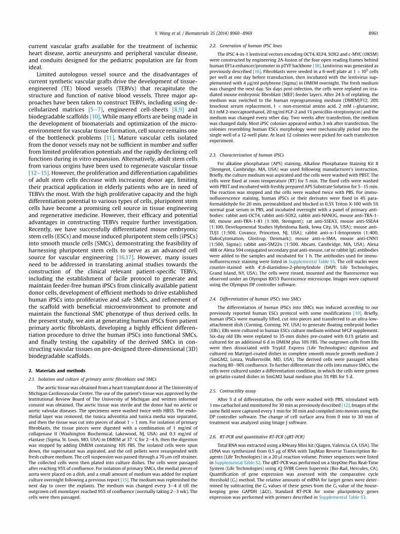
The aortic tissue was obtained from a heart transplant donor at The University ofMichigan Cardiovascular Center. The use of the patient's tissue was approved by theInstitutional Review Board of The University of Michigan and written informedconsent was obtained. The aortic tissue was sterile and the donor had no aortic oraortic valvular diseases. The specimens were washed twice with HBSS. The endo-thelial layer was removed, the tunica adventitia and tunica media was separated,and then the tissue was cut into pieces of about 1 � 1 mm. For isolation of primaryfibroblasts, the tissue pieces were digested with a combination of 1 mg/ml ofcollagenase II (Washington Biochemical, Lakewood, NJ, USA) and 0.3 mg/ml ofelastase (Sigma, St. Louis, MO, USA) in DMEM at 37 �C for 2e4 h, then the digestionwas stopped by adding DMEM containing 10% FBS. The isolated cells were spundown, the supernatant was aspirated, and the cell pellets were resuspended withfresh culture medium. The cell suspensionwas passed through a 70 mm cell strainer.The collected cells were then plated into culture dishes. The cells were passagedafter reaching 95% of confluence. For isolation of primary SMCs, the medial pieces ofaorta were placed on a dish, and a small amount of medium was added for explantculture overnight following a previous report [15]. The mediumwas replenished thenext day to cover the explants. The medium was changed every 3e4 d till theoutgrown cell monolayer reached 95% of confluence (normally taking 2e3 wk). Thecells were then passaged.
2.2. Generation of human iPSC lines
The iPSC 4-in-1 lentiviral vectors encoding OCT4, KLF4, SOX2 and c-MYC (OKSM)were constructed by engineering 2A-fusion of the four open reading frames behindhuman EF1a enhancer/promoter in pTYF backbone [18]. Lentivirus was generated aspreviously described [16]. Fibroblasts were seeded in a 6-well plate at 1 � 105 cellsper well at one day before transduction, then incubated with the lentivirus sup-plemented with 4 mg/ml polybrene (Sigma) in DMEM overnight. The fresh mediumwas changed the next day. Six days post-infection, the cells were replated on irra-diated mouse embryonic fibroblast (MEF) feeder layers. After 24 h of replating, themedium was switched to the human reprogramming medium (DMEM/F12, 20%knockout serum replacement, 1 � non-essential amino acid, 2 mM L-glutamine,0.1 mM 2-mercaptoethanol, 20 ng/ml FGF-2 and 1% penicillin-streptomycin) and themedium was changed every other day. Two weeks after transfection, the mediumwas changed daily. Most iPSC colonies appeared within 3 wk after transfection. Thecolonies resembling human ESCs morphology were mechanically picked into thesingle well of a 12-well plate. At least 12 colonies were picked for each transfectionexperiment.
2.3. Characterization of human iPSCs
For alkaline phosphatase (APS) staining, Alkaline Phosphatase Staining Kit II(Stemgent, Cambridge, MA, USA) was used following manufacturer's instruction.Briefly, the culture mediumwas aspirated and the cells were washed with PBST. Thecells were fixed at room temperature (RT) for 5 min. The fixed cells were washedwith PBSTand incubatedwith freshly prepared APS Substrate Solution for 5e15min.The reaction was stopped and the cells were washed twice with PBS. For immu-nofluorescence staining, human iPSCs or their derivates were fixed in 4% para-formaldehyde for 20 min, permeabilized and blocked in 0.5% Triton X-100 with 5%normal goat serum in PBS, and incubated overnight with a panel of primary anti-bodies: rabbit anti-OCT4, rabbit anti-SOX2, rabbit anti-NANOG, mouse anti-TRA-1-60, mouse anti-TRA-1-81 (1:100, Stemgent); rat anti-SSEA3, mouse anti-SSEA4(1:100, Developmental Studies Hybridoma Bank, Iowa City, IA, USA); mouse anti-TUJ1 (1:500, Covance, Princeton, NJ, USA); rabbit anti-a-1-fetoprotein (1:400,DakoCytomation, Glostrup, Denmark); mouse anti-a-SMA, mouse anti-CNN1(1:500, Sigma); rabbit anti-SM22a (1:500, Abcam, Cambridge, MA, USA). Alexa488 or Alexa 594 conjugated secondary goat anti-mouse, rat or rabbit IgG antibodieswere added to the samples and incubated for 1 h. The antibodies used for immu-nofluorescence staining were listed in Supplemental Table S1. The cell nuclei werecounter-stained with 40 ,6-diamidino-2-phenylindole (DAPI; Life Technologies,Grand Island, NY, USA). The cells were rinsed, mounted and the fluorescence wasobserved under an Olympus BX53 fluorescence microscope. Images were capturedusing the Olympus DP controller software.
2.4. Differentiation of human iPSCs into SMCs
The differentiation of human iPSCs into SMCs was induced according to ourpreviously reported human ESCs protocol with some modifications [19]. Briefly,human iPSCs were manually lifted, cut into pieces and transferred to an ultra-low-attachment dish (Corning, Corning, NY, USA) to generate floating embryoid bodies(EBs). EBs were cultured in human ESCs culture mediumwithout bFGF supplement.Six-day old EBs were replated to 35-mm dishes pre-coated with 0.1% gelatin andcultured for an additional 6 d in DMEM plus 10% FBS. The outgrown cells from EBswere then dissociated with TrypLE Express (Life Technologies) digestion andcultured on Matrigel-coated dishes in complete smooth muscle growth medium 2(SmGM2, Lonza, Walkersville, MD, USA). The derived cells were passaged whenreaching 80e90% confluence. To further differentiate the cells intomature SMCs, thecells were cultured under a differentiation condition, in which the cells were grownon gelatin-coated dishes in SmGM2 basal medium plus 5% FBS for 5 d.
2.5. Contractility assay
After 5 d of differentiation, the cells were washed with PBS, stimulated with1mM carbachol andmonitored for 30min as previously described [12]. Images of thesame field were captured every 1min for 30min and compiled intomovies using theDP controller software. The change of cell surface area from 0 min to 30 min oftreatment was analyzed using Image J software.
2.6. RT-PCR and quantitative RT-PCR (qRT-PCR)
Total RNAwas extracted using a RNeasyMini kit (Qiagen, Valencia, CA, USA). ThecDNA was synthesized from 0.5 mg of RNA with TaqMan Reverse Transcription Re-agents (Life Technologies) in a 20 ml reaction volume. Primer sequences were listedin Supplemental Table S2. The qRT-PCR was performed on a StepOne Plus Real-TimeSystem (Life Technologies) using iQ SYBR Green Supermix (Bio-Rad, Hercules, CA).Quantification of gene expression was assessed with the comparative cyclethreshold (Ct) method. The relative amounts of mRNA for target genes were deter-mined by subtracting the Ct values of these genes from the Ct value of the house-keeping gene GAPDH (DCt). Standard RT-PCR for some pluripotency genesexpression was performed with primers described in Supplemental Table S3.
Y. Wang et al. / Biomaterials 35 (2014) 8960e89698962
2.7. Flow cytometry
The cells were dissociated with 0.05% trypsin digestion into single cell andsuspended in PBS, then fixed with the Fixation Solution from a Fixation/Per-meabilization kit (eBiosciences, San Diego, CA, USA), and stained with primary anddetection antibodies as described by the manufacturer. Briefly, 0.5 � 106 cells wereresuspended in 400 ml of Fixation/Permeabilization Solution, kept at RT for 30 min,and washed twice with Perm/Wash Buffer. After being blocked with Blocking So-lution for 30 min, the cells were double-stained against CNN1 and a-SMA. Briefly,the cells were incubated with the primary anti-CNN1 antibody in Blocking Solutionfor 1 h at RT, then the cells were washed twice and resuspended in 100 ml of Perm/Wash Buffer with APC-conjugated secondary antibody and FITC-conjugated anti-a-SMA antibody for 1 h at RT. Then the cells were washed twice and the resuspendedcells in PBS were analyzed by flow cytometry. Isotype IgG1 and FITC-conjugatedIgG2a were used as the negative control to CNN1 antibody and FITC-conjugatedanti-a-SMA antibody, respectively. The antibodies used for flow cytometry werelisted in Supplemental Table S1.
2.8. Fabrication of three-dimensional (3D) macroporous NF poly(L-lactide) (PLLA)scaffolds
PLLA with an inherent viscosity of approximately 1.6 dl/g was purchased fromBoehinger Ingelheim (Ingelheim, Germany). PLLA in tetrahydrofuran solution (10%wt/v) was cast into an assembled sugar template (formed from bound sugar spheres,60e150 mm in diameter) under a mild vacuum. The polymer-sugar composite wasphase separated at �20 �C overnight and then immersed into cyclohexane to ex-change tetrahydrofuran for 2 d. The resulting composites were freeze-dried and thesugar spheres were leached out in distilled water and freeze-dried again to obtainhigh porous scaffolds. The scaffolds were cut into circular disks with dimensions of5 mm in diameter and 1 mm in thickness.
2.9. Construction of tissue-engineered vascular tissues
The 3D NF PLLA scaffolds were pre-wetted in 70% ethanol for 30 min, washedthree times with PBS for 30 min, and twice in the cell culture medium for 2 h each.5�105 human iPSC-derived proliferative SMCs were seeded into each scaffold. After24 h of incubation, the cell/scaffold constructs were subcutaneously implanted intonude mice. For implantation surgery, 6e8 wk old male nude mice (Charles RiverLaboratories, Wilmington, MA, USA) were used. Surgery was performed undergeneral inhalation anesthesiawith isofluorane. Twomidsagittal incisions weremadeon the dorsa and one subcutaneous pocket was created on each side of each incisionusing blunt dissection. One cell-scaffold construct was implanted into each pocket.After placement of implants, the incisions were closed with staples. At the end of
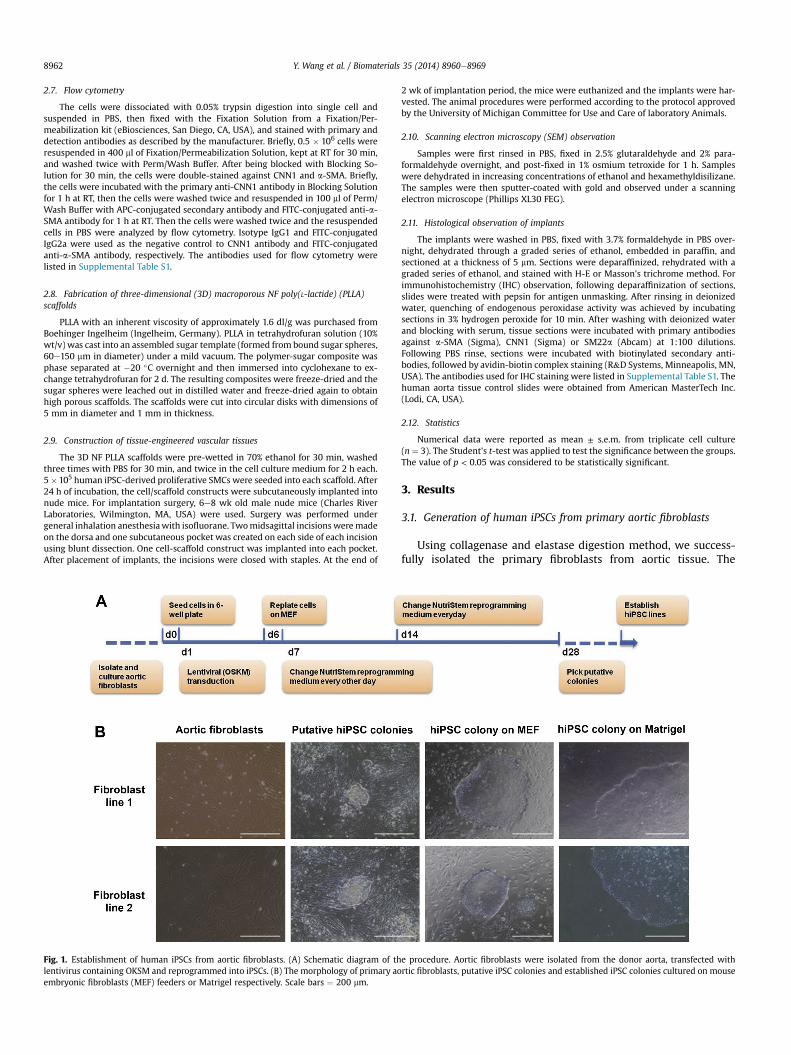
Fig. 1. Establishment of human iPSCs from aortic fibroblasts. (A) Schematic diagram of thlentivirus containing OKSM and reprogrammed into iPSCs. (B) The morphology of primary aoembryonic fibroblasts (MEF) feeders or Matrigel respectively. Scale bars ¼ 200 mm.
2 wk of implantation period, the mice were euthanized and the implants were har-vested. The animal procedures were performed according to the protocol approvedby the University of Michigan Committee for Use and Care of laboratory Animals.
2.10. Scanning electron microscopy (SEM) observation
Samples were first rinsed in PBS, fixed in 2.5% glutaraldehyde and 2% para-formaldehyde overnight, and post-fixed in 1% osmium tetroxide for 1 h. Sampleswere dehydrated in increasing concentrations of ethanol and hexamethyldisilizane.The samples were then sputter-coated with gold and observed under a scanningelectron microscope (Phillips XL30 FEG).
2.11. Histological observation of implants
The implants were washed in PBS, fixed with 3.7% formaldehyde in PBS over-night, dehydrated through a graded series of ethanol, embedded in paraffin, andsectioned at a thickness of 5 mm. Sections were deparaffinized, rehydrated with agraded series of ethanol, and stained with H-E or Masson's trichrome method. Forimmunohistochemistry (IHC) observation, following deparaffinization of sections,slides were treated with pepsin for antigen unmasking. After rinsing in deionizedwater, quenching of endogenous peroxidase activity was achieved by incubatingsections in 3% hydrogen peroxide for 10 min. After washing with deionized waterand blocking with serum, tissue sections were incubated with primary antibodiesagainst a-SMA (Sigma), CNN1 (Sigma) or SM22a (Abcam) at 1:100 dilutions.Following PBS rinse, sections were incubated with biotinylated secondary anti-bodies, followed by avidin-biotin complex staining (R&D Systems, Minneapolis, MN,USA). The antibodies used for IHC staining were listed in Supplemental Table S1. Thehuman aorta tissue control slides were obtained from American MasterTech Inc.(Lodi, CA, USA).
2.12. Statistics
Numerical data were reported as mean ± s.e.m. from triplicate cell culture(n ¼ 3). The Student's t-test was applied to test the significance between the groups.The value of p < 0.05 was considered to be statistically significant.
3. Results
3.1. Generation of human iPSCs from primary aortic fibroblasts
Using collagenase and elastase digestion method, we success-fully isolated the primary fibroblasts from aortic tissue. The
e procedure. Aortic fibroblasts were isolated from the donor aorta, transfected withrtic fibroblasts, putative iPSC colonies and established iPSC colonies cultured on mouse

Y. Wang et al. / Biomaterials 35 (2014) 8960e8969 8963
passage-2 cells of primary aortic fibroblasts were used for reprog-ramming. The protocol for reprogramming was illustrated inFig. 1A. The human ESC-like colonies appeared 3 wk after trans-fection. After several rounds of passage and expansion, primaryaortic fibroblast-derived iPSC lines were established. Each primaryaortic fibroblast line was used to establish at least three iPSC lines.The established cell lines can be maintained and expanded both onMEF feeders and Matrigel-coated surfaces (Fig. 1B).
3.2. Characterization of human iPSC colonies
At least twelve colonies were mechanically isolated from theculture of reprogrammed primary fibroblasts and passaged on freshMEF feeder. At least three iPSC lines were successfully establishedfrom each reprogramming culture and the cells can be continuouslypassaged while maintaining human ESC-like morphology. Thecolonies of the established iPSC lines showed strong positivestaining of APS activity. The pluipotency marker expression wasassessed with immunofluorescence staining. Fig. 2A showed
Fig. 2. Pluripotency analysis of human iPSCs. (A) Human iPSC line 1 and line 2 generatednofluorescence staining of pluripotency markers including OCT4, NANOG, SOX2, SSEA3, SSEAand SOX2 mRNA expression in MEFs, the two iPSC lines and H7 ESC line.
the representative iPSC colonies which strongly expressed tran-scription factors of OCT4, NANOG and SOX2 in nuclei, and otherpluripotency markers of SSEA3, SSEA4, TRA-1-60 and TRA-1-81 oncell surface. The expression of OCT4, NANOG and SOX2mRNA in thetwo human iPSC lines were also validated by RT-PCR (Fig. 2B). Toconveniently generate feeder-free cells, the iPSCs were furthermaintained with standard procedure developed for feeder-freeculture of human ESCs, and mechanically passaged on Matrigel-coated dishes with TeSR-E8 medium. Some of these cell lineshave been maintained in continuous culture for over 6 monthswithout noticeable loss of pluripotent properties (data not shown).
3.3. Pluripotency of human iPSCs
To further validate the pluripotency of established human iPSClines, EB formation assay was performed in vitro and teratoma in-duction assay was performed in vivo. All the established iPSC lineswe tested can differentiate into the three germ layers of endoderm,mesoderm and ectoderm in vitro, as Fig. 3 showed the positive
from the donor were observed with alkaline phosphatase (APS) staining and immu-4, TRA-1e60 and TRA-1e81. Scale bars ¼ 200 mm. (B) RT-PCR analysis of NANOG, OCT4
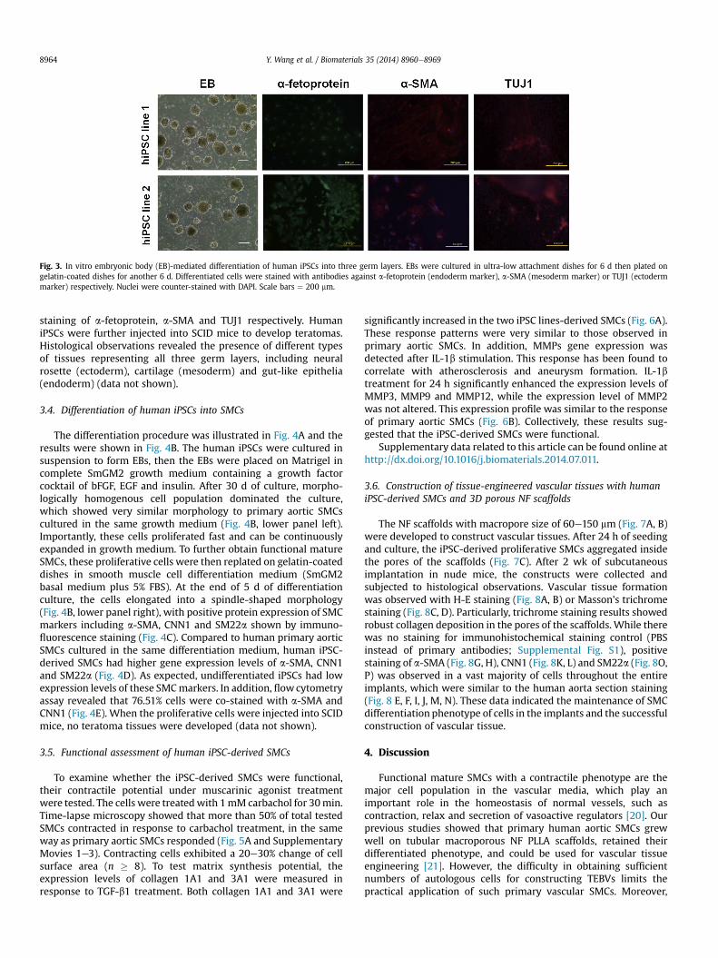
Fig. 3. In vitro embryonic body (EB)-mediated differentiation of human iPSCs into three germ layers. EBs were cultured in ultra-low attachment dishes for 6 d then plated ongelatin-coated dishes for another 6 d. Differentiated cells were stained with antibodies against a-fetoprotein (endoderm marker), a-SMA (mesoderm marker) or TUJ1 (ectodermmarker) respectively. Nuclei were counter-stained with DAPI. Scale bars ¼ 200 mm.
Y. Wang et al. / Biomaterials 35 (2014) 8960e89698964
staining of a-fetoprotein, a-SMA and TUJ1 respectively. HumaniPSCs were further injected into SCID mice to develop teratomas.Histological observations revealed the presence of different typesof tissues representing all three germ layers, including neuralrosette (ectoderm), cartilage (mesoderm) and gut-like epithelia(endoderm) (data not shown).
3.4. Differentiation of human iPSCs into SMCs
The differentiation procedure was illustrated in Fig. 4A and theresults were shown in Fig. 4B. The human iPSCs were cultured insuspension to form EBs, then the EBs were placed on Matrigel incomplete SmGM2 growth medium containing a growth factorcocktail of bFGF, EGF and insulin. After 30 d of culture, morpho-logically homogenous cell population dominated the culture,which showed very similar morphology to primary aortic SMCscultured in the same growth medium (Fig. 4B, lower panel left).Importantly, these cells proliferated fast and can be continuouslyexpanded in growth medium. To further obtain functional matureSMCs, these proliferative cells were then replated on gelatin-coateddishes in smooth muscle cell differentiation medium (SmGM2basal medium plus 5% FBS). At the end of 5 d of differentiationculture, the cells elongated into a spindle-shaped morphology(Fig. 4B, lower panel right), with positive protein expression of SMCmarkers including a-SMA, CNN1 and SM22a shown by immuno-fluorescence staining (Fig. 4C). Compared to human primary aorticSMCs cultured in the same differentiation medium, human iPSC-derived SMCs had higher gene expression levels of a-SMA, CNN1and SM22a (Fig. 4D). As expected, undifferentiated iPSCs had lowexpression levels of these SMCmarkers. In addition, flowcytometryassay revealed that 76.51% cells were co-stained with a-SMA andCNN1 (Fig. 4E). When the proliferative cells were injected into SCIDmice, no teratoma tissues were developed (data not shown).
3.5. Functional assessment of human iPSC-derived SMCs
To examine whether the iPSC-derived SMCs were functional,their contractile potential under muscarinic agonist treatmentwere tested. The cells were treatedwith 1mM carbachol for 30min.Time-lapse microscopy showed that more than 50% of total testedSMCs contracted in response to carbachol treatment, in the sameway as primary aortic SMCs responded (Fig. 5A and SupplementaryMovies 1e3). Contracting cells exhibited a 20e30% change of cellsurface area (n � 8). To test matrix synthesis potential, theexpression levels of collagen 1A1 and 3A1 were measured inresponse to TGF-b1 treatment. Both collagen 1A1 and 3A1 were
significantly increased in the two iPSC lines-derived SMCs (Fig. 6A).These response patterns were very similar to those observed inprimary aortic SMCs. In addition, MMPs gene expression wasdetected after IL-1b stimulation. This response has been found tocorrelate with atherosclerosis and aneurysm formation. IL-1btreatment for 24 h significantly enhanced the expression levels ofMMP3, MMP9 and MMP12, while the expression level of MMP2was not altered. This expression profile was similar to the responseof primary aortic SMCs (Fig. 6B). Collectively, these results sug-gested that the iPSC-derived SMCs were functional.
Supplementary data related to this article can be found online athttp://dx.doi.org/10.1016/j.biomaterials.2014.07.011.
3.6. Construction of tissue-engineered vascular tissues with humaniPSC-derived SMCs and 3D porous NF scaffolds
The NF scaffolds with macropore size of 60e150 mm (Fig. 7A, B)were developed to construct vascular tissues. After 24 h of seedingand culture, the iPSC-derived proliferative SMCs aggregated insidethe pores of the scaffolds (Fig. 7C). After 2 wk of subcutaneousimplantation in nude mice, the constructs were collected andsubjected to histological observations. Vascular tissue formationwas observed with H-E staining (Fig. 8A, B) or Masson's trichromestaining (Fig. 8C, D). Particularly, trichrome staining results showedrobust collagen deposition in the pores of the scaffolds. While therewas no staining for immunohistochemical staining control (PBSinstead of primary antibodies; Supplemental Fig. S1), positivestaining of a-SMA (Fig. 8G, H), CNN1 (Fig. 8K, L) and SM22a (Fig. 8O,P) was observed in a vast majority of cells throughout the entireimplants, which were similar to the human aorta section staining(Fig. 8 E, F, I, J, M, N). These data indicated the maintenance of SMCdifferentiation phenotype of cells in the implants and the successfulconstruction of vascular tissue.
4. Discussion
Functional mature SMCs with a contractile phenotype are themajor cell population in the vascular media, which play animportant role in the homeostasis of normal vessels, such ascontraction, relax and secretion of vasoactive regulators [20]. Ourprevious studies showed that primary human aortic SMCs grewwell on tubular macroporous NF PLLA scaffolds, retained theirdifferentiated phenotype, and could be used for vascular tissueengineering [21]. However, the difficulty in obtaining sufficientnumbers of autologous cells for constructing TEBVs limits thepractical application of such primary vascular SMCs. Moreover,

Y. Wang et al. / Biomaterials 35 (2014) 8960e8969 8965
primary vascular SMCs have low proliferation potential and sufferfrom culture senescence. SMCs have been demonstrated to bedifferentiated from alternative cell sources such as bone-marrowmesenchymal stem cells [12,13], endothelial progenitor cells [14],
Fig. 4. Differentiation of human iPSCs into vascular SMCs. (A) Schematic diagram of the prPhase contrast observation of cells at different differentiation stages. SMGM, smooth muscrescence staining showed that primary human aortic SMCs and iPSC-derived SMCs highly ereal-time PCR demonstrated that human iPSC-derived SMCs up-regulated the gene expresferentiated hiPSCs. The primary SMCs served as a positive control. (E) Flow cytometry analySMCs. Scale bars ¼ 200 mm.
adipose-derived stem cells [15], among many other adult stemcells. However, the proliferation and differentiation capabilities ofadult stem cells significantly decrease with aging donors. Recently,the generation of patient-specific iPSCs, which could differentiate
ocedure. EB, embryonic body; GM, growth medium; DM, differentiation medium. (B)le growth medium; SMDM, smooth muscle differentiation medium. (C) Immunofluo-xpressed vascular SMC markers including a-SMA, CNN1 and SM22a. (D) Quantitativesion levels of SMC markers including a-SMA, CNN1 and SM22a, compared to undif-sis of the vascular SMC marker expression of a-SMA and CNN1 in human iPSC-derived

Y. Wang et al. / Biomaterials 35 (2014) 8960e89698966
into various cell types, has brought substantial excitement to thefield of tissue engineering and regenerative medicine [22]. In aprevious report, we successfully established mouse iPSC lines andthen differentiated them into SMCs with a short-term all-transretinoid acid treatment [16]. However, obtaining patient-specificiPSC-derived SMCs to engineer human vascular grafts for clinicapplication remains a daunting challenge. In this study, we firstestablished a facile procedure to generate human iPSCs from hu-man aortic tissues and then efficiently differentiate these pluripo-tent cells into functional vascular SMCs, which could be easilyscaled up to produce large numbers of functional cells withreproducible properties. A morphologically homogenous prolifer-ative cell population was derived, which could be passaged andsequentially differentiated into functional SMCs. These proliferativeSMCs intermediates allow us to rapidly expand a large number ofcells before further inducing them into less-proliferative, contrac-tile mature cells. In addition, when these proliferative cellswere injected into SCID mice, no teratoma tissue was developed,minimizing the concern over the risk of teratoma formation inthe future clinical application. The tissue-engineered patient-spe-cific blood vessels using the patient's iPSCs have potential forthe treatment of peripheral vascular disease, which is commonly
Fig. 5. Contraction response of human iPSC-derived SMCs to muscarinic agonist. (A) Humanwere treated with 1 mM carbachol, and the images were recorded every 1 min for consecu30 min. Arrows indicated the contracting cells. (B) The change of cell surface area was calcSMCs) or 200 mm (for primary SMCs).
seen in diabetic patients and renal failure patients. The generationof iPSCs from the fibroblasts of diabetic patients and renalfailure patients need to be explored for such applications in thefuture.
Another challenge in the construction of TEBVs is to develop asuitable porous and biodegradable scaffold to support vascular cellsgrowth and differentiation, and ultimately to form functionalvascular tissue. Using a macroporous and NF PLLA scaffold and acontrol scaffold that does not have the NF feature, we found thatthe scaffold with the NF feature advantageously supported thegrowth and differentiation of mouse iPSC-derived SMCs [16]. In thisstudy, we reduced pore size of the macroporous and NF PLLAscaffold (changing from 125-250 mm to 60e150 mm in pore diam-eter), and found that this NF scaffold with the smaller pore sizebetter support human SMC growth and vascular tissue formation(unpublished data). This finding was corroborated by a recentreport that a porous scaffold fabricated from ploy(glycerol seba-cate) (PGS) with a smaller pore size resulted in a higher SMCdensity in the lumen and more ECM protein deposition comparedto the scaffold with a larger pore size [23]. The reasonably smallerpore size may facilitate cellecell interactions in the pores, possiblyresulted in the increased ECM production. However, too small a
iPSC-derived SMCs were cultured under differentiation conditions for 5 d, then the cellstive 30 min. The images represented the cells before treatment and after treatment forulated and compared to that of primary SMCs. Scale bars ¼ 100 mm (for iPSC-derived

Fig. 6. Gene expression of collagens and matrix metalloproteinases (MMPs) in human iPSC-derived SMCs with growth factor or cytokine treatment. (A) Gene expression of collagen1A1 and collagen 3A1 under 5 ng/ml of TGF-b1 treatment for 24 h. (B) Gene expression of MMP2, 3, 9 and 12 in response to 10 ng/ml IL-1b treatment for 24 h. *, P < 0.05 vs vehiclecontrol; **, P < 0.01 vs vehicle control.
Y. Wang et al. / Biomaterials 35 (2014) 8960e8969 8967
pore size leads to inefficient cell seeding inside the scaffolds due tolimited pore opening size and interconnection size between thepores. We chose a pore size of 60e150 mm because this size rangeallowed for efficient cell infiltration throughout the entire scaffoldswhile maintaining the SMC phenotype and promoting vasculartissue formation upon implantation.
Fig. 7. Seeding of human iPSC-derived proliferative SMCs into 3D scaffolds. SEM images of mmagnifications; and (C) human iPSC-derived proliferative SMCs seeded in these scaffolds fo
Construction of a whole TEBV needs the incorporation of othercell types. The blood vessels are composed of three layers, namelythe tunica intima, media and adventitia, which are mainlycomposed of endothelial cells, SMCs and fibroblasts, respectively. Inthis study, we primarily used human iPSC-derived SMCs toconstruct vascular tissues, due to the major role of tunica media in
acroporous nanofibrous (NF) scaffolds (60e150 mm pore size) at low (A) and high (B)r 24 h.
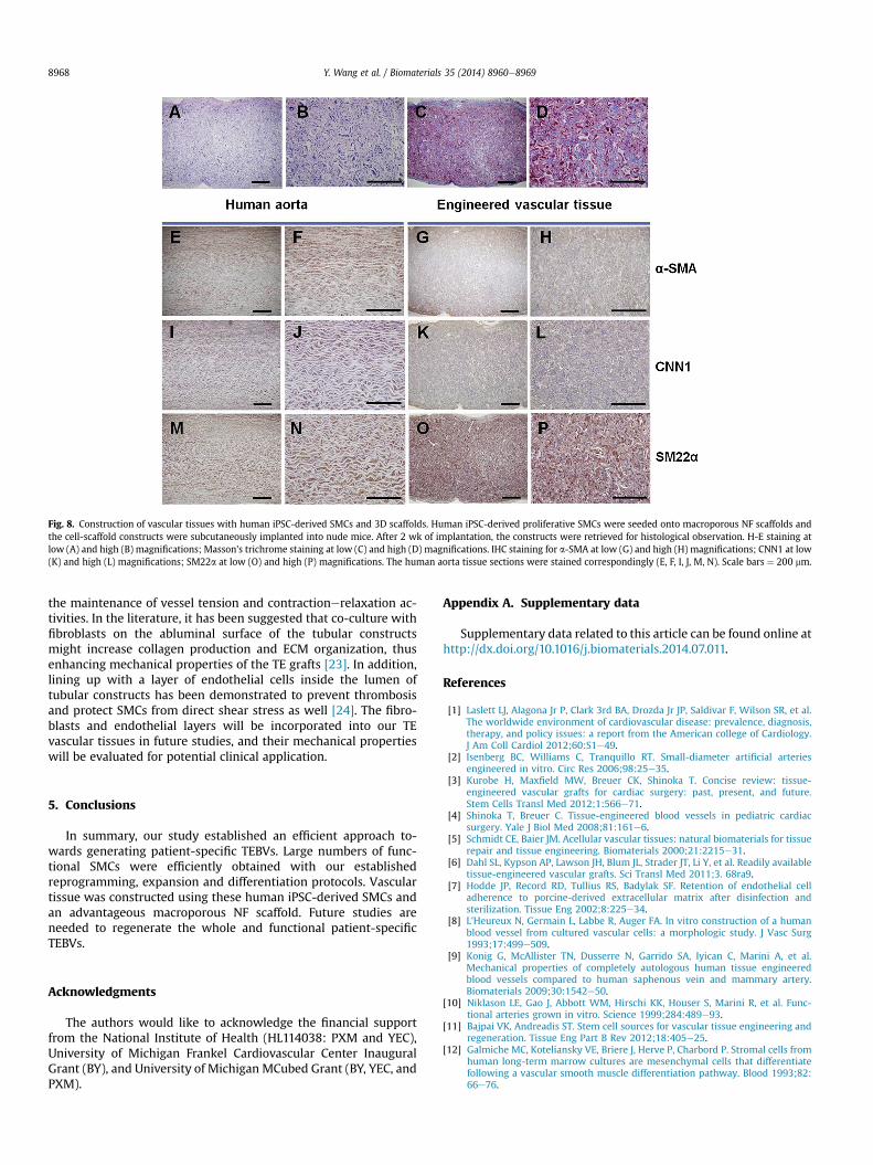
Fig. 8. Construction of vascular tissues with human iPSC-derived SMCs and 3D scaffolds. Human iPSC-derived proliferative SMCs were seeded onto macroporous NF scaffolds andthe cell-scaffold constructs were subcutaneously implanted into nude mice. After 2 wk of implantation, the constructs were retrieved for histological observation. H-E staining atlow (A) and high (B) magnifications; Masson's trichrome staining at low (C) and high (D) magnifications. IHC staining for a-SMA at low (G) and high (H) magnifications; CNN1 at low(K) and high (L) magnifications; SM22a at low (O) and high (P) magnifications. The human aorta tissue sections were stained correspondingly (E, F, I, J, M, N). Scale bars ¼ 200 mm.
Y. Wang et al. / Biomaterials 35 (2014) 8960e89698968
the maintenance of vessel tension and contractionerelaxation ac-tivities. In the literature, it has been suggested that co-culture withfibroblasts on the abluminal surface of the tubular constructsmight increase collagen production and ECM organization, thusenhancing mechanical properties of the TE grafts [23]. In addition,lining up with a layer of endothelial cells inside the lumen oftubular constructs has been demonstrated to prevent thrombosisand protect SMCs from direct shear stress as well [24]. The fibro-blasts and endothelial layers will be incorporated into our TEvascular tissues in future studies, and their mechanical propertieswill be evaluated for potential clinical application.
5. Conclusions
In summary, our study established an efficient approach to-wards generating patient-specific TEBVs. Large numbers of func-tional SMCs were efficiently obtained with our establishedreprogramming, expansion and differentiation protocols. Vasculartissue was constructed using these human iPSC-derived SMCs andan advantageous macroporous NF scaffold. Future studies areneeded to regenerate the whole and functional patient-specificTEBVs.
Acknowledgments
The authors would like to acknowledge the financial supportfrom the National Institute of Health (HL114038: PXM and YEC),University of Michigan Frankel Cardiovascular Center InauguralGrant (BY), and University of Michigan MCubed Grant (BY, YEC, andPXM).
Appendix A. Supplementary data
Supplementary data related to this article can be found online athttp://dx.doi.org/10.1016/j.biomaterials.2014.07.011.
References
[1] Laslett LJ, Alagona Jr P, Clark 3rd BA, Drozda Jr JP, Saldivar F, Wilson SR, et al.The worldwide environment of cardiovascular disease: prevalence, diagnosis,therapy, and policy issues: a report from the American college of Cardiology.J Am Coll Cardiol 2012;60:S1e49.
[2] Isenberg BC, Williams C, Tranquillo RT. Small-diameter artificial arteriesengineered in vitro. Circ Res 2006;98:25e35.
[3] Kurobe H, Maxfield MW, Breuer CK, Shinoka T. Concise review: tissue-engineered vascular grafts for cardiac surgery: past, present, and future.Stem Cells Transl Med 2012;1:566e71.
[4] Shinoka T, Breuer C. Tissue-engineered blood vessels in pediatric cardiacsurgery. Yale J Biol Med 2008;81:161e6.
[5] Schmidt CE, Baier JM. Acellular vascular tissues: natural biomaterials for tissuerepair and tissue engineering. Biomaterials 2000;21:2215e31.
[6] Dahl SL, Kypson AP, Lawson JH, Blum JL, Strader JT, Li Y, et al. Readily availabletissue-engineered vascular grafts. Sci Transl Med 2011;3. 68ra9.
[7] Hodde JP, Record RD, Tullius RS, Badylak SF. Retention of endothelial celladherence to porcine-derived extracellular matrix after disinfection andsterilization. Tissue Eng 2002;8:225e34.
[8] L'Heureux N, Germain L, Labbe R, Auger FA. In vitro construction of a humanblood vessel from cultured vascular cells: a morphologic study. J Vasc Surg1993;17:499e509.
[9] Konig G, McAllister TN, Dusserre N, Garrido SA, Iyican C, Marini A, et al.Mechanical properties of completely autologous human tissue engineeredblood vessels compared to human saphenous vein and mammary artery.Biomaterials 2009;30:1542e50.
[10] Niklason LE, Gao J, Abbott WM, Hirschi KK, Houser S, Marini R, et al. Func-tional arteries grown in vitro. Science 1999;284:489e93.
[11] Bajpai VK, Andreadis ST. Stem cell sources for vascular tissue engineering andregeneration. Tissue Eng Part B Rev 2012;18:405e25.
[12] Galmiche MC, Koteliansky VE, Briere J, Herve P, Charbord P. Stromal cells fromhuman long-term marrow cultures are mesenchymal cells that differentiatefollowing a vascular smooth muscle differentiation pathway. Blood 1993;82:66e76.

Y. Wang et al. / Biomaterials 35 (2014) 8960e8969 8969
[13] Kashiwakura Y, Katoh Y, Tamayose K, Konishi H, Takaya N, Yuhara S, et al.Isolation of bone marrow stromal cell-derived smooth muscle cells bya human SM22alpha promoter: in vitro differentiation of putativesmooth muscle progenitor cells of bone marrow. Circulation 2003;107:2078e81.
[14] Li X, Tjwa M, Moons L, Fons P, Noel A, Ny A, et al. Revascularization ofischemic tissues by PDGF-CC via effects on endothelial cells and their pro-genitors. J Clin Invest 2005;115:118e27.
[15] Rodriguez LV, Alfonso Z, Zhang R, Leung J, Wu B, Ignarro LJ. Clonogenicmultipotent stem cells in human adipose tissue differentiate into functionalsmooth muscle cells. Proc Natl Acad Sci U S A 2006;103:12167e72.
[16] Xie C, Hu J, Ma H, Zhang J, Chang LJ, Chen YE, et al. Three-dimensional growthof iPS cell-derived smooth muscle cells on nanofibrous scaffolds. Biomaterials2011;32:4369e75.
[17] Hu J, Xie C, Ma H, Yang B, Ma PX, Chen YE. Construction of vascular tissueswith macro-porous nano-fibrous scaffolds and smooth muscle cells enrichedfrom differentiated embryonic stem cells. PLoS One 2012;7. e35580.
[18] Chang LJ, Gay EE. The molecular genetics of lentiviral vectorsecurrent andfuture perspectives. Curr Gene Ther 2001;1:237e51.
[19] Xie CQ, Zhang J, Villacorta L, Cui T, Huang H, Chen YE. A highly efficientmethod to differentiate smooth muscle cells from human embryonic stemcells. Arterioscler Thromb Vasc Biol 2007;27:e311e2.
[20] Jain RK. Molecular regulation of vessel maturation. Nat Med 2003;9:685e93.[21] Hu J, Sun X, Ma H, Xie C, Chen YE, Ma PX. Porous nanofibrous PLLA scaffolds
for vascular tissue engineering. Biomaterials 2010;31:7971e7.[22] Okano H, Nakamura M, Yoshida K, Okada Y, Tsuji O, Nori S, et al. Steps toward
safe cell therapy using induced pluripotent stem cells. Circ Res 2013;112:523e33.
[23] Lee KW, Stolz DB, Wang Y. Substantial expression of mature elastin in arterialconstructs. Proc Natl Acad Sci U S A 2011;108:2705e10.
[24] Heyligers JM, Arts CH, Verhagen HJ, de Groot PG, Moll FL. Improving small-diameter vascular grafts: from the application of an endothelial cell liningto the construction of a tissue-engineered blood vessel. Ann Vasc Surg2005;19:448e56.