Repositioning the Sm-Binding Site in Saccharomyces ... - MDPI
15
non-coding RNA Article Repositioning the Sm-Binding Site in Saccharomyces cerevisiae Telomerase RNA Reveals RNP Organizational Flexibility and Sm-Directed 3 0 -End Formation Evan P. Hass 1, † and David C. Zappulla 1,2, * 1 Department of Biology, Johns Hopkins University, Baltimore, MD 21218, USA; [email protected] 2 Department of Biological Sciences, Lehigh University, Bethlehem, PA 18015, USA * Correspondence: [email protected]; Tel.:+1-(610)-758-5088 † Current address: BioFrontiers Institute, University of Colorado Boulder, Boulder, CO, USA. Received: 8 January 2020; Accepted: 27 February 2020; Published: 29 February 2020 Abstract: Telomerase RNA contains a template for synthesizing telomeric DNA and has been proposed to act as a flexible scaffold for holoenzyme protein subunits in the RNP. In Saccharomyces cerevisiae, the telomerase RNA, TLC1, is bound by the Sm 7 protein complex, which is required for stabilization of the predominant, non-polyadenylated (poly(A)–) TLC1 isoform. However, it remains unclear (1) whether Sm 7 retains this function when its binding site is repositioned within TLC1, as has been shown for other TLC1-binding telomerase subunits, and (2) how Sm 7 stabilizes poly(A)– TLC1. Here, we first show that Sm 7 can stabilize poly(A)– TLC1 even when its binding site is repositioned via circular permutation to several different positions within TLC1, further supporting the conclusion that the telomerase holoenzyme is organizationally flexible. Next, we show that when an Sm site is inserted 5 0 of its native position and the native site is mutated, Sm 7 stabilizes shorter forms of poly(A)– TLC1 in a manner corresponding to how far upstream the new site was inserted, providing strong evidence that Sm 7 binding to TLC1 controls where the mature poly(A)– 3 0 is formed by directing a 3 0 -to-5 0 processing mechanism. In summary, our results show that Sm 7 and the 3 0 end of yeast telomerase RNA comprise an organizationally flexible module within the telomerase RNP and provide insights into the mechanistic role of Sm 7 in telomerase RNA biogenesis. Keywords: telomerase; Sm 7 ;3 0 -end formation; RNA processing; TLC1; lncRNA; senescence 1. Introduction Telomeres are regions of repetitive sequences at the ends of eukaryotic chromosomes that buffer against shortening caused by the end-replication problem. In most eukaryotes, telomeres are lengthened by telomerase, an RNP enzyme that adds new telomeric repeats to the ends of telomeres via reverse transcription of an RNA template [1]. Fundamentally, this process is carried out by the two core subunits of telomerase: telomerase reverse transcriptase (TERT) and the non-coding telomerase RNA [2,3]. TERT is the catalytic protein subunit of telomerase, while the telomerase RNA contains the template for reverse transcription of telomeric repeats as well as binding sites for accessory protein subunits of the telomerase RNP [4–8]. In Saccharomyces cerevisiae, the major isoform of telomerase RNA (TLC1) is 1157 nucleotides long and is predicted to form a Y-shaped secondary structure, with the template and TERT-binding regions in the central core and binding sites for holoenzyme subunits towards the tips of each arm [9,10] (Figure 1A). Like all telomerase RNAs, yeast telomerase RNAs are evolving very rapidly in sequence and size, especially in the regions between protein-binding sites [10,11]. These intervening regions can be deleted in TLC1, Non-coding RNA 2020, 6, 9; doi:10.3390/ncrna6010009 www.mdpi.com/journal/ncrna
-
Upload
khangminh22 -
Category
Documents
-
view
1 -
download
0
Transcript of Repositioning the Sm-Binding Site in Saccharomyces ... - MDPI

non-coding
RNA
Article
Repositioning the Sm-Binding Site in Saccharomycescerevisiae Telomerase RNA Reveals RNPOrganizational Flexibility and Sm-Directed3′-End Formation
Evan P. Hass 1,† and David C. Zappulla 1,2,*1 Department of Biology, Johns Hopkins University, Baltimore, MD 21218, USA; [email protected] Department of Biological Sciences, Lehigh University, Bethlehem, PA 18015, USA* Correspondence: [email protected]; Tel.:+1-(610)-758-5088† Current address: BioFrontiers Institute, University of Colorado Boulder, Boulder, CO, USA.
Received: 8 January 2020; Accepted: 27 February 2020; Published: 29 February 2020�����������������
Abstract: Telomerase RNA contains a template for synthesizing telomeric DNA and has beenproposed to act as a flexible scaffold for holoenzyme protein subunits in the RNP. In Saccharomycescerevisiae, the telomerase RNA, TLC1, is bound by the Sm7 protein complex, which is required forstabilization of the predominant, non-polyadenylated (poly(A)–) TLC1 isoform. However, it remainsunclear (1) whether Sm7 retains this function when its binding site is repositioned within TLC1, as hasbeen shown for other TLC1-binding telomerase subunits, and (2) how Sm7 stabilizes poly(A)– TLC1.Here, we first show that Sm7 can stabilize poly(A)– TLC1 even when its binding site is repositionedvia circular permutation to several different positions within TLC1, further supporting the conclusionthat the telomerase holoenzyme is organizationally flexible. Next, we show that when an Sm siteis inserted 5′ of its native position and the native site is mutated, Sm7 stabilizes shorter forms ofpoly(A)– TLC1 in a manner corresponding to how far upstream the new site was inserted, providingstrong evidence that Sm7 binding to TLC1 controls where the mature poly(A)– 3′ is formed bydirecting a 3′-to-5′ processing mechanism. In summary, our results show that Sm7 and the 3′ end ofyeast telomerase RNA comprise an organizationally flexible module within the telomerase RNP andprovide insights into the mechanistic role of Sm7 in telomerase RNA biogenesis.
Keywords: telomerase; Sm7; 3′-end formation; RNA processing; TLC1; lncRNA; senescence
1. Introduction
Telomeres are regions of repetitive sequences at the ends of eukaryotic chromosomes that bufferagainst shortening caused by the end-replication problem. In most eukaryotes, telomeres are lengthenedby telomerase, an RNP enzyme that adds new telomeric repeats to the ends of telomeres via reversetranscription of an RNA template [1]. Fundamentally, this process is carried out by the two coresubunits of telomerase: telomerase reverse transcriptase (TERT) and the non-coding telomeraseRNA [2,3]. TERT is the catalytic protein subunit of telomerase, while the telomerase RNA contains thetemplate for reverse transcription of telomeric repeats as well as binding sites for accessory proteinsubunits of the telomerase RNP [4–8].
In Saccharomyces cerevisiae, the major isoform of telomerase RNA (TLC1) is 1157 nucleotides long andis predicted to form a Y-shaped secondary structure, with the template and TERT-binding regions in thecentral core and binding sites for holoenzyme subunits towards the tips of each arm [9,10] (Figure 1A). Likeall telomerase RNAs, yeast telomerase RNAs are evolving very rapidly in sequence and size, especiallyin the regions between protein-binding sites [10,11]. These intervening regions can be deleted in TLC1,
Non-coding RNA 2020, 6, 9; doi:10.3390/ncrna6010009 www.mdpi.com/journal/ncrna
Non-coding RNA 2020, 6, 9 2 of 15
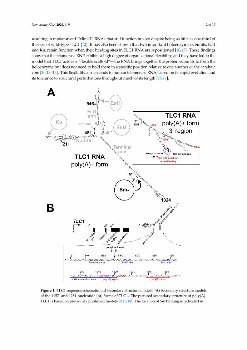
resulting in miniaturized “Mini-T” RNAs that still function in vivo despite being as little as one-third ofthe size of wild-type TLC1 [12]. It has also been shown that two important holoenzyme subunits, Est1and Ku, retain function when their binding sites in TLC1 RNA are repositioned [10,13]. These findingsshow that the telomerase RNP exhibits a high degree of organizational flexibility, and they have led to themodel that TLC1 acts as a “flexible scaffold”—the RNA brings together the protein subunits to form theholoenzyme but does not need to hold them in a specific position relative to one another or the catalyticcore [10,13–15]. This flexibility also extends to human telomerase RNA, based on its rapid evolution andits tolerance to structural perturbations throughout much of its length [16,17].
Figure 1. TLC1 sequence schematic and secondary structure models. (A) Secondary structure modelsof the 1157- and 1251-nucleotide (nt) forms of TLC1. The pictured secondary structure of poly(A)–TLC1 is based on previously published models [9,10,18]. The location of Sm binding is indicated in

Non-coding RNA 2020, 6, 9 3 of 15
black as are the four locations to which the Sm-binding site and the 5′ and 3′ ends are repositioned in theSm repositioning by circular permutation (SmCP) alleles used in Figure 2. The binding regions for Ku,Est1, and Est2, the template region, and the three arms of TLC1 are indicated in gray. The inset showsthe secondary structure model of the 3′ region of poly(A)+ TLC1 based on a previously publishedmodel [10]. The portion of the RNA used when repositioning the Sm-binding site (nucleotides 1138to 1165) is outlined in red. The locations of the Sm consensus and poly(A)– 3′ end are noted in black,and the three positions to which this Sm-binding site was repositioned in Figure 3 are indicated in red.(B) Schematic of the TLC1 gene sequence. The TLC1 regions encoding the Ku-binding site, template,Est1-binding region, pseudoknot, and Sm consensus in the RNA are denoted as black rectangles.Locations of all Sm-repositioning sites are noted by tick marks, as are the poly(A)– 3′ end and thepolyadenylation sites. The inset shows the RNA sequence of the TLC1 3′ region. The Sm consensusand poly(A)– 3′ end are noted in bold. The Nab3 and Nrd1 binding sites are bolded in blue, and theregion containing NNS termination sites is underlined in blue [19,20]. Similarly, the polyadenylationsignal is red boldface, and the region containing polyadenylation are underscored by a red line [21].
In addition to the proteins Est1, Ku, and TERT (Est2), the TLC1 RNA is also bound by the Sm7
complex [5]. The Sm7 heteroheptameric protein complex is involved in biogenesis and stabilizationof most spliceosomal snRNAs [22,23]. Sm7 binds to the consensus sequence AU5-6GR [24–27], whichis present in TLC1 at nucleotides 1143–1150 (Figure 1B) [5]. This site is located just 7 nucleotides5′ of position 1157, which is the 3′ end of poly(A)– TLC1 [28], the aforementioned most-abundant(“major”) isoform of TLC1. A less-abundant (“minor”) isoform, poly(A)+ TLC1, contains an extra ~100nucleotides of TLC1 sequence on its 3′ end, as well as a poly(A) tail [21]. In addition, there are verylow-abundance TLC1 transcripts terminated by the Nrd1-Nab3-Sen1 (NNS) complex that only have~50 extra nucleotides beyond the poly(A)– TLC1 3′ end at position 1157 [19,20], but these transcriptsare presumably not stable and are not detectable by northern blot.
In wild-type cells, the major, poly(A)– TLC1 isoform is present at ~29 molecules per cell, whilepoly(A)+ TLC1 is at only ~1 molecule per cell [29]. However, when the Sm consensus in TLC1 ismutated, only the minor poly(A)+ TLC1 isoform is detectable, likely because poly(A)– TLC1 is notstable without Sm7 bound [5]. Due to this critically low abundance of telomerase RNA, these mutantcells (tlc1-Sm– cells) display senescence-related growth defects (e.g., small or mixed colony sizes)but do not display a fully senescent phenotype, instead continuing to grow after the onset of thesedefects [5]. This “biphasic” or “near-senescent” growth phenotype (see Results Section 2.1 for a morethorough explanation of this latter term) seems to be in agreement with the fact that poly(A)+ TLC1abundance averages only ~1 molecule per cell. Because a single molecule of TLC1 per cell is the averageover a population of cells, some cells in the population probably have fewer molecules than average(i.e., none) and will eventually senesce, while others cells in the same population will have more thanone molecule of TLC1 and will be able to lengthen their telomeres enough to continue dividing.
Although it was shown 20 years ago that Sm7 binds to telomerase RNA in yeast [5], severalquestions about Sm function in the telomerase RNP remain unanswered. First, while it has beenshown that Sm7 can retain function when repositioned within Mini-T along with repositioning of the5′ and 3′ ends of the RNA (also called “circular permutation”) [30], the flexible scaffold model has notbeen tested for Sm7 in full-length TLC1 as it has been for Est1 and Ku. Additionally, there are severalopen mechanistic questions regarding Sm7 function in TLC1 biogenesis. It was proposed that TLC1 isinitially transcribed as poly(A)+ TLC1 and that this RNA is then processed into poly(A)– TLC1 [21]. Ithas also been proposed that the nuclear exosome exonucleolytically trims poly(A)+ TLC1 from 3′ to 5′
and is then sterically blocked at nucleotide 1157 by Sm7, thus generating poly(A)– TLC1 [31]. However,the hypothesis that the location of Sm7 binding defines the position of the mature 3′ end of poly(A)–TLC1 has remained untested, and it is still unclear whether poly(A)+ TLC1 is in fact the precursor ofpoly(A)– TLC1 or if NNS-terminated TLC1 transcripts are processed into the poly(A)– isoform.
Here, we show that circularly permuted full-length telomerase RNA functions in preventingsenescence and, furthermore, that the Sm7 subunit retains its function when its binding site isrepositioned in this context. This demonstrates that the Sm complex and the RNA ends are an
Non-coding RNA 2020, 6, 9 4 of 15
organizationally flexible module of the telomerase RNP’s RNA scaffold. Having shown that theSm-binding site can function at diverse positions within circularly permuted TLC1 alleles, we next usedSm-site relocation in the context of the unpermuted RNA to test the hypothesis that the Sm-bindingposition defines the 3′ end of poly(A)– telomerase RNA. When we moved the Sm site to positionsfurther 5′ in telomerase RNA, poly(A)– TLC1 RNA was still partially stabilized, and the length of thetranscript was correspondingly shorter than wild type. Additionally, when we added a second Sm site5′ of the native site in TLC1, the site at the native location still defined the mature 3′ end. This showsthat binding of the Sm7 protein complex to telomerase RNA, in addition to providing stability, dictatesformation of the mature end of the poly(A)– isoform just 3′ of its binding site, and that TLC1 processingproceeds in a 3′-to-5′ manner, likely through exonucleolytic trimming as was proposed previously.
2. Results
2.1. Sm7 Retains Function When Its Binding Site Is Repositioned by Circular Permutation in TLC1
To test whether the Sm7 protein complex retains its functions in the telomerase RNP when itsbinding site is repositioned in TLC1, we chose to reposition the Sm-binding site and the 3′ end togetherby circular permutation. Thus, Sm repositioning by circular permutation (“SmCP”) alleles allowassessment of the functions of the Sm7 complex at new locations in the RNP while retaining Sm-bindingsite location relative to the 3′ end of the RNA. We designed these TLC1-SmCP alleles using the TLC1RNA secondary structure as a guide. As shown in Figure 2A, base pair position 1134 was fused toposition 1 of the gene, thereby excising the endogenously encoded Sm site from its native locationalong with the downstream transcriptional termination sequences (Figure 1B). Then, newly encodedends were introduced at 4 different positions in the TLC1 sequence, while the Sm-binding region andtranscriptional termination sequences (nucleotide 1130 to the end of the TLC1 locus) were appendedto the new 3′ end of the gene. Additionally, to maintain endogenous expression of TLC1, the TLC1promoter and first 10 nucleotides from the wild-type 5′ end were retained at the beginning of thecircularly permuted gene. SmCP alleles were created in this manner at positions 211, 451, and 1024(Figures 1 and 2A)—the same three positions used to reposition the Est1-binding region previously [10].Furthermore, in order to test the positional flexibility of Sm function in all three arms of TLC1, we alsocreated an SmCP allele in the Est1-binding arm of TLC1, at position 546.
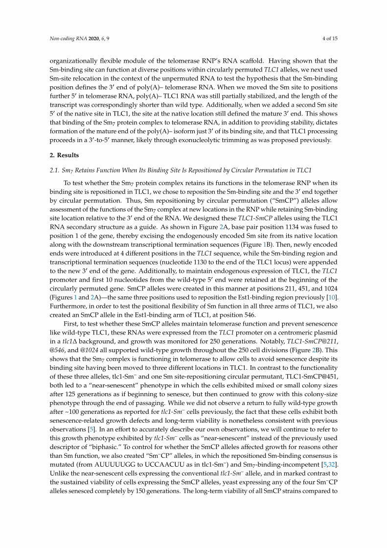
First, to test whether these SmCP alleles maintain telomerase function and prevent senescencelike wild-type TLC1, these RNAs were expressed from the TLC1 promoter on a centromeric plasmidin a tlc1∆ background, and growth was monitored for 250 generations. Notably, TLC1-SmCP@211,@546, and @1024 all supported wild-type growth throughout the 250 cell divisions (Figure 2B). Thisshows that the Sm7 complex is functioning in telomerase to allow cells to avoid senescence despite itsbinding site having been moved to three different locations in TLC1. In contrast to the functionalityof these three alleles, tlc1-Sm– and one Sm site-repositioning circular permutant, TLC1-SmCP@451,both led to a “near-senescent” phenotype in which the cells exhibited mixed or small colony sizesafter 125 generations as if beginning to senesce, but then continued to grow with this colony-sizephenotype through the end of passaging. While we did not observe a return to fully wild-type growthafter ~100 generations as reported for tlc1-Sm– cells previously, the fact that these cells exhibit bothsenescence-related growth defects and long-term viability is nonetheless consistent with previousobservations [5]. In an effort to accurately describe our own observations, we will continue to refer tothis growth phenotype exhibited by tlc1-Sm– cells as “near-senescent” instead of the previously useddescriptor of “biphasic.” To control for whether the SmCP alleles affected growth for reasons otherthan Sm function, we also created “Sm–CP” alleles, in which the repositioned Sm-binding consensus ismutated (from AUUUUUGG to UCCAACUU as in tlc1-Sm–) and Sm7-binding-incompetent [5,32].Unlike the near-senescent cells expressing the conventional tlc1-Sm– allele, and in marked contrast tothe sustained viability of cells expressing the SmCP alleles, yeast expressing any of the four Sm–CPalleles senesced completely by 150 generations. The long-term viability of all SmCP strains compared to
Non-coding RNA 2020, 6, 9 5 of 15
the senescence of all Sm–CP strains strongly suggests that the Sm7 protein complex retains its functionsat each of the four different positions to which its binding site was moved in the circularly permutedTLC1 RNAs. Furthermore, these are the first circular permutants of full-length TLC1 RNA to be tested,and it is noteworthy that the telomerase RNA ends themselves can be repositioned to any of thesefour locations while permitting telomerase functionality in vivo. However, because unpermuted TLC1with a mutated Sm site (tlc1-Sm–) does not quite cause a senescent phenotype, the fully senescentphenotype of all of the Sm–CP alleles suggests that circular permutation of the RNA interferes withTLC1 function and/or accumulation.
Figure 2. Sm7 retains function in telomerase RNA when its binding site is repositioned via circularpermutation. (A) Schematics of SmCP gene construction and expected RNA structure. The native endsof the TLC1 gene were effectively sealed off by connecting nucleotide 1134 with nucleotide 1, thusremoving the Sm site from its native position in the RNA. A dashed line connects the location of this

Non-coding RNA 2020, 6, 9 6 of 15
1134-to-1 fusion between the 4 circularly permuted alleles. The wild-type TLC1 promoter throughnucleotide 10 (green) as well as the sequence from nucleotide 1130 on (red) flank the circularlypermutated central 1–1130 region, thereby repositioning the Sm-binding site (black with red outline)along with the 5′ and 3′ ends in the encoded transcript. Black rectangles indicate the Ku-binding site,Est1-binding region, and pseudoknot (PK). A white rectangle indicates the template. The secondarystructure models of wild-type TLC1 and an example of an SmCP (TLC1-SmCP@211) are schematized tothe right, using the same coloring scheme as in the gene diagrams to the left. Details of the secondarystructure of these large RNAs are omitted in these low-resolution schematics for the sake of simplicity,but, in fact, these regions of TLC1 have wild-type sequence and predicted secondary structure verysimilar to wild type as well. (B) TLC1-SmCP alleles with a wild-type Sm-binding site support sustainedgrowth and do not cause cells to senesce. All TLC1 alleles were expressed from centromeric plasmidsin a tlc1∆ rad52∆ background, and cells were serially passaged on solid media for 250 generations (10re-streaks). (C) Sm7 confers poly(A)– telomerase RNA stabilization and appropriate 3′-end formationwhen repositioned via circular permutation. Total RNA was isolated from cells used in the passagingexperiment shown in Figure 2B and analyzed by northern blotting with TLC1 and U1 snRNA probes.Total TLC1 RNA abundance was normalized to U1 abundance to control for sample loading in each laneand then set relative to the wild-type condition. The numbers displayed below the blot are averagesof two independent biological replicates. (D) SmCP alleles support stable, short telomeres. GenomicDNA was isolated from cells in the passaging experiment in Figure 2B at 250 generations. Telomerelength was then analyzed by Southern blotting. The blot was probed for telomeric sequence and fora 1621-bp non-telomeric restriction fragment (non-telomeric control). The pairs of lanes in the blotshown are independent biological-replicate samples, and the telomere length numbers are averages ofthe two replicates except in the TLC1-SmCP@1024 condition. In this condition, the telomere lengthcould not be quantified in the first replicate due to anomalous migration of the non-telomeric control(black asterisk), and so the displayed telomere length number in this condition is a quantitation of onlythe second replicate sample.
Next, we assessed how TLC1 RNA processing and abundance was affected in the circularlypermuted SmCP alleles. For all four SmCP telomerase RNAs, the poly(A)– isoform—predicted to bejust 15 nt longer than wild type—was readily detectable by northern blotting, although at a lowerabundance than wild type (Figure 2C). This shows that Sm7 can function in TLC1 3′-end processingand (to a lesser extent) RNA accumulation when its binding site in the RNA is repositioned. Theparticularly low abundance of the SmCP@451 poly(A)– RNA probably contributes to why these cellsgrow less well than those expressing the three other SmCP alleles (Figure 2B, lower plate). Whenwe assessed TLC1 RNA abundance in the four Sm–CP alleles, we observed that poly(A)+ TLC1 wasessentially undetectable, unlike in cells containing the unpermuted tlc1-Sm– allele (Figure 2C; comparelanes 9–12 with lane 4). The fact that Sm–CP alleles have a mutated Sm-binding site at the newpositions and this results in complete loss of the poly(A)– isoform—in contrast to the SmCP allelesdescribed above which had detectable accumulation of this RNA—provides further evidence that Sm7
retains partial function in promoting RNA stability when its binding site is repositioned via circularpermutation. Additionally, this strongly suggests that while cells expressing tlc1-Sm– accumulatejust enough functional TLC1 RNA (such as the poly(A)+ isoform) to prevent complete senescence,circularly permuting tlc1-Sm– reduces the already low RNA abundance to negligible levels, resultingin senescence.
We next assessed the effect of the Sm-site repositioning and circular permutation on telomerelength. In the cases where cells did not senesce, we isolated genomic DNA from TLC1-SmCP cells atthe end of passaging and subjected the DNA to Southern blotting with a telomeric probe. The resultsshow that cells expressing SmCP@211 and @546 alleles had the longest telomeres, although all fourSmCP constructs supported telomeres substantially shorter than wild type (Figure 2D). Telomereswere longer in TLC1-SmCP@211 and @546 cells than in the near-senescent tlc1-Sm– cells, providingadditional evidence that Sm7 retains enough function when repositioned to prevent senescence. Asexpected from their near-senescent phenotype, TLC1-SmCP@451 cells had the shortest telomeres of

Non-coding RNA 2020, 6, 9 7 of 15
any of the SmCP alleles, averaging 212 bp shorter than wild type (lanes 7 and 8). Considering thatthe SmCP@451 allele supported the shortest telomeres and the lowest RNA abundance, while theSmCP@211 and @546 cells had the longest telomeres and the highest RNA levels, these results suggestthat relocated Sm7 functioned best at positions 211 and 546 and that SmCP telomerase RNAs do notsupport full-length telomeres primarily because of their low levels of accumulation.
2.2. The Sm7-Binding Site Defines the Mature 3′ End of Poly(A)– TLC1 RNA
It has been proposed that Sm7 binding to its consensus site in TLC1 (nucleotides 1143–1150)defines the mature 3′ end of poly(A)– TLC1 at nucleotide 1157 by blocking exonucleolytic trimming atthe terminus of an initially longer transcript [5,31]. Although genetic data suggest that the nuclearexosome is involved in the maturation of poly(A)– TLC1 [31], the model that Sm-binding defines themature 3′ end of poly(A)– TLC1 has not been rigorously evaluated. Having observed that Sm7 retainsfunction when its binding site is repositioned in circularly permuted telomerase RNAs, we next testedthe hypothesis that Sm7 controls 3′-end formation in poly(A)– TLC1 by moving the Sm-binding siteslightly further 5′ in TLC1 without circular permutation. If the Sm-binding site’s position defines thelocation of the 3′ end of poly(A)– TLC1, repositioning the Sm-binding site to a position further 5′ inthe RNA should result in a correspondingly truncated poly(A)– form. In the published secondarystructure model of poly(A)+ TLC1 (nts 1–1251), i.e., a possible precursor to poly(A)– TLC1 that Sm7
could bind before RNA processing, the Sm-binding consensus is on the 5′ side of an internal loop of ahairpin, while nucleotide 1157 (the poly(A)– 3′ end) is predicted to be on the opposing side of this loop,directly across from the Sm-binding consensus (Figure 1A, inset) [10]. Additionally, the nucleotides inand around this putative internal loop show higher sequence conservation [10,30], suggesting that thispredicted structure as a whole, not just the 8 nucleotide Sm-binding consensus, may be required forSm7 to perform its function. Thus, based on this conservation, predicted local secondary structure,and pilot studies involving repositioning only the minimal Sm-binding consensus sequence, we chosenucleotides 1138 to 1165 as a predicted RNA module that would be likely to function when repositionedwithin TLC1. We then inserted this Sm-binding site into tlc1-Sm– at positions 926, 1003, and 1089(Figure 1A, inset). All three of these positions are predicted to be within bulged loops located onthe distal portion of the RNA’s terminal arm, a region that is dispensable for telomerase function inS. cerevisiae [12].
TLC1 RNAs with these relocated Sm sites were expressed from centromeric plasmids in tlc1∆cells, and the cells were passaged for 250 generations to test for senescence. The strains expressingtlc1-Sm–+Sm@1089 or @926 displayed wild-type growth, whereas those expressing tlc1-Sm–+Sm@1003exhibited a near-senescent growth phenotype, similar to tlc1-Sm– cells (Figure 3A). This shows that theSm site is functional at positions 926 or 1089 (212 and 49 nts upstream of its location in wild-type TLC1RNA, respectively) in allowing telomerase to prevent cellular senescence. As for Sm-site function in TLC1RNA stability and processing, northern blotting analysis revealed that, in agreement with the observedgrowth phenotypes, the Sm-binding sites inserted at positions 1089 and 926 in tlc1-Sm– partially stabilizedpoly(A)– TLC1 (1% and 7% of wild-type poly(A)– TLC1 abundance, respectively), while the Sm-bindingsite inserted at position 1003 did not (Figure 3B). While the poly(A)– TLC1 isoform was less abundant forthese 5′-shifted Sm-site alleles than it is for wild-type, the complete absence of the poly(A)– species intlc1-Sm– cells suggests that the Sm sites at positions 1089 and 926 contribute at least some degree of functionin RNA 3′-end maturation and poly(A)– RNA stability. This result is similar to what we observed withSm site functionality in the context of circularly permuted TLC1-SmCP RNAs, which more dramaticallyrepositioned the Sm site. Additionally, the poly(A)– RNAs that were stabilized by the inserted Sm sites atpositions 1089 and 926 were shorter than wild-type poly(A)– TLC1 RNA (indicated by red arrowheads inlanes 8, 9, 12, and 13). The length of each of these poly(A)– RNAs measured from the northern blot werewithin 20 nucleotides (i.e., within ~2% of the RNA’s total length) of the expected size for each transcriptbased on the respective position of the relocated Sm site. These results provide strong evidence that theposition of the Sm-binding site in TLC1 RNA defines the mature 3′ end of poly(A)– TLC1.

Non-coding RNA 2020, 6, 9 8 of 15
Figure 3. Repositioning the Sm-binding site at two of three positions further 5′ in TLC1 results instabilization of shorter poly(A)– TLC1 RNAs. (A) Sm sites inserted in tlc1-Sm– restored robust growthat positions 1089 and 926, but not at 1003. TLC1 alleles were expressed and cells were passaged asin Figure 2. (B) Functional Sm sites inserted 5′ of the native position in tlc1-Sm– stabilized shorterpoly(A)– TLC1 RNAs. Total RNA was isolated from cells used in the passaging experiment in Figure 3Aand analyzed by northern blotting as in Figure 2C. Red arrowheads indicate the shorter poly(A)–TLC1 RNAs stabilized in tlc1-Sm–+Sm@1089 and tlc1-Sm–+Sm@926 cells. The pairs of lanes representindependent biological-replicate samples. (C) Inserting a mutated Sm site in tlc1-Sm– at position 1089causes a fully senescent phenotype. TLC1 alleles were expressed and cells were passaged as in Figure 2.(D) The native Sm site is dominant over a 5′-shifted Sm site in directing poly(A)– TLC1 3′-end formation.Total RNA was isolated from cells used in the passaging experiment in Figure 3C and analyzed bynorthern blotting as in Figure 2C. The truncated poly(A)– TLC1 RNA in tlc1-Sm–+Sm@1089 cells isindicated with a red arrowhead. Pairs of lanes represent biological duplicate samples.

Non-coding RNA 2020, 6, 9 9 of 15
Sm7 has been proposed to define the mature 3′ end of poly(A)– TLC1 specifically by halting3′-to-5′ exonucleolytic degradation of TLC1 [31]. As a further test of this model, we inserted the Smsite at position 1089 in TLC1 but did not mutate the native Sm site, resulting in an RNA with twodifferent Sm sites, both with intact Sm-binding consensus sequences. If TLC1 is processed by 3′-to-5′
exonucleolytic degradation, only the native Sm site will define the mature 3′ end of poly(A)– TLC1RNA because it is further 3′ than the Sm site at position 1089. However, if TLC1 processing occursthrough a mechanism that does not proceed in a 3′-to-5′ manner (e.g., endonucleolytic cleavage), onewould expect to observe a mixture of wild-type-length and truncated poly(A)– TLC1 due to processingat both the native and repositioned Sm sites. As controls, we also inserted a mutated Sm site at position1089 in TLC1 and in tlc1-Sm–. When we expressed these RNAs in yeast and tested for senescenceduring serial passaging, we observed wild-type growth as long as the TLC1 RNA being expressedcontained at least one wild-type Sm site (Figure 3C). In contrast, the tlc1-Sm–+Sm–@1089 controlexhibited a fully senescent phenotype, a more severe phenotype than the near-senescent phenotypeof tlc1-Sm– cells. Northern blotting showed that the Sm site at position 1089 only directed 3′-endformation if the native Sm site was mutated (Figure 3D, lanes 12 and 13). Wild-type-length poly(A)–TLC1 was present in both TLC1+Sm@1089 and TLC1+Sm–@1089 cells (Figure 3D, lanes 8–11). Theseresults suggest that Sm7 defines the mature 3′ end of poly(A)– TLC1 by halting trimming in the 3′-to-5′
direction. We also observed that inserting a Sm site at position 1089 in wild-type TLC1 reduced theabundance of poly(A)– TLC1 by 60%–70% regardless of whether or not the inserted site had an intactSm-binding consensus sequence. Additionally, inserting a mutated Sm site at position 1089 in tlc1-Sm–
reduced poly(A)+ TLC1 RNA abundance below the limit of detection, which likely is the cause ofthe fully senescent phenotype of these cells. The results from these controls suggest that the reducedabundance of the truncated poly(A)– isoform of tlc1-Sm–+Sm@1089 is partially caused by insertingSm-site sequence at 1089.
3. Discussion
The telomerase RNA differs in many ways from other large, well-studied, non-coding RNAs suchas ribosomal and spliceosomal RNAs. Although its existence is highly conserved among eukaryotes, itssequence and length vary greatly between species [33]. Considering the rapid evolution of telomeraseRNA along with experimental results has led to the model that telomerase RNA functions as a flexiblescaffold for protein subunits in the telomerase RNP [10,14,15,30]. Thus, unlike other RNP enzymes thatrequire a precise structural organization of components in the complex for function (e.g., the ribosome),the S. cerevisiae telomerase RNP has overall organization that has been shown to be strikingly flexible,which has provided a new paradigm for other RNP complexes, including those containing other longnoncoding RNAs [10,13,14,30,34].
Here, we have shown that organizational flexibility of the large yeast telomerase RNP extendsto include the Sm7 complex, which is a subunit of the RNP that binds just before the 3′ end of thetelomerase RNA’s most abundant isoform and stabilizes it. Despite repositioning of the Sm site to fourdramatically different locations across all three TLC1 RNA arms via circular permutation, Sm7 was stillable to promote processing and stabilization of the major [poly(A)–] TLC1 isoform. Although poly(A)–TLC1 RNA abundance was reduced in these SmCP alleles (discussed below), Sm7 still retained partialfunction at all positions tested compared to negative controls. This demonstrates that Sm7 and itsbinding site in telomerase RNA function as an organizationally flexible module, along with the 5′ and3′ ends, which were relocated along with the Sm site by circular permutation. Between the resultspresented here and the previously reported repositioning studies regarding the binding sites for theEst1 and Ku subunits [10,13], it is now evident that TLC1 is an organizationally flexible scaffold forthese three well-established protein subunits of the telomerase holoenzyme. While organizationalflexibility has not yet been reported for the Pop1/6/7 complex, which was recently reported to bind to anessential sequence in TLC1 near the Est1-binding site [8], the entire Est1-arm of TLC1 (with the Est1 siteas well as the adjacent Pop1/6/7-binding site) has been shown to provide its function when expressed in

Non-coding RNA 2020, 6, 9 10 of 15
trans under conditions where Est1 is artificially tethered via a heterologous binding interface to the 3′
end of TLC1 [35]. This suggests that the flexible scaffold model applies to the Pop1/6/7 proteins as well.After determining that the Sm7 subunits can function in vivo when bound to different non-native
positions within the telomerase RNP in the context of circular permutation, we next used Sm-siterepositioning to investigate mechanistic hypotheses regarding Sm function in TLC1 RNA biogenesis.We tested whether the position of Sm binding defines the mature 3′ end of poly(A)– TLC1 byrelocating the Sm-binding site further 5′ in the RNA compared to its native position. At positionswhere the 5′-shifted Sm site was functional in stabilizing poly(A)– TLC1, the stabilized species werecorrespondingly shorter RNAs, thus providing strong evidence that Sm7 does indeed direct 3′-endformation of poly(A)– TLC1. We also found that when wild-type Sm-binding sites were includedboth at the native position and at a 5′-shifted position, only the native site defined the mature 3′
end of poly(A)– TLC1, suggesting that processing proceeds in a 3′-to-5′ manner, possibly throughexonucleolytic trimming, as has been previously proposed [31].
All experiments in this work were performed using TLC1 alleles that include the endogenouspromoter and terminator regions on centromeric plasmids in a tlc1∆ strain. Unlike the strictlysingle-copy expression of chromosomally integrated alleles, CEN plasmids are maintained from equalto a few-fold higher copy number than one per cell on average and with slightly higher variability incopy number on a cell-by-cell basis [36–38]. Thus, the phenotypes that we observed may be milderthan if using chromosomally integrated genes, since transcript abundances may be slightly higher.However, given that our conclusions are based on qualities such as TLC1 RNA transcript length, whichshould not be affected by gene copy number, relative cell growth, and TLC1 abundance, in all casescompared to relevant, equally treated CEN-borne controls, the results reported herein still stronglysupport the conclusions.
The results of all of our Sm-site repositioning experiments—with or without also circularlypermuting the RNA—show that although Sm7 did retain function when its binding site was repositioned,it did not function as well as wild type in stabilizing poly(A)– TLC1. It is possible that these defectsin TLC1 RNA stabilization were caused by alterations in aspects of RNA folding. In the case of theTLC1-SmCP alleles, circular permutation may have affected local and/or global folding because ofchanges in the contact order of the RNA secondary structure and in the timing of when sequencesemerge from RNA polymerase [39–41]. Altered folding of circularly permuted TLC1 RNAs may, inturn, disrupt Sm7 binding or make the RNA more exposed to nucleases. In addition to effects ofcircular permutation on TLC1 RNA abundance, misfolding could also be deleterious to telomeraseactivity in the case of the TLC1-SmCP@451 allele, where the Sm site and RNA ends are very closeto the template-boundary element, a structure that is essential for activity in the catalytic core of thetelomerase RNP [11,42]. Thus, the near-senescent phenotype of cells expressing TLC1-SmCP@451 mayhave been caused by decreased RNA abundance as well as impaired telomerase activity.
When we repositioned the Sm site without circular permutation, multiple RNA foldingconsiderations may have contributed to decreased abundance of poly(A)– TLC1. First, as shown inFigure 3D, inserting the Sm site at position 1089 reduced RNA abundance even in wild-type TLC1,seemingly independent of Sm7 function. This may be due to disrupted folding of the region wherethe Sm site was inserted. Additionally, repositioning the Sm site without circular permutation mayhave disrupted folding of the Sm site itself. Although the portion of TLC1 that we used in the Sm-siterepositioning experiments without circular permutation was chosen for its proposed hairpin structurein poly(A)+ TLC1 [10], this may not in fact be the conformation that binds, or binds best to, the Sm7
complex. Sm-binding sites in many other RNAs have been found to be single-stranded and precededand/or followed by hairpin structures [24,25]. In agreement with this trend, an Mfold secondarystructure prediction of an 1195 nucleotide NNS-terminated TLC1 RNA [19], another possible precursorof poly(A)– TLC1, shows the Sm site adopting a largely single-stranded conformation followed by asingle hairpin (Figure S1). In contrast, Mfold secondary structure predictions of tlc1-Sm–+Sm@1089,@1003, and @926 all show the repositioned Sm sites forming the same hairpin-and-loop structure seen

Non-coding RNA 2020, 6, 9 11 of 15
in the published secondary structure model of poly(A)+ TLC1 (Figure S2). Notably, the Sm site atposition 1003, which did not stabilize poly(A)– TLC1 at all, is scored with very low Mfold pnum values(i.e., the region is “well determined” and predicted to have a high probability of forming the secondarystructure conformation shown, as indicated by red text in Figure S2) [43], suggesting that this Sm sitemay have formed a stable structure that does not permit efficient Sm7 binding. Thus, alleles withrepositioned Sm sites may not have functioned optimally because the new contexts of the Sm sites inthe landscape of the TLC1 RNA did not favor folding of the most Sm7-binding-competent structure.
The Sm7 complex has a highly conserved function in the biogenesis of certain spliceosomalsnRNAs, but why does it bind to S. cerevisiae telomerase RNA? One answer to this question mayrelate to an interesting theme that has emerged from the study of telomerase RNPs from a wide rangeof organisms. Consistent with the flexible scaffold hypothesis and the related rapid evolution oftelomerase RNAs, these RNAs have independently evolved a wide variety of ways to be processed andto allow appropriate RNP biogenesis, maturation, and probably also modes of regulation [10,13–16].Thus, the Sm7 complex binding to the 3′ end of TLC1 may simply be another variation on this broadtheme. The Sm ring is required for 3′-end maturation of some spliceosomal snRNAs in yeast, and TLC1has apparently “borrowed” this machinery for formation and stabilization of its own mature 3′ end [44].However, 3′-end formation for these snRNAs seems to involve the exosome as well as other enzymes,while thus far only the exosome has been implicated in processing of TLC1 [31,45,46]. Therefore,it seems possible either that (a) these other snRNA-processing enzymes play yet-uncharacterizedroles in maturation of TLC1, or (b) S. cerevisiae telomerase RNA has evolved a different processingmechanism from snRNAs that only requires the Sm ring and the exosome. Irrespective of these details,the recurrent observation throughout evolution that telomerase RNAs coopt proteins from other RNPcomplexes for their own biogenesis demonstrates both the myriad ways that noncoding RNAs canbe processed and stabilized and that telomerase RNAs are exceedingly flexible in which of thesemechanisms are used for their own biogenesis.
In summary, repositioning the Sm-binding site and the ends of yeast telomerase RNA has yieldedinsights about the organizationally flexible nature of the RNA and the mechanistic role of Sm7 in itsbiogenesis. Sm7 can functionally tolerate repositioning of its binding site within TLC1 along withthe RNA ends. Furthermore, the telomerase enzyme’s fundamental functions can also endure thesubstantial reorganization of the RNA that is caused by circularly permuting the telomerase RNAtranscript in vivo, despite the fact that it globally changes the RNA-folding landscape and locallybreaks the phosphodiester backbone. Sm-site repositioning also has revealed that the mature 3′ endof poly(A)– TLC1 appears to be (1) controlled by the location of Sm7 binding to the RNA and (2)generated in a 3′-to-5′ manner, lending experimental support to previously proposed models of TLC1biogenesis. The experimental approach of repositioning the Sm-binding site in order to change thelocation of the mature 3′ end could be useful for next identifying the precursor of poly(A)– TLC1 andfully characterizing telomerase RNA biogenesis in budding yeasts.
4. Materials and Methods
4.1. Construction of TLC1-SmCP Alleles
The TLC1-SmCP alleles were constructed using a plasmid containing two tandem copies of TLC1in a pRS414 backbone [47] (pDZ573, see Table S1 for full list of plasmids used). In this construct,the two copies of TLC1 are fused into a single gene such that nucleotide 1134 of the first TLC1 copy isfollowed directly by nucleotide 1 (see Figure 2A) of the second TLC1 copy, meaning that the Sm siteand transcriptional termination sequences are absent in the first TLC1 sequence. Circular permutationwas performed by PCR-amplifying specific segments of this template DNA beginning at the siteof repositioning in the first TLC1 copy and ending at the same site in the second copy (e.g., forSmCP@211, this resulted in a PCR product containing base pairs 211–1134 followed by 1–210). Thesecircularly permuted DNAs were inserted into a separate plasmid containing the natural ends of the

Non-coding RNA 2020, 6, 9 12 of 15
TLC1 gene such that sequences from nucleotide 10 upstream and from nucleotide 1130 downstream(which includes the Sm site) are retained at the ends of the new gene. As an example, the sequenceof TLC1-SmCP@211 is laid out from promoter to terminator as follows: the TLC1 promoter throughnucleotide 10, 211–1134, 1–210, and 1130 through the transcriptional terminator and natural end of theTLC1 gene (see Figure 2A). As a result of this construction scheme, nucleotides 1–10 and 1130–1134 arecontained twice in all TLC1-SmCP alleles, making the RNAs 15 nucleotides longer than wild-type TLC1.
4.2. Experiments in Yeast
All experiments were performed in the strain TCy43 (MATa ura3-53 lys2-801 ade2-101 trp1-1his3-∆200 leu2-∆1 VR::ADE2-TEL adh4::URA3-TEL tlc1::LEU2 rad52::HIS3 pTLC1-LYS2-CEN) [5]. AllTLC1 alleles were expressed from centromeric plasmids (Table S1) that were derived from pSD107(pTLC1-TRP1-CEN) [48]. TCy43 was transformed with TLC1-containing plasmids, and colonies werestreaked to minimal –TRP–LYS medium. Loss of the pTLC1-LYS2-CEN cover plasmid was selectedfor by re-streaking cells to minimal –TRP medium containing α-aminoadipate. These cells were thenserially re-streaked nine times to minimal –TRP medium and photographed after each round of growth.When estimating the number of generations at different points throughout passaging, each round ofgrowth (e.g., each colony on solid medium) after loss of the cover plasmid (including the round ofgrowth in the presence of α-aminoadipate) is approximated as 25 generations.
4.3. Northern Blotting
Northern blotting was performed as described previously [12,13,35,49]. Briefly, cells from theserial passaging plates were grown in liquid cultures to an OD600 of ~1.0 and harvested. Total RNAwas isolated using the hot phenol method [50]. In total, 15–30 µg of total RNA from each sample wasboiled, separated by urea-PAGE, and then transferred to Hybond-N+ Nylon membrane (GE). Themembrane was UV-crosslinked and probed for both TLC1 and U1 snRNA sequence using 100-foldfewer counts of U1 probe than TLC1 probe to account for the large difference in abundance between thetwo RNAs. TLC1 RNA abundance was calculated by normalizing to U1 abundance, and numbers inthe text and figures are expressed relative to the wild-type TLC1 condition. Lengths of poly(A)– TLC1RNAs in Figure 3B were calculated based on the molecular weight standard shown using ImageQuantTL (GE). The molecular weight standards used on the northern blots shown are single-stranded DNA,which migrates roughly 5% faster than single-stranded RNA. To adjust for this difference, we dividedthe known length of poly(A)– TLC1 (1157 nts) by its average apparent length relative to the DNAmolecular weight standard to obtain an expected/observed ratio. The length measurements given byImageQuant TL for the truncated forms of TLC1 were averaged between biological replicates and thenmultiplied by this ratio to produce the RNA-length measurements mentioned in the Results section.
4.4. Southern Blotting
Southern blotting was performed as described previously [12,13,35,49]. Briefly, cells were grownand harvested in the same manner as those used for northern blots, and genomic DNA was isolatedfrom these pellets (Gentra Puregene system). Roughly equal amounts of genomic DNA were digestedwith XhoI and then separated on a 1.1% agarose gel. DNA was transferred to Hybond-N+ Nylonmembrane (GE) to which it was UV-crosslinked. The membrane was probed for yeast telomericsequence and a 1621 bp non-telomeric XhoI fragment that served as a non-telomeric control band [51].Average Y′ telomere length was calculated using the weighted average mobility method describedpreviously [13]. Briefly, ImageQuant 5.2 (GE) was used to obtain the non-Gaussian distributions of Y′
telomere signal in each lane. These distributions were aligned with one another using the mobility oftheir non-telomeric control bands. The weighted average mobility (WAM) of Y′ telomeric restrictionfragments was calculated as follows using the pixel coordinates and intensity values (RFU) at eachpoint across the distribution: WAM =
∑RFU∗pixel∑
RFU . The value for WAM given by this equation in pixelcoordinates was then converted to base pairs using a trend line created based on a molecular weight

Non-coding RNA 2020, 6, 9 13 of 15
standard on the blot. The weighted-average DNA length, in base pairs, in each lane were then setrelative to the wild-type lane from their respective biological replication of the experiment, and thesenumbers were averaged between replicates, resulting in the values shown in Figure 2D.
Supplementary Materials: The following are available online at http://www.mdpi.com/2311-553X/6/1/9/s1.Figure S1: Mfold software prediction for the secondary structure of NNS-terminated TLC1 (nucleotides 1–1195);Figure S2: Mfold software predictions for the secondary structures of repositioned Sm-binding sites.
Author Contributions: E.P.H performed experiments; E.P.H. and D.C.Z. conceived and designed experiments,analyzed and interpreted data, and wrote the paper; D.C.Z. provided reagents and funding. All authors have readand agreed to the published version of the manuscript.
Funding: This research was supported in part by the National Institute of General Medical Sciences of the NationalInstitutes of Health under award number R01 GM118757 to D.C.Z., as well as funds from Lehigh University andJohns Hopkins University.
Acknowledgments: We thank members of the Zappulla laboratory for their input on these experiments, particularlyundergraduates Benjamin Ford and Timothy Kistner who helped with the design and cloning of the TLC1 allelesused in Figure 3.
Conflicts of Interest: The authors declare no conflict of interest.
References
1. Greider, C.W.; Blackburn, E.H. Identification of a specific telomere terminal transferase activity in Tetrahymenaextracts. Cell 1985, 43, 405–413. [CrossRef]
2. Greider, C.W.; Blackburn, E.H. A telomeric sequence in the RNA of Tetrahymena telomerase required fortelomere repeat synthesis. Nature 1989, 337, 331–337. [CrossRef]
3. Lingner, J.; Hughes, T.R.; Shevchenko, A.; Mann, M.; Lundblad, V.; Cech, T.R. Reverse transcriptase motifs inthe catalytic subunit of telomerase. Science 1997, 276, 561–567. [CrossRef] [PubMed]
4. Shippen-Lentz, D.; Blackburn, E.H. Functional evidence for an RNA template in telomerase. Science 1990,247, 546–552. [CrossRef] [PubMed]
5. Seto, A.G.; Zaug, A.J.; Sobel, S.G.; Wolin, S.L.; Cech, T.R. Saccharomyces cerevisiae telomerase is an Sm smallnuclear ribonucleoprotein particle. Nature 1999, 401, 177–180. [CrossRef] [PubMed]
6. Peterson, S.E.; Stellwagen, A.E.; Diede, S.J.; Singer, M.S.; Haimberger, Z.W.; Johnson, C.O.; Tzoneva, M.;Gottschling, D.E. The function of a stem-loop in telomerase RNA is linked to the DNA repair protein Ku.Nat. Genet. 2001, 27, 64–67. [CrossRef] [PubMed]
7. Seto, A.G.; Livengood, A.J.; Tzfati, Y.; Blackburn, E.H.; Cech, T.R. A bulged stem tethers Est1p to telomeraseRNA in budding yeast. Genes Dev. 2002, 16, 2800–2812. [CrossRef]
8. Lemieux, B.; Laterreur, N.; Perederina, A.; Noël, J.-F.; Dubois, M.-L.; Krasilnikov, A.S.; Wellinger, R.J.Active Yeast Telomerase Shares Subunits with Ribonucleoproteins RNase P and RNase MRP. Cell 2016, 165,1171–1181. [CrossRef]
9. Dandjinou, A.T.; Lévesque, N.; Larose, S.; Lucier, J.-F.; Abou Elela, S.; Wellinger, R.J. A phylogeneticallybased secondary structure for the yeast telomerase RNA. Curr. Biol. 2004, 14, 1148–1158. [CrossRef]
10. Zappulla, D.C.; Cech, T.R. Yeast telomerase RNA: A flexible scaffold for protein subunits. Proc. Natl. Acad.Sci. USA 2004, 101, 10024–10029. [CrossRef]
11. Tzfati, Y.; Fulton, T.B.; Roy, J.; Blackburn, E.H. Template boundary in a yeast telomerase specified by RNAstructure. Science 2000, 288, 863–867. [CrossRef] [PubMed]
12. Zappulla, D.C.; Goodrich, K.; Cech, T.R. A miniature yeast telomerase RNA functions in vivo and reconstitutesactivity in vitro. Nat. Struct. Mol. Biol. 2005, 12, 1072–1077. [CrossRef] [PubMed]
13. Zappulla, D.C.; Goodrich, K.J.; Arthur, J.R.; Gurski, L.A.; Denham, E.M.; Stellwagen, A.E.; Cech, T.R. Kucan contribute to telomere lengthening in yeast at multiple positions in the telomerase RNP. RNA 2011, 17,298–311. [CrossRef] [PubMed]
14. Zappulla, D.C.; Cech, T.R. RNA as a flexible scaffold for proteins: Yeast telomerase and beyond. Cold SpringHarb. Symp. Quant. Biol. 2006, 71, 217–224. [CrossRef] [PubMed]
15. Lebo, K.J.; Zappulla, D.C. Stiffened yeast telomerase RNA supports RNP function in vitro and in vivo. RNA2012, 18, 1666–1678. [CrossRef] [PubMed]
Non-coding RNA 2020, 6, 9 14 of 15
16. Chen, J.L.; Blasco, M.A.; Greider, C.W. Secondary structure of vertebrate telomerase RNA. Cell 2000, 100,503–514. [CrossRef]
17. Mefford, M.A.; Zappulla, D.C. Physical Connectivity Mapping by Circular Permutation of Human TelomeraseRNA Reveals New Regions Critical for Activity and Processivity. Mol. Cell. Biol. 2016, 36, 251–261. [CrossRef]
18. Niederer, R.O.; Zappulla, D.C. Refined secondary-structure models of the core of yeast and human telomeraseRNAs directed by SHAPE. RNA 2015, 21, 1053. [CrossRef]
19. Jamonnak, N.; Creamer, T.J.; Darby, M.M.; Schaughency, P.; Wheelan, S.J.; Corden, J.L. Yeast Nrd1, Nab3, andSen1 transcriptome-wide binding maps suggest multiple roles in post-transcriptional RNA processing. RNA2011, 17, 2011–2025. [CrossRef]
20. Noël, J.-F.; Larose, S.; Abou Elela, S.; Wellinger, R.J. Budding yeast telomerase RNA transcription terminationis dictated by the Nrd1/Nab3 non-coding RNA termination pathway. Nucleic Acids Res. 2012, 40, 5625–5636.[CrossRef]
21. Chapon, C.; Cech, T.R.; Zaug, A.J. Polyadenylation of telomerase RNA in budding yeast. RNA 1997, 3,1337–1351. [PubMed]
22. Jones, M.H.; Guthrie, C. Unexpected flexibility in an evolutionarily conserved protein-RNA interaction:Genetic analysis of the Sm binding site. EMBO J. 1990, 9, 2555–2561. [CrossRef] [PubMed]
23. Will, C.L.; Lührmann, R. Spliceosomal UsnRNP biogenesis, structure and function. Curr. Opin. Cell Biol.2001, 13, 290–301. [CrossRef]
24. Branlant, C.; Krol, A.; Ebel, J.P.; Lazar, E.; Haendler, B.; Jacob, M. U2 RNA shares a structural domain withU1, U4, and U5 RNAs. EMBO J. 1982, 1, 1259–1265. [CrossRef] [PubMed]
25. Liautard, J.P.; Sri-Widada, J.; Brunel, C.; Jeanteur, P. Structural organization of ribonucleoproteins containingsmall nuclear RNAs from HeLa cells. Proteins interact closely with a similar structural domain of U1, U2, U4and U5 small nuclear RNAs. J. Mol. Biol. 1982, 162, 623–643. [CrossRef]
26. Mattaj, I.W.; De Robertis, E.M. Nuclear segregation of U2 snRNA requires binding of specific snRNP proteins.Cell 1985, 40, 111–118. [CrossRef]
27. Hamm, J.; Kazmaier, M.; Mattaj, I.W. In vitro assembly of U1 snRNPs. EMBO J. 1987, 6, 3479–3485. [CrossRef]28. Bosoy, D.; Peng, Y.; Mian, I.S.; Lue, N.F. Conserved N-terminal motifs of telomerase reverse transcriptase
required for ribonucleoprotein assembly in vivo. J. Biol. Chem. 2003, 278, 3882–3890. [CrossRef]29. Mozdy, A.D.; Cech, T.R. Low abundance of telomerase in yeast: Implications for telomerase haploinsufficiency.
RNA 2006, 12, 1721–1737. [CrossRef]30. Mefford, M.A.; Rafiq, Q.; Zappulla, D.C. RNA connectivity requirements between conserved elements in the
core of the yeast telomerase RNP. EMBO J. 2013, 32, 2980–2993. [CrossRef]31. Coy, S.; Volanakis, A.; Shah, S.; Vasiljeva, L. The Sm complex is required for the processing of non-coding
RNAs by the exosome. PLoS ONE 2013, 8, e65606. [CrossRef] [PubMed]32. Raker, V.A.; Hartmuth, K.; Kastner, B.; Lührmann, R. Spliceosomal U snRNP core assembly: Sm proteins
assemble onto an Sm site RNA nonanucleotide in a specific and thermodynamically stable manner. Mol. Cell.Biol. 1999, 19, 6554–6565. [CrossRef] [PubMed]
33. Chen, J.-L.; Greider, C.W. An emerging consensus for telomerase RNA structure. Proc. Natl. Acad. Sci. USA2004, 101, 14683–14684. [CrossRef] [PubMed]
34. Guttman, M.; Donaghey, J.; Carey, B.W.; Garber, M.; Grenier, J.K.; Munson, G.; Young, G.; Lucas, A.B.; Ach, R.;Bruhn, L.; et al. lincRNAs act in the circuitry controlling pluripotency and differentiation. Nature 2011, 477,295–300. [CrossRef] [PubMed]
35. Lebo, K.J.; Niederer, R.O.; Zappulla, D.C. A second essential function of the Est1-binding arm of yeasttelomerase RNA. RNA 2015, 21, 862–876. [CrossRef]
36. Karim, A.S.; Curran, K.A.; Alper, H.S. Characterization of plasmid burden and copy number in Saccharomycescerevisiae for optimization of metabolic engineering applications. FEMS Yeast Res. 2013, 13, 107–116. [CrossRef]
37. Lee, M.E.; DeLoache, W.C.; Cervantes, B.; Dueber, J.E. A Highly Characterized Yeast Toolkit for Modular,Multipart Assembly. ACS Synth. Biol. 2015, 4, 975–986. [CrossRef]
38. Gnügge, R.; Liphardt, T.; Rudolf, F. A shuttle vector series for precise genetic engineering of Saccharomycescerevisiae. Yeast 2016, 33, 83–98. [CrossRef]
39. Pan, T.; Fang, X.; Sosnick, T. Pathway modulation, circular permutation and rapid RNA folding under kineticcontrol. J. Mol. Biol. 1999, 286, 721–731. [CrossRef]

Non-coding RNA 2020, 6, 9 15 of 15
40. Heilman-Miller, S.L.; Woodson, S.A. Perturbed folding kinetics of circularly permuted RNAs with alteredtopology. J. Mol. Biol. 2003, 328, 385–394. [CrossRef]
41. Heilman-Miller, S.L.; Woodson, S.A. Effect of transcription on folding of the Tetrahymena ribozyme. RNA2003, 9, 722–733. [CrossRef] [PubMed]
42. Seto, A.G.; Umansky, K.; Tzfati, Y.; Zaug, A.J.; Blackburn, E.H.; Cech, T.R. A template-proximal RNA pairedelement contributes to Saccharomyces cerevisiae telomerase activity. RNA 2003, 9, 1323–1332. [CrossRef] [PubMed]
43. Zuker, M.; Jacobson, A.B. Using reliability information to annotate RNA secondary structures. RNA 1998, 4,669–679. [CrossRef] [PubMed]
44. Seipelt, R.L.; Zheng, B.; Asuru, A.; Rymond, B.C. U1 snRNA is cleaved by RNase III and processed throughan Sm site-dependent pathway. Nucleic Acids Res. 1999, 27, 587–595. [CrossRef]
45. Allmang, C.; Kufel, J.; Chanfreau, G.; Mitchell, P.; Petfalski, E.; Tollervey, D. Functions of the exosome inrRNA, snoRNA and snRNA synthesis. EMBO J. 1999, 18, 5399–5410. [CrossRef] [PubMed]
46. Chanfreau, G.; Elela, S.A.; Ares, M.; Guthrie, C. Alternative 3′-end processing of U5 snRNA by RNase III.Genes Dev. 1997, 11, 2741–2751. [CrossRef]
47. Sikorski, R.S.; Hieter, P. A System of Shuttle Vectors and Yeast Host Strains Designed for Efficient Manipulationof DNA in Saccharomyces cerevisiae. Genetics 1989, 122, 19–27.
48. Diede, S.J.; Gottschling, D.E. Telomerase-mediated telomere addition in vivo requires DNA primase andDNA polymerases alpha and delta. Cell 1999, 99, 723–733. [CrossRef]
49. Hass, E.P.; Zappulla, D.C. The Ku subunit of telomerase binds Sir4 to recruit telomerase to lengthen telomeresin S. cerevisiae. eLife 2015, 4, e07750. [CrossRef]
50. Köhrer, K.; Domdey, H. Preparation of high molecular weight RNA. Meth. Enzymol. 1991, 194, 398–405.51. Friedman, K.L.; Cech, T.R. Essential functions of amino-terminal domains in the yeast telomerase catalytic
subunit revealed by selection for viable mutants. Genes Dev. 1999, 13, 2863–2874. [CrossRef] [PubMed]
© 2020 by the authors. Licensee MDPI, Basel, Switzerland. This article is an open accessarticle distributed under the terms and conditions of the Creative Commons Attribution(CC BY) license (http://creativecommons.org/licenses/by/4.0/).