A Review on Antibacterial, Antiviral, and Antifungal Activity of Curcumin
Upload
independentCategory
view
2download
0
World J Urol (2008) 26:285–291
DOI 10.1007/s00345-008-0253-4ORIGINAL ARTICLE
Curcumin protects against ischemia/reperfusion injury in rat kidneys
Omer Bayrak · Ebru Uz · Reyhan Bayrak · Faruk Turgut · Ali Fuat Atmaca · Semsettin Sahin · Mehmet Erol YÂldÂrÂm · Arif Kaya · Ersin Cimentepe · Ali Akcay
Received: 14 August 2007 / Accepted: 28 February 2008 / Published online: 29 March 2008© Springer-Verlag 2008
AbstractObjectives Renal ischemia followed by reperfusion leadsto acute renal failure in both native kidneys and renal allo-graft. We investigated the eVect of curcumin on ischemia-reperfusion (I/R) injury and the antioxidant eVects of curcu-min in rats.Methods Thirty rats were randomly divided into Wveexperimental groups (control, sham, curcumin, I/R and I/R + curcumin, n = 6 each). Curcumin was administered(200 mg kg¡1) orally to curcumin and I/R + curcumingroups for 7 days. Then, the rats were subjected to bilateralrenal ischemia for 45 min and followed by reperfusion for24 h. All rats were killed and kidney function tests, serum
and tissue nitric oxide (NO), protein carbonyl (PC), mal-ondialdehyde (MDA), superoxide dismutase (SOD) andglutathione peroxidase (GSH-Px) levels were determined.Histopathological examinations were also performed.Results Curcumin signiWcantly improved the urea andcystatin C levels in I/R + curcumin group compared to I/Rgroup (p < 0.05). Reduction of serum GSH-Px was signiW-cantly improved by curcumin (p < 0.001), but SOD enzymeactivity did not alter (p > 0.05). Treatment with curcuminalso resulted in signiWcant reduction in serum and tissueMDA, NO and PC and for tissue that were increased byrenal I/R injury (p < 0.001 for serum and p < 0.05 for tis-sue, respectively). In histological examination, the ratstreated with curcumin had nearly normal morphology of thekidney.Conclusions Based on our results, it can be concludedthat curcumin protects the kidneys against I/R injury via itsantioxidant eVects.
Keywords Antioxidant · Curcumin · Ischemia/reperfusion injury · Kidney · Oxidative stress
Introduction
Renal ischemia/reperfusion (I/R) injury is common in sev-eral clinical situations including suprarenal procedures ofaorta, renal transplantation and certain hypotensive states[1]. I/R injury is a major cause of delayed graft function inas many as 80% of grafts transplanted from dead donors.Lots of risk factors were identiWed, like donor hemody-namic compromise or prolonged cold ischemia time [2].Recent advances in the pathophysiology of delayed graftfunction point the importance of the mechanisms of I/Rinjury. From experimental studies, we have learnt that both
O. Bayrak · M. E. YÂldÂrÂm · E. CimentepeDepartment of Urology, Fatih University, School of Medicine, Ankara, Turkey
E. Uz · F. Turgut (&) · A. AkcayDepartment of Nephrology, Fatih University, School of Medicine, Hosdere cad No: 145, Y. Ayranci, 06540 Ankara, Turkeye-mail: [email protected]
R. BayrakDepartment of Pathology, Fatih University, School of Medicine, Ankara, Turkey
A. F. AtmacaDepartment of Urology, Ankara Ataturk Training and Research Hospital, Ankara, Turkey
S. SahinDepartment of Biochemistry, Gaziosmanpasa University, Tokat, Turkey
A. KayaDepartment of Internal Medicine, Fatih University, School of Medicine, Ankara, Turkey
123
286 World J Urol (2008) 26:285–291
ischemia and reinstitution of blood Xow in ischemically andreactive oxygen species play a pivotal part in the develop-ment of delayed graft function [3]. Reactive oxygen speciesare also generated in high concentration in ischemic tissueafter reperfusion [4]. Under normal conditions, naturallyoccurring antioxidant enzymes in the kidney counteract thecellular eVects of oxygen-free radicals [5]. During reperfu-sion of ischemic tissue, however, the protective ability ofthese scavengers is overwhelmed by rapid generation ofreactive oxygen species, which results in cell death byapoptosis [6]. Reduction in renal oxidative stress by dietaryor pharmacologic approaches provides an appealing targetfor therapies directed towards the retardation of progressiverenal injury.
The curcumin, an orange-yellow polyphenol present incurry spice, Curcuma longa has a long history of therapeu-tic use in the Ayurvedic and Chinese systems of medicine.It has been found to possess antioxidant, anti-inXammatory,anticancer, and several other activities. Curcumin exertsproimmune activity in several autoimmune disordersincluding Alzheimer’s disease, multiple sclerosis, cardio-vascular disease, diabetes, allergy, asthma, inXammatorybowel disease, rheumatoid arthritis, renal ischemia, psoria-sis, and scleroderma [7].
In this study, we aimed to evaluate the antioxidanteVects of curcumin and assess the protective eVects of cur-cumin against renal I/R injury in a rat model.
Materials and methods
Thirty male Wistar rats (150–200 g), bred in the central ani-mal house of Fatih University (Ankara/Turkey) were used.The animals were housed under standard conditions of lightand dark cycle with free access to food and tap water. Allthe protocols were approved by the institutional animal eth-ics committee of the Fatih University.
Experimental design and functional studies
The animals were divided into the following groups:
Group I (control, n = 6), non-operated and non-treatedanimals.Group II (Sham, n = 6), the animals in this group weresham operated with the exposure of both the renal pedi-cles, but were not subjected to any ischemia reperfusion.Group III (I/R, n = 6), animals were exposed to bilat-eral I/R, the rats in this group were subjected to 45 minof bilateral renal pedicles occlusion followed by 24 hof reperfusion.Group IV (curcumin, n = 6), animals received curcu-min [200 mg¡1 (kg day)¡1 orally] only for 7 days.
Group V (curcumin + I/R, n = 6) animals received cur-cumin (200 mg kg¡1 orally), 7 days prior to the renalischemia and the rest of procedure was same as inGroup III.
Surgery and experimental protocol
I/R, I/R + curcumin and Sham rats were anaesthetized withintramuscular injection of xylazine (10 mg kg-1) and keta-mine (70 mg kg-1). The abdominal area was prepared withbetadine, and sterile drapes were applied. A midline inci-sion was made and ischemia was induced by bilateral renalpedicle clamping for 45 min with smooth vascular clampsunder sterile conditions in I/R and I/R + curcumin groups.After removing the clamp, the kidney was inspected forrestoration of blood Xow. The abdomen was then closed intwo layers. The animals received 50 mL kg¡1 of warmsaline instilled into the abdominal cavity during the entireprocedure. The animals were then allowed to recover.Twenty-four hours after ischemia the animals were rean-aesthetized with xylazine (10 mg kg¡1) and ketamine(50 mg kg¡1) and the abdominal wall was reopened. Bilat-eral nephrectomies were carried out immediately; the leftkidney was separated and then stored at ¡80°C until theanalysis time used for further enzymatic analysis, whereasthe right kidney was stored in 10% formalin for histologicalexamination. For biochemical examination, venous bloodwas collected from descendent aorta. After that serum washarvested immediately by centrifugation at +4°C and wasstored at ¡30°C for biochemical analysis. Serum urea, cre-atinine and cystatin C (CYC) concentrations were deter-mined with an Abbott-Aeroset autoanalyzer (Chicago, IL,USA) using the original kits.
Determination of antioxidant enzymes
After weighing the samples tissues, homogenized in Wvevolumes of ice-cold tris-HCl buVer (50 mM, pH 7.4) con-taining 0.50 mL L¡1 Triton x-100, homogenization(homogenizer: IKA Ultra-Turrax t 25 Basic, Germany) wascarried out for 2 min at 13,000 rpm. All procedures wereperformed at 4°C. Homogenate, supernatant and extractedsamples were prepared and the following determinationswere made on the samples using commercial chemicalssupplied by Sigma (St Louis, USA) [8].
Catalase (CAT) activity was determined according toAebi’s method [9]. The principle of the method was basedon determination of the rate constant k (s¡1) of the H2O2
decomposition at 240 nm. Results are expressed as k g¡1
protein.Total (Cu–Zn and Mn) superoxide dismutase (SOD) (EC
1.15.1.1) activity was determined according to the methodof Sun et al. [10]. The principle of the method is based on
123

World J Urol (2008) 26:285–291 287
inhibition of nitroblue tetrazolium (NBT) reduction by thexanthine–xanthine oxidase system as a superoxide genera-tor. Activity was assessed in the ethanol phase of the super-natant after 1.0 mL of ethanol–chloroform mixture (5:3, v/v)was added to the same volume of sample and centrifuged.One unit of SOD was deWned as the amount causing 50%inhibition in the NBT reduction rate. The SOD activity isexpressed as U mg¡1 protein.
Glutathione peroxidase (GSH-Px) activity was measuredby the method of Paglia and Valentine [11]. The enzymaticreaction in the tube—containing NADPH, reduced glutathi-one, sodium azide and glutathione reductase—was initiatedby addition of H2O2 and the change in absorbance at340 nm was monitored by a spectrophotometer. Activity isexpressed as U g¡1 protein.
Determination of malondialdehyde, NO and tissue protein carbonyl content
The tissue thiobarbituric acid-reactive substance level wasdetermined based on reaction with thiobarbituric acid(TBA) at 90–100°C [12]. In the TBA test reaction, mal-ondialdehyde (MDA) or MDA-like substances and TBAreact to produce a pink pigment with absorption maximumat 532 nm. The reaction was performed at PH 2–3 and 90°Cfor 15 min. The sample was mixed with two volumes ofcold 10% (w/v) trichloroacetic acid to precipitate the pro-tein. The precipitate was pelleted by centrifugation and analiquot of the supernatant was reacted with an equal volumeof 0.67% (w/v) TBA in a boiling water-bath for 10 min.After cooling, the absorbance was read at 532 nm. Resultswere expressed as nmol per gram wet tissue, according tothe standard graphic prepared from measurements with astandard solution.
NO measurement is very diYcult in biological speci-mens; therefore tissue nitrite (NO2¡) and nitrate (NO3¡)were estimated as an index of NO production. Sampleswere initially deproteinized with Somogyi reagent. Totalnitrite (nitrite + nitrate) was measured after conversion ofnitrate to nitrite by copperized cadmium granules by a spec-trophotometer at 545 nm. A standard curve was establishedwith a set of serial dilutions (10–8–10–3 mol L¡1) ofsodium nitrite. Linear regression was carried out using thepeak area from the nitrite standard. The resulting equationwas then used to calculate the unknown sample concentra-tions [13]. Results were expressed as nmol g¡1 wet tissue.
The protein carbonyl (PC) contents were determinedspectrophotometrically (Cintra 10 E, Austria) based on thereaction of carbonyl group with 2,4-dinitrophenylhydr-azine to form 2,4-dinitrophenylhydrazone [14]. 2,4-Dini-trophenylhydrazine was the reagent originally used forproteins subjected to metal–catalyzed oxidation. The resultswere given as nanomoles of carbonyl per milligram of protein.
Estimation of total antioxidant capacity (TAC) and total oxidant status (TOS) of serum
TAC of serum was determined using a novel automatedmeasurement method, developed by Erel [15]. In thismethod, hydroxyl radical, which is the most potent biologi-cal radical, is produced. In the assay, ferrous ion solution,which is present in Reagent 1 is mixed with hydrogen per-oxide, which is present in Reagent 2. The sequentially pro-duced radicals such as brown colored dianisidinyl radicalcation, produced by the hydroxyl radical, are also potentradicals. Using this method, the antioxidative eVect of thesample against the potent free radical reactions, which isinitiated by the produced hydroxyl radical, is measured.The assay has excellent precision values lower than 3%.The results are expressed as mmol Trolox equiv L¡1.
TOS of serum was determined using a novel automatedmeasurement method, developed by Erel [16]. Oxidantspresent in the sample oxidize the ferrous ion–o-dianisidinecomplex to ferric ion. The oxidation reaction is enhancedby glycerol molecules, which are abundantly present in thereaction medium. The ferric ion makes a colored complexwith xylenol orange in an acidic medium. The color inten-sity, which can be measured spectrophotometrically, isrelated to the total amount of oxidant molecules present inthe sample. The assay is calibrated with hydrogen peroxideand the results are expressed in terms of micromolar hydro-gen peroxide equivalent per liter (�mol H2O2 equiv L¡1).
Morphological studies
Kidneys from rats of all the groups were Wxed in 10% formal-dehyde, dehydrated in graded alcohol and embedded in par-aYn. Fine sections were obtained, mounted on glass slides andcounter-stained with hematoxylin–eosin for light microscopicanalyses. Histological changes were evaluated by quantitativemeasurements of tubular cell necrosis in ten separate Welds inthe outer medulla (£400). A numerical score was used todeWne the degree of tubular cell damage: 0, no damage; 1, uni-cellular, patchy isolated necrosis; 2, tubular necrosis less than25%; 3, tubular necrosis between 25 and 50%; 4, more than50% tubular necrosis and presence of infracted tissue.
Statistical analysis
All statistical analyses were performed using the SPSS forWindows, version 11.5 (Chicago, IL, USA) and probabilityvalue of less than 0.05 was accepted as statistically signiW-cant. Data were expressed as mean § standard deviation(SD). Data were analyzed using analysis of variance(ANOVA) followed by Bonferroni’s post hoc test. TheKruskal–Wallis one-way analysis of variance by ranks wasused for a simultaneous statistical test of the pathologic
123
288 World J Urol (2008) 26:285–291
score for I/R and I/R + curcumin groups. When the nullhypothesis could be rejected, comparisons between the twogroups were made with the Mann–Whitney U non-paramet-ric test for independent samples.
Results
EVect of curcumin on renal function in rats exposed to I/R injury
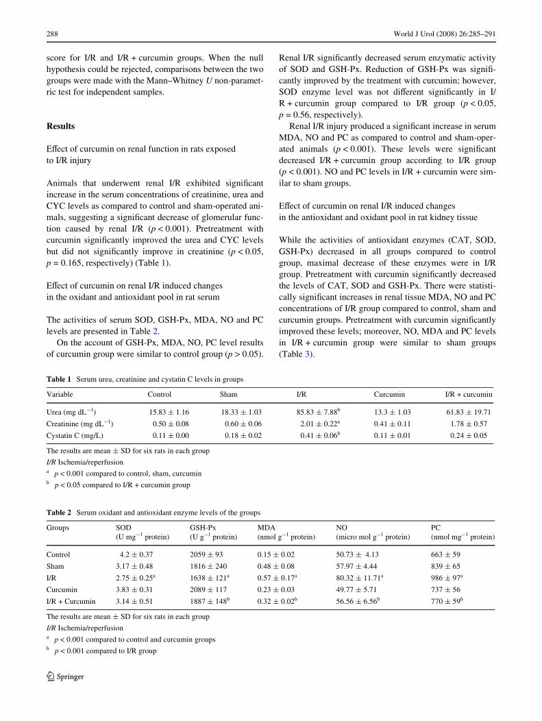
Animals that underwent renal I/R exhibited signiWcantincrease in the serum concentrations of creatinine, urea andCYC levels as compared to control and sham-operated ani-mals, suggesting a signiWcant decrease of glomerular func-tion caused by renal I/R (p < 0.001). Pretreatment withcurcumin signiWcantly improved the urea and CYC levelsbut did not signiWcantly improve in creatinine (p < 0.05,p = 0.165, respectively) (Table 1).
EVect of curcumin on renal I/R induced changes in the oxidant and antioxidant pool in rat serum
The activities of serum SOD, GSH-Px, MDA, NO and PClevels are presented in Table 2.
On the account of GSH-Px, MDA, NO, PC level resultsof curcumin group were similar to control group (p > 0.05).
Renal I/R signiWcantly decreased serum enzymatic activityof SOD and GSH-Px. Reduction of GSH-Px was signiW-cantly improved by the treatment with curcumin; however,SOD enzyme level was not diVerent signiWcantly in I/R + curcumin group compared to I/R group (p < 0.05,p = 0.56, respectively).
Renal I/R injury produced a signiWcant increase in serumMDA, NO and PC as compared to control and sham-oper-ated animals (p < 0.001). These levels were signiWcantdecreased I/R + curcumin group according to I/R group(p < 0.001). NO and PC levels in I/R + curcumin were sim-ilar to sham groups.
EVect of curcumin on renal I/R induced changes in the antioxidant and oxidant pool in rat kidney tissue
While the activities of antioxidant enzymes (CAT, SOD,GSH-Px) decreased in all groups compared to controlgroup, maximal decrease of these enzymes were in I/Rgroup. Pretreatment with curcumin signiWcantly decreasedthe levels of CAT, SOD and GSH-Px. There were statisti-cally signiWcant increases in renal tissue MDA, NO and PCconcentrations of I/R group compared to control, sham andcurcumin groups. Pretreatment with curcumin signiWcantlyimproved these levels; moreover, NO, MDA and PC levelsin I/R + curcumin group were similar to sham groups(Table 3).
Table 1 Serum urea, creatinine and cystatin C levels in groups
The results are mean § SD for six rats in each group
I/R Ischemia/reperfusiona p < 0.001 compared to control, sham, curcuminb p < 0.05 compared to I/R + curcumin group
Variable Control Sham I/R Curcumin I/R + curcumin
Urea (mg dL¡1) 15.83 § 1.16 18.33 § 1.03 85.83 § 7.88b 13.3 § 1.03 61.83 § 19.71
Creatinine (mg dL¡1) 0.50 § 0.08 0.60 § 0.06 2.01 § 0.22a 0.41 § 0.11 1.78 § 0.57
Cystatin C (mg/L) 0.11 § 0.00 0.18 § 0.02 0.41 § 0.06b 0.11 § 0.01 0.24 § 0.05
Table 2 Serum oxidant and antioxidant enzyme levels of the groups
The results are mean § SD for six rats in each group
I/R Ischemia/reperfusiona p < 0.001 compared to control and curcumin groupsb p < 0.001 compared to I/R group
Groups SOD (U mg¡1 protein)
GSH-Px (U g¡1 protein)
MDA (nmol g¡1 protein)
NO (micro mol g¡1 protein)
PC (nmol mg¡1 protein)
Control 4.2 § 0.37 2059 § 93 0.15 § 0.02 50.73 § 4.13 663 § 59
Sham 3.17 § 0.48 1816 § 240 0.48 § 0.08 57.97 § 4.44 839 § 65
I/R 2.75 § 0.25a 1638 § 121a 0.57 § 0.17a 80.32 § 11.71a 986 § 97a
Curcumin 3.83 § 0.31 2089 § 117 0.23 § 0.03 49.77 § 5.71 737 § 56
I/R + Curcumin 3.14 § 0.51 1887 § 148b 0.32 § 0.02b 56.56 § 6.56b 770 § 59b
123
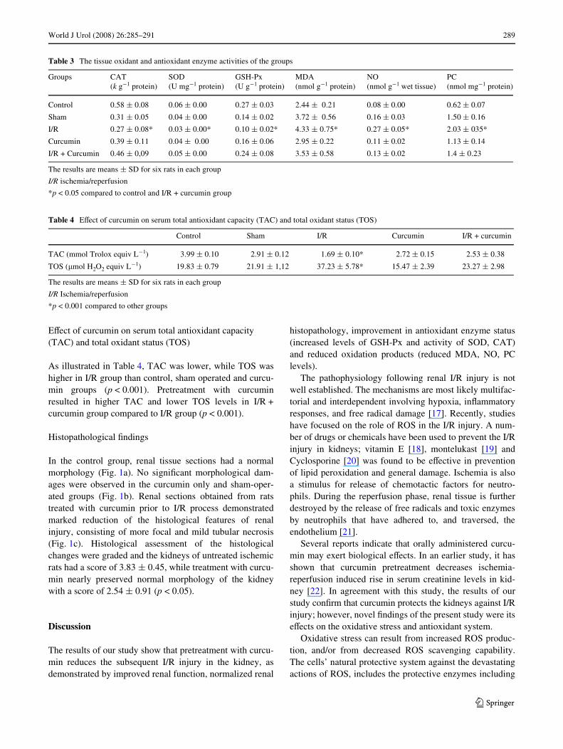
World J Urol (2008) 26:285–291 289
EVect of curcumin on serum total antioxidant capacity (TAC) and total oxidant status (TOS)
As illustrated in Table 4, TAC was lower, while TOS washigher in I/R group than control, sham operated and curcu-min groups (p < 0.001). Pretreatment with curcuminresulted in higher TAC and lower TOS levels in I/R +curcumin group compared to I/R group (p < 0.001).
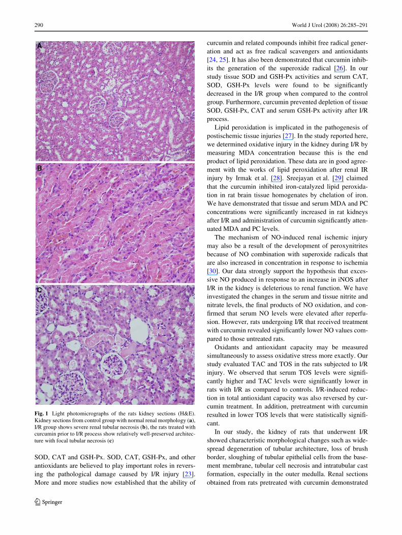
Histopathological Wndings
In the control group, renal tissue sections had a normalmorphology (Fig. 1a). No signiWcant morphological dam-ages were observed in the curcumin only and sham-oper-ated groups (Fig. 1b). Renal sections obtained from ratstreated with curcumin prior to I/R process demonstratedmarked reduction of the histological features of renalinjury, consisting of more focal and mild tubular necrosis(Fig. 1c). Histological assessment of the histologicalchanges were graded and the kidneys of untreated ischemicrats had a score of 3.83 § 0.45, while treatment with curcu-min nearly preserved normal morphology of the kidneywith a score of 2.54 § 0.91 (p < 0.05).
Discussion
The results of our study show that pretreatment with curcu-min reduces the subsequent I/R injury in the kidney, asdemonstrated by improved renal function, normalized renal
histopathology, improvement in antioxidant enzyme status(increased levels of GSH-Px and activity of SOD, CAT)and reduced oxidation products (reduced MDA, NO, PClevels).
The pathophysiology following renal I/R injury is notwell established. The mechanisms are most likely multifac-torial and interdependent involving hypoxia, inXammatoryresponses, and free radical damage [17]. Recently, studieshave focused on the role of ROS in the I/R injury. A num-ber of drugs or chemicals have been used to prevent the I/Rinjury in kidneys; vitamin E [18], montelukast [19] andCyclosporine [20] was found to be eVective in preventionof lipid peroxidation and general damage. Ischemia is alsoa stimulus for release of chemotactic factors for neutro-phils. During the reperfusion phase, renal tissue is furtherdestroyed by the release of free radicals and toxic enzymesby neutrophils that have adhered to, and traversed, theendothelium [21].
Several reports indicate that orally administered curcu-min may exert biological eVects. In an earlier study, it hasshown that curcumin pretreatment decreases ischemia-reperfusion induced rise in serum creatinine levels in kid-ney [22]. In agreement with this study, the results of ourstudy conWrm that curcumin protects the kidneys against I/Rinjury; however, novel Wndings of the present study were itseVects on the oxidative stress and antioxidant system.
Oxidative stress can result from increased ROS produc-tion, and/or from decreased ROS scavenging capability.The cells’ natural protective system against the devastatingactions of ROS, includes the protective enzymes including
Table 3 The tissue oxidant and antioxidant enzyme activities of the groups
The results are means § SD for six rats in each group
I/R ischemia/reperfusion
*p < 0.05 compared to control and I/R + curcumin group
Groups CAT (k g¡1 protein)
SOD (U mg¡1 protein)
GSH-Px (U g¡1 protein)
MDA (nmol g¡1 protein)
NO (nmol g¡1 wet tissue)
PC (nmol mg¡1 protein)
Control 0.58 § 0.08 0.06 § 0.00 0.27 § 0.03 2.44 § 0.21 0.08 § 0.00 0.62 § 0.07
Sham 0.31 § 0.05 0.04 § 0.00 0.14 § 0.02 3.72 § 0.56 0.16 § 0.03 1.50 § 0.16
I/R 0.27 § 0.08* 0.03 § 0.00* 0.10 § 0.02* 4.33 § 0.75* 0.27 § 0.05* 2.03 § 035*
Curcumin 0.39 § 0.11 0.04 § 0.00 0.16 § 0.06 2.95 § 0.22 0.11 § 0.02 1.13 § 0.14
I/R + Curcumin 0.46 § 0,09 0.05 § 0.00 0.24 § 0.08 3.53 § 0.58 0.13 § 0.02 1.4 § 0.23
Table 4 EVect of curcumin on serum total antioxidant capacity (TAC) and total oxidant status (TOS)
The results are means § SD for six rats in each group
I/R Ischemia/reperfusion
*p < 0.001 compared to other groups
Control Sham I/R Curcumin I/R + curcumin
TAC (mmol Trolox equiv L¡1) 3.99 § 0.10 2.91 § 0.12 1.69 § 0.10* 2.72 § 0.15 2.53 § 0.38
TOS (�mol H2O2 equiv L¡1) 19.83 § 0.79 21.91 § 1,12 37.23 § 5.78* 15.47 § 2.39 23.27 § 2.98
123
290 World J Urol (2008) 26:285–291
SOD, CAT and GSH-Px. SOD, CAT, GSH-Px, and otherantioxidants are believed to play important roles in revers-ing the pathological damage caused by I/R injury [23].More and more studies now established that the ability of
curcumin and related compounds inhibit free radical gener-ation and act as free radical scavengers and antioxidants[24, 25]. It has also been demonstrated that curcumin inhib-its the generation of the superoxide radical [26]. In ourstudy tissue SOD and GSH-Px activities and serum CAT,SOD, GSH-Px levels were found to be signiWcantlydecreased in the I/R group when compared to the controlgroup. Furthermore, curcumin prevented depletion of tissueSOD, GSH-Px, CAT and serum GSH-Px activity after I/Rprocess.
Lipid peroxidation is implicated in the pathogenesis ofpostischemic tissue injuries [27]. In the study reported here,we determined oxidative injury in the kidney during I/R bymeasuring MDA concentration because this is the endproduct of lipid peroxidation. These data are in good agree-ment with the works of lipid peroxidation after renal IRinjury by Irmak et al. [28]. Sreejayan et al. [29] claimedthat the curcumin inhibited iron-catalyzed lipid peroxida-tion in rat brain tissue homogenates by chelation of iron.We have demonstrated that tissue and serum MDA and PCconcentrations were signiWcantly increased in rat kidneysafter I/R and administration of curcumin signiWcantly atten-uated MDA and PC levels.
The mechanism of NO-induced renal ischemic injurymay also be a result of the development of peroxynitritesbecause of NO combination with superoxide radicals thatare also increased in concentration in response to ischemia[30]. Our data strongly support the hypothesis that exces-sive NO produced in response to an increase in iNOS afterI/R in the kidney is deleterious to renal function. We haveinvestigated the changes in the serum and tissue nitrite andnitrate levels, the Wnal products of NO oxidation, and con-Wrmed that serum NO levels were elevated after reperfu-sion. However, rats undergoing I/R that received treatmentwith curcumin revealed signiWcantly lower NO values com-pared to those untreated rats.
Oxidants and antioxidant capacity may be measuredsimultaneously to assess oxidative stress more exactly. Ourstudy evaluated TAC and TOS in the rats subjected to I/Rinjury. We observed that serum TOS levels were signiW-cantly higher and TAC levels were signiWcantly lower inrats with I/R as compared to controls. I/R-induced reduc-tion in total antioxidant capacity was also reversed by cur-cumin treatment. In addition, pretreatment with curcuminresulted in lower TOS levels that were statistically signiW-cant.
In our study, the kidney of rats that underwent I/Rshowed characteristic morphological changes such as wide-spread degeneration of tubular architecture, loss of brushborder, sloughing of tubular epithelial cells from the base-ment membrane, tubular cell necrosis and intratubular castformation, especially in the outer medulla. Renal sectionsobtained from rats pretreated with curcumin demonstrated
Fig. 1 Light photomicrographs of the rats kidney sections (H&E).Kidney sections from control group with normal renal morphology (a),I/R group shows severe renal tubular necrosis (b), the rats treated withcurcumin prior to I/R process show relatively well-preserved architec-ture with focal tubular necrosis (c)
123
World J Urol (2008) 26:285–291 291
marked reduction of the histological features of renalinjury, consisting of more focal and mild tubular necrosis.Curcumin may protect the tubular epithelium eVectivelyfrom reperfusion injury.
There are some limitations to our study. First, the samplesize used in our study was small. Larger studies might behelpful to clarify the protective eVects of curcumin on I/Rinjury. Second, some of the parameters introduced in thestudy such as NO are very sensitive to ischemia. It could bethe result of the harvesting process. Because of the suscep-tibility of ATP and its metabolites, the only accurate way todescribe possible changes is to harvest kidneys using themethod of in-vivo freeze clamping.
From the results of our study, it can be concluded thatcurcumin pretreatment protects the rat kidneys against sub-sequent I/R injury. Administration of curcumin attenuatesthe increase in markers of renal injury and oxidative dam-age. The present Wndings also imply that curcumin has anti-oxidant properties and may be used in human studies in thefuture.
ConXict of interest statement There is no conXict of interest.
References
1. Abernethy VE, Lieberthal W (2002) Acute renal failure in the crit-ically ill patient. Crit Care Clin 18:203–222
2. Tanabe K, Oshima T, Tokumoto T, Ishikawa N, Kanematsu A,Shinmura H et al (1998) Long-term renal function in nonheart-beating donor kidney transplantation: a single-center experience.Transplantation 66:1708–1713
3. Perico N, Cattaneo D, Sayegh MH, Remuzzi G (2004) Delayedgraft function in kidney transplantation. Lancet 364:1814–1827
4. Haugen E, Nath KA (1999) The involvement of oxidative stress inthe progression of renal injury. Blood Purif 17:58–65
5. Edelstein CL, Ling H, Schrier RW (1997) The nature of cell injury.Kidney Int 51:1341–1351
6. Castaneda MP, Swiatecka-Urban A, Mitsnefes MM, Feuerstein D,Kaskel FJ, Tellis V, Devarajan P (2003) Activation of mitochon-drial apoptotic pathways in human, renal allografts after ischemia-reperfusion injury. Transplantation 76:50–54
7. Jagetia GC, Aggarwal BB (2007) “Spicing up” of the immunesystem by curcumin. J Clin Immunol 27:19–35
8. Lowry OH, Rosenbrough NJ, Farr AL, Randall RJ (1951) Proteinmeasurement with the Folin phenol reagent. J Biol Chem193:265–275
9. Aebi H. (1974) Catalase In: Bergmeyer U (ed) Methods of enzy-matic analysis. Academic Press, New York
10. Sun Y, Oberley LW, Ying L (1988) A simple method for clinicalassay of superoxide dismutase. Clin Chem 34:497–500
11. Paglia DE,Valentine WN (1967) Studies on the quantitative andqualitative characterisation of erythrocyte glutathione peroxidase.J Lab Clin Med 70:158–169
12. Esterbauer H, Cheeseman KH (1990) Determination of aldehydiclipid peroxidation products: malonaldehyde and 4-hydroxynone-nal. In: Packer L, Glazer AN (eds) Oxygen radicals in biologicalsystems, methods in enzymology. Academic Pres, California, pp407–421
13. Cortas NK, Wakid NW (1990) Determination of inorganic nitratein serum and urine by a kinetic cadmium-reduction method. ClinChem 36:1440–1443
14. Levine RL, Garland D, Oliver CN, Amici A, Climent I, Lenz AG,Ahn BW, Shaltiel S, Stantman ER (1990) Determination of car-bonyl content in oxidatively modiWed proteins. In: Packer L, Glaz-er AN (eds) Methods in enzimology V 186, oxygen radicals inbiological systems. Academic Press, New York, pp 464–478
15. Erel O (2004) A novel automated method to measure total antiox-idant response against potent free radical reactions. Clin Biochem37:112–219
16. Erel O (2005) A new automated colorimetric method for measur-ing total oxidant status, Clin Biochem 38:1103–1111
17. Williams PH, Lopez DB, Chan C, Ezrin A, Hottendorf R (1997)Characterization of renal ischemia-reperfusion injury in rats. JPharmacol Toxicol Methods 37:1–7
18. Uysal F, Girgin FK, Tuzun S, Aldemir S, Sozmen EY (1998)EVect of vitamin E on antioxidant enzymes and nitric oxide inischemia-reperfused kidney injury. Biochem Mol Biol Int44:1255–1263
19. Sener G, Sehirli O, Velioflu-Ofünç A, Cetinel S, Gedik N, CanerM, Sakarcan A, Yefen BC. (2006) Montelukast protects againstrenal ischemia/reperfusion injury in rats. Pharmacol Res 54:65–71
20. Singh D, Chander V, Chopra K (2005) Cyclosporine protectsagainst ischemia/reperfusion injury in rat kidneys. Toxicology207:339–347
21. Weight SC, Bell PRF, Nicholson ML (1996) Renal ischemia-reperfusion injury. Br J Surg 83:162–170
22. Shoskes DA (1998) EVect of bioXavonoids quercetin and curcu-min on ischemic renal injury: a new class of renoprotective agents.Transplantation 66:147–152
23. Paller MS, Hoidal JR, Ferris TF (1984) Oxygen free radicals inischemic acute renal failure in the rat. J Clin Invest 74:1156–1164
24. Kunchandy E, Rao A (1990) Oxygen radical scavenging activityof curcumin. Int J Pharm 58: 237–240
25. Ruby AJ, Kuttan G, Babu KD, Rajasekharan KN, Kuttan R (1995)Anti-tumour and antioxidant activity of natural curcuminoids.Cancer Lett 94:79–83
26. Reddy AC, Lokesh BR (1994) Studies on the inhibitory eVects ofcurcumin and eugenol on the formation of reactive oxygen speciesand the oxidation of ferrous iron. Mol Cell Biochem 137:1–8
27. Eschwege P, Paradis V, Conti M, Holstege A, Richet F, Deteve J,Menager P, Legrand A, Jardin A, Bedossa P, Benoit G (1999) Insitu detection of lipid peroxidation by-products as markers of renalischemia injuries in rat kidneys. J Urol 162:553–557
28. Irmak MK, Koltuksuz U, Kutlu NO, Yagmurca M, Ozyurt H,Karaman A, Akyol O (2001) The eVect of caVeic acid phenethylester on ischemia-reperfusion injury in comparison with alpha-tocopherol in rat kidneys. Urol Res 29:190–193
29. Sreejayan N, Rao MN (1994) Curcuminoids as potent inhibitors oflipid peroxidation. J Pharm Pharmacol 46:1013–1016
30. Rhoden EL, Rhoden CR, Lucas ML, Pereira-Lima L, Zettler C,Bello-Klein A (2002) The role of nitric oxide pathway in the renalischemia-reperfusion injury in rats. Transpl Immunol 10:277–284
123
Copyright © 2022 FDOKUMEN