Fish in the Filipino Diet Fish & Fish products Fish & Fish products
Upload
khangminh22Category
view
2download
0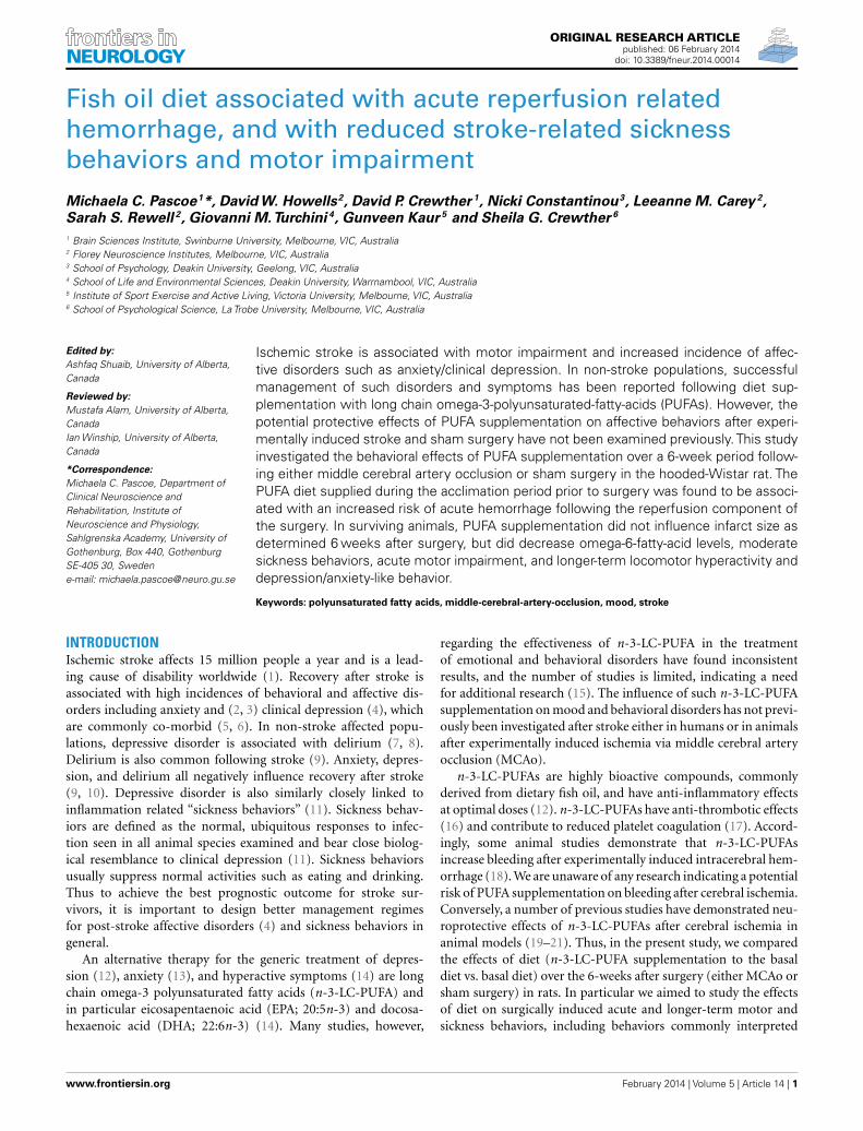
ORIGINAL RESEARCH ARTICLEpublished: 06 February 2014
doi: 10.3389/fneur.2014.00014
Fish oil diet associated with acute reperfusion relatedhemorrhage, and with reduced stroke-related sicknessbehaviors and motor impairmentMichaela C. Pascoe1*, David W. Howells2, David P. Crewther 1, Nicki Constantinou3, Leeanne M. Carey 2,Sarah S. Rewell 2, Giovanni M.Turchini 4, Gunveen Kaur 5 and Sheila G. Crewther 6
1 Brain Sciences Institute, Swinburne University, Melbourne, VIC, Australia2 Florey Neuroscience Institutes, Melbourne, VIC, Australia3 School of Psychology, Deakin University, Geelong, VIC, Australia4 School of Life and Environmental Sciences, Deakin University, Warrnambool, VIC, Australia5 Institute of Sport Exercise and Active Living, Victoria University, Melbourne, VIC, Australia6 School of Psychological Science, La Trobe University, Melbourne, VIC, Australia
Edited by:Ashfaq Shuaib, University of Alberta,Canada
Reviewed by:Mustafa Alam, University of Alberta,CanadaIan Winship, University of Alberta,Canada
*Correspondence:Michaela C. Pascoe, Department ofClinical Neuroscience andRehabilitation, Institute ofNeuroscience and Physiology,Sahlgrenska Academy, University ofGothenburg, Box 440, GothenburgSE-405 30, Swedene-mail: [email protected]
Ischemic stroke is associated with motor impairment and increased incidence of affec-tive disorders such as anxiety/clinical depression. In non-stroke populations, successfulmanagement of such disorders and symptoms has been reported following diet sup-plementation with long chain omega-3-polyunsaturated-fatty-acids (PUFAs). However, thepotential protective effects of PUFA supplementation on affective behaviors after experi-mentally induced stroke and sham surgery have not been examined previously. This studyinvestigated the behavioral effects of PUFA supplementation over a 6-week period follow-ing either middle cerebral artery occlusion or sham surgery in the hooded-Wistar rat. ThePUFA diet supplied during the acclimation period prior to surgery was found to be associ-ated with an increased risk of acute hemorrhage following the reperfusion component ofthe surgery. In surviving animals, PUFA supplementation did not influence infarct size asdetermined 6 weeks after surgery, but did decrease omega-6-fatty-acid levels, moderatesickness behaviors, acute motor impairment, and longer-term locomotor hyperactivity anddepression/anxiety-like behavior.
Keywords: polyunsaturated fatty acids, middle-cerebral-artery-occlusion, mood, stroke
INTRODUCTIONIschemic stroke affects 15 million people a year and is a lead-ing cause of disability worldwide (1). Recovery after stroke isassociated with high incidences of behavioral and affective dis-orders including anxiety and (2, 3) clinical depression (4), whichare commonly co-morbid (5, 6). In non-stroke affected popu-lations, depressive disorder is associated with delirium (7, 8).Delirium is also common following stroke (9). Anxiety, depres-sion, and delirium all negatively influence recovery after stroke(9, 10). Depressive disorder is also similarly closely linked toinflammation related “sickness behaviors” (11). Sickness behav-iors are defined as the normal, ubiquitous responses to infec-tion seen in all animal species examined and bear close biolog-ical resemblance to clinical depression (11). Sickness behaviorsusually suppress normal activities such as eating and drinking.Thus to achieve the best prognostic outcome for stroke sur-vivors, it is important to design better management regimesfor post-stroke affective disorders (4) and sickness behaviors ingeneral.
An alternative therapy for the generic treatment of depres-sion (12), anxiety (13), and hyperactive symptoms (14) are longchain omega-3 polyunsaturated fatty acids (n-3-LC-PUFA) andin particular eicosapentaenoic acid (EPA; 20:5n-3) and docosa-hexaenoic acid (DHA; 22:6n-3) (14). Many studies, however,
regarding the effectiveness of n-3-LC-PUFA in the treatmentof emotional and behavioral disorders have found inconsistentresults, and the number of studies is limited, indicating a needfor additional research (15). The influence of such n-3-LC-PUFAsupplementation on mood and behavioral disorders has not previ-ously been investigated after stroke either in humans or in animalsafter experimentally induced ischemia via middle cerebral arteryocclusion (MCAo).
n-3-LC-PUFAs are highly bioactive compounds, commonlyderived from dietary fish oil, and have anti-inflammatory effectsat optimal doses (12). n-3-LC-PUFAs have anti-thrombotic effects(16) and contribute to reduced platelet coagulation (17). Accord-ingly, some animal studies demonstrate that n-3-LC-PUFAsincrease bleeding after experimentally induced intracerebral hem-orrhage (18). We are unaware of any research indicating a potentialrisk of PUFA supplementation on bleeding after cerebral ischemia.Conversely, a number of previous studies have demonstrated neu-roprotective effects of n-3-LC-PUFAs after cerebral ischemia inanimal models (19–21). Thus, in the present study, we comparedthe effects of diet (n-3-LC-PUFA supplementation to the basaldiet vs. basal diet) over the 6-weeks after surgery (either MCAo orsham surgery) in rats. In particular we aimed to study the effectsof diet on surgically induced acute and longer-term motor andsickness behaviors, including behaviors commonly interpreted
www.frontiersin.org February 2014 | Volume 5 | Article 14 | 1
Pascoe et al. Behavioral effect of n-3-LC-PUFA post-MCAo
to reflect excessive anxiety or depressive-like behaviors in therodent model.
We hypothesized that following MCAo surgery animals wouldshow reduced food and water consumption, compared to shams.Additionally, we hypothesized that the n-3-LC-PUFA diet supple-mented animals would show less reduction in food and waterconsumption, and body weight post-surgery, compared to thebasal diet fed animals.
Acute inabilities to make co-ordinated motor movements haveregularly been reported in rats post MCAo (22, 23) and decreasein response to neuroprotective agents (24–26). Thus, we hypoth-esized that the n-3-LC-PUFA supplemented animals would alsoshow less acute MCAo related motor impairments on a battery ofwidely used neurological impairment tests (22, 23) compared tobasal diet fed rats.
We hypothesized that hyperactive locomotor behaviors assessedusing the free exploration test, would not correlate with anxiety-like behavior in this test, defined by percentage of emergencetime spent in the center of the open-field arena. Previousauthors have speculated that MCAo related changes in loco-motion might arise from an inability to habituate to unfamil-iar environments, possibly related to spatial mapping difficultiesinduced by hippocampus cell death (27–30). Therefore, spatialmapping abilities were assessed here using the spatial displace-ment recognition test, which has previously been employed tostudy spatial memory deficits post-stroke in the rodent model (28,31–33).
We hypothesized that MCAo operated animals would showmore long-term stroke-related anxiety-like and hyperactive loco-motor behaviors than sham operated animals (MCAo vs. Sham).Finally, we hypothesized that after surgery animals supplementedwith n-3-LC-PUFA would show less long-term sickness or anxiety-like and locomotor behaviors than basal diet fed animals (n-3-LC-PUFA vs. Basal). Long-term was defined as occurring at 2, 4, or6 weeks following surgery.
In summary, we anticipated that n-3-LC-PUFA diet supple-mentation would reduce acute MCAo related motor impairments,and sickness behaviors. We expected the beneficial effects ofn-3-LC-PUFA diet supplementation to persist longer term, asevidenced by a longer-term reduction in rodent behaviors, inter-preted to reflect stroke-related behavioral and affective disorders.
MATERIALS AND METHODSANIMALS AND DESIGNThe experiment used two surgery groups (MCAo and Shamgroups) and two diet groups (basal and n-3-LC-PUFA supplemen-tation) with repeated measures outcomes. Adult male hooded-Wistar rats (Laboratory Animal Services, The University of Ade-laide, Australia) commonly used in stroke research (34–36) wereseparately housed (21± 2°C, 12:12 h light–dark cycle). Food andwater were available ad libitum and consumption and body weightwere measured daily. Acclimatization to housing and diet condi-tion began 1 week prior to surgery and continued until animalsacrifice (via isoflurane anesthesia; Cat No. AHN3640-250ML,Baxter, Old Toongabbie, NSW, Australia) at 6 weeks post-surgery.Animal behavior was studied until 6 weeks post-surgery as previ-ous research, in non-stroke-related areas, has demonstrated that
6 weeks of oral supplementation with omega-3 is associated withanti-depressive-like behavioral effects in rodent models (37). Inthe week following surgery only, animals were provided withsunflower seeds and soft food (Sustagen® Everyday Nestlé, Not-ting Hill, VIC, Australia) additional to their respective diets, toencourage eating and weight gain. Surgeries and behavioral test-ing took place in the lights-on phase. The experimental protocolrequired 48 animals;j however 11 rats in the n-3-LC-PUFA MCAogroup died immediately following withdrawal of the suture thread.A further 16 rats (n-3-LC-PUFA MCAo, n= 7; Basal MCAo,n= 9) were culled due to MCAo induced motor impairmentsand clinical symptoms deemed too large for the animal to sur-vive. These animals were replaced and thus, the total number ofanimals used in the present study was 75 (n= 29 n-3-LC-PUFAMCAo; n= 22 Basal MCAo; n= 12 n-3-LC-PUFA sham; n= 12Basal Sham).
ETHICS STATEMENTEthics approval was granted by the Austin Health Research EthicsUnit (10/3865) and was conducted in accordance with the Aus-tralian Code of Practice for the use of animals for scientificpurposes (38).
DIETARY REGIMEAnimals were randomly assigned to n-3-LC-PUFA diet condition{Specialty Feeds, Cat No. SF09-109 5% Fat High N3 ModifiedRodent Diet, Glen Forest, WA, Australia [EPA 20:5 n-3 5% of totalfree fatty acid (FFA), DHA 22:6 n-3 23.8% of total fatty acids]},selected as it has previously been demonstrated to reduce oxida-tive stress in the rat model (39) or basal diet condition (SpecialtyFeeds, Cat No. AIN93G; Glen Forest, WA, Australia).
RIGHT MIDDLE CEREBRAL ARTERY OCCLUSIONWithin each diet condition, animals were randomly assigned toMCAo or sham surgery condition. Surgeries were performedaccording to the protocol of Longa (40) and its modificationsby Spratt (41). Briefly, anesthesia was induced (5% in oxygen)and maintained (2% in oxygen) with Isoflurane via a nose cone.Vital signs were monitored using iWORX (Cat No. PO2-300D,iWORX, Dover, NH, USA). Body temperature was maintained at37°C. Atropine (0.2 ml 600 µg/ml, Pfizer Australia Pty Ltd., NSW,Australia) was administered intraperitoneally to inhibit bronchialsecretions and salivation. Cerebral blood flow was measured usinglaser doppler flowmetry (LDF). The scalp was thinned using adental burr and the Laser Dopler was attached to a laboratory-made rubber probe holder attached to skull with instant adhesive(Loctite 406 instant adhesive, Cat No. 265606, Henkel, Sydney,NSW, Australia). An incision was made in the neck, small branch-ing blood vessels were cauterized, the external carotid artery wasligated, an incision made in the right external carotid artery, a0.4-mm diameter silicone tipped suture thread inserted into theincision, passed via the right external carotid and up the internalcarotid artery. MCAo was 90 min during which incisions wereclosed (4.0 silk sutures, Cat No. 90352, Dynek Pty Ltd., PortAdelaide, SA, Australia) and animals were removed from anes-thesia. For reperfusion, animals were re-anesthetized, incisionsre-opened and the MCA-occluding suture retracted. Vital signs
Frontiers in Neurology | Stroke February 2014 | Volume 5 | Article 14 | 2
Pascoe et al. Behavioral effect of n-3-LC-PUFA post-MCAo
and during reperfusion were monitored using iWORX (Cat No.PO2-300D, iWORX, Dover, NH, USA). Animal breathing wasalso monitored. Animals that died during the surgical or afterthe reperfusion procedure, or within the first 24 h after surgery,were documented and post mortem autopsy was conducted todetermine cause of death. Hemorrhage was determined at thetime of autopsy, by the presence of blood pooled around thebase of skull. Analgesia was provided as paracetamol (Cat No.569925, Sanofi-Aventis, Macquarie Park, NSW, Australia) crushedand dissolved into drinking water (0.15 mg/ml water). Saline wasinjected (i.p.) daily for 3 days following surgery [3 ml SodiumChloride Solution (0.9%), Cat No. 7647-14-5, Tocris Bioscience,Bristol, UK]. Sham animals underwent identical procedures withthe exclusion of thread insertion into the MCA. Table 1 displaysanimal groups, final successful numbers, and behavior testingtimetable.
ACUTE STROKE-RELATED MOTOR IMPAIRMENT MEASURESMotor impairments were assessed daily in the first 5 days post-surgery using the forelimb flexion test [MCAo affected rats holdleft forelimb between 45° and 90° when lifted from base of tail (22)]the torso twisting test [MCAo affected rats show curling of the headand forelimbs toward the paralytic side of the body, when held viatail (22)] the lateral push test [MCAo affected rats show weakenedresistance when pushed toward the paralytic left side (22)] and thecircling [animals with infarct damage circle toward the oppositeside of the damage (22)] and motor ability, observations (walk-ing, grooming, and rearing behaviors) which collectively measurethe acute inability to make co-ordinated motor movements, reg-ularly been reported and studied in rats post MCAo compared tosham surgery (22, 23). These motor impairments are less severein animals exposed to neuroprotective agents (24–26), thus, weaimed to determine if acute motor disability was decreased aftern-3-LC-PUFA supplementation.
LONGER-TERM ISCHEMIA ASSOCIATED BEHAVIORAL AND AFFECTIVEDISORDERSThe battery of behavioral tests designed to model ischemia-relatedaffective behaviors were administered and recorded on video inthe second, and then repeated in the fourth and sixth weeks post-stroke. The same animals were tested at each time point. Behaviorvideo was scored by an observer (NC) who was blind to ani-mal diet and surgery condition. Animal behaviors were scoredby a second observer (MP), and the inter-rater reliability correla-tion was statistically significant, r2
= 0.97. To test for longer-termsickness behaviors and particularly depression and anxious likebehaviors after surgery, we used the novel object exploration testand a modified open-field test that measures the conflict betweena rat’s mutual drives to both avoid and approach unfamiliar andpotentially fear inducing stimuli/environments (32, 42–47). Toasses the locomotor hyperactivity often reported from as earlyas 5 min to 5 weeks after MCAo surgery (29, 30, 48, 49) and inrodent models of anxiety and depression (50–54), we used a mod-ified free exploration test, which eliminates the forced explorationand related stress normally associated with the commonly usedopen-field test (55, 56). All behavior was recorded using a digitalcamcorder (Canon Legria, HG20) and was stored as a MPEG-TSvideo file. All apparatus were cleaned with 70% ethanol betweentrials (Cat No. EA043-10L, Chem-Supply, Gillman, SA, Australia).
FREE EXPLORATION TESTAnimals were habituated (15 min) to a laboratory constructedblack Plexiglass hide box (30 cm× 15 cm× 19 cm) the day priorand re-acclimated (5 min) immediately prior to testing. Theopen field was a transparent Plexiglass arena (1 m2) illuminated(~500 lux). The hide box was positioned inside one wall of theopen field. Animals were scored for locomotor hyperactivity(number of times fully emerged from the hide box, i.e., emer-gence number) and time spent fully emerged from hide box and
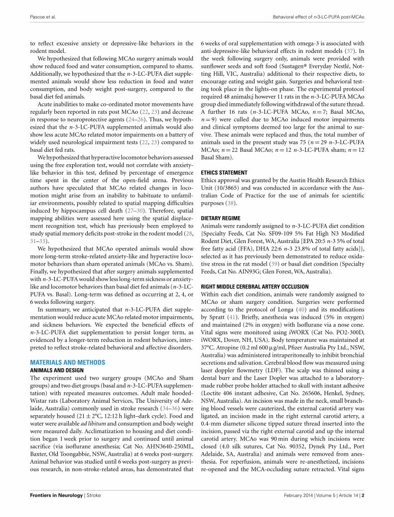
Table 1 | Experimental timeline showing order and timing of treatment and testing.
Animal surgery
and diet group
MCAo surgery and
n-3-LC-PUFA diet
MCAo surgery and
basal diet
Sham surgery and
n-3-LC-PUFA diet
Sham surgery and
basal diet
1 week prior to
surgery
Diet/housing acclimation Diet/housing acclimation Diet/housing acclimation Diet/housing acclimation
Surgery Surgery Surgery Surgery
Day 1–5
post-surgery
Acute motor impairment
testing
Acute motor impairment
testing
Acute motor impairment
testing
Acute motor impairment
testing
2 weeks
post-surgery
Free exploration, spatial
displacement recognition, and
novel object exploration testing
Free exploration, spatial
displacement recognition, and
novel object exploration testing
Free exploration, spatial
displacement recognition, and
novel object exploration testing
Free exploration, spatial
displacement recognition, and
novel object exploration testing
2 weeks
post-surgery
Behavioral testing as specified
in week 2 post-surgery
Behavioral testing as specified
in week 2 post-surgery
Behavioral testing as specified
in week 2 post-surgery
Behavioral testing as specified
in week 2 post-surgery
6 weeks
post-surgery
Behavioral testing as specified
in week 2 post-surgery
Behavioral testing as specified
in week 2 post-surgery
Behavioral testing as specified
in week 2 post-surgery
Behavioral testing as specified
in week 2 post-surgery
Sacrifice Sacrifice Sacrifice Sacrifice
MCAo, middle cerebral artery occlusion surgery condition; Sham, Sham surgery condition; Basal, basal diet fed rats; n-3-LC-PUFA, polyunsaturated fatty acid
supplemented rats.
www.frontiersin.org February 2014 | Volume 5 | Article 14 | 3

Pascoe et al. Behavioral effect of n-3-LC-PUFA post-MCAo
moving around the arena (emergence duration) for 4 min. Giventhat the MCAo procedure and the associated neurodegenerativedamage result in increased locomotor, activity per se (29, 30, 48,49) we suggest that the above-mentioned measures may not reflectanxiety-like behaviors in MCAo operated animals, but instead mayonly be another measure of locomotion hyperactivity. Animalswere however also scored for anxiety-like behavior that was definedas the avoidance of the center and quantified as the percentage oftime spent in the center area also.
SPATIAL DISPLACEMENT RECOGNITIONAnimals were acclimated (5 min) to the testing box (40 cm×40 cm× 60 cm) the day prior to testing. On the day of testing,animals were placed in the testing box, positioned with their nosefacing the mid-point of the wall opposite the objects, to prevent anunintentional bias of placing the animal in an orientation favoringa particular object (57). Object contact was defined as when themouth, nose, and/or paw touched the object or when the nose ofthe rat was within 1 cm surrounding the object. Contact judgedas accidental, such as bumping the object as the animal passedor grooming behavior was not counted. The number of objectcontacts and the duration of these were recorded.
The spatial displacement recognition testing consisted of twohabituation trials (3 min each) to two identical objects. Habitua-tion trials were separated by a 2-min interval, during which theanimal was returned to the home cage. The location of one ofthe two objects was then moved and the animal was placed in thetesting box for a single 3 min recognition trial.
NOVEL OBJECT EXPLORATIONThe novel object exploration trial took place immediately after thespatial displacement recognition trial, separated only by a 2-mininterval (animal returned to home cage). Testing took place in thesame box used for the spatial displacement recognition test. Ratswere placed in the testing box for a single 3-min trial. The familiarobject was placed in one corner of the box and the novel objectwas placed in the opposite corner. Object placement and selectionwere counterbalanced across trials and animals. Contact definitionis consistent with that described above for the spatial displacementrecognition test.
TISSUE COLLECTIONTissue was collected using two methods: (a) whole brain for totalFFA analysis (n= 5 n-3-LC-PUFA MCAo; n= 6 Basal MCAo;n= 6 n-3-LC-PUFA Sham; n= 6 Basal Sham) animals were anes-thetized via Isoflurane overdose, decapitated via guillotine, brainsremoved, snap frozen in liquid nitrogen (Liquid Nitrogen Ser-vices Pty Ltd., Melbourne, VIC, Australia), and stored at−80°. (b)Perfusion for infarct analysis (n= 6 n-3-LC-PUFA MCAo; n= 6Basal MCAo; n= 6 n-3-LC-PUFA Sham; n= 6 Basal Sham). Onceanesthetized, the rib cage and diaphragm were cut, a perfusionneedle placed into the left ventricle of the heart and the rightatrium cut. Saline (90 ml) (1.8%) was infused (5 min), followedby paraformaldehyde (PFA) (4%) (270 ml) (15 min), using a per-fusion pump (Peri-Star Pro 4-channel, high rate pump, Cat No.PERIPRO-4HS, World Precision Instruments, Hilton, SA, Aus-tralia) (18 ml/min). Brains were removed and placed directly into
PFA (4%) for 24 h, before being changed to 30% sucrose, wherethey remained until the time of paraffin embedding.
FATTY ACID ANALYSISTo confirm that the n-3-LC-PUFA supplementation resultedin changes in brain phospholipids levels of fatty acids, wholebrain frozen tissue was ground up, and tissue lipids extractedby dichloromethane/methanol (2:1) overnight, as described bySinclair et al. (58). Samples were filtered, saline added (1 ml of0.9%), and vortexed (1 min). Samples were centrifuged (1500× g,10 min) to separate the aqueous and organic phases at room tem-perature. The organic phase containing the lipid was removedand transferred to a new glass tube and evaporated under astream of nitrogen. The lipid extract was reconstituted in 200 µlof dichloromethane and lipids were then separated by thin layerchromatography (TLC). The lipid extracts were spotted onto sil-ica gel plates (silica gel 60 G, Merck, Germany) and developedin 85:15:2 (v/v) petroleum ether: diethyl ether: acetic acid inpaper-lined tanks. The lipids were visualized with 0.1% (w/v)2′,7′-dichlorofluorescein indicator in ethanol (Scharlau, Spain).The phospholipid bands from the samples were scraped off intoglass screw-capped tubes and were reacted with 5% H2SO4 in100% methanol (3 h at 80°C) to form the fatty acid methyl esters(FAMEs). FAME were isolated (100% petroleum ether) and storedin glass (−20°C). Purified FAME were isolated and identified usingan Agilent Technologies 7890A GC System (Agilent Technologies,Santa Clara, CA, USA) equipped with an Omegawax 250 cap-illary column (30 m× 0.25 mm internal diameter, 0.25 µm filmthickness, Supelco, Bellefonte, PA, USA), a flame ionization detec-tor (FID), an Agilent Technologies 7693 auto sampler, and a splitinjection system (split ratio 50:1). The injection volume was 1 µl,the injector and detector temperature were 300 and 270°C, respec-tively. The temperature program was 50–190 at 20°C min−1, thenfrom 190 to 250 at 4°C min−1, and held at 250°C for 8 min. Thecarrier gas was helium at 1.18 ml min−1, at a constant flow. Each ofthe fatty acids was identified relative to known external standards(a series of mix and individual standards from Sigma-Aldrich,Inc., St. Louis, MO, USA and from Nu-Chek Prep Inc., Elysian,MN, USA). The resulting peaks were then corrected by the theo-retical relative FID response factors (59) and quantified relative tothe internal standard.
INFARCT VOLUME ANALYSIS AND AREA OF DAMAGEBrains were cut (2 mm coronal sections) using an acrylic rat brainmatrix. Tissue was processed using a closed linear Tissue Process-ing System (Cat No. TPC 15, MEDITE GmbH, Wollenweberstr,Burgdorf, Germany). Tissue was embedded in molten paraffinwax (Cat No. Leica EG1150 H, Leica-microsystems, Ernst-Leitz-Straße,Wetzlar,Germany), and stored at room temperature. Tissuewas cut at room temperature (7 µm) using a rotary microtome(Cat No. Leica 2040, Leica-microsystems, Ernst-Leitz-Straße, Wet-zlar, Germany) suspended at 40°C in a tissue flotation bath andattached to silane coated slides (Cat No. CS2460100MK, Micro-glass, Grale Scientific, Melbourne, VIC, Australia). Tissue slideswere dried at 49°C, incubated at 32°C for 24 h, and stained withHematoxylin (5 g Hematoxylin; Cat No. 340374T, VWR Interna-tional Pty Ltd., Murarrie, QLD, Australia) and Eosin (10 g Eosin Y;
Frontiers in Neurology | Stroke February 2014 | Volume 5 | Article 14 | 4

Pascoe et al. Behavioral effect of n-3-LC-PUFA post-MCAo
Cat No. E-4382, Sigma-Aldrich, St. Louis, MO, USA). Slides werede-waxed in Histosol (Cat No. CP L HISTOSOL 08, HD Scien-tific, Wetherill Park, NSW, Australia), washed in ethanol, rinsedin dH2O, dehydrated in ethanol, followed by two Histosol washes.Slides were rehydrated in ethanol rinsed in dH2O and stained withfiltered Harris Hematoxylin. Slides were rinsed in dH2O, stained infiltered Eosin Y, washed in dH2O, and dehydrated in ethanol. Thiswas followed histosol washes and cover slipping (DPX, Cat No.1019790500, Merck KGaA, Darmstadt, Germany). Stained tissuewas examined and using bright field microscopy (Nikon Eclipse80i, Nikon Instruments Europe) with a Nikon DIGITAL SIGHTDS-U1 camera. Photographs were stored as JPEG Image files. Areasof tissue damage were analyzed using Stereo Investigator Version6 software.
DATA ANALYSISThe present study uses both parametric and non-parametric dataanalysis techniques as appropriate. This study consists of fourindependent groups [2× 2 (n-3-LC-PUFA MCAo; Basal MCAo;n-3-LC-PUFA sham; Basal Sham)] and has both single time pointand repeated measures outcomes. Where data met the assump-tion of normality, appropriate ANOVAs, as described below areused. All measures of longer-term behavioral outcomes howeverdid not meet the assumption of normality and were unable to betransformed (except spatial displacement recognition) and thusnon-parametric data analyses techniques have been used. It isimportant to note however, that IBM SPSS Statistics does not offera non-parametric equivalent to the mixed design ANOVA (theappropriate parametric analysis technique to analyze the longer-term behavioral outcome data). The non-parametric Friedman’sANOVA allows a comparison of two or more related groups, butis not appropriate for the current study, where groups are inde-pendent. The Kruskal–Wallis is appropriate for the current data,as it allows one to compare differences between two or moreindependent groups (i.e., n-3-LC-PUFA MCAo vs. Basal MCAo/n-3-LC-PUFA sham vs. Basal Sham) (60). To determine where groupdifferences exist, Mann–Whitney post hoc tests were used. In orderto control for type 1 errors, only the most important/relevantpost hoc Mann–Whitney comparisons were made, thus differencesbetween diet groups; within each of the different surgery condi-tions, at weeks 2, 4, and 6 post-surgery, have not been assessed.We determined that the most important/relevant post hoc Mann–Whitney comparisons are: (a) differences between MCAo operatedand Sham operated rats and (b) differences between n-3-LC-PUFAand basal diet fed rats, at weeks 2, 4, and 6 post-surgery, and thusthese group differences have been assessed.
For single time point outcome measures, where the assump-tions of normality were met [infarct volume and total fatty acidlevels (micrograms) in brain phospholipids] ANOVAs were con-ducted. For outcome variables measured at multiple time points[body weight, food, and water consumption (grams), acute stroke-related motor impairment and spatial displacement recognition]mixed design ANOVAs were conducted (effect of diet conditionwithin surgery condition measured at multiple time points). LSDpost hoc test with a Bonferroni correction were used. Normalityof data from behavioral tests was assessed using Q-Q plots andhistograms. Outliers were screened for using box plots and no
data was deleted, the assumptions of homogeneity was checkedusing Levene’s Test of Equality of Variance, and sphericity waschecked using Mauchly’s Test of Sphericity. A square root transfor-mation was conducted on spatial displacement recognition data toachieve a normal distribution. Spearman correlations were used todetect correlations between emergence behavior and infarct dam-age. Spearman correlations were also conducted between both thenumber of emergences from the hide box into the open field andthe duration of time spend moving around the open-field arena,and the percentage of emergence time spent in the center of theopen-field arena. Independent sample t tests were used to com-pare mean differences in animal weight and the number of animalsthat died from hemorrhage following the reperfusion componentof the surgery, as reported below, between diet groups at time ofsurgery.
RESULTSUSING PARAMETRIC DATA ANALYSIS TECHNIQUESIncreased risk of reperfusion related hemorrhage followingreperfusion among n-3-LC-PUFA supplemented animalsAn unexpected finding of the present research that has not previ-ously been reported was that 39% (N = 11) of all the n-3-LC-PUFA diet acclimated animals that underwent MCAo surgeryexperienced subarachnoid hemorrhagic bleeding following thereperfusion component of the surgery, as illustrated in Figure 1.Upon unblinding of groups, we found that this surgical compli-cation was only seen in the n-3-LC-PUFA diet acclimated animalsand did not occur in any of the basal diet fed rats. We did note anyintracerebral bleeding in animals in either of the diet groups. Inde-pendent sample t tests indicate that the difference in the rate ofreperfusion related bleeding between diet groups was significant,t (17)= 3.29, p < 0.01. These animals were obviously excludedfrom the final cohort numbers. A further 16 rats who did notexperience hemorrhagic bleeding were culled after surgery, due toMCAo symptoms too large for the animal to survive. All these ratswere replaced and thus the total number of animals used in thepresent study was 75 (n= 29 n-3-LC-PUFA MCAo; n= 22 BasalMCAo; n= 12 n-3-LC-PUFA sham; n= 12 Basal Sham). Unfortu-nately, as the death of the n-3-LC-PUFA fed animals following thereperfusion component of the surgery was unexpected we wereunable to prepare the tissue for further experimental analysis.
ANIMAL WEIGHT FROM TIME OF SURGERY UNTIL SACRIFICESurviving animal (N = 48) mean bodyweight was 337 (SD= 36.5)grams at time of surgery. There was no main effect fordiet condition in mean bodyweight at time of surgery, F(1,47)= 1.14, p= 0.29 (n-3-LC-PUFA M = 332, SE= 6.7, BasalM = 342, SE= 6.4). Between surgery conditions, sham operatedrats were slightly lighter than MCAo operated animals, F(1,47)= 14.24, p < 0.01 (MCAo M = 354, SE= 6.6, Sham M = 320,SE= 6.6). There was no interaction between diet and surgerycondition on animal body weight at the time of surgery.
FOOD AND WATER CONSUMPTION FROM TIME OF SURGERY UNTILSACRIFICERats did not differ in food and water consumption at the time ofsurgery (see Figures 2 and 3). On average at the time of surgery,
www.frontiersin.org February 2014 | Volume 5 | Article 14 | 5

Pascoe et al. Behavioral effect of n-3-LC-PUFA post-MCAo
FIGURE 1 | Number of MCAo operated rats that experiencedreperfusion related hemorrhage, between diet conditions. MCAo,middle cerebral artery occlusion surgery condition; Basal, basal diet fedrats; n-3-LC-PUFA, polyunsaturated fatty acid supplemented rats; n=11n-3-LC-PUFA MCAo; n=0 basal MCAo.
FIGURE 2 | Mean food consumption (shown with confidenceintervals), between stroke and diet conditions from week 1 until week6 post-surgery. MCAo, middle cerebral artery occlusion surgery condition;Sham, Sham surgery condition; Basal, basal diet fed rats; n-3-LC-PUFA,polyunsaturated fatty acid supplemented rats; week 1, week onepost-surgery; week 2, week two post-surgery; week 3, week threepost-surgery; week 4, week four post-surgery; week 5, week fivepost-surgery; week 6, week six post-surgery. n=11 n-3-LC-PUFA MCAo;n=13 Basal MCAo; n=12 n-3-LC-PUFA sham; n=12 Basal Sham.
sham operated n-3-LC-PUFA diet fed animals were consuming~206 mg of EPA, and 909 mg of DHA per day, per kilogram oftotal body weight. MCAo operated n-3-LC-PUFA diet fed ratswere consuming 181 mg of EPA and 800 mg of DHA per day, perkilogram of total body weight.
FIGURE 3 | Mean water consumption (shown with confidenceintervals), between surgery and diet conditions from week 1 untilweek 6 post-surgery. Note the large drop in water consumptionimmediately after surgery and the rapid recovery in drinking behavior, byweek 2 post-surgery, and by the time that ischemia associated motorimpairments were no longer obvious. MCAo, middle cerebral arteryocclusion surgery condition; Sham, Sham surgery condition; Basal, basaldiet fed rats; n-3-LC-PUFA, polyunsaturated fatty acid supplemented rats;week 1, week one post-surgery; week 2, week two post-surgery; week 3,week three post-surgery; week 4, week four post-surgery; week 5, weekfive post-surgery; week 6, week six post-surgery. n=11 n-3-LC-PUFAMCAo; n=13 Basal MCAo; n=12 n-3-LC-PUFA sham; n=12 Basal Sham.
All animals increased in bodyweight from week 1 until week 6post-surgery, F(59, 205)= 30.43, p < 0.01. Food, F(5, 215)= 5.79,p < 0.01 and water, F(5, 220)= 13.10, p < 0.01 consumptionincreased between week 1 and week 6 post-surgery. A maineffect of surgery condition was seen and as MCAo operated ani-mals consumed less food from week 1 until week 6 post-surgery,F(1, 43)= 25.46, p < 0.01 and drank less water, F(1, 44)= 4.40,p < 0.05, than sham operated animals. A main effect of diet con-dition was also seen over the same period of time, as n-3-LC-PUFAsupplemented animals consumed more food, F(1, 43)= 12.47,p < 0.01, and water, F(1, 44)= 18.06, p < 0.01 than basal diet fedrats. No interaction effects were seen.
During the 6-weeks post-surgery, sham operated n-3-LC-PUFAdiet fed animals consumed an average of 272 mg of EPA and1200 mg of DHA daily, per kilogram of total body weight. Onaverage, MCAo operated n-3-LC-PUFA diet fed animals con-sumed 189 mg of EPA and 833 mg of DHA daily, per kilogramof total body weight. Figures 2 and 3 show the mean food con-sumption in grams and mean water consumption in millilitersrespectively, between stroke and diet conditions, between week 1and 6 post-surgery.
FATTY ACID ANALYSISFatty acid levels are reported as percentage of total FAME. Therewere no main effects of surgery condition on fatty acid levels.ANOVA showed a main effect for diet condition of fatty acid levels,as n-3-LC-PUFA fed animals showed less n-6 in whole brain tissue
Frontiers in Neurology | Stroke February 2014 | Volume 5 | Article 14 | 6
Pascoe et al. Behavioral effect of n-3-LC-PUFA post-MCAo
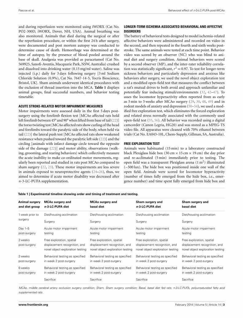
than animals fed the basal diet, evidenced by less arachidonic acid(AA; 20:4, n-6), F(1, 23)= 8.01, p < 0.05, linoleic acid (LA; 18:2,n-6), F(1, 23)= 37.41, p < 0.01 and palmitic acid (16:0) in brainphospholipids, F(1, 23)= 5.51, p < 0.05. A trend for higher levelsof eicosatrienoic acid (ETA; 22:3, n-3), F(1, 23)= 3.85, p= 0.06,among n-3-LC-PUFA fed rats than basal fed rats was seen. Nomain significant main effect of diet was seen in the amount ofDHA (22:6, n-3), F(1, 23)= 0.30, p > 0.05, or EPA (20:5, n-3),F(1, 23)= 0.72, p > 0.05. There were no significant interactioneffects. Figure 4 illustrates differences in the mean fatty acid levelsin the brain phospholipids between diet and surgery conditions.
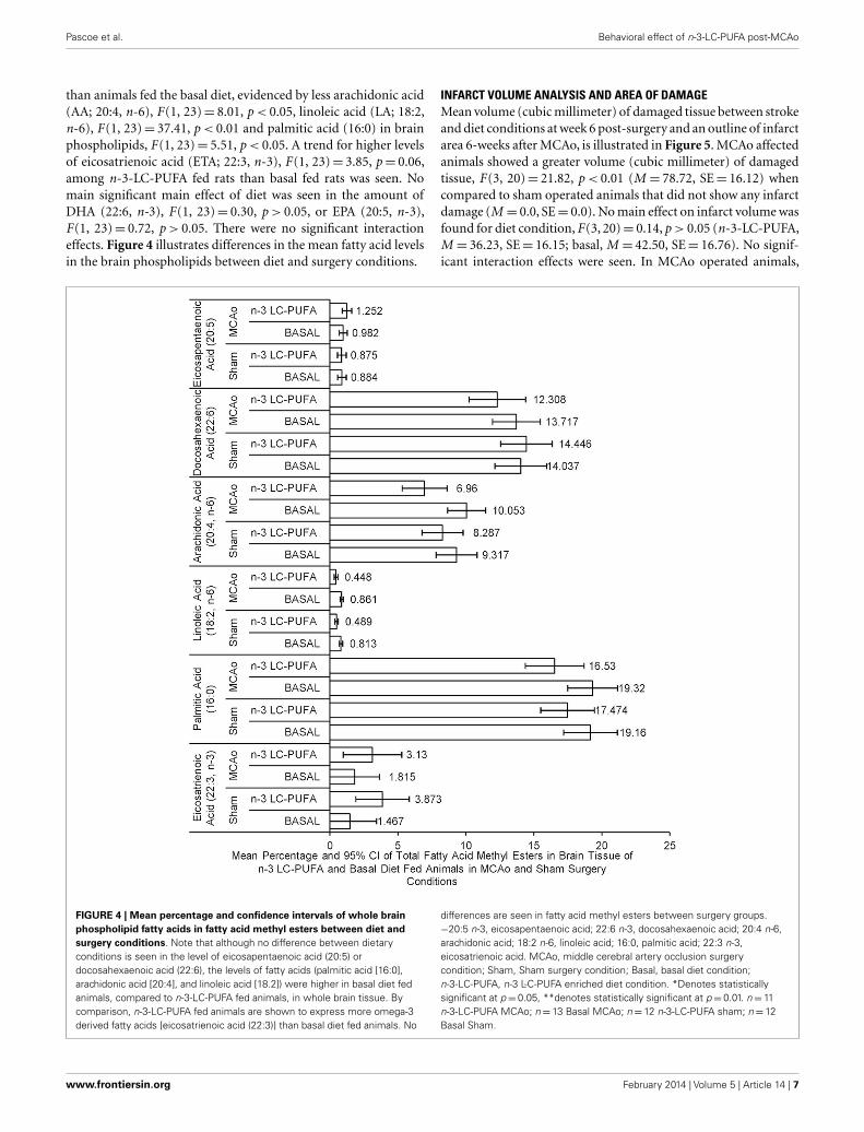
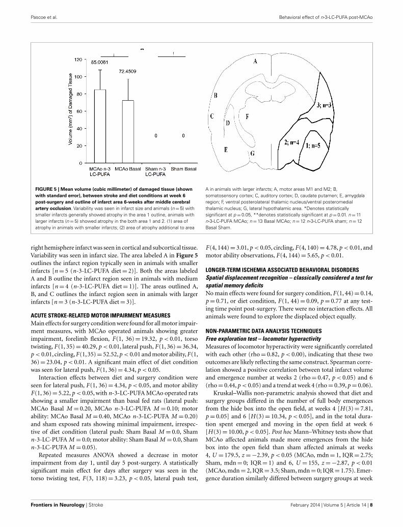
INFARCT VOLUME ANALYSIS AND AREA OF DAMAGEMean volume (cubic millimeter) of damaged tissue between strokeand diet conditions at week 6 post-surgery and an outline of infarctarea 6-weeks after MCAo, is illustrated in Figure 5. MCAo affectedanimals showed a greater volume (cubic millimeter) of damagedtissue, F(3, 20)= 21.82, p < 0.01 (M = 78.72, SE= 16.12) whencompared to sham operated animals that did not show any infarctdamage (M = 0.0, SE= 0.0). No main effect on infarct volume wasfound for diet condition, F(3, 20)= 0.14, p > 0.05 (n-3-LC-PUFA,M = 36.23, SE= 16.15; basal, M = 42.50, SE= 16.76). No signif-icant interaction effects were seen. In MCAo operated animals,
FIGURE 4 | Mean percentage and confidence intervals of whole brainphospholipid fatty acids in fatty acid methyl esters between diet andsurgery conditions. Note that although no difference between dietaryconditions is seen in the level of eicosapentaenoic acid (20:5) ordocosahexaenoic acid (22:6), the levels of fatty acids (palmitic acid [16:0],arachidonic acid [20:4], and linoleic acid [18.2]) were higher in basal diet fedanimals, compared to n-3-LC-PUFA fed animals, in whole brain tissue. Bycomparison, n-3-LC-PUFA fed animals are shown to express more omega-3derived fatty acids [eicosatrienoic acid (22:3)] than basal diet fed animals. No
differences are seen in fatty acid methyl esters between surgery groups.−20:5 n-3, eicosapentaenoic acid; 22:6 n-3, docosahexaenoic acid; 20:4 n-6,arachidonic acid; 18:2 n-6, linoleic acid; 16:0, palmitic acid; 22:3 n-3,eicosatrienoic acid. MCAo, middle cerebral artery occlusion surgerycondition; Sham, Sham surgery condition; Basal, basal diet condition;n-3-LC-PUFA, n-3 L-C-PUFA enriched diet condition. *Denotes statisticallysignificant at p=0.05, **denotes statistically significant at p=0.01. n=11n-3-LC-PUFA MCAo; n= 13 Basal MCAo; n= 12 n-3-LC-PUFA sham; n=12Basal Sham.
www.frontiersin.org February 2014 | Volume 5 | Article 14 | 7
Pascoe et al. Behavioral effect of n-3-LC-PUFA post-MCAo
FIGURE 5 | Mean volume (cubic millimeter) of damaged tissue (shownwith standard error), between stroke and diet conditions at week 6post-surgery and outline of infarct area 6-weeks after middle cerebralartery occlusion. Variability was seen in infarct size and animals (n=5) withsmaller infarcts generally showed atrophy in the area 1 outline, animals withlarger infarcts (n=5) showed atrophy in the both area 1 and 2. (1) area ofatrophy in animals with smaller infarcts; (2) area of atrophy additional to area
A in animals with larger infarcts; A, motor areas M1 and M2; B,somatosensory cortex; C, auditory cortex; D, caudate putamen; E, amygdalaregion; F, ventral posterolateral thalamic nucleus/ventral posteromedialthalamic nucleus; G, lateral hypothalamic area. *Denotes statisticallysignificant at p=0.05, **denotes statistically significant at p=0.01. n=11n-3-LC-PUFA MCAo; n=13 Basal MCAo; n=12 n-3-LC-PUFA sham; n=12Basal Sham.
right hemisphere infarct was seen in cortical and subcortical tissue.Variability was seen in infarct size. The area labeled A in Figure 5outlines the infarct region typically seen in animals with smallerinfarcts [n= 5 (n-3-LC-PUFA diet= 2)]. Both the areas labeledA and B outline the infarct region seen in animals with mediuminfarcts [n= 4 (n-3-LC-PUFA diet= 1)]. The areas outlined A,B, and C outlines the infarct region seen in animals with largerinfarcts [n= 3 (n-3-LC-PUFA diet= 3)].
ACUTE STROKE-RELATED MOTOR IMPAIRMENT MEASURESMain effects for surgery condition were found for all motor impair-ment measures, with MCAo operated animals showing greaterimpairment, forelimb flexion, F(1, 36)= 19.32, p < 0.01, torsotwisting, F(1, 35)= 40.29, p < 0.01, lateral push, F(1, 36)= 36.34,p < 0.01,circling,F(1,35)= 52.52,p < 0.01 and motor ability,F(1,36)= 23.04, p < 0.01. A significant main effect of diet conditionwas seen for lateral push, F(1, 36)= 4.34, p < 0.05.
Interaction effects between diet and surgery condition wereseen for lateral push, F(1, 36)= 4.34, p < 0.05, and motor abilityF(1, 36)= 5.22, p < 0.05, with n-3-LC-PUFA MCAo operated ratsshowing a smaller impairment than basal fed rats (lateral push:MCAo Basal M = 0.20, MCAo n-3-LC-PUFA M = 0.10; motorability: MCAo Basal M = 0.40, MCAo n-3-LC-PUFA M = 0.20)and sham exposed rats showing minimal impairment, irrespec-tive of diet condition (lateral push: Sham Basal M = 0.0, Shamn-3-LC-PUFA M = 0.0; motor ability: Sham Basal M = 0.0, Shamn-3-LC-PUFA M = 0.05).
Repeated measures ANOVA showed a decrease in motorimpairment from day 1, until day 5 post-surgery. A statisticallysignificant main effect for days after surgery was seen in thetorso twisting test, F(3, 118)= 3.23, p < 0.05, lateral push test,
F(4, 144)= 3.01, p < 0.05, circling, F(4, 140)= 4.78, p < 0.01, andmotor ability observations, F(4, 144)= 5.65, p < 0.01.
LONGER-TERM ISCHEMIA ASSOCIATED BEHAVIORAL DISORDERSSpatial displacement recognition – classically considered a test forspatial memory deficitsNo main effects were found for surgery condition, F(1, 44)= 0.14,p= 0.71, or diet condition, F(1, 44)= 0.09, p= 0.77 at any test-ing time point post-surgery. There were no interaction effects. Allanimals were found to explore the displaced object equally.
NON-PARAMETRIC DATA ANALYSIS TECHNIQUESFree exploration test – locomotor hyperactivityMeasures of locomotor hyperactivity were significantly correlatedwith each other (rho= 0.82, p < 0.00), indicating that these twooutcomes are likely reflecting the same construct. Spearman corre-lation showed a positive correlation between total infarct volumeand emergence number at weeks 2 (rho= 0.47, p < 0.05) and 6(rho= 0.44, p < 0.05) and a trend at week 4 (rho= 0.39, p= 0.06).
Kruskal–Wallis non-parametric analysis showed that diet andsurgery groups differed in the number of full body emergencesfrom the hide box into the open field, at weeks 4 [H (3)= 7.81,p= 0.05] and 6 [H (3)= 10.34, p < 0.05], and in the total dura-tion spent emerged and moving in the open field at week 6[H (3)= 10.00, p < 0.05]. Post hoc Mann–Whitney tests show thatMCAo affected animals made more emergences from the hidebox into the open field than sham affected animals at weeks4, U = 179.5, z =−2.39, p < 0.05 (MCAo, mdn= 1, IQR= 2.75;Sham, mdn= 0; IQR= 1) and 6, U = 155, z =−2.87, p < 0.01(MCAo, mdn= 2, IQR= 3.5; Sham, mdn= 0; IQR= 1.75). Emer-gence duration similarly differed between surgery groups at week
Frontiers in Neurology | Stroke February 2014 | Volume 5 | Article 14 | 8
Pascoe et al. Behavioral effect of n-3-LC-PUFA post-MCAo
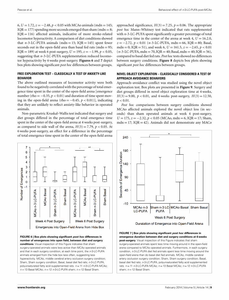
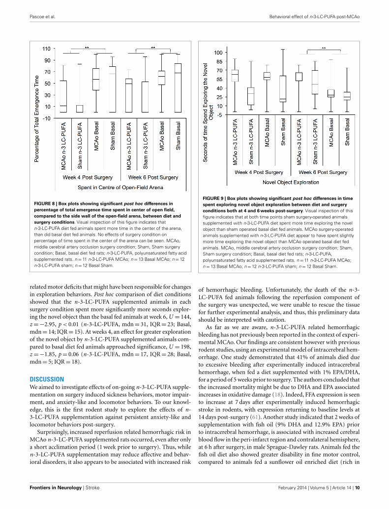
6, U = 1.72, z =−2.48, p < 0.05 with MCAo animals (mdn= 145;IQR= 177) spending more seconds emerged than sham (mdn= 8;IQR= 116) affected animals, indicative of more stroke-relatedlocomotor hyperactivity. A comparison of diet conditions showedthat n-3-LC-PUFA animals (mdn= 15; IQR= 145) spent fewerseconds out in the open-field area than basal fed rats (mdn= 95;IQR= 189) at week 6 post-surgery, U = 195, z =−1.99, p < 0.05,suggesting that n-3-LC-PUFA supplementation reduced locomo-tor hyperactivity by 6 weeks post-surgery. Figures 6 and 7 depictbox plots showing significant post hoc differences between groups.
FREE EXPLORATION TEST – CLASSICALLY A TEST OF ANXIETY-LIKEBEHAVIORThe above outlined measures of locomotor activity were bothfound to be negatively correlated with the percentage of total emer-gence time spent in the center of the open-field arena [emergencenumber (rho=−0.35, p < 0.01) and duration of time spent mov-ing in the open-field arena (rho=−0.45, p < 0.01)], indicatingthat they are unlikely to reflect anxiety-like behavior in operatedanimals.
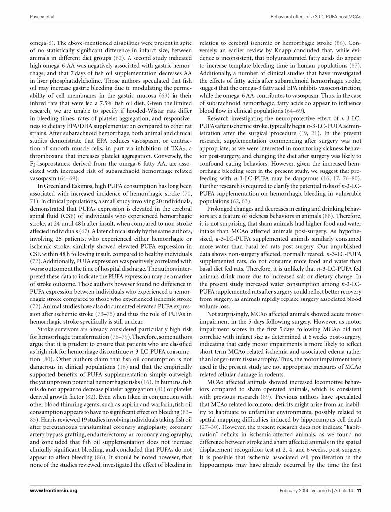
Non-parametric Kruskal–Wallis test indicated that surgery anddiet groups differed in the percentage of total emergence timespent in the center of the open-field arena at 4 weeks post-surgery,as compared to side wall of the arena, H (3)= 7.79, p < 0.05. At6 weeks post-surgery, an effect for a difference in the percentageof total emergence time spent in the center of the open field arena
FIGURE 6 | Box plots showing significant post hoc differences innumber of emergences into open field, between diet and surgeryconditions. Visual inspection of this Figure indicates that shamsurgery-operated animals were less active than MCAo operated animalsand that in each surgery condition, at each time point, the n-3-LC-PUFAanimals emerged from the hide box less often, suggesting lesshyperactivity. MCAo, middle cerebral artery occlusion surgery condition;Sham, Sham surgery condition; Basal, basal diet fed rats; n-3-LC-PUFA,polyunsaturated fatty acid supplemented rats. n=11 n-3-LC-PUFA MCAo;n=13 Basal MCAo; n=12 n-3-LC-PUFA sham; n=12 Basal Sham.
approached significance, H (3)= 7.25, p= 0.06. The appropriatepost hoc Mann–Whitney test indicated that rats supplementedwith n-3-LC-PUFA spent significantly a greater percentage of totalemergence time in the center of the arena at week 4, U = 16.2.0,z =−2.72, p < 0.01 (n-3-LC-PUFA, mdn= 66, IQR= 80; Basal,mdn= 0; IQR= 51), and week 6, U = 161.5, z =−2.65, p < 0.01(n-3-LC-PUFA, mdn= 70, IQR= 40; Basal, mdn= 40; IQR= 56),compared to basal diet fed rats. Post hoc tests showed no differencesbetween surgery conditions. Figure 8 depicts box plots showingsignificant post hoc differences between groups.
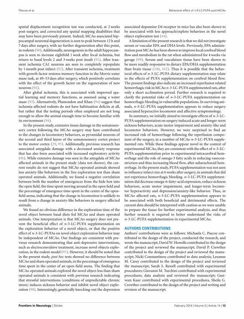
NOVEL OBJECT EXPLORATION – CLASSICALLY CONSIDERED A TEST OFAPPROACH AVOIDANCE BEHAVIORSApproach–avoidance conflict was studied using the novel objectexploration test. Box plots are presented in Figure 9. Surgery anddiet groups differed in novel object exploration time at 4 weeks,H (3)= 9.00, p < 0.01, and 6 weeks post-surgery, H (3)= 12.50,p < 0.01.
Post hoc comparisons between surgery conditions showedMCAo affected animals explored the novel object less (in sec-onds) than sham operated animals at week 4 post-surgery,U = 175, z =−2.32, p < 0.05 (MCAo, mdn= 6, IQR= 17; Sham,mdn= 17; IQR= 42). Animals did not demonstrate any MACo
FIGURE 7 | Box plots showing significant post hoc differences inemergence duration between diet and surgery conditions at 6 weekspost-surgery. Visual inspection of this Figure indicates that shamsurgery-operated animals spent less time moving around in the open-fieldarena compared to MCAo operated animals. Furthermore, in each surgerycondition, n-3-LC-PUFA diet fed animals spent less time moving around theopen-field arena than do basal diet fed animals. MCAo, middle cerebralartery occlusion surgery condition; Sham, Sham surgery condition; Basal,basal diet fed rats; n-3-LC-PUFA, polyunsaturated fatty acid supplementedrats. n= 11 n-3-LC-PUFA MCAo; n=13 Basal MCAo; n=12 n-3-LC-PUFAsham; n=12 Basal Sham.
www.frontiersin.org February 2014 | Volume 5 | Article 14 | 9
Pascoe et al. Behavioral effect of n-3-LC-PUFA post-MCAo
FIGURE 8 | Box plots showing significant post hoc differences inpercentage of total emergence time spent in center of open field,compared to the side wall of the open-field arena, between diet andsurgery conditions. Visual inspection of this figure indicates thatn-3-LC-PUFA diet fed animals spent more time in the center of the arena,than did basal diet fed animals. No effects of surgery condition onpercentage of time spent in the center of the arena can be seen. MCAo,middle cerebral artery occlusion surgery condition; Sham, Sham surgerycondition; Basal, basal diet fed rats; n-3-LC-PUFA, polyunsaturated fatty acidsupplemented rats. n=11 n-3-LC-PUFA MCAo; n=13 Basal MCAo; n=12n-3-LC-PUFA sham; n=12 Basal Sham.
related motor deficits that might have been responsible for changesin exploration behaviors. Post hoc comparison of diet conditionsshowed that the n-3-LC-PUFA supplemented animals in eachsurgery condition spent more significantly more seconds explor-ing the novel object than the basal fed animals at week 6, U = 144,z =−2.95, p < 0.01 (n-3-LC-PUFA, mdn= 31, IQR= 23; Basal,mdn= 14; IQR= 15). At weeks 4, an effect for greater explorationof the novel object by n-3-LC-PUFA supplemented animals com-pared to basal diet fed animals approached significance, U = 198,z =−1.85, p= 0.06 (n-3-LC-PUFA, mdn= 17, IQR= 28; Basal,mdn= 5; IQR= 18).
DISCUSSIONWe aimed to investigate effects of on-going n-3-LC-PUFA supple-mentation on surgery induced sickness behaviors, motor impair-ment, and anxiety-like and locomotor behaviors. To our knowl-edge, this is the first rodent study to explore the effects of n-3-LC-PUFA supplementation against persistent anxiety-like andlocomotor behaviors post-surgery.
Surprisingly, increased reperfusion related hemorrhagic risk inMCAo n-3-LC-PUFA supplemented rats occurred, even after onlya short acclimation period (1 week prior to surgery). Thus, whilen-3-LC-PUFA supplementation may reduce affective and behav-ioral disorders, it also appears to be associated with increased risk
FIGURE 9 | Box plots showing significant post hoc differences in timespent exploring novel object exploration between diet and surgeryconditions both at 4 and 6 weeks post-surgery. Visual inspection of thisfigure indicates that at both time points sham surgery-operated animalssupplemented with n-3-LC-PUFA diet spent more time exploring the novelobject than sham operated basal diet fed animals. MCAo surgery-operatedanimals supplemented with n-3-LC-PUFA diet appear to have spent slightlymore time exploring the novel object than MCAo operated basal diet fedanimals. MCAo, middle cerebral artery occlusion surgery condition; Sham,Sham surgery condition; Basal, basal diet fed rats; n-3-LC-PUFA,polyunsaturated fatty acid supplemented rats. n=11 n-3-LC-PUFA MCAo;n=13 Basal MCAo; n=12 n-3-LC-PUFA sham; n=12 Basal Sham.
of hemorrhagic bleeding. Unfortunately, the death of the n-3-LC-PUFA fed animals following the reperfusion component ofthe surgery was unexpected, we were unable to rescue the tissuefor further experimental analysis, and thus, this preliminary datashould be interpreted with caution.
As far as we are aware, n-3-LC-PUFA related hemorrhagicbleeding has not previously been reported in the context of experi-mental MCAo. Our findings are consistent however with previousrodent studies, using an experimental model of intracerebral hem-orrhage. One study demonstrated that 41% of animals died dueto excessive bleeding after experimentally induced intracerebralhemorrhage, when fed a diet supplemented with 1% EPA/DHA,for a period of 5 weeks prior to surgery. The authors concluded thatthe increased mortality might be due to DHA and EPA associatedincreases in oxidative damage (18). Indeed, FFA expression is seento increase at 7 days after experimentally induced hemorrhagicstroke in rodents, with expression returning to baseline levels at14 days post-surgery (61). Another study indicated that 2 weeks ofsupplementation with fish oil (9% DHA and 12.9% EPA) priorto intracerebral hemorrhage, is associated with increased cerebralblood flow in the peri-infarct region and contralateral hemisphere,at 6 h after surgery, in male Sprague-Dawley rats. Animals fed thefish oil diet also showed greater disability in fine motor control,compared to animals fed a sunflower oil enriched diet (rich in
Frontiers in Neurology | Stroke February 2014 | Volume 5 | Article 14 | 10
Pascoe et al. Behavioral effect of n-3-LC-PUFA post-MCAo
omega-6). The above-mentioned disabilities were present in spiteof no statistically significant difference in infarct size, betweenanimals in different diet groups (62). A second study indicatedhigh omega-6 AA was negatively associated with gastric hemor-rhage, and that 7 days of fish oil supplementation decreases AAin liver phosphatidylcholine. Those authors speculated that fishoil may increase gastric bleeding due to modulating the perme-ability of cell membranes in the gastric mucosa (63) in theirinbred rats that were fed a 7.5% fish oil diet. Given the limitedresearch, we are unable to specify if hooded-Wistar rats differin bleeding times, rates of platelet aggregation, and responsive-ness to dietary EPA/DHA supplementation compared to other ratstrains. After subarachnoid hemorrhage, both animal and clinicalstudies demonstrate that EPA reduces vasospasm, or contrac-tion of smooth muscle cells, in part via inhibition of TXA2, athromboxane that increases platelet aggregation. Conversely, theF2-isoprostanes, derived from the omega-6 fatty AA, are asso-ciated with increased risk of subarachnoid hemorrhage relatedvasospasm (64–69).
In Greenland Eskimos, high PUFA consumption has long beenassociated with increased incidence of hemorrhagic stroke (70,71). In clinical populations, a small study involving 20 individuals,demonstrated that PUFAs expression is elevated in the cerebralspinal fluid (CSF) of individuals who experienced hemorrhagicstroke, at 24 until 48 h after insult, when compared to non-strokeaffected individuals (67). A later clinical study by the same authors,involving 25 patients, who experienced either hemorrhagic orischemic stroke, similarly showed elevated PUFA expression inCSF, within 48 h following insult, compared to healthy individuals(72). Additionally, PUFA expression was positively correlated withworse outcome at the time of hospital discharge. The authors inter-preted these data to indicate the PUFA expression may be a markerof stroke outcome. These authors however found no difference inPUFA expression between individuals who experienced a hemor-rhagic stroke compared to those who experienced ischemic stroke(72). Animal studies have also documented elevated PUFA expres-sion after ischemic stroke (73–75) and thus the role of PUFAs inhemorrhagic stroke specifically is still unclear.
Stroke survivors are already considered particularly high riskfor hemorrhagic transformation (76–79). Therefore, some authorsargue that it is prudent to ensure that patients who are classifiedas high risk for hemorrhage discontinue n-3-LC-PUFA consump-tion (80). Other authors claim that fish oil consumption is notdangerous in clinical populations (16) and that the empiricallysupported benefits of PUFA supplementation simply outweighthe yet unproven potential hemorrhagic risks (16). In humans, fishoils do not appear to decrease platelet aggregation (81) or plateletderived growth factor (82). Even when taken in conjunction withother blood thinning agents, such as aspirin and warfarin, fish oilconsumption appears to have no significant effect on bleeding (83–85). Harris reviewed 19 studies involving individuals taking fish oilafter percutaneous transluminal coronary angioplasty, coronaryartery bypass grafting, endarterectomy or coronary angiography,and concluded that fish oil supplementation does not increaseclinically significant bleeding, and concluded that PUFAs do notappear to affect bleeding (86). It should be noted however, thatnone of the studies reviewed, investigated the effect of bleeding in
relation to cerebral ischemic or hemorrhagic stroke (86). Con-versely, an earlier review by Knapp concluded that, while evi-dence is inconsistent, that polyunsaturated fatty acids do appearto increase template bleeding time in human populations (87).Additionally, a number of clinical studies that have investigatedthe effects of fatty acids after subarachnoid hemorrhagic stroke,suggest that the omega-3 fatty acid EPA inhibits vasoconstriction,while the omega-6 AA, contributes to vasospasm. Thus, in the caseof subarachnoid hemorrhagic, fatty acids do appear to influenceblood flow in clinical populations (64–69).
Research investigating the neuroprotective effect of n-3-LC-PUFAs after ischemic stroke, typically begin n-3-LC-PUFA admin-istration after the surgical procedure (19, 21). In the presentresearch, supplementation commencing after surgery was notappropriate, as we were interested in monitoring sickness behav-ior post-surgery, and changing the diet after surgery was likely toconfound eating behaviors. However, given the increased hem-orrhagic bleeding seen in the present study, we suggest that pre-feeding with n-3-LC-PUFA may be dangerous (16, 17, 76–80).Further research is required to clarify the potential risks of n-3-LC-PUFA supplementation on hemorrhagic bleeding in vulnerablepopulations (62, 63).
Prolonged changes and decreases in eating and drinking behav-iors are a feature of sickness behaviors in animals (88). Therefore,it is not surprising that sham animals had higher food and waterintake than MCAo affected animals post-surgery. As hypothe-sized, n-3-LC-PUFA supplemented animals similarly consumedmore water than basal fed rats post-surgery. Our unpublisheddata shows non-surgery affected, normally reared, n-3-LC-PUFAsupplemented rats, do not consume more food and water thanbasal diet fed rats. Therefore, it is unlikely that n-3-LC-PUFA fedanimals drink more due to increased salt or dietary change. Inthe present study increased water consumption among n-3-LC-PUFA supplemented rats after surgery could reflect better recoveryfrom surgery, as animals rapidly replace surgery associated bloodvolume loss.
Not surprisingly, MCAo affected animals showed acute motorimpairment in the 5-days following surgery. However, as motorimpairment scores in the first 5 days following MCAo did notcorrelate with infarct size as determined at 6 weeks post-surgery,indicating that early motor impairments is more likely to reflectshort term MCAo related ischemia and associated edema ratherthan longer-term tissue atrophy. Thus, the motor impairment testsused in the present study are not appropriate measures of MCAorelated cellular damage in rodents.
MCAo affected animals showed increased locomotive behav-iors compared to sham operated animals, which is consistentwith previous research (89). Previous authors have speculatedthat MCAo related locomotor deficits might arise from an inabil-ity to habituate to unfamiliar environments, possibly related tospatial mapping difficulties induced by hippocampus cell death(27–30). However, the present research does not indicate “habit-uation” deficits in ischemia-affected animals, as we found nodifference between stroke and sham affected animals in the spatialdisplacement recognition test at 2, 4, and 6 weeks, post-surgery.It is possible that ischemia associated cell proliferation in thehippocampus may have already occurred by the time the first
www.frontiersin.org February 2014 | Volume 5 | Article 14 | 11
Pascoe et al. Behavioral effect of n-3-LC-PUFA post-MCAo
spatial displacement recognition test was conducted, at 2 weekspost-surgery, and corrected any spatial mapping disabilities thatmay have been previously present. Indeed, MCAo associated hip-pocampal neuronal degeneration is seen to peak between 12 h and7 days after surgery, with no further degeneration after this point,in rodents (90). Additionally, neurogenesis in the adult hippocam-pus is seen to increase eightfold 1 week after focal ischemia, butreturn to basal levels 2 and 3 weeks post insult (91). After tran-sient ischemia CA1 neurons are seen to completely repopulateby 1 month post infarct (92). After transient ischemia, treatmentwith growth factor restores memory function in the Morris watermaze task, at 49–53 days after surgery, which positively correlateswith the effect of the growth factor on the regeneration of newneurons (92).
After global ischemia, this is associated with improved spa-tial learning and memory functions, as assessed using a watermaze (93). Alternatively, Plamondon and Khan (94) suggest thatischemia-affected rodents do not have habituation deficits at all,but rather that the testing periods often employed are not longenough to allow the animal enough time to become familiar withits environment (94).
In the present study, extensive tissue damage in the somatosen-sory cortex following the MCAo surgery may have contributedto the changes in locomotory behaviors, as pyramidal neurons ofthe second and third layers of the somatosensory cortex projectto the motor cortex (29, 95). Additionally, previous research hasassociated amygdala damage with a decreased anxiety responsethat has also been associated with increased exploratory activity(96). While extensive damage was seen in the amygdala of MCAoaffected animals in the present study (data not shown), the cur-rent results do not suggest that MCAo operated animals showedless anxiety-like behaviors in the free exploration test than shamoperated animals. Additionally, we found a negative correlationbetween both the number of emergences from the hide box intothe open field, the time spent moving around in the open field andthe percentage of emergence time spent in the center of the open-field arena, indicating that the change in locomotion is unlikely toresult from a change in anxiety-like behaviors in surgery affectedanimals.
We found no obvious difference in the exploration time of thenovel object between basal diets fed MCAo and sham operatedanimals. One interpretation is that MCAo surgery does not pre-vent the beneficial effect of n-3-LC-PUFA supplementation onthe exploration behavior of a novel object, or that the positiveeffect of n-3-LC-PUFAs on novel object exploration behavior maybe independent of MCAo. Our findings are consistent with pre-vious research demonstrating that anti-depressive interventions,such as electroconvulsive treatment, increase novel objects explo-ration, in the rodent model (97). However, it should be noted thatin the present study, post hoc tests showed no difference betweenMCAo and sham operated animals, in the percentage of emergencetime spent in the center of an open-field arena. The finding thatMCAo operated animals explored the novel object less than shamoperated animals is consistent with previous research indicatingthat stressful interventions (conditions of unpredictable chronicstress) induces sickness behavior and inhibit novel object explo-ration (98). Interestingly, genetically knocking-out the depression
associated dopamine D4 receptor in mice has also been shown tobe associated with less approach/explore behaviors in the novelobject exploration test (44).
A limitation of the present research is that we did not investigateserum or vascular EPA and DHA levels. Previously, EPA adminis-tration post MCAo has been shown to improve local cerebral bloodflow and metabolism in the rat when administered for 4 weeks viagavage (99). Serum and vasculature tissue have been shown tobe more readily responsive to dietary EPA/DHA supplementationthan brain tissue (100, 101). Thus it is possible that the behav-ioral effects of n-3-LC-PUFA dietary supplementation may relateto the effects of PUFA supplementation on cerebral blood flow.The present findings also indicate an increased reperfusion relatedhemorrhagic risk in MCAo n-3-LC-PUFA supplemented rats, afteronly a short acclimation period. Further research is required toclarify the potential risks of n-3-LC-PUFA supplementation onhemorrhagic bleeding in vulnerable populations. In surviving ani-mals, n-3-LC-PUFA supplementation appears to reduce surgeryassociated hyperactive locomotor and affective symptomology.
In summary, we initially aimed to investigate effects of n-3-LC-PUFA supplementation on surgery induced acute and longer-termsickness behaviors, acute motor impairment and anxiety-like andlocomotor behaviors. However, we were surprised to find anincreased risk of hemorrhage following the reperfusion compo-nent of the surgery, in a number of MCAo n-3-LC-PUFA supple-mented rats. While these findings appear novel in the context ofexperimental MCAo, they are consistent with the effect of n-3-LC-PUFA supplementation prior to experimental intracerebral hem-orrhage and the role of omega-3 fatty acids in reducing vasocon-striction and thus increasing blood flow, after subarachnoid hem-orrhage. In the present study, PUFA supplementation was not seento influence infarct size at 6 weeks after surgery, in animals that didnot experience hemorrhagic bleeding. n-3-LC-PUFA supplemen-tation did decrease omega-6-fatty-acid expression, reduce sicknessbehaviors, acute motor impairment, and longer-term locomo-tor hyperactivity and depression/anxiety-like behavior. Thus, inMCAo affected rats, n-3-LC-PUFA supplementation appears tobe associated with both beneficial and detrimental effects. Thecurrent data should be interpreted with caution as we were unableto prepare the tissue for further experimental analysis, and thusfurther research is required to better understand the risks ofn-3-LC-PUFA supplementation in experimental MCAo.
AUTHORS CONTRIBUTIONSAuthors’ contributions were as follows: Michaela C. Pascoe con-tributed to the design of the project, conducted the research, andwrote the manuscript; David W. Howells contributed to the designof the project and reviewed the manuscript; David P. Crewthercontributed to the design of the project and reviewed the manu-script; Nicki Constantinou contributed to data analysis; LeeanneM. Carey contributed to the design of the project and reviewedthe manuscript; Sarah S. Rewell contributed with experimentalprocedures; Giovanni M. Turchini contributed with experimentalprocedures, data analysis and reviewed the manuscript; Gun-veen Kaur contributed with experimental procedures, Sheila G.Crewther contributed to the design of the project and writing andrevision of the manuscript.
Frontiers in Neurology | Stroke February 2014 | Volume 5 | Article 14 | 12
Pascoe et al. Behavioral effect of n-3-LC-PUFA post-MCAo
ACKNOWLEDGMENTSSpecial thanks to Associate Professor Trevor Norman of theDepartment of Psychiatry, University of Melbourne, Austin Hos-pital, Heidelberg, VIC, Australia for the provision of equip-ment required for behavioral testing. Special thanks to ProfessorAndrew Sinclair of Deakin University Warrnambool, VIC, Aus-tralia for the provision of facilities to conduct fatty acid analy-sis. No sources or grants contributed to the completion of thisresearch.
REFERENCES1. Mackay J, Mensah G. The Atlas of Heart Disease and Stroke. Geneva: World
Health Organization (2004). 112 p.2. Burvill PW, Johnson GA, Jamrozik KD, Anderson CS, Stewart-Wynne EG,
Chakera TM. Anxiety disorders after stroke: results from the Perth CommunityStroke Study. Br J Psychiatry (1995) 166(3):328–32. doi:10.1192/bjp.166.3.328
3. Astrom M. Generalized anxiety disorder in stroke patients. A 3-year longitudi-nal study. Stroke (1996) 27(2):270–5. doi:10.1161/01.STR.27.2.270
4. Linden T, Blomstrand C, Skoog I. Depressive disorders after 20 monthsin elderly stroke patients: a case-control study. Stroke (2007) 38(6):1860–3.doi:10.1161/STROKEAHA.106.471805
5. Kessler RC, Nelson CB, McGonagle KA, Liu J, Swartz M, Blazer DG. Comor-bidity of DSM-III-R major depressive disorder in the general population:results from the US National Comorbidity Survey. Br J Psychiatry Suppl (1996)168(30):17–30.
6. Hirschfeld RM. The comorbidity of major depression and anxiety disorders:recognition and management in primary care. Prim Care Companion J ClinPsychiatry (2001) 3(6):244–54. doi:10.4088/PCC.v03n0609
7. Pandharipande P, Cotton BA, Shintani A, Thompson J, Pun BT, Morris JAJr., et al. Prevalence and risk factors for development of delirium in surgi-cal and trauma intensive care unit patients. J Trauma (2008) 65(1):34–41.doi:10.1097/TA.0b013e31814b2c4d
8. Tully PJ, Baker RA, Winefield HR, Turnbull DA. Depression, anxiety disordersand type D personality as risk factors for delirium after cardiac surgery. AustN Z J Psychiatry (2010) 44(11):1005–11. doi:10.3109/00048674.2010.495053
9. McManus J, Pathansali R, Stewart R, Macdonald A, Jackson S. Delirium post-stroke. Age Ageing (2007) 36(6):613–8. doi:10.1093/ageing/afm140
10. Masskulpan P, Riewthong K, Dajpratham P, Kuptniratsaikul V. Anxiety anddepressive symptoms after stroke in 9 rehabilitation centers. J Med Assoc Thai(2008) 91(10):1595–602.
11. Dantzer R, O’Connor JC, Freund GG, Johnson RW, Kelley KW. From inflam-mation to sickness and depression: when the immune system subjugates thebrain. Nat Rev Neurosci (2008) 9(1):46–56. doi:10.1038/nrn2297
12. Pascoe MC, Crewther SG, Carey LM, Crewther DP. What you eat is what youare – a role for polyunsaturated fatty acids in neuroinflammation induceddepression? Clin Nutr (2011) 30(4):407–15. doi:10.1016/j.clnu.2011.03.013
13. Ross BM. Omega-3 polyunsaturated fatty acids and anxiety disorders.Prostaglandins Leukot Essent Fatty Acids (2009) 81(5–6):309–12. doi:10.1016/j.plefa.2009.10.004
14. Lavialle M, Denis I, Guesnet P, Vancassel S. Involvement of omega-3 fatty acidsin emotional responses and hyperactive symptoms. J Nutr Biochem (2010)21(10):899–905. doi:10.1016/j.jnutbio.2009.12.005
15. Appleton KM, Rogers PJ, Ness AR. Is there a role for n-3 long-chain polyun-saturated fatty acids in the regulation of mood and behaviour? A review of theevidence to date from epidemiological studies, clinical studies and interventiontrials. Nutr Res Rev (2008) 21(1):13–41. doi:10.1017/S0954422408998620
16. Bays HE. Safety considerations with omega-3 fatty acid therapy. Am J Cardiol(2007) 99(6A):35C–43C. doi:10.1016/j.amjcard.2006.11.020
17. Stone NJ. Fish consumption, fish oil, lipids, and coronary heart disease. Circu-lation (1996) 94(9):2337–40. doi:10.1161/01.CIR.94.9.2337
18. Park Y, Nam S, Yi HJ, Hong HJ, Lee M. Dietary n-3 polyunsaturated fatty acidsincrease oxidative stress in rats with intracerebral hemorrhagic stroke. Nutr Res(2009) 29(11):812–8. doi:10.1016/j.nutres.2009.10.019
19. Belayev L, Marcheselli VL, Khoutorova L, Rodriguez de Turco EB, Busto R,Ginsberg MD, et al. Docosahexaenoic acid complexed to albumin elicits high-grade ischemic neuroprotection. Stroke (2005) 36(1):118–23. doi:10.1161/01.STR.0000149620.74770.2e
20. Zhang W, Hu X, Yang W, Gao Y, Chen J. Omega-3 polyunsaturated fattyacid supplementation confers long-term neuroprotection against neonatalhypoxic-ischemic brain injury through anti-inflammatory actions. Stroke(2010) 41(10):2341–7. doi:10.1161/STROKEAHA.110.586081
21. Choi-Kwon S, Park KA, Lee HJ, Park MS, Lee JH, Jeon SE, et al. Temporalchanges in cerebral antioxidant enzyme activities after ischemia and reperfu-sion in a rat focal brain ischemia model: effect of dietary fish oil. Brain Res DevBrain Res (2004) 152(1):11–8. doi:10.1016/j.devbrainres.2004.05.004
22. Petullo D, Masonic K, Lincoln C, Wibberley L, Teliska M, Yao DL. Model devel-opment and behavioral assessment of focal cerebral ischemia in rats. Life Sci(1999) 64(13):1099–108. doi:10.1016/S0024-3205(99)00038-7
23. Bederson JB, Pitts LH, Tsuji M, Nishimura MC, Davis RL, Bartkowski H. Ratmiddle cerebral artery occlusion: evaluation of the model and development ofa neurologic examination. Stroke (1986) 17(3):472–6. doi:10.1161/01.STR.17.3.472
24. Lolic MM, Fiskum G, Rosenthal RE. Neuroprotective effects of acetyl-L-carnitine after stroke in rats. Ann Emerg Med (1997) 29(6):758–65. doi:10.1016/S0196-0644(97)70197-5
25. Akpan N, Serrano-Saiz E, Zacharia BE, Otten ML, Ducruet AF, Snipas SJ,et al. Intranasal delivery of caspase-9 inhibitor reduces caspase-6-dependentaxon/neuron loss and improves neurological function after stroke. J Neurosci(2011) 31(24):8894–904. doi:10.1523/JNEUROSCI.0698-11.2011
26. Garg P, Duncan RS, Kaja S, Zabaneh A, Chapman KD, Koulen P. Lau-roylethanolamide and linoleoylethanolamide improve functional outcome ina rodent model for stroke. Neurosci Lett (2011) 492(3):134–8. doi:10.1016/j.neulet.2011.01.073
27. Chandler MJ, DeLeo J, Carney JM. An unanesthetized-gerbil model ofcerebral ischemia-induced behavioral changes. J Pharmacol Methods (1985)14(2):137–46. doi:10.1016/0160-5402(85)90051-8
28. Wang D, Corbett D. Cerebral ischemia, locomotor activity and spatial mapping.Brain Res (1990) 533(1):78–82. doi:10.1016/0006-8993(90)91798-L
29. Kuroiwa T, Bonnekoh P, Hossmann KA. Therapeutic window of halothaneanesthesia for reversal of delayed neuronal injury in gerbils: relationship topostischemic motor hyperactivity. Brain Res (1991) 563(1–2):33–8. doi:10.1016/0006-8993(91)91511-X
30. Kuroiwa T, Bonnekoh P, Hossmann KA. Locomotor hyperactivity and hip-pocampal CA1 injury after transient forebrain ischemia of gerbils. NeurosciLett (1991) 122(2):141–4. doi:10.1016/0304-3940(91)90842-H
31. Liu P, Bilkey DK. The effect of excitotoxic lesions centered on the hippocam-pus or perirhinal cortex in object recognition and spatial memory tasks. BehavNeurosci (2001) 115(1):94–111. doi:10.1037/0735-7044.115.1.94
32. Mumby DG, Gaskin S, Glenn MJ, Schramek TE, Lehmann H. Hippocampaldamage and exploratory preferences in rats: memory for objects, places, andcontexts. Learn Mem (2002) 9(2):49–57. doi:10.1101/lm.41302
33. Save E, Poucet B, Foreman N, Buhot MC. Object exploration and reactionsto spatial and nonspatial changes in hooded rats following damage to pari-etal cortex or hippocampal formation. Behav Neurosci (1992) 106(3):447–56.doi:10.1037/0735-7044.106.3.447
34. Callaway JK, Knight MJ, Watkins DJ, Beart PM, Jarrott B. Delayedtreatment with AM-36, a novel neuroprotective agent, reduces neuronal dam-age after endothelin-1-induced middle cerebral artery occlusion in consciousrats. Stroke (1999) 30(12):2704–12; discussion 12. doi:10.1161/01.STR.30.12.2704
35. Miller AA, Dusting GJ, Roulston CL, Sobey CG. NADPH-oxidase activity iselevated in penumbral and non-ischemic cerebral arteries following stroke.Brain Res (2006) 1111(1):111–6. doi:10.1016/j.brainres.2006.06.082
36. Nieuwenhuijs VB, de Bruijn MT, Schiesser M, Morphett A, Padbury RT, BarrittGJ. Ischemic preconditioning and intermittent ischemia preserve bile flow in arat model of ischemia/reperfusion injury. Dig Dis Sci (2007) 52(11):3029–37.doi:10.1007/s10620-006-9520-7
37. Venna VR, Deplanque D, Allet C, Belarbi K, Hamdane M, Bordet R. PUFAinduce antidepressant-like effects in parallel to structural and molecularchanges in the hippocampus. Psychoneuroendocrinology (2009) 34(2):199–211.doi:10.1016/j.psyneuen.2008.08.025
38. National Health and Medical Research Council. Australian Code of Practice forthe Care and Use of Animals for Scientific Purposes. 7th ed. Canberra: AustralianGovernment (2004). 84 p.
39. Jones ML, Mark PJ, Mori TA, Keelan JA, Waddell BJ. Maternal dietaryomega-3 fatty acid supplementation reduces placental oxidative stress and
www.frontiersin.org February 2014 | Volume 5 | Article 14 | 13

Pascoe et al. Behavioral effect of n-3-LC-PUFA post-MCAo
increases fetal and placental growth in the rat. Biol Reprod (2013) 88(2):37.doi:10.1095/biolreprod.112.103754
40. Longa EZ, Weinstein PR, Carlson S, Cummins R. Reversible middle cere-bral artery occlusion without craniectomy in rats. Stroke (1989) 20(1):84–91.doi:10.1161/01.STR.20.1.84
41. Spratt NJ, Fernandez J, Chen M, Rewell S, Cox S, van Raay L, et al. Mod-ification of the method of thread manufacture improves stroke inductionrate and reduces mortality after thread-occlusion of the middle cerebralartery in young or aged rats. J Neurosci Methods (2006) 155(2):285–90.doi:10.1016/j.jneumeth.2006.01.020
42. Rygula R, Abumaria N, Flugge G, Fuchs E, Ruther E, Havemann-Reinecke U.Anhedonia and motivational deficits in rats: impact of chronic social stress.Behav Brain Res (2005) 162(1):127–34. doi:10.1016/j.bbr.2005.03.009
43. Soffie M, Buhot MC, Poucet B. Cognitive and noncognitive processesinvolved in selective object exploration: comparison between young adultand old rats. Physiol Behav (1992) 52(5):1029–35. doi:10.1016/0031-9384(92)90387-H
44. Dulawa SC, Grandy DK, Low MJ, Paulus MP, Geyer MA. Dopamine D4receptor-knock-out mice exhibit reduced exploration of novel stimuli. J Neu-rosci (1999) 19(21):9550–6.
45. Belzung C. Hippocampal mossy fibres: implication in novelty reactions or inanxiety behaviours? Behav Brain Res (1992) 51(2):149–55. doi:10.1016/S0166-4328(05)80208-6
46. Montgomery KC. The relation between fear induced by novel stimula-tion and exploratory behavior. J Comp Physiol Psychol (1955) 48(4):254–60.doi:10.1037/h0048596
47. Dember WN. Response by the rat to environmental change. J Comp PhysiolPsychol (1956) 49(1):93–5. doi:10.1037/h0045411
48. Andersen MB, Zimmer J, Sams-Dodd F. Postischemic hyperactivity in theMongolian gerbil correlates with loss of hippocampal neurons. Behav Neurosci(1997) 111(6):1205–16. doi:10.1037/0735-7044.111.6.1205
49. Kilic E, Kilic U, Bacigaluppi M, Guo Z, Abdallah NB, Wolfer DP, et al.Delayed melatonin administration promotes neuronal survival, neurogenesisand motor recovery, and attenuates hyperactivity and anxiety after mild focalcerebral ischemia in mice. J Pineal Res (2008) 45(2):142–8. doi:10.1111/j.1600-079X.2008.00568.x
50. Monteggia LM, Luikart B, Barrot M, Theoloold D, Malkovska I, Nef S, et al.Brain-derived neurotrophic factor conditional knockouts show gender differ-ences in depression-related behaviors. Biol Psychiatry (2007) 61(2):187–97.doi:10.1016/j.biopsych.2006.03.021
51. van Riezen H, Leonard BE. Effects of psychotropic drugs on the behavior andneurochemistry of olfactory bulbectomized rats. In: File SE, editor. Psychophar-macology of Anxiolytics and Antidepressants. New York: Pergamon Press (1991).p. 231–50.
52. Redmond AM, Kelly JP, Leonard BE. Behavioural and neurochemical effects ofdizocilpine in the olfactory bulbectomized rat model of depression. PharmacolBiochem Behav (1997) 58(2):355–9. doi:10.1016/S0091-3057(97)00259-1
53. Garzon J, Delrio J. Hyperactivity induced in rats by long-term isolation –further-studies on a new animal-model for the detection of anti-depressants.Eur J Pharmacol (1981) 74(4):287–94. doi:10.1016/0014-2999(81)90047-9
54. Garzon J, Fuentes JA, Delrio J. Anti-depressants selectively antagonize thehyperactivity induced in rats by long-term isolation. Eur J Pharmacol (1979)59(3–4):293–6. doi:10.1016/0014-2999(79)90293-0
55. Misslin R, Cigrang M. Does neophobia necessarily imply fear or anxiety. BehavProcesses (1986) 12(1):45–50. doi:10.1016/0376-6357(86)90069-0
56. Welker WI. Free versus forced exploration of a novel situation by rats. PsycholRep (1957) 3(1):95–108. doi:10.2466/pr0.1957.3.g.95
57. Bevins RA, Besheer J. Object recognition in rats and mice: a one-trial non-matching-to-sample learning task to study “recognition memory”. Nat Protoc(2006) 1(3):1306–11. doi:10.1038/nprot.2006.205
58. Sinclair AJ, O’Dea K, Dunstan G, Ireland PD, Niall M. Effects on plasma lipidsand fatty acid composition of very low fat diets enriched with fish or kangaroomeat. Lipids (1987) 22(7):523–9. doi:10.1007/BF02540369
59. Ackman RG. The gas chromatograph in practical analyses of commonand uncommon fatty acids for the 21st century. Anal Chim Acta (2002)465(1–2):175–92. doi:10.1016/S0003-2670(02)00098-3
60. Field A, Miles J. Discovering Statistics Using SAS. London: SAGE PublicationsLtd (2010).
61. Paik M-J, Shin JY, Lee G, Ahn YH. Monitoring of altered free fatty acid meta-bolic patterns in rat plasma following hemorrhagic stroke. Anal Lett (2011)44(7):1323–32. doi:10.1080/00032719.2010.512678
62. Clarke J, Herzberg G, Peeling J, Buist R, Corbett D. Dietary supplementa-tion of omega-3 polyunsaturated fatty acids worsens forelimb motor func-tion after intracerebral hemorrhage in rats. Exp Neurol (2005) 191(1):119–27.doi:10.1016/j.expneurol.2004.09.003
63. Huang YS, Watanabe Y, Horrobin DF, Simmons V. Fatty acid changes inliver choline and ethanolamine glycerophospholipids in aspirin-treated ratsfed linoleate, gamma-linolenate and fish oil. Clin Physiol Biochem (1989)7(2):79–86.
64. Badjatia N, Seres D, Carpenter A, Schmidt JM, Lee K, Mayer SA, et al. Free fattyacids and delayed cerebral ischemia after subarachnoid hemorrhage. Stroke(2012) 43(3):691–6. doi:10.1161/STROKEAHA.111.636035
65. Corcoran TB, Mas E, Barden AE, Durand T, Galano JM, Roberts LJ, et al.Are isofurans and neuroprostanes increased after subarachnoid hemorrhageand traumatic brain injury? Antioxid Redox Signal (2011) 15(10):2663–7.doi:10.1089/ars.2011.4125
66. Hsieh YP, Lin CL, Shiue AL,Yin H, Morrow JD, Hsu JC, et al. Correlation of F4-neuroprostanes levels in cerebrospinal fluid with outcome of aneurysmal sub-arachnoid hemorrhage in humans. Free Radic Biol Med (2009) 47(6):814–24.doi:10.1016/j.freeradbiomed.2009.06.026
67. Pilitsis JG, Coplin WM, O’Regan MH, Wellwood JM, Diaz FG, Fairfax MR,et al. Free fatty acids in human cerebrospinal fluid following subarachnoidhemorrhage and their potential role in vasospasm: a preliminary observation.J Neurosurg (2002) 97(2):272–9. doi:10.3171/jns.2002.97.2.0272
68. Shirao S, Fujisawa H, Kudo A, Kurokawa T, Yoneda H, Kunitsugu I,et al. Inhibitory effects of eicosapentaenoic acid on chronic cerebralvasospasm after subarachnoid hemorrhage: possible involvement of asphingosylphosphorylcholine-rho-kinase pathway. Cerebrovasc Dis (2008)26(1):30–7. doi:10.1159/000135650
69. Yoneda H, Shirao S, Kurokawa T, Fujisawa H, Kato S, Suzuki M. Doeseicosapentaenoic acid (EPA) inhibit cerebral vasospasm in patients afteraneurysmal subarachnoid hemorrhage? Acta Neurol Scand (2008) 118(1):54–9.doi:10.1111/j.1600-0404.2007.00983.x
70. Kromann N, Green A. Epidemiological studies in the Upernavik district, Green-land. Incidence of some chronic diseases 1950-1974. Acta Med Scand (1980)208(5):401–6. doi:10.1111/j.0954-6820.1980.tb01221.x
71. Bjerregaard P, Mulvad G, Pedersen HS. Regional variation in cardiovascular riskfactors and ischemic heart disease mortality in Greenland. Int J CircumpolarHealth (1998) 57(Suppl 1):302–5.
72. Pilitsis JG, Coplin WM, O’Regan MH, Wellwood JM, Diaz FG, Fairfax MR,et al. Measurement of free fatty acids in cerebrospinal fluid from patientswith hemorrhagic and ischemic stroke. Brain Res (2003) 985(2):198–201.doi:10.1016/S0006-8993(03)03044-0
73. Gasparovic C, Rosenberg GA, Wallace JA, Estrada EY, Roberts K, Pastuszyn A,et al. Magnetic resonance lipid signals in rat brain after experimental strokecorrelate with neutral lipid accumulation. Neurosci Lett (2001) 301(2):87–90.doi:10.1016/S0304-3940(01)01616-0
74. Nakano S, Kogure K, Abe K, Yae T. Ischemia-induced alterations in lipidmetabolism of the gerbil cerebral cortex: I. Changes in free fatty acidliberation. J Neurochem (1990) 54(6):1911–6. doi:10.1111/j.1471-4159.1990.tb04890.x
75. Narita K, Kubota M, Nakane M, Kitahara S, Nakagomi T, Tamura A, et al.Therapeutic time window in the penumbra during permanent focal ischemiain rats: changes of free fatty acids and glycerophospholipids. Neurol Res (2000)22(4):393–400.
76. Jaillard A, Cornu C, Durieux A, Moulin T, Boutitie F, Lees KR, et al. Hemor-rhagic transformation in acute ischemic stroke. The MAST-E study. MAST-EGroup. Stroke (1999) 30(7):1326–32. doi:10.1161/01.STR.30.7.1326
77. Motto C, Ciccone A, Aritzu E, Boccardi E, De Grandi C, Piana A, et al. Hem-orrhage after an acute ischemic stroke. MAST-I Collaborative Group. Stroke(1999) 30(4):761–4. doi:10.1161/01.STR.30.4.761
78. Motto C, Aritzu E, Boccardi E, De Grandi C, Piana A, Candelise L. Reliability ofhemorrhagic transformation diagnosis in acute ischemic stroke. Stroke (1997)28(2):302–6. doi:10.1161/01.STR.28.2.302
79. Alexandrov AV, Black SE, Ehrlich LE, Caldwell CB, Norris JW. Predictors ofhemorrhagic transformation occurring spontaneously and on anticoagulants
Frontiers in Neurology | Stroke February 2014 | Volume 5 | Article 14 | 14
Pascoe et al. Behavioral effect of n-3-LC-PUFA post-MCAo
in patients with acute ischemic stroke. Stroke (1997) 28(6):1198–202. doi:10.1161/01.STR.28.6.1198
80. Berliner AR, Fine DM. There’s something fishy about this bleeding. NDT Plus(2011) 4(4):270–2. doi:10.1093/ndtplus/sfr046
81. Balk E, Chung M, Lichtenstein A, Chew P, Kupelnick B, Lawrence A, et al.Effects of omega-3 fatty acids on cardiovascular risk factors and intermediatemarkers of cardiovascular disease. In: Evidence Report/Technology AssessmentNo. 93. Rockville, MD: Agency for Healthcare Research and Quality (2004).p. 1–6.
82. Wallace JM, McCabe AJ, Roche HM, Higgins S, Robson PJ, Gilmore WS, et al.The effect of low-dose fish oil supplementation on serum growth factorsin healthy humans. Eur J Clin Nutr (2000) 54(9):690–4. doi:10.1038/sj.ejcn.1601076
83. Eritsland J, Arnesen H, Gronseth K, Fjeld NB, Abdelnoor M. Effect ofdietary supplementation with n-3 fatty acids on coronary artery bypassgraft patency. Am J Cardiol (1996) 77(1):31–6. doi:10.1016/S0002-9149(97)89130-8
84. Leaf A, Jorgensen MB, Jacobs AK, Cote G, Schoenfeld DA, Scheer J, et al.Do fish oils prevent restenosis after coronary angioplasty? Circulation (1994)90(5):2248–57. doi:10.1161/01.CIR.90.5.2248
85. Bender NK, Kraynak MA, Chiquette E, Linn WD, Clark GM, Bussey HI.Effects of marine fish oils on the anticoagulation status of patients receiv-ing chronic warfarin therapy. J Thromb Thrombolysis (1998) 5(3):257–61.doi:10.1023/A:1008852127668
86. Harris WS. Expert opinion: omega-3 fatty acids and bleeding-cause for con-cern? Am J Cardiol (2007) 99(6A):44C–6C. doi:10.1016/j.amjcard.2006.11.021
87. Knapp HR. Dietary fatty acids in human thrombosis and hemostasis. Am J ClinNutr (1997) 65(5 Suppl):1687S–98S.
88. Dantzer R. Cytokine-induced sickness behaviour: a neuroimmune responseto activation of innate immunity. Eur J Pharmacol (2004) 500(1–3):399–411.doi:10.1016/j.ejphar.2004.07.040
89. Poignet H, Beaughard M, Lecoin G, Massingham R. Functional, behavioral,and histological changes induced by transient global cerebral ischemia inrats: effects of cinnarizine and flunarizine. J Cereb Blood Flow Metab (1989)9(5):646–54. doi:10.1038/jcbfm.1989.92
90. Butler TL, Kassed CA, Sanberg PR, Willing AE, Pennypacker KR. Neurode-generation in the rat hippocampus and striatum after middle cerebral arteryocclusion. Brain Res (2002) 929(2):252–60. doi:10.1016/S0006-8993(01)03371-6
91. Jin K, Minami M, Lan JQ, Mao XO, Batteur S, Simon RP, et al. Neurogen-esis in dentate subgranular zone and rostral subventricular zone after focalcerebral ischemia in the rat. Proc Natl Acad Sci U S A (2001) 98(8):4710–5.doi:10.1073/pnas.081011098
92. Nakatomi H, Kuriu T, Okabe S, Yamamoto S, Hatano O, Kawahara N, et al.Regeneration of hippocampal pyramidal neurons after ischemic brain injuryby recruitment of endogenous neural progenitors. Cell (2002) 110(4):429–41.doi:10.1016/S0092-8674(02)00862-0
93. Bendel O,Bueters T,von Euler M,Ove Ogren S,Sandin J,von Euler G. Reappear-ance of hippocampal CA1 neurons after ischemia is associated with recovery
of learning and memory. J Cereb Blood Flow Metab (2005) 25(12):1586–95.doi:10.1038/sj.jcbfm.9600153
94. Plamondon H, Khan S. Characterization of anxiety and habituation pro-file following global ischemia in rats. Physiol Behav (2005) 84(4):543–52.doi:10.1016/j.physbeh.2005.02.001
95. Zhang ZW, Deschenes M. Intracortical axonal projections of lamina VI cellsof the primary somatosensory cortex in the rat: a single-cell labeling study. JNeurosci (1997) 17(16):6365–79.
96. LaBar KS, LeDoux JE. Partial disruption of fear conditioning in rats with uni-lateral amygdala damage: correspondence with unilateral temporal lobectomyin humans. Behav Neurosci (1996) 110(5):991–7. doi:10.1037/0735-7044.110.5.991
97. Friedman A, Frankel M, Flaumenhaft Y, Merenlender A, Pinhasov A, Feder Y,et al. Programmed acute electrical stimulation of ventral tegmental area allevi-ates depressive-like behavior. Neuropsychopharmacology (2009) 34(4):1057–66.doi:10.1038/npp.2008.177
98. Rodriguez Echandia EL, Gonzalez AS, Cabrera R, Fracchia LN. A further analy-sis of behavioral and endocrine effects of unpredictable chronic stress. PhysiolBehav (1988) 43(6):789–95. doi:10.1016/0031-9384(88)90378-2
99. Katsumata T, Katayama Y, Obo R, Muramatsu H, Ohtori T, Terashi A. Delayedadministration of ethyl eicosapentate improves local cerebral blood flow andmetabolism without affecting infarct volumes in the rat focal ischemic model.Eur J Pharmacol (1999) 372(2):167–74. doi:10.1016/S0014-2999(99)00171-5
100. Arterburn LM, Hall EB, Oken H. Distribution, interconversion, and doseresponse of n-3 fatty acids in humans. Am J Clin Nutr (2006) 83(6Suppl):1467S–76S.
101. Moriguchi T, Loewke J, Garrison M, Catalan JN, Salem N Jr. Reversal of docosa-hexaenoic acid deficiency in the rat brain, retina, liver, and serum. J Lipid Res(2001) 42(3):419–27.
Conflict of Interest Statement: The authors declare that the research was conductedin the absence of any commercial or financial relationships that could be construedas a potential conflict of interest.
Received: 22 October 2013; accepted: 20 January 2014; published online: 06 February2014.Citation: Pascoe MC, Howells DW, Crewther DP, Constantinou N, Carey LM, RewellSS, Turchini GM, Kaur G and Crewther SG (2014) Fish oil diet associated with acutereperfusion related hemorrhage, and with reduced stroke-related sickness behaviors andmotor impairment. Front. Neurol. 5:14. doi: 10.3389/fneur.2014.00014This article was submitted to Stroke, a section of the journal Frontiers in Neurology.Copyright © 2014 Pascoe, Howells, Crewther, Constantinou, Carey, Rewell, Turchini,Kaur and Crewther. This is an open-access article distributed under the terms of theCreative Commons Attribution License (CC BY). The use, distribution or reproductionin other forums is permitted, provided the original author(s) or licensor are creditedand that the original publication in this journal is cited, in accordance with acceptedacademic practice. No use, distribution or reproduction is permitted which does notcomply with these terms.
www.frontiersin.org February 2014 | Volume 5 | Article 14 | 15
Copyright © 2022 FDOKUMEN