Angiogenesis selectively requires the p110α isoform of PI3K to control endothelial cell migration
7
LETTERS Angiogenesis selectively requires the p110a isoform of PI3K to control endothelial cell migration Mariona Graupera 1 , Julie Guillermet-Guibert 1 , Lazaros C. Foukas 1 , Li-Kun Phng 2 , Robert J. Cain 3 , Ashreena Salpekar 1 , Wayne Pearce 1 , Stephen Meek 4 , Jaime Millan 3 , Pedro R. Cutillas 1 , Andrew J. H. Smith 4 , Anne J. Ridley 3 , Christiana Ruhrberg 5 , Holger Gerhardt 2 & Bart Vanhaesebroeck 1 Phosphoinositide 3-kinases (PI3Ks) signal downstream of mul- tiple cell-surface receptor types. Class IA PI3K isoforms 1 couple to tyrosine kinases and consist of a p110 catalytic subunit (p110a, p110b or p110d), constitutively bound to one of five distinct p85 regulatory subunits. PI3Ks have been implicated in angiogenesis 2–5 , but little is known about potential selectivity among the PI3K isoforms and their mechanism of action in endothelial cells during angiogenesis in vivo. Here we show that only p110a activity is essential for vascular development. Ubiquitous or endothelial cell-specific inactivation of p110a led to embryonic lethality at mid-gestation because of severe defects in angiogenic sprouting and vascular remodelling. p110a exerts this critical endothelial cell-autonomous function by regulating endothelial cell migration through the small GTPase RhoA. p110a activity is particularly high in endothelial cells and prefe- rentially induced by tyrosine kinase ligands (such as vascular endothelial growth factor (VEGF)-A). In contrast, p110b in endothelial cells signals downstream of G-protein-coupled recep- tor (GPCR) ligands such as SDF-1a, whereas p110d is expressed at low level and contributes only minimally to PI3K activity in endothelial cells. These results provide the first in vivo evidence for p110-isoform selectivity in endothelial PI3K signalling during angiogenesis. p110a and p110b are ubiquitously expressed, whereas p110d is enriched in leukocytes 6,7 and present at low levels in most other cell types 4,5,8 . Ubiquitous knockout of p110a (encoded by the Pik3ca gene) causes overall embryonic growth retardation and angiogenesis defects at mid-gestation 2,9 . It is unclear, however, whether this is due to the loss of p110a or to the resulting p85 upregulation 2 which has dominant-negative effects on all p110 isoforms 10 . We devised two genetic approaches to study selectively the role of p110a activity in angiogenesis in vivo. First, we replaced endogenous p110a with a kinase-dead allele (p110a D933A ) by introduction of a germline muta- tion in the p110a gene, converting the ATP-binding DFG motif to AFG, without altering expression of p110a and p85 (ref. 11). Second, we inactivated p110a in endothelial cells by Cre/loxP-mediated recombination of a floxed p110a gene (p110a flox ; Supplementary Fig. 1). Growth retardation in p110a D933A/D933A embryos was observed from embryonic day (E) 9.5 onwards, with full arrest at E10.5 and no live embryos at E12.5 (Supplementary Fig. 2a, b). All mutant embryos at E9.5 and E10.5 showed regular heartbeat and blood flow in central vessels (Supplementary Fig. 2c, d), suggesting that a functional cardiovascular system required for haemodynamic force-dependent vascular remodelling 12 was established. However, blood accumulation in head and trunk, and the lack of large vitelline vessels (Supplementary Fig. 2e–g), indicated primary angiogenic remodelling defects as the likely cause of embryonic lethality. PI3K-dependent phosphorylation of Akt was absent in p110a D933A/D933A embryos (Supplementary Fig. 3a), with no deregu- lation of p85 expression (Supplementary Fig. 3b). At E9.5, major blood vessels such as the dorsal aorta and cardinal veins were present in p110a D933A/D933A embryos (Supplementary Fig. 4), confirming that the initial stages of vascular development occurred in the absence of functional p110a. However, the mutant dorsal aorta was thinner and the vascular plexus more primitive (Fig. 1a–d). By E10.5, vessels in the head (Fig. 1e–f) and trunk (Fig. 1g–h) were poorly remodelled and enlarged, with a primitive perineural vascular plexus and disorganized intersomitic vessels. To study the endothelial cell-autonomous function of p110a dur- ing angiogenesis, p110a flox/flox mice were crossed to mice expressing Cre under the endothelial Tie2 promoter 13 . This resulted in effective deletion of the floxed p110a allele in Tie2Cre/p110a flox/flox embryos (Supplementary Fig. 5a) and loss of expression of full-length p110a without altering the stoichiometry of PI3K complexes (assessed in mouse embryonic fibroblasts from Rosa26CreERT 2 /p110a flox/flox mice; Supplementary Fig. 5b). Tie2Cre/p110a flox/flox embryos died before E12.5 (Supplementary Fig. 6a). At E10.5, they were similar in size to littermate controls (Supplementary Fig. 6b), but developed severe vascular abnormalities similar to those of p110a D933A/D933A embryos, with defective remodelling of the primary yolk sac plexus (Supplementary Fig. 6c) and defective vascular patterning in head and trunk (Fig. 1i–p). These data suggest that the vascular defects in p110a D933A/D933A embryos occurred independently of the overall growth retardation. Thus, p110a is required in a cell-autonomous manner in endothelial cells to promote developmental angiogenesis and vascular patterning, and therefore embryonic survival. We next studied vascular development upon inactivation of p110b or p110d. Given that p110b knockout results in lethality at the blas- tocyst stage 14 , we crossed conditional p110b flox/flox mice 15 to Tie2Cre mice, resulting in reduced lipid kinase activity in p110b immuno- precipitates (Supplementary Fig. 7a, b). Tie2Cre/p110b flox/flox mice were viable and fertile (Supplementary Fig. 7c), without obvious vascular defects (Supplementary Fig. 8a–d). Likewise, mice with inactive p110d (p110d D910A/D910A ) 16 (Supplementary Fig. 9) also showed normal vascular development (Supplementary Fig. 8e–h). p110a thus plays a unique role among class IA PI3K isoforms in embryonic vascular morphogenesis and remodelling. 1 Centre for Cell Signalling, Institute of Cancer, Queen Mary, University of London, Charterhouse Square, London EC1M 6BQ, UK. 2 Vascular Biology Laboratory, Cancer Research UK London Research Institute, 44 Lincoln’s Inn Fields, London WC2A 3PX, UK. 3 King’s College London, Randall Division of Cell and Molecular Biophysics, New Hunt’s House, Guy’s Campus, London SE1 1UL, UK. 4 Gene Targeting Laboratory, The Institute for Stem Cell Research, University of Edinburgh, West Mains Road, Edinburgh EH9 3JQ, UK. 5 Department of Cell Biology, Institute of Ophthalmology, University College London, London EC1V 9EL, UK. Vol 453 | 29 May 2008 | doi:10.1038/nature06892 662 Nature Publishing Group ©2008
-
Upload
independent -
Category
Documents
-
view
1 -
download
0
Transcript of Angiogenesis selectively requires the p110α isoform of PI3K to control endothelial cell migration

LETTERS
Angiogenesis selectively requires the p110a isoformof PI3K to control endothelial cell migrationMariona Graupera1, Julie Guillermet-Guibert1, Lazaros C. Foukas1, Li-Kun Phng2, Robert J. Cain3,Ashreena Salpekar1, Wayne Pearce1, Stephen Meek4, Jaime Millan3, Pedro R. Cutillas1, Andrew J. H. Smith4,Anne J. Ridley3, Christiana Ruhrberg5, Holger Gerhardt2 & Bart Vanhaesebroeck1
Phosphoinositide 3-kinases (PI3Ks) signal downstream of mul-tiple cell-surface receptor types. Class IA PI3K isoforms1 coupleto tyrosine kinases and consist of a p110 catalytic subunit (p110a,p110b or p110d), constitutively bound to one of five distinctp85 regulatory subunits. PI3Ks have been implicated inangiogenesis2–5, but little is known about potential selectivityamong the PI3K isoforms and their mechanism of action inendothelial cells during angiogenesis in vivo. Here we show thatonly p110a activity is essential for vascular development.Ubiquitous or endothelial cell-specific inactivation of p110a ledto embryonic lethality at mid-gestation because of severe defectsin angiogenic sprouting and vascular remodelling. p110a exertsthis critical endothelial cell-autonomous function by regulatingendothelial cell migration through the small GTPase RhoA.p110a activity is particularly high in endothelial cells and prefe-rentially induced by tyrosine kinase ligands (such as vascularendothelial growth factor (VEGF)-A). In contrast, p110b inendothelial cells signals downstream of G-protein-coupled recep-tor (GPCR) ligands such as SDF-1a, whereas p110d is expressed atlow level and contributes only minimally to PI3K activity inendothelial cells. These results provide the first in vivo evidencefor p110-isoform selectivity in endothelial PI3K signalling duringangiogenesis.
p110a and p110b are ubiquitously expressed, whereas p110d isenriched in leukocytes6,7 and present at low levels in most other celltypes4,5,8. Ubiquitous knockout of p110a (encoded by the Pik3cagene) causes overall embryonic growth retardation and angiogenesisdefects at mid-gestation2,9. It is unclear, however, whether this is dueto the loss of p110a or to the resulting p85 upregulation2 which hasdominant-negative effects on all p110 isoforms10. We devised twogenetic approaches to study selectively the role of p110a activity inangiogenesis in vivo. First, we replaced endogenous p110a with akinase-dead allele (p110aD933A) by introduction of a germline muta-tion in the p110a gene, converting the ATP-binding DFG motif toAFG, without altering expression of p110a and p85 (ref. 11). Second,we inactivated p110a in endothelial cells by Cre/loxP-mediatedrecombination of a floxed p110a gene (p110aflox; SupplementaryFig. 1).
Growth retardation in p110aD933A/D933A embryos was observedfrom embryonic day (E) 9.5 onwards, with full arrest at E10.5 andno live embryos at E12.5 (Supplementary Fig. 2a, b). All mutantembryos at E9.5 and E10.5 showed regular heartbeat and bloodflow in central vessels (Supplementary Fig. 2c, d), suggesting thata functional cardiovascular system required for haemodynamic
force-dependent vascular remodelling12 was established. However,blood accumulation in head and trunk, and the lack of large vitellinevessels (Supplementary Fig. 2e–g), indicated primary angiogenicremodelling defects as the likely cause of embryonic lethality.PI3K-dependent phosphorylation of Akt was absent inp110aD933A/D933A embryos (Supplementary Fig. 3a), with no deregu-lation of p85 expression (Supplementary Fig. 3b).
At E9.5, major blood vessels such as the dorsal aorta and cardinalveins were present in p110aD933A/D933A embryos (SupplementaryFig. 4), confirming that the initial stages of vascular developmentoccurred in the absence of functional p110a. However, the mutantdorsal aorta was thinner and the vascular plexus more primitive(Fig. 1a–d). By E10.5, vessels in the head (Fig. 1e–f) and trunk(Fig. 1g–h) were poorly remodelled and enlarged, with a primitiveperineural vascular plexus and disorganized intersomitic vessels.
To study the endothelial cell-autonomous function of p110a dur-ing angiogenesis, p110aflox/flox mice were crossed to mice expressingCre under the endothelial Tie2 promoter13. This resulted in effectivedeletion of the floxed p110a allele in Tie2Cre/p110aflox/flox embryos(Supplementary Fig. 5a) and loss of expression of full-length p110awithout altering the stoichiometry of PI3K complexes (assessed inmouse embryonic fibroblasts from Rosa26CreERT2/p110aflox/flox
mice; Supplementary Fig. 5b). Tie2Cre/p110aflox/flox embryos diedbefore E12.5 (Supplementary Fig. 6a). At E10.5, they were similar insize to littermate controls (Supplementary Fig. 6b), but developedsevere vascular abnormalities similar to those of p110aD933A/D933A
embryos, with defective remodelling of the primary yolk sac plexus(Supplementary Fig. 6c) and defective vascular patterning in headand trunk (Fig. 1i–p). These data suggest that the vascular defects inp110aD933A/D933A embryos occurred independently of the overallgrowth retardation. Thus, p110a is required in a cell-autonomousmanner in endothelial cells to promote developmental angiogenesisand vascular patterning, and therefore embryonic survival.
We next studied vascular development upon inactivation of p110bor p110d. Given that p110b knockout results in lethality at the blas-tocyst stage14, we crossed conditional p110bflox/flox mice15 to Tie2Cremice, resulting in reduced lipid kinase activity in p110b immuno-precipitates (Supplementary Fig. 7a, b). Tie2Cre/p110bflox/flox micewere viable and fertile (Supplementary Fig. 7c), without obviousvascular defects (Supplementary Fig. 8a–d). Likewise, mice withinactive p110d (p110dD910A/D910A)16 (Supplementary Fig. 9) alsoshowed normal vascular development (Supplementary Fig. 8e–h).p110a thus plays a unique role among class IA PI3K isoforms inembryonic vascular morphogenesis and remodelling.
1Centre for Cell Signalling, Institute of Cancer, Queen Mary, University of London, Charterhouse Square, London EC1M 6BQ, UK. 2Vascular Biology Laboratory, Cancer Research UKLondon Research Institute, 44 Lincoln’s Inn Fields, London WC2A 3PX, UK. 3King’s College London, Randall Division of Cell and Molecular Biophysics, New Hunt’s House, Guy’sCampus, London SE1 1UL, UK. 4Gene Targeting Laboratory, The Institute for Stem Cell Research, University of Edinburgh, West Mains Road, Edinburgh EH9 3JQ, UK. 5Department ofCell Biology, Institute of Ophthalmology, University College London, London EC1V 9EL, UK.
Vol 453 | 29 May 2008 | doi:10.1038/nature06892
662Nature Publishing Group©2008

p110a and p110b were present in immortalized mouse cardiacendothelial cells (iCECs) and human umbilical vein endothelial cells(HUVECs) at levels similar to those of other cell types (Fig. 2a anddata not shown), whereas p110d expression was very low, comparedwith leukocytes (Fig. 2a)4,5. Among the class IA PI3K isoforms, p110awas the most active in endothelial cells and in highly vascularizedtissues (Fig. 2b and Supplementary Fig. 10). This is further supportedby the observation that PI3K activity associated with a phosphoTyrpeptide matrix (which binds all p85 species) or p110a immuno-precipitates was reduced by approximately 50% in p110aD933A/WT
lungs (Fig. 2c, d) and iCECs (Supplementary Fig. 11), but unaffectedin Tie2Cre/p110bflox/flox and p110dD910A/D910A lungs (Fig. 2c).Conversely, the activity of p110b and p110d was unaltered inp110aD933A/WT lungs (Supplementary Fig. 12). Thus, inhibition ofone p110 isoform did not result in compensatory activity of theothers.
We next studied the impact of inactivation of p110a or p110b onendothelial cell responses stimulated by various ligands. VEGF-A-stimulated microvessel outgrowth in aortic ring explants was reducedin p110aD933A/WT mice (Fig. 2e and Supplementary Fig. 13a), but not
5.01E
WT αD933A/D933A
5.9E
WTαD933A/D933A
WT WT
Tie2Cre/αflox/flox
a b c d
e f g h
i j k l
m n o p
**
*
Head Trunk
m
n
o
p
αflox/flox Tie2Cre/αflox/floxαflox/flox
100 µm 100 µm
100 µm100 µm
100 µm 100 µm
100 µm100 µm
100 µm100 µm 100 µm100 µm
αD933A/D933AαD933A/D933A
αD933A/D933A Tie2Cre/αflox/flox αD933A/D933A Tie2Cre/αflox/flox
Figure 1 | Inactivation of p110a in the germline(p110aD933A/D933A) or in endothelial cells(Tie2Cre/p110aflox/flox) leads to severe defectsin angiogenic sprouting and vascularremodelling. Representative images of whole-mount E9.5 (a–d) and E10.5 (e–p) embryoslabelled with endomucin (red), illustratingvascular plexus formation in the head and thetrunk. At E9.5, p110aD933A/D933A embryos(b, d) showed a rudimentary and primitivevascular network with thinner dorsal aorta (whitearrows). By E10.5, p110aD933A/D933A (f, h) andTie2Cre/p110aflox/flox (j, l) exhibited abnormallyenlarged blood vessels (white box), disorganizedpatterns (white triangle) and ectopic/irregularbranching (asterisks). m–p, Higher magnificationof the boxed areas shows highly similar featuresof immature plexus formation upon ubiquitousand conditional inactivation of p110a inendothelial cells.
iCEC
PI3
K a
ctiv
ity (a
rbitr
ary
units
)
20
10
0p110α p110β p110δ
a b
dIP: p110α
*
011p suonegodnE
011p evitc anI
iCE
CH
UV
EC
p110
Leuk
ocyt
e
iCE
CH
UV
EC
iCE
CH
UV
EC
p85
Tubulin
p110α p110β p110δ
p85
IP: p110α β δ
c
*
WTα D933A/WT
IP: pY peptide
Tie2Cre/β flox/flox
WT δ D910A/D910A
0
50
100
β flox/floxWT
α D933A/WT
Tie2Cre/β flox/flox
WT δ D910A/D910A
β flox/flox
P < 0.05
IP:
SDF
**
VEGF
f
0
30
60
90Tie2Cre/βflox/flox
βflox/flox
0
25
50
75WTαD933A/WT
VEGF SDF
*
e
Ves
sels
per
aor
tic r
ing
P < 0.01
PI3
K a
ctiv
ity (%
)
0
50
100
PI3
K a
ctiv
ity (%
)
P < 0.05
– –
P < 0.01
Ves
sels
per
aor
tic r
ing
Figure 2 | p110a is the main provider of PI3K signalling in endothelial cellsunder basal and VEGF-A-stimulated conditions. a, PI3K subunit expressionin wild-type iCECs and HUVECs. b, Lipid kinase activity and p85 contentassociated with p110 isoforms (n $ 3 for each condition). IP,immunoprecipitate; pY, phosphoTyr. c, d, Lipid kinase activity associatedwith p85 (c) and p110a (d) in adult lung (n $ 3 for each genotype). Lunghomogenates were absorbed onto an immobilized phosphoTyr peptide(which binds all p85 class IA PI3K regulatory subunits) orimmunoprecipitated using p110a antibody, followed by in vitro lipid kinaseassay. e, f, Microvessel outgrowth of aortic rings from wild-type andp110aD933A/WT (e) and control and Tie2Cre/p110bflox/flox (f) mice (n $ 6 foreach genotype and condition). Error bars, s.e.m.
NATURE | Vol 453 | 29 May 2008 LETTERS
663Nature Publishing Group©2008

in Tie2Cre/p110bflox/flox (Fig. 2f) and p110dD910A/D910A mice(Supplementary Fig. 14). In vitro tube formation was also impairedin p110aD933A/WT iCECs (Supplementary Fig. 15a, b). These pheno-types could be mimicked in wild-type aortic rings and iCECs byLY294002, a pan-PI3K inhibitor, and by PI-103 (SupplementaryTable 1), an inhibitor with selectivity for p110a (SupplementaryFigs 13c and 15c). Inhibitors of p110b (TGX-155) or p110d(IC87114) had no significant effect on VEGF-A responses in thesesystems. VEGF-A-induced Akt phosphorylation was almost comple-tely abrogated in primary CECs (pCECs; Supplementary Fig. 16) andlungs of p110aD933A/WT mice (Supplementary Fig. 17).
A possible explanation for the minimal role of p110b in VEGF-Asignalling may relate to the recent observation that p110b mainlycouples to GPCRs15. In vitro microvessel outgrowth induced by theGPCR agonists SDF-1a, IL8 or S1P was decreased in aortic rings fromTie2Cre/p110bflox/flox mice (Fig. 2f and Supplementary Fig. 18a), butnot from p110aD933A/WT mice (Fig. 2e). This was corroborated inwild-type aortic rings by pharmacological inhibition of p110b(Supplementary Fig. 18b). Moreover, SDF-1a- but not VEGF-A-induced phosphorylation of Akt was partially inhibited in Tie2Cre/p110bflox/flox pCECs (Supplementary Fig. 19).
PI3K activity is required for VEGF-A-dependent proliferation17,migration18 and survival19,20 of cultured endothelial cells. We there-fore investigated whether p110a inhibition affected any of thesecellular processes. Partial inactivation of p110a did not affect pro-liferation of iCECs and pCECs (Fig. 3a and Supplementary Fig. 20).p110aD933A/WT iCECs also showed no defects in cell viability (Fig. 3band Supplementary Fig. 21). However, clear alterations in migrationwere apparent (Supplementary Fig. 22). p110aD933A/WT iCECsshowed a 30% reduction in migration speed (12.2 6 2 versus 7.6 6
1.2 mm h21; P , 0.05) and total distance migrated (302.6 6 95 versus210 6 46.6 mm). Likewise, migration of p110aD933A/WT iCECsinto scratch wounds in cell monolayers was impaired (Fig. 3c).These phenotypes were also observed in HUVECs in which p110aexpression was downregulated effectively (greater than 75%; Fig. 3d)and selectively (Fig. 3d) using short interfering RNA (siRNA). Thisled to reduced Akt phosphorylation (Fig. 3d) and a 40% decreasein migration speed (20.34 6 11.88 versus 13.08 6 2.94 mm h21;P , 0.01) and total distance migrated (663.8 6 50 versus 374 6
32.4 mm; P , 0.01), as well as a reduced monolayer scratch woundresponse (Fig. 3e), without altering proliferation (Fig. 3f) or survival(Fig. 3g).
We next asked whether reduced p110a activity also affectedendothelial cell migration in vivo. Vascular development in thepostnatal retina21 and embryonic hindbrain22 are controlled bydirected endothelial tip-cell migration towards VEGF-A. At post-natal day five (P5), p110aD933A/WT retinas showed a significantdelay in the outgrowth of the primary vascular plexus (Fig. 3h),indicating reduced migration of endothelial tip cells23. Remodellingof arteries and veins, vessel diameter and branching density wereunaffected, implying that spatial control of endothelial cell prolifera-tion and survival was normal (Supplementary Fig. 23). Indeed, P5wild-type and p110aD933A/WT retinas showed similar endothelial5-bromodeoxyuridine (BrdU) incorporation and lack of caspase-3cleavage (Supplementary Fig. 24a, b). The presence of sprouts ori-ginating from hyaloid vessels only in the periphery of p110aD933A/WT
corroborated the delayed vascular development (Supplementary Fig.24c). A similar delay in angiogenesis was observed in p110aD933A/WT
embryonic hindbrain: at E11.5, the density of vascular sprouts enter-ing the pial side of the hindbrain and the density of the vascular
WTαD933A/WT
0 h 6 ha
[3H
]thym
idin
ein
corp
orat
ion
(c.p
.m.;
×10
–2)
* *
c
5
15
25
– 50VEGF (ng ml–1)
p11
0α In
hib
ition
in C
EC
p11
0α In
hib
ition
in H
UV
EC
p11
0α In
hib
ition
in v
ivo
0
0 h 10 hScrambled siRNA
p110α siRNA
d
h
WT WT
αD933A/WT αD933A/WT
b
Dea
d c
ells
(%)
WT
0
10
20
30
40
50
Scramble
d
p110α
p110β
p85pAkt(T308)pAkt(S473)TotalAktβ-Actin
p110α
siRNA e
P < 0.05
Scrambled siRNA
p110α siRNA
Time of siRNA Time of siRNA
500?m
WT αD933A/WT
500 µm
*
0
1
2
Rad
ial e
xpan
sion
(mm
)
WTαD933A/WT
P < 0.01
1.0 0.21
1.0 0.94
500 µm
f
0
5
10
15
20
25
Num
ber
of c
ells
(×10
–4)
24 h 48 h 72 h0
1
2
3
4
5
Dea
d c
ells
(%)
g
Scrambledp110α
siRNA
48 h
Scrambledp110α
siRNA
αD933A/WT
Figure 3 | p110a controls endothelial cellmigration in vitro and in vivo. a, VEGF-A-induced proliferation in iCECs. b, Cell viability ofiCECs after 24 h starvation of serum, assessed byflow cytometry. c, Phase-contrast images of iCECmonolayers, immediately and 6 h afterscratching. d–g, Effect of transfection of HUVECswith scrambled or p110a siRNA on PI3K subunitexpression and Akt phosphorylation (thenumbers below represent densitometricevaluation of the data, expressed as a fold overb-actin) (d), monolayer scratch wound healing(e), cell numbers (f) and viability (g). Assays wereperformed 48 h (d, g) or 24 h (e) aftertransfection. At least three independenttransfections were done for each siRNA(d–g). h, Whole-mount of P5 retinas stained forisolectin-B4 (with quantification of radial vesselexpansion; n $ 4 for each genotype). Whitearrows point towards reduced outgrowth of theprimitive vascular plexus. Error bars, s.e.m.
LETTERS NATURE | Vol 453 | 29 May 2008
664Nature Publishing Group©2008
network in the subventricular zone were reduced (SupplementaryFig. 25a), with unaltered BrdU incorporation (Supplementary Fig.25b). Thus, p110a in endothelial cells appears primarily to regulatecell migration in vitro and in vivo.
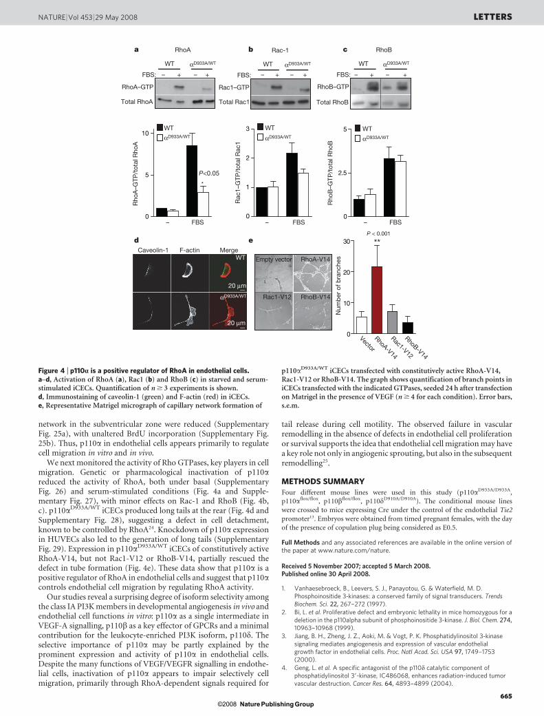
We next monitored the activity of Rho GTPases, key players in cellmigration. Genetic or pharmacological inactivation of p110areduced the activity of RhoA, both under basal (SupplementaryFig. 26) and serum-stimulated conditions (Fig. 4a and Supple-mentary Fig. 27), with minor effects on Rac-1 and RhoB (Fig. 4b,c). p110aD933A/WT iCECs produced long tails at the rear (Fig. 4d andSupplementary Fig. 28), suggesting a defect in cell detachment,known to be controlled by RhoA24. Knockdown of p110a expressionin HUVECs also led to the generation of long tails (SupplementaryFig. 29). Expression in p110aD933A/WT iCECs of constitutively activeRhoA-V14, but not Rac1-V12 or RhoB-V14, partially rescued thedefect in tube formation (Fig. 4e). These data show that p110a is apositive regulator of RhoA in endothelial cells and suggest that p110acontrols endothelial cell migration by regulating RhoA activity.
Our studies reveal a surprising degree of isoform selectivity amongthe class IA PI3K members in developmental angiogenesis in vivo andendothelial cell functions in vitro: p110a as a single intermediate inVEGF-A signalling, p110b as a key effector of GPCRs and a minimalcontribution for the leukocyte-enriched PI3K isoform, p110d. Theselective importance of p110a may be partly explained by theprominent expression and activity of p110a in endothelial cells.Despite the many functions of VEGF/VEGFR signalling in endothe-lial cells, inactivation of p110a appears to impair selectively cellmigration, primarily through RhoA-dependent signals required for
tail release during cell motility. The observed failure in vascularremodelling in the absence of defects in endothelial cell proliferationor survival supports the idea that endothelial cell migration may havea key role not only in angiogenic sprouting, but also in the subsequentremodelling25.
METHODS SUMMARY
Four different mouse lines were used in this study (p110aD933A/D933A,
p110aflox/flox, p110bflox/flox, p110dD910A/D910A). The conditional mouse lines
were crossed to mice expressing Cre under the control of the endothelial Tie2
promoter13. Embryos were obtained from timed pregnant females, with the day
of the presence of copulation plug being considered as E0.5.
Full Methods and any associated references are available in the online version ofthe paper at www.nature.com/nature.
Received 5 November 2007; accepted 5 March 2008.Published online 30 April 2008.
1. Vanhaesebroeck, B., Leevers, S. J., Panayotou, G. & Waterfield, M. D.Phosphoinositide 3-kinases: a conserved family of signal transducers. TrendsBiochem. Sci. 22, 267–272 (1997).
2. Bi, L. et al. Proliferative defect and embryonic lethality in mice homozygous for adeletion in the p110alpha subunit of phosphoinositide 3-kinase. J. Biol. Chem. 274,10963–10968 (1999).
3. Jiang, B. H., Zheng, J. Z., Aoki, M. & Vogt, P. K. Phosphatidylinositol 3-kinasesignaling mediates angiogenesis and expression of vascular endothelialgrowth factor in endothelial cells. Proc. Natl Acad. Sci. USA 97, 1749–1753(2000).
4. Geng, L. et al. A specific antagonist of the p110d catalytic component ofphosphatidylinositol 39-kinase, IC486068, enhances radiation-induced tumorvascular destruction. Cancer Res. 64, 4893–4899 (2004).
RhoB–GTP
Total RhoB
WT αD933A/WT
0
2.5
5 WT
RhoB
– –+ +FBS:
– FBS
a
0
5
10
*
Rho
A–G
TP/t
otal
Rho
A
RhoA–GTP
Total RhoA
WT αD933A/WT
– FBS
WT
αD933A/WT
b
dCaveolin-1 F-actin Merge
WT
αD933A/WT
Empty vector
RhoB-V14Rac1-V12
RhoA-V14
c
e
20 µm
1
3
+
0
2
Rac1–GTP
Total Rac1
– FBS
WT αD933A/WT
WT
– + –FBS:
Rac-1
– –+ +FBS:
RhoA
P<0.05
**
Num
ber
of b
ranc
hes
Vector
RhoA-V14
RhoB-V14
Rac1-V12
P < 0.001
0
10
20
30
αD933A/WT αD933A/WT
Rac
1–G
TP/t
otal
Rac
1
Rho
B–G
TP/t
otal
Rho
B20 µm
Figure 4 | p110a is a positive regulator of RhoA in endothelial cells.a–d, Activation of RhoA (a), Rac1 (b) and RhoB (c) in starved and serum-stimulated iCECs. Quantification of n $ 3 experiments is shown.d, Immunostaining of caveolin-1 (green) and F-actin (red) in iCECs.e, Representative Matrigel micrograph of capillary network formation of
p110aD933A/WT iCECs transfected with constitutively active RhoA-V14,Rac1-V12 or RhoB-V14. The graph shows quantification of branch points iniCECs transfected with the indicated GTPases, seeded 24 h after transfectionon Matrigel in the presence of VEGF (n $ 4 for each condition). Error bars,s.e.m.
NATURE | Vol 453 | 29 May 2008 LETTERS
665Nature Publishing Group©2008

5. Puri, K. D. et al. Mechanisms and implications of phosphoinositide 3-kinased inpromoting neutrophil trafficking into inflamed tissue. Blood 103, 3448–3456(2004).
6. Vanhaesebroeck, B. et al. P110delta, a novel phosphoinositide 3-kinase inleukocytes. Proc. Natl Acad. Sci. USA 94, 4330–4335 (1997).
7. Chantry, D. et al. p110delta, a novel phosphatidylinositol 3-kinase catalytic subunitthat associates with p85 and is expressed predominantly in leukocytes. J. Biol.Chem. 272, 19236–19241 (1997).
8. Sawyer, C. et al. Regulation of breast cancer cell chemotaxis by thephosphoinositide 3-kinase p110d. Cancer Res. 63, 1667–1675 (2003).
9. Lelievre, E. et al. Deficiency in the p110alpha subunit of PI3K results in diminishedTie2 expression and Tie2(2/2)-like vascular defects in mice. Blood 105,3935–3938 (2005).
10. Ueki, K., Algenstaedt, P., Mauvais-Jarvis, F. & Kahn, C. R. Positive and negativeregulation of phosphoinositide 3-kinase-dependent signaling pathways by threedifferent gene products of the p85a regulatory subunit. Mol. Cell. Biol. 20,8035–8046 (2000).
11. Foukas, L. C. et al. Critical role for the p110a phosphoinositide-3-OH kinase ingrowth and metabolic regulation. Nature 441, 366–370 (2006).
12. Lucitti, J. L. et al. Vascular remodeling of the mouse yolk sac requireshemodynamic force. Development 134, 3317–3326 (2007).
13. Kisanuki, Y. Y. et al. Tie2-Cre transgenic mice: a new model for endothelial cell-lineage analysis in vivo. Dev. Biol. 230, 230–242 (2001).
14. Bi, L., Okabe, I., Bernard, D. J. & Nussbaum, R. L. Early embryonic lethality in micedeficient in the p110b catalytic subunit of PI 3-kinase. Mamm. Genome 13, 169–172(2002).
15. Guillermet-Guibert, J. et al. The p110b isoform of phosphoinositide 3-kinasesignals downstream of G protein-coupled receptors and is functionally redundantwith p110c. Proc. Natl Acad. Sci. USA (in the press).
16. Okkenhaug, K. et al. Impaired B and T cell antigen receptor signaling in p110d PI3-kinase mutant mice. Science 297, 1031–1034 (2002).
17. Dayanir, V., Meyer, R. D., Lashkari, K. & Rahimi, N. Identification of tyrosineresidues in vascular endothelial growth factor receptor-2/FLK-1 involved inactivation of phosphatidylinositol 3-kinase and cell proliferation. J. Biol. Chem.276, 17686–17692 (2001).
18. Gille, H. et al. A repressor sequence in the juxtamembrane domain of Flt-1(VEGFR-1) constitutively inhibits vascular endothelial growth factor-dependentphosphatidylinositol 39-kinase activation and endothelial cell migration. EMBO J.19, 4064–4073 (2000).
19. Gerber, H. P., Dixit, V. & Ferrara, N. Vascular endothelial growth factor inducesexpression of the antiapoptotic proteins Bcl-2 and A1 in vascular endothelial cells.J. Biol. Chem. 273, 13313–13316 (1998).
20. Adini, I. et al. RhoB controls Akt trafficking and stage-specific survival ofendothelial cells during vascular development. Genes Dev. 17, 2721–2732(2003).
21. Gerhardt, H. et al. VEGF guides angiogenic sprouting utilizing endothelial tip cellfilopodia. J. Cell Biol. 161, 1163–1177 (2003).
22. Ruhrberg, C. et al. Spatially restricted patterning cues provided by heparin-bindingVEGF-A control blood vessel branching morphogenesis. Genes Dev. 16,2684–2698 (2002).
23. Gerhardt, H. & Betsholtz, C. How do endothelial cells orientate? EXS 94, 3–15(2005).
24. Worthylake, R. A., Lemoine, S., Watson, J. M. & Burridge, K. RhoA is required formonocyte tail retraction during transendothelial migration. J. Cell Biol. 154,147–160 (2001).
25. Hughes, S. & Chan-Ling, T. Roles of endothelial cell migration and apoptosis invascular remodeling during development of the central nervous system.Microcirculation 7, 317–333 (2000).
Supplementary Information is linked to the online version of the paper atwww.nature.com/nature.
Acknowledgements We thank F. Ramadani and K. Okkenhaug (BabrahamInstitute, Cambridge), E. Cernuda (Hospital Universitario Central de Asturias),T. Makinen (Cancer Research UK London Research Institute), K. Hodivala-Dilke,A. Reynolds and G. D’Amico (Institute of Cancer, Queen Mary, University ofLondon), P. Villalonga (Universitat de les Illes Balears, Spain) and members of theVanhaesebroeck laboratory (especially N. Osborne, C. See and M. Whitehead) forhelp and advice, E. Wagner (Research Institute of Molecular Pathology, Vienna),E. Dejana (Institute of Molecular Oncology, Milan), G. Balconi (Mario NegriInstitute for Pharmacological Research, Milan), M. Yanagisawa (University ofTexas Southwestern Medical Center, Dallas), D. Vestweber (Max-Planck Institute,Muenster), C. Rommel, M. Camps and T. Ruckle (Merck-Serono, Geneva) andPiramed (Slough, UK) for mice and reagents. Personal support was from EMBO(M.G., J.G.-G.), Cancer Research UK (M.G.) and the Fondation pour la RechercheMedicale and the European Union Marie Curie (J.G.-G.). Work in theVanhaesebroeck laboratory was supported by the Ludwig Institute for CancerResearch Institute, the Biotechnology and Biological Sciences Research Council(BB/C505659/1), the Association for International Cancer Research, EuropeanUnion (FP6-502935), Cancer Research UK and Barts and the London Charity. R.J.C.is supported by an Association for International Cancer Research grant to A.J.R.(07-0173). L.-K.P. and H.G. are supported by Cancer Research UK.
Author Contributions All authors designed research and analysed data. M.G.,J.G.-G., L.C.F., L.-K.P., R.J.C., A.S., W.P., S.M. and P.R.C. performed research. M.G.,H.G. and B.V. wrote the paper.
Author Information Reprints and permissions information is available atwww.nature.com/reprints. The authors declare competing financial interests:details accompany the full-text HTML version of the paper at www.nature.com/nature. Correspondence and requests for materials should be addressed to H.G.([email protected]) or B.V. ([email protected]).
LETTERS NATURE | Vol 453 | 29 May 2008
666Nature Publishing Group©2008

METHODSReagents. VEGF165 (referred to as VEGF-A in the text), SDF-1a, IL8 and S1P
were from Preprotech. HUVECs and EBM-2 medium were obtained from
Lonza. Catalogue numbers are shown between brackets. Antibodies to Akt/
PKB (9272), pT308-Akt (4056), pS473-Akt (9271) and caspase-3 (9664) were
from Cell Signalling Technologies. Antibodies to CD31 (550274), paxillin
(610051) and CD105 (550546) were from BD Pharmingen. Antibodies to
p110a were either monoclonal clone U3A or rabbit anti-p110a (made in house).
Antibodies to p110d were also made in house. Anti-pan-p85 (06-195), anti-pTyr
(4G10, catalogue number 16-101) and anti-Rac (05-389) were from Upstate.
Antibodies to p110b (sc-602), VEGFR-2 (sc-504), pErk1/2 (sc-7383), caveolin-1
(sc-894), RhoB (sc-180) and RhoA (sc-418) were from Santa Cruz
Biotechnology. Anti-isolectin B4 (L-2140), anti-laminin, anti-tubulin (T5168)
and anti-b-actin (A5441), phalloidin-TRITC (P1951) and all chemicals, unless
otherwise stated, were from Sigma-Aldrich. BrdU, anti-BrdU Alexa Fluor 488,
isolectin IB4 Alexa Fluor 568 and secondary antibodies conjugated to Alexa-488,
and Alexa-594 were from Molecular Probes. Anti-NG2 (AB5320) was from
Chemicon International. Rat anti-endomucin mouse monoclonal antibody
was provided by D. Vestweber. Secondary antibodies conjugated to fluorescein
isothiocyanate (FITC), Cy5 and TRITC were from Jackson ImmunoResearch
Labs.
Whole-mount embryo immunofluorescence. Freshly isolated embryos were
fixed overnight in 4% paraformaldehyde at 4 uC, washed in PBT (PBS, 0.1%
Tween 20) and subjected to dehydration in increasing methanol concentration
(50%, 80% methanol/PBT, 100% methanol) followed by rehydration in decreas-
ing methanol concentration (80%, 50% methanol/PBT, PBT). After washing in
Pblec (PBS pH 6.8, 1% Tween 20, 1 mM CaCl2, 1 mM MgCl2, 0.1 mM MnCl2),
the embryos were incubated overnight at 4 uC in the presence of rat anti-mouse
endomucin diluted 1:20 in Pblec. After five washes in PBT, embryos were incu-
bated overnight at 4 uC with goat anti-rat Alexa 568 diluted 1:100 in PBS, 0.5%
BSA and 0.25% Tween 20, followed by washing in PBT and post-fixation in 4%
parformaldehyde (PFA) before analysis. Confocal laser scanning microscopy was
performed with an LSM 510 (Zeiss) mounted over an Axioplan microscope
(Zeiss) using a 310 objective.
PI3K isoform expression and lipid kinase assay. PBS was perfused through the
heart of terminally anaesthetized mice, followed by harvesting and freezing of the
tissues in liquid nitrogen. PI3K isoform expression and lipid kinase assays were
performed as previously described26. Three independent samples for each geno-
type were used.
Aortic ring assay. Thoracic aortic rings (1 mm; n $ 6 for each genotype and
condition) were embedded on top of 50-ml rat collagen R from rat tail (Serva)
and overlaid with 100ml of OptiMEM (GIBCO), with or without VEGF-A
(30 ng ml21), SDF-1a (30 ng ml21), IL8 (30 ng ml21) or S1P (30 ng ml21).
Microvessel outgrowth was visualized by time-lapse phase contrast microscopy,
and the number of vessels growing from each aortic ring was counted on day 8.
Representative images were taken using a KPM1E/K-S10 CCD camera (Hitachi
Denshi). For PI3K inhibitor studies, aortic rings were incubated with inhibitors
or vehicle for the whole duration of the study. Medium was changed every other
day in the presence of fresh PI3K inhibitors. Quantification was performed as
shown in Supplementary Fig. 13b.
CEC isolation. CECs were isolated from 8- to 12-week-old mice as described27.
Three independent cell lines were derived for each genotype. Briefly, hearts were
aseptically removed, diced and digested with collagenase A (Roche). CECs were
isolated by immunoselection using anti-CD105 (MJ7/18 monoclonal antibody)-
conjugated beads, followed by plating of the cell suspension on 0.1% gelatin-
coated dishes in DMEM (Invitrogen) supplemented with 10% fetal bovine
serum (Helena Biosciences), 10 U ml21 heparin and 50 mg ml21 endothelial cell
growth factor. Two days later, the cells were infected for 4 h with retrovirus
encoding polyoma middle T antigen. After overnight incubation in endothelial
cell culture medium, a second 4 h round of infection with polyoma middle T
antigen retrovirus was performed. Cultures were then washed, incubated for 2
weeks, and cells subjected to a second round of immuno-isolation as described
above. Expression of various markers was assessed by PCR, or by fluorescence-
activated cell sorting in the case of CD105. CECs of both genotypes were found to
be positive for VEGFR1, VEGFR2, TIE1, TIE2, VE-cadherin, CD34 and CD31
(data not shown). Primary CEC isolation was as described above, with a single
positive selection and assessment of CD105 expression by immunofluorescence.
Stimulation of pCECs with soluble ligands. For stimulation, early passage (P4)
pCECs were seeded in 6 cm dishes (2.5 3 105 cells per dish), starved in DMEM
without fetal bovine serum (FBS) and antibiotics for 24 h, followed by stimu-
lation with VEGF (50 ng ml21 and 100 ng ml21), SDF-1a (30 ng ml21) or FBS
(10%) for 10 min, unless stated otherwise. Quantification of at least three inde-
pendent experiments was performed.
In vitro endothelial tube formation assay. Growth-factor-reduced Matrigel
matrix (BD Pharmingen, catalogue number 354230) was plated evenly in a 12-
well plate (150ml per well) and incubated at 37 uC for 30 min before adding
75,000 CECs per well. Cells were overlaid with 2% serum medium with or
without 50 ng ml21 VEGF-A. Sixteen hours after seeding, photographs of
representative 310 fields were taken (n $ 5 for each condition and genotype),
and endothelial tubes were quantified by counting branches.
In vivo stimulation with VEGF-A. Saline or mouse VEGF-A (0.2mg g21) was
injected into the tail vein of 8- to 12-week-old mice (n $ 6 for each genotype and
condition), followed 5 min later by harvesting and freezing of the tissues in liquid
nitrogen, as described28. Tissues were homogenized and re-suspended in differ-
ent buffers according to the application. After removal of insoluble material by
centrifugation at 12,000g for 10 min at 4 uC, 100mg cell lysate was analysed by
western blot analysis, or 1 mg of lysate used to immunoprecipitate pTyr followed
by western blot analysis. The Akt activity assay was performed as described29.
Proliferation assay. Immortalized CECs and primary CECs were incubated at
105 and 2 3 105 cells, respectively, per well in 96-well plate, deprived of FBS for
16 h, followed by stimulation with or without 50 ng ml21 VEGF in medium
(DMEM, 2% FBS, penicillin/streptomycin) containing [3H]thymidine.
Twenty-four hours later, the cells were harvested and [3H]thymidine incorpo-
rated into DNA measured by scintillation counting.
Cell viability assays. Cells, grown at 5 3 105 cells per 10-cm dish for 24 h, were
starved of FBS for 20 h, followed by analysis of cell viability by flow cytometry
using the Annexin V-FITC apoptosis kit I (BD Pharmingen).
Caspase activity assay. Cells, grown at 3 3 105 cells per 10-cm dish for 24 h, were
starved of FBS for 20 h, followed by executioner caspase activity measurement
using the Quantipak kit (Biomol). This was performed on 50mg cytosolic cell
extracts using 200mM of the caspase-3 chromogenic substrate Z-Asp-Glu-Val-
Asp-para-nitroaniline (DEVD-pNA), according to the manufacturer’s instruc-
tions. Samples containing no cell extract were used as negative controls.
Time-lapse microscopy. To study cell migration, CECs were seeded on 35-mm
dishes in DMEM growth medium, followed 1 h later by filming of the cells for
12 h at 37 uC. Images were collected with a KPM1E/K-S10 CCD camera (Hitachi
Denshi) every 10 min using Tempus software (Kinetic Imaging Ltd, now Andor
Technology). Time-lapse images from the whole duration of the experiment
were then displayed as a movie, and cells from each frame tracked using
Motion Analysis software (Andor/Kinetic Imaging). The mean migration speed
of the population was determined using Mathematica 6.0 workbooks30.
Scratch wound assay. iCECs or HUVECs 24 h after transfection were seeded at
high density in DMEM growth medium and EBM-2 medium, respectively, fol-
lowed one day later by wounding of the confluent monolayer by scraping a
pipette tip across.
siRNA knockdown experiments. HUVECs (passage 2; Lonza) were plated in
six-well dishes at 1 3 105 cells per well, 24 h before transfection. Four siRNA
oligonucleotides (Dharmacon) targeting the human p110a catalytic subunit,
were used individually or in combination: D-003018-5 (sense sequence (GCUA-
UCAUCUGAACAAUUAUU), antisense sequence (59P UAAUUGUUCAGA-
UGAUAGCUU)), D-003018-6 (sense sequence (GGAUAGAGGCCAAAUAA-
UAUU), antisense sequence (59P UAUUAUUUGGCCUCUAUCCUU)),
D-003018-7 (sense sequence (GGACAACUGUUUCAUAUAGUU), antisense
sequence (59P CUAUAUGAAACAGUUGUCCUU)) and D-003018-8 (sense
sequence (GCCAGUACCUCAUGGAUUAUU), antisense sequence (59PUAAUCCAUGAGGUACUGGCUU)).
The siRNA pool gave the strongest knockdown (data not shown) and was used
in all subsequent experiments. Cells were transfected with Oligofectamine
(Invitrogen), and after 24 h cells were trypsinized and plated at sub-confluence
and allowed to adhere (8 h) on fibronectin-coated coverslips for immunofluo-
rescence analysis (5 3 104 cells per well) or at confluence in 24-well plates for
time-lapse microscopy or lysis and western blot analysis (105 cells per well). Cell
proliferation was analysed by Casy Counter analysis 24, 48 and 72 h after trans-
fection. For cell viability analysis, cells were trypsinized 48 h after transfection,
followed by staining with propidium iodide. Samples were analysed by flow
cytometry.
Retinal angiogenesis. Postnatal P5 eyes (n $ 4 for each genotype) were fixed in4% PFA in PBS at 4 uC overnight and washed in PBS. Retinas were dissected,
stained with biotinylated isolectin-B4 and flat-mounted as previously described
or processed for multiple labelling21.
Endothelial cell proliferation in vivo. BrdU was injected at 50 mg kg21 body
weight intraperitoneally into P5 pups 2 h before harvesting of the eyes. P5 eyes
were fixed overnight in 4% PFA in PBS at 4 uC and washed in PBS. Retinas were
dissected, incubated with proteinase K (10mg ml21 in PBT) for 30 min, followed
by 10 min in glycine (2 mg ml21 in PBT), two washes in PBT and post-fixation
for 20 min in 4% PFA/0.1% glutaraldehyde in PBT. After washing in PBT,
samples were equilibrated in DNase I buffer (50 mM Tris.HCl, 10 mM MgCl2
doi:10.1038/nature06892
Nature Publishing Group©2008

at pH 7.5) and incubated for 2 h in DNase I (0.1 U ml21) at 37 uC. DNase washeat-inactivated for 10 min at 70 uC in 50 mM Tris-HCl (pH 7.5), followed by
washes in ice-cold Tris-HCl. After two washes in PBT, unspecific binding of
antibodies was blocked by 1% BSA/0.5% Tween 20 in PBS for 30 min. Samples
were labelled using isolectin-B4 and mouse anti-BrdU as described above.
Samples were flat-mounted and cover-slipped using Moviol.
Hindbrain angiogenesis. E11.5 hindbrains (n $ 5 for each genotype), dissected
clean from surrounding tissue, were fixed in 4% formaldehyde in PBS for 2 h on
ice, permeabilized by two washes in PBS containing 0.1% Triton X-100 (PBX),
incubated for 5 min in 50% methanol/PBX then incubated in methanol. Samples
were rehydrated for 30 min at room temperature, stained with CD31 and flat-
mounted as previously described. Littermate samples were photographed at the
same magnification. For each genotype, the average number of branch points
was determined in five randomly chosen 500mm2 regions22.
BrdU incorporation. E11.5 timed pregnant females were injected intraperito-
neally with 50 mg kg21 body weight BrdU in PBS and killed 2 h later. Dissected
embryonic hindbrains were processed for immunostaining as described above,
except they were incubated for 30 min in 4 N HCl, followed by neutralization in
100 mM sodium tetraborate for 5 min before the staining procedure. Sampleswere labelled using isolectin-B4 and mouse anti-BrdU as described above.
Samples were flat-mounted and cover-slipped using Moviol. The analysis was
performed in four or five hindbrains per genotype.
Measurement of Rho activity. The capacity of Rac1–GTP to bind GST–PBD
(p21-activated kinase-binding domain), or RhoB–GTP and RhoA–GTP to bind
to GST–rhotekin–RBD, was used to determine the cellular levels of active
GTPases. Rho/Rac–GTP pull-down assays were performed by incubation of
cleared lysates with glutathione-Sepharose 4B beads coupled to GST–PBD or
GST–rhotekin–RBD (Upstate Biotechnology) for 1 h at 4 uC, followed by four
washes of the beads in extraction buffer (50 mM Tris-HCl pH 7.5, 1 mM EDTA,
500 mM NaCl, 10 mM MgCl2, 1% Triton X-100, 0.5% sodium deoxycholate,
0.1% SDS, 10% glycerol, 0.5% 2-mercaptoethanol). Bound proteins were solu-
bilized by the addition of 25 ml of SDS–polyacrylamide gel electrophoresis (SDS–
PAGE) Laemmli loading buffer, followed by separation on 12.5% SDS–PAGE
gels and western blotting for Rac, RhoB or RhoA31.
Immunofluorescence. Cells grown on coverslips were fixed with 4% para-
formaldehyde for 20 min, blocked with TBS containing 1% BSA (25 mM
Tris.HCl pH 7.4, 150 mM NaCl) for 10 min, permeabilized for 5 min with PBScontaining 0.2% Triton X-100 at 4 uC, and incubated at 37 uC for 30 min with
antibodies or 1 mg ml21 TRITC-labelled phalloidin. Specimens were mounted in
DAKO fluorescent mounting medium (Ely). Confocal laser scanning micro-
scopy was performed with an LSM 510 (Zeiss) mounted over an Axioplan
microscope (Zeiss) using a 340 1.3 NA oil immersion objective.
Transient transfection. p110aD933A/WT iCECs were transfected using
Lipofectamine 2000 (GIBCO) according to the manufacturer’s instructions.
Plasmids used were pCMV5–myc–Rac1–V12, pCMV5–Flag–RhoA–V14 and
pCMV5–HA–RhoB–V14 or empty vector (pCMV-Myc; Clontech, catalogue
number K6003-1). Twenty-four hours after transfection, the cells were trypsin-
ized and seeded on Matrigel for analysis of tube formation as described above.
Statistical analysis. Means were compared between two groups using the non-
parametric Mann–Whitney U-test. For a three-group comparison, the analysis
of variance (ANOVA) test was used.
26. Bilancio, A. et al. Key role of the p110delta isoform of PI3K in B-cell antigen and IL-4 receptor signaling: comparative analysis of genetic and pharmacologicinterference with p110d function in B cells. Blood 107, 642–650 (2006).
27. Lidington, E. A. et al. Conditional immortalization of growth factor-responsivecardiac endothelial cells from H-2K(b)-tsA58 mice. Am. J. Physiol. Cell Physiol.282, C67–C74 (2002).
28. Weis, S. et al. Src blockade stabilizes a Flk/cadherin complex, reducing edema andtissue injury following myocardial infarction. J. Clin. Invest. 113, 885–894 (2004).
29. Cutillas, P. R. et al. Ultrasensitive and absolute quantification of thephosphoinositide 3-kinase/Akt signal transduction pathway by massspectrometry. Proc. Natl Acad. Sci. USA 103, 8959–8964 (2006).
30. Wells, C. M. & Ridley, A. J. Analysis of cell migration using the Dunn chemotaxischamber and time-lapse microscopy. Methods Mol. Biol. 294, 31–41 (2005).
31. Ren, X. D. & Schwartz, M. A. Determination of GTP loading on Rho. MethodsEnzymol. 325, 264–272 (2000).
doi:10.1038/nature06892
Nature Publishing Group©2008