The Relationship between Gene Isoform Multiplicity, Number of Exons and Protein Divergence
14
The Relationship between Gene Isoform Multiplicity, Number of Exons and Protein Divergence Jordi Morata 1 , Santi Be ´ jar 1 , David Talavera 2. , Casandra Riera 3. , Sergio Lois 3 , Gemma Mas de Xaxars 4 , Xavier de la Cruz 1,3,5 * 1 Department of Structural Biology, Institut de Biologia Molecular de Barcelona (IBMB)-Consejo Superior de Investigaciones Cientı ´ficas (CSIC), Barcelona, Spain, 2 Faculty of Life Sciences, Manchester University, Manchester, United Kingdom, 3 Laboratory of Translational Bioinformatics in Neuroscience, Vall d’Hebron Institute of Research (VHIR), Barcelona, Spain, 4 Laboratori de Bota `nica, Facultat de Farma `cia, Universitat de Barcelona, Barcelona, Spain, 5 Institucio ´ Catalana de Recerca i Estudis Avanc ¸ats (ICREA), Barcelona, Spain Abstract At present we know that phenotypic differences between organisms arise from a variety of sources, like protein sequence divergence, regulatory sequence divergence, alternative splicing, etc. However, we do not have yet a complete view of how these sources are related. Here we address this problem, studying the relationship between protein divergence and the ability of genes to express multiple isoforms. We used three genome-wide datasets of human-mouse orthologs to study the relationship between isoform multiplicity co-occurrence between orthologs (the fact that two orthologs have more than one isoform) and protein divergence. In all cases our results showed that there was a monotonic dependence between these two properties. We could explain this relationship in terms of a more fundamental one, between exon number of the largest isoform and protein divergence. We found that this last relationship was present, although with variations, in other species (chimpanzee, cow, rat, chicken, zebrafish and fruit fly). In summary, we have identified a relationship between protein divergence and isoform multiplicity co-occurrence and explained its origin in terms of a simple gene-level property. Finally, we discuss the biological implications of these findings for our understanding of inter-species phenotypic differences. Citation: Morata J, Be ´jar S, Talavera D, Riera C, Lois S, et al. (2013) The Relationship between Gene Isoform Multiplicity, Number of Exons and Protein Divergence. PLoS ONE 8(8): e72742. doi:10.1371/journal.pone.0072742 Editor: Gil Ast, Tel Aviv University, Israel Received February 27, 2013; Accepted July 14, 2013; Published August 30, 2013 Copyright: ß 2013 Morata et al. This is an open-access article distributed under the terms of the Creative Commons Attribution License, which permits unrestricted use, distribution, and reproduction in any medium, provided the original author and source are credited. Funding: Work supported by the Spanish Ministerio de Economı ´a (gran number: BIO2012–40133) Spanish Ministerio de Educacio ´ n y Ciencia (grant number: BFU2009-11527); the Spanish Ministerio de Ciencia e Innovacio ´ n (grant number BIO2006–15557); and the Consejo Superior de Investigaciones Cientı ´ficas (CSIC) (grant number 200420E578). The funders had no role in study design, data collection and analysis, decision to publish, or preparation of the manuscript. Competing Interests: The authors have declared that no competing interests exist. * E-mail: [email protected] . These authors contributed equally to this work. Introduction Understanding the molecular basis of phenotypic differences (PheDif) between organisms is a fundamental problem in modern biology. A large body of evidence shows that changes in phenotype can arise from gene regulation and protein divergence [1–3]. Recent data show that this is also the case for other processes, particularly those leading to gene isoform multiplicity (IM), such as alternative splicing or alternative translation initiation and/or termination [1,2,4–7]. However, and in spite of its relevance, we still know little about the interplay between all these processes and how it leads to PheDif between species [1,3]. In the present work we address this problem in the case of human and mouse, focusing on the relationship between protein divergence and isoform multiplicity co-occurrence (IMco) (Figure 1). IM, a gene property reflecting the fact that genes can express more than one isoform, can result from alternative splicing of pre- mRNA or alternative translation initiation and/or termination, although it is generally accepted that alternative splicing is the most important source [8]. Alternative splicing has such a potential to sample protein function space [8–10] that it has been postulated as an important contributor to complexity differences between organisms [11,12]. The idea that variations in alternative splicing patterns play such a role is supported by a large number of studies relating alternative splicing and disease [13–15]. Also, at a more general level, this idea has been explored and tested by many researchers who have looked at differences on the amount of alternative splicing between organisms [5], checked the conserva- tion of alternative splicing events between species [7,16–28] and of AS structural/functional impact [29], explored the role of premature termination codons [30], differential regulation [31], etc. The results of these studies indeed point to a clear relationship between interspecies differences and the distribution of alternative splicing properties. Mechanistically different from alternative splicing, but related to it, alternative transcription initiation and termination are two additional sources of multiple gene isoforms [6,32]. They can result in proteins with different N- or C-terminal ends, or more substantial sequence changes [6,32], and may have functional properties similar to those of alternative splicing isoforms, e.g. behave as dominant-negatives or have new cellular locations [32]. Recent data indicate that the relevance of these mechanisms may be comparable to that of AS, and that both phenomena are tightly related [6,33]. PLOS ONE | www.plosone.org 1 August 2013 | Volume 8 | Issue 8 | e72742
-
Upload
manchester -
Category
Documents
-
view
4 -
download
0
Transcript of The Relationship between Gene Isoform Multiplicity, Number of Exons and Protein Divergence

The Relationship between Gene Isoform Multiplicity,Number of Exons and Protein DivergenceJordi Morata1, Santi Bejar1, David Talavera2., Casandra Riera3., Sergio Lois3, Gemma Mas de Xaxars4,
Xavier de la Cruz1,3,5*
1 Department of Structural Biology, Institut de Biologia Molecular de Barcelona (IBMB)-Consejo Superior de Investigaciones Cientıficas (CSIC), Barcelona, Spain, 2 Faculty of
Life Sciences, Manchester University, Manchester, United Kingdom, 3 Laboratory of Translational Bioinformatics in Neuroscience, Vall d’Hebron Institute of Research
(VHIR), Barcelona, Spain, 4 Laboratori de Botanica, Facultat de Farmacia, Universitat de Barcelona, Barcelona, Spain, 5 Institucio Catalana de Recerca i Estudis Avancats
(ICREA), Barcelona, Spain
Abstract
At present we know that phenotypic differences between organisms arise from a variety of sources, like protein sequencedivergence, regulatory sequence divergence, alternative splicing, etc. However, we do not have yet a complete view of howthese sources are related. Here we address this problem, studying the relationship between protein divergence and theability of genes to express multiple isoforms. We used three genome-wide datasets of human-mouse orthologs to study therelationship between isoform multiplicity co-occurrence between orthologs (the fact that two orthologs have more thanone isoform) and protein divergence. In all cases our results showed that there was a monotonic dependence betweenthese two properties. We could explain this relationship in terms of a more fundamental one, between exon number of thelargest isoform and protein divergence. We found that this last relationship was present, although with variations, in otherspecies (chimpanzee, cow, rat, chicken, zebrafish and fruit fly). In summary, we have identified a relationship betweenprotein divergence and isoform multiplicity co-occurrence and explained its origin in terms of a simple gene-level property.Finally, we discuss the biological implications of these findings for our understanding of inter-species phenotypicdifferences.
Citation: Morata J, Bejar S, Talavera D, Riera C, Lois S, et al. (2013) The Relationship between Gene Isoform Multiplicity, Number of Exons and ProteinDivergence. PLoS ONE 8(8): e72742. doi:10.1371/journal.pone.0072742
Editor: Gil Ast, Tel Aviv University, Israel
Received February 27, 2013; Accepted July 14, 2013; Published August 30, 2013
Copyright: � 2013 Morata et al. This is an open-access article distributed under the terms of the Creative Commons Attribution License, which permitsunrestricted use, distribution, and reproduction in any medium, provided the original author and source are credited.
Funding: Work supported by the Spanish Ministerio de Economıa (gran number: BIO2012–40133) Spanish Ministerio de Educacion y Ciencia (grant number:BFU2009-11527); the Spanish Ministerio de Ciencia e Innovacion (grant number BIO2006–15557); and the Consejo Superior de Investigaciones Cientıficas (CSIC)(grant number 200420E578). The funders had no role in study design, data collection and analysis, decision to publish, or preparation of the manuscript.
Competing Interests: The authors have declared that no competing interests exist.
* E-mail: [email protected]
. These authors contributed equally to this work.
Introduction
Understanding the molecular basis of phenotypic differences
(PheDif) between organisms is a fundamental problem in modern
biology. A large body of evidence shows that changes in phenotype
can arise from gene regulation and protein divergence [1–3].
Recent data show that this is also the case for other processes,
particularly those leading to gene isoform multiplicity (IM), such as
alternative splicing or alternative translation initiation and/or
termination [1,2,4–7]. However, and in spite of its relevance, we
still know little about the interplay between all these processes and
how it leads to PheDif between species [1,3]. In the present work
we address this problem in the case of human and mouse, focusing
on the relationship between protein divergence and isoform
multiplicity co-occurrence (IMco) (Figure 1).
IM, a gene property reflecting the fact that genes can express
more than one isoform, can result from alternative splicing of pre-
mRNA or alternative translation initiation and/or termination,
although it is generally accepted that alternative splicing is the
most important source [8]. Alternative splicing has such a
potential to sample protein function space [8–10] that it has been
postulated as an important contributor to complexity differences
between organisms [11,12]. The idea that variations in alternative
splicing patterns play such a role is supported by a large number of
studies relating alternative splicing and disease [13–15]. Also, at a
more general level, this idea has been explored and tested by many
researchers who have looked at differences on the amount of
alternative splicing between organisms [5], checked the conserva-
tion of alternative splicing events between species [7,16–28] and of
AS structural/functional impact [29], explored the role of
premature termination codons [30], differential regulation [31],
etc. The results of these studies indeed point to a clear relationship
between interspecies differences and the distribution of alternative
splicing properties.
Mechanistically different from alternative splicing, but related to
it, alternative transcription initiation and termination are two
additional sources of multiple gene isoforms [6,32]. They can
result in proteins with different N- or C-terminal ends, or more
substantial sequence changes [6,32], and may have functional
properties similar to those of alternative splicing isoforms, e.g.
behave as dominant-negatives or have new cellular locations [32].
Recent data indicate that the relevance of these mechanisms may
be comparable to that of AS, and that both phenomena are tightly
related [6,33].
PLOS ONE | www.plosone.org 1 August 2013 | Volume 8 | Issue 8 | e72742

As mentioned before, we still do not know the relationship
between protein divergence and IM. Are they independent? Do
they covary? And if so, in which manner? Answering these
questions would constitute an important advance towards
understanding the molecular basis of inter-species PheDif. Here
we address this problem by studying how IM co-occurrence
(IMco) between human-mouse orthologs (Figure 1; see Materials
and Methods) varies as a function of protein divergence (PD). Note
that instead of using IM co-occurrence we could have used
another IM-related property, like isoform number or nature, or
conservation of IM signals at the gene level. However, there is an
already large number of studies addressing these problems [7,16–
29,34–41].
We have divided our work in three parts. First, description of
the IMco-PD relationship for the human-mouse case. Second,
explanation of this relationship in terms of a more fundamental
relationship, between number of exons of the largest isoform and
PD. Third, given the interest of this last relationship, we explored
whether it was present in other species.
Our results show (Figure 2) that IMco and PD are monoton-
ically related for human-mouse orthologs. This observation could
be rationalized combining two facts: (i) the connection between IM
and the number of exons of the largest isoform, a fact expected
from previous work [42–44]; and the relationship between number
of exons of the largest isoform and PD, an unexpected fact. Finally,
we find that this second relationship is also present in other species,
although with some variations (the most extreme being for fruit fly,
for which the monotonic trend was reversed). In the Discussion
section, we first explore the origin of these relationships; then, for
the human-mouse case, we propose how the IMco-PD relationship
can be used to understand the relative contribution of PD and
IMco to the generation of PheDif between these species.
Materials and Methods
The Annotated Ortholog DatasetsIn this work we have used ortholog sets to study the IMco-PD
relationship in human-mouse, to trace its origin, and to explore the
relationship between number of exons and PD in different species.
We therefore had to obtain a set of gene orthologs for each
human-species case, as well as transcript annotations for all the
genes. We describe below how this was done. Note that we treat
separately the human-mouse case, which constituted the core of
this work, because more data were available for its study.
1. Human-mouse. We produced three datasets of annotated
orthologs, to ensure the robustness of our results.
Datasets 1 and 2. Here the orthology relationships were
obtained with InParanoid [45], using as starting point a set of
human and mouse sequences obtained by combining data from
two manually curated databases, UniProt/SwissProt [46,47] and
RefSeq [48], following a five-step protocol (we explain it for
human only, the same steps were applied to mouse). First, we
obtained all human genes from UniProt/SwissProt (release 54.7)
and RefSeq (release 27). Second, we established the equivalence
between the two versions of the same gene using the GeneId
identifier. Third, we eliminated those genes for which sequence
conflicts were more than expected by random; this was done
computing the frequency of sequence conflicts from UniProt/
SwissProt sequence data, and using it in a Poisson model for the
distribution of sequence conflicts (analogous to that described in
Altmann et al. [49]) to exclude those genes for which the number
of conflicts had a probability lower than 0.05 (this was done for
UniProt/SwissProt data only). Fourth, we eliminated those
isoforms for which the RefSeq record started by either XP_ or
ZP_. Fifth, from the final set of human and mouse genes we
eliminated those cases for which no correspondence could be
established between UniProt/SwissProt and Ensembl records (this
was required to use isoform annotations from Ensembl).
We used the longest isoform for each gene to find the orthology
relationships between human and mouse. We eliminated those
cases for which InParanoid [45] found no orthologs in one of the
species, or for which no unique human-mouse orthology
relationship was available. At the end of this process we obtained
a set of 11969 pairs of human-mouse orthologs that was the
starting point for the subsequent analyses.
Transcript annotations for the genes in this list defined the
differences between dataset 1 and 2. For dataset 1 they were
obtained from Ensembl [50] (version 65, GRCh37.p5 and
NCBIM37 for human and mouse respectively); for dataset 2 they
were obtained from VEGA [51] (version 45, GRCh37.p5 and
NCBIM37 for human and mouse respectively), without applying
any filtering in neither case.
Figure 1. Description of isoform multiplicity co-occurrence (IMco). The figure illustrates how IMco is computed. We start from a set ofhuman-mouse orthologs for which isoform annotations are available in ENSEMBL (or VEGA), as shown in (A). We will say that one gene has isoformmultiplicity (IM) when it has more than one isoform. The scheme shows different instances that cover all possible IM combinations for human-mouseortholog pairs: both orthologs have IM, only one has IM, and no ortholog has IM. (B) IMco will correspond to the fraction of cases for which bothorthologs have IM; it is computed as shown in the table.doi:10.1371/journal.pone.0072742.g001
Isoform Multiplicity and Protein Divergence
PLOS ONE | www.plosone.org 2 August 2013 | Volume 8 | Issue 8 | e72742

In summary, we obtained two annotated sets human-mouse
orthologs (11969 pairs for dataset 1 and 6795 pairs for dataset 2,
numbers resulting from matching Uniprot/SwissProt records and
Ensembl and VEGA records, respectively), with a fraction of IM
of: 86% (human) and 72% (mouse), dataset 1; and 85% (human)
and 84% (mouse), dataset 2. These datasets were used to produce
the results in Figures 2A (dataset 1) and 2B (dataset 2).
Dataset 3. The list of human-mouse orthologs was obtained
with EnsemblCompara [52]. We used only Ensembl (version 69)
data for genes and transcripts, imposing that both had to be
labelled as KNOWN.
In summary, we obtained one annotated set of 15134 human-
mouse ortholog pairs, with a fraction of IM of 80% (human) and
54% (mouse). This dataset was used to produce the results in
Figure 2C.
2. IM-annotated gene sets for other species. For a set of 6
species (chimpanzee, cow, rat, chicken, zebrafish and fruit fly) we
characterized the relationship between number of exons of the
longest species and PD. We used only Ensembl (version 69) [50]
data for genes and transcripts, additionally imposing that both had
to be labelled as KNOWN. The lists of orthologs were obtained
with the standard program, EnsemblCompara [52]. The statistics
of the resulting datasets are summarized in Table 1.
In all cases, including human and mouse, exon annotations
were obtained from Ensembl.
IM co-occurrence (IMco)IMco, explained in Figure 1, was defined as the fraction of pairs
for which both orthologs had more than one isoform. We
represented this fraction as a function of the percentage of
sequence identity between ortholog proteins, which is a measure of
PD (see section ‘‘Computation of PD’’ below). IMco can also be
expressed in a more formal view as a probability, P(IMH, IMM|x)
(Note: to express joint events we use commas instead of >, as in
Bishop [53], e.g.):
P(IMH ,IMM Dx)~P IMH~YESð Þ , IMM~YESð Þ½ �Dpsi~xf gð1Þ
where IMH is an indicator variable with two values: YES, when
human orthologs have more than one isoform, and NO, when
they only have one. IMM is the mouse equivalent of IMH. psi is a
variable that corresponds to protein sequence identity, and x to its
actual value. Note: here we use the human-mouse case to explain
IMco, but an exactly analogous definition applies to other cases.
In summary, IMco is a similarity measure between sets of
human-mouse paired orthologs. It varies between 0 and 1; 0
happens when there is no ortholog pair with IM for both genes, 1
happens when all ortholog pairs have IM for both genes.
Using basic probability results [54] we can express P(IMH,
IMM|x) (Equation 1) in terms of the product of the two species-
specific IM, P(IMH|x) and P(IMM|x):
Figure 2. Relationship between isoform multiplicity co-occurrence (IMco) and protein divergence (PD). In (A), (B) and (C) we plot IMco(the fraction of (IM, IM) ortholog pairs, see Figure 1 and Materials and Methods) as a function of PD (measured using the percentage of sequenceidentity). Black dots are used to display the raw, unprocessed data; a blue line is used for the smoothed data and in grey we show the envelope (seeResults section). In both cases we can see that there is a monotonically increasing relationship between IMco and PD (see text). Outliers from thistrend define two lines, one at 0 and the other at 1; these outliers result from IMco estimates obtained with less than 5 observations. They essentiallydisappear when eliminating these poorer estimates (Figure S1). (A) and (B) where obtained with a Uniprot/SwissProt/RefSeq-based sequence datasetand Ensembl and VEGA isoform annotations, respectively; (C) was obtained using Ensembl data for both genes and transcripts. The monotonic trendis comparable in the three figures, although in (C) the curve shows a slight shift towards lower IMco values resulting from lower amount of geneswith IM.doi:10.1371/journal.pone.0072742.g002
Table 1. Statistics of the datasets used in this work: number of ortholog pairs and percentage of genes with IM (the latter refersonly to the model species, not to human).
Species Human Chimpanzee Human Cow Human Mouse Human Rat Human Chicken Human Zebrafish Human Fruit fly
Number of orthologpairs
15954 10790 15134 14150 4238 6714 3108
% genes with IM(non-human species)
5.0 8.5 54.2 27.4 37.1 34.0 31.0
doi:10.1371/journal.pone.0072742.t001
Isoform Multiplicity and Protein Divergence
PLOS ONE | www.plosone.org 3 August 2013 | Volume 8 | Issue 8 | e72742

P IMH ,IMH Dxð Þ~P IMH Dxð Þ:P IMM Dxð Þ:QHM ð2Þ
where QHM={[P(IMH|IMM,x)?P(IMM|IMH,x)]/[P(IMH|x)?tP(IMM|x)]}1/2.
If the contribution of IMH and IMM to IMco is independent,
then QHM = 1 and P(IMH, IMM|x) = P(IMH|x)?P(IMM|x). We
used Equation 2 as a starting point to explore the relationship
between P(IMH, IMM|x) and gene properties.
Computation of PDAs a measure of PD we used the percentage of sequence identity
between ortholog proteins. It was computed after global sequence
alignment (using a standard dynamic programming algorithm
[55]) of the longest isoform from each species, and was equal to the
number of identical residue pairs (nid) in the alignment divided by
the average of the human (nhuman) and mouse (nmouse) protein
lengths: 2.nid/(nhuman+nmouse). To avoid any confusion in the
interpretation of our results, it must be noted that high sequence
identities correspond to low PD, and low sequence identities to
high PD.
The longest isoform was used for consistency with Inparanoid
[45] and EnsemblCompara [52] orthology computations, where it
is also used. The sequence of the longest isoform has also been
used in different works, e.g. in evolutionary studies of genomic
duplication [56] and of mammalian gene families [57], in gene
morbidity classification [58], etc.
Statistical ComputationsStatistical computations were carried with R [59]. For
illustration purposes, in most of the figures we include the
smoothed version of the data, with a shaded envelope. This
representation was also obtained with R; it corresponds to a
LOESS smoothing, where the envelope reflects data dispersion (its
size is inversely associated to sample size: the more sample, the
tighter the envelope).
Mouse IM Annotations from RNA-seq DataRNA-seq data for mouse [28] were retrieved from NCBI’s
GEO database [60]: accession number GSE41637. We processed
these data to obtain IM annotations for the mouse genome using
Cuffcompare [61], with Ensembl as a reference and default
parameters. After parsing the resulting output we got a list of genes
with their isoforms, according to the RNA-seq experiment. These
annotations were obtained for each tissue sample and individual.
To carry our analysis, we first collapsed IM annotations from
different tissues of an individual as follows: a gene was annotated
as having IM when it had IM in at least one tissue. Annotations
from the three individuals were subsequently collapsed in two
different ways: a gene was annotated as having IM when (i) it had
IM in at least one individual or (ii) in each individual.
Results
In the following we describe the three parts of our work: (i) the
description of the relationship between IMco (Figure 1) and PD in
human-mouse orthologs; (ii) the explanation of this relationship in
terms of another, more fundamental relationship, between
number of exons of the longest isoform and PD; and (iii)
identification of this second relationship in other species (chim-
panzee, rat, cow, chicken, zebrafish and fruit fly). The first two
parts are presented in section ‘‘A. The human-mouse case’’, and
the third part in section ‘‘B. The relationship between number of
exons of the largest isoform and PD in other species’’.
Note 1: as explained before, we define IMco as the fraction of
human-mouse ortholog pairs for which both genes have more than
one isoform (Figure 1, and Equation 1 in Materials and Methods). For
simplicity, sometimes we will also refer to IMco as P(IMH,
IMM|x), and to species-specific IM (the fraction of genes with IM
in one species) as P(IMH|x) and P(IMM|x), for human and mouse,
respectively. In all cases x is the percentage protein sequence
identity, used as a measure of PD (see Materials and Methods).
Note 2: as explained before we measure PD using percentage of
protein sequence identity (see Materials and Methods). In the text,
sometimes we will refer to PD, sometimes to sequence identity
(mostly when describing the figures). The global meaning will be
the same, with one subtle difference that appears when considering
monotonic trends. Because low PD corresponds to high sequence
identities, and high PD corresponds to low sequence identities, an
increasing monotonic trend involving sequence identity will
correspond to a decreasing monotonic trend involving PD.
A. The Human-mouse CaseThis case was treated separately because the genes of these two
species are more extensively annotated in the databases used
(Ensembl, VEGA and UniProt/SwissProt, RefSeq).
A.1. The IMco-PD relationship. As mentioned in the
Materials and Methods section, we used three different datasets to
obtain the IMco-PD relationship and check its robustness. We
start the section with the results for datasets 1 and 2, for which
sequences came from UniProt/SwissProt-RefSEq, orthology
relationships from InParanoid and transcript annotations from
either Ensembl (dataset 1) or VEGA (dataset 2). We finish the
section with the results for dataset 3, for which all the sequence
and transcript data came from Ensembl and the orthology
relationships from EnsemblCompara.
For dataset 1, when plotting IMco against percentage of protein
sequence indentity (Figure 2A) we observed a monotonically
increasing (decreasing, if we think in terms of PD) relationship
between these two variables (Spearman’s rank correlation = 0.3, p-
value = 10212. Note: in the following we will use rho to refer to
Spearman’s rank correlation). Simply stated, we observed that as
sequence identity grows (or PD decreases) it is easier to find
ortholog pairs where both genes have IM. It has to be noted that
IMco was estimated from the data available at each individual
value of sequence identity, no data clustering was applied. At low
sequence identities, because there were less human-mouse
ortholog pairs IMco estimates were noisier. In the most extreme
case, only one ortholog pair was available, thus leading to outlier
points populating the two extreme IMco values, 0 and 1, for
sequence identity below 60%–70% (Figure 2A). To correct for this
sample effect, we followed two different approaches. In the first
one, we computed the smoothed version of the data (see Materials
and Methods), where this effect is alleviated. The resulting curve
(continuous line in Figure 2A) confirmed the monotonic trend
found using raw data. In the second approach we discarded, from
the original dataset, any IMco estimate obtained with less than 5
observations. The results (Figure S1A) show the existence of the
monotonically increasing relationship with a correlation
higher than for the non-pruned, original dataset (rho = 0.43,
p-value,10221); for completeness, we also applied the smoothing
procedure to this filtered dataset with essentially the same result
(Figure S1A).
Next, we reproduced our analyses with dataset 2, finding the
same monotonic relationship between IMco and sequence identity
(Figure 2B; rho = 0.12, p-value,0.004). Here the sampling
problem was more severe, as the number of ortholog pairs
(6795) was smaller than for the dataset 1 (11969). Application of
Isoform Multiplicity and Protein Divergence
PLOS ONE | www.plosone.org 4 August 2013 | Volume 8 | Issue 8 | e72742

the smoothing procedure (Figure 2B) supported the existence of
the monotonically increasing relationship. This was also confirmed
when plotting IMco vs. sequence identity, after eliminating from
the original dataset those IMco estimates obtained with less than 5
observations (Figure S1B; rho = 0.35, p-value,10211). Again,
application, for completeness, to this filtered dataset of the
smoothing procedure gave the monotonic relationship (Figure
S1B).
Finally, we reproduced the previous results with dataset 3. As
can be seen in Figure 2C, we observe again the monotonic
relationship between IMco and sequence identity (rho = 0.4; p-
value,10222). This is relevant because we used this simpler data
retrieving protocol for the other cases studied (see section ‘‘B. The
relationship between number of exons of the largest isoform and
PD in other species’’ below).
A.2. Understanding the Gene-level Origin of the IMco-PD
Relationship: the Connection Between Number of Exons
and PD. This explanatory analysis was carried using datasets 1
and 2. As mentioned in the Materials and Methods section, IMco
depends on the product of species-specific IM (Equation 2). This
could be clearly seen when comparing P(IMH, IMM|x) (that is,
IMco) and P(IMH|x)?P(IMM|x) for dataset 1: using raw data we
observed (Figure 3A) an important overlap between the corre-
sponding data clouds. This similarity was more clearly seen when
plotting the smoothed versions of P(IMH|x)?P(IMM|x) and P(IMH,
IMM|x): as expected, the monotonically increasing behavior of the
resulting curves was essentially the same (Figure 3B); there was
only a small shift between both curves. The same result was
obtained when using dataset 2 (Figure S2).
On the basis of the previous considerations we decided to study
separately the behavior of the human and mouse species-specific
IM, P(IMH|x) and P(IMM|x) respectively, as a function of PD. In
both cases we observed a monotonic relationship between species-
specific IM and PD (Figure 4), also present when using dataset 2
(Figure S3) and mouse RNA-seq data [28] (Figure S4). Because
alternative splicing is a main contributor to IM and depends on
the gene’s number of exons [42–44] (Figure 5), we checked if there
was a relationship between sequence identity and number of exons
of the largest isoform. We found (Figure 6A; rho = 0.6 and 0.6, p-
value,10251 and ,10258, for human and mouse, respectively)
that this was indeed the case, with larger genes (in terms of number
of exons) being more abundant at higher than at lower sequence
identities. The same result was obtained with dataset 2 (Figure S5;
rho = 0.5 and 0.5, p-value, 10233 and ,10238 for (A); rho = 0.4
and 0.4, p-value,10216 and ,10217, (B); for both (A) and (B) rho
and p-values are given first for human and then mouse data) and
dataset 3 (Figure 6B; rho = 0.6 and 0.6, p-value = ,10259 and
,10280, for human and mouse, respectively).
The value of this unexpected relationship lies on two facts. First,
it provides a gene-level explanation for the IMco-PD relationship
(Figure 2); to add further support to this explanation we showed
that IMco depends on the number of exons of the human and
mouse longest isoforms (Figure 7; rho = 0.5, p-value,10237).
Second, and particularly if present in other species, this
relationship provides a simple way to integrate and understand
at the gene level two molecular sources of PheDif such as IM and
PD.
B. The Relationship between Number of Exons of theLargest Isoform and PD in Other Species
Given its interest, we explored if this relationship was present in
other species (chimpanzee, cow, rat, chicken, zebrafish, and fruit
fly), finding that this was always the case (Figure 8). However,
there was a difference between vertebrates and fruit fly. For
vertebrates, the curves for all the species had similar features,
including a common monotonic behavior in the central sequence
identity range (roughly between 50% and 80%–90%) and
deviations at the extremes. For fruit fly the relationship was also
significant (rho = 20.3, p-value,10214), but the monotonic trend
was opposite to vertebrates (Figure 8).
Figure 3. The contribution of species-specific isoform multiplicity (IM) to isoform multiplicity co-occurrence (IMco). Here we compareIMco with P(IMH|x)?P(IMM|x), the product of species-specific IM and a term of IMco, as shown in Equation 2 (see Materials and Methods). In (A) we showthe raw data representation: we can observe an important overlap between both data clouds, as well as a similar monotonic trend, somethingconfirmed in (B) where we show the smoothed data. In (A) we used black and grey for IMco and P(IMH|x)?P(IMM|x), respectively; in (B) we used darkand light blue, respectively.doi:10.1371/journal.pone.0072742.g003
Isoform Multiplicity and Protein Divergence
PLOS ONE | www.plosone.org 5 August 2013 | Volume 8 | Issue 8 | e72742

In summary, our results show that for all the species studied
there was a relationship between number of exons of the largest
isoform and PD, although its monotonic nature changed from
vertebrates to fruit fly.
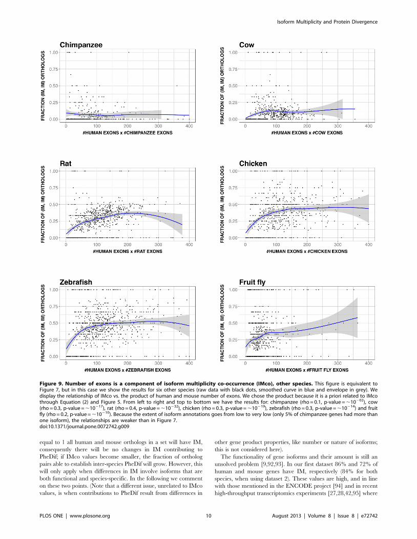
As before (in Figure 7), we explored to which extent number of
exons and IMco were connected. It has to be noted that in this
case IMco estimates were subject to a larger error, given that IM
annotations were less extensive (Table 1). Nonetheless, our results
showed (Figure 9) that number of exons was a component of IMco.
In some cases the relationship was weak, something probably due
to the following causes: low IM coverage, and/or existence of a
bias in the human-animal model ortholog gene set, a bias which
could either be of technical (e.g. low IM coverage in chimpanzee)
or of biological (e.g. smaller number of orthologs for human-fruit
fly) origin.
Discussion
Recent years have witnessed important advances in the
identification of the molecular sources of PheDif between
organisms [4,7,62]. However, we still do not know how these
molecular sources relate [3,4]. For example, in general we ignore
whether and to which extent they cooperate [3,63], or if there is
some degree of equivalence between them (comparable to that
proposed for alternative splicing and gene duplication [64,65]),
etc. A series of studies have started to clarify this issue for PD,
promoter-level divergence and gene expression [62,66–70]. Here
we advance in this direction by analyzing the relationship between
IMco and PD (Figure 1).
To relate IMco and PD we first have to take into account that
IM can be studied from two different sides: (i) the isoform/signal
side, in which isoforms, or their associated gene-level signals, are
compared between species; and (ii) the pattern of IM presence/
Figure 4. Species-specific isoform mutiplicity (IM) vs. proteindivergence (PD). Here we show the relationship between species-specific IM and PD, for both human and mouse genes. Black and greydots are used for human and mouse, respectively. We observe the samemonotonic trend for both species, a result that provides a simpleexplanation for the also monotonic behavior of P(IMH|x)?P(IMM|x) (seeFigure 3), the product of species-specific IM. In addition, this result is animportant intermediate step that will allow us to trace back the result inFigure 2 to a simple gene-level property (see text and Figure 6): thenumber of exons of the longest gene isoform. The continuous linesrepresent the smoothed version of the raw data (yellow and red forhuman and mouse, respectively; grey for the envelope) and lead to thesame interpretation.doi:10.1371/journal.pone.0072742.g004
Figure 5. The relationship between isoform multiplicity (IM) and number of exons of the largest isoform. The figure illustrates therelationship between these two properties for both human and mouse genes (A), and for these and other species (B). (A) and (B) differ in the dataorigin: (A) was obtained using UniProt/SwissProt/RefSeq sequences and Ensembl/VEGA transcript annotations; (B) was obtained using only Ensembldata. In general, we see the same trend: an increasing monotonic relationship which approaches 1 asymptotically, indicating that the larger thenumber of exons of the gene, the larger the number of isoforms of this gene. The fluctuations observed are due to a combination of factors: irregularisoform annotations (e.g. fruit fly), or low sample effects (particularly, for number of exons.30). Chimpanzee is an exception due to a very lowpercentage of transcript annotations (only 5% of the genes had multiple isoforms in the version of Ensembl used).doi:10.1371/journal.pone.0072742.g005
Isoform Multiplicity and Protein Divergence
PLOS ONE | www.plosone.org 6 August 2013 | Volume 8 | Issue 8 | e72742

absence between orthologs. An important volume of work has
been devoted to the first topic. For example, Modrek and Lee [20]
found, using a bioinformatics approach, that exons constitutively
spliced or present in major isoforms were conserved between
species, while those present in minor isoforms were mainly species-
specific. This result was experimentally confirmed and extended
by Pan et al. [22,37] using specifically designed microarrays and
splicing prediction methods. Sorek and Ast [71] and Sugnet et al.
[72] have focused on the differences in the nature of the flanking
intronic sequences between constitutive and alternatively spliced
exons; several other aspects of splicing signals and their
conservation have been also addressed [25,34,36,38–40,73]. In
all these studies gene sequence similarity was related in one way or
another to IM conservation between species, thus providing a
good basis for understanding the coordinated contribution of these
factors to PheDif.
The situation is different when we consider IM from the point of
view of its pattern of presence/absence in orthologs. We know that
presence of IM implies the introduction of a new regulatory level
in gene expression [8]. Therefore, simple switching from IM to
noIM between orthologs may imply an important change, even
before taking into consideration any possible difference between
isoforms. At the genome level, this suggests that differences
between organisms in the fraction of genes with IM may
contribute to explain their PheDif [5,11,74,75]. While initial tests
of this hypothesis led to controversial results [74,75], it was
subsequently shown that IM could indeed play the proposed role
[5]. Our work can be seen as a natural extension of this research:
here the results of IM co-occurrence are broken down along the
PD range, to see what is the relationship between these two
variables and whether it can be explained in terms of some gene
property.
Using a set of 11969 human-mouse orthologs we found that the
IMco-PD relationship was monotonic (Figure 2A), a result
confirmed with a second (Figure 2B) and third (Figure 2C)
datasets. Overlap between coding region and alternative splicing
signals [76–80] could provide a possible explanation for the
monotonic nature of the relationship. However, comparison of
P(IMH, IMM|x) and P(IMH|x)?P(IMM|x) showed that species-
specific IM is a major contributor to this relationship (Figure 3),
and that it monotonically depends on PD (Figure 4). Interestingly,
a relationship similar to the latter had also been found in different
species by Su et al. [65] for duplicated genes. From this point, and
with the link between IM and number of exons in mind (Figure 5),
we were able to trace back the monotonic behavior of IMco to a
relationship between number of exons and PD (Figures 6). This
relationship was a priori unexpected, although a related result,
involving the size of protein alignments instead of the number of
exons, had been described by Makalowski et al. [81] for a small set
of human-mouse orthologs. We then decided to check if number
of exons and PD were also related in other species, finding that this
was the case (Figure 8). Actually, the monotonic behavior was
comparable for vertebrates, although inverted for fruit fly.
The connection between number of exons and PD may be used
to suggest a biological reason for the origin of the IMco-PD
relationship. We know that the number of exons of an isoform is
directly related to the length of the resulting protein. Interestingly,
a few years ago Lipman et al. [82] found a link between protein
conservation and sequence length: conserved proteins tended to be
larger than non-conserved ones. Although their definition of
conservation was discrete (two classes: conserved and non-
conserved) and based on taxonomic considerations, their use of
sequence comparisons made it relatively similar to our PD. When
replacing sequence length by number of exons, the finding of
Lipman et al. [82] qualitatively corresponds to our observed
relationship between number of exons and PD (Figure 6). These
authors explain their results in terms of a balance between
different constraints [82]. Functional constraints would be higher
Figure 6. Number of exons vs. protein divergence (PD). Here we show the relationship between number of exons of the largest isoform andPD, for both human and mouse genes. Black and grey dots are used for human and mouse, respectively. We observe the same monotonicallyincreasing trend for both species, which provides a natural explanation for the behavior of species-specific IM seen in Figure 4. The smoothed versionis shown with a continuous line (yellow and red for human and mouse, respectively; grey for the envelope). The relevance of this result is that wehave identified a simple gene property contributing to the relationship between IM co-occurrence, IMco, and PD shown in Figure 2. (A) was obtainedusing UniProt/SwissProt/RefSeq sequences and Ensembl transcript annotations; (B) was obtained using only Ensembl data.doi:10.1371/journal.pone.0072742.g006
Isoform Multiplicity and Protein Divergence
PLOS ONE | www.plosone.org 7 August 2013 | Volume 8 | Issue 8 | e72742

for conserved proteins, therefore limiting any drastic sequence
change, like deletions. On the contrary, for non-conserved
proteins functional constraints would be weaker, and gradually
replaced by the pressure to minimize translation costs. The latter,
combined with a higher frequency of sequence deletions over
insertions, would result in smaller proteins. This explanation can
be naturally transferred to the monotonic dependence between
number of exons and PD (Figure 6) and extended, with caution, to
the IMco-PD relationship, which would then result from a balance
between gene function and protein synthesis costs. Finally, it has to
be mentioned that a combination of recent results from two groups
[83,84] suggest the existence of a relationship between number of
exons and evolutionary rate consistent with our work.
Using the PD-IMco Relationship to Improve OurUnderstanding of the Molecular Basis of Inter-speciesPheDif
The final goal of integrating the different sources of inter-species
PheDif is to reach a better understanding of the molecular basis of
these PheDif [4]. Within this context we will discuss the
explanatory power of the IMco-PD relationship and what are its
present limitations. To this end we need to see how changes in PD
and IMco relate to changes in molecular function, something that
Figure 7. Number of exons is a component of isoform multiplicity co-occurrence (IMco). We display (black dots) the relationship of IMcovs. the product of human and mouse number of exons. We chose the product because it is a priori related to IMco through Equation (2) and Figure 5.The result shows the existence of a monotonically increasing relationship. Shown in blue is the smoothed version of the raw data, and the envelopein grey.doi:10.1371/journal.pone.0072742.g007
Isoform Multiplicity and Protein Divergence
PLOS ONE | www.plosone.org 8 August 2013 | Volume 8 | Issue 8 | e72742

will require different approaches, as PD is a single-gene-based
measure, and IMco is multigene. In the case of PD we know that
most features of protein function are conserved, on the average,
above a certain sequence identity threshold: 60–70% for enzyme
function [85–87]; 30–40% for the overall geometry of protein
interactions [88]; 50% for quaternary structure [89]; above 65%
for protein partner conservation [90]; and between 60–80% for a
series of function related properties [91]. That is, there is a
sequence identity region between 50% and 70% defining the
boundaries of protein function conservation: above this region
protein function will be generally conserved, and therefore will be
unlikely to contribute to PheDif. On the contrary, below 50%
sequence identity protein function will generally vary and
therefore will be more likely to contribute to PheDif.
In the case of IMco, we know that its values are obtained for sets
of genes (see also Materials and Methods and Figure 1). When IMco is
Figure 8. Number of exons vs. protein divergence (PD) in other species. This figure is equivalent to Figure 6, but in this case we show theresults for six other species. In each plot we represent the data for human (black dots; smoothed version in yellow) and the other species (grey dots;smoothed version in red); the envelopes of the smoothed versions are shown in grey. From left to right and top to bottom we have the results for:chimpanzee (rho = 0.3 and 0.4, p-value,10210 and ,10217, for human and chimpanzee, respectively), cow (rho = 0.6 and 0.6, p-value,10259 and,10266), rat (rho = 0.4 and 0.6, p-value,10225 and ,10266), chicken (rho = 0.3 and 0.4, p-value,10221 and ,10227), zebrafish (rho = 0.0 and 0.1, p-value,0.39 and ,10205) and fruit fly (rho = 20.3 and20.3, p-value,10214 and ,10214). In all cases we observe a nearly monotonic relationship,which is increasing for vertebrates, and decreasing for fruit fly.doi:10.1371/journal.pone.0072742.g008
Isoform Multiplicity and Protein Divergence
PLOS ONE | www.plosone.org 9 August 2013 | Volume 8 | Issue 8 | e72742
equal to 1 all human and mouse orthologs in a set will have IM,
consequently there will be no changes in IM contributing to
PheDif; if IMco values become smaller, the fraction of ortholog
pairs able to establish inter-species PheDif will grow. However, this
will only apply when differences in IM involve isoforms that are
both functional and species-specific. In the following we comment
on these two points. (Note that a different issue, unrelated to IMco
values, is when contributions to PheDif result from differences in
other gene product properties, like number or nature of isoforms;
this is not considered here).
The functionality of gene isoforms and their amount is still an
unsolved problem [9,92,93]. In our first dataset 86% and 72% of
human and mouse genes have IM, respectively (84% for both
species, when using dataset 2). These values are high, and in line
with those mentioned in the ENCODE project [94] and in recent
high-throughput transcriptomics experiments [27,28,42,95] where
Figure 9. Number of exons is a component of isoform multiplicity co-occurrence (IMco), other species. This figure is equivalent toFigure 7, but in this case we show the results for six other species (raw data with black dots, smoothed curve in blue and envelope in grey). Wedisplay the relationship of IMco vs. the product of human and mouse number of exons. We chose the product because it is a priori related to IMcothrough Equation (2) and Figure 5. From left to right and top to bottom we have the results for: chimpanzee (rho = 0.1, p-value = ,10202), cow(rho = 0.3, p-value = ,10217), rat (rho = 0.4, p-value = ,10232), chicken (rho = 0.3, p-value = ,10219), zebrafish (rho = 0.3, p-value = ,10214) and fruitfly (rho = 0.2, p-value = ,10210). Because the extent of isoform annotations goes from low to very low (only 5% of chimpanzee genes had more thanone isoform), the relationships are weaker than in Figure 7.doi:10.1371/journal.pone.0072742.g009
Isoform Multiplicity and Protein Divergence
PLOS ONE | www.plosone.org 10 August 2013 | Volume 8 | Issue 8 | e72742

a large majority of human multi-exon genes (and probably those
from other mammal species) is found to have multiple isoforms.
Are all these isoforms functional? That is, when expressed do they
contribute a new function to the cell proteome or play a regulatory
role? Because the experimental approach required to provide an
answer for the thousands of known isoforms is so complex, these
questions are still open. On one side, studies carried in specific
systems [96–100] support the functional role of IM. This is also
supported by large-scale studies. Using a quantitative microarray
Pan et al. [37] established a link between tissue-specific alternative
splicing and functional effects; Ellis et al. [101] and Buljan et al.
[102] have characterized the relationship between tissue-specific
alternative splicing, protein-protein interactions and protein
disorder, a result supported by Barbosa-Morais et al. [27]; and
Merkin et al. [28] have related alternative splicing and phosphor-
ylatability.
On the other side, a growing amount of data indicates that not
all expressed isoforms are functional [9,92,93,103–105]. In
particular, it has been found that abundance and nature of
transcript data are consistent with the existence of noisy splicing
[103,104,106,107]. Recently, Pickrell et al. [93] have used RNA-
seq to show that indeed an important amount of alternative
isoforms result from noisy splicing. On the same line, Hon et al.
[92] have used RNA-seq in E.histolytica to show that a majority of
alternative splicing and polyadenylation isoforms are the result of
stochastic processes and therefore unlikely to play a functional
role. Reinforcing these results, recent proteomics studies [105]
show that a fraction of transcripts do not reach the protein level,
and for this reason are less likely to be functional.
We mentioned before that the second requirement that species-
specific IM must fulfill to contribute to inter-species PheDif is that
it must not correspond to genetically-driven, or individual, IM.
The latter will contribute to intra-species, but not to inter-species,
differences. At present it is well accepted that this variability exists
[108–112], although its proportion is yet unknown.
In summary, from the previous considerations it is clear that a
certain amount of the isoforms contributing to IMco will have no
impact on inter-species PheDif. For this reason, phenotypically
relevant IMco values will be lower than those observed in Figure 2.
We can now go back to our original question: how can we use
the IMco-PD relationship to improve our view of the molecular
basis of human-mouse PheDif. In Figure 10 we reproduce Figure 2,
adding the functional threshold for PD values and highlighting the
fact that the present result constitutes an upper threshold for
phenotypically relevant IMco. In Figure 10 we highlight in grey
the region between 50% and 70%, which corresponds to the
protein function threshold (see above). Above this threshold IMco
values are already below one, indicating that in general IM
differences but not PD will contribute to PheDif between human
and mouse.
For the grey region between 50% and 70% sequence identity
little can be said. However, below this region the situation is
complex, with functional changes resulting from sequence
divergence being accompanied by phenotypically-relevant changes
in IMco. This points to a rich scenario in which both phenomena
would frequently cooperate to originate PheDif between species.
Conclusions
To understand the interplay between the molecular contributors
to PheDif we studied the relationship between IMco and PD, using
three sets of human-mouse orthologs. We found that there was a
monotonic dependence between IMco and PD that could be
traced to a more fundamental relationship: the link between exon
number of the longest gene isoform and PD. Given its interest, we
explored the existence of this second relationship in other species,
finding that this was the case, although with variations in the
Figure 10. The interplay between isoform multiplicity co-occurrence (IMco) and protein divergence (PD) in the generation ofhuman-mouse phenotypic differences. Here we plot the same graph as in Figure 2A with two additions: a grey-shaded area corresponding tothe 50%–70% zone that separates functional from non-functional PD (see Discussion); a red-shaded area below the black line, highlighting the factthat the latter is an upper threshold for IMco values obtained after exclusion of non-relevant isoforms. We can see that phenotypically-relevant IMcovalues (red-shaded area) are always lower than 1, indicating that differences in isoform multiplicity can contribute to human-mouse phenotypicdifferences all over the PD range. To the right of the grey-shaded area this contribution will be more relevant than that of PD; to the left bothphenomena will very likely cooperate to generate phenotypic changes; the situation is unclear within the 50%–70% zone.doi:10.1371/journal.pone.0072742.g010
Isoform Multiplicity and Protein Divergence
PLOS ONE | www.plosone.org 11 August 2013 | Volume 8 | Issue 8 | e72742

monotonic behavior (for the fruit fly case). Using previous results
from the literature we could provide a plausible explanation for
this relationship in terms of the balance between functional and
cost of synthesis constraints. Finally, we show how the IMco-PD
relationship could be used to analyze the molecular basis of inter-
species PheDif.
Supporting Information
Figure S1 Relationship between isoform multiplicityco-occurrence (IMco) and protein divergence (PD). This
figure is equivalent to Figure 2, with the difference that IMco
estimates obtained from less than 5 ortholog pairs have been
eliminated. (A) and (B) correspond to Ensembl and VEGA data. In
both figures black dots are used to show the raw, unprocessed
data; a blue line is used for the smoothed data, which is shown
with its envelope in grey.
(TIF)
Figure S2 The contribution of species-specific isoformmultiplicity (IM) to isoform multiplicity co-occurrence(IMco) (VEGA data). This figure is equivalent to Figure 3,
except in that we have used VEGA, instead of Ensembl, isoform
data. Here we compare IMco with the product of species-specific
IM, P(IMH|x)?P(IMM|x), a term of IMco, as shown in Equation 2
(see Materials and Methods). In (A) we show the raw data
representation: we can observe an important overlap between
both data clouds, as well as a similar monotonic trend, something
confirmed in (B) where we show the smoothed data. In (A) the
color code is: black for IMco and grey for P(IMH|x)?P(IMM|x);
in (B) we have dark blue for IMco, and light blue for
P(IMH|x)?P(IMM|x).
(TIF)
Figure S3 Species-specific isoform mutiplicity (IM) vs.protein divergence (PD) (VEGA data). This figure is
equivalent to Figure 4, except in that we have used VEGA,
instead of Ensembl, isoform data. We show the relationship
between species-specific IM and PD, for both human and mouse
genes. In both (A) and (B) we plot raw data, with black black and
grey dots for human and mouse, respectively. Also, in both (A) and
(B) we plot a smoothed version of these raw data: yellow and red
for human and mouse, respectively, and grey for the correspond-
ing envelopes. Finally, (A) differs from (B) in that for the latter we
have eliminated those estimates of IMco obtained from less than 5
observations. We observe the same monotonically increasing trend
for both species, a result that provides a simple explanation for the
also monotonically increasing behavior of P(IMH|x)?P(IMM|x)
(Figures 3 and S2), the product of species-specific IM.
(TIF)
Figure S4 Mouse-specific isoform mutiplicity (IM) vs.protein divergence (PD) (RNA-seq data). This figure is
equivalent to Figures 4 and S3, except in that we have restricted
our analysis to the mouse case and used RNA-seq, instead of
Ensembl, isoform annotations (data for three individuals from
Merkin et al. [28]). We show the relationship between species-
specific IM and PD for mouse genes, under scenarios that combine
different quality controls for RNA-seq data (mild, FPKM.0; less
permissive, FPKM$1) and the effect of individual variability in
RNA-seq data: (A) lax quality control, individual variability
ignored (results from three samples considered together); (B) lax
quality control, individual variability considered (to annotate a
given gene as having IM, it had to display IM in at least one tissue
for each of three individuals); (C) less permissive quality control,
individual variability ignored; and (D) less permissive quality
control, individual variability considered. In all the plots we have
two curves: in green we have the results obtained with RNA-seq
data only, in brown we have the results obtained with RNA-seq
data enriched with Ensembl annotations (as a result more genes
are annotated as having IM). Dots represent the raw data and
continuous lines are used for the smoothed data, which are shown
with the corresponding envelope in grey. As a minimum quality
control, IM is estimated when more than 5 observations are
available. In all four plots we observe a monotonically increasing
trend between mouse IM and PD (Spearman rank correlation,
rho, and p-values from Figures S4A to S4D: rho = 0.15, p-
val = 0.002; rho = 0.22, p-val = 1.961026; rho = 0.28, p-
val = 2.6821029; rho = 0.36, p-val = 1.3610214), in accordance
with the results found in Figures 4 and S3.
(TIF)
Figure S5 Number of exons vs. protein divergence (PD)(VEGA data). This figure is equivalent to Figure 6, except in that
we have used VEGA, instead of Ensembl, isoform data. We show
the relationship between number of exons of the largest isoform
and PD, for both human and mouse genes. In both (A) and (B) we
plot raw data, with black and grey dots for human and mouse,
respectively. Also, in both (A) and (B) we plot a smoothed version
of these raw data: yellow and red for human and mouse,
respectively, and grey for the corresponding envelopes. Finally, (A)
differs from (B) in that for the latter we have eliminated those
estimates of IMco obtained from less than 5 observations.
(TIF)
Acknowledgments
The authors wish to acknowledge Mar Alba, Cinta Pegueroles, William
Valdar and Adam Hospital for helpful comments.
Author Contributions
Conceived and designed the experiments: XDC. Performed the experi-
ments: JM CR GMX SL. Analyzed the data: JM SB CR DT XDC. Wrote
the paper: XDC.
References
1. Alonso CR, Wilkins AS (2005) The molecular elements that underlie
developmental evolution. Nat Rev Genet 6: 709–715.
2. Hoekstra HE, Coyne JA (2007) The locus of evolution: evo devo and the
genetics of adaptation. Evolution 61: 995–1016.
3. Copley RR (2008) The animal in the genome: comparative genomics and
evolution. Philos Trans R Soc Lond B Biol Sci 363: 1453–1461.
4. Gharib WH, Robinson-Rechavi M (2011) When orthologs diverge between
human and mouse. Brief Bioinform 12: 436–441.
5. Kim E, Magen A, Ast G (2007) Different levels of alternative splicing among
eukaryotes. Nucleic Acids Res 35: 125–131.
6. Shabalina SA, Spiridonov AN, Spiridonov NA, Koonin EV (2010) Connec-
tions between alternative transcription and alternative splicing in mammals.
Genome Biol Evol 2: 791–799.
7. Mudge JM, Frankish A, Fernandez-Banet J, Alioto T, Derrien T, et al. (2011)
The origins, evolution, and functional potential of alternative splicing in
vertebrates. Mol Biol Evol 28: 2949–2959.
8. Nilsen TW, Graveley BR (2010) Expansion of the eukaryotic proteome by
alternative splicing. Nature 463: 457–463.
9. Kim E, Goren A, Ast G (2008) Alternative splicing: current perspectives.
Bioessays 30: 38–47.
10. Talavera D, Vogel C, Orozco M, Teichmann SA, de la Cruz6 (2007) The
(in)dependence of alternative splicing and gene duplication. PLoS computa-
tional biology 3: e33.
11. Lander ES, Linton LM, Birren B, Nusbaum C, Zody MC, et al. (2001) Initial
sequencing and analysis of the human genome. Nature 409: 860–921.
Isoform Multiplicity and Protein Divergence
PLOS ONE | www.plosone.org 12 August 2013 | Volume 8 | Issue 8 | e72742

12. Marden JH (2008) Quantitative and evolutionary biology of alternativesplicing: how changing the mix of alternative transcripts affects phenotypic
plasticity and reaction norms. Heredity 100: 111–120.
13. Garcia-Blanco MA, Baraniak AP, Lasda EL (2004) Alternative splicing in
disease and therapy. Nat Biotechnol 22: 535–546.
14. Wang GS, Cooper TA (2007) Splicing in disease: disruption of the splicing code
and the decoding machinery. Nat Rev Genet 8: 749–761.
15. Tazi J, Bakkour N, Stamm S (2009) Alternative splicing and disease. BiochimBiophys Acta 1792: 14–26.
16. Artamonova II, Gelfand MS (2007) Comparative genomics and evolution ofalternative splicing: the pessimists’ science. Chem Rev 107: 3407–3430.
17. Takeda J, Suzuki Y, Sakate R, Sato Y, Seki M, et al. (2008) Low conservationand species-specific evolution of alternative splicing in humans and mice:
comparative genomics analysis using well-annotated full-length cDNAs.Nucleic Acids Res 36: 6386–6395.
18. Calarco JA, Superina S, O’Hanlon D, Gabut M, Raj B, et al. (2009) Regulationof vertebrate nervous system alternative splicing and development by an SR-
related protein. Cell 138: 898–910.
19. Sorek R, Shamir R, Ast G (2004) How prevalent is functional alternative
splicing in the human genome? Trends Genet 20: 68–71.
20. Modrek B, Lee CJ (2003) Alternative splicing in the human, mouse and ratgenomes is associated with an increased frequency of exon creation and/or loss.
Nat Genet 34: 177–180.
21. Nurtdinov RN, Artamonova II, Mironov AA, Gelfand MS (2003) Low
conservation of alternative splicing patterns in the human and mouse genomes.
Hum Mol Genet 12: 1313–1320.
22. Pan Q, Bakowski MA, Morris Q, Zhang W, Frey BJ, et al. (2005) Alternativesplicing of conserved exons is frequently species-specific in human and mouse.
Trends Genet 21: 73–77.
23. Thanaraj TA, Clark F, Muilu J (2003) Conservation of human alternative
splice events in mouse. Nucleic Acids Res 31: 2544–2552.
24. Calarco JA, Xing Y, Caceres M, Calarco JP, Xiao X, et al. (2007) Global
analysis of alternative splicing differences between humans and chimpanzees.Genes Dev 21: 2963–2975.
25. Ohler U, Shomron N, Burge CB (2005) Recognition of unknown conservedalternatively spliced exons. PLoS computational biology 1: 113–122.
26. Yeo GW, Van Nostrand E, Holste D, Poggio T, Burge CB (2005) Identification
and analysis of alternative splicing events conserved in human and mouse. Proc
Natl Acad Sci U S A 102: 2850–2855.
27. Barbosa-Morais NL, Irimia M, Pan Q, Xiong HY, Gueroussov S, et al. (2012)The evolutionary landscape of alternative splicing in vertebrate species. Science
338: 1587–1593.
28. Merkin J, Russell C, Chen P, Burge CB (2012) Evolutionary dynamics of gene
and isoform regulation in Mammalian tissues. Science 338: 1593–1599.
29. Valenzuela A, Talavera D, Orozco M, de la Cruz X (2004) Alternative splicing
mechanisms for the modulation of protein function: conservation betweenhuman and other species. J Mol Biol 335: 495–502.
30. Wetterbom A, Gyllensten U, Cavelier L, Bergstrom TF (2009) Genome-wideanalysis of chimpanzee genes with premature termination codons. BMC
Genomics 10: 56.
31. Blekhman R, Marioni JC, Zumbo P, Stephens M, Gilad Y (2010) Sex-specific
and lineage-specific alternative splicing in primates. Genome Res 20: 180–189.
32. Landry JR, Mager DL, Wilhelm BT (2003) Complex controls: the role of
alternative promoters in mammalian genomes. Trends Genet 19: 640–648.
33. Latchman D (2008) Gene Regulation. A Eukaryotic Perspective: Springer.
34. Yeo G, Hoon S, Venkatesh B, Burge CB (2004) Variation in sequence and
organization of splicing regulatory elements in vertebrate genes. Proc NatlAcad Sci U S A 101: 15700–15705.
35. Ara T, Lopez F, Ritchie W, Benech P, Gautheret D (2006) Conservation ofalternative polyadenylation patterns in mammalian genes. BMC Genomics 7:
189.
36. Baek D, Green P (2005) Sequence conservation, relative isoform frequencies,
and nonsense-mediated decay in evolutionarily conserved alternative splicing.Proc Natl Acad Sci U S A 102: 12813–12818.
37. Pan Q, Shai O, Misquitta C, Zhang W, Saltzman AL, et al. (2004) Revealingglobal regulatory features of mammalian alternative splicing using a
quantitative microarray platform. Mol Cell 16: 929–941.
38. Plass M, Agirre E, Reyes D, Camara F, Eyras E (2008) Co-evolution of the
branch site and SR proteins in eukaryotes. Trends Genet 24: 590–594.
39. Plass M, Eyras E (2006) Differentiated evolutionary rates in alternative exonsand the implications for splicing regulation. BMC Evol Biol 6: 50.
40. Schwartz SH, Silva J, Burstein D, Pupko T, Eyras E, et al. (2008) Large-scalecomparative analysis of splicing signals and their corresponding splicing factors
in eukaryotes. Genome Res 18: 88–103.
41. Morata J, Riera C, de la Cruz X (2012) Alternative Splicing as a Source of
Phenotypic Differences Between Species: Protein-level Mechanisms; PontarottiP, editor. Heidelberg: Springer.
42. Pan Q, Shai O, Lee LJ, Frey BJ, Blencowe BJ (2008) Deep surveying ofalternative splicing complexity in the human transcriptome by high-throughput
sequencing. Nat Genet 40: 1413–1415.
43. Koralewski TE, Krutovsky KV (2011) Evolution of exon-intron structure and
alternative splicing. PLoS One 6: e18055.
44. Rasche A, Herwig R (2010) ARH: predicting splice variants from genome-wide
data with modified entropy. Bioinformatics 26: 84–90.
45. Ostlund G, Schmitt T, Forslund K, Kostler T, Messina DN, et al. (2010)InParanoid 7: new algorithms and tools for eukaryotic orthology analysis.
Nucleic Acids Res 38: D196–203.
46. Consortium TU (2010) The Universal Protein Resource (UniProt) in 2010.Nucleic Acids Res 38: D142–148.
47. Boeckmann B, Bairoch A, Apweiler R, Blatter MC, Estreicher A, et al. (2003)The SWISS-PROT protein knowledgebase and its supplement TrEMBL in
2003. Nucleic Acids Res 31: 365–370.
48. Pruitt KD, Tatusova T, Maglott DR (2007) NCBI reference sequences
(RefSeq): a curated non-redundant sequence database of genomes, transcripts
and proteins. Nucleic Acids Res 35: D61–65.
49. Altmann A, Weber P, Quast C, Rex-Haffner M, Binder EB, et al. (2011) vipR:
variant identification in pooled DNA using R. Bioinformatics 27: i77–84.
50. Flicek P, Amode MR, Barrell D, Beal K, Brent S, et al. (2011) Ensembl 2011.
Nucleic Acids Res 39: D800–806.
51. Wilming LG, Gilbert JG, Howe K, Trevanion S, Hubbard T, et al. (2008) Thevertebrate genome annotation (Vega) database. Nucleic Acids Res 36: D753–
760.
52. Vilella AJ, Severin J, Ureta-Vidal A, Heng L, Durbin R, et al. (2009)
EnsemblCompara GeneTrees: Complete, duplication-aware phylogenetic trees
in vertebrates. Genome Res 19: 327–335.
53. Bishop CM (2006) Pattern Recognition and Machine Learning. New York:
Springer.
54. Feller W (1970) An Introduction to Probability Theory and Its Applications.
New Jersey: John Wiley & Sons.
55. Needleman SB, Wunsch CD (1970) A general method applicable to the searchfor similarities in the amino acid sequence of two proteins. J Mol Biol 48: 443–
453.
56. McLysaght A, Hokamp K, Wolfe KH (2002) Extensive genomic duplication
during early chordate evolution. Nat Genet 31: 200–204.
57. Demuth JP, De Bie T, Stajich JE, Cristianini N, Hahn MW (2006) Theevolution of mammalian gene families. PLoS One 1: e85.
58. Kondrashov FA, Ogurtsov AY, Kondrashov AS (2004) Bioinformatical assay ofhuman gene morbidity. Nucleic Acids Res 32: 1731–1737.
59. RCoreTeam (2013) R: A language and environment for statistical computing.
Vienna, Austria: R fundation for statistical computing.
60. Barrett T, Wilhite SE, Ledoux P, Evangelista C, Kim IF, et al. (2013) NCBI
GEO: archive for functional genomics data sets–update. Nucleic Acids Res 41:D991–995.
61. Trapnell C, Roberts A, Goff L, Pertea G, Kim D, et al. (2010) Differential geneand transcript expression analysis of RNA-seq experiments with TopHat and
Cufflinks. Nat Protoc 7: 562–578.
62. Castillo-Davis CI, Hartl DL, Achaz G (2004) cis-Regulatory and proteinevolution in orthologous and duplicate genes. Genome Res 14: 1530–1536.
63. Lemos B, Bettencourt BR, Meiklejohn CD, Hartl DL (2005) Evolution ofproteins and gene expression levels are coupled in Drosophila and are
independently associated with mRNA abundance, protein length, and number
of protein-protein interactions. Mol Biol Evol 22: 1345–1354.
64. Kopelman NM, Lancet D, Yanai I (2005) Alternative splicing and gene
duplication are inversely correlated evolutionary mechanisms. Nat Genet 37:588–589.
65. Su Z, Wang J, Yu J, Huang X, Gu X (2006) Evolution of alternative splicing
after gene duplication. Genome Res 16: 182–189.
66. Chiba H, Yamashita R, Kinoshita K, Nakai K (2008) Weak correlation
between sequence conservation in promoter regions and in protein-codingregions of human-mouse orthologous gene pairs. BMC Genomics 9: 152.
67. Wang Y, Rekaya R (2009) A comprehensive analysis of gene expression
evolution between humans and mice. Evol Bioinform Online 5: 81–90.
68. Liao BY, Zhang J (2006) Evolutionary conservation of expression profiles
between human and mouse orthologous genes. Mol Biol Evol 23: 530–540.
69. Tirosh I, Weinberger A, Bezalel D, Kaganovich M, Barkai N (2008) On the
relation between promoter divergence and gene expression evolution. Mol Syst
Biol 4: 159.
70. Wall DP, Hirsh AE, Fraser HB, Kumm J, Giaever G, et al. (2005) Functional
genomic analysis of the rates of protein evolution. Proc Natl Acad Sci U S A102: 5483–5488.
71. Sorek R, Ast G (2003) Intronic sequences flanking alternatively spliced exonsare conserved between human and mouse. Genome Res 13: 1631–1637.
72. Sugnet CW, Kent WJ, Ares MJ, Haussler D (2004) Transcriptome and genome
conservation of alternative splicing events in humans and mice. Pac SympBiocomput: 66–77.
73. Ast G (2004) How did alternative splicing evolve? Nat Rev Genet 5: 773–782.
74. Brett D, Pospisil H, Valcarcel J, Reich J, Bork P (2002) Alternative splicing and
genome complexity. Nat Genet 30: 29–30.
75. Kim H, Klein R, Majewski J, Ott J (2004) Estimating rates of alternativesplicing in mammals and invertebrates. Nat Genet 36: 915–916; author reply
916–917.
76. Fichant GA (1992) Constraints acting on the exon positions of the splice site
sequences and local amino acid composition of the protein. Hum Mol Genet 1:
259–267.
77. Whamond GS, Thornton JM (2006) An analysis of intron positions in relation
to nucleotides, amino acids, and protein secondary structure. J Mol Biol 359:238–247.
Isoform Multiplicity and Protein Divergence
PLOS ONE | www.plosone.org 13 August 2013 | Volume 8 | Issue 8 | e72742

78. Goren A, Ram O, Amit M, Keren H, Lev-Maor G, et al. (2006) Comparative
analysis identifies exonic splicing regulatory sequences–The complex definitionof enhancers and silencers. Mol Cell 22: 769–781.
79. Schaal TD, Maniatis T (1999) Multiple distinct splicing enhancers in the
protein-coding sequences of a constitutively spliced pre-mRNA. Mol Cell Biol19: 261–273.
80. Smith CW, Valcarcel J (2000) Alternative pre-mRNA splicing: the logic ofcombinatorial control. Trends Biochem Sci 25: 381–388.
81. Makalowski W, Zhang J, Boguski MS (1996) Comparative analysis of 1196
orthologous mouse and human full-length mRNA and protein sequences.Genome Res 6: 846–857.
82. Lipman DJ, Souvorov A, Koonin EV, Panchenko AR, Tatusova TA (2002)The relationship of protein conservation and sequence length. BMC Evol Biol
2: 20.83. Neme R, Tautz D (2013) Phylogenetic patterns of emergence of new genes
support a model of frequent de novo evolution. BMC Genomics 14: 117.
84. Toll-Riera M, Bosch N, Bellora N, Castelo R, Armengol L, et al. (2009) Originof primate orphan genes: a comparative genomics approach. Mol Biol Evol 26:
603–612.85. Devos D, Valencia A (2000) Practical limits of function prediction. Proteins 41:
98–107.
86. Rost B (2002) Enzyme function less conserved than anticipated. J Mol Biol 318:595–608.
87. Tian W, Skolnick J (2003) How well is enzyme function conserved as a functionof pairwise sequence identity? J Mol Biol 333: 863–882.
88. Aloy P, Ceulemans H, Stark A, Russell RB (2003) The relationship betweensequence and interaction divergence in proteins. J Mol Biol 332: 989–998.
89. Levy ED, Boeri Erba E, Robinson CV, Teichmann SA (2008) Assembly reflects
evolution of protein complexes. Nature 453: 1262–1265.90. Yu H, Luscombe NM, Lu HX, Zhu X, Xia Y, et al. (2004) Annotation transfer
between genomes: protein-protein interologs and protein-DNA regulogs.Genome Res 14: 1107–1118.
91. Devos D, Valencia A (2001) Intrinsic errors in genome annotation. Trends
Genet 17: 429–431.92. Hon CC, Weber C, Sismeiro O, Proux C, Koutero M, et al. (2013)
Quantification of stochastic noise of splicing and polyadenylation inEntamoeba histolytica. Nucleic Acids Res 41: 1936–1952.
93. Pickrell JK, Pai AA, Gilad Y, Pritchard JK (2010) Noisy splicing drives mRNAisoform diversity in human cells. PLoS Genet 6: e1001236.
94. Djebali S, Davis CA, Merkel A, Dobin A, Lassmann T, et al. (2012) Landscape
of transcription in human cells. Nature 489: 101–108.95. Wang ET, Sandberg R, Luo S, Khrebtukova I, Zhang L, et al. (2008)
Alternative isoform regulation in human tissue transcriptomes. Nature 456:470–476.
96. Stamm S, Ben-Ari S, Rafalska I, Tang Y, Zhang Z, et al. (2005) Function of
alternative splicing. Gene 344: 1–20.
97. Lois S, Blanco N, Martinez-Balbas M, de la Cruz X (2007) The functional
modulation of epigenetic regulators by alternative splicing. BMC Genomics 8:
252.
98. Romero PR, Zaidi S, Fang YY, Uversky VN, Radivojac P, et al. (2006)
Alternative splicing in concert with protein intrinsic disorder enables increased
functional diversity in multicellular organisms. Proc Natl Acad Sci U S A 103:
8390–8395.
99. Fuxreiter M (2012) Fuzziness: linking regulation to protein dynamics. Mol
Biosyst 8: 168–177.
100. Barbany M, Morata J, Meyer T, Lois S, Orozco M, et al. (2012)
Characterization of the impact of alternative splicing on protein dynamics:
the cases of glutathione S-transferase and ectodysplasin-A isoforms. Proteins
80: 2235–2249.
101. Ellis JD, Barrios-Rodiles M, Colak R, Irimia M, Kim T, et al. (2012) Tissue-
specific alternative splicing remodels protein-protein interaction networks. Mol
Cell 46: 884–892.
102. Buljan M, Chalancon G, Eustermann S, Wagner GP, Fuxreiter M, et al. (2012)
Tissue-specific splicing of disordered segments that embed binding motifs
rewires protein interaction networks. Mol Cell 46: 871–883.
103. Chern TM, van Nimwegen E, Kai C, Kawai J, Carninci P, et al. (2006) A
simple physical model predicts small exon length variations. PLoS Genet 2:
e45.
104. Melamud E, Moult J (2009) Stochastic noise in splicing machinery. Nucleic
Acids Res 37: 4873–4886.
105. Ezkurdia I, del Pozo A, Frankish A, Rodriguez JM, Harrow J, et al. (2012)
Comparative proteomics reveals a significant bias toward alternative protein
isoforms with conserved structure and function. Mol Biol Evol 29: 2265–2283.
106. Melamud E, Moult J (2009) Structural implication of splicing stochastics.
Nucleic Acids Res 37: 4862–4872.
107. Zhang Z, Xin D, Wang P, Zhou L, Hu L, et al. (2009) Noisy splicing, more
than expression regulation, explains why some exons are subject to nonsense-
mediated mRNA decay. BMC Biol 7: 23.
108. Nembaware V, Wolfe KH, Bettoni F, Kelso J, Seoighe C (2004) Allele-specific
transcript isoforms in human. FEBS Lett 577: 233–238.
109. Graveley BR (2008) The haplo-spliceo-transcriptome: common variations in
alternative splicing in the human population. Trends Genet 24: 5–7.
110. Hull J, Campino S, Rowlands K, Chan MS, Copley RR, et al. (2007)
Identification of common genetic variation that modulates alternative splicing.
PLoS Genet 3: e99.
111. Kwan T, Benovoy D, Dias C, Gurd S, Serre D, et al. (2007) Heritability of
alternative splicing in the human genome. Genome Res 17: 1210–1218.
112. de Souza JE, Ramalho RF, Galante PA, Meyer D, de Souza SJ (2011)
Alternative splicing and genetic diversity: silencers are more frequently
modified by SNVs associated with alternative exon/intron borders. Nucleic
Acids Res 39: 4942–4948.
Isoform Multiplicity and Protein Divergence
PLOS ONE | www.plosone.org 14 August 2013 | Volume 8 | Issue 8 | e72742