Duplication of Growth Hormone Gene and Accelerated Evolution in Passerine Birds
Upload
un-lincolnCategory
view
1download
0
Gene Duplication and the Evolution of Hemoglobin IsoformDifferentiation in Birds*□S
Received for publication, April 24, 2012, and in revised form, September 6, 2012 Published, JBC Papers in Press, September 8, 2012, DOI 10.1074/jbc.M112.375600
Michael T. Grispo‡, Chandrasekhar Natarajan‡, Joana Projecto-Garcia‡, Hideaki Moriyama‡, Roy E. Weber§,and Jay F. Storz‡1
From the ‡School of Biological Sciences, University of Nebraska, Lincoln, Nebraska 68588 and §Zoophysiology, Institute forBioscience, Aarhus University, DK-8000 Aarhus C, Denmark
Background: The functional significance of hemoglobin heterogeneity remains a mystery.Results: In adult birds, the HbD isoform (related to embryonic hemoglobin) exhibits distinct oxygenation properties relative tothe major HbA isoform.Conclusion: Substitutions that distinguish HbD from HbA are not shared with embryonic hemoglobin.Significance: Differences between isoforms stem from derived (nonancestral) changes in duplicated genes, not from theretention of an ancestral condition.
The majority of bird species co-express two functionally dis-tinct hemoglobin (Hb) isoforms in definitive erythrocytes as fol-lows: HbA (the major adult Hb isoform, with �-chain subunitsencoded by the �A-globin gene) and HbD (the minor adult Hbisoform, with�-chain subunits encoded by the�D-globin gene).The �D-globin gene originated via tandem duplication of anembryonic �-like globin gene in the stem lineage of tetrapodvertebrates,which suggests the possibility that functional differ-entiation between the HbA and HbD isoforms may be attribut-able to a retained ancestral character state in HbD that harkensback to a primordial, embryonic function. To investigate thispossibility, we conducted a combined analysis of protein bio-chemistry and sequence evolution to characterize the structuraland functional basis of Hb isoform differentiation in birds.Functional experiments involving purified HbA and HbD iso-forms from 11 different bird species revealed that HbD is char-acterized by a consistently higher O2 affinity in the presence ofallosteric effectors such as organic phosphates and Cl� ions. Inthe case of both HbA andHbD, analyses of oxygenation proper-ties under the two-state Monod-Wyman-Changeux allostericmodel revealed that the pH dependence of Hb-O2 affinity stemsprimarily from changes in the O2 association constant of deoxy(T-state)-Hb. Ancestral sequence reconstructions revealed thatthe amino acid substitutions that distinguish the adult-ex-pressed Hb isoforms are not attributable to the retention of anancestral (pre-duplication) character state in the �D-globingene that is shared with the embryonic �-like globin gene.
Hemoglobin (Hb) is one of the most extensively studied pro-teins in terms of structure-function relationships, and compar-ative studies of Hbs from non-human animals have madeimportant contributions to this knowledge base (1–4). Despitethis detailed understanding, a number of vexing questionsabout Hb function continue to challenge comparative bio-chemists and physiologists. One such question concerns thefunctional and adaptive significance of co-expressing multiple,structurally distinct Hb isoforms (isoHbs)2 (2, 5–9). Most ver-tebrate species express functionally distinct isoHbs during dif-ferent stages of pre-natal development, and inmany groups it isalso common to co-express different isoHbs during postnatallife. Themajority of birds and nonavian reptiles co-express twofunctionally distinct isoHbs in definitive erythrocytes as fol-lows: HbA (the major adult isoHb, with �-chain subunitsencoded by the �A-globin gene) and HbD (the minor adultisoHb, with �-chains encoded by the �D-globin gene). HbDtypically accounts for �10–30% of total Hb in definitiveerythrocytes, and available evidence indicates that it is gen-erally characterized by an elevated O2 affinity relative toHbA (10–20).Insights into the physiological division of labor between the
HbA andHbD isoformsmay help to explain why the duplicated�A- and �D-globin genes have been retained and why the adultexpression of �D-globin has persisted in the majority of avianlineages. Because theHbAandHbD isoforms exhibit consistentdifferences in O2-binding properties, regulatory changes inintra-erythrocytic isoHb stoichiometry could provide a mech-anism for modulating blood-O2 affinity in response to changesin O2 availability or changes in internal metabolic demand (11,20, 21).Another distinction between the two avian isoHbs is that
tetrameric HbDs self-associate upon deoxygenation, whichresults in super-cooperativity (Hill coefficients �4.0) becausehigher order allosteric interactions betweenHbD tetramers aresuperimposed on the subunit-subunit interaction within
* This work was supported, in whole or in part, by National Institutes of HealthGrants R01 HL087216 and HL087216-S1 from NHLBI. This work was alsosupported by National Science Foundation Grant IOS-0949931 and theScience Faculty, Aarhus University.
□S This article contains supplemental Tables S1 and S2 and additionalreferences.
The nucleotide sequence(s) reported in this paper has been submitted to the Gen-BankTM/EBI Data Bank with accession number(s) JQ405307–JQ405317,JQ405319 –JQ405326, JQ697045–JQ 97070, JQ405317, and JQ824132.
1 To whom correspondence should be addressed: School of Biological Sci-ences, University of Nebraska, Lincoln, NE 68588. Tel.: 402-472-1114; Fax:402-472-2083; E-mail: [email protected].
2 The abbreviations used are: isoHb, Hb isoforms; IHP, inositol hexaphos-phate; CBD, constant-but-different; RACE, rapid amplification of cDNA end.
THE JOURNAL OF BIOLOGICAL CHEMISTRY VOL. 287, NO. 45, pp. 37647–37658, November 2, 2012© 2012 by The American Society for Biochemistry and Molecular Biology, Inc. Published in the U.S.A.
NOVEMBER 2, 2012 • VOLUME 287 • NUMBER 45 JOURNAL OF BIOLOGICAL CHEMISTRY 37647
at UN
IV O
F N
EB
RA
SK
A - Lincoln, on N
ovember 7, 2012
ww
w.jbc.org
Dow
nloaded from
http://www.jbc.org/content/suppl/2012/09/08/M112.375600.DC1.html Supplemental Material can be found at:

tetramers (16, 22–24). In principle, the enhanced cooperativitycould facilitate efficientO2 unloading over an especially narrowrange of blood O2 tensions (25).It is also possible that the physiological benefits of Hb het-
erogeneity are unrelated to O2-binding properties. If isoHbswith different isoelectric points co-occur in the same erythro-cytes, then Hb heterogeneity may enhance solubility (andhence, corpuscular Hb concentration), thereby increasing theO2-carrying capacity of the blood (26–28). isoHbs with differ-ent charges would also indirectly influence the distribution ofprotons and other ions across the red cellmembrane by alteringthe Donnan equilibrium, and this could play an important rolein the allosteric regulation ofHb-O2 affinity and cellularmetab-olism (29, 30). These considerations led Ingermann (8) to con-clude that it “…seems unlikely that the presence of electropho-retically distinguishable Hb multiplicity represents selectivelyneutral variations in Hb structure and function.”Available evidence suggests that the tetrapod common
ancestor possessed three tandemly linked gene duplicates thatencode the �-chain subunits of the �2�2 Hb tetramer, 5�-�E–�D–�A-3� (31–33). In the stem lineage of tetrapods, the proto-�E- and �A-globin genes originated via tandem duplication ofan ancestral �-like globin gene, and �D-globin originated sub-sequently via tandem duplication of the proto-�E-globin gene(31). In tetrapod vertebrates, the �E-globin gene is exclusivelyexpressed in larval/embryonic erythroid cells, and the �A-glo-bin gene is expressed in definitive erythroid cells during laterstages of prenatal development and postnatal life. In birds andnonavian reptiles that have been studied to date, the �D-globingene is expressed in both primitive anddefinitive erythroid cells(34–36). HbD does not appear to be expressed in the definitiveerythrocytes of crocodilians (37–39), and the �D-globin genehas been inactivated or deleted independently in amphibiansand mammals (31, 40, 41).Given that HbA and HbD share the same �-chain subunits,
functional differences between the two isoHbs must be attrib-utable to amino acid substitutions in the �A- and/or �D-globingenes. In light of what is known about the phylogenetic history
of the �-like globin genes in tetrapods (31, 32), functional dif-ferentiation between theHbA andHbD isoformsmay be attrib-utable to post-duplication substitutions that occurred in the�A-globin and/or �D-globin gene lineages (Fig. 1, A–C), theycould be attributable to substitutions that occurred in the singlecopy, pre-duplication ancestor of the �E- and �D-globin genes(Fig. 1D), or they could be attributable to a combination of pre-and post-duplication substitutions (Fig. 1, E and F). Becauseembryonic and adult-expressed Hbs exhibit a number of con-sistent functional differences (42), the scenarios depicted in Fig.1, D–F, suggest the possibility that HbA/D isoform differentia-tion may be attributable to a retained ancestral character statein HbD that harkens back to a primordial, embryonic function.To investigate this possibility and to examine the functionalevolution of the �-like globin genes, we conducted a combinedanalysis of protein biochemistry and sequence evolution tocharacterize the structural and functional basis of Hb isoformdifferentiation in birds. The main objectives were as follows: (i)to characterize the O2-binding properties of HbA and HbD inspecies that are representative of several major avian lineages;(ii) to gain insight into the structural basis of the observed func-tional differentiation between the HbA and HbD isoforms; and(iii) to determine whether functional differentiation betweenthe HbA and HbD isoforms is primarily attributable to post-duplication substitutions or the retention of ancestral characterstates shared by HbD and embryonic Hb. Functional experi-ments involving purified HbA and HbD isoforms from 11 dif-ferent bird species confirmed that HbD is characterized by aconsistently higher O2 affinity in the presence of allostericeffectors such as organic phosphates and Cl� ions. Results ofthe comparative sequence analysis revealed that isoHb differ-entiation is attributable to roughly equal numbers of post-du-plication amino acid substitutions that occurred in the �A- and�D-globin genes.
EXPERIMENTAL PROCEDURES
Experimental Measures of Hemoglobin Function—To char-acterize the nature of isoHb differentiation in birds, we mea-
FIGURE 1. Hypothetical scenarios depicting the phylogenetic distribution of amino acid substitutions that are responsible for functional differenti-ation between the co-expressed HbA and HbD isoforms in birds. The phylogeny represented in each panel depicts the known branching relationshipsamong the �A-, �D-, and �E-globin genes. At any given site, fixed differences between the �A- and �D-globin genes could be attributable to a substitution thatoccurred on the branch leading to �A-globin (A), a substitution that occurred on the post-duplication branch leading to �D-globin (B), substitutions thatoccurred on the branch leading to �A-globin and on the post-duplication branch leading to �D-globin (C), a substitution that occurred on the pre-duplicationbranch leading to the single-copy progenitor of �D- and �E-globin (D), substitutions that occurred on the branch leading to �A-globin and on the pre-duplication branch leading to the �D/�E ancestor (E), or substitutions that occurred on the pre-duplication branch leading to the �D/�E ancestor and on thepost-duplication branch leading to �D-globin (F).
Hemoglobin Isoform Differentiation
37648 JOURNAL OF BIOLOGICAL CHEMISTRY VOLUME 287 • NUMBER 45 • NOVEMBER 2, 2012
at UN
IV O
F N
EB
RA
SK
A - Lincoln, on N
ovember 7, 2012
ww
w.jbc.org
Dow
nloaded from

sured O2-binding properties of purified HbA and HbD isoformsfrom a total of 11 avian species representing each of six ordersas follows: griffon vulture,Gyps fulvus (Accipitriformes, Accip-itridae); greylag goose, Anser anser (Anseriformes, Anatidae);amazilia hummingbird, Amazilia amazilia (Apodiformes,Trochilidae); green-and-white hummingbird, Amazilia viridi-cauda (Apodiformes, Trochilidae); violet-throated starfrontlet,Coeligena violifer (Apodiformes, Trochilidae); giant humming-bird, Patagona gigas (Apodiformes, Trochilidae); great-billedhermit, Phaethornis malaris (Apodiformes, Trochilidae); com-mon pheasant, Phasianus colchicus (Galliformes, Phasianidae);rook, Corvus frugilegus (Passeriformes, Corvidae); house wren,Troglodytes aedon (Passeriformes, Troglodytidae); and ostrich,Struthio camelus (Struthioformes:, Struthionidae). We exam-ined Hbs from a disproportionate number of hummingbirdsbecause Apodiformes is the most speciose order of birds afterthe passerines, and because this group is under-represented inpreviously published studies of avian Hb function.Blood samples were obtained according to methods
described byWeber et al. (20) and Nothum et al. (14). Washedred cells were frozen at �70 °C and were subsequently thawedby adding 2 volumes of distilled water and 1⁄3 volume of 1 M
Tris/HCl buffer, pH 7.5. For each individual specimen, Hb iso-form composition was characterized by means of alkalinePAGE and/or thin layer isoelectric focusing (PhastSystem, GEHealthcare Biosciences). Depending on the species, the HbAand HbD isoforms were separated by fast protein liquid chro-matography (FPLC), DEAE anion-exchange chromatography,CM-Sepharose cation-exchange chromatography, and/or pre-parative electrofocusing, as described previously (11, 36, 43,44). The separate isoHbswere further stripped of organic phos-phate and other ions by passing the samples through a mixedbed resin column (MB-1 AG501-X8; Bio-Rad) using FPLC. Hbsolutions were then saturated with carbon monoxide and dia-lyzed at 5 °C for at least 24 h against three changes of CO-satu-rated 0.01 M Tris/HCl buffer, pH 7.5, containing 0.5mM EDTA.In cases where partial oxidation (metHb formation) was evi-dent, Hb was reduced by adding sodium dithionite, followed bydialysis against Tris buffer containing EDTA, as described byWeber et al. (20). For the Hbs of the house wrens and the fivehummingbird species, we used 10 mM HEPES as the dialysisbuffer.We measured O2 equilibrium curves for purified HbA and
HbD isoforms using a modified gas diffusion chamber coupledto cascadedWosthoff pumps formixing pureN2 (99.998%), O2,and atmospheric air (9). Changes in the absorbance spectra ofthin layer Hb solutions (4 �l) were measured in conjunctionwith stepwise changes in the partial pressure of O2 (PO2) insidethe chamber. Values of P50 (the PO2 at which heme is 50%saturated) and n50 (Hill’s cooperativity coefficient at 50% satu-ration) were interpolated from linear plots of log(Y/(Y � 1))versus logPO2 for at least four values of Y (fractional saturation)between 0.25 and 0.75. To assess variation in the sensitivity ofHb-O2 affinity to allosteric effectors (ligands that alter Hb-O2affinity by reversibly binding to sites remote from the activesite), we measured O2 equilibrium curves of Hbs suspended in0.10 M NaHEPES buffer, in the absence of added effectors(“stripped”), in the presence of inositol hexaphosphate (IHP
(IHP/Hb tetramer ratio � 2.0)), in the presence of 0.10 M Cl�ions (added as potassium chloride, KCl), and in the presence ofboth effectors ([heme], 0.3mM, unless otherwise specified). IHPis a chemical analog of inositol pentaphosphate, which is themost potent allosteric effector molecule in avian red cells (21,45).In the case of pheasant HbA and HbD, we conducted a
detailed analysis of allosteric interactions based on measure-ments of O2 equilibria that included extremely high andextremely low saturation values. This allowed us to analyze thedata in terms of the two-stateMonod-Wyman-Changeux allos-teric model (46), which relates Hb-O2 saturation (Y) to the par-tial pressure ofO2 (P), theO2 association constants for “R-state”oxyHb and “T-state” deoxy-Hb (KR and KT, respectively), theallosteric constant (L), and the number of interacting O2-bind-ing sites (q) as shown in Equation 1.
Y �LKTP�1 � KTP��q � 1 � KRP�1 � KRP��q � 1
L�1 � KTPq � �1 � KRPq (Eq. 1)
This equationwas fit to the data in the form log (Y/1�Y) versuslogP (end-weighting), and parameterswere estimated using thecurve-fitting procedure described by Weber et al. (47). In sep-arate analyses, values of qwere estimated from the data or werefixed at 4, as applies to tetrameric Hb. The two-state Monod-Wyman-Changeux parameters derived for q � 4 were used tocalculate the intrinsic Adair constants that characterize theaffinities of four successive heme oxygenation steps (48, 49) asshown in Equation 2.
k1 � �KR � LKT��1 � L
k2 � �KR2 � LKT
2��KR � LKT
k3 � �KR3 � LKT
3��KR2 � LKT
2 (Eq. 2)
k4 � �KR4 � LKT
4��KR3 � LKT
3
The half-saturation value, P50, was calculated as the PO2 at log(Y/1�Y)� 0, and themedianPO2,Pm, was calculated as shownin Equation 3,
Pm � � 1
KR�� L � 1
Lcq � 1�1/q
(Eq. 3)
where c � KT/KR (50). The maximum slope of the log-log plot,nmax, was calculated by first solving for PO2 in Equation 4,
d2�log� s
1 � s��d{log�PO2}
2 � 0 (Eq. 4)
and then using that value to calculate d(log[Y/(1 � Y))/d(logPO2). The free energy of cooperativity,G, was calculatedas in Equation 5.
G �RTln{�L � 1�Lcq � 1}
�Lc � 1�Lcq � 1 � 1(Eq. 5)
OurO2 binding data are not strictly comparablewith those ofsome previously published studies because some workers
Hemoglobin Isoform Differentiation
NOVEMBER 2, 2012 • VOLUME 287 • NUMBER 45 JOURNAL OF BIOLOGICAL CHEMISTRY 37649
at UN
IV O
F N
EB
RA
SK
A - Lincoln, on N
ovember 7, 2012
ww
w.jbc.org
Dow
nloaded from

measured O2 equilibria using ionic buffers that change Hb-O2affinity via pH-dependent perturbations in the free concentra-tion of allosteric effectors (51). Nonetheless, measurements ofO2-binding properties for HbA and HbD should be internallyconsistent within a given study, so it is possible to comparerelative levels of isoHb differentiation among studies. Thus, forthe purpose of making broad scale comparisons of isoHb dif-ferentiation among species, we surveyed published studies ofavian Hbs and compiled measures of the difference in log-transformed P50 values between HbA and HbD in the presenceand absence of IHP.Molecular Modeling—We built homology-based structural
models of pheasant HbA and HbD isoforms using SWISS-MODEL (52). We used deoxyhemoglobin (Protein Data BankID, 2HHB) as a template to maintain consistency with theresults of Riccio et al. (53) and Tamburrini et al. (18). The rootmean square deviations between the templates and models ofthe �A-, �D-, and �-chains were less than 0.08, 0.09, and 0.10 Å,respectively. These structures were used to calculate surfacepotentials using the PBEQ solver found on the CHARMMGUIserver (54). We used the Swiss Institute of BioinformaticsExPASy proteomics server (55) to estimate the isoelectric point(pI) of the observed and reconstructed �-chain globin struc-tures. Finally, we conductedmolecular dynamics simulations topredictO2 and IHPbinding energies usingAutoDockVina (56).The search box for IHP (3HXN)was 25Å cubic centered on the�-chain dyad cleft and that for O2 (1DN2) was 5 Å cubic cen-tered on the O2-binding heme iron.Taxon Sampling for the Molecular Evolution Analysis—Our
phylogenetic survey of amino acid divergence among the avian�-like globin genes included a total of 54 species, including 10 ofthe 11 species that were used as subjects for the experimentalstudies of Hb function (supplemental Table S1).We cloned andsequenced the �A-, �D-, and/or �E-globin genes from 28 of thebird species, and we retrieved the remaining sequences frompublic databases. We also included 98 homologous �-like glo-bin sequences from species that are representative of the othermain tetrapod lineages (amphibians, nonavian reptiles, andmammals) as well as teleost fish (supplemental Table S2).Molecular Cloning and Sequencing—Genomic DNAwas iso-
lated from frozen liver tissues using the DNeasy kit, and RNAwas isolated from frozenwhole blood or frozen pectoralmuscleusing the RNeasy kit (Qiagen, Valencia, CA). We designedparalog-specific PCRprimer combinations for the�E-,�D-, and�A-globin genes by using multispecies alignments of ortholo-gous sequences that have been annotated in avian genomeassemblies (32, 41, 57). For each species, the�E-globin genewasPCR-amplified from genomic DNA using the Invitrogen Taqpolymerase native kit (Invitrogen) and the following thermalcycling protocol: 94 °C (10 min) initial denaturing (94 °C (30 s),54–62.5 °C (30 s), and 72 °C (1 min)) for 34 cycles, followed bya final extension at 72 °C (7 min). The �D- and �A-globin geneswere PCR-amplified from genomic DNA, as described above,or cDNAswere amplified fromRNAusing theQiagenOneStepRT-PCR kit (Qiagen, Valencia, CA). Reverse transcriptase(RT)-PCRs were conducted according to the following thermalcycling protocol: 50 °C (30 min) followed by a 94 °C (15 min)
initial denaturing, (94 °C (30 s), 55 °C (30 s), 72 °C (1 min)) for34 cycles, followed by a final extension at 72 °C (3 min).For some species, we sequenced the �A- and �D-globin cod-
ing regions by using rapid amplification of cDNA ends (RACE).Primers were designed in the conserved exonic regions of the�A- and �D-globin genes, and the first strand synthesis wascarried out using SuperScriptTM II reverse transcriptase (Invit-rogen). Both 5�- and 3�-RACEwere performed according to themanufacturer’s protocol. Sequences of all PCR, RT-PCR, andRACE primers are available upon request.PCR products were electrophoretically separated on a 1.2%
agarose gel (100 volts) and were then excised and eluted fromthe gel following the protocol in theQIAquick gel extraction kit(Qiagen, Valencia, CA). PCR amplicons were cloned intopCR4-TOPOvector (Invitrogen), whichwas thenused to trans-fect One-shot TOP10 chemically competent Escherichia colicells (Invitrogen). Positive clones were sequenced on an ABI3730XL high throughput capillary DNA analyzer (AppliedBiosystems, Foster City, CA) using internal T7/T3 primers.All sequences were deposited in GenBankTM underthe accession numbers: JQ405307–JQ405317, JQ405319–JQ405326, JQ697045–JQ697070, JQ405318, and JQ824132.Prediction of Functionally Divergent Sites—To nominate
candidate sites for functional divergence between the avian �A-and �D-globin sequences, we identified residue positions thatwere highly conserved within each paralogous clade but dif-fered between the two clades. Such sites were termed “con-stant-but-different” (CBD) sites byGribaldo et al. (58) andweretermed “type II” divergent sites by Gu (59). Our analysis offunctional divergence was based on a total of 92 sequences (47avian �A-globin sequences and 45 avian �D-globin sequences).To quantify the conservation of physicochemical properties ateach residue position within the separate sets of �A- and�D-globin sequences, we calculated site-specific entropy values(60) as shown in Equation 6,
Hj � ��j
i
pjlogbpj (Eq. 6)
where pj is the frequency of a particular physicochemical stateat site j, and b � 8 such that calculated values fall within theinterval (0, 1). In addition to considering single-residue inser-tions or deletions, we considered eight possible physicochemi-cal states for each residue position as follows: hydrophobic (Ala,Val, Ile, and Leu), hydrophilic (Ser, Thr, Asn, and Gln), sulfur-containing (Met and Cys), glycine (Gly), proline (Pro), acidic(Asp and Glu), basic (His, Lys, and Arg), and aromatic (Phe,Trp, and Tyr). We identified CBD sites as residue positionsthat were highly conserved within each set of orthologoussequences (Hi �0.5) but exhibited a consistent physicochemi-cal difference between the �A- and �D-globin sequences. CBDsites do not necessarily represent fixed amino acid differencesbetween the �A- and �D-globin paralogs, because a given sitecould be variable for an interchangeable set of isomorphousresidues within each set of orthologous sequences.Ancestral Sequence Reconstruction—To infer the phyloge-
netic distribution of amino acid substitutions that contributed
Hemoglobin Isoform Differentiation
37650 JOURNAL OF BIOLOGICAL CHEMISTRY VOLUME 287 • NUMBER 45 • NOVEMBER 2, 2012
at UN
IV O
F N
EB
RA
SK
A - Lincoln, on N
ovember 7, 2012
ww
w.jbc.org
Dow
nloaded from

to functional differentiation between the avian HbA and HbDisoforms,we reconstructed ancestral sequences at four separatenodes in the phylogeny of �-like globin genes as follows: (i) thesingle-copy proto-�-globin gene in the stem lineage of tetrapodvertebrates; (ii) the ancestral �A-globin in the stem lineage ofbirds; (iii) the single-copy ancestor of the �E- and �D-globinparalogs in the stem lineage of tetrapods; and (iv) the ancestral�D-globin in the stem lineage of birds. To reconstruct ancestral�-chain sequences, we applied the maximum likelihoodapproach of Yang et al. (61) using theWAG� Fmodel of aminoacid substitution (62, 63) as implemented in PAML 4.4 (64).Amino acid sequences were aligned using the default parame-ters inMuscle (65). The ancestral sequence reconstruction wasbased on a phylogeny of �-like globin sequences from a repre-sentative set ofmammals, birds, nonavian reptiles, and amphib-ians, and the tree was rooted with �-globin sequences fromteleost fishes. Ancestral states of individual sites were recon-structed independently, and we restricted the analysis to sitesthat had posterior probabilities �0.8 for a given residue orphysicochemical property.In the phylogeny of �-like globin genes, (�A (�D and �E)),
site-specific amino acid differences between the avian �A- and�D-globin sequences could be attributable to substitutions on(i) the branch leading to �A, (ii) the post-duplication branchleading to �D, and/or (iii) the pre-duplication branch leading tothe single copy ancestor of �D and �E (Fig. 1). Accordingly, weidentified all amino acid substitutions that distinguish the avian�A- and �D-globin paralogs, and after reconstructing ancestralstates at relevant nodes of the phylogeny, we mapped theobserved substitutions onto the branches mentioned above.Mapping charge-changing substitutions onto branches of thephylogeny also allowed us to reconstruct the causes of diver-gence in isoelectric point (pI) between the HbA and HbDisoforms.
RESULTS
Relative Abundance of HbA and HbD—Ten of the bird spe-cies included in our study expressed twomainHb isoforms thatwere clearly referable to HbA and HbD. The griffon vulture,G.fulvus, expressed three isoHbs, one of which is clearly identifi-able as HbD, and the other two incorporated the products ofduplicated �A-globin genes (HbA and HbA�). The species thatwe examined generally expressed theHbAandHbD isoforms ina 2:1–4:1 ratio, which is consistent with results from previousstudies (8). The pheasant represented the sole exception to thispattern, as HbD was present at a higher concentration thanHbA (69 versus 31%). HbD expression has been secondarily lostin representatives of six avian orders (Ciconiiformes, Columbi-formes, Coraciformes, Cuculiformes, Psittaciformes, and Sphe-nisciformes), and parsimony-based character-state mapping(using the phylogeny ofHackett et al. (66)) suggests that each ofthese losses occurred independently (data not shown).Functional Properties of Avian HbA and HbD Isoforms—O2-
equilibrium measurements revealed consistent functional dif-ferences between the HbA andHbD isoforms, as HbDwas gen-erally characterized by a higher O2 affinity (lower P50) in thepresence of IHP (2-fold molar excess over Hb) and in the pres-ence of IHP� 0.1 M Cl� (Table 1). This pattern of isoHb differ-
entiation is consistent with previously published results foravian Hbs (Table 2). HbD was generally characterized by ahigher intrinsic O2 affinity than HbA, but there were severalexceptions. In the griffon vulture, HbD showed a much lowerO2 affinity than HbA in the absence of effectors but a higheraffinity in the presence of IHP (Table 1). Likewise, in 4 of the 5hummingbird species that we examined (amazilia humming-bird, green-and-white hummingbird, violet-throated starfront-let, and great-billed hermit), the HbA isoform exhibited aslightly higher intrinsic O2 affinity than HbD. In all cases thesedifferences in O2 affinity were reversed in the presence of IHP(Table 2). Vandecasserie et al. (19) also reported that the HbAisoforms of mallard duck, pheasant, and turkey had higherintrinsic O2 affinities than the co-expressed HbD isoforms, andagain, this was reversed in the presence of anionic effectors(Table 2). Contrary to the results reported by Vandecasserie etal. (19), our O2-equilibrium measurements on pheasant Hbsrevealed a lower O2 affinity in HbA than in co-expressed HbDin the presence and in the absence of anionic effectors, as foundin most other species that express both isoforms (Tables 1 and2). Whereas our O2-binding experiments were carried outusing zwitterionic HEPES buffer, those by Vandecasserie et al.(19) were carried out using an ionic Tris/HCl buffer that mayperturb the measurements of O2 affinity by reducing the con-centration of free anionic effectors in a pH-dependent manner(51). Our results and those of Vandecasserie et al. (19) are inagreement that theO2 affinity of HbA is lower than that of HbDin the presence of anionic effectors, the state that is most rele-vant to in vivo conditions.The O2 affinities of pheasant HbA and HbDwere modulated
by pH in a similar fashion, as estimated Bohr factors were vir-tually identical for both isoHbs (� � log P50/pH � �0.43 at25 °C and pH 7.0–7.5). The Bohr factor was slightly reduced at37 °C (� forHbA� �0.37), in accordancewith the temperaturedependence of proton dissociation, but it was stronglyincreased in the presence IHP (� � �0.63 at 37 °C), which isconsistent with the induction of basic proton binding groups bythis anionic effector (67). When stripped of allosteric effectors,HbA and HbD exhibited similar cooperativity coefficients (n50�2.0 at 37 °C and pH 7.0–7.5) that increased in the presence ofIHP (n50�2.6). Themixture of purifiedHbAandHbD isoformsexhibited P50 values that were intermediate to those of the indi-vidual isoHbs at physiological pH (Fig. 2), which indicates theabsence of functionally significant intracellular interactionbetween the two isoforms, as observed previously for chickenHb at high Hb concentration (16). This lack of interaction, inconjunction with the observed symmetry of O2-binding curves(reflected by the correspondence between nmax and n50 valuesand between Pm and P50 values; Table 3), justifies the quantifi-cation of the allosteric interactions of both isoforms in terms ofshifts in P50 values (68).
Extended Hill plots for the HbA and HbD isoforms (Fig. 3)and estimates of the Monod-Wyman-Changeux parameters(Table 3) elucidate the allosteric control mechanisms thatunderlie the observed hetero- and homotropic effects. Whenmeasured at the samepH, extendedHill plots forHbAandHbDare almost superimposed, revealing nearly identical associationconstants in the deoxygenated and oxygenated states (KT and
Hemoglobin Isoform Differentiation
NOVEMBER 2, 2012 • VOLUME 287 • NUMBER 45 JOURNAL OF BIOLOGICAL CHEMISTRY 37651
at UN
IV O
F N
EB
RA
SK
A - Lincoln, on N
ovember 7, 2012
ww
w.jbc.org
Dow
nloaded from

KR, which can be interpolated from the intercepts of the lowerand the upper asymptotes, respectively, of the extended Hillplots with the vertical line at log PO2 � 0; Fig. 3A). Thus, bothisoHbs are characterized by similar free energies of Hb cooper-ativity (G � �8.4 and �8.0 for HbA and HbD, respectively,with q � free at pH �7.5; Table 3). Whereas increased protonactivity (decreased pH) reducesO2 affinity by loweringKTwith-out markedly affecting KR, IHP decreases O2 affinity by lower-ing KT more than KR, such that both effectors raise G. Theeffect of IHP on both KT and KR (Fig. 3B), indicates that thiseffector binds to the deoxy as well as the oxy structures. Similareffects of IHP have been documented in human and fish Hbs,although physiological levels of the autochthonous phosphateeffectors (2,3-diphosphoglycerate and ATP, respectively) pri-marily modulate KT (69, 70). As shown in Fig. 3B, increasedtemperature lowers KR more than KT, thereby decreasing thefree energy of heme-heme cooperativity.The allosteric T-state3R-state transitions of pheasant HbA
and HbD and their dependence on modulating factors are fur-ther illustrated by the Adair association constants (k1–4) for thefour successive oxygenation steps. In strippedHbA andHbD atpH �7.5, the similar k1 and k2 constants and the markedlyincreased k3 and k4 values indicate that the allosteric transitionoccurs only after binding the second and third O2 molecules(Fig. 4). This also applies at low pH (�7.0), where lower k1 andk2 values show that proton binding reduces the affinities forbinding the 1st and 2nd O2 molecules. In the presence of IHP,
the even lower values of k1, k2, and k3 combined with a drasti-cally increased k4 (Fig. 4) indicate that IHP suppresses the affin-ities of unliganded hemes for the 1st, 2nd, and 3rdO2moleculesbut has little effect on the affinity of the remaining unligandedheme. This indicates that IHP-binding delays the T-state 3R-state transition in quaternary structure until the final oxy-genation step.Insights into the Evolutionary Origins of Hb Isoform
Differentiation—Comparison of avian �A and �D sequencesyielded a Poisson-corrected amino acid divergence of 35.6%.We identified a total of 39 candidate sites that may contributeto functional divergence between the avian �A- and �D-globingenes (Fig. 5), 33 of which are CBD sites (both paralogs havingHi values �0.50; sites 1, 8, 9, 11, 12, 28, 53, 57, 67, 68, 71, 72, 75,77, 78, 82, 85, 89, 90, 102, 106, 113–117, 124, 125, 130, 133, 134,137, and 138). The remaining six sitesweremore variable in oneor both sets of orthologous sequences (sites 5, 15, 18, 21, 30, and50). Ancestral sequence reconstructions revealed that roughlyequal numbers of substitutions occurred on the post-duplica-tion branches leading to �A- and �D-globin. Substitutions at 11sites were consistent with the scenario depicted in Fig. 1A; sub-stitutions at 12 sites were consistent with Fig. 1B, and substitu-tions at five sites were consistent with Fig. 1C. None of thedivergent sites between the �D- and �A-globin sequences wereconsistent with the scenarios depicted in Fig. 1, D and E or F.The pattern was similar when we considered the complete setof 39 substitutions (including sites for which ancestral state
TABLE 1O2 affinities (P50, torr) and cooperativity coefficients (n50) of purified HbA and HbD isoforms from 11 bird speciesO2 equilibria were measured in 0.1 mM HEPES buffer at pH 7.4 ( 0.01) and 37 °C in the absence (stripped) and presence of allosteric effectors (�Cl��, 0.1 M; �HEPES�, 0.1M; IHP/Hb tetramer ratio, 2.0. P50 and n50 values were derived from single O2 equilibrium curves, where each value was interpolated from linear Hill plots (correlationcoefficient r� 0.995) based on four or more equilibrium steps between 25 and 75% saturation.
Stripped � KCl � IHP � KCl � IHPSpecies IsoHb �Heme�, mM P50 N50 P50 n50 P50 n50 P50 n50
AccipitriformesGyps fulvus HbA 0.30 6.46 1.62 28.84 1.98
HbD 0.07 15.86 1.82 26.61 1.99AnseriformesAnser anser HbA 1.00 4.78 2.51 43.95a 3.00a
HbD 0.72 3.59 1.90 29.79a 2.51a
ApodiformesA. amazilia HbA 0.30 3.14 1.38 5.28 1.90 36.77 2.16 29.84 2.42
HbD 0.30 3.36 1.70 4.79 2.08 28.61 2.63 23.20 2.40A. viridicauda HbA 0.30 2.62 1.43 4.47 1.81 28.49 2.13 24.24 2.07
HbD 0.30 2.78 1.34 3.90 1.64 21.83 2.22 20.36 2.29C. violifer HbA 0.30 2.12 1.29 3.74 1.65 23.55 1.96 19.12 1.70
HbD 0.30 2.48 1.40 3.65 1.80 17.70 2.30 17.01 2.46P. gigas HbA 0.30 2.52 1.46 4.14 1.63 29.97 2.28 25.86 2.49
HbD 0.30 2.45 1.41 3.19 1.97 17.44 2.24 16.56 2.56P. malaris HbA 0.30 2.83 1.39 4.70 1.83 37.00 2.27 28.13 2.04
HbD 0.30 3.06 1.62 5.02 2.11 26.03 2.47 24.92 2.72GalliformesPhasianus colchicus HbA 0.08 5.62 1.86 44.67a 2.31a
HbD 0.11 5.54 1.73HbA 0.60 4.12b 2.47b 29.51b 2.55bHbD 0.60 3.50b 2.38b 24.24a,b 2.46a,b
PasseriformesCorvus frugilegus HbA 0.06 5.60 1.50
HbD 0.04 4.15 1.46Troglodytes aedon HbA 0.30 2.80 1.48 4.57 1.91 33.90 1.98 25.87 2.11
HbD 0.30 1.58 1.47 2.67 1.92 22.59 2.39 16.28 2.36StruthioformesStruthio camelus HbA 0.58 3.55 1.90 32.73a 2.85
HbD 0.58 2.63 1.75 22.90a 2.44a Saturating IHP/Hb4 ratio is �20.b These values are taken from Table 3 (Monod-Wyman-Changeux parameters), temperature � 25 °C.
Hemoglobin Isoform Differentiation
37652 JOURNAL OF BIOLOGICAL CHEMISTRY VOLUME 287 • NUMBER 45 • NOVEMBER 2, 2012
at UN
IV O
F N
EB
RA
SK
A - Lincoln, on N
ovember 7, 2012
ww
w.jbc.org
Dow
nloaded from

reconstructions had posterior probabilities �0.8). The soleexception is that the inferred history of substitution at site �9was consistent with the scenario depicted in Fig. 1D.Charge-changing substitutions at 17 solvent-exposed resi-
due positions account for the observed difference in net surfacecharge between HbA (mean pI � 8.67) and HbD (mean pI �7.09). Substitutions at eight sites were consistent with the sce-nario depicted in Fig. 1A (71, 89, 90, 116, 117, 130, and 138),substitutions at eight sites were consistent with the scenariodepicted in Fig. 1B (11, 30, 53, 68, 75, 82, 85, and 115), andsubstitutions at two sites were consistent with the scenariodepicted in Fig. 1C (8 and 15).Insights into the Structural Basis of Hb Isoform Differ-
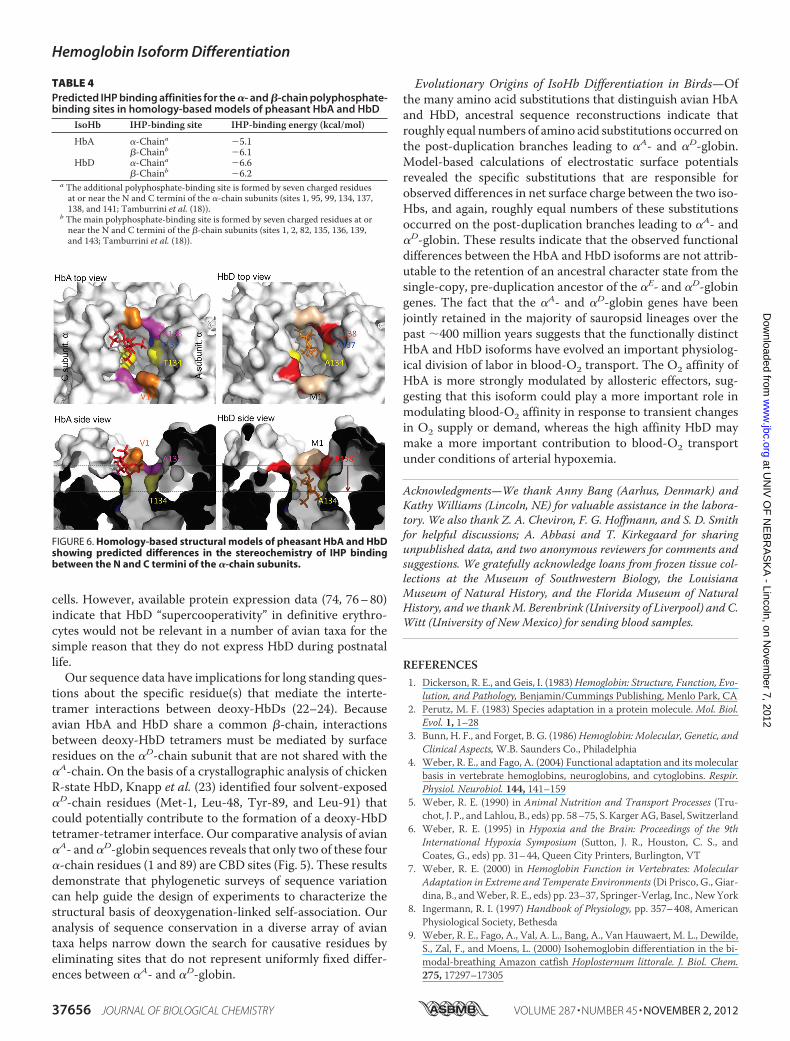
entiation—The simulation-based autodocking experimentspredicted a slightly lower O2-binding energy (and hence higherO2 affinity) for the �-chain heme groups of HbD relative tothose of HbA, �0.5 kcal/mol versus �0.4 kcal/mol, respec-tively. The molecular dynamics simulations also predicted thatthe “additional” �-chain phosphate-binding site of HbD (sensuTamburrini et al. (18) and Riccio et al. (53)) has a slightly lowerIHP-binding energy relative to that of HbA (Table 4) and thatthe bound IHP molecule is lodged more deeply in the �-chainbinding cleft of HbD (Fig. 6). This isoform difference in thestereochemistry of IHP binding ismainly attributable to substi-tutions at three symmetry-related pairs of amino acid residuesas follows: Met-�D1 (which reduces electrostatic repulsion rel-
FIGURE 2. O2 affinity and cooperativity (P50 and n50, respectively) ofpheasant HbA and HbD as a function of pH, temperature, and in theabsence and presence of IHP (IHP/H4B ratio � 23.5). O2 equilibria weremeasured in 0.1 M NaHEPES buffer containing 0.1 M KCl. Heme concentration,0.08 mM (HbA) and 0.11 mM (HbD) and 0.10 (HbA �D).
TABLE 2O2 affinity differences between avian HbA and HbD isoforms in the absence of allosteric effectors (stripped) and in the presence of IHPIHP was present at saturating concentrations (IHP/Hb4 ratio �20), except where indicated.
�logP50 (HbA - HbD)Species Stripped/�KCl �IHP °C pH Buffer Ref.
AccipitriformesGyps fulvus �0.37a 0.06a 37 7.4 0.1 M NaHEPES, 0.1 M KCl This study
�0.41b 37 7.4 0.1 M NaHEPES, 0.1 M KCl This studyGyps ruppellii 0.20a �0.40a 38 7.5 0.1 M NaHEPES, 0.1 M KCl Weber et al. (20)
0.09b �0.20b 38 7.5 0.1 M NaHEPES, 0.1 M KCl Weber et al. (20)Trigonoceps occipitalis 0.10 0.06 38 7.5 0.1 M NaHEPES, 0.1 M KCl Hiebl et al. (12)
AnseriformesAnas platyrhinchos �0.32 0.45c 20 7.0 0.025 M TrisHCl/0.1 M NaCl Vandecasserie et al. (19)Anser anser 0.17 37 7.4 0.1 M NaHEPES, 0.1 M KCl Present studyApus apus 0.23 0.54 38 7.5 0.02 M TrisHCl/0.1 M NaCl Nothum et al. (14)
ApodiformesA. amazilia �0.03 0.11 37 7.4 0.1 M NaHEPES, 0.1 M KCl Present studyA. viridicauda �0.03 0.12 37 7.4 0.1 M NaHEPES, 0.1 M KCl Present studyC. violifer �0.07 0.12 37 7.4 0.1 M NaHEPES, 0.1 M KCl Present studyP. gigas 0.01 0.24 37 7.4 0.1 M NaHEPES, 0.1 M KCl Present studyP. malaris �0.03 0.15 37 7.4 0.1 M NaHEPES, 0.1 M KCl Present study
CharadriiformesCatharacta maccormicki 0.20 �0.15 37 7.5 0.1 M NaHepes /0.1 M NaCl Tamburrini et al. (18)
GalliformesGallus gallus 0.14 0.61c 20 7.0 0.025 M TrisHCl/0.1 M NaCl Vandecasserie et al. (19)
0.55 0.88 25 7.0 0.1 M NaHEPES, 0.1 M KCl Weber et al. (44)Meleagris gallopavo �0.27 0.35c 20 7.0 0.025 M TrisHCl/0.1 M NaCl Vandecasserie et al. (19)Phasianus colchicus �0.37 0.20c 20 7.0 0.025 M TrisHCl/0.1 M NaCl Vandecasserie et al. (19)
0.01 37 7.4 0.1 M NaHEPES, 0.1 M KCl Present study0.07 0.09 25 7.5 0.1 M NaHEPES, 0.1 M KCl Present study
PasseriformesCorvus frugilegus 0.13 37 7.4 0.1 M NaHEPES, 0.1 M KCl Present studyTroglodytes aedon 0.25 0.18 37 7.4 0.1 M NaHEPES, 0.1 M KCl Present study
PhoenicopteriformesPhoenicopterus roseus 0.24 �0.50 20 7.5 0.05 M TrisHCl/0.1 M NaCl Sanna et al. (17)
StruthioniformesStruthio camelus 0.30 0.48c 37 7.4 0.05 M TrisHCl/0.2 M NaCl Oberthür et al. (15)
0.13 0.16 37 7.4 0.1 M NaHEPES, 0.1 M KCl Present studya Comparison was between HbA and HbD.b Comparison was between HbA� and HbD.c IHP/Hb tetramer ratio is 1:1.
Hemoglobin Isoform Differentiation
NOVEMBER 2, 2012 • VOLUME 287 • NUMBER 45 JOURNAL OF BIOLOGICAL CHEMISTRY 37653
at UN
IV O
F N
EB
RA
SK
A - Lincoln, on N
ovember 7, 2012
ww
w.jbc.org
Dow
nloaded from

ative to Val-�A1), Glu-�D138 (which increases electrostaticattraction relative to Ala-�A138), and Ala-�D134 (which, rela-tive toThr-�A134, reduces steric hindrance in the cleft betweenthe �1 and �2 subunits).
DISCUSSION
IsoHb Composition of Avian Red Cells—In contrast to thevariable patterns of Hb heterogeneity in fishes and other ecto-thermic vertebrate groups (5, 7–9, 71–73), the two-componentHbA/HbD system of birds is remarkably consistent. A numberof bird species are known to express three or four structurallydistinct isoHbs in definitive erythrocytes (11, 14, 15, 74, 75), butin all such cases HbA and HbD (i.e. tetrameric assemblies thatincorporate the products of �A- and �D-globin, respectively)represent the twomain isoforms. HbD expression has also beensecondarily lost in a number of avian taxa (e.g. pigeons, para-keets, cuckoos, jays, herons, storks, and penguins (74, 76–80)).The same appears to be true for crocodilians (30–32), the sistergroup to Aves.
Structural and Functional Differentiation between HbA andHbD—It is difficult to pinpoint specific substitutions that maybe responsible for isoHb differences in intrinsic O2 affinity,although substitutions at intersubunit contact surfaces aregood candidates: three of the CBD sites (114, 115, and 117)represent �1�1 “packing” contacts. It is also possible that isoHbdifferences in intrinsic O2 affinity are attributable to differentcombinations of substitutions in different species. Studies ofisoHb differentiation in the tufted duck, common swift, Rup-pell’s griffon, and goshawk suggested that the higher O2 affinityof HbD may be attributable to the possession of Gln-�D38 orThr instead of Pro-�A38 (11, 14, 20, 81–83). In Hbs with�Gln-38 or Thr, the R-state (oxy) structure is stabilized by twohydrogen bonds with �His-97 and �Asp-99, whereas only thelatter hydrogen bond is possible in the T-state. Thus, HbDwithGln-�D38 or Thr is more highly stabilized in the R-state, andthe allosteric equilibrium is shifted in favor of this high affinityquaternary structure. This structural mechanism may contrib-ute to O2 affinity differences between HbA and HbD in the
TABLE 3Parameter estimates derived from O2 equilibrium measurements of pheasant HbA and HbD under the two state Monod-Wyman-Changeux(MWC) allosteric model (compare Fig. 3)In separate analyses, the number of O2-binding sites was freely estimated (q � free) or fixed at 4 (q � 4).
isoHb °C pH IHP/Hb4 P50 n50 nmax Pm KT KR L �G q
torr torr torr�1 ( S.E.) torr�1 ( S.E.) kJ mol�1
HbAq � free 25 7.487 4.13 2.60 2.63 3.89 0.0663 0.0054 2.0777 0.3396 1.3�104 8.38 4.54
25 7.050 7.12 2.61 2.65 6.73 0.0316 0.0008 2.1798 0.1974 3.5�104 10.10 3.89fixed q � 4 25 7.487 4.12 2.44 2.47 3.9 0.0637 0.0046 2.4835 0.3419 8.8�103 8.74 4.00
25 7.050 7.13 2.64 2.68 6.73 0.0320 0.0007 2.0496 0.1014 3.6�104 9.99 4.00HbDq � free 25 7.492 3.51 2.47 2.49 3.36 0.0752 0.0042 2.0168 0.1706 4.2�103 7.99 4.36
25 7.496 23.5 26.14 2.71 3.02 22.05 0.0162 0.0008 1.2308 0.8497 7.5�107 10.20 5.4937 7.433 6.05 2.10 2.12 5.72 0.0575 0.0046 1.0665 0.2236 1.7�103 7.070 4.10
fixed q � 4 25 7.492 3.50 2.38 2.39 3.36 0.0721 0.0034 2.1948 0.1497 3.0�103 8.20 4.0025 7.496 23.5 24.2 2.46 2.61 21.5 0.0143 0.0010 2.7�105 3.6�1010 1.1�1027 11.70 4.0037 7.433 6.05 2.08 2.10 5.72 0.0572 0.0037 1.1053 0.1196 1.6�103 7.12 4.00
FIGURE 3. Extended Hill plots of O2 equilibria (where Y � fractional O2 saturation) for pheasant HbA and HbD. A, HbA and HbD at 25 °C; B, HbD at 25 and37 °C and in the absence and presence of saturating IHP concentration (IHP/Hb ratio � 23.5). In each plot, the intercept of the lower asymptote with thehorizontal line at logY/(Y � 1) � 0 provides an estimate of KT, the O2 association constant of T-state deoxy-Hb, and the intercept of the upper asymptote withthe same line provides an estimate of KR, the O2 association constant of R-state oxyHb. Heme concentration 0.60 (HbA and HbD); other conditions are asdescribed in the legend for Fig. 2.
Hemoglobin Isoform Differentiation
37654 JOURNAL OF BIOLOGICAL CHEMISTRY VOLUME 287 • NUMBER 45 • NOVEMBER 2, 2012
at UN
IV O
F N
EB
RA
SK
A - Lincoln, on N
ovember 7, 2012
ww
w.jbc.org
Dow
nloaded from

particular species mentioned above, but it does not provide ageneral explanation for the observed patterns of functional dif-ferentiation between avian HbA andHbD because the majorityof bird species retain the ancestral Gln residue at this intersub-unit contact site in both �A- and �D-globin.
In addition to the isoHb differences in intrinsic O2 affinity,HbD also exhibits a consistently higher O2 affinity in the pres-ence of IHP (Tables 1 and 2). This indicates that HbD is lessresponsive to the inhibitory effects of IHP, a potent allostericeffector that preferentially binds and stabilizes the low affinityT-state quaternary structure of the Hb tetramer. The uniformdifference in IHP sensitivity between HbA and HbD is surpris-ing because themain polyphosphate-binding site is formed by acluster of positively charged �-chain residues that line the inte-rior of the central cavity (18, 84). Because HbA and HbD shareidentical�-chain subunits (and thus share the same phosphate-binding sites), the observed isoform differences in IHP sensitiv-ity must be attributable to one or more substitutions between
�A- and �D-globin that do not directly affect the main phos-phate-binding site. Experimental evidence suggests that anadditional polyphosphate-binding site is formed by seven resi-dues from each �-chain (sites 1, 95, 99, 134, 137, 138, and 141),which stabilize IHP via charge-charge interactions (18, 85, 86).Specifically,�Lys-99 and charged residues at the�-chainN andC termini of avian HbA and HbD are predicted to form six saltbridges with the negatively charged phosphate groups of IHP(18, 53). This additional phosphate-binding site is hypothesizedto serve as an “entry/leaving site,” which modulates Hb-O2affinity by enhancing phosphate uptake and transfer to themain oxygenation-linked binding site between the�-chain sub-units (18, 53). Of the seven �-chain residues that compose thisadditional phosphate-binding site, four represent CBD sitesthat distinguish avian�A- and�D-globin sequences (1, 134, 137,and 138). The role of �Val-1 in this additional phosphate-bind-ing site is implicated by the fact that carbamylation of the�-chain N termini produces a 40% reduction in IHP affinity(85). However, even though HbD exhibits a consistently higherO2 affinity than HbA in the presence of IHP (Tables 1 and 2),ourmolecular dynamics simulations predict that the additionalphosphate-binding site of HbD actually has a slightly lower IHPbinding energy (and hence, higher IHP affinity) than that ofHbA (Table 4). An alternative hypothesis suggested by resultsof themolecular dynamics simulations (Fig. 6) is that IHP bind-ing between the �1 and �2 subunits of HbD produces a second-order perturbation of quaternary structure that is propagatedto the main phosphate-binding site between the �-chainsubunits.Deoxygenation-linked Self-association of HbD—Our mea-
sures of Hb-O2 equilibria were conducted under standard con-ditions ([heme] � 0.3 mM (50)) where intrinsic functional dif-ferences between tetrameric HbA and HbD were not obscuredby possible effects of deoxygenation-linked self-association.Measurements of oxygenation properties under these condi-tionsmoreover permitmeaningful comparisons with data frompreviously published studies (Tables 1 and 2). The Hb concen-trations used in our experiments greatly exceeded the thresholdat which tetrameric vertebrate Hbs dissociate to dimers andmonomers (87) but were below the threshold at which deoxy-genation-linked self-association of HbD is expected to occur.Thus, our experimental data donot shed light on the prevalenceor physiological relevance of HbD self-association in avian red
FIGURE 4. Adair constants (k1, k2, k3, and k4) for pheasant HbA and HbD asa function of temperature, pH, and the absence and presence of IHP(derived from data shown in Fig. 2).
FIGURE 5. Reconstructed ancestral states of 39 sites that distinguish the �A- and �D-globin polypeptides. As shown in the inset phylogeny of �-like globingenes, ancestral states for each of the 39 sites were reconstructed for four separate nodes in the tree.
Hemoglobin Isoform Differentiation
NOVEMBER 2, 2012 • VOLUME 287 • NUMBER 45 JOURNAL OF BIOLOGICAL CHEMISTRY 37655
at UN
IV O
F N
EB
RA
SK
A - Lincoln, on N
ovember 7, 2012
ww
w.jbc.org
Dow
nloaded from
cells. However, available protein expression data (74, 76–80)indicate that HbD “supercooperativity” in definitive erythro-cytes would not be relevant in a number of avian taxa for thesimple reason that they do not express HbD during postnatallife.Our sequence data have implications for long standing ques-
tions about the specific residue(s) that mediate the interte-tramer interactions between deoxy-HbDs (22–24). Becauseavian HbA and HbD share a common �-chain, interactionsbetween deoxy-HbD tetramers must be mediated by surfaceresidues on the �D-chain subunit that are not shared with the�A-chain. On the basis of a crystallographic analysis of chickenR-state HbD, Knapp et al. (23) identified four solvent-exposed�D-chain residues (Met-1, Leu-48, Tyr-89, and Leu-91) thatcould potentially contribute to the formation of a deoxy-HbDtetramer-tetramer interface. Our comparative analysis of avian�A- and�D-globin sequences reveals that only two of these four�-chain residues (1 and 89) are CBD sites (Fig. 5). These resultsdemonstrate that phylogenetic surveys of sequence variationcan help guide the design of experiments to characterize thestructural basis of deoxygenation-linked self-association. Ouranalysis of sequence conservation in a diverse array of aviantaxa helps narrow down the search for causative residues byeliminating sites that do not represent uniformly fixed differ-ences between �A- and �D-globin.
Evolutionary Origins of IsoHb Differentiation in Birds—Ofthe many amino acid substitutions that distinguish avian HbAand HbD, ancestral sequence reconstructions indicate thatroughly equal numbers of amino acid substitutions occurred onthe post-duplication branches leading to �A- and �D-globin.Model-based calculations of electrostatic surface potentialsrevealed the specific substitutions that are responsible forobserved differences in net surface charge between the two iso-Hbs, and again, roughly equal numbers of these substitutionsoccurred on the post-duplication branches leading to �A- and�D-globin. These results indicate that the observed functionaldifferences between the HbA and HbD isoforms are not attrib-utable to the retention of an ancestral character state from thesingle-copy, pre-duplication ancestor of the �E- and �D-globingenes. The fact that the �A- and �D-globin genes have beenjointly retained in the majority of sauropsid lineages over thepast �400 million years suggests that the functionally distinctHbA and HbD isoforms have evolved an important physiolog-ical division of labor in blood-O2 transport. The O2 affinity ofHbA is more strongly modulated by allosteric effectors, sug-gesting that this isoform could play a more important role inmodulating blood-O2 affinity in response to transient changesin O2 supply or demand, whereas the high affinity HbD maymake a more important contribution to blood-O2 transportunder conditions of arterial hypoxemia.
Acknowledgments—We thank Anny Bang (Aarhus, Denmark) andKathy Williams (Lincoln, NE) for valuable assistance in the labora-tory. We also thank Z. A. Cheviron, F. G. Hoffmann, and S. D. Smithfor helpful discussions; A. Abbasi and T. Kirkegaard for sharingunpublished data, and two anonymous reviewers for comments andsuggestions. We gratefully acknowledge loans from frozen tissue col-lections at the Museum of Southwestern Biology, the LouisianaMuseum of Natural History, and the Florida Museum of NaturalHistory, and we thankM. Berenbrink (University of Liverpool) and C.Witt (University of New Mexico) for sending blood samples.
REFERENCES1. Dickerson, R. E., and Geis, I. (1983)Hemoglobin: Structure, Function, Evo-
lution, and Pathology, Benjamin/Cummings Publishing, Menlo Park, CA2. Perutz, M. F. (1983) Species adaptation in a protein molecule. Mol. Biol.
Evol. 1, 1–283. Bunn, H. F., and Forget, B. G. (1986)Hemoglobin: Molecular, Genetic, and
Clinical Aspects,W.B. Saunders Co., Philadelphia4. Weber, R. E., and Fago, A. (2004) Functional adaptation and its molecular
basis in vertebrate hemoglobins, neuroglobins, and cytoglobins. Respir.Physiol. Neurobiol. 144, 141–159
5. Weber, R. E. (1990) in Animal Nutrition and Transport Processes (Tru-chot, J. P., and Lahlou, B., eds) pp. 58–75, S. Karger AG, Basel, Switzerland
6. Weber, R. E. (1995) in Hypoxia and the Brain: Proceedings of the 9thInternational Hypoxia Symposium (Sutton, J. R., Houston, C. S., andCoates, G., eds) pp. 31–44, Queen City Printers, Burlington, VT
7. Weber, R. E. (2000) in Hemoglobin Function in Vertebrates: MolecularAdaptation in Extreme and Temperate Environments (Di Prisco, G., Giar-dina, B., andWeber, R. E., eds) pp. 23–37, Springer-Verlag, Inc., NewYork
8. Ingermann, R. I. (1997) Handbook of Physiology, pp. 357–408, AmericanPhysiological Society, Bethesda
9. Weber, R. E., Fago, A., Val, A. L., Bang, A., Van Hauwaert, M. L., Dewilde,S., Zal, F., and Moens, L. (2000) Isohemoglobin differentiation in the bi-modal-breathing Amazon catfish Hoplosternum littorale. J. Biol. Chem.275, 17297–17305
FIGURE 6. Homology-based structural models of pheasant HbA and HbDshowing predicted differences in the stereochemistry of IHP bindingbetween the N and C termini of the �-chain subunits.
TABLE 4Predicted IHP binding affinities for the �- and �-chain polyphosphate-binding sites in homology-based models of pheasant HbA and HbD
IsoHb IHP-binding site IHP-binding energy (kcal/mol)
HbA �-Chaina �5.1�-Chainb �6.1
HbD �-Chaina �6.6�-Chainb �6.2
a The additional polyphosphate-binding site is formed by seven charged residuesat or near the N and C termini of the �-chain subunits (sites 1, 95, 99, 134, 137,138, and 141; Tamburrini et al. (18)).
b The main polyphosphate-binding site is formed by seven charged residues at ornear the N and C termini of the �-chain subunits (sites 1, 2, 82, 135, 136, 139,and 143; Tamburrini et al. (18)).
Hemoglobin Isoform Differentiation
37656 JOURNAL OF BIOLOGICAL CHEMISTRY VOLUME 287 • NUMBER 45 • NOVEMBER 2, 2012
at UN
IV O
F N
EB
RA
SK
A - Lincoln, on N
ovember 7, 2012
ww
w.jbc.org
Dow
nloaded from
10. Baumann, R., Fischer, J., and Engelke, M. (1987) Functional properties ofprimitive and definitive red cells from chick embryo. Oxygen-bindingcharacteristics, pH, and membrane potential and response to hypoxia. J.Exp. Zool. Suppl. 1, 227–238
11. Hiebl, I., Weber, R. E., Schneeganss, D., Kosters, J., and Braunitzer, G.(1988) High altitude respiration of birds. Structural adaptations in themajor and minor hemoglobin component of adult Ruppell’s griffon (Gypsrueppellii, Aegypiinae). A new molecular pattern for hypoxic tolerance.Biol. Chem. Hoppe-Seyler 369, 217–232
12. Hiebl, I., Weber, R. E., Schneeganss, D., and Braunitzer, G. (1989) Highaltitude respiration of Falconiformes. The primary structure and func-tional properties of the major and minor hemoglobin components of theadult white-headed vulture (Trigonoceps occipitalis, Aegypiinae). Biol.Chem. Hoppe-Seyler 370, 699–706
13. Isaacks, R. E., Harkness, D. R., Adler, J. L., and Goldman, P. H. (1976)Studies on avian erythrocyte metabolism. Effect of organic phosphates onoxygen affinity of embryonic and adult-type hemoglobins of the chickembryo. Arch. Biochem. Biophys. 173, 114–120
14. Nothum, R., Weber, R. E., Kosters, J., Schneeganss, D., and Braunitzer, G.(1989) Amino acid sequences and functional differentiation of hemoglo-bins A and D from swift (Apus, Apodiformes). Biol. Chem. Hoppe-Seyler370, 1197–1207
15. Oberthur, W., Braunitzer, G., Baumann, R., and Wright, P. G. (1983) Pri-mary structures of the �- and �-chains from the major hemoglobin com-ponent of the ostrich (Struthio camelus) and American rhea (Rhea amer-icana) (Struthioformes). Aspects of respiratory physiology and taxonomy.Hoppe-Seyler’s Z. Physiol. Chem. 363, 119–134
16. Rana,M. S., Knapp, J. E., Holland, R.A., andRiggs, A. F. (2008)ComponentD of chicken hemoglobin and the hemoglobin of the embryonic Tammarwallaby (Macropus eugenii) self-associate upon deoxygenation. Effect onoxygen binding. Proteins 70, 553–561
17. Sanna, M. T., Manconi, B., Podda, G., Olianas, A., Pellegrini, M., Castag-nola, M., Messana, I., and Giardina, B. (2007) Alkaline Bohr effect of birdhemoglobins. The case of the flamingo. Biol. Chem. 388, 787–795
18. Tamburrini, M., Riccio, A., Romano, M., Giardina, B., and di Prisco, G.(2000) Structural and functional analysis of the two hemoglobins of theAntarctic seabird Catharacta maccormicki. Characterization of an addi-tional phosphate-binding site by molecular modeling. Eur. J. Biochem.267, 6089–6098
19. Vandecasserie, C., Paul, C., Schnek, A. G., and Leonis, J. (1973) Oxygenaffinity of avian hemoglobins. Comp. Biochem. Physiol. A 44, 711–718
20. Weber, R. E., Hiebl, I., and Braunitzer, G. (1988) High altitude and hemo-globin function in the vultures Gyps rueppellii and Aegypius monachus.Biol. Chem. Hoppe-Seyler 369, 233–240
21. Lutz, P. L. (1980) On the oxygen affinity of bird blood. Am. Zool. 20,187–198
22. Cobb, J. A., Manning, D., Kolatkar, P. R., Cox, D. J., and Riggs, A. F. (1992)Deoxygenation-linked association of a tetrameric component of chickenhemoglobin. J. Biol. Chem. 267, 1183–1189
23. Knapp, J. E., Oliveira, M. A., Xie, Q., Ernst, S. R., Riggs, A. F., and Hackert,M. L. (1999) The structural and functional analysis of the hemoglobin Dcomponent from chicken. J. Biol. Chem. 274, 6411–6420
24. Rana, M. S., and Riggs, A. F. (2011) Indefinite noncooperative self-associ-ation of chicken deoxy hemoglobin D. Proteins 79, 1499–1512
25. Riggs, A. F. (1998) Self-association, cooperativity, and supercooperativityof oxygen binding by hemoglobins. J. Exp. Biol. 201, 1073–1084
26. Perutz, M. F., Steinkraus, L. K., Stockell, A., and Bangham, A. D. (1959)Chemical and crystallographic study of the two fractions of adult horsehemoglobin. J. Mol. Biol. 1, 402–404
27. Riggs, A. (1976) Factors in the evolution of hemoglobin function. Fed.Proc. 35, 2115–2118
28. Riggs, A. (1979) Studies of the hemoglobins of Amazonian fishes. Over-view. Comp. Biochem. Physiol. 62, 257–272
29. Nikinmaa, M. (2001) Hemoglobin function in vertebrates. Evolutionarychanges in cellular regulation in hypoxia. Respir. Physiol. 128, 317–329
30. Jensen, F. B. (2004) Red blood cell pH, the Bohr effect, and other oxygen-ation-linked phenomena in blood O2 and CO2 transport. Acta Physiol.Scand. 182, 215–227
31. Hoffmann, F. G., and Storz, J. F. (2007) The �D-globin gene originated viaduplication of an embryonic �-like globin gene in the ancestor of tetrapodvertebrates.Mol. Biol. Evol. 24, 1982–1990
32. Hoffmann, F. G., Storz, J. F., Gorr, T. A., and Opazo, J. C. (2010) Lineage-specific patterns of functional diversification in the �- and �-globin genefamilies of tetrapod vertebrates.Mol. Biol. Evol. 27, 1126–1138
33. Storz, J. F., Opazo, J. C., and Hoffmann, F. G. (2011) Phylogenetic diversi-fication of the globin gene superfamily in chordates. IUBMB Life 63,313–322
34. Cirotto, C., Panara, F., and Arangi, I. (1987) The minor hemoglobins ofprimitive and definitive erythrocytes of the chicken embryo. Evidence forhemoglobin L. Development 101, 805–813
35. Alev, C., Shinmyozu, K., McIntyre, B. A., and Sheng, G. (2009) Genomicorganization of zebra finch �- and �-globin genes and their expression inprimitive and definitive blood in comparison with globins in chicken.Dev.Genes Evol. 219, 353–360
36. Storz, J. F., Hoffmann, F. G., Opazo, J. C., Sanger, T. J., and Moriyama, H.(2011) Developmental regulation of hemoglobin synthesis in the greenanole lizard, Anolis carolinensis. J. Exp. Biol. 214, 575–581
37. Weber, R. E., and White, F. N. (1986) Oxygen binding in alligator bloodrelated to temperature, diving, and “alkaline tide.” Am. J. Physiol. 251,R901–R908
38. Weber, R., and White, F. (1994) Chloride-dependent organic phosphatesensitivity of the oxygenation reaction in crocodilian hemoglobins. J. Exp.Biol. 192, 1–11
39. Grigg, G. C., Wells, R. M. G., and Beard, L. A. (1993) Allosteric control ofoxygen binding by hemoglobin during development in the crocodileCrocodylus porosus. The role of red cell organic phosphates and carbondioxide. J. Exp. Biol. 175, 15–32
40. Hoffmann, F. G., Opazo, J. C., and Storz, J. F. (2008) Rapid rates of lineage-specific gene duplication and deletion in the �-globin gene family. Mol.Biol. Evol. 25, 591–602
41. Hoffmann, F. G., Opazo, J. C., and Storz, J. F. (2011) Differential loss andretention of myoglobin, cytoglobin, and globin-E during the radiation ofvertebrates. Genome Biol. Evol. 3, 588–600
42. Brittain, T. (2002) Molecular aspects of embryonic hemoglobin function.Mol. Aspects Med. 23, 293–342
43. Weber, R. E., Ostojic, H., Fago, A., Dewilde, S., Van Hauwaert, M. L.,Moens, L., and Monge, C. (2002) Novel mechanism for high altitude ad-aptation in hemoglobin of the Andean frog Telmatobius peruvianus.Am. J. Physiol. Regul. Integr. Comp. Physiol. 283, R1052–R1060
44. Weber, R. E., Voelter, W., Fago, A., Echner, H., Campanella, E., and Low,P. S. (2004) Modulation of red cell glycolysis. Interactions between verte-brate hemoglobins and cytoplasmic domains of band 3 red cell membraneproteins. Am. J. Physiol. Regul. Integr. Comp. Physiol. 287, R454–R464
45. Brygier, J., and Paul, C. (1976) Oxygen equilibrium of chicken hemoglobinin the presence of organic phosphates. Biochimie 58, 755–756
46. Monod, J., Wyman, J., and Changeux, J. P. (1965) On the nature of allos-teric transitions. A plausible model. J. Mol. Biol. 12, 88–118
47. Weber, R. E., Malte, H., Braswell, E. H., Oliver, R. W., Green, B. N.,Sharma, P. K., Kuchumov, A., and Vinogradov, S. N. (1995)Mass spectro-metric composition, molecular mass, and oxygen binding ofMacrobdelladecora hemoglobin and its tetramer and monomer subunits. J. Mol. Biol.251, 703–720
48. Adair, G. S. (1925) The hemoglobin system. IV. The oxygen dissociationcurve of hemoglobin. J. Biol. Chem. 63, 529–545
49. Ferry, M. F., and Green, A. A. (1929) Studies in the chemistry of hemoglo-bin. III. The equilibrium between oxygen and hemoglobin and its relationto changing hydrogen ion activity. J. Biol. Chem. 81, 175–203
50. Imai, K. (1982) Allosteric Effects in Hemoglobin, Cambridge University,Press, Cambridge, UK
51. Weber, R. E. (1992) Use of ionic and zwitterionic (Tris/BisTris andHEPES) buffers in studies on hemoglobin function. J. Appl. Physiol. 72,1611–1615
52. Arnold, K., Bordoli, L., Kopp, J., and Schwede, T. (2006) The SWISS-MODELWorkspace. A web-based environment for protein structure ho-mology modeling. Bioinformatics 22, 195–201
53. Riccio, A., Tamburrini,M., Giardina, B., and di PriscoG. (2001)Molecular
Hemoglobin Isoform Differentiation
NOVEMBER 2, 2012 • VOLUME 287 • NUMBER 45 JOURNAL OF BIOLOGICAL CHEMISTRY 37657
at UN
IV O
F N
EB
RA
SK
A - Lincoln, on N
ovember 7, 2012
ww
w.jbc.org
Dow
nloaded from

dynamics analysis of a second phosphate site in the hemoglobins of theseabird, south polar skua. Is there a site-site migratory mechanism alongthe central cavity? Biophys. J. 81, 1938–1946
54. Jo, S., Kim, T., Iyer, V. G., and Im, W. (2008) CHARMM-GUI. A web-based graphical user interface for CHARMM. J. Comput. Chem. 29,1859–1865
55. Gasteiger, E., Gattiker, A., Hoogland, C., Ivanyi, I., Appel, R. D., and Bai-roch, A. (2003) ExPASy. The proteomics server for in-depth proteinknowledge and analysis. Nucleic Acids Res. 31, 3784–3788
56. Trott, O., and Olson, A. J. (2010) AutoDock Vina. Improving the speedand accuracy of docking with a new scoring function, efficient optimiza-tion, and multithreading. J. Comput. Chem. 31, 455–461
57. Hoffmann, F. G., Opazo, J. C., and Storz, J. F. (2012) Whole-genome du-plication spurred the functional diversification of the globin gene super-family in vertebrates.Mol. Biol. Evol. 29, 303–312
58. Gribaldo, S., Casane, D., Lopez, P., and Philippe, H. (2003) Functionaldivergence prediction from evolutionary analysis. A case study of verte-brate hemoglobin.Mol. Biol. Evol. 20, 1754–1759
59. Gu, X. (2001) Maximum-likelihood approach for gene family evolutionunder functional divergence.Mol. Biol. Evol. 18, 453–464
60. Shannon, C. E. (1948) A mathematical theory of communication. BellSystem Tech. J. 27, 379–423, 623–656
61. Yang, Z., Kumar, S., and Nei, M. (1995) A new method of inference ofancestral nucleotide and amino acid sequences.Genetics 141, 1641–1650
62. Cao, Y., Adachi, J., Janke, A., Paabo, S., and Hasegawa, M. (1994) Phyloge-netic relationships among eutherian orders estimated from inferred se-quences of mitochondrial proteins. Instability of a tree based on a singlegene. J. Mol. Evol. 39, 519–527
63. Whelan, S., and Goldman, N. (2001) A general empirical model of proteinevolution derived from multiple protein families using a maximum-like-lihood approach.Mol. Biol. Evol. 18, 691–699
64. Yang, Z. (2007) PAML 4. Phylogenetic analysis by maximum likelihood.Mol. Biol. Evol. 24, 1586–1591
65. Edgar, R. C. (2004) MUSCLE. Multiple sequence alignment with highaccuracy and high throughput. Nucleic Acids Res. 32, 1792–1797
66. Hackett, S. J., Kimball, R. T., Reddy, S., Bowie, R. C., Braun, E. L., Braun,M. J., Chojnowski, J. L., Cox, W. A., Han, K. L., Harshman, J., Huddleston,C. J., Marks, B. D., Miglia, K. J., Moore, W. S., Sheldon, F. H., Steadman,D. W., Witt, C. C., and Yuri, T. (2008) A phylogenomic study of birdsreveals their evolutionary history. Science 320, 1763–1768
67. Gill, S. J., Gaud, H. T., and Barisas, B. G. (1980) Calorimetric studies ofcarbon monoxide and inositol hexaphosphate binding to hemoglobin A.J. Biol. Chem. 255, 7855–7857
68. Wyman, J., Jr. (1964) Linked functions and reciprocal effects in hemoglo-bin. A second look. Adv. Protein Chem. 19, 223–286
69. Tyuma, I., Imai, K., and Shimizu, K. (1973) Analysis of oxygen equilibriumof hemoglobin and control mechanism of organic phosphates. Biochem-istry 12, 1491–1498
70. Weber, R. E., Jensen, F. B., and Cox, R. P. (1987) Analysis of teleost hemo-globin byAdair andMonod-Wyman-Changeuxmodels. Effects of nucleo-side triphosphates and pH on oxygenation of tench hemoglobin. J. Comp.Physiol. B 157, 145–152
71. Binotti, I., Giovenco, S., Giardina, B., Antonini, E., Brunori, M., and Wy-man, J. (1971) Studies on the functional properties of fish hemoglobins. II.
The oxygen equilibrium of the isolated hemoglobin components fromtrout blood. Arch. Biochem. Biophys. 142, 274–280
72. Weber, R. E., and Jensen, F. B. (1988) Functional adaptations in hemoglo-bins from ectothermic vertebrates. Annu. Rev. Physiol. 50, 161–179
73. Weber, R. E. (1996) in Physiology and Biochemistry of the Fishes of theAmazon (Val, A. L., Almeida-Val, V.M., and Randall, D. J., eds) pp. 75–90,INPA, Brazil
74. Saha, A., and Ghosh, J. (1965) Comparative studies on avian hemoglobins.Comp. Biochem. Physiol. 15, 217–235
75. Lee, K. S., Huang, P. C., andCohen, B.H. (1976) Further resolution of adultchick hemoglobins by isoelectric focusing in polyacrylamide gel. Biochim.Biophys. Acta 427, 178–196
76. Godovac-Zimmermann, J., and Braunitzer, G. (1984) Hemoglobin of theadult white stork (Ciconia, Ciconiiformes). The primary structure of �A-and �-chains from the only present hemoglobin component. Hoppe-Sey-ler’s Z. Physiol. Chem. 365, 1107–1113
77. Godovac-Zimmermann, J., and Braunitzer, G. (1985) The primary struc-ture of �A- and �-chains from blue-and-yellow macaw (Ara ararauna,Psittaci) hemoglobin. No evidence for expression of �D-chains. Biol.Chem. Hoppe-Seyler 366, 503–508
78. Oberthur, W., Godovac-Zimmermann, J., and Braunitzer, G. (1986) Theexpression of �D-chains in the hemoglobin of adult ostrich (Struthio cam-elus) and American rhea (Rhea americana). The different evolution ofadult bird �A-, �D-, and �-chains. Biol. Chem. Hoppe-Seyler 367, 507–514
79. Sultana, C., Abbasi, A., and Zaidi, Z. H. (1989) Primary structure of hemo-globin �-chain of Columba livia (gray wild pigeon). J. Protein Chem. 8,629–646
80. Tamburrini, M., Condo, S. G., di Prisco, G., and Giardina, B. (1994) Ad-aptation to extreme environments. Structure-function relationships inemperor penguin hemoglobin. J. Mol. Biol. 237, 615–621
81. Hiebl, I., Kosters, J., and Braunitzer, G. (1987) The primary structures ofthe major and minor hemoglobin component of adult goshawk (Accipitergentilis, Accipitrinae). Biol. Chem. Hoppe-Seyler 368, 333–342
82. Abbasi, A., and Lutfullah, G. (2002) Molecular basis of bird respiration.Primary hemoglobin structure component from tufted duck (Aythya ful-igula, Anseriformes). Role of �Arg-99 in formation of a complex saltbridge network. Biochem. Biophys. Res. Commun. 291, 176–184
83. Lutfullah, G., Ali, S. A., and Abbasi, A. (2005) Molecular mechanism ofhigh altitude respiration. Primary structure of a minor hemoglobin com-ponent from tufted duck (Aythya fuligula, Anseriformes). Biochem. Bio-phys. Res. Commun. 326, 123–130
84. Arnone, A., and Perutz, M. F. (1974) Structure of inositol hexaphosphate-human deoxyhemoglobin complex. Nature 249, 34–36
85. Zuiderweg, E. R., Hamers, L. F., Rollema, H. S., de Bruin, S. H., andHilbers,C. W. (1981) 31P NMR study of the kinetics of binding of myo-inositolhexakisphosphate to human hemoglobin. Observation of fast exchangekinetics in high affinity systems. Eur. J. Biochem. 118, 95–104
86. Amiconi, G., Bertollini, A., Bellelli, A., Coletta, M., Condo, S. G., andBrunori, M. (1985) Evidence for two oxygen-linked binding sites for poly-anions in dromedary hemoglobin. Eur. J. Biochem. 150, 387–393
87. Mills, F. C., Johnson, M. L., and Ackers, G. K. (1976) Oxygenation-linkedsubunit interactions in human hemoglobin. Experimental studies on theconcentration dependence of oxygenation curves. Biochemistry 15,5350–5362
Hemoglobin Isoform Differentiation
37658 JOURNAL OF BIOLOGICAL CHEMISTRY VOLUME 287 • NUMBER 45 • NOVEMBER 2, 2012
at UN
IV O
F N
EB
RA
SK
A - Lincoln, on N
ovember 7, 2012
ww
w.jbc.org
Dow
nloaded from
Copyright © 2022 FDOKUMEN