Identification and Functional Analysis of the Vision-Specific BBS3 (ARL6) Long Isoform
13
Identification and Functional Analysis of the Vision- Specific BBS3 (ARL6) Long Isoform Pamela R. Pretorius 1,2,3 , Lisa M. Baye 1 , Darryl Y. Nishimura 2,3 , Charles C. Searby 2,3 , Kevin Bugge 2,3 , Baoli Yang 4 , Robert F. Mullins 5 , Edwin M. Stone 2,5 , Val C. Sheffield 2,3 *, Diane C. Slusarski 1 1 Department of Biology, University of Iowa, Iowa City, Iowa, United States of America, 2 Howard Hughes Medical Institute, Chevy Chase, Maryland, United States of America, 3 Department of Pediatrics, University of Iowa, Iowa City, Iowa, United States of America, 4 Department of Obstetrics and Gynecology, University of Iowa, Iowa City, Iowa, United States of America, 5 Department of Ophthalmology and Visual Sciences, University of Iowa, Iowa City, Iowa, United States of America Abstract Bardet-Biedl Syndrome (BBS) is a heterogeneous syndromic form of retinal degeneration. We have identified a novel transcript of a known BBS gene, BBS3 (ARL6), which includes an additional exon. This transcript, BBS3L, is evolutionally conserved and is expressed predominantly in the eye, suggesting a specialized role in vision. Using antisense oligonucleotide knockdown in zebrafish, we previously demonstrated that bbs3 knockdown results in the cardinal features of BBS in zebrafish, including defects to the ciliated Kupffer’s Vesicle and delayed retrograde melanosome transport. Unlike bbs3, knockdown of bbs3L does not result in Kupffer’s Vesicle or melanosome transport defects, rather its knockdown leads to impaired visual function and mislocalization of the photopigment green cone opsin. Moreover, BBS3L RNA, but not BBS3 RNA, is sufficient to rescue both the vision defect as well as green opsin localization in the zebrafish retina. In order to demonstrate a role for Bbs3L function in the mammalian eye, we generated a Bbs3L-null mouse that presents with disruption of the normal photoreceptor architecture. Bbs3L-null mice lack key features of previously published Bbs-null mice, including obesity. These data demonstrate that the BBS3L transcript is required for proper retinal function and organization. Citation: Pretorius PR, Baye LM, Nishimura DY, Searby CC, Bugge K, et al. (2010) Identification and Functional Analysis of the Vision-Specific BBS3 (ARL6) Long Isoform. PLoS Genet 6(3): e1000884. doi:10.1371/journal.pgen.1000884 Editor: Akihiro Ikeda, University of Wisconsin-Madison, United States of America Received December 18, 2009; Accepted February 15, 2010; Published March 19, 2010 Copyright: ß 2010 Pretorius et al. This is an open-access article distributed under the terms of the Creative Commons Attribution License, which permits unrestricted use, distribution, and reproduction in any medium, provided the original author and source are credited. Funding: This work was supported by US National Institutes of Health Grants R01EY110298 and R01EY017168 to VCS and EMS, R01CA112369 to DCS (http:// www.nih.gov/), and an Iowa Cardiovascular Center Institutional Research Fellowship to LMB. VCS and EMS are Investigators of the Howard Hughes Medical Institute. The funders had no role in study design, data collection and analysis, decision to publish, or preparation of the manuscript. Competing Interests: The authors have declared that no competing interests exist. * E-mail: [email protected] Introduction Visual impairment and blindness have far reaching implications for society. Hundreds of individually rare, but collectively common Mendelian disorders can cause blindness. One of these disorders is a heterogeneous syndromic form of retinal degeneration, Bardet- Biedl Syndrome (BBS, OMIM 209900). This pleiotropic disorder is characterized by retinal degeneration, obesity, polydactyly, renal abnormalities, hypogenitalism and cognitive impairment [1–4]. Additionally, BBS is associated with an increased incidence of hypertension, diabetes mellitus and heart defects [1,2,5]. Although there is variability in the ocular phenotype between individuals, BBS patients typically present with early and progressive photoreceptor degeneration, leading to both central and periph- eral vision loss by the third decade of life [1,6–13]. To date, 14 genes (BBS1-14) have been implicated in BBS [14–28]. Analysis of mouse models of BBS (Bbs1 M390R/M390R , Bbs2 2/2 , Bbs4 2/2 and Bbs6 2/2 ) reveals that these mice have major components of the human phenotype including retinal degeneration, obesity, renal cysts and neurological deficits [29– 32]. Multiple lines of evidence suggest that BBS phenotypes involve cilia dysfunction in a range of tissues, including the retina. The vertebrate retina contains photoreceptors, highly polarized cells with a modified cilium (connecting cilium) that joins the photosensitive outer segment (OS) to the protein synthesizing inner segment (IS). The connecting cilium transports cellular components from the IS to the OS that are necessary for the structure and function of the OS [33,34]. Intraflagellar transport (IFT) proteins are important in this intraphotoreceptor transport process as they play a key role in both assembly and maintenance of photoreceptor cells [35–37]. Loss of IFT genes in vertebrates leads to abnormal OS development, retinal degeneration and mislocalization of photopigments [36,38,39]. Retina phenotypes observed in Bbs-null mice are similar to those seen with loss of IFT genes, indicating that BBS proteins play a role in transporting proteins through the connecting cilium into the OS of the photoreceptor. For instance, characterization of the retinal phenotype in the mouse model has shown that photoreceptor death is preceded by mislocalization of rhodopsin [29–32,40]. Recent work with two independently generated Bbs4-null mice indicates that Bbs4 proteins play an important role in establishing both correct structure as well as proper transport of phototransduction proteins [40,41]. In the zebrafish model system, individual knockdown of bbs genes results in defects in the ciliated Kupffer’s Vesicle (KV) and delayed retrograde transport within the melanosome [26,42,43]. Moreover, work in Caenor- habditis elegans has shown that bbs1, bbs3, bbs5, bbs7 and bbs8 localize to the basal body of ciliated cells and are involved in IFT [23,44]. Taken together, these data strongly support a role for BBS proteins in intracellular transport and cilia; thus further PLoS Genetics | www.plosgenetics.org 1 March 2010 | Volume 6 | Issue 3 | e1000884
Transcript of Identification and Functional Analysis of the Vision-Specific BBS3 (ARL6) Long Isoform

Identification and Functional Analysis of the Vision-Specific BBS3 (ARL6) Long IsoformPamela R. Pretorius1,2,3, Lisa M. Baye1, Darryl Y. Nishimura2,3, Charles C. Searby2,3, Kevin Bugge2,3, Baoli
Yang4, Robert F. Mullins5, Edwin M. Stone2,5, Val C. Sheffield2,3*, Diane C. Slusarski1
1 Department of Biology, University of Iowa, Iowa City, Iowa, United States of America, 2 Howard Hughes Medical Institute, Chevy Chase, Maryland, United States of
America, 3 Department of Pediatrics, University of Iowa, Iowa City, Iowa, United States of America, 4 Department of Obstetrics and Gynecology, University of Iowa, Iowa
City, Iowa, United States of America, 5 Department of Ophthalmology and Visual Sciences, University of Iowa, Iowa City, Iowa, United States of America
Abstract
Bardet-Biedl Syndrome (BBS) is a heterogeneous syndromic form of retinal degeneration. We have identified a noveltranscript of a known BBS gene, BBS3 (ARL6), which includes an additional exon. This transcript, BBS3L, is evolutionallyconserved and is expressed predominantly in the eye, suggesting a specialized role in vision. Using antisenseoligonucleotide knockdown in zebrafish, we previously demonstrated that bbs3 knockdown results in the cardinal featuresof BBS in zebrafish, including defects to the ciliated Kupffer’s Vesicle and delayed retrograde melanosome transport. Unlikebbs3, knockdown of bbs3L does not result in Kupffer’s Vesicle or melanosome transport defects, rather its knockdown leadsto impaired visual function and mislocalization of the photopigment green cone opsin. Moreover, BBS3L RNA, but not BBS3RNA, is sufficient to rescue both the vision defect as well as green opsin localization in the zebrafish retina. In order todemonstrate a role for Bbs3L function in the mammalian eye, we generated a Bbs3L-null mouse that presents withdisruption of the normal photoreceptor architecture. Bbs3L-null mice lack key features of previously published Bbs-null mice,including obesity. These data demonstrate that the BBS3L transcript is required for proper retinal function and organization.
Citation: Pretorius PR, Baye LM, Nishimura DY, Searby CC, Bugge K, et al. (2010) Identification and Functional Analysis of the Vision-Specific BBS3 (ARL6) LongIsoform. PLoS Genet 6(3): e1000884. doi:10.1371/journal.pgen.1000884
Editor: Akihiro Ikeda, University of Wisconsin-Madison, United States of America
Received December 18, 2009; Accepted February 15, 2010; Published March 19, 2010
Copyright: � 2010 Pretorius et al. This is an open-access article distributed under the terms of the Creative Commons Attribution License, which permitsunrestricted use, distribution, and reproduction in any medium, provided the original author and source are credited.
Funding: This work was supported by US National Institutes of Health Grants R01EY110298 and R01EY017168 to VCS and EMS, R01CA112369 to DCS (http://www.nih.gov/), and an Iowa Cardiovascular Center Institutional Research Fellowship to LMB. VCS and EMS are Investigators of the Howard Hughes MedicalInstitute. The funders had no role in study design, data collection and analysis, decision to publish, or preparation of the manuscript.
Competing Interests: The authors have declared that no competing interests exist.
* E-mail: [email protected]
Introduction
Visual impairment and blindness have far reaching implications
for society. Hundreds of individually rare, but collectively common
Mendelian disorders can cause blindness. One of these disorders is
a heterogeneous syndromic form of retinal degeneration, Bardet-
Biedl Syndrome (BBS, OMIM 209900). This pleiotropic disorder
is characterized by retinal degeneration, obesity, polydactyly, renal
abnormalities, hypogenitalism and cognitive impairment [1–4].
Additionally, BBS is associated with an increased incidence of
hypertension, diabetes mellitus and heart defects [1,2,5]. Although
there is variability in the ocular phenotype between individuals,
BBS patients typically present with early and progressive
photoreceptor degeneration, leading to both central and periph-
eral vision loss by the third decade of life [1,6–13].
To date, 14 genes (BBS1-14) have been implicated in BBS
[14–28]. Analysis of mouse models of BBS (Bbs1M390R/M390R,
Bbs22/2, Bbs42/2 and Bbs62/2) reveals that these mice have
major components of the human phenotype including retinal
degeneration, obesity, renal cysts and neurological deficits [29–
32]. Multiple lines of evidence suggest that BBS phenotypes
involve cilia dysfunction in a range of tissues, including the retina.
The vertebrate retina contains photoreceptors, highly polarized
cells with a modified cilium (connecting cilium) that joins the
photosensitive outer segment (OS) to the protein synthesizing
inner segment (IS). The connecting cilium transports cellular
components from the IS to the OS that are necessary for the
structure and function of the OS [33,34]. Intraflagellar transport
(IFT) proteins are important in this intraphotoreceptor transport
process as they play a key role in both assembly and maintenance
of photoreceptor cells [35–37]. Loss of IFT genes in vertebrates
leads to abnormal OS development, retinal degeneration and
mislocalization of photopigments [36,38,39].
Retina phenotypes observed in Bbs-null mice are similar to
those seen with loss of IFT genes, indicating that BBS proteins
play a role in transporting proteins through the connecting cilium
into the OS of the photoreceptor. For instance, characterization
of the retinal phenotype in the mouse model has shown that
photoreceptor death is preceded by mislocalization of rhodopsin
[29–32,40]. Recent work with two independently generated
Bbs4-null mice indicates that Bbs4 proteins play an important role
in establishing both correct structure as well as proper transport
of phototransduction proteins [40,41]. In the zebrafish model
system, individual knockdown of bbs genes results in defects in the
ciliated Kupffer’s Vesicle (KV) and delayed retrograde transport
within the melanosome [26,42,43]. Moreover, work in Caenor-
habditis elegans has shown that bbs1, bbs3, bbs5, bbs7 and bbs8
localize to the basal body of ciliated cells and are involved in IFT
[23,44]. Taken together, these data strongly support a role for
BBS proteins in intracellular transport and cilia; thus further
PLoS Genetics | www.plosgenetics.org 1 March 2010 | Volume 6 | Issue 3 | e1000884

substantiating a critical role for BBS genes in the specialized
connecting cilium of the photoreceptor for cell maintenance and
function.
BBS3 is a member of the Ras superfamily of small GTP-binding
proteins, which is subdivided into ADP-ribosylation factor (ARF)
and ARF-like (ARL) subgroups [45]. The precise function of ARL
proteins is unknown, but it has been proposed that they play a role
in membrane and/or vesicular trafficking [45]. Work in C. elegans
indicates that ARL6 is specifically expressed in ciliated cells and
undergoes IFT along the ciliary axoneme [17]. Here we report the
identification of a second transcript of BBS3, BBS3L, and
determine that the BBS3L protein product plays an important
role in eye structure and function. BBS3L is evolutionally
conserved and is unique among BBS gene products as it is
expressed predominantly in the eye, suggesting a specialized role
in vision. We have established both mouse and zebrafish models to
study the function of BBS3L, and determined that BBS3L is
specifically required for retinal organization and function.
Results
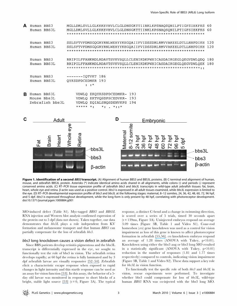
Identification of a second BBS3 transcript in human,mouse, and zebrafish
Expressed sequence tag (EST) data for human BBS3 was
compared to the known coding region of the gene. Although most
of the ESTs were virtually identical to the BBS3 reference
sequence, a few were found to contain 13 extra base pairs.
Interestingly, all ESTs that contained this alternative sequence
originated from retina or whole eye libraries, suggesting that this
second longer transcript, BBS3L, has an expression pattern that is
limited to the eye.
BBS3L results from differential splicing that leads to the
inclusion of a 13 base pair exon and a shift in the open reading
frame generating different C-terminal regions (Figure 1A). The
striking conservation of the C-terminal region of the long isoform
in human, mouse and zebrafish strongly suggests that bbs3L has
functional relevance (Figure 1B). To determine if the bbs3 and
bbs3L transcripts have similar tissue-specific expression in other
species, RT-PCR of zebrafish and mouse tissues was performed.
Zebrafish bbs3 is expressed in all adult tissues examined, while
bbs3L expression is limited to the eye (Figure 1C). Similar tissue
expression patterns for Bbs3 and Bbs3L were seen in the mouse
with the addition of low levels of Bbs3L mRNA expression in the
brain (Figure S1). A developmental profile in zebrafish embryos
reveals that while bbs3 is expressed throughout development, bbs3L
is not expressed until 48 hours post fertilization (hpf). This is a time
when retinal neuroepithelial cells are exiting the cell cycle and
differentiating into photoreceptor cells, the light sensing cells of the
retina [46] (Figure 1D).
Knockdown of bbs3 results in characteristic BBSphenotypes
To determine the functional role of bbs3L in development and
to distinguish the individual roles of the two bbs3 protein products,
we utilized antisense oligonucleotide mediated gene knockdown
(morpholinos, MO) in zebrafish. Two independent MOs were
utilized: one targeting the splice junction specific to the long
transcript (bbs3 long MO) and the other a previously described
MO targeting both transcripts (bbs3 aug MO) through blocking of
the translational start site [43] (Figure 2A). RT-PCR was used to
determine the knockdown efficiency of the bbs3 long MO on
staged embryos and demonstrated knockdown of the long
transcript through at least 5 days post fertilization (dpf) (Figure 2B).
Knockdown of bbs function in zebrafish generates two
prototypical defects: reduction of the size of the Kupffer’s vesicle
(KV) as well as retrograde transport defects [26,42,43]. As
previously demonstrated, alterations in the formation of the
ciliated KV was the earliest observable phenotype resulting from
knockdown of both bbs3 transcripts by the bbs3 aug MO [43]. At
the 8–10 somite stage (12–14 hpf) in wild-type and control injected
embryos the KV has formed in the posterior tailbud. The KV
diameter is approximately 50 mm and is larger than the width of
the notochord (Figure 2C and 2D). Injection of the bbs3 aug MO
resulted in a reduction of KV size to a width less than that of the
notochord (Figure 2E). Knockdown of both bbs3 transcripts by the
aug MO results in a statistically significant increase in embryos
with KV defects (Fisher’s exact test, p,0.001) (Figure 2F). Of note,
injection of the bbs3 long MO does not lead to KV defects
(Figure 2F).
The second prototypical phenotype observed in bbs MO-
injected embryos (morphants) is delayed trafficking of melano-
somes. Zebrafish are able to adapt to their surroundings through
intracellular trafficking of melanosomes within melanophores in
response to light and hormonal stimuli [47–50]. To test the rate of
this movement, 5-day old zebrafish were dark adapted, to
maximally disperse the melanosomes (Figure 2G and 2H) and
then treated with epinephrine to chemically stimulate the
retrograde transport of melanosomes [42,43,51] (Figure 2I).
Wild-type and control injected embryos show rapid movement
of melanosomes to a perinuclear location averaging 1.4 minutes,
whereas bbs3 aug MO injected embryos demonstrated a
statistically significant delay averaging 2.1 minutes (ANOVA with
Tukey, p,0.01) (Figure 2J). In contrast, the rate of melanosome
movement in bbs3 long knockdown embryos is statistically the
same as control embryos, averaging 1.5 minutes (Figure 2J).
To test for MO-specificity as well as differential function of the
two bbs3 transcripts, human RNAs of both BBS3 and BBS3L were
used. Embryos co-injected with a combination of MO and RNA
were evaluated for suppression of MO induced KV and
melanosome transport defects. Co-injection of BBS3 RNA with
the aug MO did not rescue the KV defect but was sufficient to
suppress the melanosome transport delay; however, co-injection of
the aug MO with BBS3L RNA was not able to suppress either
Author Summary
Retinitis pigmentosa (RP), a disorder of retinal degenera-tion resulting in blindness, occurs due to mutations indozens of different genes encoding proteins with highlydiverse functions. To date, there are no effective therapiesto delay or arrest retinal degeneration. RP places a largeburden on affected families and on society as a whole. Wehave studied a syndromic form of RP known as Bardet-Biedl Syndrome (BBS), which leads to degeneration of thephotoreceptor cells and is associated with non-visionabnormalities including obesity, hypertension, diabetes,and congenital abnormalities of the kidney, heart, andlimbs. In this study we utilized two model systems, thezebrafish and mouse, to evaluate the function of a specificform of BBS (BBS3). We have identified a novel proteinproduct of the BBS3 gene and demonstrated thatfunctional and structural abnormalities of the eye occurwhen this form of BBS3 is absent. This finding is ofsignificance because it indicates that BBS3 mutations canlead to non-syndromic blindness, as well as blindnessassociated with other clinical features. This work alsoindicates that treatment of BBS3 blindness will requirereplacement of a specific form of the BBS3 gene.
Vision-Specific Role of BBS3 (ARL6) Long Isoform
PLoS Genetics | www.plosgenetics.org 2 March 2010 | Volume 6 | Issue 3 | e1000884
MO-induced defect (Table S1). Myc-tagged BBS3 and BBS3L
RNA injection and Western blot analysis confirmed expression of
the protein out to 5 dpf (data not shown). Taken together, our data
demonstrates that bbs3L plays a role independent from KV
formation and melanosome transport and that human BBS3 can
partially compensate for the loss of zebrafish bbs3.
bbs3 long knockdown causes a vision defect in zebrafishSince BBS patients develop retinitis pigmentosa and the bbs3L
transcript is differentially expressed in the eye, we sought to
functionally test the role of bbs3 in vision. The zebrafish retina
develops rapidly; at 60 hpf the retina is fully laminated and by 3
dpf zebrafish larvae are visually responsive [52–54]. Zebrafish
elicit a characteristic escape response when exposed to rapid
changes in light intensity and this startle response can be used as
an assay for vision function [53]. In this assay, the behavior of a 5-
day old larvae was monitored in response to short blocks of a
bright, stable light source [53] (t = 0, Figure 3A). The typical
response, a distinct C-bend and a change in swimming direction,
is scored over a series of 5 trials, timed 30 seconds apart
(t = 139ms, Figure 3A). Uninjected embryos respond on average
3.09 times (Figure 3B, Table 1 and Video S1). Cone-rod
homeobox (crx) gene knockdown was used as a control for vision
impairment as loss of this gene is known to affect photoreceptor
formation in zebrafish [55,56]. crx knockdown embryos respond
an average of 1.28 times (ANOVA with Tukey, p,0.01).
Knockdown using either the bbs3 aug or bbs3 long MO resulted
in a statistically significant (ANOVA with Tukey, p,0.01)
reduction in the number of responses (1.81 and 1.77 times
respectively) compared to controls, indicating vision impairment
(Figure 3B, Table 1 and Video S2). These data support a key role
for bbs3L in vision function.
To functionally test the specific role of both bbs3 and bbs3L in
vision, rescue experiments were performed. To investigate
whether bbs3 could compensate for loss of bbs3L, wild-type
human BBS3 RNA was co-injected with the bbs3 long MO.
Figure 1. Identification of a second BBS3 transcript. (A) Alignment of human BBS3 and BBS3L proteins. (B) C-terminal end alignment of human,mouse, and zebrafish BBS3L protein. Asterisks (*) indicate identical amino acids shared in all alignments, while colons (:) and periods (.) representconserved amino acids. (C) RT–PCR tissue expression profile of zebrafish bbs3 and bbs3L transcripts in wild-type adult zebrafish tissues: fat, brain,heart, whole eye and retina. b-actin was used as a positive control. bbs3 is expressed in all adult tissues examined, while bbs3L expression is limited tothe eye. (D) RT–PCR developmental expression profile of bbs3 and bbs3L at the following stages: maternal, 8–12 somites, 24, 36, 42, 48, 60, 72, 96 hpf,and 5 dpf. bbs3 is expressed throughout development, while the long form is only present by 48 hpf, correlating with photoreceptor development.doi:10.1371/journal.pgen.1000884.g001
Vision-Specific Role of BBS3 (ARL6) Long Isoform
PLoS Genetics | www.plosgenetics.org 3 March 2010 | Volume 6 | Issue 3 | e1000884

Vision-Specific Role of BBS3 (ARL6) Long Isoform
PLoS Genetics | www.plosgenetics.org 4 March 2010 | Volume 6 | Issue 3 | e1000884
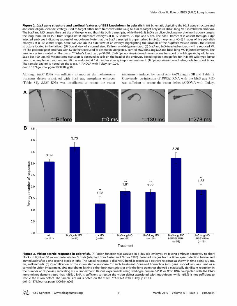
Although BBS3 RNA was sufficient to suppress the melanosome
transport delays associated with bbs3 aug morphant embryos
(Table S1), BBS3 RNA was insufficient to rescue the vision
impairment induced by loss of only bbs3L (Figure 3B and Table 1).
Conversely, co-injection of BBS3L RNA with the bbs3 aug MO
was sufficient to rescue the vision defect (ANOVA with Tukey,
Figure 2. bbs3 gene structure and cardinal features of BBS knockdown in zebrafish. (A) Schematic depicting the bbs3 gene structure andantisense oligonucleotide strategy used to target either both transcripts (bbs3 aug MO) or to target only bbs3L (bbs3 long MO) in zebrafish embryos.The bbs3 aug MO targets the start site of the gene and thus hits both transcripts, while the bbs3L MO is a splice-blocking morpholino that only targetsthe long form. (B) RT-PCR from staged bbs3L morphant embryos at 8–12 somites, 72 hpf, and 5 dpf. The bbs3L transcript is absent through 5 dpfinjected embryos indicating successful knockdown. Note that the bbs3 transcript is unperturbed in bbs3L morphants. (C–E) Images of live zebrafishembryos at 8–10 somite stage. Scale bar 200 mm. (C) Side view of an embryo highlighting the location of the Kupffer’s Vesicle (circle), the ciliatedstructure located in the tailbud. (D) Dorsal view of a normal sized KV from a wild-type embryo. (E) bbs3 aug MO–injected embryos with a reduced KV.(F) The percentage of embryos with KV defects (reduced or absent) in uninjected, control MO, bbs3 aug MO and bbs3 long MO injected embryos. Thesample size (n) is noted on the x-axis. **Fisher’s Exact test, p,0.001. (G–I) Epinephrine-induced melanosome transport of wild-type 6-day old larvae.Scale bar 100 mm. (G) Melanosome transport is observed in cells on the head of the embryos. Boxed region is magnified for (H,I). (H) Wild-type larvaeprior to epinephrine treatment and (I) the endpoint at 1.4 minutes after epinephrine treatment. (J) Epinephrine-induced retrograde transport times.The sample size (n) is noted on the x-axis. **ANOVA with Tukey, p,0.01.doi:10.1371/journal.pgen.1000884.g002
Figure 3. Vision startle response in zebrafish. (A) Vision function was assayed in 5-day old embryos by testing embryos sensitivity to shortblocks in light at 30 second intervals for 5 trials (adapted from Easter and Nicola 1996). Selected images from a time-lapse collection before andimmediately after a one second block in light. The typical response, a distinct C-bend, is scored as a positive response as shown in time point 139 ms.ms, milliseconds. (B) Quantification of the vision startle response for each treatment. Cone-rod homeobox (crx) gene knockdown was used as acontrol for vision impairment. bbs3 morphants lacking either both transcripts or only the long transcript showed a statistically significant reduction inthe number of responses, indicating visual impairment. Rescue experiments using wild-type human BBS3L or BBS3 RNA co-injected with the bbs3morpholinos demonstrated that hBBS3L RNA is sufficient to rescue the vision defect associated with knockdown, while hBBS3 is not sufficient torescue the vision defect. The sample size (n) is noted on the x-axis. **ANOVA with Tukey, p,0.01.doi:10.1371/journal.pgen.1000884.g003
Vision-Specific Role of BBS3 (ARL6) Long Isoform
PLoS Genetics | www.plosgenetics.org 5 March 2010 | Volume 6 | Issue 3 | e1000884

p,0.01) (Figure 3B and Table 1). Based on these rescue
experiments, bbs3L is necessary and sufficient for vision function.
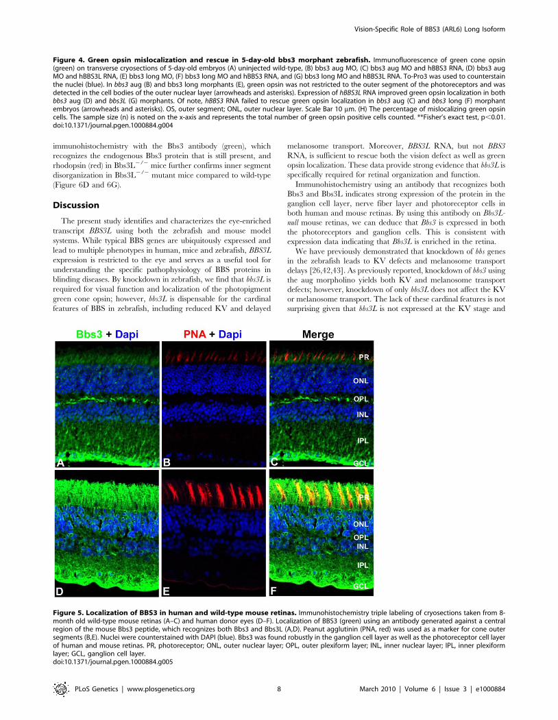
hBBS3L is sufficient to rescue green opsin mislocalizationin bbs3 morphant zebrafish
Previous work has demonstrated that Bbs1 M390R knockin,
Bbs2, Bbs4 and Bbs6 mutant mice initially form photoreceptors;
however, the photoreceptors subsequently show a mislocalization
of rhodopsin, a photopigment protein, to the cell bodies of the
outer nuclear layer (ONL) and undergo progressive photoreceptor
degeneration [29–32,40]. By gross histology, wild-type, bbs3 aug
and bbs3 long morphant zebrafish embryo retinas displayed a fully
laminated retina at 5 dpf (data not shown). Ganglion cell
outgrowth and optic nerve formation was evaluated using the
ath5:GFP [Tg(atoh7:GFP)] transgenic line, a marker of ganglion
cell and axon outgrowth [57]. We found that gross retinal ganglion
axon trajectories were not perturbed in bbs3 aug or long
morphants (data not shown).
While the overall architecture of the retina appeared morpho-
logically normal at 5 dpf, we investigated photopigment
localization in bbs3 morphants. Photopigments are known to
localize to the outer segment of the zebrafish photoreceptor;
therefore, we assessed opsin localization using an antibody specific
to green cone opsin [58]. In the wild-type retina, green opsin is
found in the outer-segment of the green cone photoreceptor
(Figure 4A). In bbs3 aug and bbs3 long morphants green opsin
expression was not restricted to the outer segments of the
photoreceptors; rather, green opsin was also detected in the cell
bodies of the outer nuclear layer throughout the entire retina
(Figure 4B and 4E).
To determine whether there is a functional difference between
BBS3 and BBS3L in its ability to rescue the green opsin localization
in the photoreceptors of MO-injected embryos rescue experiments
were performed. The first question we addressed was if BBS3L
RNA was sufficient to rescue green opsin localization in morphant
embryos. Expression of wild-type human BBS3L RNA led to
improved green opsin localization in both bbs3 aug and bbs3L
morphant embryos (Figure 4D and 4G). The percentage of cells
mislocalizing green opsin was quantified and indeed BBS3L RNA
was able to statistically rescue the green opsin defect in bbs3 aug
morphants (Fisher’s exact test, p,0.01) (Figure 4H and Table 1).
We next investigated whether BBS3 could compensate for loss of
bbs3L in the zebrafish retina. Co-injection of wild-type human
BBS3 RNA failed to rescue green opsin localization in bbs3 aug
and bbs3L morphant embryos (Figure 4C, 4F, and 4H and
Table 1). These data are consistent with the vision startle response
rescue data and supports the hypothesis that BBS3L has an eye
specific role. Moreover, these data support a specific role for bbs3L
in the retina and for localization of proteins within the
photoreceptor cell.
Bbs3 is expressed in ganglion and photoreceptor cells inmouse and human retinas
A polyclonal antibody against a central region of the mouse
Bbs3 peptide, which is conserved across human and mouse, was
generated to recognize both isoforms of Bbs3. Cellular localization
of Bbs3 was assessed in donor human and mouse retinal tissue.
Immunohistochemistry was performed on transverse cryosections
from adult human and adult mouse eyes using the Bbs3 antibody.
Staining revealed expression of Bbs3 (green) in the ganglion cell
layer and the nerve fiber layer as well as the photoreceptor cells of
both mouse (Figure 5A) and human retinal tissue (Figure 5D).
Additionally, peanut agglutinin (PNA, red) was used as a marker
for cone outer segments in both mouse (Figure 5B) and human
retinal sections (Figure 5E). The merge represents the co-
localization of Bbs3 (green) and PNA (red) in the photoreceptor
cells of both mouse (Figure 5C) and human (Figure 5F). The
specificity of the BBS3 antibody for immunohistochemistry was
confirmed through peptide blocking of the antibody on wild-type
mouse retina (Figure S2).
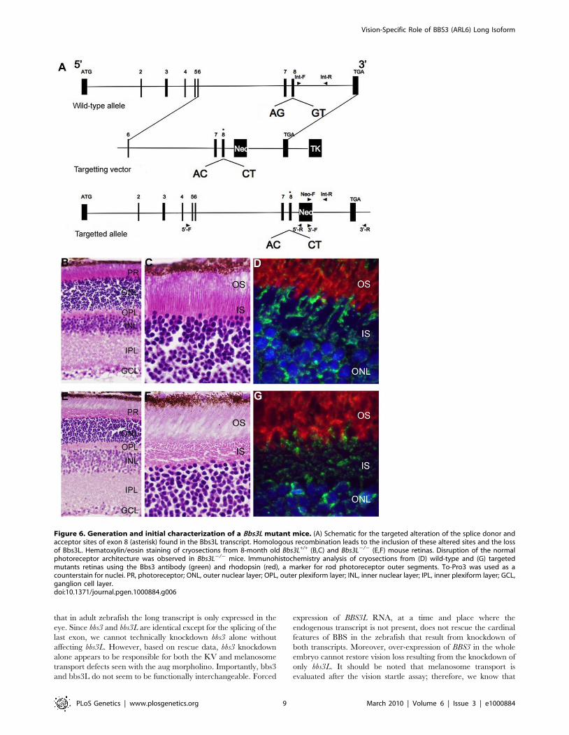
Bbs3L2/2 mice display structural abnormalitiesTo characterize the effects of loss of Bbs3L on mammalian
photoreceptors a targeted knockin of the long form of Bbs3 was
carried out by altering the splice donor and acceptor sites flanking
exon 8, leading to the exclusion of exon 8 upon homologous
recombination (Figure 6A). This approach leads to the preserva-
tion of Bbs3 expression in the Bbs3L-null mice. RT-PCR confirmed
the generation and transmission of the Bbs3L allele in +/2 and
2/2 mice (Figure S3). Unlike previously generated BBS knockout
mice, which are obese by 7 months of age, Bbs3L2/2 mice do not
become obese (data not shown) [29–32]. This supports the idea
that Bbs3L function is restricted to the retina and is consistent with
the zebrafish knockdown studies.
Gross histological examination of 8-month-old wild-type and
homozygous (Bbs3L2/2) mutant mice revealed that while all cell
layers were present (Figure 6B and 6E), the inner segments of the
photoreceptors were disrupted in a majority of the mutant mice as
compared to wild-type (Figure 6C and 6F). In wild-type mice, the
inner segment layer is arranged in a parallel array; while in the
Bbs3L-null mice the parallel arrangement of the IS was eccentric
with individual inner segments randomly oriented. Additionally,
Table 1. Vision startle assay and percentage of green opsin mislocalization.
Treatment Vision (# of responses) n Mislocalized green opsin (%) n
wt 3.09 181 10.6 585
bbs3 aug MO 1.81** 168 19.1++ 555
bbs3 aug MO + hBBS3 RNA 2.13** 54 20.4++ 496
bbs3 aug MO + hBBS3L RNA 3.25 53 12.78 227
bbs3 long MO 1.77** 158 21.0++ 905
bbs3 long MO + hBBS3 RNA 1.88** 40 18.8++ 330
bbs3 long MO + hBBS3L RNA 2.7 53 10.9 357
** ANOVA and Tukey test, p,0.01 as compared to wt.++ Fisher’s Exact test, p,0.01 as compared to wt.doi:10.1371/journal.pgen.1000884.t001
Vision-Specific Role of BBS3 (ARL6) Long Isoform
PLoS Genetics | www.plosgenetics.org 6 March 2010 | Volume 6 | Issue 3 | e1000884

Vision-Specific Role of BBS3 (ARL6) Long Isoform
PLoS Genetics | www.plosgenetics.org 7 March 2010 | Volume 6 | Issue 3 | e1000884
immunohistochemistry with the Bbs3 antibody (green), which
recognizes the endogenous Bbs3 protein that is still present, and
rhodopsin (red) in Bbs3L2/2 mice further confirms inner segment
disorganization in Bbs3L2/2 mutant mice compared to wild-type
(Figure 6D and 6G).
Discussion
The present study identifies and characterizes the eye-enriched
transcript BBS3L using both the zebrafish and mouse model
systems. While typical BBS genes are ubiquitously expressed and
lead to multiple phenotypes in human, mice and zebrafish, BBS3L
expression is restricted to the eye and serves as a useful tool for
understanding the specific pathophysiology of BBS proteins in
blinding diseases. By knockdown in zebrafish, we find that bbs3L is
required for visual function and localization of the photopigment
green cone opsin; however, bbs3L is dispensable for the cardinal
features of BBS in zebrafish, including reduced KV and delayed
melanosome transport. Moreover, BBS3L RNA, but not BBS3
RNA, is sufficient to rescue both the vision defect as well as green
opsin localization. These data provide strong evidence that bbs3L is
specifically required for retinal organization and function.
Immunohistochemistry using an antibody that recognizes both
Bbs3 and Bbs3L indicates strong expression of the protein in the
ganglion cell layer, nerve fiber layer and photoreceptor cells in
both human and mouse retinas. By using this antibody on Bbs3L-
null mouse retinas, we can deduce that Bbs3 is expressed in both
the photoreceptors and ganglion cells. This is consistent with
expression data indicating that Bbs3L is enriched in the retina.
We have previously demonstrated that knockdown of bbs genes
in the zebrafish leads to KV defects and melanosome transport
delays [26,42,43]. As previously reported, knockdown of bbs3 using
the aug morpholino yields both KV and melanosome transport
defects; however, knockdown of only bbs3L does not affect the KV
or melanosome transport. The lack of these cardinal features is not
surprising given that bbs3L is not expressed at the KV stage and
Figure 4. Green opsin mislocalization and rescue in 5-day-old bbs3 morphant zebrafish. Immunofluorescence of green cone opsin(green) on transverse cryosections of 5-day-old embryos (A) uninjected wild-type, (B) bbs3 aug MO, (C) bbs3 aug MO and hBBS3 RNA, (D) bbs3 augMO and hBBS3L RNA, (E) bbs3 long MO, (F) bbs3 long MO and hBBS3 RNA, and (G) bbs3 long MO and hBBS3L RNA. To-Pro3 was used to counterstainthe nuclei (blue). In bbs3 aug (B) and bbs3 long morphants (E), green opsin was not restricted to the outer segment of the photoreceptors and wasdetected in the cell bodies of the outer nuclear layer (arrowheads and asterisks). Expression of hBBS3L RNA improved green opsin localization in bothbbs3 aug (D) and bbs3L (G) morphants. Of note, hBBS3 RNA failed to rescue green opsin localization in bbs3 aug (C) and bbs3 long (F) morphantembryos (arrowheads and asterisks). OS, outer segment; ONL, outer nuclear layer. Scale Bar 10 mm. (H) The percentage of mislocalizing green opsincells. The sample size (n) is noted on the x-axis and represents the total number of green opsin positive cells counted. **Fisher’s exact test, p,0.01.doi:10.1371/journal.pgen.1000884.g004
Figure 5. Localization of BBS3 in human and wild-type mouse retinas. Immunohistochemistry triple labeling of cryosections taken from 8-month old wild-type mouse retinas (A–C) and human donor eyes (D–F). Localization of BBS3 (green) using an antibody generated against a centralregion of the mouse Bbs3 peptide, which recognizes both Bbs3 and Bbs3L (A,D). Peanut agglutinin (PNA, red) was used as a marker for cone outersegments (B,E). Nuclei were counterstained with DAPI (blue). Bbs3 was found robustly in the ganglion cell layer as well as the photoreceptor cell layerof human and mouse retinas. PR, photoreceptor; ONL, outer nuclear layer; OPL, outer plexiform layer; INL, inner nuclear layer; IPL, inner plexiformlayer; GCL, ganglion cell layer.doi:10.1371/journal.pgen.1000884.g005
Vision-Specific Role of BBS3 (ARL6) Long Isoform
PLoS Genetics | www.plosgenetics.org 8 March 2010 | Volume 6 | Issue 3 | e1000884
that in adult zebrafish the long transcript is only expressed in the
eye. Since bbs3 and bbs3L are identical except for the splicing of the
last exon, we cannot technically knockdown bbs3 alone without
affecting bbs3L. However, based on rescue data, bbs3 knockdown
alone appears to be responsible for both the KV and melanosome
transport defects seen with the aug morpholino. Importantly, bbs3
and bbs3L do not seem to be functionally interchangeable. Forced
expression of BBS3L RNA, at a time and place where the
endogenous transcript is not present, does not rescue the cardinal
features of BBS in the zebrafish that result from knockdown of
both transcripts. Moreover, over-expression of BBS3 in the whole
embryo cannot restore vision loss resulting from the knockdown of
only bbs3L. It should be noted that melanosome transport is
evaluated after the vision startle assay; therefore, we know that
Figure 6. Generation and initial characterization of a Bbs3L mutant mice. (A) Schematic for the targeted alteration of the splice donor andacceptor sites of exon 8 (asterisk) found in the Bbs3L transcript. Homologous recombination leads to the inclusion of these altered sites and the lossof Bbs3L. Hematoxylin/eosin staining of cryosections from 8-month old Bbs3L+/+ (B,C) and Bbs3L2/2 (E,F) mouse retinas. Disruption of the normalphotoreceptor architecture was observed in Bbs3L2/2 mice. Immunohistochemistry analysis of cryosections from (D) wild-type and (G) targetedmutants retinas using the Bbs3 antibody (green) and rhodopsin (red), a marker for rod photoreceptor outer segments. To-Pro3 was used as acounterstain for nuclei. PR, photoreceptor; ONL, outer nuclear layer; OPL, outer plexiform layer; INL, inner nuclear layer; IPL, inner plexiform layer; GCL,ganglion cell layer.doi:10.1371/journal.pgen.1000884.g006
Vision-Specific Role of BBS3 (ARL6) Long Isoform
PLoS Genetics | www.plosgenetics.org 9 March 2010 | Volume 6 | Issue 3 | e1000884

over-expressed BBS3 is functional at the time of the vision assay.
Although bbs3 may have some effect on vision that is below the
detection level of our assay, we have demonstrated that bbs3L
function is both necessary for vision and sufficient to rescue vision
loss in the zebrafish.
Similar to the zebrafish results, a Bbs3L-null mouse lacks the
observed phenotypes of previously published Bbs-null mice, such as
obesity [29–32]. The effect of Bbs3 in the mouse retina may be
more significant as Bbs3L-null mice present with only a variable
mild disruption of the normal architecture. This indicates that in
the mouse retina, Bbs3 is able to partially compensate for loss of
Bbs3L. Moreover, the difference in phenotype between zebrafish
and mouse could potentially be due to the ratio of cones and rods
found in each model system. One hypothesis is that bbs3L plays a
major functional role in cones, but only a minor role in rods. At
the stages examined in the zebrafish, cones are the only functional
photoreceptors in the retina, whereas mice have a rod-dominated
retina [59,60]. These attributes are important to consider when
looking at the role of BBS in human disease progression, as
humans rely on their fovea, a specialized cone-dominant structure
in the center of the macula, for visual acuity. Continued
characterization of the Bbs3L-null mouse may shed more light on
this difference between the mouse and zebrafish system, as well as
elucidate a more definitive role for BBS3L in the retina.
Taken together, these date demonstrate that the BBS3L
transcript is specifically required for retinal organization and
function. While we have identified a second transcript of BBS3, a
gene known to cause BBS, we would not expect patients with
mutations affecting only BBS3L to present with BBS. Based on our
findings in both a zebrafish and mouse model of BBS3L, patients
with mutations in BBS3L alone would present with a non-
syndromic retinal disease, characterized by photoreceptor dys-
function and death. Indeed, recent homozygosity mapping of a
consanguineous Saudi family has identified a missense mutation in
BBS3 that leads to non-syndromic RP [61]. Functional charac-
terization of this mutation in the zebrafish may provide additional
clues to the role of BBS3 in the eye. Thus this eye specific
transcript, BBS3L, will serve as a useful tool for understanding the
pathophysiology of other blinding diseases. In addition, our data
indicate that expression of BBS3L, rather than BBS3, would be
needed for gene therapy aimed at treatment of blindness in BBS3
patients.
Materials and Methods
Ethics statementAll animal work in this study was approved by the by the
University Animal Care and Use Committee at the University of
Iowa.
ESTExpressed sequence tag (EST) data for human and mouse BBS3
was downloaded from NCBI and compared to the known coding
region as represented by the NCBI reference sequence
(NM_177976.1 and NM_032146.3 for human and NM_019665.3
for mouse).
Danio rerioRT–PCR. RNA was extracted from a pool of 10–20 embryos
at the following stages: 8–12 somites, 24, 36, 42, 48, 60, 72, 96 hpf
and 5 dpf. Additionally, RNA was extracted from the following
adult tissues: fat, brain, heart, whole eye and retina. cDNA was
synthesized using oligo dT primers and bbs3 primer pair 1
recognizing both bbs3 transcripts were used to evaluate expression.
b-actin expression served as a control.
Primers:
bbs3 primer pair 1-F: 59-AAGGACAAACCATGGCATATC-39
bbs3 primer pair 1-R: 59-TTACGTTTTCATCGCTCTGAT-39
b-actin-F: 59-TCAGCCATGGATGATGAAAT-39
b-actin-R: 59-GGTCAGGATCTTCATGAGGT-39
Morpholino injections and knockdown efficiency.
Antisense morpholinos (MO) were designed and purchased from
Gene Tools.
bbs3_aug [43]: AGCTTGTCAAAAAGCCCCATTTGCT
bbs3_long: ATTTCAGCTTCAGTACTTACAGTGC
control MO bbs3_6mis: AaCTTGTgAAAtAGCgCCATaTGaT
crx: GGCTGCTTTATGTAGGACATCATTC
MOs (12 ng) were air-pressure-injected into one- to four-cell
staged embryos. Transcript knockdown efficiency was assessed by
RT-PCR as described above using bbs3 long splice-blocking
morphants at the following stages: 8–12 somites, 72 hpf and 5 dpf.
Primers recognizing both bbs3 (bbs3 primer pair 1) transcripts were
used to assess knockdown efficiency.
Human BBS3 cloning and RNA synthesis. Wild-type
human BBS3 and BBS3L constructs were generated by TA
cloning into the Gateway vector system (Invitrogen), and
subsequently subcloned into Gateway expression vectors with a
C-terminal mCherry or myc tag (generous gift from Chien and
Lawson Lab).
Primers:
-hBBS3-F: ATGGGATTGCTAGACAGACTTTC
-hBBS3-R: TGTCTTCACAGTCTGGATCTG
-hBBS3L-R: TCTTTTCATGTCTTCACAGTC
For rescue experiments, MO-resistant C-terminally tagged
mCherry RNA was synthesized using the mMessage mMachine
transcription kit (Ambion). hBBS3 or hBBS3L RNA (8 pg) was co-
injected with the appropriate MO into one- to four-cell staged
embryos.
Analysis of Kupffer’s Vesicle. Embryos with KVs smaller
than the width of the notochord (less than approximately 50 mm in
diameter) were considered reduced, while embryos in which KVs
could not be morphologically identified were scored as absent.
Live embryos were photographed on a stereoscope with a Zeiss
Axiocam camera.
Melanosome transport assay. The melanosome transport
assay was performed as previously described [26,42,43]. Dark-
adapted 5-day-post fertilization larvae were treated with
epinephrine (50 mg/ml, Sigma, E4375) added to egg water [62]
for a final concentration of 500 mg/ml. Melanosome retraction
time was monitored under the microscope. Live embryos were
photographed on a stereoscope with a Zeiss Axiocam camera.
Vision startle response assay. A visually evoked startle
response behavioral assay was modified from a previously
described assay [53]. Prior to experimentation, 5-day-old
zebrafish larvae were light adapted for 1 hour. A visual stimulus
was applied by performing rapid changes (approximately 1 second)
in white light intensity through abruptly opening and closing the
shutter located between the light source and the animal. An abrupt
movement of the zebrafish within one second of visual stimuli
application was scored as a positive response. After performing five
visual stimuli trials spaced at 30 seconds apart, the mechanical
stimulus response was evaluated by probing embryos with the tip
of a blunt needle. Embryos that failed to respond to the
mechanical stimulation, although rare, were not included in the
analysis.
Immunohistochemistry. Five day post-fertilized larvae
were fixed overnight at 4uC with 4% paraformaldehyde (PFA)
Vision-Specific Role of BBS3 (ARL6) Long Isoform
PLoS Genetics | www.plosgenetics.org 10 March 2010 | Volume 6 | Issue 3 | e1000884

prepared in BT buffer (4% sucrose, 0.1M CaCl2 in 0.1M PO4,
pH 7.3). Embryos were rinsed with phosphate-buffer saline (PBS)
and infiltrated at 4uC with 15% sucrose, 30% sucrose and
overnight in 100% optimal cutting temperature compound (OCT,
Sakura). Embryos were cryosectioned at 221uC. Sections were
collected at 12 mm and were allowed to dry for 1 hour at 25uC.
The tissues were incubated with blocking solution (5% normal
donkey serum, 0.1% tween-20, 1% DMSO in PBS) for 2 hours
and then incubated overnight at 4uC with mouse-anti-green cone
opsin diluted in blocking solution (1:500, generous gift from the
Hyde lab). Following washes with PBDT (PBS, 1% DMSO, 0.1%
tween-20) sections were incubated for 1.5 hours at 25uC with goat-
anti-mouse Alexa 488 (1:400, Molecular Probes) diluted in
blocking solution. Nuclei were counterstained with To-Pro3
(1:1000, Molecular Probes) diluted in PBS. Sections were
mounted in Vectashield mounting medium (Vector Laboratories)
and analyzed using a Leica SP2 laser confocal microscope system
with 636magnification and 3x zoom. Images are representative of
maximum projections of multiple focal planes (z-series).
Green opsin cell counts. The ratio of mislocalized green
opsin cells to total green opsin positive cells was determined from a
12 mm thick central retina image taken of a single eye.
Mislocalization of green opsin was defined as the presence of
green opsin present in the outer nuclear layer (ONL) of the retina.
The number of independent fish retinas counted per group were as
follows: wt n = 9, bbs3 aug MO n = 10, bbs3 long MO n = 15, bbs3
aug MO+ hBBS3L RNA n = 5, bbs3 long MO+hBBS3 RNA n = 5.
Scorers were masked to the genotype of the embryos.
Mus musculusGeneration of Bbs3L mutant mice. A targeting plasmid
was constructed by amplifying the 59 and 39 regions of Bbs3L using
genomic DNA isolated from the 129/SvJ mouse strain. The
consensus splice sites were ablated to alter the slice donor
(59- CTGCTGTCACAAAAAACAGTACACTAAGTATCTG-39)
and splice acceptor (59- CAGATACTTAGTGTACTGTTTT-
TTGTGACAGCAG-39) sites flanking exon 8, the exon
responsible for the long transcript. Following mutagenesis,
these regions were cloned into the targeting vector
pOSDUPDEL (a gift from O. Smithies, University of North
Carolina, Chapel Hill, NC, USA). The targeting construct was
linearized with NotI and electroporated into R1 embryonic stem
(ES) cells (129 X 1/SvJ3 129S1/Sv). Double selection of ES
cells was carried out for the presence of the neomycin gene
(Neo) and the absence of the thymidine kinase gene (TK). To
identify Bbs3L-targeted ES cells, G418-resistnt clones were
screened for by PCR. One ES cell line was used to produce
chimeras that were bred with C57BL/6J mice to generate
Bbs3L heterozygous (Bbs3L+/2) mice on a mixed background.
These mixed background offspring were evaluated for germline
transmission by PCR, and the resulting Bbs3L heterozygotes
(Bbs3L+/2) crossed to pure 129/SvEv mice for seven generations
to enrich for the pure 129/SvEv background. Heterozygous
mice were intercrossed and the progeny genotyped by PCR
using primers to identify the presence of the targeted allele.
Presence of the wild-type and mutant allele was determined by
using a three primer pool: forward primer specific to the wild-
type allele (59-TTGGAGATTTGTCTCCCTCTG-39), forward
primer specific to the mutant allele (59-GCTACCCGTGAT-
ATTGCTGAA-39) and a reverse primer that recognizes both
alleles (59-AAAAGGGCATAAAAGCACCTC-39).
Histological analysis of Bbs3L2/2 mice. Enucleated eyes
were fixed in 4% PFA in PBS (pH 7.4). Following 2–4 hours of
fixation, the anterior chamber and lens of the eye was removed and
the eyecup allowed to fix further overnight at 4uC in 4% PFA. The
eyecups were rinsed with PBS and cryoprotected through a series of
5%:20% sucrose incubations (2:1, 1:1, 1:2) before an overnight
incubation at 4uC in 20% sucrose. Eyecups were infiltrated with 2
parts 20% sucrose in 1 part OCT (Sakura) for thirty minutes at
room temperature. Sections were collected at 7 mm and were
allowed to dry for at least 1 hour at 25uC. Gross morphology of the
retinas was evaluated with hematoxylin/eosin staining.
Immunohistochemistry of Bbs3L2/2 mice. Immunohis-
tochemistry was performed on cryosections from Bbs3L+/+ and
Bbs3L2/2 mouse retinas. The sections were blocked with bovine
serum albumin (BSA, 1 mg/ml) in PBS for 15 min and then
incubated for 1 hour at room temperature with either rabbit anti-
mouse Bbs3 (1:100), biotinylated peanut agglutinin (1:100, PNA,
Vector Laboratories) or monoclonal mouse rhodopsin (1:1000,
RET-P1, NeoMarker) in PBS. Following washes with PBS,
sections were incubated for 30 minutes at room temperature
with a species specific secondary antibody: goat-anti-rabbit Alexa
488 (1:200, Molecular Probes), Texas Red Avidin D (1:200,
Vector Laboratories) or goat-anti-mouse Alexa 546 (1:200,
Molecular Probes). Nuclei were counter stained with either 49,
6-diamidino-2-phentlindole (DAPI, Molecular Probes) or To-Pro-
3 (1:1000, Molecular Probes). Sections were mounted in Aqua
Mount (Lerner Laboratories) and analyzed using either an
Olympus BX-41 microscope with a SPOT RT digital camera
(Diagnostic Instruments) or a Bio-Rad 1024 confocal microscope
system. Images from the confocal are representative of multiple
focal planes (z-series).
Supporting Information
Figure S1 Expression of Bbs3 and Bbs3L in wild-type mouse
tissues. RT-PCR run on a silver stained denaturing gel used to
initially identify the long transcript of Bbs3 in mouse tissues.
Found at: doi:10.1371/journal.pgen.1000884.s001 (0.22 MB TIF)
Figure S2 Bbs3 antibody blocking with peptide. Peptide
blocking was used to further confirm the specificity of the Bbs3
antibody on wild-type mouse tissue. PR, photoreceptor; ONL,
outer nuclear layer; OPL, outer plexiform layer; INL, inner
nuclear layer; IPL, inner plexiform layer; GCL, ganglion cell layer.
Found at: doi:10.1371/journal.pgen.1000884.s002 (0.67 MB TIF)
Figure S3 Expression of Bbs3 and Bbs3L in Bbs3L-targeted mice.
RT-PCR analysis of Bbs3 and Bbs3L expression in the whole eye
from heterozygous (+/2), homozygous (2/2) and wild-type (+/+)
mice.
Found at: doi:10.1371/journal.pgen.1000884.s003 (0.32 MB TIF)
Table S1 Percentage of abnormal KV and melanosome
transport times.
Found at: doi:10.1371/journal.pgen.1000884.s004 (0.09 MB TIF)
Video S1 A response wild-type zebrafish embryo. Real-time
imaging of 5-day-old zebrafish embryos during the vision startle
response assay. The dark frames correspond to the lights being
turned off. Wild-type embryo demonstrating an immediate
response to the change in light intensity.
Found at: doi:10.1371/journal.pgen.1000884.s005 (6.07 MB AVI)
Video S2 A non-responsive bbs3L morphant embryo. Real-time
imaging of 5-day-old zebrafish embryos during the vision startle
response assay. The dark frames correspond to the lights being
turned off. bbs3 long knockdown embryo that does not respond to
the change in light intensity, but does respond to mechanical
stimulation with a blunt needle.
Found at: doi:10.1371/journal.pgen.1000884.s006 (7.31 MB AVI)
Vision-Specific Role of BBS3 (ARL6) Long Isoform
PLoS Genetics | www.plosgenetics.org 11 March 2010 | Volume 6 | Issue 3 | e1000884

Acknowledgments
The authors thank M. Andrews, G. Beck, J. Beck, V. Buffard, G-H. Kim,
R. Swiderski, J. Grabouski, X. Patrinostro, S. Swaminathan, and T.
Westfall for technical assistance and D. Aguiar Crouch for administrative
assistance. We also thank Dr. David Hyde at the University of Notre Dame
for his generous donation of antibodies. We acknowledge The University of
Iowa Carver Center for Imaging and the Central Microscopy Research
Facility (CMRF) for Imaging assistance and the Gene Targeting Core
Facility at the University of Iowa for assistance with gene targeting in the
mouse. We also thank the Foundation Fighting Blindness, the Carver
Endowment for Molecular Ophthalmology and the Grousbeck Family
Foundation
Author Contributions
Conceived and designed the experiments: PRP LMB DYN VCS DCS.
Performed the experiments: PRP CCS KB BY. Analyzed the data: PRP
LMB RFM DCS. Contributed reagents/materials/analysis tools: PRP
CCS KB BY RFM EMS VCS DCS. Wrote the paper: PRP LMB DYN
RFM EMS VCS DCS.
References
1. Green JS, Parfrey PS, Harnett JD, Farid NR, Cramer BC, et al. (1989) The
cardinal manifestations of Bardet-Biedl syndrome, a form of Laurence-Moon-
Biedl syndrome. N Engl J Med 321: 1002–1009.
2. Harnett JD, Green JS, Cramer BC, Johnson G, Chafe L, et al. (1988) Thespectrum of renal disease in Laurence-Moon-Biedl syndrome. N Engl J Med
319: 615–618.
3. Bardet G (1995) On congenital obesity syndrome with polydactyly and retinitis
pigmentosa (a contribution to the study of clinical forms of hypophyseal obesity).1920. Obes Res 3: 387–399.
4. Biedl A (1995) A pair of siblings with adiposo-genital dystrophy. 1922. Obes Res
3: 404.
5. Elbedour K, Zucker N, Zalzstein E, Barki Y, Carmi R (1994) Cardiac
abnormalities in the Bardet-Biedl syndrome: echocardiographic studies of 22patients. Am J Med Genet 52: 164–169.
6. Leys MJ, Schreiner LA, Hansen RM, Mayer DL, Fulton AB (1988) Visual
acuities and dark-adapted thresholds of children with Bardet-Biedl syndrome.Am J Ophthalmol 106: 561–569.
7. Riise R (1987) Visual function in Laurence-Moon-Bardet-Biedl syndrome. Asurvey of 26 cases. Acta Ophthalmol Suppl 182: 128–131.
8. Jacobson SG, Borruat FX, Apathy PP (1990) Patterns of rod and cone
dysfunction in Bardet-Biedl syndrome. Am J Ophthalmol 109: 676–688.
9. Beales PL, Warner AM, Hitman GA, Thakker R, Flinter FA (1997) Bardet-Biedl
syndrome: a molecular and phenotypic study of 18 families. J Med Genet 34:92–98.
10. Carmi R, Elbedour K, Stone EM, Sheffield VC (1995) Phenotypic differences
among patients with Bardet-Biedl syndrome linked to three differentchromosome loci. Am J Med Genet 59: 199–203.
11. Riise R, Andreasson S, Borgastrom MK, Wright AF, Tommerup N, et al. (1997)Intrafamilial variation of the phenotype in Bardet-Biedl syndrome.
Br J Ophthalmol 81: 378–385.
12. Fulton AB, Hansen RM, Glynn RJ (1993) Natural course of visual functions inthe Bardet-Biedl syndrome. Arch Ophthalmol 111: 1500–1506.
13. Heon E, Westall C, Carmi R, Elbedour K, Panton C, et al. (2005) Ocularphenotypes of three genetic variants of Bardet-Biedl syndrome. Am J Med
Genet A 132A: 283–287.
14. Mykytyn K, Nishimura DY, Searby CC, Shastri M, Yen HJ, et al. (2002)Identification of the gene (BBS1) most commonly involved in Bardet-Biedl
syndrome, a complex human obesity syndrome. Nat Genet 31: 435–438.
15. Nishimura DY, Searby CC, Carmi R, Elbedour K, Van Maldergem L, et al.
(2001) Positional cloning of a novel gene on chromosome 16q causing Bardet-Biedl syndrome (BBS2). Hum Mol Genet 10: 865–874.
16. Chiang AP, Nishimura D, Searby C, Elbedour K, Carmi R, et al. (2004)
Comparative genomic analysis identifies an ADP-ribosylation factor-like gene as
the cause of Bardet-Biedl syndrome (BBS3). Am J Hum Genet 75: 475–484.
17. Fan Y, Esmail MA, Ansley SJ, Blacque OE, Boroevich K, et al. (2004) Mutationsin a member of the Ras superfamily of small GTP-binding proteins causes
Bardet-Biedl syndrome. Nat Genet 36: 989–993.
18. Mykytyn K, Braun T, Carmi R, Haider NB, Searby CC, et al. (2001)
Identification of the gene that, when mutated, causes the human obesitysyndrome BBS4. Nat Genet 28: 188–191.
19. Li JB, Gerdes JM, Haycraft CJ, Fan Y, Teslovich TM, et al. (2004) Comparative
genomics identifies a flagellar and basal body proteome that includes the BBS5human disease gene. Cell 117: 541–552.
20. Katsanis N, Beales PL, Woods MO, Lewis RA, Green JS, et al. (2000) Mutationsin MKKS cause obesity, retinal dystrophy and renal malformations associated
with Bardet-Biedl syndrome. Nat Genet 26: 67–70.
21. Slavotinek AM, Stone EM, Mykytyn K, Heckenlively JR, Green JS, et al. (2000)Mutations in MKKS cause Bardet-Biedl syndrome. Nat Genet 26: 15–16.
22. Badano JL, Ansley SJ, Leitch CC, Lewis RA, Lupski JR, et al. (2003)Identification of a novel Bardet-Biedl syndrome protein, BBS7, that shares
structural features with BBS1 and BBS2. Am J Hum Genet 72: 650–658.
23. Ansley SJ, Badano JL, Blacque OE, Hill J, Hoskins BE, et al. (2003) Basal bodydysfunction is a likely cause of pleiotropic Bardet-Biedl syndrome. Nature 425:
628–633.
24. Nishimura DY, Swiderski RE, Searby CC, Berg EM, Ferguson AL, et al. (2005)
Comparative genomics and gene expression analysis identifies BBS9, a newBardet-Biedl syndrome gene. Am J Hum Genet 77: 1021–1033.
25. Stoetzel C, Laurier V, Davis EE, Muller J, Rix S, et al. (2006) BBS10 encodes avertebrate-specific chaperonin-like protein and is a major BBS locus. Nat Genet
38: 521–524.
26. Chiang AP, Beck JS, Yen HJ, Tayeh MK, Scheetz TE, et al. (2006)
Homozygosity mapping with SNP arrays identifies TRIM32, an E3 ubiquitin
ligase, as a Bardet-Biedl syndrome gene (BBS11). Proc Natl Acad Sci U S A 103:6287–6292.
27. Stoetzel C, Muller J, Laurier V, Davis EE, Zaghloul NA, et al. (2007)Identification of a novel BBS gene (BBS12) highlights the major role of a
vertebrate-specific branch of chaperonin-related proteins in Bardet-Biedl
syndrome. Am J Hum Genet 80: 1–11.
28. Leitch CC, Zaghloul NA, Davis EE, Stoetzel C, Diaz-Font A, et al. (2008)
Hypomorphic mutations in syndromic encephalocele genes are associated withBardet-Biedl syndrome. Nat Genet 40: 443–448.
29. Mykytyn K, Mullins RF, Andrews M, Chiang AP, Swiderski RE, et al. (2004)
Bardet-Biedl syndrome type 4 (BBS4)-null mice implicate Bbs4 in flagellaformation but not global cilia assembly. Proc Natl Acad Sci U S A 101:
8664–8669.
30. Nishimura DY, Fath M, Mullins RF, Searby C, Andrews M, et al. (2004) Bbs2-
null mice have neurosensory deficits, a defect in social dominance, and
retinopathy associated with mislocalization of rhodopsin. Proc Natl AcadSci U S A 101: 16588–16593.
31. Fath MA, Mullins RF, Searby C, Nishimura DY, Wei J, et al. (2005) Mkks-nullmice have a phenotype resembling Bardet-Biedl syndrome. Hum Mol Genet 14:
1109–1118.
32. Davis RE, Swiderski RE, Rahmouni K, Nishimura DY, Mullins RF, et al. (2007)A knockin mouse model of the Bardet-Biedl syndrome 1 M390R mutation has
cilia defects, ventriculomegaly, retinopathy, and obesity. Proc Natl Acad Sci U S A104: 19422–19427.
33. Young RW (1967) The renewal of photoreceptor cell outer segments. J Cell Biol
33: 61–72.
34. Besharse JC, Horst CJ (1990) The photoreceptor connecting cilium. A model for
for the transition zone. In: Bloodgood RA, ed. Ciliary and Flagellar Membranes.New York: Plenum Publishing Corp. pp 389–417.
35. Krock BL, Perkins BD (2008) The intraflagellar transport protein IFT57 is
required for cilia maintenance and regulates IFT-particle-kinesin-II dissociationin vertebrate photoreceptors. J Cell Sci 121: 1907–1915.
36. Pazour GJ, Baker SA, Deane JA, Cole DG, Dickert BL, et al. (2002) Theintraflagellar transport protein, IFT88, is essential for vertebrate photoreceptor
assembly and maintenance. J Cell Biol 157: 103–113.
37. Luby-Phelps K, Fogerty J, Baker SA, Pazour GJ, Besharse JC (2008) Spatialdistribution of intraflagellar transport proteins in vertebrate photoreceptors.
Vision Res 48: 413–423.
38. Tsujikawa M, Malicki J (2004) Intraflagellar transport genes are essential fordifferentiation and survival of vertebrate sensory neurons. Neuron 42: 703–716.
39. Sukumaran S, Perkins BD (2009) Early defects in photoreceptor outer segmentmorphogenesis in zebrafish ift57, ift88 and ift172 Intraflagellar Transport
mutants. Vision Res 49: 479–489.
40. Abd-El-Barr MM, Sykoudis K, Andrabi S, Eichers ER, Pennesi ME, et al.(2007) Impaired photoreceptor protein transport and synaptic transmission in a
mouse model of Bardet-Biedl syndrome. Vision Res 47: 3394–3407.
41. Swiderski RE, Nishimura DY, Mullins RF, Olvera MA, Ross JL, et al. (2007)
Gene expression analysis of photoreceptor cell loss in bbs4-knockout mice
reveals an early stress gene response and photoreceptor cell damage. InvestOphthalmol Vis Sci 48: 3329–3340.
42. Yen HJ, Tayeh MK, Mullins RF, Stone EM, Sheffield VC, et al. (2006) Bardet-Biedl syndrome genes are important in retrograde intracellular trafficking and
Kupffer’s vesicle cilia function. Hum Mol Genet 15: 667–677.
43. Tayeh MK, Yen HJ, Beck JS, Searby CC, Westfall TA, et al. (2008) Geneticinteraction between Bardet-Biedl syndrome genes and implications for limb
patterning. Hum Mol Genet.
44. Blacque OE, Reardon MJ, Li C, McCarthy J, Mahjoub MR, et al. (2004) Loss of
C. elegans BBS-7 and BBS-8 protein function results in cilia defects and
compromised intraflagellar transport. Genes Dev 18: 1630–1642.
45. Pasqualato S, Renault L, Cherfils J (2002) Arf, Arl, Arp and Sar proteins: a
family of GTP-binding proteins with a structural device for ’front-back’communication. EMBO Rep 3: 1035–1041.
Vision-Specific Role of BBS3 (ARL6) Long Isoform
PLoS Genetics | www.plosgenetics.org 12 March 2010 | Volume 6 | Issue 3 | e1000884

46. Hu M, Easter SS (1999) Retinal neurogenesis: the formation of the initial central
patch of postmitotic cells. Dev Biol 207: 309–321.47. Skold HN, Aspengren S, Wallin M (2002) The cytoskeleton in fish melanophore
melanosome positioning. Microsc Res Tech 58: 464–469.
48. Barral DC, Seabra MC (2004) The melanosome as a model to study organellemotility in mammals. Pigment Cell Res 17: 111–118.
49. Marks MS, Seabra MC (2001) The melanosome: membrane dynamics in blackand white. Nat Rev Mol Cell Biol 2: 738–748.
50. Blott EJ, Griffiths GM (2002) Secretory lysosomes. Nat Rev Mol Cell Biol 3:
122–131.51. Nascimento AA, Roland JT, Gelfand VI (2003) Pigment cells: a model for the
study of organelle transport. Annu Rev Cell Dev Biol 19: 469–491.52. Schmitt EA, Dowling JE (1999) Early retinal development in the zebrafish,
Danio rerio: light and electron microscopic analyses. J Comp Neurol 404:515–536.
53. Easter SS, Jr., Nicola GN (1996) The development of vision in the zebrafish
(Danio rerio). Dev Biol 180: 646–663.54. Branchek T (1984) The development of photoreceptors in the zebrafish,
brachydanio rerio. II. Function. J Comp Neurol 224: 116–122.55. Liu Y, Shen Y, Rest JS, Raymond PA, Zack DJ (2001) Isolation and
characterization of a zebrafish homologue of the cone rod homeobox gene.
Invest Ophthalmol Vis Sci 42: 481–487.
56. Shen YC, Raymond PA (2004) Zebrafish cone-rod (crx) homeobox gene
promotes retinogenesis. Dev Biol 269: 237–251.
57. Masai I, Lele Z, Yamaguchi M, Komori A, Nakata A, et al. (2003) N-cadherin
mediates retinal lamination, maintenance of forebrain compartments and
patterning of retinal neurites. Development 130: 2479–2494.
58. Vihtelic TS, Doro CJ, Hyde DR (1999) Cloning and characterization of six
zebrafish photoreceptor opsin cDNAs and immunolocalization of their
corresponding proteins. Vis Neurosci 16: 571–585.
59. Bilotta J, Saszik S, Sutherland SE (2001) Rod contributions to the
electroretinogram of the dark-adapted developing zebrafish. Dev Dyn 222:
564–570.
60. Young RW (1985) Cell differentiation in the retina of the mouse. Anat Rec 212:
199–205.
61. Abu Safieh L, Aldahmesh M, Shamseldin H, Hashem M, Shaheen R, et al.
(2009) Clinical and Molecular Characterization of Bardet-Biedl Syndrome in
Consanguineous Populations: The Power of Homozygosity Mapping. J Med
Genet.
62. Westerfield M (1993) The Zebrafish book. A guide for the laboratory use of
zebrafish (Brachydanio rerio). Eugene, OR: University of Oregon Press.
Vision-Specific Role of BBS3 (ARL6) Long Isoform
PLoS Genetics | www.plosgenetics.org 13 March 2010 | Volume 6 | Issue 3 | e1000884