Gene Duplication and the Evolution of Hemoglobin Isoform ...
Upload
uni-saarlandCategory
view
5download
0
The FASEB Journal • Research Communication
ATP modulates Ca2� uptake by TRPV6 and iscounteracted by isoform-specific phosphorylation
Dalia Al-Ansary,*,† Ivan Bogeski,† Barbara M. J. Disteldorf,† Ute Becherer,‡
and Barbara A. Niemeyer*,†,1
*Department of Pharmacology and Toxicology, †Department of Biophysics, and ‡Department ofPhysiology, University of Saarland, Homburg, Germany
ABSTRACT Ca2� homeostasis requires balanced up-take and extrusion, and dysregulation leads to disease.TRPV6 channels are homeostasis regulators, are up-regulated in certain cancers, and show an unusual allele-specific evolution in humans. To understand how Ca2�
uptake can be adapted to changes in metabolic status,we investigate regulation of Ca2�-influx by ATP andphosphorylation. We show that ATP binds to TRPV6,reduces whole-cell current increments, and preventschannel rundown with an EC50 of 380 �M. By usingboth biochemical binding studies and patch-clamp anal-yses of wild-type and mutant channels, we have mappedone relevant site for regulation by ATP to residueswithin the ankyrin repeat domain (ARD) and identify anadditional C-terminal binding region. Stimulation ofPKC largely prevented the effects of ATP. This regula-tion requires PKC�II and defined phosphorylation siteswithin the ARD and the C-terminus. Both regulatorysites act synergistically to constitute a novel mechanismby which ATP stabilizes channel activity and acts as ametabolic switch for Ca2� influx. Decreases in ATPconcentration or activation of PKC�II disable regula-tion of the channels by ATP, rendering them moresusceptible to inactivation and rundown and preventingCa2� overload.—Al-Ansary, D., Bogeski, I., Disteldorf,B. M. J., Becherer, U., Niemeyer, B. A. ATP modulatesCa2� uptake by TRPV6 and is counteracted by isoform-specific phosphorylation. FASEB J. 24, 425–435 (2010).www.fasebj.org
Key Words: Ca2� homeostasis � PKC beta II � TRP channels� ATP regulation � allele-specific selection � polymorphism
Transcellular Ca2� transport is a highly energy-consuming process because Ca2� efflux has to beaccomplished against an �10,000-fold concentrationgradient and an �40–60 mV membrane potentialdifference. Ca2� influx, in contrast, follows this electro-chemical gradient and must be tightly regulated toavoid toxic Ca2� overload. TRPV5 and TRPV6 arehighly selective for Ca2�, and their vitamin D-regulatedexpression in Ca2�-transporting epithelia (e.g., placen-tal tissues and epithelial cells of the intestine andkidney) and in exocrine tissues such as pancreaticacinar and salivary gland cells (TRPV6) make them
candidates for selective Ca2� entry (1, 2). In expressionsystems they are constitutively active at low intracellularCa2� concentrations (3–5). Ca2�-dependent negativefeedback regulation has been studied in detail for bothchannels (6, 7). TRPV6 shows both a rapid, Ca2�-dependent but calmodulin-independent regulationwith involvement of the first intracellular loop (8, 9)and a slower calmodulin-dependent regulation thatinvolves binding of calmodulin to the distal C-terminalregion of the protein (10, 11). Depletion of PIP2 alsointerferes with Ca2�-dependent inactivation (12) andwith permeation block by Mg2�, as shown for TRPV5(13). But are these the signals that keep channels openif more Ca2� uptake is required? Which regulatorypathways close the channels when sufficient Ca2� hasbeen transferred?
Many ion channels and transporters are regulated byATP-dependent mechanisms (14–20), but the mecha-nism and binding sites for Ca2� channels are unknown.For the capsaicin receptor TRPV1 it has recently beenshown that binding of ATP within its N-terminalankyrin repeat domains (ARDs) can prevent capsaicin-induced tachyphylaxis. Here binding of ATP competeswith binding of calmodulin (21). Although TRPV6contains a similar number and a related structure ofARDs as TRPV1, it neither binds calmodulin within itsARDs nor is the ATP binding pocket of TRPV1 con-served (21, 22).
TRPV6 also exhibits a highly unusual coupled poly-morphism within the human population (5): althoughthe ancestral allele (TRPV6a: R157, V378, T681) ispredominant among African populations, the derivedallele (TRPV6b: C157, M378, and M681) is predomi-nant in all other tested population groups, suggestingthat this haplotype conferred a temporally or geograph-ically selective advantage (23). The reason for thisselection has remained elusive, and no obvious differ-ences in ion channel properties have been found (24).TRPV6 expression correlates with the invasiveness ofcertain cancers such as prostate cancer and may serve asa prognostic marker for its outcome (25). Whether the
1 Correspondence: Department of Biophysics, University ofSaarland, Kirrberger Str., Geb. 58, 66421 Homburg, Ger-many. E-mail: [email protected]
doi: 10.1096/fj.09-141481
4250892-6638/10/0024-0425 © FASEB

increased incidence of prostate cancer among AfricanAmericans (26) correlates with the TRPV6 polymor-phism is unknown. To understand how channels areregulated by the metabolic status, we investigated theinfluence and the mechanism of ATP regulation. Be-cause ATP is expected to fuel protein kinases andbecause the 2 alleles differ in the number of potentialPKC phosphorylation sites, we also focused on theregulation by protein kinases.
MATERIALS AND METHODS
Cell culture
HEK293 cells (CRL-1573; American Type Culture Collection,Manassas, VA, USA) and TRPV6 stable cell lines were main-tained in a 37°C, 5%CO2 humidified incubator in Dulbecco’smodified Eagle medium (DMEM) containing 10% fetal calfserum and 4 mM l-glutamine and supplemented with 200 U/mlpenicillin-G-sodium and 100 �g/ml streptomycin or hygromy-cin-b (Invitrogen, Carlsbad, CA, USA), respectively. Cells werepassaged by treatment with trypsin. Stable cell lines were createdfrom TRPV6a- or -b-expressing vector (pcDNA5/FRT) trans-fected into Flp-In™-293 cells (Invitrogen) using SuperfectTransfection-Reagent (Qiagen, Valencia, CA, USA). Transienttransfections of constructs cloned into pCAGGS_IRES_EGFPplasmids were made with Fugene Transfection Reagent (RocheDiagnostics, Mannheim, Germany). Cells were trypsinized andseeded at low density after 24 h and recorded from after �48 h.
Electrophysiology
TRPV6a (AJ243500) or TRPV6b (AJ243501) stable HEK293(D11, C3) cells or transiently transfected wild-type or mutantconstructs in pCAGGS-IRES-GFP were used for patch-clamprecordings. Recordings were performed at room temperature inthe tight-seal whole-cell configuration using electrodes of 2–4M� resistance. Cell capacitance and access resistance weremonitored before each voltage ramp, and measurements werediscarded when � access resistance was �5 M�. Series resistancewas compensated to 80%. Membrane currents were filtered at1.5 kHz and digitized at a sampling rate of 5–10 kHz. Currentswere recorded with an EPC-9 patch-clamp amplifier controlledby Pulse 8.3 software (HEKA Electronics, Lambrecht/Pfalz,Germany). Immediately after establishing whole-cell configura-tion, linear voltage ramps from �100 to �100 mV within 100 mswere applied every 10 s from a holding potential of �70 mV for10 or 20 min as indicated. Currents at �80 mV ramp potentialwere divided by cell capacitance, normalized (CD/CDmax,where CD�current density), and plotted against time. Thepipette solution contained the following (in mM): 140 cesiumaspartate, 10 EGTA, 10 NaCl, 10 HEPES, and 16 �M MgCl2 or 1mM MgCl2 with 6 mM Na2ATP (pH 7.2 with CsOH). Theresulting free Mg2� concentration of 11 �M was kept constantfor the different concentrations of ATP or other nucleotides byadjusting total [Mg2�] using CaBuf software (ftp://ftp.cc.kuleuven.ac.be/pub/droogmans/cabuf.zip). The bath solution con-tained the following (in mM): 115 tetraethylammoniumchloride, 10 CsCl, 2.8 KCl, 2 MgCl2, 30 CaCl2, 10 glucose, and20 HEPES (pH 7.4 with NaOH). ATP concentration-responsecurves were fitted by Eq. 1:
Y � base �max � base
1 � X1/2 /Xrate (1)
Statistical analysis was performed using an unpaired,2-tailed Student’s t test. Values of P � 0.05 wereconsidered significant.
Fluorescence-based Ca2� imaging
Imaging of free intracellular Ca2� concentration ([Ca2�]i)was performed using an Olympus IX 70 inverted microscope(Olympus Deutschland, Hamburg, Germany) equipped withan �20 (UApo/ 340, N.A. 0.75) objective, a Polychrome VMonochromator, and a charge-coupled device camera (TILLImago; TILL Photonics, Grafelfing, Germany). HEK cellswere loaded with 1 �M Fura-2/AM in DMEM growth mediumat 37°C for 15 min in a dark chamber. Images were analyzedusing TILL Vision software. Bath solutions contained (inmM): 150 NaCl, 2.8 KCl, 1 MgCl2, 1 CaCl2, 20 glucose, and 10HEPES (pH 7.4 with NaOH) or with 135 NaCl and 10 CaCl2.In the ATP depletion medium NaCl was reduced to 140 mM,glucose was substituted by 20 mM deoxyglucose, and 10 mMsodium azide (NaN3) was added. The absolute intracellularCa2�-concentration was calculated as described by Grynk-iewicz et al. (27).
Fluorescence-based H2O2 detection
Intracellular H2O2 was detected with the same equipmentused for Ca2� imaging. HEK cells were loaded with 1 �MCM-H2DCFDA [2,7-dichlorofluorescin (DCF); Invitrogen]for 30 min at room temperature. DCF fluorescence wasmeasured at 485 nm excitation and 535 nm emission. GFPfluorescence was used to detect the transiently transfectedcells.
Site-directed mutagenesis
Quickchange Site-Directed Mutagenesis Kit (Stratagene, LaJolla, CA, USA) was used to introduce the desired mutations(namely, R153Q/R154P and R606Q) and to mutate thepotential phosphorylation sites identified by the prosite soft-ware (http://www.expasy.ch/prosite) or the NetPhosK(http://www.cbs.dtu.dk/services/NetPhosK/) by replacingthe serine or threonine codons at the specified sites withalanine codons (S144A, T150A, T298A, T299A, S318A,T688A, T702A). The mutated fragments were confirmed bydouble-stranded sequencing. Unless otherwise noted, muta-tions were done in the hTRPV6b background.
Cloning and expression of His-tagged proteins
The sequences coding for different fragments of the humanTRPV6 N- and C-termini were PCR amplified from hTRPV6bcDNA and subcloned into the BamHI or XhoI site of pET19bvector (10 N-term HIS). The fusion protein covering aa 1-117was made from mouse TRPV6. Protein expression was carriedout in transformed Escherichia coli BL21 strains. Overnightcultures were diluted 1:20, grown until an OD600 of 0.5, andprotein production was induced with 100 �M of IPTG for 4 hat 30°C. Cells were pelleted by centrifugation at 6000 g for 15min, and pellets were initially resuspended in 5 mM imida-zole, 500 mM NaCl, and 20 mM Tris (pH 7.9). Lysates weresonicated 3 � 30 s and centrifuged at 13,000 g for 20 min, andpellets were resuspended in the above buffer with a repeat ofsonication and centrifugation. The final resuspension bufferalso contained 6 M urea (BBU). Lysates were then incubatedwith gentle rotation at 4°C for 1 h and pottered with aDounce homogenizer. Finally, lysates were ultracentrifuged at24,000 g for 25 min at 4°C, and supernatants were incubated
426 Vol. 24 February 2010 AL-ANSARY ET AL.The FASEB Journal � www.fasebj.org
overnight with Ni-NTA beads (Qiagen). Columns werewashed once with BBU, then with BBU containing 15 mMimidazole. Proteins were eluted with BBU with 1 M imidazole.The integrity and size of the purified proteins were checkedby Coomassie-stained PAGE gels, and protein concentrationwas determined by BCA (Pierce, Rockford, IL, USA). Fornative protein expression, ARD (42–266) and CT (633–725)were cloned into a modified pET21b vector (28), and bacteriawere induced at an OD600 of 0.5 with 75 �M IPTG for 16 h at25°C. Pellets were resuspended in 20 mM Tris-HCL (pH 8.0),300 mM NaCl, 20 mM Imidazole, 1 mM PMSF, 0.1% Triton-X-100, 0.2 mg/ml lysozyme, 50 �g/ml RNase, and 25 �g/mlDNase and treated as above. The cleared (20 min at 30,000 g)lysates were incubated with Ni-NTA beads for 2 h at 4°C andwashed with 20 mM Tris-HCl (pH 8), 300 mM NaCl, and 20 mMimidazole. For elution, imidazole was increased to 200 mM.
ATP-agarose binding
Different His-tagged proteins (40 �g) were diluted to 900 �lwith binding buffer: 10 mM Tris (pH 7.5), 50 mM NaCl, 1 mMDTT (no DTT for native proteins), and 0.15% w/v dodecyl- -D-maltopyranoside (see also ref. 21) with or without 6/15mM ATP or adenosine, and incubated at 4°C first for 20 min,then with 75 �l of prewashed 50% slurry of ATP-agarose(bound by a C6-spacer to C-8 of ATP; Sigma-Aldrich, St Louis,MO, USA) for 2 h with gentle rotation. Afterward, beads weresedimented, washed 4 times with 1 ml binding buffer, andresuspended in the corresponding loading buffer, dependingon the type of gel and buffer used. Probes were denatured at37°C for 30 min and separated by 15% SDS-PAGE or 10–20%TRIS/Tricine PAGE. Samples were divided, and gels were
either incubated with Coomassie-stain or used to blot theproteins to nitrocellulose or PVDF membranes followed bydetection with our own affinity purified antibody against theHis-tag. Binding experiments were repeated at least 3 timesalso with different batches of expressed protein.
RESULTS
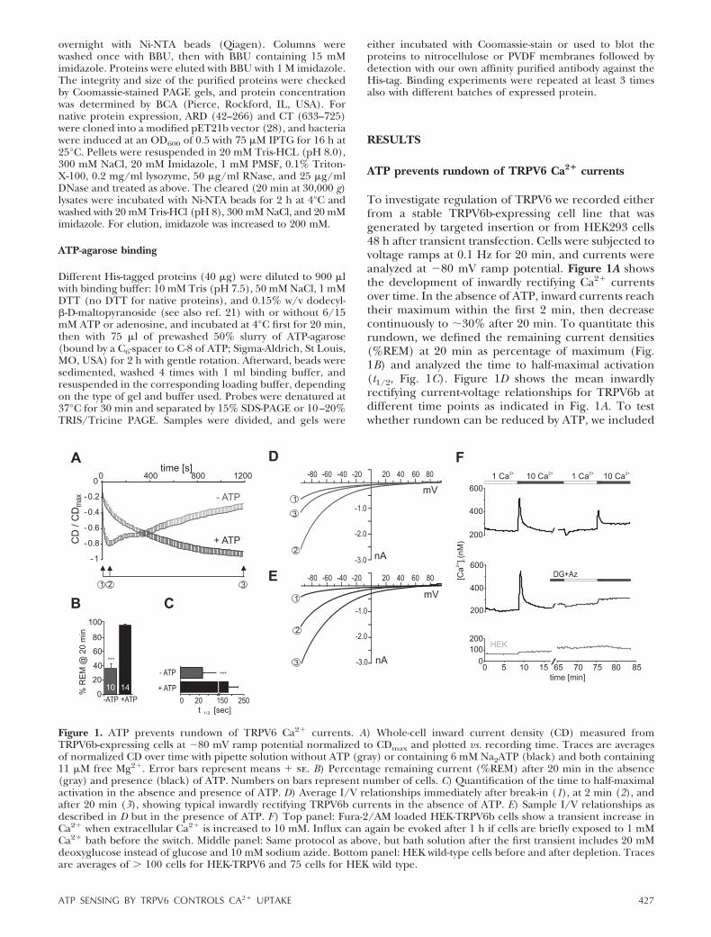
ATP prevents rundown of TRPV6 Ca2� currents
To investigate regulation of TRPV6 we recorded eitherfrom a stable TRPV6b-expressing cell line that wasgenerated by targeted insertion or from HEK293 cells48 h after transient transfection. Cells were subjected tovoltage ramps at 0.1 Hz for 20 min, and currents wereanalyzed at �80 mV ramp potential. Figure 1A showsthe development of inwardly rectifying Ca2� currentsover time. In the absence of ATP, inward currents reachtheir maximum within the first 2 min, then decreasecontinuously to �30% after 20 min. To quantitate thisrundown, we defined the remaining current densities(%REM) at 20 min as percentage of maximum (Fig.1B) and analyzed the time to half-maximal activation(t1/2, Fig. 1C). Figure 1D shows the mean inwardlyrectifying current-voltage relationships for TRPV6b atdifferent time points as indicated in Fig. 1A. To testwhether rundown can be reduced by ATP, we included
Figure 1. ATP prevents rundown of TRPV6 Ca2� currents. A) Whole-cell inward current density (CD) measured fromTRPV6b-expressing cells at �80 mV ramp potential normalized to CDmax and plotted vs. recording time. Traces are averagesof normalized CD over time with pipette solution without ATP (gray) or containing 6 mM Na2ATP (black) and both containing11 �M free Mg2�. Error bars represent means � se. B) Percentage remaining current (%REM) after 20 min in the absence(gray) and presence (black) of ATP. Numbers on bars represent number of cells. C) Quantification of the time to half-maximalactivation in the absence and presence of ATP. D) Average I/V relationships immediately after break-in (1), at 2 min (2), andafter 20 min (3), showing typical inwardly rectifying TRPV6b currents in the absence of ATP. E) Sample I/V relationships asdescribed in D but in the presence of ATP. F) Top panel: Fura-2/AM loaded HEK-TRPV6b cells show a transient increase inCa2� when extracellular Ca2� is increased to 10 mM. Influx can again be evoked after 1 h if cells are briefly exposed to 1 mMCa2� bath before the switch. Middle panel: Same protocol as above, but bath solution after the first transient includes 20 mMdeoxyglucose instead of glucose and 10 mM sodium azide. Bottom panel: HEK wild-type cells before and after depletion. Tracesare averages of � 100 cells for HEK-TRPV6 and 75 cells for HEK wild type.
427ATP SENSING BY TRPV6 CONTROLS CA2� UPTAKE

6 mM Na2ATP in the recording pipette and clamped[Mg2�]i to that of the control solution (11 �M).Because [Mg2�]i is below the IC50 for Mg2� block ofthe pore (29), we can exclude confounding influenceson pore properties. Inclusion of ATP led to alteredkinetics of current development (Fig. 1A): initial cur-rent densities were reduced to �16.3 � 3.4 pA/pF(n�15) compared to �39.7 � 15 pA/pF (n�11,P�0.5), and subsequent increments in current ampli-tude were also reduced, leading to current densitiesthat did not decrease within the recording time (Fig.1A), thus resulting in �100% REM after 20 min (Fig.1B). To test whether the initial reduction in currentdensity was due to an effect of external perfusion ofATP while approaching the cell with the patch pipette,we recorded from cells with no ATP in the patchpipette but locally perfused ATP from a second pipetteimmediately before and after break-in. However, initialcurrent densities were not reduced (�41.3�6.6 pA/pF,n�4), likely excluding activation of purinergic recep-tors as the cause of current reduction.
Mean IV relationships in the presence of ATP (Fig.1E) indicate that no other major ATP-induced conduc-tances were observed with our recording conditions butrevealed a minor decrease in the steepness of inwardrectification. A shift in the same direction is seen in theabsence of ATP when EGTA is replaced by BAPTA(data not shown), which may indicate a decrease in theCa2�-dependent feedback mechanism when addingATP to the EGTA pipette solution. To exclude thepossibility that ATP is preventing rundown solely byincreasing the fast calcium-buffering capacity of thepipette solution, we confirmed that ATP still had sig-nificant effects in the presence of 20 mM BAPTA (datanot shown). Prevention of rundown by ATP has alsobeen shown in Ca2�-free conditions (30). The maximalcurrent densities for TRPV6b after 20 min with ATPwere �153 � 11 pA/pF, which were not different whencompared to the maximal current densities withoutATP (�160�27 pA/pF).
The effects of ATP on both kinetics of currentdevelopment and on rundown were also seen withtransient expression of TRPV6b. Rundown and t1/2were analyzed at 10 min recording time, as transientlyexpressing cells were less stable. Whereas in the ab-sence of ATP the kinetics of current development,maximal current density, and the degree of rundownwere comparable for transient vs. stable transfectedcells (Supplemental Fig. S1A), in the presence of ATPthe slowdown of current development was also signifi-cant, but less pronounced than with stable TRPV6expression (t1/2 57�10 vs. 117�22 s for transient andstable expressed TRPV6b, respectively; SupplementalFig. S1A, B). Because oxidation has been shown tomodulate binding of ATP to chloride channels (31), weused the fluorescent indicator DCF to test whethertransient expression influences the redox state of thecells. Indeed, Supplemental Fig. S1C shows that tran-siently transfected cells showed higher levels of intra-cellular oxidants than stable cells. We thus investigated
the influence of oxidants (H2O2) on the time course ofTRPV6 in the stable expression system. SupplementalFig. S1D shows that increasing oxidative stress throughaddition of H2O2, probably by modifying reactive cys-teines of TRPV6, leads to larger current increments inthe presence of ATP and can thus explain the fastercurrent development in cells transiently expressingTRPV6. However, even in the presence of a strongoxidant, ATP is still able to prevent rundown (Supple-mental Fig. S1D). For further analyses, we have focusedon the effects of ATP on the percentage of rundown(%REM) and compared rundown at 20 min for stableand at 10 min for transient expression.
To mimic a more physiological condition, we testedthe influence of ATP depletion (32) on Ca2� transientsin unperturbed cells. Figure 1F shows that TRPV6 cellsrespond to a change in [Ca2�]e with a transient in-crease in [Ca2�]i. This transient increase can again beevoked after �50 min (Fig. 1F, top panel). However,when TRPV6 cells are ATP depleted, the second tran-sient peak is absent (Fig. 1F, middle panel), confirmingthat either a sufficient concentration of ATP or of ametabolite is needed for the ability to respond tosudden changes in [Ca2�]e. HEK cells show a lowerresting Ca2� and do not show a transient increase in[Ca2�]i on exposure to 10 mM [Ca2�]e (Fig. 1F, bottompanel). Interestingly, in HEK cells, the change back to 1mM [Ca2�]e does not lead to a drop in [Ca2�]i (Fig. 1F,top panel), which may be due to Ca2�-ATPase inactivity atlow resting [Ca2�]i (Km 0.4–0.5 �M; 33).
Thyagarajan et al. (12) showed that hydrolysis of PIP2mediated Ca2�-induced inactivation of TRPV6 chan-nels (12). To test the potential involvement of PIP2 inthe ATP effect, we replaced ATP with 50 �M diC8PIP2,which was unable to prevent rundown in the absence ofATP (Fig. 2A, B). We also included a nonhydrolyzableATP analog, AMP-PCP, in the pipette solution. Thisanalog also prevented rundown and slowed currentdevelopment (Fig. 2A, B). Because R599 was identifiedas a critical residue responsible for mediating PIP2-dependent channel activity in TRPV5 (34), we checkedwhether this highly conserved arginine mediates theATP effect in TRPV6 (R599 in rbTRPV5�R606 inhTRPV6). We mutated R606 to Q and recorded thetransiently expressed mutant in the presence and ab-sence of ATP. Figure 2C, D shows that ATP is still ableto prevent rundown in the mutant. Together with theevidence seen in Fig. 2A, the results suggest that ATPprevents rundown of TRPV6 in a PIP2-independentmanner.
Nucleotide specificity
To further test the specificity of the ATP effect, weincluded different nucleotides in the pipette solution at6 mM each. Figure 3A shows currents over time forsome of the tested nucleotides, and Fig. 3B the percent-age REM for all nucleotides tested. Although adenosineand cAMP were unable to prevent rundown, otherphosphorylated nucleotides had intermediate effects,
428 Vol. 24 February 2010 AL-ANSARY ET AL.The FASEB Journal � www.fasebj.org

with AMP being more potent than GTP and slightlymore potent than UTP, which suggests that both nu-cleotide moiety and number of negatively chargedphosphate groups are essential (Fig. 3A). To test thedependence of rundown on the ATP concentration, wecalculated the dose response relationship after record-ing currents with different free ATP concentrations,keeping free [Mg2�] constant. Figure 3C shows sampletraces of currents with 6, 304, and 609 �M and 3.7 mMfree ATP in the pipette solution. Data points were fittedwith a sigmoidal dose-response function of variableslope, where %REMmax is the maximal remaining cur-rent obtained after addition of 3.7 mM free ATP, EC50is the half-maximal concentration of free ATP, and n isthe Hill slope. As shown in Fig. 3D, the mean EC50obtained from the graph was 378 �M free ATP, and theHill slope was 4.8. The EC50 value obtained is one orderof magnitude greater than that normally required for akinase-mediated phosphorylation reaction (14); how-ever, we also investigated the crosstalk between run-down and PKC-mediated phosphorylation.
Regulation by protein kinase C and identification of afunctionally relevant ATP binding site
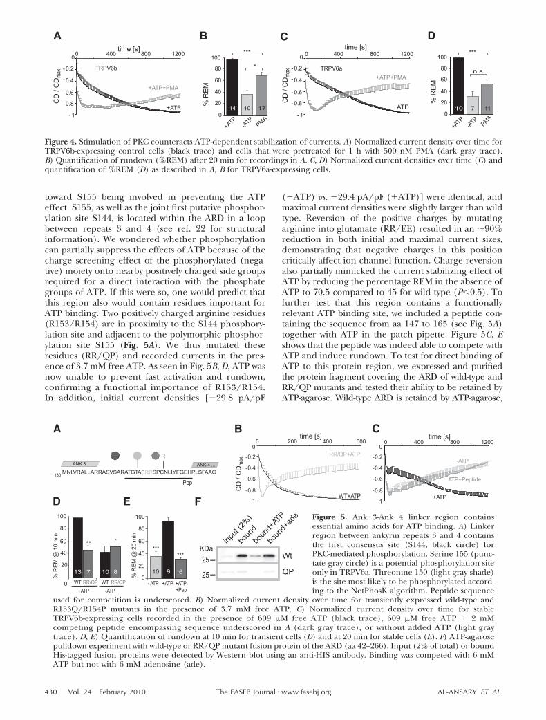
To measure TRPV6 currents in the presence of ATPand after activation of protein kinases, we incubatedcells for 1 h with 500 nM PMA in serum-free mediumand then recorded with PMA also present in the bathsolution. Figure 4A shows that incubation with PMA wasable to partially prevent the effects of ATP: AlthoughATP was included, current size increased much faster,reached maximal values within the first 2 min, andentered rundown (Fig. 4A, B).
Because the ancestral polymorphic allele (TRPV6a)contains an additional potential PKC phosphorylationsite (SPR157) compared to SPC for TRPV6b, we alsotested the effects of PMA treatment on TRPV6a andfound a slightly more efficient prevention of the ATPeffect (Fig. 4C, D). In the absence of ATP, PMAtreatment had little effect on rundown (SupplementalFig. S2A, B). The finding that the polymorphs differslightly in the extent of the PMA effect may point
Figure 3. Nucleotide specificity and dose-response curve for ATP. A) Normalized currentdensity over time in the presence of different intracellular nucleotides at 6 mM. Error barsrepresent means � se. B) Quantification of percentage REM for all nucleotides tested.Asterisks indicate significant differences of percentage REM against the no-added-ATPcontrol. Numbers on bars represent number of cells. Recordings were obtained in stableTRPV6b cells. C) Normalized current density over time for selected concentrations ofintracellular free ATP. D) Mean relationship between [free ATP] and the degree ofrundown after 20 min (%REM). Data points (number of cells above) were best fitted to Eq.1. EC50, ATP concentration at which prevention of rundown is half-maximal; nH, Hill slope.
Figure 2. Effects of PIP2 and a nonhydrolyzable ATP analog on rundown. A) Normalized current density over time in theabsence of ATP (gray trace), in the presence of 50 �M diC8PIP2, or in the presence of 6 mM AMP-PCP in TRPV6b stable cellline. B) Percentage REM for traces in A. C) R606Q mutants retain the ATP effect. Normalized current density over time fortransiently expressed TRPV6_R606Q mutants with and without ATP in the recording pipette. D) Percentage REM after 10 minrecording time; see also Fig. 5B for transiently expressed wild type.
429ATP SENSING BY TRPV6 CONTROLS CA2� UPTAKE
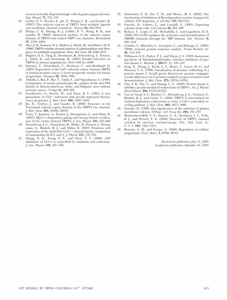
toward S155 being involved in preventing the ATPeffect. S155, as well as the joint first putative phosphor-ylation site S144, is located within the ARD in a loopbetween repeats 3 and 4 (see ref. 22 for structuralinformation). We wondered whether phosphorylationcan partially suppress the effects of ATP because of thecharge screening effect of the phosphorylated (nega-tive) moiety onto nearby positively charged side groupsrequired for a direct interaction with the phosphategroups of ATP. If this were so, one would predict thatthis region also would contain residues important forATP binding. Two positively charged arginine residues(R153/R154) are in proximity to the S144 phosphory-lation site and adjacent to the polymorphic phosphor-ylation site S155 (Fig. 5A). We thus mutated theseresidues (RR/QP) and recorded currents in the pres-ence of 3.7 mM free ATP. As seen in Fig. 5B, D, ATP wasnow unable to prevent fast activation and rundown,confirming a functional importance of R153/R154.In addition, initial current densities [�29.8 pA/pF
(�ATP) vs. �29.4 pA/pF (�ATP)] were identical, andmaximal current densities were slightly larger than wildtype. Reversion of the positive charges by mutatingarginine into glutamate (RR/EE) resulted in an �90%reduction in both initial and maximal current sizes,demonstrating that negative charges in this positioncritically affect ion channel function. Charge reversionalso partially mimicked the current stabilizing effect ofATP by reducing the percentage REM in the absence ofATP to 70.5 compared to 45 for wild type (P�0.5). Tofurther test that this region contains a functionallyrelevant ATP binding site, we included a peptide con-taining the sequence from aa 147 to 165 (see Fig. 5A)together with ATP in the patch pipette. Figure 5C, Eshows that the peptide was indeed able to compete withATP and induce rundown. To test for direct binding ofATP to this protein region, we expressed and purifiedthe protein fragment covering the ARD of wild-type andRR/QP mutants and tested their ability to be retained byATP-agarose. Wild-type ARD is retained by ATP-agarose,
Figure 4. Stimulation of PKC counteracts ATP-dependent stabilization of currents. A) Normalized current density over time forTRPV6b-expressing control cells (black trace) and cells that were pretreated for 1 h with 500 nM PMA (dark gray trace).B) Quantification of rundown (%REM) after 20 min for recordings in A. C, D) Normalized current densities over time (C) andquantification of %REM (D) as described in A, B for TRPV6a-expressing cells.
A B C
E
DC/
DC
max
WT+ATP
RR/QP+ATP
-1
-
-
-
-
0
0.8
0.6
0.4
0.2
time [s]200 400 6000 0 400 800 1200
time [s]
-1
-
-
-
-
0
0.8
0.6
0.4
0.2
- ATP +ATP+Pep
+ATP0
20
40
60
80
100
nim
02@
MER
%
******
0
20
40
60
80
100
RR/QP
+ATP
WTRR/QPWT
-ATP
nim
0 1@
MER
%
**
7 813 10 10 9 6
F
MNLVRALLARRASVSARATGTAF SPCNLIYFGEHPLSFAACRR
... ANK 3 ANK 4 ...
Pep
R
130
D
bound+ade
input(2%)
bound
bound+ATP
Wt25
KDa
QP25
ATP+Peptide
+ATP
-ATP
Figure 5. Ank 3-Ank 4 linker region containsessential amino acids for ATP binding. A) Linkerregion between ankyrin repeats 3 and 4 containsthe first consensus site (S144, black circle) forPKC-mediated phosphorylation. Serine 155 (punc-tate gray circle) is a potential phosphorylation siteonly in TRPV6a. Threonine 150 (light gray shade)is the site most likely to be phosphorylated accord-ing to the NetPhosK algorithm. Peptide sequence
used for competition is underscored. B) Normalized current density over time for transiently expressed wild-type andR153Q/R154P mutants in the presence of 3.7 mM free ATP. C) Normalized current density over time for stableTRPV6b-expressing cells recorded in the presence of 609 �M free ATP (black trace), 609 �M free ATP � 2 mMcompeting peptide encompassing sequence underscored in A (dark gray trace), or without added ATP (light graytrace). D, E) Quantification of rundown at 10 min for transient cells (D) and at 20 min for stable cells (E). F) ATP-agarosepulldown experiment with wild-type or RR/QP mutant fusion protein of the ARD (aa 42–266). Input (2% of total) or boundHis-tagged fusion proteins were detected by Western blot using an anti-HIS antibody. Binding was competed with 6 mMATP but not with 6 mM adenosine (ade).
430 Vol. 24 February 2010 AL-ANSARY ET AL.The FASEB Journal � www.fasebj.org

and binding can be competed for by free ATP or UTP(data not shown), but not by adenosine (Fig. 5F). Incontrast, RR/QP-ARD showed a significant reduction inbinding to ATP-agarose (Fig. 5F). Together, these resultssuggest that the ARD 3–4 region with residues R153/154mediate binding of ATP’s negatively charged moietiesand is responsible for its physiological effects. Phosphor-ylation at S144, and possibly at S155 in the case ofTRPV6a, may prevent binding either by charge hindranceor by a conformational change.
If phosphorylation of TRPV6 mediates the PMAeffect, we should be able to define functionallyrelevant phosphorylation sites by mutating the con-sensus site serine or threonine residues. The first site,S144, is solvent exposed (22) (Fig. 6A), and S144Amutants were resistant to rundown in the presence ofPMA (Fig. 6B, C), confirming the hypothesis thatATP binding may be sterically hindered by phosphor-ylation at the ARD 3,4 linker region. The fastercurrent development of S144A mutants (�ATP) ispartially due to transient expression (see Supplemen-tal Fig. S1). Currents in the absence of ATP wereidentical to wild type (%REM; Fig. 6C).
To test whether other PKC phosphorylation sitesare also involved, we mutated single sites detected bythe prosite algorithm (35) and investigated the ef-fects of PMA. Figure 6A shows the relative location,and Fig. 6D–F shows average current densities overtime for 3 additional mutants. Three other sites(T150A, S318A, and T702A) showed clear PMA-
induced rundown (Fig. 6D, E, G). Mutating the dualphosphorylation site T298A/T299A resulted in aweaker effect of PMA, but mutation of the C-terminalsite T688A was as efficient as the S144A mutation inblocking the PMA effect (Fig. 6F, G). All mutantsdisplayed comparable rundown to wild type in theabsence of added ATP (data not shown).We have alsoconfirmed ATP-mediated prevention of rundown formTRPV5 and for mTRPV6. Although other putativePKC sites are conserved between species, T688 is nota consensus site within mTRPV5, and PMA wasunable to oppose the effects of ATP in mTRPV5 (seeSupplemental Fig. S2C–F). These results point to-ward a synergistic role of the C-terminal phosphory-lation site in mediating the effect of PMA and raisethe possibility that a C-terminal region could also beinvolved in binding of ATP.
Isozyme specificity
To address the specificity of the PMA effect, weincubated TRPV6b cells with an inactive analog ofPMA, 4�PDD. This inactive analog was unable tomimic the effects of PMA (Fig. 7A, B), confirming alikely effect on activation of PKCs. We tried to blockthe effect of PMA by pretreatment with both BIS andGo6983, inhibitors of classical PKCs, and then addedPMA for 1 h. Surprisingly, these inhibitors wereunable to prevent the effect of PMA on current
Figure 6. Mutational analysis of PKC phosphorylation sites. A) Schematic representation of putative PKC phosphorylation sitesas found by the Prosite algorithm. B) Normalized current density over time for TRPV6_S144A mutants with and without PMA(500 nM for 1 h) treatment in the presence of ATP. C) Quantification of rundown for transiently expressed TRPV6 wild-typeand TRPV6 S144A mutants. D–F) Normalized current density over time for transiently expressed mutants T150A (D), S318A (E),and T688A (F). G) Bar graph showing quantification of rundown in the presence of 3.7 mM free ATP with (gray) or without(black) PMA preincubation.
431ATP SENSING BY TRPV6 CONTROLS CA2� UPTAKE

development (Fig. 7A, B). Because neither BIS norGo6983 has a defined IC50 for the PKC II isoform, wetested whether Ro-31-8322, with an IC50 of 14 nM forPKC II (36), was able to prevent PMA from counter-acting the ATP effects. Figure 7A, B shows thatRo-31-8322 was indeed able to suppress the effects ofPMA, suggesting that PMA exerts its effects throughactivation of PKC II. Ro-31-8322 alone did not haveeffects on ATP-modulated TRPV6b currents in theabsence of PMA (data not shown). One concern thatremained was that long incubation times were neces-sary to see the effects of PMA, whereas translocationof cPKCs happens within minutes. However, HEK293cells express only low levels of endogenous PKC II protein(37); therefore, we overexpressed GFP-tagged PKC II orGFP-tagged PKC� and tested whether PMA was now ableto exert its effects within minutes. Indeed, with expressionof PKC II, but not with overexpression of PKC�, TRPV6currents now showed PMA-induced rundown whenadded to the bath solution without preincubation (Fig.7C, D). Both PKC isoforms showed translocation to theplasma membrane after stimulation with PMA (Fig. 7Eand data not shown). To test if PKC II expression isfound in similar tissue types as TRPV6, we checkedfor its presence in human placenta cDNA (5) using
isoform-specific PCR primers and were indeed ableto amplify DNA of the correct size (Fig. 7F).
ATP binding sites
To screen for additional ATP binding sites, we createdfusion proteins (10xHIS at N-term) covering all intra-cellular protein domains and tested their ability to beretained by ATP-agarose. We found several additionalpotential binding regions within TRPV6: although thevery N-terminal region of the protein containing ARD 1and 2 (Supplemental Fig. S3B) was not retained byATP-agarose, thus demonstrating that the HIS tagalone does not confer unspecific binding, the ARDregion showed weak binding (see also Fig. 5). TheN-terminal region between aa 267 and TM1 was alsoretained (Supplemental Fig. S3D). Its sequence con-tains a basic KKR region as well as a potential PHdomain and the dual phosphorylation site T298A/T299A. The proximal region (aa 584-632) of the C-terminal domain containing the TRP motif showedbinding that is not efficiently competed for by freeATP, and thus not as specific (Supplemental Fig. S3E).However, the distal region (aa 633 to 725; CT) is wellretained by ATP-agarose, and binding is effectively
Figure 7. PMA-mediated effects are PKC isoform specific. A) Normalized current density over time for TRPV6b-expressing cellsthat were pretreated either for 1 h with PMA (dark blue) or 4-�-PDD (green) or for 30 min with 500 nM each of inhibitorsGo6983/BIS (light blue) or Ro-31-8220 (orange) and then with inhibitors and 500 nM PMA for 1 h. B) Percentage REM for PMAand inhibitors. C) Normalized current densities recorded from TRPV6b stable cells with transient expression of GFP-PKC II orGFP-PKC�. D) Percentage REM with acute (�10 min in bath) PMA treatment for TRPV6b (V6) cells alone or V6 cells transfectedwith either GFP-PKC II or GFP-PKC�. A–D) Internal solution with 3.7 mM free ATP. E) Fluorescent images from TRPV6b cellsexpressing GFP-PKC II before and after PMA-stimulation. F) PKC II differs from PKC I by 162 specific C-terminal nucleotides.These were amplified with II specific primers proportional to the amount of template (human placental cDNA or fromrecombinant PKC II vector).
432 Vol. 24 February 2010 AL-ANSARY ET AL.The FASEB Journal � www.fasebj.org

competed for by free ATP (Supplemental Fig. S3F). Wealso checked retention of the ARD region of rat TRPV1that has been shown to bind ATP (21) (SupplementalFig. S3G). Considering that T688 is a potential phos-phorylation site preventing ATP binding (see Fig. 6F, Gand Supplemental Fig. S2) and the finding that theC-terminal fusion protein encompassing T688 is wellretained by ATP-agarose, we wanted to confirm bindingto ATP agarose for ARD and CT proteins under nativepurification conditions. Because proteins containingthe N-terminal 10xHIS tag could be solubilized onlywith urea, we constructed ARD and CT fusion proteinswith a C-terminal 6xHIS Tag (Supplemental Fig. S3H)and repeated binding experiments after native purifi-cation. ARD was retained by ATP-agarose with bindingefficiencies of �10% in several experiments (Coomas-sie and Western blot), and CT showed binding efficien-cies of �15%, comparable to the proteins purified inthe presence of urea. In addition, in vitro translatedfull-length TRPV6 was retained by ATP-agarose in theabsence of ATP in the binding buffer, but significantlyless in the presence of ATP (data not shown).
Because mutations in individual phosphorylationsites either in the N- or in the C-terminal region (S144,T688) did not show rundown despite PMA incubationand because both fusion proteins encompassing thesesites were also retained by ATP-agarose, we suggest amodel where ATP directly binds to TRPV6 and mightact as a stabilizing factor between the intracellular Nand C termini, with residues in both the N- andC-terminal domains necessary for efficient binding inthe native protein (see model, Fig. 8). The Hill slopeof 4.8 as estimated from the dose response (Fig. 3D)strengthens this model for a tetrameric channel.Phosphorylation near the N- and C-terminal bindingsites destabilizes the protein and tags channels forrundown.
DISCUSSION
Stringent regulation of Ca2� influx through TRPV6 is anecessary requirement to avoid Ca2� overload. TRPV6is inactivated by several Ca2�-dependent pathways suchas regulation by calmodulin, breakdown of PIP2 andprobably constitutive exo- and endocytotic mechanisms(38, 39). However, there are also Ca2�-independentmechanisms that lead to channel rundown. Our workidentifies ATP as an intracellular ligand that stabilizesthe channel and prevents rundown. Because bothabsolute and normalized maximal current densities arenot significantly different within the recording timewith or without ATP, ATP does not seem to increase thenumber of channels in the membrane, although it mayprevent the reduction of the number of channels thatcould lead to rundown. The prevention of rundowndoes not necessitate hydrolysis of ATP, shows nucleo-tide specificity (although other phosphorylated nucle-otides can partially substitute), and cannot be mimickedby PIP2. Stabilization of current size shows an EC50 of
�380 �M free ATP. This value corresponds well withphysiological levels of free ATP and allows the channels torespond to small decreases in ATP concentration toprevent Ca2� overload while the Ca2�-ATPases in theplasma membrane (EC50 for ATP 0.1–1 �M; see ref. 40)are still functional. We identified R153 and R154 asbeing essential for mediating the functional ATP effect.Disruption of this site both by mutagenesis or compe-tition by a peptide resembling this linker sequencedisables ATP from decreasing current increments andpreventing rundown. Because the RR/QP mutants stillshow normal currents in the absence of ATP, thesemutations did not disturb essential functions not re-lated to the ATP effect. However, charge reversion atthis site by creation of RR/EE mutant channels severelyreduced current size, which mimics to some extentadding negative charges from ATP’s phosphate moi-eties, but is unable to substitute the adenosine moietyof ATP, which may have its own binding site (see Figs.3A, B and 8). Evidence from ATP-agarose pulldownexperiments support a hypothesis that ATP interactswith this N-terminal ARD domain through its negativelycharged phosphate groups. Because Phelps et al. (22)did not find retention of mTRPV6 ARD by ATP-agarose, we also expressed and purified ARD and CT ofhTRPV6 with a similar C-terminal 6xHIS tag undernative conditions. ARD still showed binding to ATP-agarose, indicating that species differences or storageconditions (10 mM EDTA, 2.5 mM 2-mercaptoethanol;see ref. 22) may account for differences in binding. Theeffects of ATP on both current stabilization and activa-
Figure 8. Model of regulatory mechanisms on TRPV6 by ATPand phosphorylation. To simplify graph, only one pore-forming subunit is represented. In the absence of ATP, thechannel is prone to both Ca2�-dependent (calmodulin) andCa2�-independent regulation and rundown. Sufficient ATPstabilizes N and C termini and prevents channel rundown.Phosphorylation hinders ATP binding and thus reduces itsstabilizing effect.
433ATP SENSING BY TRPV6 CONTROLS CA2� UPTAKE

tion kinetics were disrupted when TRPV6 cells (stableor transient expression) were incubated with PMA. Thiseffect is PKC specific and requires activation of PKC II,a PKC isoform that is not strongly expressed in HEK293cells but is present together with TRPV6 in placenta.Overexpression of PKC II, but not of PKC�, obliteratesthe need for PMA preincubation. The results obtainedfrom mutating individual phosphorylation sites arguefor a direct effect, where phosphorylation of TRPV6might hinder binding of ATP. Because both the N-terminal and the distal TRPV6 C-terminal fusion pro-teins are retained by ATP-agarose and because mu-tagenesis of putative phosphorylation sites withinthese domains (S144A and T688A) are able to sup-press the PMA effect, we propose a synergistic effectof both binding sites. ATP might form a bridgebetween both domains and thereby alter channelproperties (Fig. 8), leading to slowed current devel-opment and reduced rundown. To fully understandthe dual effects of ATP, single-channel analyses willbe necessary. Low-resolution freeze fracture dataobtained for TRPV1 (41) may suggest an interactionbetween N- and C-terminal regions. Because a mu-tant that lacks the C-terminal calmodulin binding siteof TRPV6 still shows an effect of ATP on rundown(data not shown), the C-terminal ATP binding sitedoes not overlap with the calmodulin binding site,unlike the multiligand binding site of TRPV1 (21).
A small drop in cellular-free ATP levels (i.e., by a 1–2mM increase in Mg2�; see ref. 42) leads to channels lessstable and more prone to both Ca2�-dependent andCa2�-independent rundown phenomena and thus areduction in overall Ca2� influx. Activation of PKC II
prevents stabilization by ATP, and channels show morerundown and Ca2�-dependent inactivation. The up-stream GPCR that activates PKC II in vivo may thus beinvolved in feedback regulation when sufficient Ca2�
has been taken up by TRPV6 and may also be respon-sible for conferring an allele-specific advantage. It willbe interesting to analyze if and how both kinasesregulate the native channels. Unfortunately, thesestudies are hampered by the difficulty in obtaininghuman cells in which native TRPV6 currents can bemeasured.
We especially thank Dr. Veit Flockerzi and Dr. UlrichWissenbach (University of Saarland, Homburg, Germany) forthe stable TRPV6 cell lines, for antibodies against TRPV6 andthe HIS tag, and, together with Dr. Hoth, for discussions andcritical reading of the manuscript. We also thank Dr. YusufHannun (Medical University of South Carolina, Charleston,SC, USA) and Dr. Peter Lipp (University of Saarland) forkindly providing us with the cDNA for GFP-PKC II andGFP-PKC�; Dr. Marcel Meissner (University of Saarland) forfusion proteins of mTRPV6; Heidi Lohr, Anja Ludes, andBettina Strauß for cell culture work; and members of the labsfor critical input. This work was supported, in part, by theDeutsche Forschungsgemeinschaft (DFG; NI 671 to B.N.)and HOMFOR (B.N.). D.A.-A. is a member of the DFG-sponsored graduate program GK 1326.
REFERENCES
1. Suzuki, Y., Landowski, C. P., and Hediger, M. A. (2008) Mech-anisms and regulation of epithelial Ca2� absorption in healthand disease. Annu. Rev. Physiol. 70, 257–271
2. Van de Graaf, S. F., Bindels, R. J., and Hoenderop, J. G. (2007)Physiology of epithelial Ca2� and Mg2� transport. Rev. Physiol.Biochem. Pharmacol. 158, 77–160
3. Nilius, B., Vennekens, R., Prenen, J., Hoenderop, J. G., Bindels,R. J., and Droogmans, G. (2000) Whole-cell and single channelmonovalent cation currents through the novel rabbit epithelialCa2� channel ECaC. J. Physiol. 527(Pt. 2), 239–248
4. Vennekens, R., Hoenderop, J. G., Prenen, J., Stuiver, M.,Willems, P. H., Droogmans, G., Nilius, B., and Bindels, R. J.(2000) Permeation and gating properties of the novel epithelialCa(2�) channel. J. Biol. Chem. 275, 3963–3969
5. Wissenbach, U., Niemeyer, B. A., Fixemer, T., Schneidewind, A.,Trost, C., Cavalie, A., Reus, K., Meese, E., Bonkhoff, H., andFlockerzi, V. (2001) Expression of CaT-like, a novel calcium-selective channel, correlates with the malignancy of prostatecancer. J. Biol. Chem. 276, 19461–19468
6. Nilius, B., Prenen, J., Vennekens, R., Hoenderop, J. G., Bindels,R. J., and Droogmans, G. (2001) Modulation of the epithelialcalcium channel, ECaC, by intracellular Ca2�. Cell Calcium 29,417–428
7. Bodding, M., Wissenbach, U., and Flockerzi, V. (2002) Therecombinant human TRPV6 channel functions as Ca2� sensorin human embryonic kidney and rat basophilic leukemia cells.J. Biol. Chem. 277, 36656–36664
8. Nilius, B., Prenen, J., Hoenderop, J. G., Vennekens, R., Hoefs,S., Weidema, A. F., Droogmans, G., and Bindels, R. J. (2002)Fast and slow inactivation kinetics of the Ca2� channels ECaC1and ECaC2 (TRPV5 and TRPV6): role of the intracellular looplocated between transmembrane segments 2 and 3. J. Biol. Chem.277, 30852–30858
9. Hoenderop, J. G., Voets, T., Hoefs, S., Weidema, F., Prenen, J.,Nilius, B., and Bindels, R. J. (2003) Homo- and heterotetramericarchitecture of the epithelial Ca2� channels TRPV5 andTRPV6. EMBO J. 22, 776–785
10. Niemeyer, B. A., Bergs, C., Wissenbach, U., Flockerzi, V., andTrost, C. (2001) Competitive regulation of CaT-like-mediatedCa2� entry by protein kinase C and calmodulin. Proc. Natl.Acad. Sci. U. S. A. 98, 3600–3605
11. Derler, I., Hofbauer, M., Kahr, H., Fritsch, R., Muik, M.,Kepplinger, K., Hack, M. E., Moritz, S., Schindl, R., Groschner,K., and Romanin, C. (2006) Dynamic but not constitutiveassociation of calmodulin with rat TRPV6 channels enables finetuning of Ca2�-dependent inactivation. J. Physiol. 577, 31–44
12. Thyagarajan, B., Lukacs, V., and Rohacs, T. (2008) Hydrolysis ofphosphatidylinositol 4,5-bisphosphate mediates calcium-inducedinactivation of TRPV6 channels. J. Biol. Chem. 283, 14980–14987
13. Lee, J., Cha, S. K., Sun, T. J., and Huang, C. L. (2005) PIP2activates TRPV5 and releases its inhibition by intracellularMg2�. J. Gen. Physiol. 126, 439–451
14. Hilgemann, D. W. (1997) Cytoplasmic ATP-dependent regula-tion of ion transporters and channels: mechanisms and messen-gers. Annu. Rev. Physiol. 59, 193–220
15. Craig, T. J., Ashcroft, F. M., and Proks, P. (2008) How ATP inhibitsthe open K(ATP) channel. J. Gen. Physiol. 132, 131–144
16. Dhar-Chowdhury, P., Malester, B., Rajacic, P., and Coetzee,W. A. (2007) The regulation of ion channels and transporters byglycolytically derived ATP. Cell. Mol. Life Sci. 64, 3069–3083
17. Takahashi, M., Kondou, Y., and Toyoshima, C. (2007) Interdo-main communication in calcium pump as revealed in the crystalstructures with transmembrane inhibitors. Proc. Natl. Acad. Sci.U. S. A. 104, 5800–5805
18. Kameyama, M., Kameyama, A., Takano, E., and Maki, M. (1998)Run-down of the cardiac L-type Ca2� channel: partial restora-tion of channel activity in cell-free patches by calpastatin.Pflugers Arch. 435, 344–349
19. Romanin, C., Grosswagen, P., and Schindler, H. (1991) Cal-pastatin and nucleotides stabilize cardiac calcium channel activ-ity in excised patches. Pflugers Arch. 418, 86–92
20. Okashiro, T., Tokuno, H., Fukumitsu, T., Hayashi, H., andTomita, T. (1992) Effects of intracellular ATP on calcium
434 Vol. 24 February 2010 AL-ANSARY ET AL.The FASEB Journal � www.fasebj.org
current in freshly dispersed single cells of guinea-pig portal vein.Exp. Physiol. 77, 719–731
21. Lishko, P. V., Procko, E., Jin, X., Phelps, C. B., and Gaudet, R.(2007) The ankyrin repeats of TRPV1 bind multiple ligandsand modulate channel sensitivity. Neuron 54, 905–918
22. Phelps, C. B., Huang, R. J., Lishko, P. V., Wang, R. R., andGaudet, R. (2008) Structural analyses of the ankyrin repeatdomain of TRPV6 and related TRPV ion channels. Biochemistry47, 2476–2484
23. Akey, J. M., Swanson, W. J., Madeoy, J., Eberle, M., and Shriver, M. D.(2006) TRPV6 exhibits unusual patterns of polymorphism and diver-gence in worldwide populations. Hum. Mol. Genet. 15, 2106–2113
24. Hughes, D. A., Tang, K., Strotmann, R., Schoneberg, T., Prenen,J., Nilius, B., and Stoneking, M. (2008) Parallel selection onTRPV6 in human populations. PLoS ONE 3, e1686
25. Fixemer, T., Wissenbach, U., Flockerzi, V., and Bonkhoff, H.(2003) Expression of the Ca2�-selective cation channel TRPV6in human prostate cancer: a novel prognostic marker for tumorprogression. Oncogene 22, 7858–7861
26. Abdalla, I., Ray, P., Ray, V., Vaida, F., and Vijayakumar, S. (1998)Comparison of serum prostate-specific antigen levels and PSAdensity in African-American, white, and Hispanic men withoutprostate cancer. Urology 51, 300–305
27. Grynkiewicz, G., Poenie, M., and Tsien, R. Y. (1985) A newgeneration of Ca2� indicators with greatly improved fluores-cence properties. J. Biol. Chem. 260, 3440–3450
28. Jin, X., Touhey, J., and Gaudet, R. (2006) Structure of theN-terminal ankyrin repeat domain of the TRPV2 ion channel.J. Biol. Chem. 281, 25006–25010
29. Voets, T., Janssens, A., Prenen, J., Droogmans, G., and Nilius, B.(2003) Mg(2�)-dependent gating and strong inward rectifica-tion of the cation channel TRPV6. J. Gen. Physiol. 121, 245–260
30. Hoenderop, J. G., Vennekens, R., Muller, D., Prenen, J., Droog-mans, G., Bindels, R. J., and Nilius, B. (2001) Function andexpression of the epithelial Ca(2�) channel family: comparisonof mammalian ECaC1 and 2. J. Physiol. 537, 747–761
31. Zhang, X. D., Tseng, P. Y., and Chen, T. Y. (2008) ATPinhibition of CLC-1 is controlled by oxidation and reduction.J. Gen. Physiol. 132, 421–428
32. Schwoebel, E. D., Ho, T. H., and Moore, M. S. (2002) Themechanism of inhibition of Ran-dependent nuclear transport bycellular ATP depletion. J. Cell Biol. 157, 963–974
33. Guerini, D., Coletto, L., and Carafoli, E. (2005) Exportingcalcium from cells. Cell Calcium 38, 281–289
34. Rohacs, T., Lopes, C. M., Michailidis, I., and Logothetis, D. E.(2005) PI(4,5)P2 regulates the activation and desensitization ofTRPM8 channels through the TRP domain. Nat. Neurosci. 8,626–634
35. Combet, C., Blanchet, C., Geourjon, C., and Deleage, G. (2000)NPS@: network protein sequence analysis. Trends Biochem. Sci.25, 147–150
36. Wilkinson, S. E., Parker, P. J., and Nixon, J. S. (1993) Isoenzymespecificity of bisindolylmaleimides, selective inhibitors of pro-tein kinase C. Biochem. J. 294(Pt. 2), 335–337
37. Feng, X., Zhang, J., Barak, L. S., Meyer, T., Caron, M. G., andHannun, Y. A. (1998) Visualization of dynamic trafficking of aprotein kinase C betaII/green fluorescent protein conjugatereveals differences in G protein-coupled receptor activation anddesensitization. J. Biol. Chem. 273, 10755–10762
38. Cha, S. K., Wu, T., and Huang, C. L. (2008) Protein kinase Cinhibits caveolae-mediated endocytosis of TRPV5. Am. J. Physiol.Renal Physiol. 294, F1212–F1221
39. Van de Graaf, S. F., Rescher, U., Hoenderop, J. G., Verkaart, S.,Bindels, R. J., and Gerke, V. (2008) TRPV5 is internalized viaclathrin-dependent endocytosis to enter a Ca2�-controlled re-cycling pathway. J. Biol. Chem. 283, 4077–4086
40. Guerini, D. (1998) The significance of the isoforms of plasmamembrane calcium ATPase. Cell Tissue Res. 292, 191–197
41. Moiseenkova-Bell, V. Y., Stanciu, L. A., Serysheva, I. I., Tobe,B. J., and Wensel, T. G. (2008) Structure of TRPV1 channelrevealed by electron cryomicroscopy. Proc. Natl. Acad. Sci.U. S. A. 105, 7451–7455
42. Romani, A. M., and Scarpa, A. (2000) Regulation of cellularmagnesium. Front. Biosci. 5, D720–D734
Received for publication July 14, 2009.Accepted for publication September 10, 2009.
435ATP SENSING BY TRPV6 CONTROLS CA2� UPTAKE
Copyright © 2022 FDOKUMEN