Ischemic Brain Damage in Mice After Selectively Modifying BDNF or NT4 Gene Expression
Upload
independentCategory
view
0download
0
PINK1 Is Selectively Stabilized on Impaired Mitochondriato Activate ParkinDerek P. Narendra1, Seok Min Jin1, Atsushi Tanaka1, Der-Fen Suen1, Clement A. Gautier2, Jie Shen2,
Mark R. Cookson3, Richard J. Youle1*
1 Biochemistry Section, Surgical Neurology Branch, National Institute of Neurological Disorders and Stroke, National Institutes of Health, Bethesda, Maryland, United States
of America, 2 Center for Neurologic Diseases, Brigham and Women’s Hospital, Program in Neuroscience, Harvard Medical School, Boston, Massachusetts, United States of
America, 3 Cell Biology and Gene Expression Unit, Laboratory of Neurogenetics, National Institute on Aging, Bethesda, Maryland, United States of America
Abstract
Loss-of-function mutations in PINK1 and Parkin cause parkinsonism in humans and mitochondrial dysfunction in modelorganisms. Parkin is selectively recruited from the cytosol to damaged mitochondria to trigger their autophagy. How Parkinrecognizes damaged mitochondria, however, is unknown. Here, we show that expression of PINK1 on individualmitochondria is regulated by voltage-dependent proteolysis to maintain low levels of PINK1 on healthy, polarizedmitochondria, while facilitating the rapid accumulation of PINK1 on mitochondria that sustain damage. PINK1 accumulationon mitochondria is both necessary and sufficient for Parkin recruitment to mitochondria, and disease-causing mutations inPINK1 and Parkin disrupt Parkin recruitment and Parkin-induced mitophagy at distinct steps. These findings provide abiochemical explanation for the genetic epistasis between PINK1 and Parkin in Drosophila melanogaster. In addition, theysupport a novel model for the negative selection of damaged mitochondria, in which PINK1 signals mitochondrialdysfunction to Parkin, and Parkin promotes their elimination.
Citation: Narendra DP, Jin SM, Tanaka A, Suen D-F, Gautier CA, et al. (2010) PINK1 Is Selectively Stabilized on Impaired Mitochondria to Activate Parkin. PLoSBiol 8(1): e1000298. doi:10.1371/journal.pbio.1000298
Academic Editor: Douglas R. Green, St. Jude Children’s Research Hospital, United States of America
Received July 8, 2009; Accepted December 18, 2009; Published January 26, 2010
This is an open-access article distributed under the terms of the Creative Commons Public Domain declaration which stipulates that, once placed in the publicdomain, this work may be freely reproduced, distributed, transmitted, modified, built upon, or otherwise used by anyone for any lawful purpose.
Funding: This work was supported by a National Institutes of Health (NIH)-Cambridge scholarship (to DPN), a JSPS Research Fellowship for Japanese Biomedicaland Behavioral Researchers (to AT), NIH grant R01NS41779 (to JS), and the intramural research program at the NIH. The funders had no role in study design, datacollection and analysis, decision to publish, or preparation of the manuscript.
Competing Interests: The authors have declared that no competing interests exist.
Abbreviations: mtDNA, mitochondrial DNA; MTG, Mitotracker Green; shRNA, short hairpin RNA; SD, standard deviation; SN, substantia nigra
* E-mail: [email protected]
Introduction
Parkinson disease is a common neurodegenerative disorder with
no disease-modifying therapy presently available for its treatment
[1]. Study of recessive forms of familial Parkinson disease, such as
those resulting from mutations in the E3 ubiquitin ligase Parkin
(GeneID: 5071) or the mitochondrial kinase PINK1 (GeneID:
65018), may reveal disease mechanisms important to the
development of disease in these families as well as those suffering
from sporadic Parkinson disease.
Although the cause of sporadic Parkinson disease is likely
complex, several lines of evidence link mitochondrial dysfunction
to its pathogenesis. Mitochondria within the substantia nigra (SN),
a midbrain region that is preferentially affected in Parkinson
disease, have a higher somatic mitochondrial DNA (mtDNA)
mutation rate than all other regions of the brain examined [2].
Increased mitochondrial damage in the SN, particularly to
mtDNA, has been associated with sporadic Parkinson disease
[3–5], and mitochondrial dysfunction is sufficient to cause
parkinsonism in patients with rare multiple mtDNA deletion
syndromes and in animal models with decreased mtDNA
expression [6–8]. In addition, toxins such as MPTP and rotenone,
which are believed to increase reactive oxygen species from
complex I of the electron transport chain, can induce a
parkinsonian syndrome in humans and animal models [9,10].
Since neurons in the SN are postmitotic, any mitochondrial
damage they acquire could accumulate over an organism’s
lifetime, leading to progressive mitochondrial dysfunction—
including increased oxidative stress, decreased calcium buffering
capacity, loss of ATP, and, eventually, cell death—unless quality
control processes eliminate the damaged mitochondria.
Recent studies have linked Parkin and PINK1 in a pathway
critical for the maintenance of mitochondrial integrity and
function. Loss of either protein in Drosophila results in a similar
phenotype, with mitochondrial damage preceding muscle degen-
eration, as well as disrupted spermatogenesis and death of
dopaminergic neurons [11–15]. Interestingly, overexpression of
Parkin can partially compensate for PINK1 loss, but PINK1
overexpression cannot compensate for Parkin loss, suggesting that
PINK1 functions upstream of Parkin in a common pathway.
Additionally, mice null for either Parkin or PINK1 exhibit
increased oxidative damage and decreased mitochondrial function
in the striatium (which receives projections from dopaminergic
neurons) [16,17]; and primary cells from patients with loss-of-
function mutations in Parkin or PINK1 have similar abnormalities
[18–20]. Together these findings suggest that Parkin and PINK1
may function in an evolutionarily conserved pathway critical for
the maintenance of mitochondrial integrity and function. We
recently reported that Parkin is selectively recruited to dysfunc-
tional mitochondria with low membrane potential and, subse-
PLoS Biology | www.plosbiology.org 1 January 2010 | Volume 8 | Issue 1 | e1000298

quently, promotes their autophagic degradation [21]. This
suggests that Parkin may limit mitochondrial damage by acting
in a pathway that identifies and eliminates damaged mitochondria
from the mitochondrial network. How mitochondrial dysfunction
is signaled to Parkin, however, is unknown.
Here, we show that full-length PINK1 accumulates selectively
on dysfunctional mitochondria, and that Parkin recruitment to
depolarized mitochondria and subsequent Parkin-induced mito-
phagy are strictly dependent on PINK1’s mitochondrial targeting
signal and depolarization-induced accumulation. Together, these
results strongly support a novel model for signaling between
PINK1 and Parkin in response to mitochondrial damage. In this
model, mitochondrial PINK1 is rapidly turned over on bioener-
getically well-coupled mitochondria by proteolysis, but is selec-
tively stabilized on mitochondria with low membrane potential.
Selective accumulation of PINK1 on the impaired mitochondria
recruits Parkin, and Parkin, in turn, induces the degradation of the
damaged mitochondria. In this model, PINK1 and Parkin form a
pathway for sensing and selectively eliminating damaged mito-
chondria from the mitochondrial network. Disease-causing
mutations in PINK1 and/or Parkin disrupt this pathway at
distinct steps, consistent with the pathway’s importance for
preventing early-onset parkinsonism.
Results
PINK1 Accumulates following MitochondrialDepolarization
Parkin is selectively recruited to damaged mitochondria that
have lost their membrane potential, but how Parkin distin-
guishes dysfunctional mitochondria with low membrane poten-
tial from healthy mitochondria is unknown. Since PINK1 is
genetically upstream of Parkin, we tested whether PINK1’s
activity might be activated by mitochondrial depolarization.
Remarkably, levels of endogenous mitochondrial PINK1
respond robustly to changes in mitochondrial membrane
potential. When HeLa cells are treated with CCCP, which
depolarizes mitochondria by increasing membrane permeability
to H+, a large increase in endogenous full-length PINK1
(,63 kDa) is seen beginning by 30 min and continuing for at
least 3 h (Figure 1A). This ,63-kDa band also increases in the
mitochondria-rich membrane fraction following treatment with
valinomycin, which, unlike CCCP, depolarizes mitochondria by
permeabilizing the membrane to K+ (Figure S1A). By contrast,
no band increases in the cytosolic fraction following depolar-
ization with CCCP (Figure S1B).
To verify that the ,63-kDa band is in fact PINK1, we
immunoblotted for endogenous PINK1 in M17 cells stably
transduced with control short hairpin RNA (shRNA) or PINK
shRNA. We found that the ,63-kDa band increases following
CCCP treatment in control shRNA cells, but does not increase in
the PINK1 shRNA cells, demonstrating that this ,63-kDa band is
endogenous PINK1 (Figure 1B). Similar results were found in
PINK12/2 cells transfected with PINK1-myc or left untransfected
(Figure S1C). We also tested whether PINK1 similarly accumu-
lates in primary rat cortical neurons following depolarization with
CCCP. Although we (and others) failed to detect endogenous rat
or mouse PINK1 with the available commercial antibodies ([22]
and unpublished data), we observed PINK1-V5 increases in
cortical neurons following treatment with 1 mM of CCCP for 6 h
(Figure 1C). With CCCP treatment, PINK1 may accumulate
more slowly in primary neurons than in HeLa cells, because,
unlike HeLa cells [23], neurons rely almost exclusively on
oxidative phosphorylation for ATP production [24].
To explore the kinetics of PINK1 accumulation at the single-cell
level, we fused YFP to PINK1 and imaged cells live following
depolarization with CCCP. Consistent with results obtained by
Western blotting, we found that PINK1-YFP expression steadily
increases from 1–5 min, when an increase is first detectable, until
at least 70 min (Figure 1D and Video S1).
PINK1 Accumulates Preferentially on DepolarizedMitochondria in a Single Cell
To examine the selectivity of PINK1 accumulation on
uncoupled mitochondria within single cells, we first investigated
its expression in mouse embryonic fibroblasts (MEFs) null for
mitochondrial fusion proteins mitofusin-1 and mitofusin-2 (Mfn1/
2). The Mfn1/2 null MEFs have a heterogeneous population of
mitochondria, some of which are bioenergetically uncoupled and
some of which are well coupled [25]. We found that, similar to
YFP-Parkin [21], PINK1-YFP accumulates selectively on mito-
chondria with low membrane potential, demonstrating that
PINK1 is selectively stabilized on the depolarized mitochondria
within a bioenergetically diverse population of mitochondria
(Figure 1E and 1F).
Treatment with paraquat, a pesticide that has been linked to
Parkinsonism, also results in a heterogeneous population of
mitochondria, likely due to stochastic damage of mitochondria by
reactive oxygen species [26]. We treated HeLa cells overnight
with a high dose of paraquat (2 mM). Similar to results with
Parkin reported previously [21], we found that PINK1-YFP
accumulates preferentially on damaged mitochondria with low
membrane potential (Figure 1G). Although PINK1-YFP coloca-
lizes with cytochrome c, which is present in all mitochondria
(average Pearson coefficient = 0.5860.11), PINK1-YFP does not
colocalize with MTR (average Pearson coefficient = 0.2660.13),
which accumulates only in bioenergetically active mitochondria
(p-value ,0.001 for PINK1/cytochrome c vs. PINK1/MTR,
paired Student t-test). These data suggest that PINK1-YFP
accumulates selectively on depolarized mitochondria that have
been damaged by oxidative stress (Figure 1H).
Next, we examined whether Parkin is recruited to the same
depolarized mitochondria that accumulate PINK1 following
treatment with paraquat. This relationship is difficult to test
Author Summary
Mutations in the PINK1 or Parkin genes lead to an inheritedform of Parkinson disease. Understanding how theproducts of these genes work may give us insights intowhat goes wrong in these patients and in Parkinsondisease more generally. Previous studies in flies and mice,and in human cells suggest that PINK1 and Parkin are partof a common pathway that protects against damagedmitochondria; these organelles power the cell whenhealthy but can produce harmful reactive oxygen specieswhen damaged. Exactly how PINK1 and Parkin worktogether to protect against damaged mitochondria isunclear. The findings we report in this paper suggest anew model in which PINK1 and Parkin together sensemitochondria in distress and selectively target them fordegradation. In this pathway, PINK1 acts as a flag thataccumulates on dysfunctional mitochondria and thensignals to Parkin, which tags these mitochondria fordestruction. Since disease-causing mutations in PINK1 orParkin disrupt this pathway, patients with these mutationsmay not be able to clean up their damaged mitochondria,leading to the neuronal damage typical of parkinsonism.
PINK1 Accumulation Promotes Parkin Recruitment
PLoS Biology | www.plosbiology.org 2 January 2010 | Volume 8 | Issue 1 | e1000298

PINK1 Accumulation Promotes Parkin Recruitment
PLoS Biology | www.plosbiology.org 3 January 2010 | Volume 8 | Issue 1 | e1000298

directly, because overexpression of PINK1 appears to accel-
erate the kinetics of Parkin recruitment to mitochondria (as
shown later) [27], and so we used a kinase-deficient version of
PINK1 (PINK1 KD) [28], as a reporter for endogenous
PINK1 accumulation in the HeLa cells. We find PINK1 KD
expression is regulated by mitochondrial voltage similarly to
wild-type PINK1 (Figure S1D); but unlike wild-type PINK1,
PINK1 KD does not enhance Parkin recruitment when
overexpressed (as shown later). After treatment with paraquat
overnight, we find that PINK1 KD accumulates selectively on
depolarized mitochondria, in a pattern similar to wild-type
PINK1 (Figure 1I). In addition, a substantial subset of the
mitochondria that accumulated PINK1 KD (and so likely
accumulated endogenous PINK1, as well) recruited Parkin
(Figure 1I). Although Parkin and PINK1 KD colocalize in
paraquat-treated cells (average Pearson coefficient =
0.4560.13), PINK1KD does not colocalize with MTR
(average Pearson coefficient = 0.2260.13; p-value = 0.002 for
PINK1 KD/Parkin vs. PINK1 KD/MTR) (Figure 1J).
Considered together, these results demonstrate that PINK1
selectively accumulates on dysfunctional mitochondria with low
membrane potentials.
PINK1 Cleavage Is Inhibited by Loss of MembranePotential, Leading to Its Accumulation on the OuterMitochondrial Membrane
Regulation of PINK1 expression at the level of transcription
or translation would likely not be selective for a subpopulation
of mitochondria, and so we assessed whether increased PINK1
expression on damaged mitochondria is achieved by the
selective removal of PINK1 from functional mitochondria.
Full-length PINK1 (,63 kDa), which is anchored in the
mitochondrial membrane, is proteolytically cleaved into an
,52-kDa cytosolic fragment that can be degraded by the
proteasome [22,28–30]. To test whether PINK1 accumulation
following CCCP treatment is due to inhibition of its proteolytic
cleavage, we assessed the effect of CCCP washout on PINK1
cleavage. HeLa cells were treated with vehicle (DMSO) or
CCCP for 3 h, after which CCCP was either washed out or left
in for an additional 30 min. Cycloheximide was either added
or left out during the final hour of treatment to control for de
novo PINK1 synthesis during the washout period. Following
PINK1 accumulation in the continuous presence of CCCP for
3 h, the addition of cycloheximide for 1 h has little effect on
the abundance of full-length PINK1, suggesting that once it
has accumulated, the ,63-kDa PINK1 is relatively stable on
depolarized mitochondria (Figure 2A, lanes 4 vs. lane 6).
However, within 30 min of CCCP washout, ,63-kDa PINK1
abundance falls dramatically, consistent with its being cleaved
and maintained at low abundance on polarized, undamaged
mitochondria (Figure 2A, lanes 4–7 vs. lanes 8–11). The
residual full-length PINK1 seen following CCCP washout
largely represents PINK1 that had accumulated during the 3-h
CCCP treatment, as the addition of cycloheximide prior to
washout has little effect on its level (Figure 2A, lane 8 vs.
lane 10).
To further assess the stability of PINK1 under depolarizing
conditions, we performed the same set of experiments in the
presence of MG132, an inhibitor of proteasomal degradation.
When MG132 is added during the final hour of treatment in
HeLa cells treated with vehicle, an ,52-kDa band appears,
consistent with the cleavage product of full-length Parkin
described in previous reports [22,28,30] (Figure 2A, lane 1 vs.
lane 2). The accumulation of this short form of PINK1
following treatment with MG132 suggests that it is unstable
under basal conditions, as has been observed previously
[22,30] (Figure 2A, lane 1 vs. lane 2). Interestingly, levels of
the short form of PINK1 in the presence of MG132 decrease
following depolarization with CCCP for 3.5 hrs, as levels of
full-length PINK1 rise (Figure 2A, lane 2 vs. lane 5); but
increase following CCCP washout, as levels of full-length
PINK1 fall (Figure 2A, lane 2 vs. lane 9). This pattern indicates
that the cleavage of full-length PINK1 into the unstable short
form is blocked by mitochondrial depolarization and reinstated
upon CCCP washout. Taken together, these results support a
two-step model for the processing of PINK1: first, full-length
PINK1 is cleaved into the ,52-kDa short form in a voltage-
dependent, proteasome-independent manner, and second, the
short form of PINK1 is rapidly degraded by the proteasome
(Figure 2B). The voltage-dependent processing of PINK1
maintains low levels of PINK1 on healthy polarized mito-
chondria, but allows for the rapid accumulation of PINK1 on
depolarized mitochondria.
Although these experiments suggest that the increased expres-
sion of PINK1 is due at least in part to inhibition of PINK1
cleavage, it is possible that increased transcription of PINK1
following depolarization might also be contributing to the increase
in PINK1 abundance. To assess whether PINK1 transcription is
Figure 1. PINK1 selectively accumulates on depolarized mitochondria. (A) HeLa cells stably expressing YFP-Parkin were treated with10 mM CCCP in serum at time point 0, fractionated, and carbonate extracted. The carbonate-extracted pellet, which is enriched in integralmitochondrial proteins, was run on SDS gels and immunoblotted for endogenous PINK1 and the mitochondrial protein VDAC. HeLa cells stablyexpressing YFP-Parkin were used in our initial experiments because it was unclear whether the stability of PINK1 would be affected by the absenceof Parkin, as has been reported previously [29]. (B) M17 human neuroblastoma cells stably transduced with control shRNA or PINK1 shRNA weretreated with 20 mM CCCP in serum and fractionated. The mitochondria-rich membrane fraction was run on SDS gels and immunoblotted as in (A).(C) E18 rat cortical neurons (7 d in vitro) were transfected with PINK1-V5. The next day the cells were treated with 1 mM CCCP for 6 h. Whole-celllysates were run on SDS page gels and immunoblotted as in (A). (D) Live-cell imaging of HeLa cells transfected with PINK1-YFP (green) were treatedwith 10 mM CCCP in serum at time point 0. Mitochondria were labeled by pulsing with Mitotracker Red (MTR) (red) before depolarization withCCCP. (E) Mfn1/2 null MEFs transfected with PINK1-YFP (green). All mitochondria were stained with antibody against cytochrome c (Cyto C; white)and bioenergetically coupled mitochondria were stained by pulsing cells with Mitotracker Red (MTR) (red). (F) Average MTR intensity/pixel forPINK1-negative mitochondria (PINK12 Mito) and PINK1-positive mitochondria (PINK1+ Mito), respectively, was measured in eight or more cells intwo independent experiments. Data from a representative experiment are shown. a.u., arbitrary units. (G) HeLa cells transfected with PINK1-YFP(green) and treated for 16 h with 2 mM paraquat. Cells were pulsed with MTR (red), fixed, and immunostained for cytochrome c (white). (H) ThePearson coefficient indexes between PINK1-YFP intensity and cytochrome c intensity, and PINK1-YFP intensity and MTR intensity were determinedfor eight or more cells in two independent experiments. Data from a representative experiment are shown. (I) HeLa cells were transfected with CFP-Parkin (red) and PINK1KD-YFP (green), which served as a reporter for endogenous PINK1 accumulation, and treated for 16 h with 2 mM paraquat.Cells were pulsed with MTR (white) and fixed. (J) The Pearson coefficient indexes between PINK1 KD-YFP intensity and CFP-Parkin intensity andPINK1 KD-YFP intensity and MTR intensity were determined for seven or more cells in two independent experiments. Data from a representativeexperiment are shown.doi:10.1371/journal.pbio.1000298.g001
PINK1 Accumulation Promotes Parkin Recruitment
PLoS Biology | www.plosbiology.org 4 January 2010 | Volume 8 | Issue 1 | e1000298

also regulated by membrane potential, we performed quantitative
RT-PCR (qRT-PCR) of PINK1 levels in HeLa cells treated with
DMSO or CCCP for 1 h. We found that whereas exogenous
expression of PINK1 causes a significant increase in PINK1
transcription relative to untransfected HeLa cells, PINK1
transcription does not significantly increase following depolariza-
tion with CCCP (p = 0.4499). These data confirm that the increase
in PINK1 expression following depolarization is not driven by an
increase PINK1 transcription (Figure 2C).
Finally, to test the localization of accumulated PINK1 on
depolarized mitochondria, we performed a protease protection
assay, using an antibody raised against PINK1’s kinase domain.
Consistent with results from a recent study of PINK1’s topology
when it is ectopically expressed [22], we found that the kinase
domain of endogenous PINK1 faces the cytosol following
depolarization (Figure S1E).
PINK1 Accumulation on Depolarized Mitochondria IsIndependent of the Protease PARL
The protease responsible for PINK1 cleavage in mammalian
cells is unknown, but in Drosophila cells, the intramembrane serine
protease, Rhomboid-7, appears to be required for PINK1 cleavage
[31]. To examine whether the mammalian ortholog of Rhomboid-
7, PARL, is responsible for PINK1 cleavage in mammalian cells,
we tested whether PINK1-V5 accumulates in HeLa cells
transfected with PARL shRNA and treated with CCCP. Although
endogenous PARL could not be detected in HeLa cells, PARL
shRNA inhibited expression of overexpressed PARL (Figure S2A
and S2B). Knockdown of PARL did not appreciably change basal
levels of endogenous PINK1 or augment the depolarization-
induced accumulation of endogenous PINK1 in HeLa cells (Figure
S2B). Likewise, PINK1-V5 levels were similar in PARL2/2 and
PARL+/+ MEFs, under basal conditions and following depolar-
Figure 2. PINK1 accumulates following inhibition of voltage-sensitive cleavage. (A) HeLa cells stably expressing YFP-Parkin were treatedwith DMSO for 3.5 h, 2 mM CCCP for 3.5 h, or CCCP for 3 h followed by washout of CCCP for 0.5 h in the absence of serum. 50 mM MG132 and/or100 mM cyclohexamide were added for the last 1 h of treatment. Whole-cell lysates (WCL) run on SDS gels and immunoblotted for endogenous PINK1and tubulin. (B) Model depicting the two-step processing of PINK1. (C) Quantitative RT-PCR was used to measure relative PINK1 mRNA expression inHeLa cells treated with DMSO or CCCP for 1 h. The graph represents the results from four independent experiments. As a positive control, relativePINK1 mRNA levels were also measured in HeLa cells following exogenous expression of PINK1. PINK1 mRNA expression levels were normalized tothe housekeeping gene b-actin. WT, wild type.doi:10.1371/journal.pbio.1000298.g002
PINK1 Accumulation Promotes Parkin Recruitment
PLoS Biology | www.plosbiology.org 5 January 2010 | Volume 8 | Issue 1 | e1000298

ization with CCCP (Figure S2C). Together, these results suggest
that PARL is dispensable for PINK1 cleavage.
PINK1 Accumulation on Depolarized Mitochondria IsIndependent of Parkin Expression
Previous studies in Drosophila and mammalian cells indicate that
PINK1 functions genetically upstream of Parkin [11–13,20,32],
although the molecular mechanism of this genetic interaction
remains unexplained. To test whether PINK1 accumulation on
mitochondria is upstream of Parkin recruitment to depolarized
mitochondria, we assessed the dependence of PINK1 accumula-
tion on Parkin expression. Endogenous PINK1 accumulates
similarly in HeLa cells, which display little or no endogenous
Parkin expression, and HeLa cells stably expressing YFP-Parkin
(Figure S2D). Consistent with these findings, we observed that
exogenous PINK1-myc accumulates similarly in immortalized
Parkin2/2 and Parkin+/+ MEFs (Figure S2E). Together, these
results show that PINK1 accumulation is upstream of Parkin
recruitment to depolarized mitochondria and independent of
Parkin expression.
PINK1 Expression Is Required for Parkin Recruitment toDepolarized Mitochondria and Parkin-InducedMitophagy
Next, we tested whether Parkin recruitment to depolarized
mitochondria is dependent on PINK1 expression. We found that,
although YFP-Parkin is recruited to mitochondria in 43.368.1%
(mean 6 standard deviation [SD]) of PINK1+/+ primary MEFs after
3-h exposure to 20 mM CCCP, it is not detectably recruited to
mitochondria in PINK12/2 MEFs, as assessed by confocal
microscopy (Figure 3A and 3B). We also failed to detect YFP-
Parkin recruitment at 24 h following CCCP in PINK12/2 MEFs
(unpublished data), suggesting that little or no recruitment of YFP-
Parkin to depolarized mitochondria occurs in the absence of
PINK1. YFP-Parkin recruitment could be reconstituted in
PINK12/2 MEFs by expression of wild-type PINK1, but not by
PINK1 DN lacking its mitochondrial targeting N-terminus (1–155)
[22], suggesting that mitochondrial targeting of PINK1 is required
for Parkin recruitment to mitochondria (Figure 3A and 3B). A
kinase-deficient (KD) version of PINK1 [28] also failed to
reconstitute Parkin recruitment to mitochondria (Figure 3A and 3B).
We further tested the dependence of Parkin recruitment on
PINK1 in a SV40-transformed MEF cell line, which was derived
from an independently generated PINK12/2 mouse [29]
(Figure 3C and Figure S3A). Similar to the primary PINK12/2
MEFs, no recruitment is seen in the transformed PINK12/2 cells,
whereas Parkin is recruited to mitochondria in 60.767.7% of
PINK1+/+ cells upon CCCP treatment. Likewise, Parkin recruit-
ment in the transformed PINK12/2 cells is reconstituted following
exogenous expression of PINK1 (72.867.7% vs. 0.060.0%, p-
value ,0.001), but not PINK1 DN or PINK1 KD.
Finally, we tested the dependence of Parkin recruitment in a
human neuroblastoma cell line (M17) [33,34]. In M17 cells stably
transduced with PINK1 shRNA, YFP-Parkin translocated to
mitochondria in 4.761.2% of CCCP-treated cells, whereas
67.363.1% of control shRNA M17 cells displayed mitochondrial
YFP-Parkin after treatment with 10 mM CCCP for 3 h (p-value ,
0.001) (Figure 3D and 3E). Vehicle treatment failed to induce
YFP-Parkin translocation to mitochondria in both cell lines. The
necessity of PINK1 expression for Parkin recruitment to
membranes was also examined in the M17 cell line by
immunoblotting. In control shRNA cells, YFP-Parkin levels
increase in the mitochondria-rich membrane fraction and decrease
in the supernatant following treatment with CCCP, consistent with
Parkin translocation to mitochondria (Figure 3F, upper panel, and
Figure S3B). YFP-Parkin was expressed less in the PINK1 shRNA
cells compared to control shRNA cells, possibly because the
transfection efficiency is lower in these cells and/or because Parkin
is less stable in the absence of PINK1, as has been observed
previously [29]. Nonetheless, we failed to see Parkin increase in the
membrane fraction either under equal loading conditions or when
loading was adjusted so that total Parkin was approximately equal
in the two cell populations, further indicating that Parkin is not
recruited to uncoupled mitochondria in the absence of PINK1
(Figure 3F, lower panel, and Figure S3B).
We previously reported that ectopic Parkin can induce the
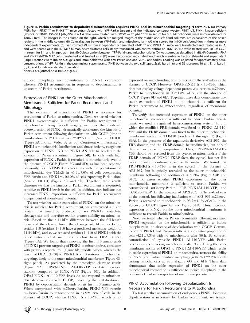
autophagy of depolarized mitochondria [21]. To test whether
PINK1 is necessary for Parkin-induced mitophagy, we treated
primary PINK12/2 and PINK1+/+ MEFs transiently expressing
YFP-Parkin with 20 mM CCCP for 24 h (Figure 4A and 4B).
Whereas no mitochondria can be detected in 66.1616.8% of
PINK1+/+ MEFs, all PINK2/2 MEFs retain their mitochondria.
Parkin-dependent mitophagy is reconstituted by exogenous
PINK1 expression in the PINK2/2 MEFs, with 65.565.0% of
reconstituted PINK2/2 cells displaying undetectable mitochon-
dria following CCCP treatment.
We found that Parkin-induced mitophagy is also dependent on
PINK1 expression in the M17 human neuroblastoma cell line.
Whereas in 27.168.6% of control shRNA M17 cells displayed
complete loss of mitochondria after 24 h, less than 5% of cells lost
mitochondria in the PINK1 shRNA cells (Figure 4C and 4D).
These results suggest that PINK1 is necessary for the mitophagy of
depolarized mitochondria following overexpression of Parkin.
To test whether PINK1 expression affects mitochondrial
turnover in the presence of endogenous levels of Parkin, we
treated the control shRNA and PINK1 shRNA M17 cells (which
express moderate levels of Parkin) with DMSO or CCCP for 24 h
and measured their relative mitochondrial mass by Mitotracker
Green (MTG) staining and flow cytometry. MTG, a sensitive
measure of mitochondrial mass, stains mitochondrial lipid in a
membrane potential–independent manner and has been used to
measure mitochondrial mass of depolarized mitochondria previ-
ously [35,36]. Control shRNA M17 cells exhibit a decrease in
mitochondrial mass (CCCP vs. DMSO, 222.4612.6%) following
CCCP treatment, whereas PINK1 shRNA M17 cells exhibit an
increase in mitochondrial mass (CCCP vs. DMSO, 43.5620.0%)
following depolarization (p-value = 0.008 for change in mitochon-
drial mass control shRNA vs. PINK shRNA) (Figure 4E). These
results are consistent with endogenous PINK1 promoting
mitochondrial degradation in the context of continued (or
increased) mitochondrial biogenesis [37,38]. To more directly
assay mitochondrial turnover in control and PINK1 shRNA M17
cells, we pulsed the cells with MTG and tracked loss of MTG
intensity at 0, 16, and 24 h in the presence of CCCP. Consistent
with the hypothesis that endogenous PINK1 promotes the
degradation of depolarized mitochondria, MTG intensity decreas-
es more slowly in PINK1 shRNA cells when compared with
control shRNA cells, treated with CCCP (0.5860.07 vs.
0.3360.07 relative MTG intensity at 24 h) (Figure 4F). These
data suggest that PINK1 promotes mitophagy in the context of
endogenous levels of Parkin. Additionally, these results suggest that
the selective turnover of dysfunctional mitochondria may be
balanced by the biogenesis of new mitochondria, allowing
exchange of damaged, dysfunctional mitochondria for healthy,
functional mitochondria.
Consistent with genetic studies in Drosophila, these findings show
that Parkin translocation to depolarized mitochondria and Parkin-
PINK1 Accumulation Promotes Parkin Recruitment
PLoS Biology | www.plosbiology.org 6 January 2010 | Volume 8 | Issue 1 | e1000298
PINK1 Accumulation Promotes Parkin Recruitment
PLoS Biology | www.plosbiology.org 7 January 2010 | Volume 8 | Issue 1 | e1000298
induced mitophagy are downstream of PINK1 expression,
whereas PINK1 accumulation in response to depolarization is
upstream of Parkin recruitment.
Expression of PINK1 on the Outer MitochondrialMembrane Is Sufficient for Parkin Recruitment andMitophagy
The expression of mitochondrial PINK1 is necessary for
recruitment of Parkin to mitochondria. Next, we tested whether
PINK1 overexpression is sufficient for Parkin recruitment to
mitochondria. Using live-cell imaging, we found that moderate
overexpression of PINK1 dramatically accelerates the kinetics of
Parkin recruitment following depolarization with CCCP (time to
translocation 5.061.5 min vs. 32.065.4 min, p-value ,0.001)
(Figure 5A and 5B; Video S2 vs. S3). Consistent with necessity of
PINK1’s mitochondrial localization and kinase activity, exogenous
expression of PINK1 KD or PINK1 DN fails to accelerate the
kinetics of Parkin recruitment (Figure 5B). In cells with high
expression of PINK1, Parkin is recruited to mitochondria even in
the absence of CCCP (Figure 5C and 5D), as has been reported
previously [27]. YFP-Parkin colocalizes with the potentiometric
mitochondrial dye TMRE in 45.367.6% of cells coexpressing
YFP-Parkin and PINK1 vs. 060% of cells expressing Parkin alone
(p-value ,0.001) (Figure 5C and 5D). Together, these results
demonstrate that the kinetics of Parkin recruitment is exquisitely
sensitive to PINK1 levels in the cell. In addition, they indicate that
increased PINK1 expression is sufficient for Parkin recruitment
independent of membrane potential.
To test whether stable expression of PINK1 on the mitochon-
dria is sufficient for Parkin recruitment, we constructed a fusion
protein that would be predicted to lack PINK1’s proteolytic
cleavage site and therefore exhibit greater stability on mitochon-
dria. Based on the ,11-kDa difference between the full-length
form and the cleaved form, the cleavage site likely lies before
residue 110 (residues 1–110 have a predicted molecular weight of
11.54 kDa), and so we replaced residues 1–110 of PINK1 with the
outer mitochondrial membrane anchor from OPA3 (1–30)
(Figure 6A). We found that removing the first 110 amino acids
of PINK1 prevents targeting of PINK1 to mitochondria, consistent
with previous reports [39] (Figure 6B, middle panel); whereas the
fusion of OPA3 (1–30) to PINK1 D1-110 restores mitochondrial
targeting, likely to the outer mitochondrial membrane (Figure 6B,
right panel). As predicted by the proteolytic cleavage results
(Figure 2A), OPA3-PINK1 D1-110-YFP exhibits increased
stability compared to PINK1-YFP (Figure 6C). In addition,
OPA3-PINK1 D1-110-YFP levels do not respond to mitochon-
drial depolarization with CCCP, indicating that stabilization of
PINK1 by depolarization depends on its first 110 amino acids.
When coexpressed with mCherry-Parkin, PINK1-YFP recruits
mCherry-Parkin to mitochondria in 57.961.8% of cells in the
absence of CCCP; whereas PINK1 D1-110-YFP, which is not
expressed on mitochondria, fails to recruit mCherry-Parkin in the
absence of CCCP. However, OPA3-PINK1 D1-110-YFP, which
does not display voltage dependent proteolysis, recruits mCherry-
Parkin to mitochondria in 9861.8% of cells in the absence of
CCCP (Figure 6D and 6E). Together, these data demonstrate that
stable expression of PINK1 on mitochondria is sufficient for
Parkin recruitment to mitochondria, regardless of membrane
potential.
To verify that increased expression of PINK1 on the outer
mitochondrial membrane is sufficient to induce Parkin recruit-
ment, we used a regulated heterodimerization system [40], in
which the modified FRB domain was fused to PINK1 D1-110-
YFP and the FKBP domain was fused to the outer mitochondrial
membrane anchor of TOM20 (residues 1 through 33) (Figure
S4A). In the presence of the rapamycin derivative AP21967, the
FRB domain and the FKBP domain heterodimerize, but only if
they are in the same compartment. Thus, FRB-PINK1D1-110-
YFP should be recruited from the cytosol to mitochondria if the
FKBP domain of TOM20-FKBP faces the cytosol but not if it
faces the inter membrane space or the matrix. We found that
FRB-PINK1D1-110-YFP is in the cytosol in the absence of
AP21967, but is quickly recruited to the outer mitochondrial
membrane following the addition of AP21967 (Figure S4B and
S4C). To assess whether PINK1 expression on the outer
mitochondrial membrane is sufficient to recruit Parkin, we
cotransfected mCherry-Parkin, FRB-PINK1D1-110-YFP, and
TOM20-FKBP. In the absence of AP21967, mCherry-Parkin is
in the cytosol, but following incubation with AP21967 mCherry-
Parkin is recruited to mitochondria in 96.764.1% of cells, in the
absence of CCCP (Figure 6F and Figure S4D). Thus, increased
expression of PINK1 on the outer mitochondrial membrane is
sufficient to recruit Parkin to mitochondria.
Next, we tested whether Parkin recruitment following increased
PINK1 expression on the mitochondria is sufficient to induce
mitophagy in the absence of depolarization with CCCP. Cotrans-
fection of PINK1 and Parkin results in a substantial proportion of
cells (42.167.3%) with no mitochondria after 96 h. By contrast,
cotransfection of cytosolic PINK1 D1-110-YFP with Parkin
produces no cells lacking mitochondria after 96 h. Fusing the outer
membrane anchor of OPA3 to PINK1 D1-110-YFP, which results
in stable expression of PINK1 on mitochondria, restores the ability
of PINK1 and Parkin to induce mitophagy ,with 76.462.2% of cells
lacking mitochondria at 96 h (Figure 6G and 6H). These data
demonstrate that stable expression of PINK1 on the outer
mitochondrial membrane is sufficient to induce mitophagy in the
presence of Parkin, irrespective of membrane potential.
PINK1 Accumulation following Depolarization IsNecessary for Parkin Recruitment to Mitochondria
To test whether accumulation of endogenous PINK1 following
depolarization is necessary for Parkin recruitment, we treated
Figure 3. Parkin recruitment to depolarized mitochondria requires PINK1 and its mitochondrial targeting N-terminus. (A) PrimaryMEFs from PINK1+/+ or PINK12/2 mice cotransfected with YFP-Parkin (green) and the indicated construct (vector, PINK1-V5, PINK1 kinase-deficient[KD]-V5, or PINK1 156–581 [DN]-V5) in a 1:4 ratio were treated with DMSO or 20 mM CCCP in serum for 3 h. Mitochondria were immunostained forTom20 (red). The images in the column on the right, which are merged images of the middle and left-hand columns, are expansions of the boxedregions in the middle column. (B) Colocalization between YFP-Parkin and mitochondria in (A) was scored for $100 cells/condition in three or moreindependent experiments. (C) Transformed MEFs from independently generated PINK1+/+ and PINK12/2 mice were transfected and treated as in (A)and were scored as in (B). (D) M17 human neuroblastoma cells stably transduced with control shRNA or PINK1 shRNA were treated with 10 mM CCCPin serum for 3 h and imaged as in (A). (E) Colocalization between YFP-Parkin and mitochondria in (D) was scored as described in (B). (F) Control shRNAand PINK1 shRNA M17 cells transfected and treated as in (D) were fractionated into mitochondria-rich membrane fraction (Memb) and supernatant(Sup). Fractions were run on SDS gels and immunoblotted with anti-Parkin and anti-VDAC antibodies. Loading was adjusted for approximately equalconcentrations of YFP-Parkin in the postnuclear supernatants (PNS) between the two cell types. Scale bars in (A and D) represent 10 mm. Error bars in(B, C, and E) indicate standard deviation.doi:10.1371/journal.pbio.1000298.g003
PINK1 Accumulation Promotes Parkin Recruitment
PLoS Biology | www.plosbiology.org 8 January 2010 | Volume 8 | Issue 1 | e1000298
Figure 4. PINK1 is required for Parkin-induced autophagy of depolarized mitochondria. (A) Primary MEFs from PINK1+/+ or PINK12/2 micecotransfected with YFP-Parkin were treated with DMSO or 20 mM CCCP in serum for 24 h. Mitochondria were stained with an anti-Tom20 antibody.(B) Percentage of cells with no detectable mitochondria in (A) was scored for .150 cells/condition in three or more independent experiments. (C)
PINK1 Accumulation Promotes Parkin Recruitment
PLoS Biology | www.plosbiology.org 9 January 2010 | Volume 8 | Issue 1 | e1000298
HeLa cells with CCCP alone (for 60 min) or with CCCP plus
cycloheximide, a general inhibitor of protein synthesis (cyclohex-
imide was added 30 min before CCCP and maintained through-
out the 60-min CCCP treatment). Treatment of HeLa cells for
90 min with cycloheximide blocked the depolarization-induced
accumulation of endogenous PINK1 in whole-cell lysates as well as
in the mitochondria-rich membrane fraction (Figure 7A and 7B).
A 90-min treatment with cycloheximide, likewise, blocked Parkin
recruitment to depolarized mitochondria by confocal microscopy
(96.063.5% vs. 11.364.2%) (Figure 7C and 7D). By contrast, 90-
min treatment with actinomycin D, an inhibitor of transcription,
had a modest effect on Parkin recruitment to uncoupled
mitochondria by confocal microscopy (Figure 7C and 7D),
suggesting that new transcription of PINK1 is not required for
Parkin recruitment. This is consistent with the absence of PINK1
mRNA up-regulation following uncoupling (Figure 2C). Cyclo-
heximide likewise blocked YFP-Parkin accumulation in the
mitochondria-enriched heavy membrane fraction by immunoblot-
ting (Figure 7E). Although these findings do not prove new PINK1
synthesis is required for Parkin recruitment (cycloheximide blocks
de novo synthesis of all proteins and thus may inhibit Parkin
recruitment independently of PINK1 accumulation), they suggest
that PINK1 accumulation and Parkin recruitment may be casually
related.
Threonines 175 and 217 in Parkin May Not Be Involved inParkin Recruitment to Mitochondria
It has been proposed that PINK1 may induce mitochondrial
recruitment of Parkin through phosphorylation of threonines 175
and 217 in a highly conserved region/domain of Parkin, which has
been recently named RING0 (Figure S5A) [27,41]. We found that
although mutation of T175 and T217 to alanine blocked
recruitment of Parkin to mitochondria, as was reported previously,
the phosphomimetic mutants T175E, T217E, and T175, 217E do
not translocate to mitochondria spontaneously. In addition, these
phosphomimetic mutants appear to inhibit CCCP-induced
recruitment of Parkin. Although these findings do not rule out
the possibility that phosphorylation of these sites by PINK1 or
another kinase induces Parkin recruitment, they suggest that these
threonines are more likely to play an important structural role
(Figure S5B and S5C).
Patient Mutations in PINK1 and Parkin Disrupt PINK1/Parkin Pathway at Distinct Steps
We assessed the ability of disease-causing mutations in PINK1
to reconstitute YFP-Parkin recruitment to mitochondria in
PINK12/2 primary MEFs. Following exogenous PINK1 WT
expression in PINK12/2 MEFs, YFP-Parkin was recruited to
mitochondria in 78.663.9% of cells after 20 mM CCCP treatment
for 3 h (Figure 8A and 8B). We found that the L347P patient
mutant of PINK1 is unstable (Figure 8C and Figure S6A), as was
reported previously [28], and that L347P failed to reconstitute
YFP-Parkin recruitment to depolarized mitochondria (Figure 8A
and 8B). Of the patient mutations that exhibited stable expression,
A168P and H271Q also failed to reconstitute YFP-Parkin
recruitment at 3 h, whereas G309D only partially reconstituted
YFP-Parkin recruitment (30.7616.7%) (Figure 6A and 6B). The
polymorphism G411S, which to date has only been found in cases
heterozygous for the mutation [42], reconstituted YFP-Parkin
recruitment to a similar extent as wild-type PINK1 (74.265.4%),
suggesting that PINK1 containing this polymorphism may be
functional in the PINK1/Parkin pathway (Figure 8A and 8B). This
is consistent with the idea that G411S may represent a natural
variant and may not be a true disease-causing mutation. Protein
levels of all PINK1 mutants accumulated upon exposure of cells to
CCCP (Figure 8C and Figure S6A).
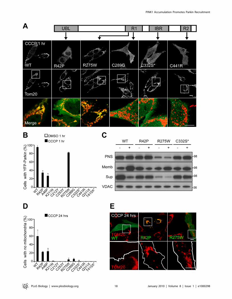
Next, we tested patient mutations in Parkin to see if they
would affect Parkin recruitment to mitochondria and/or
Parkin-induced mitophagy. Parkin has an N-terminal ubiqui-
tin-like domain (UBL) and a C-terminal RING-between-RING
(RBR) superdomain, which consists of three atypical RING
domains (Figure 9A). The fold of the N-terminal RING1 most
closely resembles that of traditional RING domains, such as
that of c-CBL, whereas the In-Between-RING (IBR) and the
C-terminal RING2 likely have unique folds [43–45]. The RBR
domain is responsible for Parkin’s ubiquitin ligase activity,
whereas its UBL domain is thought to mediate interactions
between Parkin and proteins with ubiquitin-binding domains
(UBDs) [46].
As was reported previously, wild-type YFP-Parkin is recruited to
mitochondria in the majority of HeLa cells (94.765.8%) by
confocal microscopy, following treatment with 10 mM CCCP for
1 h (Figure 9A and 9B). Pathogenic mutations in the UBL domain
(R42P and R46P), deletion of the UBL, or mutation of a key
residue (I44A) in the interaction of UBLs with UBDs [47], all
cause a moderate deficit in Parkin recruitment to depolarized
mitochondria (3465.3% and 26.566.6% for R42P and R46P,
respectively) (Figure 9A and 9B and Figure S7A–S7E). Mutations
in conserved cysteines of the RING domains (the patient
mutations C253Y, C289G, and C441R and the engineered
mutation C332S) completely disrupt recruitment at 1 h of CCCP
treatment, as do mutations (patient mutation Q311X and
engineered mutation T415X) that result in loss of RING2
(Figure 9A and 9B). Mutations K211N and C212Y, which lie
within a highly conserved region of Parkin that is likely a novel
RING-like domain [41] (Figure S5A), similarly blocked the
recruitment of Parkin to mitochondria (Figure 9B), consistent
with the importance of this region for Parkin’s activity.
Mitochondrial recruitment was seen for several of the conserved
cysteine RING mutants (C289G, C332S, and C441R) after 24 h
of CCCP exposure, suggesting that recruitment is not completely
disrupted with these mutations (Figure S8A and S8B). Interest-
ingly, the R275W mutation in RING1 exhibited only a mild
deficit in recruitment (81.762.1%) (Figure 9A and 9B). The
recruitment of YFP-Parkin R275W was verified in a live-cell
imaging experiment (Figure S8C). Although under control
conditions some mutants formed visible aggregates (Figure S8D),
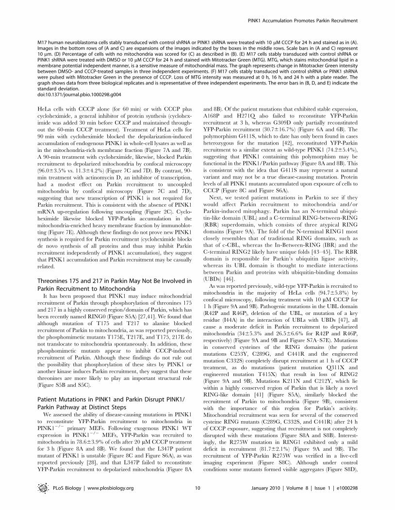
M17 human neuroblastoma cells stably transduced with control shRNA or PINK1 shRNA were treated with 10 mM CCCP for 24 h and stained as in (A).Images in the bottom rows of (A and C) are expansions of the images indicated by the boxes in the middle rows. Scale bars in (A and C) represent10 mm. (D) Percentage of cells with no mitochondria was scored for (C) as described in (B). (E) M17 cells stably transduced with control shRNA orPINK1 shRNA were treated with DMSO or 10 mM CCCP for 24 h and stained with Mitotracker Green (MTG). MTG, which stains mitochondrial lipid in amembrane potential independent manner, is a sensitive measure of mitochondrial mass. The graph represents change in Mitotracker Green intensitybetween DMSO- and CCCP-treated samples in three independent experiments. (F) M17 cells stably transduced with control shRNA or PINK1 shRNAwere pulsed with Mitotracker Green in the presence of CCCP. Loss of MTG intensity was measured at 0 h, 16 h, and 24 h with a plate reader. Thegraph shows data from three biological replicates and is representative of three independent experiments. The error bars in (B, D, and E) indicate thestandard deviation.doi:10.1371/journal.pbio.1000298.g004
PINK1 Accumulation Promotes Parkin Recruitment
PLoS Biology | www.plosbiology.org 10 January 2010 | Volume 8 | Issue 1 | e1000298

Figure 5. Kinetics of Parkin recruitment are modulated by PINK1 expression. (A) HeLa cells transfected with mCherry-Parkin (red) alone ormCherry-Parkin (red) and PINK1-YFP in a 1:1 ratio were imaged live following the addition of 10 mM CCCP in serum at time point 0 min. (B) HeLa cellstransfected with mCherry-Parkin and the indicated construct in a 1:1 ratio were treated as in (A) and imaged live (one frame/minute) following theaddition of CCCP. Time to the beginning of Parkin translocation was defined as the first appearance of puncta in two or more quadrants of the cell fortwo or more consecutive images for six or more cells in a minimum of three independent experiments. N.S., nonsignificant. (C) Live confocal image ofHeLa cells transfected with YFP-Parkin (green) or YFP-Parkin (green) and PINK1-myc (in a 1:4 ratio). Cells were loaded with TMRE (red) to stainpolarized mitochondria. Cells were not treated with CCCP. Scale bar in last image represents 10 mm. Images in the middle and right-hand panels are
PINK1 Accumulation Promotes Parkin Recruitment
PLoS Biology | www.plosbiology.org 11 January 2010 | Volume 8 | Issue 1 | e1000298
no mutant, including R275W, colocalized with mitochondria
(Figure 9B and Figure S8E).
Next, we assessed recruitment of Parkin mutants to depolarized
mitochondria by immunoblotting. As with our previous results
[21], we found some background YFP-Parkin signal in the
membrane fraction under control conditions. Following treatment
with CCCP for 1 h, levels of wild-type Parkin increase in the
mitochondria-rich membrane fraction and decrease in the
supernatant (Figure 9C). Although expression of Parkin R275W
was moderately less than wild type, it also increases localization in
the membrane fraction and decreases in the supernatant upon
CCCP treatment, consistent with the mitochondrial translocation
seen for this mutation by confocal microscopy (Figure 9C). No
membrane translocation was detectable by Western blotting for
either R42P or C332S, however, suggesting that translocation of
these mutants is substantially lower than wild type and consistent
with the deficit in mitochondrial translocation seen by confocal
microscopy (Figure 9C).
We assessed the ability of Parkin mutants to induce mitophagy.
As we found previously [21], expression of wild-type Parkin in
HeLa cells that do not detectably express endogenous Parkin
completely eliminates mitochondria in greater than half of the cells
(59.0615.1%) following treatment with CCCP for 24 h (Figure 9D
and 9E). Mutations in the UBL of Parkin exhibit a moderate loss
in mitophagy activity (22.062.0% and 23.168.4% of cells
exhibited no mitochondria for R42P and R46P, respectively);
whereas mutations in the conserved cysteines of the RBR or
truncations that resulted in loss of RING2 exhibit a severe
mitophagy deficit (060% to 5.362.3%, depending on the
mutation) (Figure 9D and 9E). In addition, patient mutations
R211N and C212Y caused a similar deficit in mitophagy,
supporting the notion that this may be an atypical RING domain
similar to RING1, IBR, and RING2 (Figure 9D). Interestingly, the
R275W mutation in RING1 also exhibited a severe deficit in
mitophagy (4.064%) (Figure 9D and 9E) even though it appears
to largely retain its ability to translocate to uncoupled mitochon-
dria (Figure 9A and 9B). This pattern of findings suggests that
recruitment of Parkin to mitochondria and its induction of
mitophagy are dissociable events.
Discussion
We recently reported that the Parkinson disease-linked E3
ubiquitin ligase, Parkin, is selectively recruited to dysfunctional
mitochondria with low membrane potential to promote their
autophagic degradation, suggesting that a deficiency of mitochon-
drial quality control may underlie the observed mitochondrial
dysfunction in Parkin knockout Drosophila and mice [11–15,17,21].
How Parkin is able to distinguish damaged, depolarized
mitochondria from healthy, polarized mitochondria, however,
was unknown.
Here, we show that PINK1 selectively accumulates on
depolarized mitochondria that have sustained damage. This
selective accumulation is achieved by a novel mechanism, in
which PINK1 is constitutively synthesized and imported into all
mitochondria, but cleaved from healthy mitochondria by voltage-
sensitive proteolysis (Figure S9). On damaged mitochondria that
have lost their membrane potential, however, PINK1 cleavage is
inhibited, leading to high PINK1 expression on the dysfunctional
mitochondria. Expression of mitochondrial PINK1 is required for
the recruitment of Parkin to the dysfunctional mitochondria and
for their selective elimination by Parkin. In addition, increased
expression of PINK1 on the outer mitochondrial membrane is
sufficient for Parkin recruitment and Parkin-induced mitophagy,
suggesting that loss of membrane potential activates Parkin
recruitment primarily through the up-regulation of mitochondrial
PINK1.
This model offers a parsimonious explanation for several
observations that have been made previously. Full-length mito-
chondrial PINK1 (,63 kDa) is cleaved into a short ,52-kDa
form, but the short, primarily cytosolic form is unstable, raising the
questions: why is PINK1 found both on the mitochondria and in
the cytosol, and which form of PINK1 is active in the PINK1/
Parkin pathway [22,28–30]. Our results suggest that full-length
mitochondrial PINK1 is the active form in the PINK1/Parkin
pathway, and that cleavage of PINK1 into an unstable cytosolic
form maintains low levels of PINK1 on healthy mitochondria in
order to suppress the PINK1/Parkin pathway in the absence of
mitochondrial damage. Additionally, this model provides an
explanation for the observation that the uncoupler valinomycin
(which can inhibit the TIM22/23 mitochondrial import pathway)
blocks PINK1 processing but fails to block PINK1 import [30].
Our model suggests that membrane potential is not required for
PINK1 import but is required to selectively maintain low PINK1
expression on healthy mitochondria. This mechanism couples the
collapse of mitochondrial voltage potential following mitochon-
drial damage to selective PINK1 accumulation on damaged
mitochondria.
At present, it is unclear which protease(s) mediate the cleavage
of PINK1 in mammalian cells. Although the intramembrane
serine protease Rhomboid-7 appears to be required for PINK1
cleavage in Drosophila [31], our results suggest that PARL, its
mammalian ortholog, is not required for PINK1 cleavage in
mammalian cells. This situation is similar to that of OPA1, which
also requires Rhomboid-7 for cleavage in Drosophila, but does not
require PARL for cleavage in mammalian cells [48].
In addition, determining how PINK1 cleavage is modulated by
membrane potential will require further study. The protease itself
may be sensitive to membrane potential and/or the PINK1
cleavage site may be available to the protease only in the presence
of a membrane potential. Alternatively, the regulation of PINK1
cleavage by membrane potential may be indirect. That inhibition
of PINK1 cleavage by mitochondrial depolarization up-regulates
the PINK1/Parkin mitophagy pathway also raises the possibility
that inhibitors of PINK1’s protease might up-regulate the pathway
and have some therapeutic benefit.
Our results suggest that PINK1 induces Parkin recruitment to a
particular subset of mitochondria, following its accumulation, and
there are several models for how PINK1 might induce Parkin
recruitment. In the simplest, as PINK1 accumulates, Parkin may
be recruited to mitochondria through a direct interaction with the
accumulated PINK1. In support of this model, PINK1 appears to
directly bind Parkin at least in some contexts [29]. Alternatively,
PINK1 may need to phosphorylate Parkin, a substrate of Parkin,
or an adaptor between PINK1 and Parkin, and thereby increase
Parkin’s affinity for a substrate or receptor on mitochondria.
Consistent with a role for phosphorylation in the activation of
Parkin, we found a kinase-deficient version of PINK1 fails to
rescue Parkin recruitment to mitochondria in PINK1 null MEFs
(even though PINK1 KD appears to be processed identically to
expansions of the boxed regions in the panels on the left. (D) Cells treated as described in (C) were scored for colocalization between YFP-Parkin andTMRE. $50 cells/experiment were scored in three or more independent experiments.doi:10.1371/journal.pbio.1000298.g005
PINK1 Accumulation Promotes Parkin Recruitment
PLoS Biology | www.plosbiology.org 12 January 2010 | Volume 8 | Issue 1 | e1000298

PINK1 Accumulation Promotes Parkin Recruitment
PLoS Biology | www.plosbiology.org 13 January 2010 | Volume 8 | Issue 1 | e1000298

wild-type PINK1). We were unable to replicate findings suggesting
that phosphorylation of two threonines in a conserved region of
Parkin is sufficient to induce Parkin recruitment to mitochondria
[27], but it is possible that Parkin may be phosphorylated by
PINK1 elsewhere. If direct phosphorylation is sufficient to induce
Parkin recruitment to mitochondria, however, it seems difficult to
explain how Parkin can be targeted to a particular subset of
mitochondria, as appears to occur in cells with bioenergetically
diverse populations of mitochondria [21].
Mutations in Parkin and PINK1 are inherited primarily in a
recessive manner, and loss of their function is thought to cause
early-onset Parkinson disease. We find that patient mutations in
PINK1 and Parkin disrupt the PINK1/Parkin mitochondrial
turnover pathway at distinct steps, consistent with the potential
relevance of this pathway for the development of Parkinson
disease.
Mutations in Parkin’s UBL or its deletion caused a moderate
deficit in Parkin recruitment to depolarized mitochondria and
induction of mitophagy. That deletion of the UBL only partially
inhibited the recruitment of Parkin to mitochondria suggests that
whereas this domain promotes the recruitment of Parkin to
mitochondria, it is not absolutely necessary for recruitment or
subsequent mitophagy. The UBL likely promotes recruitment of
Parkin through interaction with a protein containing a ubiquitin-
binding domain, as mutating residue isoleucine 44, which is
critical for the interaction between UBLs and UBDs [47], to
alanine resulted in a recruitment deficit similar to that caused by
deletion of the UBL domain. The disease-causing mutations
R42P, which causes global unfolding by NMR [49], and A46P lie
on either ends of the beta-pleated sheet containing I44A,
suggesting that these mutations may inhibit Parkin recruitment
by disrupting the interaction between Parkin and UBD-containing
proteins (Figure S7A and S7B).
Mutations in key cysteine residues in the RBR domain or
deletion of RING2, which is responsible for Parkin’s ubiquitin
ligase activity, severely disrupt both the recruitment of Parkin to
mitochondria and its induction of mitophagy. Interestingly, the
R275W mutation in RING1 of Parkin causes only a minor
disturbance of Parkin recruitment to depolarized mitochondria
but severely disrupts mitophagy, suggesting that recruitment and
mitophagy can be experimentally disassociated.
The R275W polymorphism in Parkin and the G411S
polymorphism in PINK1 have only been identified as heterozy-
gous polymorphisms in cases of Parkinson disease [42,50]. For this
reason, the pathogenicity of these polymorphisms has been a
matter of controversy. Our results show that the R275W Parkin
mutation, which affects a highly conserved arginine residue, causes
a significant loss of Parkin function in our mitophagy assay. This is
consistent with in vivo data in Drosophila melanogaster, demonstrating
that Parkin R275W, unlike wild-type Parkin, fails to compensate
for loss of endogenous Parkin. By contrast, we found that PINK1
containing the G411S polymorphism, which is conserved in
vertebrates, but not invertebrates, could compensate for loss of
endogenous PINK1, consistent with the view that PINK1 G411S
may be a natural variant and not a disease-causing mutation.
The stringent dependence of Parkin recruitment on PINK1
under depolarizing conditions is a little surprising given that, when
overexpressed, Parkin can partially compensate for PINK1 loss in
Drosophila and in mammalian cells [11–13,20]. How Parkin
overexpression compensates for PINK1 loss is not known, but
there are several possible explanations. First, there may be
mechanisms independent of PINK1 and depolarization that can
recruit Parkin to dysfunctional mitochondria. Alternatively, Parkin
may serve other functions in the cell that are independent of
PINK1 and protect against mitochondrial dysfunction indirectly;
or Parkin may function to some degree upon overexpression
independently of mitochondrial docking, perhaps effecting mito-
phagy or other mitochondrial changes from the cytosolic
compartment.
Stable loss or knockdown of PINK1 in mammalian cellular
models and mice leads to a number of mitochondria-related
abnormalities. Mitochondria in these cells or tissues exhibit
electron transport chain (ETC) dysfunction, diminished mem-
brane potential, increased reactive oxygen species production,
mitochondrial fragmentation, and calcium dysregulation, among
other abnormalities [20,32,34,51]. Although some of these
abnormalities may be a reversible consequence of others—for
instance, mitochondrial fragmentation may be due to low
membrane potential [34], and ETC dysfunction and decreased
membrane potential may be, in part, a functional consequence of
calcium dysregulation [51]—other abnormalities may be due to
irreversible dysfunction of specific mitochondrial proteins or
protein complexes. For instance, Complex I and the putative
Na+/Ca2+ transporter seem to be dysfunctional in cultured cells
following PINK1 knockdown [51], whereas Complex I and II
appear to be dysfunctional in the striatum of mice lacking PINK1
[16].
Although the proximate cause of these abnormalities in PINK1
null cells remains obscure, one explanation may be the failure of
PINK1/Parkin pathway to eliminate oxidatively damaged mito-
chondria, which accumulate over time as a natural consequence of
metabolism and other cellular stresses. That Parkin null cells and
tissues appear to share some of the same mitochondrial defects as
PINK1 null cells and tissues supports the view that these
abnormalities may be due to loss of a common PINK1/Parkin
pathway [17,18]. We cannot rule out that PINK1 may actively
Figure 6. Stable expression of PINK1 on the outer mitochondrial membrane is sufficient for Parkin recruitment. (A) Schematic diagramdepicting the construction of PINK1-YFP (green), PINK1 (111–581)-YFP (green), and OPA3-PINK1 (111–581)-YFP (green). (B) Confocal images depictingthe localization of PINK1-YFP, PINK1 (111–581)-YFP, and OPA3-PINK1 (111–581)-YFP in HeLa cells. Mitochondria are stained with the potentiometricdye TMRE (red). (C) HeLa cells were transfected with PINK1-YFP, PINK1 (111–581)-YFP, or Opa3-PINK1 (111–581)-YFP and treated with DMSO or 2 mMCCCP in serum-free medium for 3 h. Whole-cell lysates (WCL) were run on SDS gels and immunoblotted for PINK1, GFP, and tubulin. (D) Confocalimages of HeLa cells cotransfected with mCherry-Parkin (red) and PINK1-YFP (green), PINK1 (111–581)-YFP (green), or OPA3-PINK1 (111–581)-YFP(green). Cells were not treated with CCCP. (E) HeLa cells in (D) were scored for mCherry-Parkin forming puncta characteristic of mitochondria in $150cells in three or more independent experiments. Cells were not treated with CCCP. (F) HeLa cells were transfected with FRB-PINK1 (111–581)-YFP,which is in the cytosol, TOM20(1–33)-FKBP, which is on mitochondria, and mCherry-Parkin. In the presence of the rapamycin analog, AP21967, theFRB and FKBP domains of the respective fusion proteins (PINK1 (111–581) and TOM20’s outer mitochondrial membrane anchor) heterodimerize, ifthey have access to the same compartment (e.g., the cytosol). Cells treated with vehicle or 250 nM AP21967 for 8 h were scored for mCherry-Parkin inpuncta characteristic of mitochondria in $150 cells in three or more independent experiments. (G) Confocal images of HeLa cells transfected withPINK1-YFP (green), PINK1 (111–581)-YFP (green), or OPA3-PINK1 (111–581)-YFP (green) with or without ECFP-Parkin and cultured for 96 h in theabsence of CCCP. Cells were immunostained for Tom20 (red). To aid in visualizing cells that lack mitochondria, some individual cells have beenoutlined. (H) Cells treated as in (G) were scored for the absence of detectable mitochondria in $150 cells in three or more independent experiments.Scale bars in all images represent 10 mm.doi:10.1371/journal.pbio.1000298.g006
PINK1 Accumulation Promotes Parkin Recruitment
PLoS Biology | www.plosbiology.org 14 January 2010 | Volume 8 | Issue 1 | e1000298

Figure 7. PINK1 accumulation following depolarization with CCCP may be required for Parkin recruitment. (A) HeLa cells stablyexpressing YFP-Parkin were treated with 2 mM CCCP 1 h alone or CCCP 1 h + 2 mM CHX (30-min pretreatment and 1-h treatment) in the absence ofserum. Whole-cell lysates were run on SDS gels and immunoblotted for endogenous PINK1 and the loading control GAPDH. (B) Cells treated as in (A)were fractionated. The mitochondria-enriched membrane fraction (Memb) was run on SDS gels and immunoblotted for endogenous PINK1 andVDAC. (C) HeLa cells were transfected with YFP-Parkin (green) and treated with 10 mM CCCP 1 h alone, CCCP + 10 mM of actinomycin (30-minpretreatment and 1-h treatment), or CCCP 1 h + 100 mM CHX (30-min pretreatment and 1-h treatment) in the presence of serum and immunostainedfor Tom20 (red). (D) Colocalization between YFP-Parkin and mitochondria in (C) was scored for $150 cells/condition in three or more independent
PINK1 Accumulation Promotes Parkin Recruitment
PLoS Biology | www.plosbiology.org 15 January 2010 | Volume 8 | Issue 1 | e1000298

prevent mitochondrial damage and dysfunction, in addition to its
signaling role in the PINK1/Parkin pathway. PINK1’s interaction
with HtrA2/OMI, for instance, appears to be independent of
Parkin function in Drosophila [31,52,53].
Loss of PINK1 and Parkin affects some cell populations, like
substantia nigral neurons, more than others, even though PINK1
and Parkin appear to be more widely expressed. Why some tissues
are more vulnerable to loss of PINK1/Parkin than others is
unclear, but it may relate to the degree of damage mitochondria
sustain within that tissue (e.g., mitochondria in the SN are subject
to greater oxidative stress than those in other neural tissues [2]);
the existence of redundant mitophagy pathways (e.g., mammalian
tissues may contain pathways orthologous to those recently
identified in yeast [54,55]); the ability of the tissue to mitigate
the damage by other means (a tissue composed of mitotic cells may
be able to manage mitochondrial damage through cellular
turnover rather than mitochondrial turnover); and mitochondrial
demand within a particular tissue (neurons have high, local
metabolic demands, and dopaminergic neurons are subject to
especially high calcium fluxes that need to be buffered by
mitochondria [56]). Some or all of these factors may contribute
to the special reliance of SN neurons on PINK1 and Parkin.
PINK1 and Parkin are a significant cause of autosomal recessive
parkinsonism and have been genetically linked to a pathway that
protects against progressive mitochondrial damage and dysfunc-
tion. We have found that PINK1 levels and subsequently Parkin
recruitment to mitochondria are dramatically regulated by the
bioenergetic state of individual mitochondria, and that this unique
regulation may allow PINK1 and Parkin to promote the selective
and efficient turnover of mitochondria that have become
damaged. Loss of PINK1 or Parkin function due to pathogenic
mutations can disrupt this mitochondrial turnover pathway which
may lead to the accumulation of dysfunctional mitochondria in
vulnerable tissues—with a resultant increase in oxidative stress,
depression of metabolism, and, eventually, accelerated cell death,
which has been observed in Drosophila and, to a lesser extent, in
mouse models of the disease [11–17]. Together, these findings
provide a biochemical explanation for the genetic epistasis
between PINK1 and Parkin observed in Drosophila, and support
a novel, testable model of how loss of PINK1 and Parkin function
may lead to autosomal recessive parkinsonism.
Materials and Methods
Cell CultureHeLa YFP-Parkin, E18 Rat cortical neurons, PINK1+/+ SV40-
transformed MEF cells, PINK12/2 SV40-transformed MEF, M17
neuroblastoma control shRNA, M17 neuroblastoma PINK1,
Mfn1/22/2 MEF, and Parl2/2 MEF cell lines have been
described previously [21,25,29,33,57,58]. PINK1+/+ and
PINK12/2 primary MEFs were isolated from embryos using a
standard protocol [16]. Parkin+/+- and Parkin2/2-transformed
MEFs were created by isolation of primary cells from embryos of
B6.129S4-Park2tm1Shn/J mice (Jackson Labs), using a standard
protocol [16], followed by retroviral transduction of SV40
(Applied Biological Materials). YFP-Parkin, YFP-Parkin mutants,
mCherry-Parkin, PINK1-YFP, PINK1KD-YFP, PINK1 D1-110-
YFP, and Opa3-PINK1 D1-110-YFP are in C1 or N1 Clontech
vectors. PINK1WT-V5, PINK1KD-V5, and PINK1 D1-156-V5
are in pDest40 vector (Invitrogen). PINK1 patient mutations are
in the pLenti-V5 vector (Invitrogen). PINK1-myc is in a
pCMBTNT vector (Promega). The PARL shRNA construct
targeting (59-CCAACTTGGAGCTTCTAGTAAGTTCTCTA-
CTAGAAGCTCCAAGTTGG-39) is in the pSuper-GFP vector
(OligoEngine). To make FRB-PINK1 (111–581)-YFP and Tom20
(1–33)-FKBP, PCR fragments containing PINK1 (111–581)-YFP
and Tom20 (1–33) were cloned into the BamHI site of the pC4-
RHE vector and the EcoRI and XbaI sites of pC4M-F2E vectors,
respectively (ARIAD Pharmaceuticals). The rapamycin analog
AP21967 was obtained from ARIAD Pharmaceuticals.
Confocal MicroscopyConfocal microscopy of fixed samples, scoring of Parkin
recruitment and Parkin-induced mitophagy, and live-cell imaging
were performed as described previously [21]. Experiments in
Mfn1/2 null cells were performed as described previously, with
minor modifications, as described in the supplemental materials
and methods (Text S1) [21].
Immunoblotting and ImmunocytochemistryFor PINK1 experiments, cells were fractionated using the
Mitochondria Isolation Kit (Pierce), according to manufacturer’s
specifications, with slight modifications, as described in the
supplemental methods (Text S1). To isolate integral membrane
proteins, membrane fractions obtained as above were carbonate
extracted with 0.1 M Na2CO3 fresh cold buffer, and membranes
were pelleted, as described in the supplemental materials and
methods (Text S1). For Parkin experiments, cells were fractionated
as described previously, with minor modifications, as described in
the supplemental materials and methods (Text S1) [21]. The
protease protection assay was performed as described previously
[22]. Cells were fixed and immunostained as described previously
[21]. The following primary antibodies were used: anti-Parkin
(PRK8) monoclonal (Santa Cruz Biotechnology), anti-Tom20
polyclonal (Santa Cruz Biotechnology.), anti-cytochrome c
monoclonal (BD Biosciences), anti-PINK1 polyclonal (Novus
Biologicals), anti-VDAC monoclonal (Calbiochem), anti-GAPDH
polyclonal (Sigma-Aldrich), anti-Tubulin monoclonal (Sigma-
Aldrich), anti-V5 monoclonal (Invitrogen), anti-GFP polyclonal
(Invitrogen), anti-TIM23 monoclonal (BD Biosciences), and anti-
Hsp60 monoclonal (Stressgen).
Quantitative RT-PCRqRT-PCR of PINK1 mRNA levels was performed as described
in detail previously [34].
Supporting Information
Figure S1 Mitochondrial PINK1 accumulates on theouter mitochondrial membrane following mitochondrialdepolarization. (A) HeLa cells treated with 1 mM of valinomy-
cin without serum at time point 0 were fractionated, and
carbonate extracted. The carbonate extracted pellet, which is
enriched for proteins integral to mitochondria, was run on SDS
gels and immunoblotted for endogenous PINK1 and mitochon-
drial protein VDAC. (B) HeLa cells stably expressing YFP-Parkin
were treated with 2 mM of CCCP without serum at time point 0
and fractionated. The mitochondria-rich membrane fraction
(lanes 1 and 2) and the cytosolic enriched postmembrane fraction
(lanes 4–9) were run on SDS gels and immunoblotted for PINK1,
experiments. (E) HeLa cells stably expressing YFP-Parkin were treated as in (A) and fractionated. The mitochondria-rich fraction was run on an SDS geland immunostained for Parkin. Scale bars in all images represent 10 mm.doi:10.1371/journal.pbio.1000298.g007
PINK1 Accumulation Promotes Parkin Recruitment
PLoS Biology | www.plosbiology.org 16 January 2010 | Volume 8 | Issue 1 | e1000298

tubulin, and VDAC. (C) PINK12/2 MEFs transfected with
PINK1-myc or left untransfected were treated with 2 mM CCCP
without serum for 3 h and fractionated. Mitochondrial-rich
membrane fraction was run on SDS gels and immunoblotted for
PINK1 and VDAC. (D) HeLa cells transfected with PINK1-YFP
or a kinase-deficient version of PINK1 (PINK1KD-YFP) were
treated as in (B). Whole-cell lysates were run on SDS gels and
immunoblotted for PINK1 and tubulin. Arrow indicates the
predicted molecular weight (MW) of full-length PINK1-YFP. (E)
HeLa cells stably expressing YFP-Parkin were treated with 10 mM
CCCP for 3 h and fractionated. The mitochondria-enriched
membrane fraction was aliquoted. Each aliquot was treated with 0
Figure 8. Disease-causing PINK1 mutants fail to reconstitute Parkin recruitment to depolarized mitochondria. (A) Primary MEFs fromPINK12/2 mice cotransfected with YFP-Parkin (green) and indicated V5-tagged PINK1 constructs in a 1:4 ratio were treated with DMSO or 20 mM CCCP inserum for 3 h. Mitochondria were stained with an anti-Tom20 antibody (red). Scale bar in images represents 10 mm. Images in the middle and bottomrows are expansions of the images indicated by the boxes in the top row. (B) Colocalization between YFP-Parkin and mitochondria in (A) was scored for.150 cells/condition in three or more independent experiments. Error bars indicate standard deviation. (C) HeLa cells stably expressing YFP-Parkin weretransfected with the indicated V5-tagged constructs, treated with DMSO or 2 mM CCCP for 3 h in serum-free medium, and fractionated. Themitochondria-rich membrane fraction was run on an SDS gel and immunoblotted for PINK1, the V5 tag, and the mitochondrial protein VDAC.doi:10.1371/journal.pbio.1000298.g008
PINK1 Accumulation Promotes Parkin Recruitment
PLoS Biology | www.plosbiology.org 17 January 2010 | Volume 8 | Issue 1 | e1000298
PINK1 Accumulation Promotes Parkin Recruitment
PLoS Biology | www.plosbiology.org 18 January 2010 | Volume 8 | Issue 1 | e1000298

to 100 mg/ml protease K and immunoblotted for endogenous
PINK1, the outer membrane protein TOM20, the inner
membrane protein Tim23, and matrix protein Hsp60.
Found at: doi:10.1371/journal.pbio.1000298.s001 (2.72 MB TIF)
Figure S2 PINK1 accumulates independently of PARLand Parkin expression. (A) HeLa cells cotransfected with
PARL-Flag and either PARL shRNA or control shRNA were
depolarized with 10 mM CCCP for 3 h. Whole-cell lysates were
run on SDS gels and immunoblotted with antibodies against the
N-terminus of PARL, the C-terminus of PARL, and tubulin. (B)
HeLa cells mock transfected or transfected with shRNA PARL
were treated with DMSO or CCCP for 3 h and fractionated. The
mitochondria-enriched membrane fraction (left) and whole-cell
lysates (right) were run on SDS gels and immunoblotted for
PINK1, the C-terminus of PARL, VDAC, and/or tubulin. (C)
Wild-type or PARL-null MEFs were transfected with PINK1-V5,
treated as in (B), and fractionated. The mitochondria-enriched
heavy membrane fraction was run on SDS gels and immuno-
blotted for PINK1, the N-terminus of PARL, the C-terminus of
PARL, and Hsp60. (D) Untransfected HeLa cells or HeLa cells
stably expressing YFP-Parkin (HeLa/Parkin) were treated with
DMSO or 2 mM CCCP without serum for 1 h. Whole-cell lysates
(WCL) were run on SDS gels and immunoblotted for endogenous
PINK1 and tubulin. (E) Parkin+/+ or Parkin2/2 MEFs transfected
with PINK1-myc were treated with 2 mM CCCP in the absence of
serum and fractionated. The mitochondria-rich membrane
fraction was immunoblotted for PINK1 and VDAC. Scale bars
in all images represent 10 mm.
Found at: doi:10.1371/journal.pbio.1000298.s002 (2.32 MB TIF)
Figure S3 PINK1 is required for Parkin recruitment tomitochondria. (A) SV40-transformed PINK12/2 MEFs co-
transfected with YFP-Parkin (green) and vector, PINK1, or
PINK1 KD were treated with 20 mM CCCP for 3 h. Cells were
immunostained for Tom20 (red). (B) M17 neuroblastoma cells
stably transduced with control shRNA or PINK1 shRNA and
transfected with YFP-Parkin were treated with DMSO or 10 mM
CCCP for 3 h and fractionated into postnuclear supernatant
(PNS), mitochondria-rich heavy membrane fraction (HMF), and
supernatant (Sup). Fractions were run on SDS gels and
immunoblotted for Parkin, PINK1, and VDAC. Scale bars in
images represent 10 mm.
Found at: doi:10.1371/journal.pbio.1000298.s003 (1.39 MB TIF)
Figure S4 Increased expression of PINK1 on the outermitochondrial membrane is sufficient for Parkin re-cruitment. (A) A schematic demonstrating the construction of
PINK1-YFP, FRB-PINK1 (111–581)-YFP, and Tom20(1–33)-
FKBP. The FRB and FKBP domains heterodimerize in the
presence of the rapamycin analog AP21967 if they are in the same
compartment. (B) HeLa cell were transfected with PINK1 (111–
581)-YFP and Tom20 (1–33)-FKBP, and imaged live. The
rapamycin analog, AP21967 (250 nM), was added at time point
0. (C) Confocal image depicting the localization of FRB-PINK1
(111–581)-YFP (green) cotransfected with Tom20 (1–33)-FKBP
following treatment with vehicle or 250 nM of AP21967 for
30 min. Mitochondria are labeled with the potentiometric dye
TMRE (red). (D) Confocal image depicting the localization of
FRB-PINK1 (111–581)-YFP (green) and mCherry-Parkin (red)
following treatment with vehicle or 250 nM of AP21967 for 8 h.
Scale bars in all images represent 10 mm.
Found at: doi:10.1371/journal.pbio.1000298.s004 (2.16 MB TIF)
Figure S5 Putative PINK1 phosphorylation sites onParkin, T175, and T217 are not sufficient for Parkinrecruitment. (A) Alignment of highly conserved Parkin unique
region/domain containing T175 and T217. Arrows on top show
positions of threonines 175 and 217 and the disease-causing
mutation C212. Brackets on the bottom of the alignment point to
the conserved cysteine and histidine residues forming putative
zinc-binding sites I and II of the RING0 domain. (B) HeLa cells
were transfected with YFP-Parkin (white) containing the indicated
point mutations and treated with DMSO or CCCP for 1 h.
Mitochondria were labeled with anti-Tom20 antibody. (C)
Colocalization between YFP-Parkin and mitochondria in (A) was
scored for .150 cells/condition in three or more independent
experiments. Scale bars in all images represent 10 mm.
Found at: doi:10.1371/journal.pbio.1000298.s005 (2.17 MB TIF)
Figure S6 Disease-causing mutations in PINK1 do notaffect PINK1-induced accumulation. (A) HeLa cells stably
expressing Parkin transfected with the indicated V5-tagged PINK1
constructs were treated with DMSO or 2 mM CCCP in serum-free
medium. Whole-cell lysates were run on SDS gels and immuno-
blotted for PINK1, the V5 tag, and tubulin.
Found at: doi:10.1371/journal.pbio.1000298.s006 (0.67 MB TIF)
Figure S7 Mutations in Parkin’s ubiquitin-like domain(UBL) partially disrupt Parkin recruitment to mitochon-dria. (A) Alignment of part of the UBL amino acid sequences
from orthologous Parkin proteins. An asterisk (*) indicates position
of patient mutations (R42P and R46) and engineered mutation
(I44A) examined below. Red box indicates position of beta-pleated
sheet containing I44, a key residue for interactions between UBL
domains and ubiquitin-binding domains. (B) Structure of UBL
(PDB 1IYF) with position of patient mutations (R42P and A46P)
highlighted in blue and position of engineered mutation I44A
highlighted in red. (C and D) HeLa cells transfected with YFP-
Parkin containing indicated mutations (green) and treated with
CCCP for 1 h (C) or 24 h (D). Mitochondria were labeled with
Tom20 antibody (red). (E) Colocalization between YFP-Parkin
and mitochondria in (C) scored for $150 cells/condition in three
or more independent experiments. (F) Percentage of cells with no
mitochondria transfected and treated as described in (D) scored for
$150 cells/condition in three or more independent experiments.
Scale bars in all images represent 10 mm.
Found at: doi:10.1371/journal.pbio.1000298.s007 (3.70 MB TIF)
Figure S8 Mutations in Parkin disrupt Parkin recruit-ment and/or Parkin-mediated mitophagy. (A) HeLa cells
transfected with YFP-Parkin (green) containing the indicated
Figure 9. Disease-causing mutations in Parkin disrupt Parkin recruitment to mitochondria and/or Parkin-induced mitophagy. (A)HeLa cells were transfected with YFP-Parkin (white and green) containing indicated mutations and treated with CCCP for 1 h. Mitochondria labeledwith an anti-Tom20 antibody (red). Images in the bottom row are expansions of the regions indicated by the boxes in the middle row. WT, wild type.(B) Colocalization between YFP-Parkin and mitochondria in (A) scored for $150 cells/condition in three or more independent experiments. (C) HeLacells transfected and treated as in (A) and fractionated into postnuclear supernatant (PNS), mitochondria-rich heavy membrane fraction (HMF), andsupernatant (Sup). Fractions run on SDS gels and immunoblotted for Parkin and VDAC. (D and E) HeLa cells transfected as in (A) and treated withCCCP or DMSO for 24 h. (D) Number of HeLa cells with no mitochondria scored for $150 cells/condition in three or more independent experiments.(E) Images of WT, R42P, and R275W Parkin (green) stained as in (A). An asterisk (*) indicates engineered mutation; all others have been linked toParkinson disease. Scale bars in all images represent 10 mm. Error bars in (B and D) indicate standard deviation.doi:10.1371/journal.pbio.1000298.g009
PINK1 Accumulation Promotes Parkin Recruitment
PLoS Biology | www.plosbiology.org 19 January 2010 | Volume 8 | Issue 1 | e1000298

missense mutations were treated with 10 mM CCCP in serum for
24 h were scored for Parkin colocalizing with mitochondria in
$150 cells/condition in three or more independent experiments.
Mitochondria were immunostained for Tom20 (red). (B) Confocal
images representing (A). (C) HeLa cells transfected with YFP-
Parkin R275W (green) were imaged live. CCCP (10 mM) was
added at time point 0. (D) HeLa cells transfected with the
indicated constructs were treated with DMSO or 10 mM CCCP
for 24 h and scored for the percentage of cells with visible
aggregates in $150 cells/condition in three or more independent
experiments. (E) HeLa cells transfected with YFP-Parkin WT
(green) or YFP-Parkin R275W (green) were treated with DMSO
or CCCP for 1 h and imaged live. Mitochondria were stained with
the potentiometric dye TMRE (red). Scale bars in all images
represent 10 mm.
Found at: doi:10.1371/journal.pbio.1000298.s008 (2.40 MB TIF)
Figure S9 Model depicting regulation of PINK1 stabilityon healthy and dysfunctional mitochondria by mem-brane potential. On healthy mitochondria PINK1 is constitu-
tively imported, proteolytically cleaved into a cytosolic form, and
degraded by the proteasome, resulting in low levels of mitochon-
drial PINK1. On damaged mitochondria with low membrane
potentials (DY), however, PINK1 cleavage is blocked, leading to
accumulation of mitochondrial PINK1 on the dysfunctional
mitochondria. Accumulated PINK1 recruits Parkin to damaged
mitochondria, which Parkin marks, likely by ubiquitination, for
autophagic degradation.
Found at: doi:10.1371/journal.pbio.1000298.s009 (0.39 MB TIF)
Text S1 Supplemental experimental methods.Found at: doi:10.1371/journal.pbio.1000298.s010 (0.03 MB
DOC)
Video S1 PINK1-YFP accumulates on mitochondria.HeLa cells transiently transfected with PINK1-YFP (green) were
incubated in CO2-independent medium (Gibco) for 1 h and then
treated with 10 mM CCCP at time point 0. Images of cells were
taken every 1 min for 40 min, using a PerkinElmer UltraView
LCI (Live Cell Imaging) System at 35uC.
Found at: doi:10.1371/journal.pbio.1000298.s011 (2.67 MB
MOV)
Video S2 mCherry-Parkin is recruited to mitochondria.HeLa cells transiently transfected with mCherry-Parkin (red) +empty vector in a 1:1 ratio were incubated in CO2-independent
medium (Gibco) for 1 h and then treated with 10 mM CCCP at
time point 0. Images of cells were taken every 1 min for 68 min,
using a PerkinElmer UltraView LCI (Live Cell Imaging) System at
35uC.
Found at: doi:10.1371/journal.pbio.1000298.s012 (2.00 MB
MOV)
Video S3 Kinetics of mCherry-Parkin recruitment tomitochondria are enhanced by exogenous PINK1 ex-pression. HeLa cells transiently transfected with mCherry-
Parkin (red) + PINK1-YFP in a 1:1 ratio were incubated in CO2-
independent medium (Gibco) for 1 h and then treated with 10 mM
CCCP at time point 0. Images of cells were taken every 1 min for
51 min, using a PerkinElmer UltraView LCI (Live Cell Imaging)
System at 35uC.
Found at: doi:10.1371/journal.pbio.1000298.s013 (4.11 MB
MOV)
Acknowledgments
We thank C. Smith for help with confocal microscopy, S. Smith for
assistance with cell culture, A. Beilina for help with PINK1 constructs and
for performing the qRT-PCR experiments, and C. Blackstone for his
insights and careful reading of the manuscript. We thank D. C. Chan for
Mfn1/22/2 MEFs, Z. Zhang for SV40-transformed PINK1+/+ and
PINK12/2 MEFs, Luca Pellegrini for Parl+/+ and Parl2/2 MEFs, and
Luca Pellegrini and Stephan Frank for the Parl shRNA construct.
Author Contributions
The author(s) have made the following declarations about their
contributions: Conceived and designed the experiments: DPN SMJ AT
DFS RJY. Performed the experiments: DPN SMJ AT. Analyzed the data:
DPN SMJ AT MRC RJY. Contributed reagents/materials/analysis tools:
CAG JS RJY. Wrote the paper: DPN RJY.
References
1. Schapira AH (2009) Molecular and clinical pathways to neuroprotection of
dopaminergic drugs in Parkinson disease. Neurology 72: S44–50.
2. Soong NW, Hinton DR, Cortopassi G, Arnheim N (1992) Mosaicism for a
specific somatic mitochondrial DNA mutation in adult human brain. Nat Genet
2: 318–323.
3. Schapira AH (2008) Mitochondria in the aetiology and pathogenesis of
Parkinson’s disease. Lancet Neurol 7: 97–109.
4. Bender A, Krishnan KJ, Morris CM, Taylor GA, Reeve AK, et al. (2006) High
levels of mitochondrial DNA deletions in substantia nigra neurons in aging and
Parkinson disease. Nat Genet 38: 515–517.
5. Kraytsberg Y, Kudryavtseva E, McKee AC, Geula C, Kowall NW, et al. (2006)
Mitochondrial DNA deletions are abundant and cause functional impairment in
aged human substantia nigra neurons. Nat Genet 38: 518–520.
6. Baloh RH, Salavaggione E, Milbrandt J, Pestronk A (2007) Familial
parkinsonism and ophthalmoplegia from a mutation in the mitochondrial
DNA helicase twinkle. Arch Neurol 64: 998–1000.
7. Luoma P, Melberg A, Rinne JO, Kaukonen JA, Nupponen NN, et al. (2004)
Parkinsonism, premature menopause, and mitochondrial DNA polymerase
gamma mutations: clinical and molecular genetic study. Lancet 364:
875–882.
8. Ekstrand MI, Terzioglu M, Galter D, Zhu S, Hofstetter C, et al. (2007)
Progressive parkinsonism in mice with respiratory-chain-deficient dopamine
neurons. Proc Natl Acad Sci U S A 104: 1325–1330.
9. Langston JW, Ballard P, Tetrud JW, Irwin I (1983) Chronic Parkinsonism in
humans due to a product of meperidine-analog synthesis. Science 219: 979–980.
10. Betarbet R, Sherer TB, MacKenzie G, Garcia-Osuna M, Panov AV, et al.
(2000) Chronic systemic pesticide exposure reproduces features of Parkinson’s
disease. Nat Neurosci 3: 1301–1306.
11. Yang Y, Gehrke S, Imai Y, Huang Z, Ouyang Y, et al. (2006) Mitochondrial
pathology and muscle and dopaminergic neuron degeneration caused by
inactivation of Drosophila Pink1 is rescued by Parkin. Proc Natl Acad Sci U S A
103: 10793–10798.
12. Clark IE, Dodson MW, Jiang C, Cao JH, Huh JR, et al. (2006) Drosophila pink1
is required for mitochondrial function and interacts genetically with parkin.
Nature 441: 1162–1166.
13. Park J, Lee SB, Lee S, Kim Y, Song S, et al. (2006) Mitochondrial dysfunction in
Drosophila PINK1 mutants is complemented by parkin. Nature 441:
1157–1161.
14. Greene JC, Whitworth AJ, Kuo I, Andrews LA, Feany MB, et al. (2003)
Mitochondrial pathology and apoptotic muscle degeneration in Drosophila
parkin mutants. Proc Natl Acad Sci U S A 100: 4078–4083.
15. Whitworth AJ, Theodore DA, Greene JC, Benes H, Wes PD, et al. (2005)
Increased glutathione S-transferase activity rescues dopaminergic neuron loss in
a Drosophila model of Parkinson’s disease. Proc Natl Acad Sci U S A 102:
8024–8029.
16. Gautier CA, Kitada T, Shen J (2008) Loss of PINK1 causes mitochondrial
functional defects and increased sensitivity to oxidative stress. Proc Natl Acad
Sci U S A 105: 11364–11369.
17. Palacino JJ, Sagi D, Goldberg MS, Krauss S, Motz C, et al. (2004)
Mitochondrial dysfunction and oxidative damage in parkin-deficient mice.
J Biol Chem 279: 18614–18622.
18. Mortiboys H, Thomas KJ, Koopman WJ, Klaffke S, Abou-Sleiman P, et al.
(2008) Mitochondrial function and morphology are impaired in parkin-mutant
fibroblasts. Ann Neurol 64: 555–565.
19. Muftuoglu M, Elibol B, Dalmizrak O, Ercan A, Kulaksiz G, et al. (2004)
Mitochondrial complex I and IV activities in leukocytes from patients with
parkin mutations. Mov Disord 19: 544–548.
20. Exner N, Treske B, Paquet D, Holmstrom K, Schiesling C, et al. (2007) Loss-of-
function of human PINK1 results in mitochondrial pathology and can be
rescued by parkin. J Neurosci 27: 12413–12418.
PINK1 Accumulation Promotes Parkin Recruitment
PLoS Biology | www.plosbiology.org 20 January 2010 | Volume 8 | Issue 1 | e1000298

21. Narendra D, Tanaka A, Suen DF, Youle RJ (2008) Parkin is recruited selectively
to impaired mitochondria and promotes their autophagy. J Cell Biol 183:
795–803.
22. Zhou C, Huang Y, Shao Y, May J, Prou D, et al. (2008) The kinase domain of
mitochondrial PINK1 faces the cytoplasm. Proc Natl Acad Sci U S A 105:
12022–12027.
23. Eguchi Y, Shimizu S, Tsujimoto Y (1997) Intracellular ATP levels determine cell
death fate by apoptosis or necrosis. Cancer Res 57: 1835–1840.
24. Budd SL, Nicholls DG (1996) A reevaluation of the role of mitochondria in
neuronal Ca2+ homeostasis. J Neurochem 66: 403–411.
25. Chen H, Chomyn A, Chan DC (2005) Disruption of fusion results in
mitochondrial heterogeneity and dysfunction. J Biol Chem 280: 26185–26192.
26. Cocheme HM, Murphy MP (2008) Complex I is the major site of mitochondrial
superoxide production by paraquat. J Biol Chem 283: 1786–1798.
27. Kim Y, Park J, Kim S, Song S, Kwon SK, et al. (2008) PINK1 controls
mitochondrial localization of Parkin through direct phosphorylation. Biochem
Biophys Res Commun 377: 975–980.
28. Beilina A, Van Der Brug M, Ahmad R, Kesavapany S, Miller DW, et al. (2005)
Mutations in PTEN-induced putative kinase 1 associated with recessive
parkinsonism have differential effects on protein stability. Proc Natl Acad
Sci U S A 102: 5703–5708.
29. Xiong H, Wang D, Chen L, Choo YS, Ma H, et al. (2009) Parkin, PINK1, and
DJ-1 form a ubiquitin E3 ligase complex promoting unfolded protein
degradation. J Clin Invest 119: 650–660.
30. Lin W, Kang UJ (2008) Characterization of PINK1 processing, stability, and
subcellular localization. J Neurochem 106: 464–474.
31. Whitworth AJ, Lee JR, Ho VM, Flick R, Chowdhury R, et al. (2008) Rhomboid-
7 and HtrA2/Omi act in a common pathway with the Parkinson’s disease
factors Pink1 and Parkin. Dis Model Mech 1: 168–174; discussion 173.
32. Dagda RK, Cherra SJ 3rd, Kulich SM, Tandon A, Park D, et al. (2009) Loss of
PINK1 function promotes mitophagy through effects on oxidative stress and
mitochondrial fission. J Biol Chem 284: 13843–13855.
33. Weihofen A, Thomas KJ, Ostaszewski BL, Cookson MR, Selkoe DJ (2009)
Pink1 Forms a Multiprotein Complex with Miro and Milton, Linking Pink1
Function to Mitochondrial Trafficking (dagger). Biochemistry 48: 2045–2052.
34. Sandebring A, Thomas KJ, Beilina A, van der Brug M, Cleland MM, et al.
(2009) Mitochondrial alterations in PINK1 deficient cells are influenced by
calcineurin-dependent dephosphorylation of dynamin-related protein 1. PLoS
One 4: e5701. doi:10.1371/journal.pone.0005701.
35. Rodriguez-Enriquez S, Kim I, Currin RT, Lemasters JJ (2006) Tracker dyes to
probe mitochondrial autophagy (mitophagy) in rat hepatocytes. Autophagy 2:
39–46.
36. Kundu M, Lindsten T, Yang CY, Wu J, Zhao F, et al. (2008) Ulk1 plays a
critical role in the autophagic clearance of mitochondria and ribosomes during
reticulocyte maturation. Blood 112: 1493–1502.
37. Li B, Holloszy JO, Semenkovich CF (1999) Respiratory uncoupling induces
delta-aminolevulinate synthase expression through a nuclear respiratory factor-
1-dependent mechanism in HeLa cells. J Biol Chem 274: 17534–17540.
38. Rossmeisl M, Barbatelli G, Flachs P, Brauner P, Zingaretti MC, et al. (2002)
Expression of the uncoupling protein 1 from the aP2 gene promoter stimulates
mitochondrial biogenesis in unilocular adipocytes in vivo. Eur J Biochem 269:
19–28.
39. Haque ME, Thomas KJ, D’Souza C, Callaghan S, Kitada T, et al. (2008)
Cytoplasmic Pink1 activity protects neurons from dopaminergic neurotoxin
MPTP. Proc Natl Acad Sci U S A 105: 1716–1721.
40. Belshaw PJ, Ho SN, Crabtree GR, Schreiber SL (1996) Controlling protein
association and subcellular localization with a synthetic ligand that inducesheterodimerization of proteins. Proc Natl Acad Sci U S A 93: 4604–4607.
41. Hristova VA, Beasley SA, Rylett RJ, Shaw GS (2009) Identification of a novel
Zn2+-binding domain in the autosomal recessive juvenile parkinson’s related E3ligase parkin. J Biol Chem 284: 14978–14986.
42. Abou-Sleiman PM, Muqit MM, McDonald NQ, Yang YX, Gandhi S, et al.(2006) A heterozygous effect for PINK1 mutations in Parkinson’s disease? Ann
Neurol 60: 414–419.
43. Eisenhaber B, Chumak N, Eisenhaber F, Hauser MT (2007) The ring betweenring fingers (RBR) protein family. Genome Biol 8: 209.
44. Beasley SA, Hristova VA, Shaw GS (2007) Structure of the Parkin in-between-ring domain provides insights for E3-ligase dysfunction in autosomal recessive
Parkinson’s disease. Proc Natl Acad Sci U S A 104: 3095–3100.45. Capili AD, Edghill EL, Wu K, Borden KL (2004) Structure of the C-terminal
RING finger from a RING-IBR-RING/TRIAD motif reveals a novel zinc-
binding domain distinct from a RING. J Mol Biol 340: 1117–1129.46. Fallon L, Belanger CM, Corera AT, Kontogiannea M, Regan-Klapisz E, et al.
(2006) A regulated interaction with the UIM protein Eps15 implicates parkin inEGF receptor trafficking and PI(3)K-Akt signalling. Nat Cell Biol 8: 834–842.
47. Hurley JH, Lee S, Prag G (2006) Ubiquitin-binding domains. Biochem J 399:
361–372.48. Griparic L, Kanazawa T, van der Bliek AM (2007) Regulation of the
mitochondrial dynamin-like protein Opa1 by proteolytic cleavage. J Cell Biol178: 757–764.
49. Safadi SS, Shaw GS (2007) A disease state mutation unfolds the parkinubiquitin-like domain. Biochemistry 46: 14162–14169.
50. Abbas N, Lucking CB, Ricard S, Durr A, Bonifati V, et al. (1999) A wide variety
of mutations in the parkin gene are responsible for autosomal recessiveparkinsonism in Europe. French Parkinson’s Disease Genetics Study Group and
the European Consortium on Genetic Susceptibility in Parkinson’s Disease.Hum Mol Genet 8: 567–574.
51. Gandhi S, Wood-Kaczmar A, Yao Z, Plun-Favreau H, Deas E, et al. (2009)
PINK1-associated Parkinson’s disease is caused by neuronal vulnerability tocalcium-induced cell death. Mol Cell 33: 627–638.
52. Yun J, Cao JH, Dodson MW, Clark IE, Kapahi P, et al. (2008) Loss-of-functionanalysis suggests that Omi/HtrA2 is not an essential component of the PINK1/
PARKIN pathway in vivo. J Neurosci 28: 14500–14510.53. Tain LS, Chowdhury RB, Tao RN, Plun-Favreau H, Moisoi N, et al. (2009)
Drosophila HtrA2 is dispensable for apoptosis but acts downstream of PINK1
independently from Parkin. Cell Death Differ 16: 1118–1125.54. Kanki T, Wang K, Cao Y, Baba M, Klionsky DJ (2009) Atg32 is a
mitochondrial protein that confers selectivity during mitophagy. Dev Cell 17:98–109.
55. Okamoto K, Kondo-Okamoto N, Ohsumi Y (2009) Mitochondria-anchored
receptor Atg32 mediates degradation of mitochondria via selective autophagy.Dev Cell 17: 87–97.
56. Chan CS, Guzman JN, Ilijic E, Mercer JN, Rick C, et al. (2007) ‘Rejuvenation’protects neurons in mouse models of Parkinson’s disease. Nature 447:
1081–1086.57. Kitada T, Pisani A, Porter DR, Yamaguchi H, Tscherter A, et al. (2007)
Impaired dopamine release and synaptic plasticity in the striatum of PINK1-
deficient mice. Proc Natl Acad Sci U S A 104: 11441–11446.58. Cipolat S, Rudka T, Hartmann D, Costa V, Serneels L, et al. (2006)
Mitochondrial rhomboid PARL regulates cytochrome c release during apoptosisvia OPA1-dependent cristae remodeling. Cell 126: 163–175.
PINK1 Accumulation Promotes Parkin Recruitment
PLoS Biology | www.plosbiology.org 21 January 2010 | Volume 8 | Issue 1 | e1000298
Copyright © 2022 FDOKUMEN