TigarB causes mitochondrial dysfunction and neuronal loss in PINK1 deficiency
Upload
independentCategory
view
1download
0
RESEARCH COMMUNICATION
Roles of PINK1, mTORC2,and mitochondria in preservingbrain tumor-forming stem cellsin a noncanonical Notchsignaling pathwayKyu-Sun Lee,1,2,8 Zhihao Wu,1,8 Yan Song,1,3,4
Siddhartha S. Mitra,5,6 Abdullah H. Feroze,5,6,7
Samuel H. Cheshier,5,6,7 and Bingwei Lu1,9
1Department of Pathology, Stanford University Schoolof Medicine, Stanford, California 94305, USA;2BioNanotechnology Research Center, Korea Research Instituteof Bioscience and Biotechnology, Daejeon 305-806, Korea;3School of Life Sciences, 4Peking-Tsinghua Center for LifeSciences, Peking University, Beijing 100871, China; 5Institute ofStem Cell Biology and Regenerative Medicine, 6Stanford LudwigCenter for Cancer Stem Cell Research and Medicine,7Department of Neurosurgery, Stanford University Schoolof Medicine, Stanford, California 94305, USA
The self-renewal versus differentiation choice of Dro-sophila and mammalian neural stem cells (NSCs) re-quires Notch (N) signaling. How N regulates NSC behavioris not well understood. Here we show that canonical Nsignaling cooperates with a noncanonical N signalingpathway to mediate N-directed NSC regulation. In thenoncanonical pathway, N interacts with PTEN-inducedkinase 1 (PINK1) to influence mitochondrial function,activating mechanistic target of rapamycin complex 2(mTORC2)/AKT signaling. Importantly, attenuating non-canonical N signaling preferentially impaired the mainte-nance of Drosophila and human cancer stem cell-liketumor-forming cells. Our results emphasize the impor-tance of mitochondria to N and NSC biology, with im-portant implications for diseases associated with aberrantN signaling.
Supplemental material is available for this article.
Received June 22, 2013; revised version accepted November6, 2013.
Maintaining a delicate balance between self-renewal anddifferentiation is a hallmark of all stem cells (Morrisonand Kimble 2006; Doe 2008; Zhong and Chia 2008).Impairments of such balance can lead to lineage depletionor tumorigenesis. The self-renewal versus differentiationdecision of mammalian neural stem cells (NSCs) andDrosophila neuroblasts (NBs), an excellent model forNSC biology (Doe 2008; Knoblich 2010; Sousa-Nuneset al. 2010), requires Notch (N) signaling (Wang et al.
2006; Artavanis-Tsakonas and Muskavitch 2010; Anderssonet al. 2011). In the type II NB lineages of the Drosophilalarval central brain, which contain transit-amplifyingintermediate progenitors (IPs) and are similar to mamma-lian NSCs in lineage hierarchy (Fig. 1A), inhibition of Nsignaling leads to NB loss, whereas N activation causes thededifferentiation of IPs into ectopic NBs (Bowman et al.2008; Weng et al. 2010; Song and Lu 2011), reminiscent ofthe cell of origin of brain tumors in mammals (Dirks 2010;Liu and Zong 2012). The mechanism by which N signalingmaintains NB lineage homeostasis is not well defined. Ncan signal through Suppressor of Hairless [Su(H)] to tran-scriptionally regulate its target gene, Myc, whose regu-lation of cell growth is critical for the maintenance ofNSCs and cancer stem cell (CSC)-like cells in Drosophila.However, overexpression of Myc alone is insufficient tomimic the effect of N in promoting ectopic NSC formation(Song and Lu 2011), suggesting the involvement of otherpathways.
Here we show that a novel noncanonical N signalingpathway is involved in N-directed NSC regulation. Inthis pathway, N interacts with PTEN-induced kinase 1(PINK1) to influence mitochondrial function, activatingmechanistic target of rapamycin complex 2 (mTORC2)/AKT signaling and enhancing NB growth and prolifera-tion. Importantly, attenuating the noncanonical N sig-naling pathway preferentially impaired the maintenanceof Drosophila and human brain CSC-like cells. CanonicalN signaling, which promotes nucleolar growth, actedtogether with the newly identified noncanonical N sig-naling pathway to maintain normal NBs. Moreover,coactivation of canonical and noncanonical N signalingwas sufficient to induce the dedifferentiation of IPs intoectopic NBs, recapitulating the effect of N activation. Ourresults identify a noncanonical N signaling pathwaypreferentially required by brain CSC-like cells, empha-size the underappreciated importance of mitochondriain N and stem cell biology, and have important implica-tions for cancer and other diseases caused by aberrant Nsignaling.
Results and Discussion
To test whether canonical N signaling is sufficient toaccount for the full effect of N on NB lineage homeo-stasis, we used the NB-specific 1407-Gal4, type II NB-specific Pnt-Gal4, or IP-specific Erm-Gal4 drivers tooverexpress Su(H) and mastermind (Mam), key genes inthe canonical N pathway, and Myc, a transcriptionaltarget of Su(H) (Song and Lu 2011). Compared with thecontrols, there was no significant change in the numberof central brain NBs after these genetic manipulations(Fig. 1B,C; data not shown). Since overexpression of Su(H)or Mam was sufficient to activate canonical N signaling,as indicated by up-regulation of E(spl) expression (Sup-plemental Fig. S1A), these results suggest that activationof canonical N signaling under these conditions is in-
� 2013 Lee et al. This article is distributed exclusively by Cold SpringHarbor Laboratory Press for the first six months after the full-issuepublication date (see http://genesdev.cshlp.org/site/misc/terms.xhtml). Aftersix months, it is available under a Creative Commons License (Attribution-NonCommercial 3.0 Unported), as described at http://creativecommons.org/licenses/by-nc/3.0/.
[Keywords: Notch; PINK1; mTORC2/AKT signaling; mitochondria; neuralstem cells; cancer stem cells]8These authors contributed equally to this work.9Corresponding authorE-mail [email protected] is online at http://www.genesdev.org/cgi/doi/10.1101/gad.225169.113.
2642 GENES & DEVELOPMENT 27:2642–2647 Published by Cold Spring Harbor Laboratory Press; ISSN 0890-9369/13; www.genesdev.org
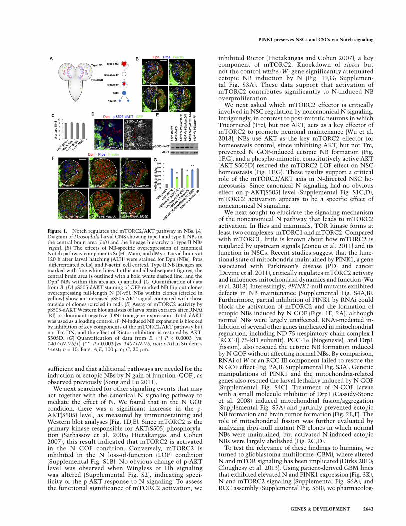
sufficient and that additional pathways are needed for theinduction of ectopic NBs by N gain of function (GOF), asobserved previously (Song and Lu 2011).
We next searched for other signaling events that mayact together with the canonical N signaling pathway tomediate the effect of N. We found that in the N GOFcondition, there was a significant increase in the p-AKT(S505) level, as measured by immunostaining andWestern blot analyses (Fig. 1D,E). Since mTORC2 is theprimary kinase responsible for AKT(S505) phosphoryla-tion (Sarbassov et al. 2005; Hietakangas and Cohen2007), this result indicated that mTORC2 is activatedin the N GOF condition. Conversely, mTORC2 isinhibited in the N loss-of-function (LOF) condition(Supplemental Fig. S1B). No obvious change of p-AKTlevel was observed when Wingless or Hh signalingwas altered (Supplemental Fig. S2), indicating speci-ficity of the p-AKT response to N signaling. To assessthe functional significance of mTORC2 activation, we
inhibited Rictor (Hietakangas and Cohen 2007), a keycomponent of mTORC2. Knockdown of rictor butnot the control white (W) gene significantly attenuatedectopic NB induction by N (Fig. 1F,G; Supplemen-tal Fig. S3A). These data support that activation ofmTORC2 contributes significantly to N-induced NBoverproliferation.
We next asked which mTORC2 effector is criticallyinvolved in NSC regulation by noncanonical N signaling.Intriguingly, in contrast to post-mitotic neurons in whichTricornered (Trc), but not AKT, acts as a key effector ofmTORC2 to promote neuronal maintenance (Wu et al.2013), NBs use AKT as the key mTORC2 effector forhomeostasis control, since inhibiting AKT, but not Trc,prevented N GOF-induced ectopic NB formation (Fig.1F,G), and a phospho-mimetic, constitutively active AKT(AKT-S505D) rescued the mTORC2 LOF effect on NSChomeostasis (Fig. 1F,G). These results support a criticalrole of the mTORC2/AKT axis in N-directed NSC ho-meostasis. Since canonical N signaling had no obviouseffect on p-AKT(S505) level (Supplemental Fig. S1C,D),mTORC2 activation appears to be a specific effect ofnoncanonical N signaling.
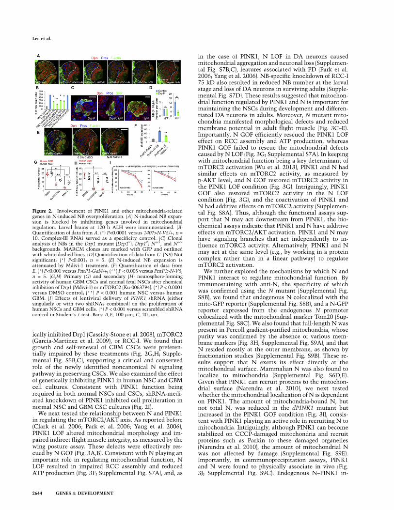
We next sought to elucidate the signaling mechanismof the noncanonical N pathway that leads to mTORC2activation. In flies and mammals, TOR kinase forms atleast two complexes: mTORC1 and mTORC2. Comparedwith mTORC1, little is known about how mTORC2 isregulated by upstream signals (Zoncu et al. 2011) and itsfunction in NSCs. Recent studies suggest that the func-tional state of mitochondria maintained by PINK1, a geneassociated with Parkinson’s disease (PD) and cancer(Devine et al. 2011), critically regulates mTORC2 activityand influences mitochondrial dynamics and function (Wuet al. 2013). Interestingly, dPINK1-null mutants exhibiteddefects in NB maintenance (Supplemental Fig. S4A,B).Furthermore, partial inhibition of PINK1 by RNAi couldblock the activation of mTORC2 and the formation ofectopic NBs induced by N GOF (Figs. 1E, 2A), althoughnormal NBs were largely unaffected. RNAi-mediated in-hibition of several other genes implicated in mitochondrialregulation, including ND-75 (respiratory chain complex-I[RCC-I] 75-kD subunit), PGC-1a (biogenesis), and Drp1(fission), also rescued the ectopic NB formation inducedby N GOF without affecting normal NBs. By comparison,RNAi of W or an RCC-III component failed to rescue theN GOF effect (Fig. 2A,B; Supplemental Fig. S3A). Geneticmanipulations of PINK1 and the mitochondria-relatedgenes also rescued the larval lethality induced by N GOF(Supplemental Fig. S4C). Treatment of N-GOF larvaewith a small molecule inhibitor of Drp1 (Cassidy-Stoneet al. 2008) induced mitochondrial fusion/aggregation(Supplemental Fig. S5A) and partially prevented ectopicNB formation and brain tumor formation (Fig. 2E,F). Therole of mitochondrial fission was further evaluated byanalyzing drp1-null mutant NB clones in which normalNBs were maintained, but activated N-induced ectopicNBs were largely abolished (Fig. 2C,D).
To test the relevance of these findings to humans, weturned to glioblastoma multiforme (GBM), where alteredN and mTOR signaling has been implicated (Dirks 2010;Cloughesy et al. 2013). Using patient-derived GBM linesthat exhibited elevated N and PINK1 expression (Fig. 3K),N and mTORC2 signaling (Supplemental Fig. S6A), andRCC assembly (Supplemental Fig. S6B), we pharmacolog-
Figure 1. Notch regulates the mTORC2/AKT pathway in NBs. (A)Diagram of Drosophila larval CNS showing type I and type II NBs inthe central brain area (left) and the lineage hierarchy of type II NBs(right). (B) The effects of NB-specific overexpression of canonicalNotch pathway components Su(H), Mam, and dMyc. Larval brains at120 h after larval hatching (ALH) were stained for Dpn (NBs), Pros(differentiated cells), and F-actin (cell cortex). Type II NB lineages aremarked with fine white lines. In this and all subsequent figures, thecentral brain area is outlined with a bold white dashed line, and theDpn+ NBs within this area are quantified. (C) Quantification of datafrom B. (D) pS505-dAKT staining of GFP-marked NB flip-out clonesoverexpressing full-length N (N-v5). NBs within clones (circled inyellow) show an increased pS505-AKT signal compared with thoseoutside of clones (circled in red). (E) Assay of mTORC2 activity bypS505-dAKT Western blot analysis of larva brain extracts after RNAi(RI) or dominant-negative (DN) transgene expression. Total dAKTwas used as a loading control. (F) N-induced NB expansion is blockedby inhibition of key components of the mTORC2/AKT pathway butnot Trc-DN, and the effect of Rictor inhibition is restored by AKT-S505D. (G) Quantification of data from E. (*) P < 0.0003 (vs.1407>N-V5/+); (**) P < 0.002 (vs. 1407>N-V5; rictor-RI) in Student’st-test; n = 10. Bars: A,E, 100 mm; C, 20 mm.
PINK1 preserves NSCs and CSCs via Notch signaling
GENES & DEVELOPMENT 2643
ically inhibited Drp1 (Cassidy-Stone et al. 2008), mTORC2(Garcia-Martinez et al. 2009), or RCC-I. We found thatgrowth and self-renewal of GBM CSCs were preferen-tially impaired by these treatments (Fig. 2G,H; Supple-mental Fig. S5B,C), supporting a critical and conservedrole of the newly identified noncanonical N signalingpathway in preserving CSCs. We also examined the effectof genetically inhibiting PINK1 in human NSC and GBMcell cultures. Consistent with PINK1 function beingrequired in both normal NSCs and CSCs, shRNA-medi-ated knockdown of PINK1 inhibited cell proliferation innormal NSC and GBM CSC cultures (Fig. 2I).
We next tested the relationship between N and PINK1in regulating the mTORC2/AKT axis. As reported before(Clark et al. 2006; Park et al. 2006; Yang et al. 2006),PINK1 LOF altered mitochondrial morphology and im-paired indirect flight muscle integrity, as measured by thewing posture assay. These defects were effectively res-cued by N GOF (Fig. 3A,B). Consistent with N playing animportant role in regulating mitochondrial function, NLOF resulted in impaired RCC assembly and reducedATP production (Fig. 3F; Supplemental Fig. S7A), and, as
in the case of PINK1, N LOF in DA neurons causedmitochondrial aggregation and neuronal loss (Supplemen-tal Fig. S7B,C), features associated with PD (Park et al.2006; Yang et al. 2006). NB-specific knockdown of RCC-I75 kD also resulted in reduced NB number at the larvalstage and loss of DA neurons in surviving adults (Supple-mental Fig. S7D). These results suggested that mitochon-drial function regulated by PINK1 and N is important formaintaining the NSCs during development and differen-tiated DA neurons in adults. Moreover, N mutant mito-chondria manifested morphological defects and reducedmembrane potential in adult flight muscle (Fig. 3C–E).Importantly, N GOF efficiently rescued the PINK1 LOFeffect on RCC assembly and ATP production, whereasPINK1 GOF failed to rescue the mitochondrial defectscaused by N LOF (Fig. 3G; Supplemental S7A). In keepingwith mitochondrial function being a key determinant ofmTORC2 activation (Wu et al. 2013), PINK1 and N hadsimilar effects on mTORC2 activity, as measured byp-AKT level, and N GOF restored mTORC2 activity inthe PINK1 LOF condition (Fig. 3G). Intriguingly, PINK1GOF also restored mTORC2 activity in the N LOFcondition (Fig. 3G), and the coactivation of PINK1 andN had additive effects on mTORC2 activity (Supplemen-tal Fig. S8A). Thus, although the functional assays sup-port that N may act downstream from PINK1, the bio-chemical assays indicate that PINK1 and N have additiveeffects on mTORC2/AKT activation. PINK1 and N mayhave signaling branches that act independently to in-fluence mTORC2 activity. Alternatively, PINK1 and Nmay act at the same level (e.g., by working in a proteincomplex rather than in a linear pathway) to regulatemTORC2 activation.
We further explored the mechanisms by which N andPINK1 interact to regulate mitochondrial function. Byimmunostaining with anti-N, the specificity of whichwas confirmed using the N mutant (Supplemental Fig.S8B), we found that endogenous N colocalized with themito-GFP reporter (Supplemental Fig. S8B), and a N-GFPreporter expressed from the endogenous N promotercolocalized with the mitochondrial marker Tom20 (Sup-plemental Fig. S8C). We also found that full-length N waspresent in Percoll gradient-purified mitochondria, whosepurity was confirmed by the absence of various mem-brane markers (Fig. 3H; Supplemental Fig. S9A), and thatN resided mostly at the outer membrane, as shown byfractionation studies (Supplemental Fig. S9B). These re-sults support that N exerts its effect directly at themitochondrial surface. Mammalian N was also found tolocalize to mitochondria (Supplemental Fig. S6D,E).Given that PINK1 can recruit proteins to the mitochon-drial surface (Narendra et al. 2010), we next testedwhether the mitochondrial localization of N is dependenton PINK1. The amount of mitochondria-bound N, butnot total N, was reduced in the dPINK1 mutant butincreased in the PINK1 GOF condition (Fig. 3I), consis-tent with PINK1 playing an active role in recruiting N tomitochondria. Intriguingly, although PINK1 can becomestabilized on CCCP-damaged mitochondria and recruitproteins such as Parkin to these damaged organelles(Narendra et al. 2010), the amount of mitochondrial Nwas not affected by damage (Supplemental Fig. S9E).Importantly, in coimmunoprecipitation assays, PINK1and N were found to physically associate in vivo (Fig.3J; Supplemental Fig. S9C). Endogenous N–PINK1 in-
Figure 2. Involvement of PINK1 and other mitochondria-relatedgenes in N-induced NB overproliferation. (A) N-induced NB expan-sion is blocked by inhibiting genes involved in mitochondrialregulation. Larval brains at 120 h ALH were immunostained. (B)Quantification of data from A. (*) P<0.0001 versus 1407>N-V5/+; n =10. Complex-III RNAi served as a specificity control. (C) Clonalanalysis of NBs in the Drp1 mutant (Drp12), Drp12; Nact, and Nact
backgrounds. MARCM clones are marked with GFP and outlinedwith white dashed lines. (D) Quantification of data from C. (NS) Notsignificant; (*) P<0.001; n = 5. (E) N-induced NB expansion isattenuated by Mdivi-1 treatment. (F) Quantification of data fromE. (*) P<0.001 versus PntP1-Gal4/+; (**) P < 0.005 versus PntP1>N-V5;n = 5. (G,H) Primary (G) and secondary (H) neurosphere-formingactivity of human GBM CSCs and normal fetal NSCs after chemicalinhibition of Drp1 (Mdivi-1) or mTORC2 (Ku-0063794). (*) P < 0.0001versus DMSO control; (**) P < 0.001 human NSC versus humanGBM. (I) Effects of lentiviral delivery of PINK1 shRNA (eithersingularly or with two shRNAs combined) on the proliferation ofhuman NSCs and GBM cells. (*) P < 0.001 versus scrambled shRNAcontrol in Student’s t-test. Bars: A,E, 100 mm; C, 20 mm.
Lee et al.
2644 GENES & DEVELOPMENT
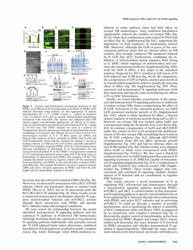
teraction was also observed in human GBM cells (Fig. 3K).Moreover, we detected N association with a RCC-I 30-kDsubunit, which was previously shown to interact withPINK1 (Wu et al. 2013), but no N association with theRCC-III or RCC-IV subunits (Fig. 3J). These results reveala conserved noncanonical mechanism whereby N regu-lates mitochondrial function and mTORC2 activitythrough direct interaction with PINK1 and specificRCC subunits under physiological conditions.
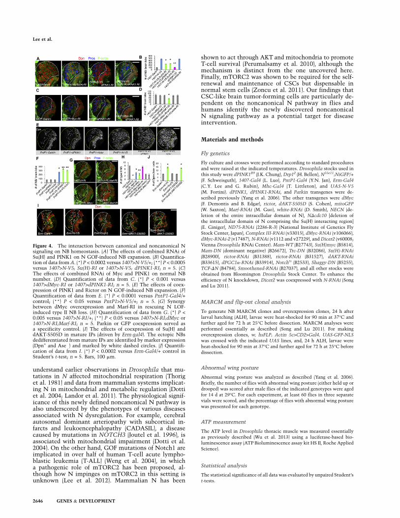
We next tested the relationship between the newlyidentified noncanonical N signaling pathway and thecanonical N pathway in N-directed NB homeostasis.Although knocking down the canonical or noncanonicalN signaling pathway individually only partially rescuedthe N GOF-induced brain tumor phenotype, combinedknockdown of both pathways resulted in nearly completerescue (Fig. 4A,B). Strikingly, while RNAi-mediated in-
hibition of either pathway alone had little effect onnormal NB maintenance, their combined knockdownsignificantly reduced the number of normal NBs (Fig.4C,D), while their combination with control W RNAi hadno effect (Fig. 4C; Supplemental Fig. S3C), supporting theimportance of both pathways in maintaining normalNBs. Moreover, although the GOF of genes in the non-canonical pathway alone had no obvious effect on NBnumber, they strongly enhanced NB expansion inducedby N GOF (Fig. 4E,F). Furthermore, combining the in-hibition of mitochondrial fusion regulator Marf (Denget al. 2008), which impinges on mitochondria and acti-vates the noncanonical pathway (Supplemental Fig. S9D),with the GOF of dMyc, a key target of the canonicalpathway (Song and Lu 2011), resulted in full rescue of NLOF-induced type II NB loss (Fig. 4G,H). By comparison,the coexpression of GFP or Parkin, another gene involvedin mitochondrial regulation, failed to modify the rescuingeffect of dMyc (Fig. 4H; Supplemental Fig. S9F). Thus,canonical and noncanonical N signaling pathways bothplay important and specific roles in mediating the effectsof N on NSC homeostasis.
Finally, we tested whether coactivation of the canon-ical and noncanonical N signaling pathways is sufficientto induce ectopic NBs, hence recapitulating the effect ofN GOF. Activation of the canonical pathway via Su(H)GOF promoted nucleolar growth of IPs (SupplementalFig. S1D), which is likely mediated by dMyc, a knownmaster regulator of nucleolar growth (Song and Lu 2011).However, no ectopic NB was formed when Su(H) wasoverexpressed using the IP-specific Erm-Gal4 driver (Fig.4I,J). In contrast, coexpression of Su(H) and AKT-S505Dunder the control of Erm-Gal4 promoted the dedifferen-tiation of IPs into ectopic NBs resembling those found inthe N GOF condition (Fig. 4I,J), although AKT-S505Doverexpression alone did not affect nucleolar growth(Supplemental Fig. S1E) and had no obvious effect ontype II NB number (Fig. 4I,J). Similar results were obtainedwhen Su(H) or Mam were coexpressed with PINK1(Supplemental Fig. S10A,C). We also used a NDcdc10construct defective in Su(H) binding and thus canonical Nsignaling (Lawrence et al. 2000) but capable of noncanon-ical N signaling (Supplemental Fig. S1A). Coexpression ofSu(H) and NDcdc10 also induced ectopic NBs (Supple-mental Fig. S10B,D). These results suggest that non-canonical and canonical N signaling mediate distinctaspects of N function and act coordinately to regulateNSC behavior.
Our results uncover a novel mechanism of N inregulating NSC self-renewal and maintenance througha noncanonical signaling pathway involving PINK1,mTORC2, and AKT. A central feature of this noncanon-ical N signaling pathway is specific mitochondrial roles ofN in regulating RCC function through direct interactionswith PINK1 and select RCC subunits and in activatingmTORC2. N could act through a number of possiblemechanisms, such as facilitating the import or assemblyof RCC components (Ades and Butow 1980), as suggestedby its interaction with complex I subunits (Fig. 3J), ordirecting the quality control of mitochondria, as has beenimplicated for PINK1 (Rugarli and Langer 2012). Futurestudies will determine the exact domains of N involvedin PINK1 interaction and whether noncanonical N sig-naling is ligand-dependent. Although the exact mecha-nism remains to be determined, our results will help us to
Figure 3. Genetic and biochemical interactions between N andPINK1. (A,B) Effects of N overexpression or N RNAi on PINK1 LOF-induced abnormal wing posture (A) and aggregation of musclemitochondria (B). (*) P < 0.0001 versus w� control in Student’st-test. (C) Effects of N LOF on muscle mitochondrial morphologymonitored with mito-GFP. The uneven and enhanced mito-GFPsignals suggest mitochondrial aggregation. (D) Effects of N muta-tions on mitochondrial membrane potential (indicated by JC-1 signalat 575–625 nm). The PINK1 mutant was used as a control. (E)Transmission electron microscopy analysis of mitochondrial cristaemorphology in N mutants. Bar, 100 nm. N[55e11] refers to N[55e11]/+heterozygous females in C–E. (F) Blue native gel (BNG) andWestern blot analyses of RCC assembly in PINK1 and N LOF andGOF backgrounds. Actin and the complex IV subunit 1 served asloading controls. (G) Effects of N GOF on RCC assembly and pS505-dAKT level changes caused by PINK1 LOF. (H,I) Fractionation assayshowing enrichment of full-length N at mitochondria (H) andincreased mitochondrial N in the PINK1 overexpression condition(I). (J) Coimmunoprecipitation assays using NiGFP fly head tissue todemonstrate N interaction with PINK1 and complex I 30 kD. Thecomplex III subunit served as a negative control. (K) The interactionbetween hNotch1 and PINK1 in the mitochondria of human NSCsand GBM cells. The complex III subunit served as a negative control.GBM cells show elevated expression of PINK1 and both the full-length and the intracellular domain of Notch1.
PINK1 preserves NSCs and CSCs via Notch signaling
GENES & DEVELOPMENT 2645
understand earlier observations in Drosophila that mu-tations in N affected mitochondrial respiration (Thoriget al. 1981) and data from mammalian systems implicat-ing N in mitochondrial and metabolic regulation (Dottiet al. 2004; Landor et al. 2011). The physiological signif-icance of this newly defined noncanonical N pathway isalso underscored by the phenotypes of various diseasesassociated with N dysregulation. For example, cerebralautosomal dominant arteriopathy with subcortical in-farcts and leukoencephalopathy (CADASIL), a diseasecaused by mutations in NOTCH3 (Joutel et al. 1996), isassociated with mitochondrial impairment (Dotti et al.2004). On the other hand, GOF mutations of Notch1 areimplicated in over half of human T-cell acute lympho-blastic leukemia (T-ALL) (Weng et al. 2004), in whicha pathogenic role of mTORC2 has been proposed, al-though how N impinges on mTORC2 in this setting isunknown (Lee et al. 2012). Mammalian N has been
shown to act through AKT and mitochondria to promoteT-cell survival (Perumalsamy et al. 2010), although themechanism is distinct from the one uncovered here.Finally, mTORC2 was shown to be required for the self-renewal and maintenance of CSCs but dispensable innormal stem cells (Zoncu et al. 2011). Our findings thatCSC-like brain tumor-forming cells are particularly de-pendent on the noncanonical N pathway in flies andhumans identify the newly discovered noncanonicalN signaling pathway as a potential target for diseaseintervention.
Materials and methods
Fly genetics
Fly culture and crosses were performed according to standard procedures
and were raised at the indicated temperatures. Drosophila stocks used in
this study were dPINK1B9 (J.K. Chung), Drp12 (H. Bellen), N55e11;NiGFP/+
(F. Schweisguth), 1407-Gal4 (L. Luo), PntP1-Gal4 (Y.N. Jan), Erm-Gal4
(C.Y. Lee and G. Rubin), Mhc-Gal4 (T. Littleton), and UAS-N-V5
(M. Fortini). dPINK1, dPINK1-RNAi, and Parkin transgenes were de-
scribed previously (Yang et al. 2006). The other transgenes were dMyc
(F. Demontis and B. Edgar), rictor, dAKT-S505D (S. Cohen), mitoGFP
(W. Saxton), Marf-RNAi (M. Guo), white-RNAi (D. Smith), NECN (de-
letion of the entire intracellular domain of N), NDcdc10 [deletion of
the intracellular domain of N comprising the Su(H) interacting region]
(E. Giniger), ND75-RNAi (2286-R-3) (National Institute of Genetics Fly
Stock Center, Japan), Complex III-RNAi (v33015), dMyc-RNAi (v106066),
dMyc-RNAi-2 (v17487), N-RNAi (v1112 and v27229), and Dicer2 (v60008;
Vienna Drosophila RNAi Center). Mam-WT (B27743), Su(H)myc (B5814),
Mam-DN (dominant negative) (B26672), Trc-DN (B32086), Su(H)-RNAi
(B28900), rictor-RNAi (B31388), rictor-RNAi (B31527), dAKT-RNAi
(B33615), dPGC1a-RNAi (B33914), Notchts (B2533), Shaggy-DN (B5255),
TCF-DN (B4784), Smoothened-RNAi (B27037), and all other stocks were
obtained from Bloomington Drosophila Stock Center. To enhance the
efficiency of N knockdown, Dicer2 was coexpressed with N-RNAi (Song
and Lu 2011).
MARCM and flip-out clonal analysis
To generate NB MARCM clones and overexpression clones, 24 h after
larval hatching (ALH), larvae were heat-shocked for 90 min at 37°C and
further aged for 72 h at 25°C before dissection. MARCM analyses were
performed essentially as described (Song and Lu 2011). For making
overexpression clones, w, hsFLP; Actin 5c>CD2>Gal4, UAS-GFP-NLS
was crossed with the indicated UAS lines, and, 24 h ALH, larvae were
heat-shocked for 90 min at 37°C and further aged for 72 h at 25°C before
dissection.
Abnormal wing posture
Abnormal wing posture was analyzed as described (Yang et al. 2006).
Briefly, the number of flies with abnormal wing posture (either held up or
drooped) was scored after male flies of the indicated genotypes were aged
for 14 d at 29°C. For each experiment, at least 60 flies in three separate
vials were scored, and the percentage of flies with abnormal wing posture
was presented for each genotype.
ATP measurement
The ATP level in Drosophila thoracic muscle was measured essentially
as previously described (Wu et al. 2013) using a luciferase-based bio-
luminescence assay (ATP Bioluminescence assay kit HS II, Roche Applied
Science).
Statistical analysis
The statistical significance of all data was evaluated by unpaired Student’s
t-tests.
Figure 4. The interaction between canonical and noncanonical Nsignaling on NB homeostasis. (A) The effects of combined RNAi ofSu(H) and PINK1 on N GOF-induced NB expansion. (B) Quantifica-tion of data from A. (*) P < 0.0002 versus 1407>N-V5/+; (**) P < 0.0005versus 1407>N-V5; Su(H)-RI or 1407>N-V5; dPINK1-RI; n = 5. (C)The effects of combined RNAi of Myc and PINK1 on normal NBnumber. (D) Quantification of data from C. (*) P < 0.001 versus1407>dMyc-RI or 1407>dPINK1-RI; n = 5. (E) The effects of coex-pression of PINK1 and Rictor on N GOF-induced NB expansion. (F)Quantification of data from E. (*) P < 0.0001 versus PntP1-Gal4/+control; (**) P < 0.05 versus PntP1>N-V5/+; n = 5. (G) Synergybetween dMyc overexpression and Marf-RI in rescuing N LOF-induced type II NB loss. (H) Quantification of data from G. (*) P <0.005 versus 1407>N-RI/+; (**) P < 0.05 versus 1407>N-RI;dMyc or1407>N-RI;Marf-RI; n = 5. Parkin or GFP coexpression served asa specificity control. (I) The effects of coexpression of Su(H) anddAKT-S505D in mature IPs (driven by Erm-gal4). The ectopic NBsdedifferentiated from mature IPs are identified by marker expression(Dpn+ and Ase�) and marked by white dashed circles. (J) Quantifi-cation of data from I. (*) P < 0.0002 versus Erm-Gal4/+ control inStudent’s t-test; n = 5. Bars, 100 mm.
Lee et al.
2646 GENES & DEVELOPMENT

Acknowledgments
We are grateful to Dr. J.K. Chung, Dr. H. Bellen, Dr. E. Giniger, Dr.
F. Schweisguth, Dr. L. Luo, Dr. Y.N. Jan, Dr. C.Y. Lee, Dr. G. Rubin, Dr.
T. Littleton, Dr. M. Fortini, Dr. F. Demontis, Dr. B. Edgar, Dr. S. Cohen,
Dr. W. Saxton, Dr. M. Guo, Dr. J. Skeath, Dr. L. Pallanck, University of
Iowa Developmental Studies Hybridoma Bank, Bloomington Drosophila
Stock Center, National Institute of Genetics Fly Stock Center Japan, and
Vienna Drosophila RNAi Center for fly stocks and reagents. We thank
J. Gaunce and G. Silverio for technical assistance, and members of the Lu
laboratory for discussions and help. This work was supported by National
Institutions of Health grants (R01 NS043167 and R01 AR054926) to B.L.,
and a Stanford University School of Medicine Dean’s Post-doctoral
Fellowship to Z.W.
References
Ades IZ, Butow RA. 1980. The products of mitochondria-bound cyto-
plasmic polysomes in yeast. J Biol Chem 255: 9918–9924.
Andersson ER, Sandberg R, Lendahl U. 2011. Notch signaling: Sim-
plicity in design, versatility in function. Development 138: 3593–
3612.
Artavanis-Tsakonas S, Muskavitch MA. 2010. Notch: The past, the
present, and the future. Curr Top Dev Biol 92: 1–29.
Bowman SK, Rolland V, Betschinger J, Kinsey KA, Emery G, Knoblich
JA. 2008. The tumor suppressors Brat and Numb regulate transit-
amplifying neuroblast lineages in Drosophila. Dev Cell 14: 535–
546.
Cassidy-Stone A, Chipuk JE, Ingerman E, Song C, Yoo C, Kuwana T,
Kurth MJ, Shaw JT, Hinshaw JE, Green DR, et al. 2008. Chemical
inhibition of the mitochondrial division dynamin reveals its role in
Bax/Bak-dependent mitochondrial outer membrane permeabilization.
Dev Cell 14: 193–204.
Clark IE, Dodson MW, Jiang C, Cao JH, Huh JR, Seol JH, Yoo SJ, Hay BA,
Guo M. 2006. Drosophila pink1 is required for mitochondrial
function and interacts genetically with parkin. Nature 441: 1162–
1166.
Cloughesy TF, Cavenee WK, Mischel PS. 2013. Glioblastoma: From
molecular pathology to targeted treatment. Annu Rev Pathol doi:
10.1146/annurev-pathol-01111-130324.
Deng H, Dodson MW, Huang H, Guo M. 2008. The Parkinson’s disease
genes pink1 and parkin promote mitochondrial fission and/or inhibit
fusion in Drosophila. Proc Natl Acad Sci 105: 14503–14508.
Devine MJ, Plun-Favreau H, Wood NW. 2011. Parkinson’s disease and
cancer: Two wars, one front. Nat Rev Cancer 11: 812–823.
Dirks PB. 2010. Brain tumor stem cells: The cancer stem cell hypothesis
writ large. Mol Oncol 4: 420–430.
Doe CQ. 2008. Neural stem cells: Balancing self-renewal with differen-
tiation. Development 135: 1575–1587.
Dotti MT, De Stefano N, Bianchi S, Malandrini A, Battisti C, Cardaioli E,
Federico A. 2004. A novel NOTCH3 frameshift deletion and mito-
chondrial abnormalities in a patient with CADASIL. Arch Neurol 61:
942–945.
Garcia-Martinez JM, Moran J, Clarke RG, Gray A, Cosulich SC, Chresta
CM, Alessi DR. 2009. Ku-0063794 is a specific inhibitor of the
mammalian target of rapamycin (mTOR). Biochem J 421: 29–42.
Hietakangas V, Cohen SM. 2007. Re-evaluating AKT regulation: Role of
TOR complex 2 in tissue growth. Genes Dev 21: 632–637.
Joutel A, Corpechot C, Ducros A, Vahedi K, Chabriat H, Mouton P,
Alamowitch S, Domenga V, Cecillion M, Marechal E, et al. 1996.
Notch3 mutations in CADASIL, a hereditary adult-onset condition
causing stroke and dementia. Nature 383: 707–710.
Knoblich JA. 2010. Asymmetric cell division: Recent developments and
their implications for tumour biology. Nat Rev Mol Cell Biol 11: 849–
860.
Landor SK, Mutvei AP, Mamaeva V, Jin S, Busk M, Borra R, Gronroos TJ,
Kronqvist P, Lendahl U, Sahlgren CM. 2011. Hypo- and hyperacti-
vated Notch signaling induce a glycolytic switch through distinct
mechanisms. Proc Natl Acad Sci 108: 18814–18819.
Lawrence N, Klein T, Brennan K, Martinez Arias A. 2000. Structural
requirements for Notch signalling with Delta and serrate during the
development and patterning of the wing disc of Drosophila. De-
velopment 127: 3185–3195.
Lee K, Nam KT, Cho SH, Gudapati P, Hwang Y, Park DS, Potter R, Chen
J, Volanakis E, Boothby M. 2012. Vital roles of mTOR complex 2 in
Notch-driven thymocyte differentiation and leukemia. J Exp Med
209: 713–728.
Liu C, Zong H. 2012. Developmental origins of brain tumors. Curr Opin
Neurobiol 22: 844–849.
Morrison SJ, Kimble J. 2006. Asymmetric and symmetric stem-cell
divisions in development and cancer. Nature 441: 1068–1074.
Narendra DP, Jin SM, Tanaka A, Suen DF, Gautier CA, Shen J, Cookson MR,
Youle RJ. 2010. PINK1 is selectively stabilized on impaired mito-
chondria to activate parkin. PLoS Biol 8: e1000298.
Park J, Lee SB, Lee S, Kim Y, Song S, Kim S, Bae E, Kim J, Shong M, Kim JM,
et al. 2006. Mitochondrial dysfunction in Drosophila PINK1 mu-
tants is complemented by parkin. Nature 441: 1157–1161.
Perumalsamy LR, Nagala M, Sarin A. 2010. Notch-activated signaling
cascade interacts with mitochondrial remodeling proteins to regulate
cell survival. Proc Natl Acad Sci 107: 6882–6887.
Rugarli EI, Langer T. 2012. Mitochondrial quality control: A matter of life
and death for neurons. EMBO J 31: 1336–1349.
Sarbassov DD, Guertin DA, Ali SM, Sabatini DM. 2005. Phosphorylation
and regulation of Akt/PKB by the rictor–mTOR complex. Science
307: 1098–1101.
Song Y, Lu B. 2011. Regulation of cell growth by Notch signaling and its
differential requirement in normal vs. tumor-forming stem cells in
Drosophila. Genes Dev 25: 2644–2658.
Sousa-Nunes R, Cheng LY, Gould AP. 2010. Regulating neural prolifer-
ation in the Drosophila CNS. Curr Opin Neurobiol 20: 50–57.
Thorig GE, Heinstra PW, Scharloo W. 1981. The action of the notchlocus
in Drosophila melanogaster. II. Biochemical effects of recessive
lethals on mitochondrial enzymes. Genetics 99: 65–74.
Wang H, Somers GW, Bashirullah A, Heberlein U, Yu F, Chia W. 2006.
Aurora-A acts as a tumor suppressor and regulates self-renewal of
Drosophila neuroblasts. Genes Dev 20: 3453–3463.
Weng AP, Ferrando AA, Lee W, Morris JPt, Silverman LB, Sanchez-Irizarry
C, Blacklow SC, Look AT, Aster JC. 2004. Activating mutations of
NOTCH1 in human T cell acute lymphoblastic leukemia. Science
306: 269–271.
Weng M, Golden KL, Lee CY. 2010. dFezf/Earmuff maintains the re-
stricted developmental potential of intermediate neural progenitors
in Drosophila. Dev Cell 18: 126–135.
Wu Z, Sawada T, Shiba K, Liu S, Kanao T, Takahashi R, Hattori N, Imai Y,
Lu B. 2013. Tricornered/NDR kinase signaling mediates PINK1-
directed mitochondrial quality control and tissue maintenance.
Genes Dev 27: 157–162.
Yang Y, Gehrke S, Imai Y, Huang Z, Ouyang Y, Wang JW, Yang L, Beal MF,
Vogel H, Lu B. 2006. Mitochondrial pathology and muscle and
dopaminergic neuron degeneration caused by inactivation of Dro-
sophila Pink1 is rescued by Parkin. Proc Natl Acad Sci 103: 10793–
10798.
Zhong W, Chia W. 2008. Neurogenesis and asymmetric cell division.
Curr Opin Neurobiol 18: 4–11.
Zoncu R, Efeyan A, Sabatini DM. 2011. mTOR: From growth signal
integration to cancer, diabetes and ageing. Nat Rev Mol Cell Biol 12:
21–35.
PINK1 preserves NSCs and CSCs via Notch signaling
GENES & DEVELOPMENT 2647
Copyright © 2022 FDOKUMEN