Constitutive CD40 signaling in B cells selectively activates the noncanonical NF- B pathway and...
13
The Journal of Experimental Medicine ARTICLE © 2008 Hömig-Hölzel et al. The Rockefeller University Press $30.00 J. Exp. Med. Vol. 205 No. 6 1317-1329 www.jem.org/cgi/doi/ 1317 10.1084/jem.20080238 CD40, which is a member of the TNF receptor superfamily, is expressed on virtually all mature B lymphocytes and on most of their neoplastic counterparts. Upon interaction with CD40 ligand (CD40L), which is predominantly expressed on activated CD4 + T cells, CD40 promotes B cell activation, proliferation, survival, and up-regu- lation of surface molecules involved in antigen presentation (1). The finding that patients suf- fering from “X-linked hyper-IgM syndrome” have a defect in expression of CD40L was in- strumental in the discovery that CD40–CD40L interactions play a crucial role in T cell–depen- dent (TD) immune response (2, 3), contributing to germinal center (GC) formation, memory B cell development, Ig isotype switching, and affinity maturation (4, 5). The cytoplasmic tail of CD40, which lacks intrinsic catalytic activity, delivers signals to the cells by recruitment of TNF receptor–associated factors 1, 2, 3, 5, and 6. Interaction with its li- gand leads to clustering of CD40 in lipid rafts and, finally, to the recruitment of TNF receptor– associated factor molecules (6, 7). These events CORRESPONDENCE Ursula Zimber-Strobl: [email protected] Abbreviations used: Erk, extra- cellular signal–regulated kinase; ES, embryonic stem; GC, ger- minal center; HE, hematoxylin and eosin; LMP, latent mem- brane protein; MAPK, mitogen- activated protein kinase; TD, T cell dependent. C. Hömig-Hölzel and C. Hojer contributed equally to this paper. S. Casola’s present address is IFOM-The Foundation for Cancer Research Institute of Molecular Oncology, 20139 Milan, Italy. C. Hömig-Hölzel’s present address is Division of Molecular Genetics, Netherlands Cancer Institute, Amsterdam, Netherlands. J. Rastelli’s present address is Whitehead Institute for Bio- medical Research, Cambridge, MA 02142. The online version of this article contains supplemental material. Constitutive CD40 signaling in B cells selectively activates the noncanonical NF- B pathway and promotes lymphomagenesis Cornelia Hömig-Hölzel, 1 Caroline Hojer, 1 Julia Rastelli, 1 Stefano Casola, 2 Lothar J. Strobl, 1 Werner Müller, 3 Leticia Quintanilla-Martinez, 4 Andreas Gewies, 5 Jürgen Ruland, 5 Klaus Rajewsky, 2 and Ursula Zimber-Strobl 1 1 Institute of Clinical Molecular Biology and Tumor Genetics, Helmholtz Center Munich, German Research Center for Environment and Health, D-81377 Munich, Germany 2 Immune Disease Institute, Harvard Medical School, Boston, MA 02115 3 The Department of Experimental Immunology, Helmholtz Center for Infectious Research, D-38124 Braunschweig, Germany 4 Institute of Pathology, Helmholtz Center Munich, German Research Center for Environment and Health, D-85764 Neuherberg, Germany 5 III Med. Department, Technical University of Munich, Klinikum rechts der Isar, D-81675 Munich, Germany CD40, a member of the tumor necrosis factor (TNF) receptor family, plays an essential role in T cell–dependent immune responses. Because CD40 is widely expressed on the surface of tumor cells in various B cell malignancies, deregulated CD40 signaling has been suggested to contribute to lymphomagenesis. In this study, we show that B cell-specific expression of a constitutively active CD40 receptor, in the form of a latent membrane protein 1 (LMP1)/ CD40 chimeric protein, promoted an increase in the number of follicular and marginal zone B cells in secondary lymphoid organs in transgenic mice. The B cells displayed an activated phenotype, prolonged survival and increased proliferation, but were significantly impaired in T cell-dependent immune responses. Constitutive CD40 signaling in B cells induced selective and constitutive activation of the noncanonical NF- B pathway and the mitogen- activated protein kinases Jnk and extracellular signal–regulated kinase. LMP1/CD40- expressing mice older than 12 mo developed B cell lymphomas of mono- or oligoclonal origin at high incidence, thus showing that the interplay of the signaling pathways induced by constitutive CD40 signaling is sufficient to initiate a tumorigenic process, ultimately leading to the development of B cell lymphomas.
-
Upload
uni-tuebingen1 -
Category
Documents
-
view
0 -
download
0
Transcript of Constitutive CD40 signaling in B cells selectively activates the noncanonical NF- B pathway and...

The
Journ
al o
f Exp
erim
enta
l M
edic
ine
ARTICLE
© 2008 H ö mig-H ö lzel et al.
The Rockefeller University Press $30.00
J. Exp. Med. Vol. 205 No. 6 1317-1329 www.jem.org/cgi/doi/
1317
10.1084/jem.20080238
CD40, which is a member of the TNF receptor superfamily, is expressed on virtually all mature B lymphocytes and on most of their neoplastic counterparts. Upon interaction with CD40 ligand (CD40L), which is predominantly expressed on activated CD4 + T cells, CD40 promotes B cell activation, proliferation, survival, and up-regu-
lation of surface molecules involved in antigen presentation ( 1 ). The fi nding that patients suf-fering from “ X-linked hyper-IgM syndrome ” have a defect in expression of CD40L was in-strumental in the discovery that CD40 – CD40L interactions play a crucial role in T cell – depen-dent (TD) immune response ( 2, 3 ), contributing to germinal center (GC) formation, memory B cell development, Ig isotype switching, and affi nity maturation ( 4, 5 ).
The cytoplasmic tail of CD40, which lacks intrinsic catalytic activity, delivers signals to the cells by recruitment of TNF receptor – associated factors 1, 2, 3, 5, and 6. Interaction with its li-gand leads to clustering of CD40 in lipid rafts and, fi nally, to the recruitment of TNF receptor – associated factor molecules ( 6, 7 ). These events
CORRESPONDENCE
Ursula Zimber-Strobl:
Abbreviations used: Erk, extra-
cellular signal – regulated kinase;
ES, embryonic stem; GC, ger-
minal center; HE, hematoxylin
and eosin; LMP, latent mem-
brane protein; MAPK, mitogen-
activated protein kinase; TD,
T cell dependent. C. H ö mig-H ö lzel and C. Hojer contributed equally to this
paper.
S. Casola ’ s present address is IFOM-The Foundation
for Cancer Research Institute of Molecular Oncology,
20139 Milan, Italy.
C. H ö mig-H ö lzel ’ s present address is Division of Molecular
Genetics, Netherlands Cancer Institute, Amsterdam,
Netherlands.
J. Rastelli ’ s present address is Whitehead Institute for Bio-
medical Research, Cambridge, MA 02142.
The online version of this article contains supplemental material.
Constitutive CD40 signaling in B cells selectively activates the noncanonical NF- � B pathway and promotes lymphomagenesis
Cornelia H ö mig-H ö lzel, 1 Caroline Hojer, 1 Julia Rastelli, 1 Stefano Casola, 2 Lothar J. Strobl, 1 Werner M ü ller, 3 Leticia Quintanilla-Martinez, 4 Andreas Gewies, 5 J ü rgen Ruland, 5 Klaus Rajewsky, 2 and Ursula Zimber-Strobl 1
1 Institute of Clinical Molecular Biology and Tumor Genetics, Helmholtz Center Munich, German Research Center
for Environment and Health, D-81377 Munich, Germany
2 Immune Disease Institute, Harvard Medical School, Boston, MA 02115
3 The Department of Experimental Immunology, Helmholtz Center for Infectious Research, D-38124 Braunschweig, Germany
4 Institute of Pathology, Helmholtz Center Munich, German Research Center for Environment and Health,
D-85764 Neuherberg, Germany
5 III Med. Department, Technical University of Munich, Klinikum rechts der Isar, D-81675 Munich, Germany
CD40, a member of the tumor necrosis factor (TNF) receptor family, plays an essential role
in T cell – dependent immune responses. Because CD40 is widely expressed on the surface of
tumor cells in various B cell malignancies, deregulated CD40 signaling has been suggested
to contribute to lymphomagenesis. In this study, we show that B cell-specifi c expression of
a constitutively active CD40 receptor, in the form of a latent membrane protein 1 (LMP1)/
CD40 chimeric protein, promoted an increase in the number of follicular and marginal zone
B cells in secondary lymphoid organs in transgenic mice. The B cells displayed an activated
phenotype, prolonged survival and increased proliferation, but were signifi cantly impaired
in T cell-dependent immune responses. Constitutive CD40 signaling in B cells induced
selective and constitutive activation of the noncanonical NF- � B pathway and the mitogen-
activated protein kinases Jnk and extracellular signal – regulated kinase. LMP1/CD40-
expressing mice older than 12 mo developed B cell lymphomas of mono- or oligoclonal
origin at high incidence, thus showing that the interplay of the signaling pathways induced
by constitutive CD40 signaling is suffi cient to initiate a tumorigenic process, ultimately
leading to the development of B cell lymphomas.
1318 CONSTITUTIVELY ACTIVE CD40 PROMOTES LYMPHOMAGENESIS | H ö mig-H ö lzel et al.
protein is able to deliver a ligand-independent constitutive CD40 signal by promoting self-aggregation in the plasma membrane ( 23, 24 ). Constitutive CD40 signaling in B cells led to a strong accumulation of follicular and marginal zone B cells in secondary lymphoid organs, accompanied by a signifi cant enlargement of follicles. These B cells displayed an activated phenotype, but failed to form GCs in response to immuniza-tion with TD antigens. Ultimately, LMP1/CD40-expressing mice developed mono- or oligoclonal B cell lymphomas at high incidence, demonstrating that deregulated CD40 signal-ing is suffi cient to initiate B cell transformation.
RESULTS
Generation of transgenic mice expressing a conditional
LMP1/CD40 transgene
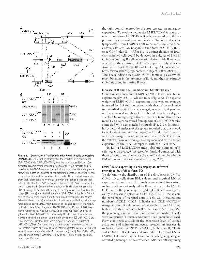
To study constitutive CD40 signaling in vivo, mice expressing a fusion protein comprising the signaling domain of the hu-man CD40 and the transmembrane domain of LMP1 (LMP1/CD40) were generated. It had been shown earlier that human CD40 can rescue the phenotype of CD40 -defi cient mice, in-dicating that the receptors of both species are functionally equivalent ( 25 ). A copy of the LMP1/CD40 chimeric gene was inserted into the murine rosa26 genomic locus of BALB/c-derived embryonic stem (ES) cells by homologous recombi-nation ( Fig. 1 A ). To restrict expression of LMP1/CD40 in a cell type – and stage-specifi c manner, we inserted a loxP-fl anked transcription and translation termination sequence (stop cassette) upstream of the LMP1/CD40 coding sequence. After Cre-mediated excision of the stop-cassette, the LMP1/CD40 transgene is placed under the control of the ubiqui-tously active rosa26 promoter. Correctly targeted ES cell clones were identifi ed by Southern blot analysis and used to establish the LMP1/CD40 fl STOP mouse strain.
The LMP1/CD40 fl STOP mice were crossed to CD19-Cre and CD21-Cre mice to activate expression of the chimeric pro-tein from earlier (pro – /pre – ) or later (mature) stages of B cell development, respectively ( 26, 27 ). Because off spring of both crosses revealed the same immunological phenotype, results are presented for mice obtained from LMP1/CD40 fl STOP ;CD19-Cre crosses only. In all biochemical experiments, only LMP1/CD40 fl STOP ;CD19-Cre were analyzed. For simplicity, mice carrying one LMP1/CD40 knock-in allele and one CD19-Cre allele will be referred to as LMP1/CD40 mice. As con-trols, either LMP1/CD40 fl STOP or CD19-Cre mice were used. These control strains had an identical phenotype. In LMP1/CD40 mice, deletion of the stop cassette could be detected in about half of BM and in almost all splenic B cells ( Fig. 1 B ). This is consistent with previous reports showing a gradual in-crease in Cre-mediated recombination in CD19-Cre mice as B cells proceed through their diff erentiation ( 27 ). Expression of the LMP1/CD40 protein in splenic cells after removal of the stop cassette was analyzed by Western blot analysis ( Fig. 1 C ). LMP1/CD40 protein expression was detected in splenocytes isolated from LMP1/CD40 mice, but not in control mice ( Fig. 1 C ), indicating that LMP1/CD40 is expressed after Cre-mediated recombination and confi rming
initiate downstream signaling, resulting in activation of phospho-inositide 3 kinase, phospholipase C � , mitogen-activated pro-tein kinases (MAPKs), and NF- � B ( 1, 6 ). Additionally, the Janus family kinase 3 (JAK3), which is associated with the cyto-plasmic tail of CD40, undergoes phosphorylation, result-ing in the activation of the signal transducer and activator of transcription 3 ( 8 ).
The following three subfamilies of structurally related MAPKs are activated by CD40: extracellular signal-regulated kinase 1 (Erk1) and Erk2, the c-jun kinases Jnk1 and Jnk2, and the kinase p38/MAPK ( 9 – 11 ). MAPKs are serine/threo-nine protein kinases, which are activated by dual phosphory-lation on a specifi c tyrosine and threonine residue, respectively. After activation, MAPKs phosphorylate nuclear substrates such as the transcription factors c-Jun, Elk-1, Egr-1, and Atf-2, which in turn bind to specifi c DNA sequences and thus modulate transcription ( 1, 6 ).
CD40 signaling activates both the canonical and the non-canonical NF- � B signaling pathways ( 12, 13 ). In mammals, the NF- � B family consists of fi ve genes coding for NF- � B1 (p105/p50), NF- � B2 (p100/p52), RelA (p65), RelB, and c-Rel ( 14 ). In unstimulated cells, the DNA-binding activity of NF- � B dimers is inhibited through the interaction with proteins of the inhibitor of the NF- � B (I � B) family. The ac-tivation of the canonical NF- � B signaling pathway largely depends on ubiquitin-dependent degradation of small I � B proteins, leading to the nuclear translocation of NF- � B het-erodimers p50/p65 and p50/c-Rel. The liberated heterodi-mers are capable of binding DNA, and thereby activating gene expression. The noncanonical pathway depends on proteolytic cleavage of the precursor p100, liberating mainly p52/RelB heterodimers for nuclear translocation. Thus, ca-nonical and noncanonical NF- � B signaling pathways induce the formation and nuclear translocation of distinct NF- � B heterodimers that activate distinct target genes ( 15 – 17 ).
Whereas CD40 plays a decisive role in TD immune re-sponses, aberrant CD40 signaling is suspected to play a critical role in the oncogenic processes of various cell types, includ-ing B cells ( 18 ). CD40 is widely expressed in B cell lymphomas and in selected carcinomas. Additionally, aberrant CD40L expression, which may lead to constitutive CD40 engagement, has been observed in several malignancies, including chronic lymphocytic leukemia, mantle cell lymphoma, follicular lym-phoma, Burkitt ’ s lymphoma, and breast cancer ( 19 – 22 ). Thus, a contribution of this receptor to tumor pathogenesis has been suggested.
However, it is unclear whether aberrant CD40 signaling is suffi cient to drive lymphomagenesis or whether it is only in-volved in the maintenance of established malignancies. Ade-quate experimental systems to explore this question are still missing. To study the biological and potential pathological ef-fects of a constitutive CD40 activation, we generated knock-in mice expressing in a conditional manner a chimeric protein consisting of the transmembrane domain of the Epstein-Barr viral latent membrane protein 1 (LMP1) and the intracellular signaling domain of CD40 (LMP1/CD40). This chimeric
JEM VOL. 205, June 9, 2008
ARTICLE
1319
the tight control exerted by the stop cassette on transgene expression. To study whether the LMP1/CD40 fusion pro-tein can substitute for CD40 in B cells, we tested its ability to promote Ig class switch recombination. We isolated splenic lymphocytes from LMP1/CD40 mice and stimulated them ex vivo with anti-CD40 agonistic antibody ( � -CD40), IL-4, or � -CD40 plus IL-4. After 5 d, a distinct fraction of IgG1 class-switched cells could be detected in cultures of LMP1/CD40-expressing B cells upon stimulation with IL-4 only, whereas in the controls, IgG1 + cells appeared only after co-stimulation with � -CD40 and IL-4 (Fig. S1, available at http://www.jem.org/cgi/content/full/jem.20080238/DC1). These data indicate that LMP1/CD40 induces Ig class switch recombination in the presence of IL-4, and thus constitutive CD40 signaling in murine B cells.
Increase of B and T cell numbers in LMP1/CD40 mice
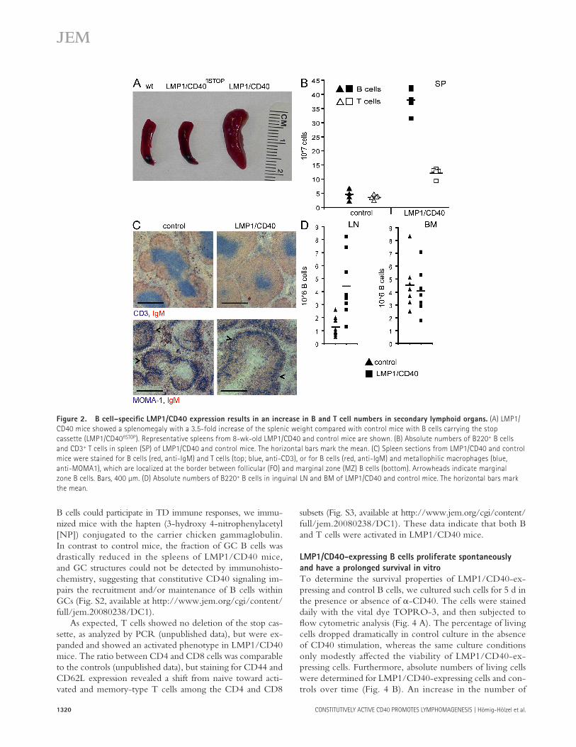
Conditional expression of LMP1/CD40 in B cells resulted in a splenomegaly in 8 – 16-wk-old mice ( Fig. 2 A ). The splenic weight of LMP1/CD40-expressing mice was, on average, increased by 3.5-fold compared with that of control mice (unpublished data). The splenomegaly was largely dependent on the increased number of B cells and, to a lesser degree, T cells. On average, eight times more B cells and three times more T cells were recovered from spleens of LMP1/CD40 mice compared with age-matched controls ( Fig. 2 B ). Immuno-histochemical analysis of the spleen revealed that the overall follicular structure with the respective B and T cell zones, as well as the marginal zone, was retained ( Fig. 2 C ). The size of the follicles, however, was signifi cantly increased, with a larger expansion of the B cell compared with the T cell zone.
In LNs of LMP1/CD40 mice, absolute numbers of B cells were, on average, increased by fourfold compared with those of control mice, whereas absolute B cell numbers in the BM of mutant mice were unaff ected ( Fig. 2 D ).
LMP1/CD40-expressing B cells display an activated
phenotype, but fail to form GCs
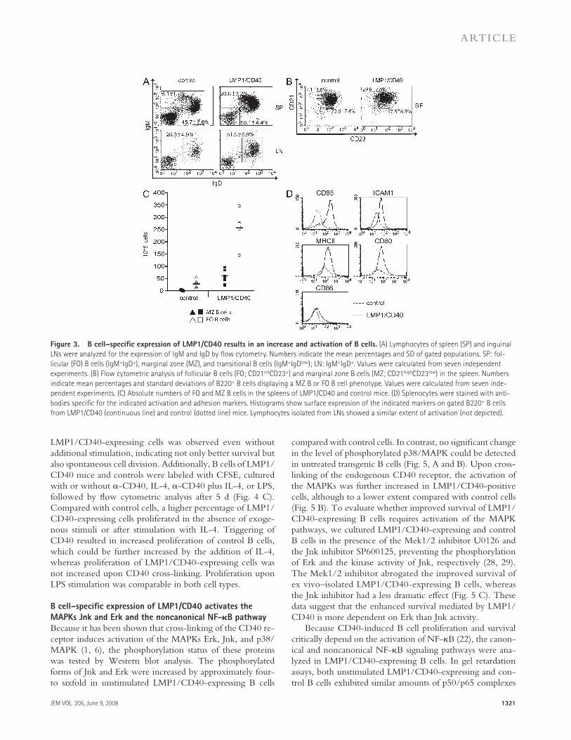
To determine the distribution of B cell subsets in LMP1/CD40 mice, cells from BM, spleen, and inguinal LNs of experimental and control animals were stained for various surface markers and analyzed by fl ow cytometry. In LMP1/CD40 mice, the percentage of IgM + IgD + B cells was signifi -cantly increased in spleen and LN ( Fig. 3 A ). In the spleen, the percentage of marginal zone B cells was increased and numbers of CD21 + CD23 + follicular and CD21 high CD23 low marginal zone B cells were, respectively, 8 and 12 times higher than those of controls ( Fig. 3, B and C ). In the BM, the percentages of pro – , pre – , immature, and mature B cells were comparable in mutant and control mice (unpublished data). Flow cytometric analysis of the expression level of various activation and adhesion molecules revealed an increase in surface expression of CD95, ICAM-1, MHC class II, CD80, and CD86 in B cells isolated from the spleen and LN of LMP1/CD40 mice ( Fig. 3 D and not depicted), suggesting an activated phenotype. To test whether LMP1/CD40- expressing
Figure 1. Generation of transgenic mice conditionally expressing
LMP1/CD40. (A) Targeting strategy for the insertion of a conditional
LMP1/CD40 allele (LMP1/CD40 fl STOP ) into the murine rosa26 -locus. Cre-
mediated recombination leads to deletion of the stop cassette and ex-
pression of LMP1/CD40 under transcriptional control of the endogenous
rosa26 -promoter. The scheme of the targeting construct shows the EcoRI
recognition sites and the location of the probe. The expected fragments
after EcoRI digestion and hybridization with the labeled probe are indi-
cated by the thin lines. SAS, splice acceptor site; STOP, Stop cassette, XbaI,
site of insertion. (B) Southern blot analysis of EcoRI-digested genomic
DNA showing the deletion effi ciency of the stop cassette in B cells of the
spleen (SP; lane 3) and the BM (lane 6) of LMP1/CD40 mice. DNA from B
cells of control mice (lanes 2 and 5) and mice heterozygous for LMP1/
CD40 fl STOP (lane 1 and 4) was included. B cells were purifi ed by using mag-
netic beads against CD19. After deletion of the stop cassette, the rosa26
probe detects a 5.2-kb fragment (LMP1/CD40). The 15- and 7.1-kb frag-
ments represent the wild-type rosa26 allele ( rosa26 locus) and the tar-
geted allele ( LMP1/CD40 fl STOP ), respectively. The deletion effi ciency was
� 50% in the BM and almost complete in the spleen. (C) LMP1/CD40 pro-
tein expression. Western blots were prepared from spleen lysates of
LMP1/CD40;CD19-Cre (lanes 1 and 2) and control mice (lane 3). As con-
trol, protein lysates of 293 cells transiently transfected with a LMP1/CD40
expression vector were included in the analysis (lane 4). The 56-kD LMP1/
CD40 chimeric protein was detected by an anti – human CD40 antibody.
ns, nonspecifi c band.
1320 CONSTITUTIVELY ACTIVE CD40 PROMOTES LYMPHOMAGENESIS | H ö mig-H ö lzel et al.
subsets (Fig. S3, available at http://www.jem.org/cgi/content/full/jem.20080238/DC1). These data indicate that both B and T cells were activated in LMP1/CD40 mice.
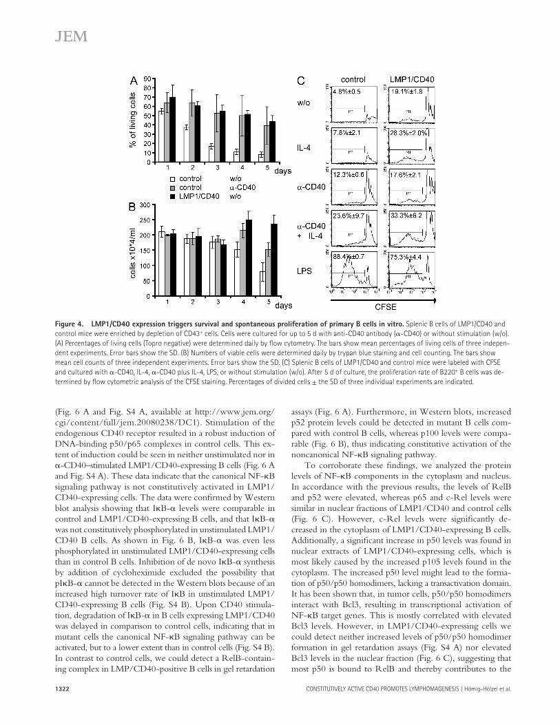
LMP1/CD40-expressing B cells proliferate spontaneously
and have a prolonged survival in vitro
To determine the survival properties of LMP1/CD40-ex-pressing and control B cells, we cultured such cells for 5 d in the presence or absence of � -CD40. The cells were stained daily with the vital dye TOPRO-3, and then subjected to fl ow cytometric analysis ( Fig. 4 A ). The percentage of living cells dropped dramatically in control culture in the absence of CD40 stimulation, whereas the same culture conditions only modestly aff ected the viability of LMP1/CD40-ex-pressing cells. Furthermore, absolute numbers of living cells were determined for LMP1/CD40-expressing cells and con-trols over time ( Fig. 4 B ). An increase in the number of
B cells could participate in TD immune responses, we immu-nized mice with the hapten (3-hydroxy 4-nitrophenylacetyl [NP]) conjugated to the carrier chicken gammaglobulin. In contrast to control mice, the fraction of GC B cells was drastically reduced in the spleens of LMP1/CD40 mice, and GC structures could not be detected by immunohisto-chemistry, suggesting that constitutive CD40 signaling im-pairs the recruitment and/or maintenance of B cells within GCs (Fig. S2, available at http://www.jem.org/cgi/content/full/jem.20080238/DC1).
As expected, T cells showed no deletion of the stop cas-sette, as analyzed by PCR (unpublished data), but were ex-panded and showed an activated phenotype in LMP1/CD40 mice. The ratio between CD4 and CD8 cells was comparable to the controls (unpublished data), but staining for CD44 and CD62L expression revealed a shift from naive toward acti-vated and memory-type T cells among the CD4 and CD8
Figure 2. B cell – specifi c LMP1/CD40 expression results in an increase in B and T cell numbers in secondary lymphoid organs. (A) LMP1/
CD40 mice showed a splenomegaly with a 3.5-fold increase of the splenic weight compared with control mice with B cells carrying the stop
cassette (LMP1/CD40 fl STOP ). Representative spleens from 8-wk-old LMP1/CD40 and control mice are shown. (B) Absolute numbers of B220 + B cells
and CD3 + T cells in spleen (SP) of LMP1/CD40 and control mice. The horizontal bars mark the mean. (C) Spleen sections from LMP1/CD40 and control
mice were stained for B cells (red, anti-IgM) and T cells (top; blue, anti-CD3), or for B cells (red, anti-IgM) and metallophilic macrophages (blue,
anti-MOMA1), which are localized at the border between follicular (FO) and marginal zone (MZ) B cells (bottom). Arrowheads indicate marginal
zone B cells. Bars, 400 μ m. (D) Absolute numbers of B220 + B cells in inguinal LN and BM of LMP1/CD40 and control mice. The horizontal bars mark
the mean.
JEM VOL. 205, June 9, 2008
ARTICLE
1321
compared with control cells. In contrast, no signifi cant change in the level of phosphorylated p38/MAPK could be detected in untreated transgenic B cells ( Fig. 5, A and B ). Upon cross-linking of the endogenous CD40 receptor, the activation of the MAPKs was further increased in LMP1/CD40-positive cells, although to a lower extent compared with control cells ( Fig. 5 B ). To evaluate whether improved survival of LMP1/CD40-expressing B cells requires activation of the MAPK pathways, we cultured LMP1/CD40-expressing and control B cells in the presence of the Mek1/2 inhibitor U0126 and the Jnk inhibitor SP600125, preventing the phosphorylation of Erk and the kinase activity of Jnk, respectively ( 28, 29 ). The Mek1/2 inhibitor abrogated the improved survival of ex vivo – isolated LMP1/CD40-expressing B cells, whereas the Jnk inhibitor had a less dramatic eff ect ( Fig. 5 C ). These data suggest that the enhanced survival mediated by LMP1/CD40 is more dependent on Erk than Jnk activity.
Because CD40-induced B cell proliferation and survival critically depend on the activation of NF- � B ( 22 ), the canon-ical and noncanonical NF- � B signaling pathways were ana-lyzed in LMP1/CD40-expressing B cells. In gel retardation assays, both unstimulated LMP1/CD40-expressing and con-trol B cells exhibited similar amounts of p50/p65 complexes
LMP1/CD40-expressing cells was observed even without additional stimulation, indicating not only better survival but also spontaneous cell division. Additionally, B cells of LMP1/CD40 mice and controls were labeled with CFSE, cultured with or without � -CD40, IL-4, � -CD40 plus IL-4, or LPS, followed by fl ow cytometric analysis after 5 d ( Fig. 4 C ). Compared with control cells, a higher percentage of LMP1/CD40-expressing cells proliferated in the absence of exoge-nous stimuli or after stimulation with IL-4. Triggering of CD40 resulted in increased proliferation of control B cells, which could be further increased by the addition of IL-4, whereas proliferation of LMP1/CD40-expressing cells was not increased upon CD40 cross-linking. Proliferation upon LPS stimulation was comparable in both cell types.
B cell – specifi c expression of LMP1/CD40 activates the
MAPKs Jnk and Erk and the noncanonical NF- � B pathway
Because it has been shown that cross-linking of the CD40 re-ceptor induces activation of the MAPKs Erk, Jnk, and p38/MAPK ( 1, 6 ), the phosphorylation status of these proteins was tested by Western blot analysis. The phosphorylated forms of Jnk and Erk were increased by approximately four- to sixfold in unstimulated LMP1/CD40-expressing B cells
Figure 3. B cell – specifi c expression of LMP1/CD40 results in an increase and activation of B cells. (A) Lymphocytes of spleen (SP) and inguinal
LNs were analyzed for the expression of IgM and IgD by fl ow cytometry. Numbers indicate the mean percentages and SD of gated populations. SP: fol-
licular (FO) B cells (IgM + IgD + ), marginal zone (MZ), and transitional B cells (IgM + IgD low ); LN: IgM + IgD + . Values were calculated from seven independent
experiments. (B) Flow cytometric analysis of follicular B cells (FO; CD21 int CD23 + ) and marginal zone B cells (MZ; CD21 high CD23 low ) in the spleen. Numbers
indicate mean percentages and standard deviations of B220 + B cells displaying a MZ B or FO B cell phenotype. Values were calculated from seven inde-
pendent experiments. (C) Absolute numbers of FO and MZ B cells in the spleens of LMP1/CD40 and control mice. (D) Splenocytes were stained with anti-
bodies specifi c for the indicated activation and adhesion markers. Histograms show surface expression of the indicated markers on gated B220 + B cells
from LMP1/CD40 (continuous line) and control (dotted line) mice. Lymphocytes isolated from LNs showed a similar extent of activation (not depicted).
1322 CONSTITUTIVELY ACTIVE CD40 PROMOTES LYMPHOMAGENESIS | H ö mig-H ö lzel et al.
assays ( Fig. 6 A ). Furthermore, in Western blots, increased p52 protein levels could be detected in mutant B cells com-pared with control B cells, whereas p100 levels were compa-rable ( Fig. 6 B ), thus indicating constitutive activation of the noncanonical NF- � B signaling pathway.
To corroborate these fi ndings, we analyzed the protein levels of NF- � B components in the cytoplasm and nucleus. In accordance with the previous results, the levels of RelB and p52 were elevated, whereas p65 and c-Rel levels were similar in nuclear fractions of LMP1/CD40 and control cells ( Fig. 6 C ). However, c-Rel levels were signifi cantly de-creased in the cytoplasm of LMP1/CD40-expressing B cells. Additionally, a signifi cant increase in p50 levels was found in nuclear extracts of LMP1/CD40-expressing cells, which is most likely caused by the increased p105 levels found in the cytoplasm. The increased p50 level might lead to the forma-tion of p50/p50 homodimers, lacking a transactivation domain. It has been shown that, in tumor cells, p50/p50 homodimers interact with Bcl3, resulting in transcriptional activation of NF- � B target genes. This is mostly correlated with elevated Bcl3 levels. However, in LMP1/CD40-expressing cells we could detect neither increased levels of p50/p50 homodimer formation in gel retardation assays (Fig. S4 A) nor elevated Bcl3 levels in the nuclear fraction ( Fig. 6 C ), suggesting that most p50 is bound to RelB and thereby contributes to the
( Fig. 6 A and Fig. S4 A, available at http://www.jem.org/cgi/content/full/jem.20080238/DC1). Stimulation of the endogenous CD40 receptor resulted in a robust induction of DNA-binding p50/p65 complexes in control cells. This ex-tent of induction could be seen in neither unstimulated nor in � -CD40 – stimulated LMP1/CD40-expressing B cells ( Fig. 6 A and Fig. S4 A). These data indicate that the canonical NF- � B signaling pathway is not constitutively activated in LMP1/CD40-expressing cells. The data were confi rmed by Western blot analysis showing that I � B- � levels were comparable in control and LMP1/CD40-expressing B cells, and that I � B- � was not constitutively phosphorylated in unstimulated LMP1/CD40 B cells. As shown in Fig. 6 B, I � B- � was even less phosphorylated in unstimulated LMP1/CD40-expressing cells than in control B cells. Inhibition of de novo I � B- � synthesis by addition of cycloheximide excluded the possibility that pI � B- � cannot be detected in the Western blots because of an increased high turnover rate of I � B in unstimulated LMP1/CD40-expressing B cells (Fig. S4 B). Upon CD40 stimula-tion, degradation of I � B- � in B cells expressing LMP1/CD40 was delayed in comparison to control cells, indicating that in mutant cells the canonical NF- � B signaling pathway can be activated, but to a lower extent than in control cells (Fig. S4 B). In contrast to control cells, we could detect a RelB-contain-ing complex in LMP/CD40-positive B cells in gel retardation
Figure 4. LMP1/CD40 expression triggers survival and spontaneous proliferation of primary B cells in vitro. Splenic B cells of LMP1/CD40 and
control mice were enriched by depletion of CD43 + cells. Cells were cultured for up to 5 d with anti-CD40 antibody ( � -CD40) or without stimulation (w/o).
(A) Percentages of living cells (Topro negative) were determined daily by fl ow cytometry. The bars show mean percentages of living cells of three indepen-
dent experiments. Error bars show the SD. (B) Numbers of viable cells were determined daily by trypan blue staining and cell counting. The bars show
mean cell counts of three independent experiments. Error bars show the SD. (C) Splenic B cells of LMP1/CD40 and control mice were labeled with CFSE
and cultured with � -CD40, IL-4, � -CD40 plus IL-4, LPS, or without stimulation (w/o). After 5 d of culture, the proliferation rate of B220 + B cells was de-
termined by fl ow cytometric analysis of the CFSE staining. Percentages of divided cells ± the SD of three individual experiments are indicated.

JEM VOL. 205, June 9, 2008
ARTICLE
1323
centage of LMP1/CD40-expressing mice older than 12 mo showed obvious signs of disease. Those mice were further ana-lyzed and showed an extreme splenomegaly with a 20 – 40-fold increase in weight, signifi cantly enlarged inguinal LNs, hepatomegaly, and nodular infi ltrates in the kidney, lung, and liver (Fig. S5, available at http://www.jem.org/cgi/content/full/jem.20080238/DC1).
To confi rm lymphoma development in these mice, splenic cells were analyzed for mono- or oligoclonality by Southern blot analysis using a probe spanning the J H 3-4 region of the mouse IgH locus. In contrast to the controls, all spleen cell
activity of the noncanonical NF- � B signaling pathway. Col-lectively, these results indicate that constitutive activation of CD40 favors the activation of the noncanonical NF- � B sig-naling pathway.
LMP1/CD40 mice develop B cell lymphomas
Because LMP1/CD40-expressing B cells exhibited sponta-neous proliferation and enhanced survival in vitro, which might ultimately lead to the development of malignancies, we monitored the mice for the occurrence of B cell lym-phomas by regularly palpating the abdomen. A high per-
Figure 5. LMP1/CD40 expression induces activity of the Jnk- and Erk-signaling pathways. (A) Whole-cell extracts of splenic B cells of LMP1/
CD40 (L/C) and control (co) mice from three independent preparations (1 – 3) were loaded on one gel and analyzed for Jnk (Jnk2, 54 kD; Jnk1, 46 kD), Erk
(Erk1. 44kD; Erk2, 42 kD), p38/MAPK (38 kD) and the corresponding phosphorylated forms with specifi c antibodies. Equal protein loading was controlled
by Ponceau S staining. The graph shows the mean values of the fold induction of the signals from the unphosphorylated and phosphorylated forms of
Jnk, Erk, and p38/MAPK compared with the signals in control cells. Mean values and SDs were calculated from at least fi ve independent experiments. SDs
are shown by error bars. *, P < 0.05; **, P < 0.001, calculated by the two-tailed Student ’ s t test. (B) After CD40 triggering, LMP1/CD40-expressing cells are
damped in activation compared with control cells. Splenic B cells of LMP1/CD40 and control mice were stimulated with � -CD40 antibody for the indi-
cated time points, and Western blots were performed as described in A. Equal protein loading was controlled by tubulin staining. One representative ex-
periment out of three is shown. (C) The improved survival of LMP1/CD40-expressing B cells is dependent on continuous Erk phosphorylation. Splenic B
cells of LMP1/CD40 and control mice (CD19-Cre) were cultured for up to 5 d with the Mek1/2 inhibitor U0126 (dotted line) and the Jnk-inhibitor
SP600125 (dashed line; triangle and square, respectively). As control, B cells from LMP1/CD40 and control mice were cultured in the presence of DMSO
(continuous lines, rectangle, and circle, respectively). Percentages of living cells (Topro negative) were determined by fl ow cytometry at day 1, 3, and 5.
The bars show mean percentages of living cells of three independent experiments. Error bars show the SD.

1324 CONSTITUTIVELY ACTIVE CD40 PROMOTES LYMPHOMAGENESIS | H ö mig-H ö lzel et al.
samples of diseased LMP1/CD40 mice showed beside the germline IgH band one or more distinct additional bands, indicating a mono- or oligoclonal outgrowth of B cells ( Fig. 7 A ). This was further confi rmed by the sequence analysis of the CDR3 region (Fig. S6, available at http://www.jem.org/cgi/content/full/jem.20080238/DC1). Whereas several rearrangements could be amplifi ed in wild-type cells, only one or two rearrangements were amplifi ed from the tumor samples. None of the lymphomas carried IgH somatic mu-tations. The tumors were further characterized by immuno-histopathology and FACS analysis. Hematoxylin and eosin (HE) staining of spleen sections showed a nodular infi ltrate characterized by the presence of small and large lymphoid cells that could not be seen in the controls ( Fig. 7 B ). Nine tumors were further analyzed by immunohistochemical stainings. Staining for B220 and CD3 surface markers dem-onstrated that in seven cases, the expanded population was
Figure 6. LMP1/CD40 expression induces activity of the nonca-
nonical NF- � B pathway . (A) Splenic B cells of LMP1/CD40 and control
(co) mice were cultured with (+) or without ( � ) � -CD40 for 1 h. Nuclear
extracts were incubated with radioactively labeled DNA oligonucleotides
containing a NF- � B binding site and separated by gel electrophoresis. For
supershift experiments, the indicated antibodies were added to the ex-
tracts. (B) Whole-cell extracts of splenic B cells of LMP1/CD40 and control
(CD19-Cre) mice from three independent preparations (1 – 3) were loaded
on one gel and analyzed by immunoblot. The expression levels of p100
and p52 as well as of I � B � and pI � B � are shown. Equal protein loading
was verifi ed by Ponceau S staining. (C) Cytoplasmic (CP) and nuclear (N)
levels of NF- � B components of splenic B cells from LMP1/CD40 and con-
trol (CD19-Cre) mice were analyzed by immunoblot. Purity of cytoplasmic
and nuclear extracts was verifi ed by � -tubulin and � -Bcl3 staining, re-
spectively. Equal protein loading was controlled by Ponceau S staining.
The experiment was performed three times. Figure 7. LMP1/CD40 mice develop mono- and oligoclonal lym-
phomas. (A) Southern blot analysis to examine IgH gene confi guration
with an IgH-specifi c probe. Genomic DNA was prepared and digested
with EcoRI from splenic cells of LMP1/CD40 mice that developed neopla-
sias (34, 142, 176, 197, 230, 247, 365, and 221), one young LMP1/CD40
mouse (900; 3 mo of age), as well as a control mouse (905). The sequence
analysis of the amplifi ed IgH genes is shown in Fig. S6. (B) Representative
histological analyses of diseased LMP1/CD40 mice and age matched con-
trol mice. (1) HE-stained normal spleen. (2) HE-stained spleen representa-
tive for diseased LMP1/CD40 mice, showing prominent nodular tumor
infi ltrates. (3) Lymphoma infi ltrate within the spleen of a representative
diseased LMP1/CD40 mouse. B cells were visualized by immunohisto-
chemistry using an antibody specifi c for B220. (4) Higher magnifi cation
of section described in 3. (5) Dispersed reactive T cells within the lym-
phoma infi ltrate were detected by an anti-CD3 – specifi c antibody.
A representative highly magnifi ed section is shown. Bars: (1 and 2) 400
μ m; (3) 100 μ m; (4, 5, and inset) 25 μ m. (C) Flow cytometric analysis of
splenic cells for the expression of IgM and IgD. The gates were set ac-
cording to the age-matched controls. For the diseased LMP1/CD40 mice,
a shift toward one or two main populations could be observed, as shown
for three representative samples. Fig. S6 is available at http://www.jem
.org/cgi/content/full/jem.20080238/DC1.
JEM VOL. 205, June 9, 2008
ARTICLE
1325
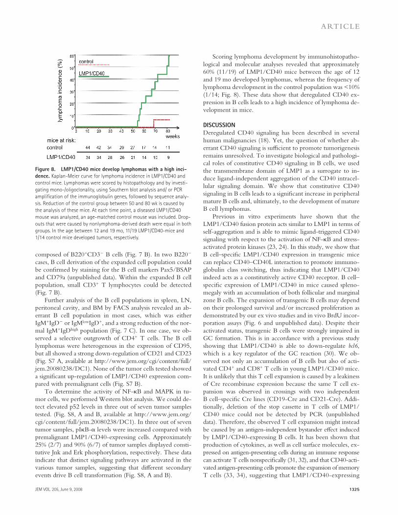
Scoring lymphoma development by immunohistopatho-logical and molecular analyses revealed that approximately 60% (11/19) of LMP1/CD40 mice between the age of 12 and 19 mo developed lymphomas, whereas the frequency of lymphoma development in the control population was < 10% (1/14; Fig. 8 ). These data show that deregulated CD40 ex-pression in B cells leads to a high incidence of lymphoma de-velopment in mice.
DISCUSSION
Deregulated CD40 signaling has been described in several human malignancies ( 18 ). Yet, the question of whether ab-errant CD40 signaling is suffi cient to promote tumorigenesis remains unresolved. To investigate biological and pathologi-cal roles of constitutive CD40 signaling in B cells, we used the transmembrane domain of LMP1 as a surrogate to in-duce ligand-independent aggregation of the CD40 intracel-lular signaling domain. We show that constitutive CD40 signaling in B cells leads to a signifi cant increase in peripheral mature B cells and, ultimately, to the development of mature B cell lymphomas.
Previous in vitro experiments have shown that the LMP1/CD40 fusion protein acts similar to LMP1 in terms of self-aggregation and is able to mimic ligand-triggered CD40 signaling with respect to the activation of NF- � B and stress-activated protein kinases ( 23, 24 ). In this study, we show that B cell – specifi c LMP1/CD40 expression in transgenic mice can replace CD40 – CD40L interaction to promote immuno-globulin class switching, thus indicating that LMP1/CD40 indeed acts as a constitutively active CD40 receptor. B cell – specifi c expression of LMP1/CD40 in mice caused spleno-megaly with an accumulation of both follicular and marginal zone B cells. The expansion of transgenic B cells may depend on their prolonged survival and/or increased proliferation as demonstrated by our ex vivo studies and in vivo BrdU incor-poration assays ( Fig. 6 and unpublished data). Despite their activated status, transgenic B cells were strongly impaired in GC formation. This is in accordance with a previous study showing that LMP1/CD40 is able to down-regulate bcl6 , which is a key regulator of the GC reaction ( 30 ). We ob-served not only an accumulation of B cells but also of acti-vated CD4 + and CD8 + T cells in young LMP1/CD40 mice. It is unlikely that this T cell expansion is caused by a leakiness of Cre recombinase expression because the same T cell ex-pansion was observed in crossings with two independent B cell – specifi c Cre lines (CD19-Cre and CD21-Cre). Addi-tionally, deletion of the stop cassette in T cells of LMP1/CD40 mice could not be detected by PCR (unpublished data). Therefore, the observed T cell expansion might instead be caused by an antigen-independent bystander eff ect induced by LMP1/CD40-expressing B cells. It has been shown that production of cytokines, as well as cell surface molecules, ex-pressed on antigen-presenting cells during an immune response can activate T cells nonspecifi cally ( 31, 32 ), and that CD40-acti-vated antigen-presenting cells promote the expansion of memory T cells ( 33, 34 ), suggesting that LMP1/CD40-expressing
composed of B220 + CD3 � B cells ( Fig. 7 B ). In two B220 � cases, B cell derivation of the expanded cell population could be confi rmed by staining for the B cell markers Pax5/BSAP and CD79a (unpublished data). Within the expanded B cell population, small CD3 + T lymphocytes could be detected ( Fig. 7 B ).
Further analysis of the B cell populations in spleen, LN, peritoneal cavity, and BM by FACS analysis revealed an ab-errant B cell population in most cases, which was either IgM + IgD � or IgM low IgD + , and a strong reduction of the nor-mal IgM + IgD high population ( Fig. 7 C ). In one case, we ob-served a selective outgrowth of CD4 + T cells. The B cell lymphomas were heterogenous in the expression of CD95, but all showed a strong down-regulation of CD21 and CD23 (Fig. S7 A, available at http://www.jem.org/cgi/content/full/jem.20080238/DC1). None of the tumor cells tested showed a signifi cant up-regulation of LMP1/CD40 expression com-pared with premalignant cells (Fig. S7 B).
To determine the activity of NF- � B and MAPK in tu-mor cells, we performed Western blot analysis. We could de-tect elevated p52 levels in three out of seven tumor samples tested. (Fig. S8, A and B, available at http://www.jem.org/cgi/content/full/jem.20080238/DC1). In three out of seven tumor samples, pI � B- � levels were increased compared with premalignant LMP1/CD40-expressing cells. Approximately 25% (2/7) and 90% (6/7) of tumor samples displayed consti-tutive Jnk and Erk phosphorylation, respectively. These data indicate that distinct signaling pathways are activated in the various tumor samples, suggesting that diff erent secondary events drive B cell transformation (Fig. S8, A and B).
Figure 8. LMP1/CD40 mice develop lymphomas with a high inci-
dence. Kaplan-Meier curve for lymphoma incidence in LMP1/CD40 and
control mice. Lymphomas were scored by histopathology and by investi-
gating mono-/oligoclonality, using Southern blot analysis and/ or PCR
amplifi cation of the immunoglobulin genes, followed by sequence analy-
sis. Reduction of the control group between 50 and 80 wk is caused by
the analysis of these mice. At each time point, a diseased LMP1/CD40
mouse was analyzed, an age-matched control mouse was included. Drop-
outs that were caused by nonlymphoma-derived death were equal in both
groups. In the age between 12 and 19 mo, 11/19 LMP1/CD40-mice and
1/14 control mice developed tumors, respectively.
1326 CONSTITUTIVELY ACTIVE CD40 PROMOTES LYMPHOMAGENESIS | H ö mig-H ö lzel et al.
even less active in the mutant cells than in unstimulated con-trol B cells. Additionally, in comparison to control B cells, p105 levels were increased, whereas c-Rel levels were de-creased, in the cytoplasm of LMP1/CD40-expressing B cells. Whether this alteration is caused by the constitutive activa-tion of CD40 or a direct consequence of the constitutively activated noncanonical NF- � B pathway remains to be deter-mined. The question arises why LMP1/CD40 expression in B cells leads selectively to the activation of the noncanonical NF- � B pathway. In wild-type B cells, CD40 ligation results in a fast and robust activation of the canonical NF- � B activity, which is rapidly counteracted by multiple negative-feedback mechanisms, whereas the noncanonical NF- � B pathway re-sponds more slowly and appears to lack strong negative-feed-back mechanisms ( 39 ). This might explain the preferential chronic activation of the noncanonical NF- � B pathway in LMP1/CD40-expressing B cells. Recently, two studies have provided compelling evidence that the noncanonical NF- � B pathway plays a critical role in multiple myeloma (MM) patho-genesis ( 40, 41 ). Thus, 20% of patients with MM were found to carry mutations in essential NF- � B components, leading mainly to the activation of the noncanonical NF- � B pathway ( 41 ). Of note, in some cases of MM, a strong up-regulation of CD40 was observed. As shown for CD30 ( 42 ), CD40 over-expression might lead to self-aggregation of CD40, resulting in ligand-independent constitutive CD40 signaling, and thus creating a primary oncogenic event similar to that seen in our experimental system.
In addition to the noncanonical NF- � B pathway, the MAPKs Erk and Jnk were constitutively phosphorylated in unstimulated LMP1/CD40-expressing B cells. The impor-tance of Erk phosphorylation for the improved survival of ex vivo – isolated LMP1/CD40-expressing B cells was supported by the fact that the survival advantage by LMP1/CD40 ex-pression was abrogated in the presence of the Mek1/2 inhibi-tor UO126. The Ras – Raf – Mek – Erk pathway has been implicated in malignant transformation of various cell types and has been associated with proliferation and survival of he-matopoietic cells ( 43 ). A basal constitutive activity of Erk, which has been attributed to CD40, CD30, and RANK sig-naling, has also been described in the Hodgkin-Reed Stern-berg tumor cells of Hodgkin disease ( 44 ). Treatment of Hodgkin disease cell lines with the MEK inhibitor UO126 resulted in the inhibition of proliferation, underlining the importance of constitutive Erk phosphorylation for the pro-liferation of Hodgkin lymphoma cell lines. Unlike premalig-nant LMP1/CD40 B cells, constitutive Erk phosphorylation was not consistently detected in all independent primary tu-mors derived from LMP1/CD40-expressing B cells. This re-sult suggests the existence of distinct oncogenic partners involved in CD40-driven lymphomagenesis, some of which may bypass the need for Erk activation.
Our results provide direct evidence that constitutive CD40 signaling leads to the selective activation of the nonca-nonical NF- � B pathway and the MAPKs Jnk and Erk. The concerted action of these signaling pathways ultimately leads
B cells may be able to generate an activating environment for T cells.
Deregulated CD40 signaling induced the development of lymphomas in > 60% of mice older than 1 yr. Consistent with the B cell – specifi c expression of LMP1/CD40 and the eff ects of constitutive CD40 signaling on B cell survival and prolif-eration, most of these tumors were of clonal B cell origin. Only 1 out of 11 lymphomas was T cell derived. In all other tumors, the expanded population carried clonal IgH rear-rangements and expressed B cell markers. The nodular growth pattern of the lymphomas was reminiscent of follicular lym-phomas ( 35 ). However, in contrast to follicular lymphomas, the variable regions of the immunoglobulin genes cloned from LMP1/CD40-expressing B lymphoma cells were unmutated, indicating a pre-GC origin. The late onset of tumor develop-ment, the heterogeneity in surface marker expression, and the activity of distinct signaling pathways suggest that multiple independent secondary oncogenic hits cooperate with CD40 activation to promote lymphomagenesis.
A role for CD40 signaling in the pathogenesis of human B cell lymphomas and carcinomas has previously been sug-gested. We provide strong evidence that constitutive CD40 signaling exerts transforming activity in vivo. The oncogenic property of constitutive CD40 signaling appears in contrast with the previously reported therapeutic eff ects of CD40 triggering in hematopoietic malignancies ( 18, 36 ). Indeed, triggering the CD40 receptor can promote growth arrest and cell death of malignant CD40-expressing cells and can induce an autologous antitumor T cell response by up-regulating the costimulatory molecules B7.1 and B7.2 ( 37 ). Our data sug-gest that this therapeutic approach might not be risk free. The level and duration of the CD40 signal may critically infl uence the balance between the apparently contrasting biological ef-fects triggered by CD40 signaling. Indeed, low-level consti-tutive engagement of CD40 was shown to induce proliferation of lymphoma cells, whereas treatment of malignant cells with high doses of agonistic anti-CD40 antibodies or CD40L re-sulted in cell cycle arrest and apoptosis ( 20 ).
Our gel retardation and nuclear fractionation experiments revealed that, compared to control B cells, RelB-contain-ing complexes are increased in the nuclei of LMP1/CD40-expressing B cells, indicating constitutive activation of the noncanonical NF- � B pathway. We suggest that in LMP1/CD40-expressing B cells, RelB dimerizes with either p50 or p52, which were both found to be increased in the nuclear fraction. This is in accordance with previous reports showing that the noncanonical NF- � B pathway based on p100 pro-cessing leads to the nuclear translocation of not only RelB/p52 but also of RelB/p50 dimers ( 17, 38 ). In contrast to the noncanonical pathway, the canonical NF- � B pathway was not found to be activated in LMP1/CD40-expressing B cells, as indicated by similar p65 and c-Rel levels in the nuclear fractions of unstimulated LMP1/CD40-expressing B cells and control B cells. The reduced basal phosphorylation of I � B- � in LMP1/CD40-expressing B cells compared with control B cells suggests that the canonical NF- � B pathway is
JEM VOL. 205, June 9, 2008
ARTICLE
1327
CFSE (fi nal concentration 5 μ M; Invitrogen) for 10 min at 37 ° C. CFSE-la-
beled cells were cultured for up to 5 d in 96-well plates (5 × 10 5 /well) in B
cell medium and analyzed by fl ow cytometry. For treatment with small
chemical inhibitors, B cells were cultured with 2.5 μ M SP600125 (Ag Sci-
entifi c), 10 μ M UO126 (Cell Signaling Technology), or DMSO (Calbio-
chem) in B cell medium for up to 5 d.
For Western blots and gel retardation assays, B cells were purifi ed from
spleen, using magnetic beads against CD43 (MACS; Miltenyi Biotec), and
stimulated from 5 min for up to 24 h with agonistic anti-CD40 anti-
bodies (10 μ g/ml; J Bioscience [HM40-3]). For I � B- � degradation assays,
B cells were treated with 10 μ M cycloheximide (Sigma-Aldrich) 30 min
before stimulation.
Tumor cells. Primary tumor cells were frozen in RPMI, 10% DMSO, and
20% FCS. Because all tumor cells are CD43 + , we were not able to purify
them with magnetic beads as we did for premalignant cells. Therefore, after
thawing, cells were cultured for 2 d to obtain a purer tumor cell population
before Western blots extracts were prepared.
Flow cytometry. Single-cell suspensions prepared from various lymphoid
organs were surface stained with combinations of FITC, PE, Cy-Chrome
(Cyc), APC, and Cy5.5-conjugated monoclonal antibodies. Antibodies to
B220, CD4, CD5, CD8, CD21, CD23, CD43, CD44, CD80, CD95, IgD,
IgG1, and IgM were purchased from BD Biosciences and the antibody to
CD62L was purchased from Immunotech. Additional monoclonal anti-
bodies to CD54 and CD86 were provided by J. Mysliwietz (Helmholtz
Center, Munich, Germany). Peanut agglutinin – FITC was purchased from
Vector Laboratories. All analyses were made with a FACSCalibur (BD Bio-
sciences) and results were analyzed using CellQuest software. Data were ana-
lyzed from 3 × 10 4 viable lymphocyte-gated cells as determined by forward
and side scatter and propidium iodide or Topro-3 (Invitrogen) staining.
Immunohistochemistry. Spleens were embedded in OCT Tissue-Tek
(Sakura), frozen on dry ice, and cut into 8- μ m-thick sections. The sections
were thawed, air dried, fi xed in acetone, and incubated for 30 min at 22 ° C
in a humidifi ed chamber with anti-biotin, anti-avidin solution (Avidin/Bio-
tin blocking kit; Vector Laboratories).
Sections were stained using peroxidase-conjugated anti – mouse IgM
(Sigma-Aldrich), rat anti – mouse CD3 (provided by E. Kremmer, Helmholtz
Center, Munich, Germany), and rat anti – mouse MOMA-1 (T-2011; BMA)
antibodies. Biotin-conjugated mouse anti – rat IgG1 (Jackson ImmunoRe-
search Laboratories) was used to detect MOMA-1 or CD3 antibodies, and
streptavidin coupled to alkaline phosphatase (Sigma-Aldrich) to detect the
biotin-conjugated mouse anti – rat IgG1 antibody. Streptavidin-coupled anti-
bodies were detected by the reaction with an alkaline phosphate substrate kit
(Vector Laboratories), and peroxidase-coupled antibodies were detected by
the reaction with 3-amino-9-ethylcarbazole (peroxidase substrate kit; Vector
Laboratories). All incubation steps were performed at 22 ° C in humidifi ed
chamber, followed by three washing steps with PBS. Slides were analyzed
with a microscope (Carl Zeiss, Inc.); pictures were obtained with a digital
camera (RS Photometrics) and processed with Openlab (Improvision) and
Photoshop (Adobe) software. Immunohistochemical staining of paraffi n sec-
tions was performed on an automated immunostainer (Ventana Medical Sys-
tems) according to the manufacturer ’ s protocols. Antigen retrieval was
performed with a microwave pressure cooker in 0.01 M citrate buff er (pH
6.0). Incubation with the primary antibodies was performed overnight at
room temperature. The rest of the procedure was completed on the Ventana
immunostainer. The antibodies used included B220 (BD Biosciences), CD3
(Dako), Pax5 (BD Biosciences), and CD79a (clone HM57; Dako). Positive
controls for all the antibodies investigated were used to confi rm the adequacy
of the staining.
Electrophoretic mobility shift assay. Nuclear protein extracts were pre-
pared from stimulated and unstimulated cells, as previously described ( 47 ). The
electrophoretic mobility shift assay was performed with a NF- � B – specifi c
to B cell lymphomagenesis. The mouse model described in this paper provides a tool to dissect the contribution of these signaling pathways to lymphomagenesis in vivo.
MATERIALS AND METHODS Generation of the transgenic mouse line LMP1/CD40. The LMP1/
CD40 chimeric gene was isolated from the plasmid p1778.16 encoding aa
1 – 190 from LMP1 fused to aa 223 – 280 of human CD40 ( 23 ). The adenine
at position 243 bp after the ATG of the LMP1 gene was replaced by a cyto-
sine, introducing a silent mutation, to destroy a potential splice acceptor side.
To insert the LMP1/CD40 fusion gene into the rosa26 -locus, the vector
pRosa26-1 was used ( 45 ). Before introducing LMP1/CD40 , pRosa26-1
was modifi ed by introducing a loxP-fl anked region, consisting of a stop cas-
sette containing a transcription and translation termination site ( 46 ), the gene
encoding the red fl uorescent protein (not expressed in this context), and a
neomycin-resistance gene fl anked by frt sites. The LMP1/CD40 fusion gene
was cloned downstream of the stop cassette. The fi nal targeting vector was
electroporated into BALB/c-derived ES cells. The targeted ES cells were
screened for homologous recombination by Southern blot analysis. The
DNA was digested with EcoRI and hybridized with a specifi c rosa26 probe
( 45 ). Recombinant ES cells were injected into C57BL/6 blastocysts, which
were then transferred into foster mothers to obtain chimeric mice.
Mice. Mice carrying the LMP1/CD40 fl STOP allele were crossed either to
the CD19-Cre or the CD21-Cre mouse strain (on a C57BL/6 background)
to generate mice expressing the transgene in early B cell stages in the
BM ([LMP1/CD40 fl STOP ;CD19-Cre] F1) or in mature B cells ([LMP1/
CD40 fl STOP ;CD21-Cre] F1), respectively. Off spring were routinely screened
by PCR using primer specifi c for LMP1/CD40 (5 � LMP1, 5 � -AGGAGCC-
CTCCTTGTCCTCTA-3 � ; 3 � CD40, 5 � -CTGAGATGCGACTCTCTTT-
GCCAT-3 � ) and CD19-Cre or CD21-Cre ( 26, 27 ).
Only mice of the F1 generation were used in this study and analyzed at
8 – 16 wk of age unless stated otherwise. As controls, LMP1/CD40 fl STOP and
CD19-Cre – or CD21-Cre – expressing mice were used. All mice were bred
and maintained in specifi c pathogen-free conditions, and the experiments
were performed in compliance with the German animal welfare law and
have been approved by the institutional committee on animal experimenta-
tion and the government of Upper Bavaria.
Western blot. 3 × 10 6 cells were lysed in Laemmli buff er or NP40 lysis
buff er, and lysates were separated on a SDS-PAGE gel and transferred to a
nitrocellulose or PVDF membrane. LMP1/CD40 protein was detected by
the anti – human CD40 antibody (sc975), which was like anti – I � B- � (sc-
371), purchased from Santa Cruz Biotechnology, Inc. MAPKs and their
phosphorylated forms were detected by anti-p38/MAPK, anti – phospho-
p38/MAPK (pT180/pY182), anti-Erk (p44/42), anti – phospho-Erk (pT202/
pY204), anti-Jnk (56G8), and anti – phospho-Jnk (pT183/pY185), all pur-
chased from Cell Signaling Technology. The phosphorylated form of I � B- �
was detected by anti – phospho-I � B- � (Cell Signaling Technology). Signals
were visualized with ECL Western Blotting Detection Reagents (GE
Healthcare). The amount of loaded protein was standardized against tubulin
using an anti-tubulin antibody (Dianova). After scanning of the fi lms with an
Expression 1680Pro scanner (Epson), quantifi cation of bands was performed
with the TINA software package (Raytest). Protein expression values of con-
trol mice were set to 1 to achieve comparability between diff erent Western
blots. P values were determined by applying the two-tailed Student ’ s t test.
In vitro cultures. Splenic cells were cultured for up to 5 d in 96-well plates
(5 × 10 5 cells/well). Stimuli included lipopolysaccharide (20 μ g/ml; Esche-
richia coli 055:B5; Sigma-Aldrich), IL-4 (10 ng/ml; mouse recombinant;
Sigma-Aldrich), and anti-CD40 antibody (10 μ g/ml; BD Biosciences; 3/23).
Cells were stained and analyzed with a FACSCalibur or counted using try-
pan blue to exclude dead cells. For proliferation assays, splenic cells (5 × 10 6
cells/ml) were labeled by incubation in serum-free RPMI media containing

1328 CONSTITUTIVELY ACTIVE CD40 PROMOTES LYMPHOMAGENESIS | H ö mig-H ö lzel et al.
3 . Korthauer , U. , D. Graf , H.W. Mages , F. Briere , M. Padayachee , S. Malcolm , A.G. Ugazio , L.D. Notarangelo , R.J. Levinsky , and R.A. Kroczek . 1993 . Defective expression of T-cell CD40 ligand causes X-linked immunodefi ciency with hyper-IgM. Nature . 361 : 539 – 541 .
4 . Kawabe , T. , T. Naka , K. Yoshida , T. Tanaka , H. Fujiwara , S. Suematsu , N. Yoshida , T. Kishimoto , and H. Kikutani . 1994 . The immune re-sponses in CD40-defi cient mice: impaired immunoglobulin class switching and germinal center formation. Immunity . 1 : 167 – 178 .
5 . Xu , J. , T.M. Foy , J.D. Laman , E.A. Elliott , J.J. Dunn , T.J. Waldschmidt , J. Elsemore , R.J. Noelle , and R.A. Flavell . 1994 . Mice defi cient for the CD40 ligand. Immunity . 1 : 423 – 431 .
6 . Grammer , A.C. , and P.E. Lipsky . 2000 . CD40-mediated regulation of immune responses by TRAF-dependent and TRAF-independent sig-naling mechanisms. Adv. Immunol. 76 : 61 – 178 .
7 . Hostager , B.S. , I.M. Catlett , and G.A. Bishop . 2000 . Recruitment of CD40 and tumor necrosis factor receptor-associated factors 2 and 3 to membrane microdomains during CD40 signaling. J. Biol. Chem. 275 : 15392 – 15398 .
8 . Hanissian , S.H. , and R.S. Geha . 1997 . Jak3 is associated with CD40 and is critical for CD40 induction of gene expression in B cells. Immunity . 6 : 379 – 387 .
9 . Craxton , A. , G. Shu , J.D. Graves , J. Saklatvala , E.G. Krebs , and E.A. Clark . 1998 . p38 MAPK is required for CD40-induced gene expression and proliferation in B lymphocytes. J. Immunol. 161 : 3225 – 3236 .
10 . Li , Y.Y. , M. Baccam , S.B. Waters , J.E. Pessin , G.A. Bishop , and G.A. Koretzky . 1996 . CD40 ligation results in protein kinase C-independent activation of ERK and JNK in resting murine splenic B cells. J. Immunol. 157 : 1440 – 1447 .
11 . Sutherland , C.L. , A.W. Heath , S.L. Pelech , P.R. Young , and M.R. Gold . 1996 . Diff erential activation of the ERK, JNK, and p38 mitogen-activated protein kinases by CD40 and the B cell antigen receptor. J. Immunol. 157 : 3381 – 3390 .
12 . Berberich , I. , G.L. Shu , and E.A. Clark . 1994 . Cross-linking CD40 on B cells rapidly activates nuclear factor-kappa B. J. Immunol. 153 : 4357 – 4366 .
13 . Coope , H.J. , P.G. Atkinson , B. Huhse , M. Belich , J. Janzen , M.J. Holman , G.G. Klaus , L.H. Johnston , and S.C. Ley . 2002 . CD40 regulates the processing of NF-kappaB2 p100 to p52. EMBO J. 21 : 5375 – 5385 .
14 . Bonizzi , G. , and M. Karin . 2004 . The two NF-kappaB activation path-ways and their role in innate and adaptive immunity. Trends Immunol. 25 : 280 – 288 .
15 . Bonizzi , G. , M. Bebien , D.C. Otero , K.E. Johnson-Vroom , Y. Cao , D. Vu , A.G. Jegga , B.J. Aronow , G. Ghosh , R.C. Rickert , and M. Karin . 2004 . Activation of IKKalpha target genes depends on recogni-tion of specifi c kappaB binding sites by RelB:p52 dimers. EMBO J. 23 : 4202 – 4210 .
16 . Dejardin , E. , N.M. Droin , M. Delhase , E. Haas , Y. Cao , C. Makris , Z.W. Li , M. Karin , C.F. Ware , and D.R. Green . 2002 . The lympho-toxin-beta receptor induces diff erent patterns of gene expression via two NF-kappaB pathways. Immunity . 17 : 525 – 535 .
17 . Derudder , E. , E. Dejardin , L.L. Pritchard , D.R. Green , M. Korner , and V. Baud . 2003 . RelB/p50 dimers are diff erentially regulated by tumor necrosis factor-alpha and lymphotoxin-beta receptor activation: critical roles for p100. J. Biol. Chem. 278 : 23278 – 23284 .
18 . Eliopoulos , A.G. , and L.S. Young . 2004 . The role of the CD40 path-way in the pathogenesis and treatment of cancer. Curr. Opin. Pharmacol. 4 : 360 – 367 .
19 . Baxendale , A.J. , C.W. Dawson , S.E. Stewart , V. Mudaliar , G. Reynolds , J. Gordon , P.G. Murray , L.S. Young , and A.G. Eliopoulos . 2005 . Constitutive activation of the CD40 pathway promotes cell transforma-tion and neoplastic growth. Oncogene . 24 : 7913 – 7923 .
20 . Challa , A. , A.G. Eliopoulos , M.J. Holder , A.S. Burguete , J.D. Pound , A. Chamba , G. Grafton , R.J. Armitage , C.D. Gregory , H. Martinez-Valdez , et al . 2002 . Population depletion activates autonomous CD154-dependent survival in biopsylike Burkitt lymphoma cells. Blood . 99 : 3411 – 3418 .
21 . Furman , R.R. , Z. Asgary , J.O. Mascarenhas , H.C. Liou , and E.J. Schattner . 2000 . Modulation of NF-kappa B activity and apoptosis in chronic lymphocytic leukemia B cells. J. Immunol. 164 : 2200 – 2206 .
22 . Pham , L.V. , A.T. Tamayo , L.C. Yoshimura , P. Lo , N. Terry , P.S. Reid , and R.J. Ford . 2002 . A CD40 signalosome anchored in lipid rafts leads to
double-stranded DNA probe, as previously described ( 48 ). For supershifts,
2 μ g of specifi c antibodies against p50 (sc114), p65 (c20), c-Rel (sc70) p52
(sc298), and RelB (sc226) from Santa Cruz Biotechnology, Inc. were added
to the binding reaction.
B cell fractionation. 2 × 10 7 B cells were incubated in 200 μ l buff er A
(10 mM Hepes, pH 7.9, 10 mM KCl, 0.1 mM EDTA, 0.1 mM EGTA,
1 mM DTT, and 1x complete protease inhibitors [Roche]) for 15 min on ice.
After addition of 12.5 μ l 10% NP-40 and shaking for 5 min at 4 ° C, nuclei
were spun down at 15,000 rpm for 15 min, and the supernatant (cytoplasmic
fraction) was saved. Nuclei were washed once with buff er A before lysis in
buff er C (20 mM Hepes, pH 7.9, 0.4 M NaCl, 1 mM EDTA, 1 mM EGTA,
1 mM DTT, and 1x complete protease inhibitors). After shaking at 4 ° C for
30 min and centrifugation at 15,000 rpm for 15 min, the supernatant (nuclear
fraction) was saved.
Analysis of lymphomas. Animals were kept under observation and pal-
pated regularly to detect lymphoma development. Animals that were obvi-
ously sick were killed and analyzed. All animals were killed and analyzed at
the age of 19 mo at the latest. Lymphomas were scored by histopathology and
by investigating mono-/oligoclonality, using Southern blot analysis and/or
PCR amplifi cation of the immunoglobulin genes, followed by sequence
analysis. Lymphoma incidences were estimated using the Kaplan-Meier
method. Killed control mice or mice that died from causes unrelated to tumor
development were defi ned as early dropouts. Statistical analysis was performed
using the Statistical Analysis Software (SAS) version 6.12.
Analysis of IgH gene rearrangements. Genomic DNA was isolated from
splenic cells, and Southern blot analysis with EcoRI-digested DNA was per-
formed. To detect the IgH region, a radioactive-labeled J H Probe was used
representing the HindIII- and EcoRI-fl anked 1.6-kb fragment spanning the
J H 3-4 region of the mouse IgH locus.
Online supplemental material. Fig. S1 shows that LMP1/CD40 induces
CSR to IgG1 in combination with IL-4. Fig. S2 shows that LMP1/CD40 im-
pairs GC responses. Fig. S3 shows T cell activation by B cell – specifi c LMP1/
CD40 expression. Fig. S4 shows preferential activation of the noncanonical
NF- � B pathway by LMP1/CD40. Fig. S5 shows sequence analysis of the IgH
CDR3 region. Fig. S6 shows tumors in LMP1/CD40 mice. Fig. S7 shows
surface marker expression in LMP1/CD40 positive tumors. Fig. S8 shows sig-
naling pathways in LMP1/CD40 + tumors. The online version of this article is
available at http://www.jem.org/cgi/content/full/jem.20080238/DC1.
We thank G.W. Bornkamm, N. Uyttersprot, and B. Jungnickel for critically reading
the manuscript; D. H ö lzel and M. H ö lzel for help with the statistical analysis;
W. Hammerschmidt for the plasmid p1778.16; G. Marschall for excellent technical
assistance; and M. Schmidt-Supprian and V. Heissmeyer for helpful discussions.
The injection of ES cells into blastocytes was done by M. Hafner and M. Ebel of
the Gesellschaft f ü r Biotechnologische Forschung Braunschweig (supported by
Nationales Genomforschungsnetz Deutschland).
This work was supported by the Deutsche Forschungsgemeinschaft STR-461/3-
2, and Deutsche Krebshilfe. J. Rastelli was supported by the Boehringer Ingelheim
Fonds. K. Rajewsky is supported by grants from the National Institutes of Health
and the Leukemia and Lymphoma Society, and K. Rajewsky and W. Mueller are
supported by the European Union through MUGEN.
The authors declare that they have no confl icting fi nancial interests.
Submitted: 4 February 2008
Accepted: 16 April 2008
REFERENCES 1 . van Kooten , C. , and J. Banchereau . 2000 . CD40-CD40 ligand. J. Leukoc.
Biol. 67 : 2 – 17 . 2 . DiSanto , J.P. , J.Y. Bonnefoy , J.F. Gauchat , A. Fischer , and G. de Saint
Basile . 1993 . CD40 ligand mutations in x-linked immunodefi ciency with hyper-IgM. Nature . 361 : 541 – 543 .

JEM VOL. 205, June 9, 2008
ARTICLE
1329
constitutive activation of NF-kappaB and autonomous cell growth in B cell lymphomas. Immunity . 16 : 37 – 50 .
23 . Gires , O. , U. Zimber-Strobl , R. Gonnella , M. Ueffi ng , G. Marschall , R. Zeidler , D. Pich , and W. Hammerschmidt . 1997 . Latent membrane protein 1 of Epstein-Barr virus mimics a constitutively active receptor molecule. EMBO J. 16 : 6131 – 6140 .
24 . Hatzivassiliou , E. , W.E. Miller , N. Raab-Traub , E. Kieff , and G. Mosialos . 1998 . A fusion of the EBV latent membrane protein-1 (LMP1) transmembrane domains to the CD40 cytoplasmic domain is similar to LMP1 in constitutive activation of epidermal growth factor receptor expression, nuclear factor-kappa B, and stress-activated protein kinase. J. Immunol. 160 : 1116 – 1121 .
25 . Yasui , T. , M. Muraoka , Y. Takaoka-Shichijo , I. Ishida , N. Takegahara , J. Uchida , A. Kumanogoh , S. Suematsu , M. Suzuki , and H. Kikutani . 2002 . Dissection of B cell diff erentiation during primary immune re-sponses in mice with altered CD40 signals. Int. Immunol. 14 : 319 – 329 .
26 . Kraus , M. , M.B. Alimzhanov , N. Rajewsky , and K. Rajewsky . 2004 . Survival of resting mature B lymphocytes depends on BCR signaling via the Igalpha/beta heterodimer. Cell . 117 : 787 – 800 .
27 . Rickert , R.C. , J. Roes , and K. Rajewsky . 1997 . B lymphocyte-specifi c, Cre-mediated mutagenesis in mice. Nucleic Acids Res. 25 : 1317 – 1318 .
28 . Bennett , B.L. , D.T. Sasaki , B.W. Murray , E.C. O ’ Leary , S.T. Sakata , W. Xu , J.C. Leisten , A. Motiwala , S. Pierce , Y. Satoh , et al . 2001 . SP600125, an anthrapyrazolone inhibitor of Jun N-terminal kinase. Proc. Natl. Acad. Sci. USA . 98 : 13681 – 13686 .
29 . Favata , M.F. , K.Y. Horiuchi , E.J. Manos , A.J. Daulerio , D.A. Stradley , W.S. Feeser , D.E. Van Dyk , W.J. Pitts , R.A. Earl , F. Hobbs , et al . 1998 . Identifi cation of a novel inhibitor of mitogen-activated protein kinase kinase. J. Biol. Chem. 273 : 18623 – 18632 .
30 . Panagopoulos , D. , P. Victoratos , M. Alexiou , G. Kollias , and G. Mosialos . 2004 . Comparative analysis of signal transduction by CD40 and the Epstein-Barr virus oncoprotein LMP1 in vivo. J. Virol. 78 : 13253 – 13261 .
31 . Bangs , S.C. , A.J. McMichael , and X.N. Xu . 2006 . Bystander T cell acti-vation – implications for HIV infection and other diseases. Trends Immunol. 27 : 518 – 524 .
32 . Ehl , S. , J. Hombach , P. Aichele , H. Hengartner , and R.M. Zinkernagel . 1997 . Bystander activation of cytotoxic T cells: studies on the mechanism and evaluation of in vivo signifi cance in a transgenic mouse model. J. Exp. Med. 185 : 1241 – 1251 .
33 . Koschella , M. , D. Voehringer , and H. Pircher . 2004 . CD40 ligation in vivo induces bystander proliferation of memory phenotype CD8 T cells. J. Immunol. 172 : 4804 – 4811 .
34 . Taraban , V.Y. , T.F. Rowley , and A. Al-Shamkhani . 2004 . Cutting edge: a critical role for CD70 in CD8 T cell priming by CD40-licensed APCs. J. Immunol. 173 : 6542 – 6546 .
35 . Morse , H.C. III , M.R. Anver , T.N. Fredrickson , D.C. Haines , A.W. Harris , N.L. Harris , E.S. Jaff e , S.C. Kogan , I.C. MacLennan , P.K. Pattengale , and J.M. Ward . 2002 . Bethesda proposals for classifi cation of lymphoid neoplasms in mice. Blood . 100 : 246 – 258 .
36 . Vonderheide , R.H. , J.P. Dutcher , J.E. Anderson , S.G. Eckhardt , K.F. Stephans , B. Razvillas , S. Garl , M.D. Butine , V.P. Perry , R.J. Armitage , et al . 2001 . Phase I study of recombinant human CD40 ligand in cancer patients. J. Clin. Oncol. 19 : 3280 – 3287 .
37 . Fiumara , P. , and A. Younes . 2001 . CD40 ligand (CD154) and tumour necrosis factor-related apoptosis inducing ligand (Apo-2L) in haemato-logical malignancies. Br. J. Haematol. 113 : 265 – 274 .
38 . Basak , S. , H. Kim , J.D. Kearns , V. Tergaonkar , E. O ’ Dea , S.L. Werner , C.A. Benedict , C.F. Ware , G. Ghosh , I.M. Verma , and A. Hoff mann . 2007 . A fourth IkappaB protein within the NF-kappaB signaling mod-ule. Cell . 128 : 369 – 381 .
39 . Hoff mann , A. , and D. Baltimore . 2006 . Circuitry of nuclear factor kap-paB signaling. Immunol. Rev. 210 : 171 – 186 .
40 . Annunziata , C.M. , R.E. Davis , Y. Demchenko , W. Bellamy , A. Gabrea , F. Zhan , G. Lenz , I. Hanamura , G. Wright , W. Xiao , et al . 2007 . Frequent engagement of the classical and alternative NF-kappaB pathways by diverse genetic abnormalities in multiple myeloma. Cancer Cell . 12 : 115 – 130 .
41 . Keats , J.J. , R. Fonseca , M. Chesi , R. Schop , A. Baker , W.J. Chng , S. Van Wier , R. Tiedemann , C.X. Shi , M. Sebag , et al . 2007 . Promiscuous mutations activate the noncanonical NF-kappaB pathway in multiple myeloma. Cancer Cell . 12 : 131 – 144 .
42 . Horie , R. , T. Watanabe , Y. Morishita , K. Ito , T. Ishida , Y. Kanegae , I. Saito , M. Higashihara , S. Mori , M.E. Kadin , and T. Watanabe . 2002 . Ligand-independent signaling by overexpressed CD30 drives NF-kappaB activation in Hodgkin-Reed-Sternberg cells. Oncogene . 21 : 2493 – 2503 .
43 . McCubrey , J.A. , L.S. Steelman , W.H. Chappell , S.L. Abrams , E.W. Wong , F. Chang , B. Lehmann , D.M. Terrian , M. Milella , A. Tafuri , et al . 2007 . Roles of the Raf/MEK/ERK pathway in cell growth, malignant transformation and drug resistance. Biochim. Biophys. Acta . 1773 : 1263 – 1284 .
44 . Zheng , B. , P. Fiumara , Y.V. Li , G. Georgakis , V. Snell , M. Younes , J.N. Vauthey , A. Carbone , and A. Younes . 2003 . MEK/ERK pathway is aberrantly active in Hodgkin disease: a signaling pathway shared by CD30, CD40, and RANK that regulates cell proliferation and survival. Blood . 102 : 1019 – 1027 .
45 . Soriano , P. 1999 . Generalized lacZ expression with the ROSA26 Cre reporter strain. Nat. Genet. 21 : 70 – 71 .
46 . Lakso , M. , B. Sauer , B. Mosinger Jr ., E.J. Lee , R.W. Manning , S.H. Yu , K.L. Mulder , and H. Westphal . 1992 . Targeted oncogene activa-tion by site-specifi c recombination in transgenic mice. Proc. Natl. Acad. Sci. USA . 89 : 6232 – 6236 .
47 . Ruland , J. , G.S. Duncan , A. Elia , I. del Barco Barrantes , L. Nguyen , S. Plyte , D.G. Millar , D. Bouchard , A. Wakeham , P.S. Ohashi , and T.W. Mak . 2001 . Bcl10 is a positive regulator of antigen receptor-induced activation of NF-kappaB and neural tube closure. Cell . 104 : 33 – 42 .
48 . Ruland , J. , G.S. Duncan , A. Wakeham , and T.W. Mak . 2003 . Diff erential requirement for Malt1 in T and B cell antigen receptor signaling. Immunity . 19 : 749 – 758 .


![Preparation and Structural Characterization of Three Types of Homo and Heterotrinuclear Boron Complexes: Salen{[B−O−B][O 2 BOH]}, Salen{[B−O−B][O 2 BPh]}, and Salen{[B−O−B][O](https://static.fdokumen.com/doc/165x107/631bb28ea906b217b906972f/preparation-and-structural-characterization-of-three-types-of-homo-and-heterotrinuclear.jpg)