PI3K/AKT pathway regulates phosphorylation of steroid receptors, hormone independence and tumor...
10
Carcinogenesis vol.0 no.0 pp.1–10, 2011 doi:10.1093/carcin/bgr303 Advance Access publication December 17, 2011 PI3K/AKT pathway regulates phosphorylation of steroid receptors, hormone independence and tumor differentiation in breast cancer Marina Riggio, Marı ´a Laura Polo, Matı ´as Blaustein 1,2 , Alejandro Colman-Lerner 1,2 , Isabel Lu ¨thy, Claudia Lanari and Virginia Novaro Laboratorio de Carcinoge ´nesis Hormonal, Instituto de Biologı ´a y Medicina Experimental and 1 Instituto de Fisiologı ´a, Biologı ´a Molecular y Neurociencias, Consejo Nacional de Investigaciones Cientı ´ficas y Te ´cnicas (IBYME-CONICET), Vuelta de Obligado 2490, C1428ADN Buenos Aires, Argentina and 2 Departamento de Fisiologı ´a, Biologı ´a Molecular y Celular, Facultad de Ciencias Exactas y Naturales, Universidad de Buenos Aires, C1428EHA Buenos Aires, Argentina To whom correspondence should be addressed. Tel: þ054 011 4783 2869; Fax: þ054 011 4786 2564; Email: [email protected] Using a model of medroxyprogesterone acetate (MPA)-induced mouse mammary tumors that transit through different stages of hormone dependence, we previously reported that the activation of the phosphatidylinositol 3-kinase (PI3K)/AKT (protein kinase B) pathway is critical for the growth of hormone-independent (HI) mammary carcinomas but not for the growth of hormone-dependent (HD) mammary carcinomas. The objective of this work was to ex- plore whether the activation of the PI3K/AKT pathway is responsi- ble for the changes in tumor phenotype and for the transition to autonomous growth. We found that the inhibition of the PI3K/ AKT/mTOR (mammalian target of rapamycin) pathway suppresses HI tumor growth. In addition, we were able to induce mammary tumors in mice in the absence of MPA by inoculating HD tumor cells expressing a constitutively active form of AKT1, myristoylated AKT1 (myrAKT1). These tumors were highly differentiated and displayed a ductal phenotype with laminin-1 and cytokeratin 8 ex- pression patterns typical of HI tumors. Furthermore, myrAKT1 in- creased the tumor growth of IBH-6 and IBH-7 human breast cancer cell lines. In the estrogen-dependent IBH-7 cell line, myrAKT1 in- duced estrogen-independent growth accompanied by the expression of the adhesion markers focal adhesion kinase and E-cadherin. Fi- nally, we found that cells expressing myrAKT1 exhibited increased phosphorylation of the progesterone receptor at Ser190 and Ser294 and of the estrogen receptor a at Ser118 and Ser167, independently of exogenous MPA or estrogen supply. Our results indicate that the activation of the PI3K/AKT/mTOR pathway promotes tissue archi- tecture remodeling and the activation of steroid receptors. Introduction The activation of the serine–threonine protein kinase AKT/PKB (protein kinase B) is initiated when cytoplasmic AKT binds phosphatidylinositol (3,4,5)-trisphosphate at the membrane. Phosphatidylinositol (3,4,5)- trisphosphate is the product of the activity of phosphatidylinositol 3- kinase (PI3K), which is activated by different membrane receptors (1). AKT is phosphorylated by phosphatidylinositol-dependent kinase 1 at Thr308 and by the mammalian target of rapamycin complex 2 [mam- malian target of rapamycin C2 (mTORC2), also known as the rictor- mTOR complex] at Ser473 (2). In cells with basal PI3K activity, AKT can also be activated by the deletion of the lipid phosphatase activity of the tumor suppressor tensin homolog (PTEN) on chromosome 10. AKT is a mediator of the protumorigenic effects of hormones and growth factors and of the anchorage-mediated survival of epithelial cells. Its activation stimulates glucose metabolism, cell cycle progression, sur- vival and metabolism through the phosphorylation of many substrates, such as GSK3, BAX, mTOR/p70S6K, S6, TSC1 and Tpl2 (3,4). There is increasing evidence that the PI3K/AKT signaling pathway is implicated in human breast cancer (5,6) and in resistance to endo- crine (7,8), chemotherapy and radiotherapy (9,10). Consequently, in- hibitors of AKT/mTOR signaling are currently being tested in clinical cancer trials (11,12). Despite increasing evidence linking mutations at the level of PI3K and PTEN with protumorigenic behavior (13,14), the direct role of AKT in cancer progression has not been genetically defined. Likewise, the contribution of AKT activation to tumorigenesis is derived from loss-of-function studies. Little is known about the tu- morigenic phenotypes caused by gain-of-function mutants of AKT signaling. It has recently been shown that whereas the AKT1 isoform accelerates mammary tumor incidence, the AKT2 isoform promotes the metastasis of tumor cells without affecting the latency of tumor de- velopment (15). It is well accepted that over time, the molecular phe- notypes of breast cancers can change, which impacts the design of an efficient antitumor therapy. Thus, the main goal of our study was to analyze whether the overactivation of AKT1 is directly involved in breast cancer progression and, in particular, in the acquisition of auton- omous tumor growth by using two well-studied models of breast cancer. The syngeneic experimental mouse model of mammary carcino- genesis induced by the prolonged administration of medroxyproges- terone acetate (MPA) displays all the stages of breast cancer progression, from hormone dependence to hormone independence and eventually to endocrine resistance (16). The clinical significance of the MPA-induced model lies in its similarity to the most common human breast cancers. MPA-induced mammary tumors are typically ductal adenocarcinomas, estrogen receptor (ER)- and progesterone receptor (PR)-positive and metastatic to the lung and the lymph nodes. Furthermore, we previously reported that the PI3K/AKT signaling pathway appeared to play an important role in this model of tumor progression because hormone-independent (HI) C4-HI tumors ex- hibited high sensitivity to anti-PI3K/AKT therapy. In contrast, hor- mone-dependent (HD) C4-HD tumors were more sensitive to endocrine therapy with RU-486 and ICI 182780 (17). The human cell lines IBH-6 and IBH-7, which are derived from primary invasive ductal mammary carcinomas, are good models to study in vivo human breast cancer because they express steroid hormone receptors and are invasive (18). In vivo IBH-6 cells grow independently of the administration of 17-b-estradiol (E 2 ), whereas IBH-7 cells require continuous E 2 treatment to grow (19). In the present study, we showed that the activation of the PI3K/ AKT/mTOR signaling pathway was necessary and sufficient for the transition to highly differentiated and exogenous MPA- or estrogen- independent mammary tumors in a mouse model and in IBH-7 cells, respectively. Materials and methods Animals Two-month-old virgin female BALB/c (IBYME Animal Facility) and nude (nu/nu, University of La Plata Animal Facility) mice were used. Animal care and animal procedures were approved by the Institutional guidelines in agreement with the Guide for the Care and Use of Laboratory Animals (20). Tumors and cell lines C4-HD is maintained by serial subcutaneous transplantation into MPA-treated BALB/c female mice. C4-HI is the HI variant of C4-HD, which arose sponta- neously in an untreated mouse [reviewed in ref. (16)]. IBH-6 and IBH-7 cell lines were developed from invasive ductal breast carcinomas from two different patients (18). Abbreviations: AKT/PKB, protein kinase B; DMEM, Dulbecco’s modified Eagle’s medium; ER, estrogen receptor; FAK, focal adhesion kinase; FCS, fetal calf serum; HD, hormone-dependent; HI, hormone-independent; MPA, medroxyprogesterone acetate; mTOR, mammalian target of rapamycin; PI3K, phosphatidylinositol 3-kinase; p-PTEN, phosphorylated PTEN; PR, progester- one receptor; 3D, three-dimensional. Ó The Author 2011. Published by Oxford University Press. All rights reserved. For Permissions, please email: [email protected] 1 Carcinogenesis Advance Access published January 19, 2012 at SUNY Health Science Center at Brooklyn on January 20, 2012 http://carcin.oxfordjournals.org/ Downloaded from
Transcript of PI3K/AKT pathway regulates phosphorylation of steroid receptors, hormone independence and tumor...
Carcinogenesis vol.0 no.0 pp.1–10, 2011doi:10.1093/carcin/bgr303Advance Access publication December 17, 2011
PI3K/AKT pathway regulates phosphorylation of steroid receptors, hormoneindependence and tumor differentiation in breast cancer
Marina Riggio, Marıa Laura Polo, Matıas Blaustein1,2,Alejandro Colman-Lerner1,2, Isabel Luthy, Claudia Lanariand Virginia Novaro�
Laboratorio de Carcinogenesis Hormonal, Instituto de Biologıa y MedicinaExperimental and 1Instituto de Fisiologıa, Biologıa Molecular yNeurociencias, Consejo Nacional de Investigaciones Cientıficas y Tecnicas(IBYME-CONICET), Vuelta de Obligado 2490, C1428ADN Buenos Aires,Argentina and 2Departamento de Fisiologıa, Biologıa Molecular y Celular,Facultad de Ciencias Exactas y Naturales, Universidad de Buenos Aires,C1428EHA Buenos Aires, Argentina
�To whom correspondence should be addressed. Tel: þ054 011 4783 2869;Fax: þ054 011 4786 2564;Email: [email protected]
Using a model of medroxyprogesterone acetate (MPA)-inducedmouse mammary tumors that transit through different stages ofhormone dependence, we previously reported that the activation ofthe phosphatidylinositol 3-kinase (PI3K)/AKT (protein kinase B)pathway is critical for the growth of hormone-independent (HI)mammary carcinomas but not for the growth of hormone-dependent(HD) mammary carcinomas. The objective of this work was to ex-plore whether the activation of the PI3K/AKT pathway is responsi-ble for the changes in tumor phenotype and for the transition toautonomous growth. We found that the inhibition of the PI3K/AKT/mTOR (mammalian target of rapamycin) pathway suppressesHI tumor growth. In addition, we were able to induce mammarytumors in mice in the absence ofMPA by inoculating HD tumor cellsexpressing a constitutively active form of AKT1, myristoylatedAKT1 (myrAKT1). These tumors were highly differentiated anddisplayed a ductal phenotype with laminin-1 and cytokeratin 8 ex-pression patterns typical of HI tumors. Furthermore, myrAKT1 in-creased the tumor growth of IBH-6 and IBH-7 human breast cancercell lines. In the estrogen-dependent IBH-7 cell line, myrAKT1 in-duced estrogen-independent growth accompanied by the expressionof the adhesion markers focal adhesion kinase and E-cadherin. Fi-nally, we found that cells expressing myrAKT1 exhibited increasedphosphorylation of the progesterone receptor at Ser190 and Ser294and of the estrogen receptor a at Ser118 and Ser167, independentlyof exogenous MPA or estrogen supply. Our results indicate that theactivation of the PI3K/AKT/mTOR pathway promotes tissue archi-tecture remodeling and the activation of steroid receptors.
Introduction
The activation of the serine–threonine protein kinase AKT/PKB (proteinkinase B) is initiated when cytoplasmic AKT binds phosphatidylinositol(3,4,5)-trisphosphate at the membrane. Phosphatidylinositol (3,4,5)-trisphosphate is the product of the activity of phosphatidylinositol 3-kinase (PI3K), which is activated by different membrane receptors (1).AKT is phosphorylated by phosphatidylinositol-dependent kinase 1 atThr308 and by the mammalian target of rapamycin complex 2 [mam-malian target of rapamycin C2 (mTORC2), also known as the rictor-mTOR complex] at Ser473 (2). In cells with basal PI3K activity, AKTcan also be activated by the deletion of the lipid phosphatase activity ofthe tumor suppressor tensin homolog (PTEN) on chromosome 10. AKT
is a mediator of the protumorigenic effects of hormones and growthfactors and of the anchorage-mediated survival of epithelial cells. Itsactivation stimulates glucose metabolism, cell cycle progression, sur-vival and metabolism through the phosphorylation of many substrates,such as GSK3, BAX, mTOR/p70S6K, S6, TSC1 and Tpl2 (3,4).
There is increasing evidence that the PI3K/AKT signaling pathwayis implicated in human breast cancer (5,6) and in resistance to endo-crine (7,8), chemotherapy and radiotherapy (9,10). Consequently, in-hibitors of AKT/mTOR signaling are currently being tested in clinicalcancer trials (11,12). Despite increasing evidence linking mutations atthe level of PI3K and PTEN with protumorigenic behavior (13,14),the direct role of AKT in cancer progression has not been geneticallydefined. Likewise, the contribution of AKT activation to tumorigenesisis derived from loss-of-function studies. Little is known about the tu-morigenic phenotypes caused by gain-of-function mutants of AKTsignaling. It has recently been shown that whereas the AKT1 isoformaccelerates mammary tumor incidence, the AKT2 isoform promotes themetastasis of tumor cells without affecting the latency of tumor de-velopment (15). It is well accepted that over time, the molecular phe-notypes of breast cancers can change, which impacts the design of anefficient antitumor therapy. Thus, the main goal of our study was toanalyze whether the overactivation of AKT1 is directly involved inbreast cancer progression and, in particular, in the acquisition of auton-omous tumor growth by using two well-studied models of breast cancer.
The syngeneic experimental mouse model of mammary carcino-genesis induced by the prolonged administration of medroxyproges-terone acetate (MPA) displays all the stages of breast cancerprogression, from hormone dependence to hormone independenceand eventually to endocrine resistance (16). The clinical significanceof the MPA-induced model lies in its similarity to the most commonhuman breast cancers. MPA-induced mammary tumors are typicallyductal adenocarcinomas, estrogen receptor (ER)- and progesteronereceptor (PR)-positive and metastatic to the lung and the lymph nodes.Furthermore, we previously reported that the PI3K/AKT signalingpathway appeared to play an important role in this model of tumorprogression because hormone-independent (HI) C4-HI tumors ex-hibited high sensitivity to anti-PI3K/AKT therapy. In contrast, hor-mone-dependent (HD) C4-HD tumors were more sensitive toendocrine therapy with RU-486 and ICI 182780 (17).
The human cell lines IBH-6 and IBH-7, which are derived fromprimary invasive ductal mammary carcinomas, are good models tostudy in vivo human breast cancer because they express steroidhormone receptors and are invasive (18). In vivo IBH-6 cells growindependently of the administration of 17-b-estradiol (E2), whereasIBH-7 cells require continuous E2 treatment to grow (19).
In the present study, we showed that the activation of the PI3K/AKT/mTOR signaling pathway was necessary and sufficient for thetransition to highly differentiated and exogenous MPA- or estrogen-independent mammary tumors in a mouse model and in IBH-7 cells,respectively.
Materials and methods
Animals
Two-month-old virgin female BALB/c (IBYME Animal Facility) and nude(nu/nu, University of La Plata Animal Facility) mice were used. Animalcare and animal procedures were approved by the Institutional guidelines inagreement with the Guide for the Care and Use of Laboratory Animals (20).
Tumors and cell lines
C4-HD is maintained by serial subcutaneous transplantation into MPA-treatedBALB/c female mice. C4-HI is the HI variant of C4-HD, which arose sponta-neously in an untreated mouse [reviewed in ref. (16)]. IBH-6 and IBH-7cell lines were developed from invasive ductal breast carcinomas from twodifferent patients (18).
Abbreviations: AKT/PKB, protein kinase B; DMEM, Dulbecco’s modifiedEagle’s medium; ER, estrogen receptor; FAK, focal adhesion kinase; FCS,fetal calf serum; HD, hormone-dependent; HI, hormone-independent; MPA,medroxyprogesterone acetate; mTOR, mammalian target of rapamycin; PI3K,phosphatidylinositol 3-kinase; p-PTEN, phosphorylated PTEN; PR, progester-one receptor; 3D, three-dimensional.
� The Author 2011. Published by Oxford University Press. All rights reserved. For Permissions, please email: [email protected] 1
Carcinogenesis Advance Access published January 19, 2012 at SU
NY
Health Science C
enter at Brooklyn on January 20, 2012
http://carcin.oxfordjournals.org/D
ownloaded from
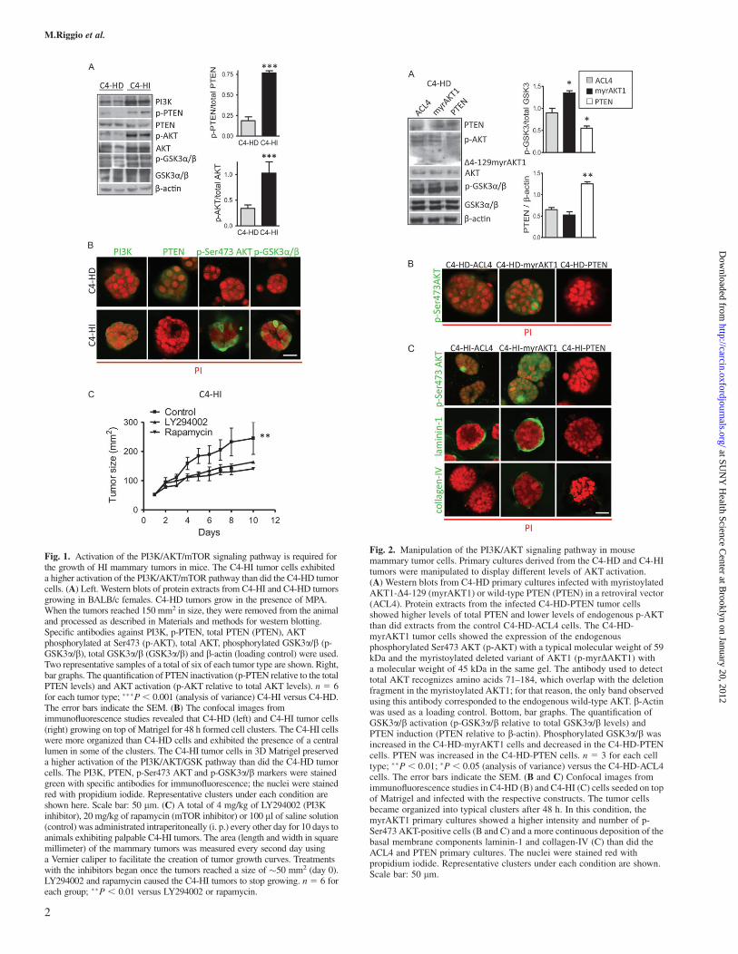
Fig. 1. Activation of the PI3K/AKT/mTOR signaling pathway is required forthe growth of HI mammary tumors in mice. The C4-HI tumor cells exhibiteda higher activation of the PI3K/AKT/mTOR pathway than did the C4-HD tumorcells. (A) Left. Western blots of protein extracts from C4-HI and C4-HD tumorsgrowing in BALB/c females. C4-HD tumors grow in the presence of MPA.When the tumors reached 150 mm2 in size, they were removed from the animaland processed as described in Materials and methods for western blotting.Specific antibodies against PI3K, p-PTEN, total PTEN (PTEN), AKTphosphorylated at Ser473 (p-AKT), total AKT, phosphorylated GSK3a/b (p-GSK3a/b), total GSK3a/b (GSK3a/b) and b-actin (loading control) were used.Two representative samples of a total of six of each tumor type are shown. Right,bar graphs. The quantification of PTEN inactivation (p-PTEN relative to the totalPTEN levels) and AKT activation (p-AKT relative to total AKT levels). n 5 6for each tumor type; ���P, 0.001 (analysis of variance) C4-HI versus C4-HD.The error bars indicate the SEM. (B) The confocal images fromimmunofluorescence studies revealed that C4-HD (left) and C4-HI tumor cells(right) growing on top of Matrigel for 48 h formed cell clusters. The C4-HI cellswere more organized than C4-HD cells and exhibited the presence of a centrallumen in some of the clusters. The C4-HI tumor cells in 3D Matrigel preserveda higher activation of the PI3K/AKT/GSK pathway than did the C4-HD tumorcells. The PI3K, PTEN, p-Ser473 AKT and p-GSK3a/b markers were stainedgreen with specific antibodies for immunofluorescence; the nuclei were stainedred with propidium iodide. Representative clusters under each condition areshown here. Scale bar: 50 lm. (C) A total of 4 mg/kg of LY294002 (PI3Kinhibitor), 20 mg/kg of rapamycin (mTOR inhibitor) or 100 ll of saline solution(control) was administrated intraperitoneally (i. p.) every other day for 10 days toanimals exhibiting palpable C4-HI tumors. The area (length and width in squaremillimeter) of the mammary tumors was measured every second day usinga Vernier caliper to facilitate the creation of tumor growth curves. Treatmentswith the inhibitors began once the tumors reached a size of �50 mm2 (day 0).LY294002 and rapamycin caused the C4-HI tumors to stop growing. n 5 6 foreach group; ��P , 0.01 versus LY294002 or rapamycin.
Fig. 2. Manipulation of the PI3K/AKT signaling pathway in mousemammary tumor cells. Primary cultures derived from the C4-HD and C4-HItumors were manipulated to display different levels of AKT activation.(A) Western blots from C4-HD primary cultures infected with myristoylatedAKT1-D4-129 (myrAKT1) or wild-type PTEN (PTEN) in a retroviral vector(ACL4). Protein extracts from the infected C4-HD-PTEN tumor cellsshowed higher levels of total PTEN and lower levels of endogenous p-AKTthan did extracts from the control C4-HD-ACL4 cells. The C4-HD-myrAKT1 tumor cells showed the expression of the endogenousphosphorylated Ser473 AKT (p-AKT) with a typical molecular weight of 59kDa and the myristoylated deleted variant of AKT1 (p-myrDAKT1) witha molecular weight of 45 kDa in the same gel. The antibody used to detecttotal AKT recognizes amino acids 71–184, which overlap with the deletionfragment in the myristoylated AKT1; for that reason, the only band observedusing this antibody corresponded to the endogenous wild-type AKT. b-Actinwas used as a loading control. Bottom, bar graphs. The quantification ofGSK3a/b activation (p-GSK3a/b relative to total GSK3a/b levels) andPTEN induction (PTEN relative to b-actin). Phosphorylated GSK3a/b wasincreased in the C4-HD-myrAKT1 cells and decreased in the C4-HD-PTENcells. PTEN was increased in the C4-HD-PTEN cells. n 5 3 for each celltype; ��P , 0.01; �P , 0.05 (analysis of variance) versus the C4-HD-ACL4cells. The error bars indicate the SEM. (B and C) Confocal images fromimmunofluorescence studies in C4-HD (B) and C4-HI (C) cells seeded on topof Matrigel and infected with the respective constructs. The tumor cellsbecame organized into typical clusters after 48 h. In this condition, themyrAKT1 primary cultures showed a higher intensity and number of p-Ser473 AKT-positive cells (B and C) and a more continuous deposition of thebasal membrane components laminin-1 and collagen-IV (C) than did theACL4 and PTEN primary cultures. The nuclei were stained red withpropidium iodide. Representative clusters under each condition are shown.Scale bar: 50 lm.
M.Riggio et al.
2
at SUN
Y H
ealth Science Center at B
rooklyn on January 20, 2012http://carcin.oxfordjournals.org/
Dow
nloaded from
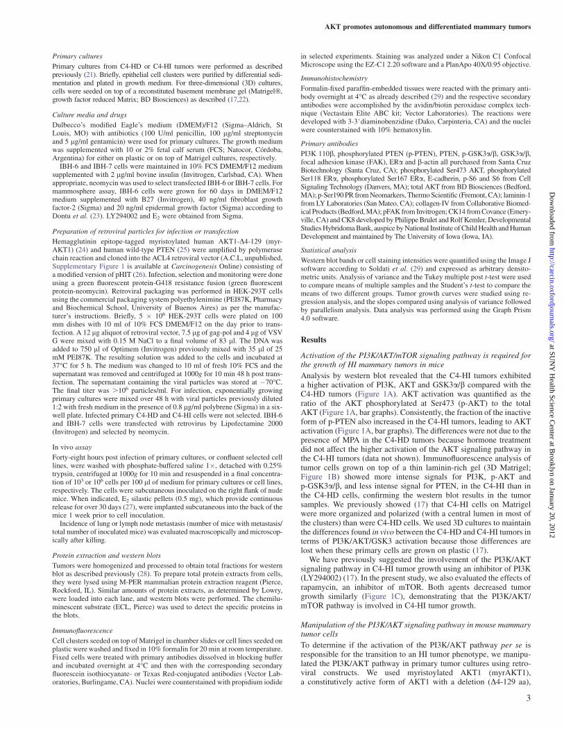
Primary cultures
Primary cultures from C4-HD or C4-HI tumors were performed as describedpreviously (21). Briefly, epithelial cell clusters were purified by differential sedi-mentation and plated in growth medium. For three-dimensional (3D) cultures,cells were seeded on top of a reconstituted basement membrane gel (Matrigel�,growth factor reduced Matrix; BD Biosciences) as described (17,22).
Culture media and drugs
Dulbecco’s modified Eagle’s medium (DMEM)/F12 (Sigma–Aldrich, StLouis, MO) with antibiotics (100 U/ml penicillin, 100 lg/ml streptomycinand 5 lg/ml gentamicin) were used for primary cultures. The growth mediumwas supplemented with 10 or 2% fetal calf serum (FCS; Natocor, Cordoba,Argentina) for either on plastic or on top of Matrigel cultures, respectively.
IBH-6 and IBH-7 cells were maintained in 10% FCS DMEM/F12 mediumsupplemented with 2 lg/ml bovine insulin (Invitrogen, Carlsbad, CA). Whenappropriate, neomycin was used to select transfected IBH-6 or IBH-7 cells. Formammosphere assay, IBH-6 cells were grown for 60 days in DMEM/F12medium supplemented with B27 (Invitrogen), 40 ng/ml fibroblast growthfactor-2 (Sigma) and 20 ng/ml epidermal growth factor (Sigma) according toDontu et al. (23). LY294002 and E2 were obtained from Sigma.
Preparation of retroviral particles for infection or transfection
Hemagglutinin epitope-tagged myristoylated human AKT1-D4-129 (myr-AKT1) (24) and human wild-type PTEN (25) were amplified by polymerasechain reaction and cloned into the ACL4 retroviral vector (A.C.L, unpublished,Supplementary Figure 1 is available at Carcinogenesis Online) consisting ofa modified version of pHIT (26). Infection, selection and monitoring were doneusing a green fluorescent protein-G418 resistance fusion (green fluorescentprotein-neomycin). Retroviral packaging was performed in HEK-293T cellsusing the commercial packaging system polyethylenimine (PEI87K, Pharmacyand Biochemical School, University of Buenos Aires) as per the manufac-turer’s instructions. Briefly, 5 � 106 HEK-293T cells were plated on 100mm dishes with 10 ml of 10% FCS DMEM/F12 on the day prior to trans-fection. A 12 lg aliquot of retroviral vector, 7.5 lg of gag-pol and 4 lg of VSVG were mixed with 0.15 M NaCl to a final volume of 83 ll. The DNA wasadded to 750 ll of Optimem (Invitrogen) previously mixed with 35 ll of 25mM PEI87K. The resulting solution was added to the cells and incubated at37�C for 5 h. The medium was changed to 10 ml of fresh 10% FCS and thesupernatant was removed and centrifuged at 1000g for 10 min 48 h post trans-fection. The supernatant containing the viral particles was stored at �70�C.The final titer was .106 particles/ml. For infection, exponentially growingprimary cultures were mixed over 48 h with viral particles previously diluted1:2 with fresh medium in the presence of 0.8 lg/ml polybrene (Sigma) in a six-well plate. Infected primary C4-HD and C4-HI cells were not selected. IBH-6and IBH-7 cells were transfected with retrovirus by Lipofectamine 2000(Invitrogen) and selected by neomycin.
In vivo assay
Forty-eight hours post infection of primary cultures, or confluent selected celllines, were washed with phosphate-buffered saline 1�, detached with 0.25%trypsin, centrifuged at 1000g for 10 min and resuspended in a final concentra-tion of 103 or 106 cells per 100 ll of medium for primary cultures or cell lines,respectively. The cells were subcutaneous inoculated on the right flank of nudemice. When indicated, E2 silastic pellets (0.5 mg), which provide continuousrelease for over 30 days (27), were implanted subcutaneous into the back of themice 1 week prior to cell inoculation.
Incidence of lung or lymph node metastasis (number of mice with metastasis/total number of inoculated mice) was evaluated macroscopically and microscop-ically after killing.
Protein extraction and western blots
Tumors were homogenized and processed to obtain total fractions for westernblot as described previously (28). To prepare total protein extracts from cells,they were lysed using M-PER mammalian protein extraction reagent (Pierce,Rockford, IL). Similar amounts of protein extracts, as determined by Lowry,were loaded into each lane, and western blots were performed. The chemilu-minescent substrate (ECL, Pierce) was used to detect the specific proteins inthe blots.
Immunofluorescence
Cell clusters seeded on top of Matrigel in chamber slides or cell lines seeded onplastic were washed and fixed in 10% formalin for 20 min at room temperature.Fixed cells were treated with primary antibodies dissolved in blocking bufferand incubated overnight at 4�C and then with the corresponding secondaryfluorescein isothiocyanate- or Texas Red-conjugated antibodies (Vector Lab-oratories, Burlingame, CA). Nuclei were counterstained with propidium iodide
in selected experiments. Staining was analyzed under a Nikon C1 ConfocalMicroscope using the EZ-C1 2.20 software and a PlanApo 40X/0.95 objective.
Immunohistochemistry
Formalin-fixed paraffin-embedded tissues were reacted with the primary anti-body overnight at 4�C as already described (29) and the respective secondaryantibodies were accomplished by the avidin/biotin peroxidase complex tech-nique (Vectastain Elite ABC kit; Vector Laboratories). The reactions weredeveloped with 3-3#diaminobenzidine (Dako, Carpinteria, CA) and the nucleiwere counterstained with 10% hematoxylin.
Primary antibodies
PI3K 110b, phosphorylated PTEN (p-PTEN), PTEN, p-GSK3a/b, GSK3a/b,focal adhesion kinase (FAK), ERa and b-actin all purchased from Santa CruzBiotechnology (Santa Cruz, CA); phosphorylated Ser473 AKT, phosphorylatedSer118 ERa, phosphorylated Ser167 ERa, E-cadherin, p-S6 and S6 from CellSignaling Technology (Danvers, MA); total AKT from BD Biosciences (Bedford,MA); p-Ser190 PR from Neomarkers, Thermo Scientific (Fremont, CA); laminin-1from LY Laboratories (San Mateo, CA); collagen-IV from Collaborative Biomed-ical Products (Bedford, MA); pFAK from Invitrogen; CK14 from Covance (Emery-ville, CA) and CK8 developed by Philippe Brulet and Rolf Kemler, DevelopmentalStudies Hybridoma Bank, auspice by National Institute of Child Health and HumanDevelopment and maintained by The University of Iowa (Iowa, IA).
Statistical analysis
Western blot bands or cell staining intensities were quantified using the Image Jsoftware according to Soldati et al. (29) and expressed as arbitrary densito-metric units. Analysis of variance and the Tukey multiple post t-test were usedto compare means of multiple samples and the Student’s t-test to compare themeans of two different groups. Tumor growth curves were studied using re-gression analysis, and the slopes compared using analysis of variance followedby parallelism analysis. Data analysis was performed using the Graph Prism4.0 software.
Results
Activation of the PI3K/AKT/mTOR signaling pathway is required forthe growth of HI mammary tumors in mice
Analysis by western blot revealed that the C4-HI tumors exhibiteda higher activation of PI3K, AKT and GSK3a/b compared with theC4-HD tumors (Figure 1A). AKT activation was quantified as theratio of the AKT phosphorylated at Ser473 (p-AKT) to the totalAKT (Figure 1A, bar graphs). Consistently, the fraction of the inactiveform of p-PTEN also increased in the C4-HI tumors, leading to AKTactivation (Figure 1A, bar graphs). The differences were not due to thepresence of MPA in the C4-HD tumors because hormone treatmentdid not affect the higher activation of the AKT signaling pathway inthe C4-HI tumors (data not shown). Immunofluorescence analysis oftumor cells grown on top of a thin laminin-rich gel (3D Matrigel;Figure 1B) showed more intense signals for PI3K, p-AKT andp-GSK3a/b, and less intense signal for PTEN, in the C4-HI than inthe C4-HD cells, confirming the western blot results in the tumorsamples. We previously showed (17) that C4-HI cells on Matrigelwere more organized and polarized (with a central lumen in most ofthe clusters) than were C4-HD cells. We used 3D cultures to maintainthe differences found in vivo between the C4-HD and C4-HI tumors interms of PI3K/AKT/GSK3 activation because those differences arelost when these primary cells are grown on plastic (17).
We have previously suggested the involvement of the PI3K/AKTsignaling pathway in C4-HI tumor growth using an inhibitor of PI3K(LY294002) (17). In the present study, we also evaluated the effects ofrapamycin, an inhibitor of mTOR. Both agents decreased tumorgrowth similarly (Figure 1C), demonstrating that the PI3K/AKT/mTOR pathway is involved in C4-HI tumor growth.
Manipulation of the PI3K/AKT signaling pathway in mouse mammarytumor cells
To determine if the activation of the PI3K/AKT pathway per se isresponsible for the transition to an HI tumor phenotype, we manipu-lated the PI3K/AKT pathway in primary tumor cultures using retro-viral constructs. We used myristoylated AKT1 (myrAKT1),a constitutively active form of AKT1 with a deletion (D4-129 aa),
AKT promotes autonomous and differentiated mammary tumors
3
at SUN
Y H
ealth Science Center at B
rooklyn on January 20, 2012http://carcin.oxfordjournals.org/
Dow
nloaded from

to maintain AKT1 at the cell membrane and the overexpression ofPTEN to inhibit AKT activation. Each construct was cloned into theACL4 retroviral vector (Supplementary Figure 1 is available at Car-cinogenesis Online).
Although the efficiency of infection of the primary cultures wasrelatively low (�30%, as determined by green fluorescent protein-positive cells, data not shown), it was sufficient to result in changesdetectable by western blot in both the C4-HD (Figure 2A) and the C4-HI (data not shown) cells. The C4-HD cells infected with PTENshowed higher levels of total PTEN and lower phosphorylation levelsof endogenous AKT and its downstream effector GSK3a/b than didC4-HD cells infected with the empty vector ACL4 (Figure 2A). Incontrast, the C4-HD cells infected with myrAKT1 exhibited the ap-propriate p-myrDAKT1 band and an increase in p-GSK3a/b(Figure 2A).
Similar results were obtained using immunofluorescence in 3Dcultures. Increased p-Ser473 AKT signals were observed in both theC4-HD-myrAKT1 (Figure 2B) and the C4-HI-myrAKT1 cultures(Figure 2C) as compared with the respective ACL4 cultures. Almostno p-Ser473 AKT staining was observed in the C4-HD-PTEN andC4-HI-PTEN cultures. As expected, the basal intensity of p-AKT andthe number of positive cells were higher in the C4-HI-ACL4 than inthe C4-HD-ACL4 cultures. Furthermore, the C4-HI-myrAKT1 3Dcultures displayed a more continuous deposition of the basal mem-brane components laminin-1 and collagen-IV than did the emptyvector or the PTEN cultures (Figure 2C), suggesting that the hyper-activation of AKT1 correlates with the in vitro basal reorganization ofthe basement membrane components.
Overactivation of AKT1 in mammary tumor cells leads toMPA-independent tumors in mice
To determine if the activation of the PI3K/AKT pathway also causeschanges in tumor growth in vivo, we inoculated nude mice withC4-HD or C4-HI cells that had been previously infected with retro-viral constructs expressing myrAKT1, PTEN or the empty vectorACL4.
In the absence of MPA, myrAKT1 caused the C4-HD (P , 0.001;Figure 3A left) and C4-HI cells (P, 0.05; Figure 3A right) to increasetumor growth as compared with ACL4. In contrast, PTEN inhibited C4-HI tumor growth mainly at the early time points (P , 0.05) withouta significant effect on the C4-HD tumors. In the absence of MPA, thenaive C4-HD tumor cells required .3 months post inoculation to gen-erate tumors (data not shown), which suggested that retrovirus infectionper se induced a protumorigenic effect, on top of which the effect ofmyrAKT1 was still appreciable. This effect was stronger in the C4-HD(Figure 3A and B) than in the C4-HI cells probably because the C4-HIcells already had an active PI3K/AKT pathway.
Immunohistochemical studies showed, as expected, a higher signalfor p-Ser473 AKT in myrAKT1 than in the control or PTEN-expressingtumors, both in the C4-HD and in the C4-HI samples (Figure 3C andD), although the effect was less remarkable in the C4-HI tumors prob-ably because of the high levels of endogenous p-Ser473 AKT. Nuclearand cytoplasmic staining was observed in the myrAKT1 tumors(Figure 3C and D, middle). Nuclear staining likely corresponded toendogenous AKT, which was usually observed in nuclei of uninfectedcells. These results indicated that during the progression to hormoneindependence in this model, the activation of AKT1 constitutes a keyevent.
The overactivation of AKT1 in myrAKT1 tumors did not increasethe incidence or size of the lung or node metastases with respect toACL4 tumors (data not shown), suggesting that the hyperactivation ofAKT1 was not involved in cell invasiveness.
To further confirm the participation of the downstream effectors ofthe PI3K/AKT/mTOR pathway in the determination of tumor pheno-type, we evaluated PTEN levels and the phosphorylation of S6 kinase(p-S6) in C4-HD tumors. As shown in Figure 3E, PTEN was in-creased in the C4-HD-PTEN tumors, whereas S6 was activated in theC4-HD-myrAKT1 tumors and inhibited in the C4-HD-PTEN tumors.
MPA-independent mammary tumors originating from theoveractivation of AKT1 are luminal-type and highly differentiated
To characterize the MPA-independent mammary carcinomas inducedby the overactivation of AKT1, we first evaluated their histology. Anincrease in differentiation (the ductal-like structures indicated byblack arrows in Supplementary Figure 2 is available at Carcinogen-esis Online and Figure 4A) was observed in the C4-HD-myrAKT1tumors compared with the C4-HD-ACL4 and C4-HD-PTEN tumors.The C4-HI-ACL4 tumors were already ductal-like and differentiated(Supplementary Figure 2 is available atCarcinogenesis Online), similarto the naive C4-HI tumors (17) (Figure 4B); PTEN overexpression didnot change their morphology (Figure 3C and D and SupplementaryFigure 2 is available at Carcinogenesis Online).
To further characterize these tumors, we evaluated the expressionof cytokeratins 8 (CK8; luminal) and 14 (CK14; basal), as well as thatof laminin-1, by immunohistochemistry. Increases in CK8 stainingand in the interstitial deposition of laminin-1 were observed in theC4-HD-myrAKT1 tumors as compared with the C4-HD-ACL4 tu-mors, whereas no difference was observed in CK14 expression, whichshowed mild staining in both cases (Figure 4A). No evident alterationin CK8 or laminin-1 staining was observed in the C4-HD-PTENtumors compared with the C4-HD-ACL4 tumors (data not shown).Interestingly, the C4-HD-myrAKT1 tumors exhibited tissue organi-zation that was more similar to that of the naive C4-HI tumors than tothat of the C4-HD tumors (Figure 4B). C4-HD-myrAKT1 and C4-HIwere highly differentiated tumors, comprising abundant glandularstructures surrounded by the deposition of laminin-1 and displayinghigh expression of CK8. These findings, consistent with the in vitroresults shown in Figure 2D, indicated that independent of MPAsupply, the overactivation of PI3K/AKT leads to the reorganizationof the basement membrane and the tissue architecture in mammarytumor cells.
Overactivation of AKT1 in tumor cells results in phosphorylation ofthe PR
To determine the involvement of the PR in the MPA-independenttumor growth mediated by AKT1, we evaluated the phosphorylationat serine 190 (p-Ser190) of isoforms A (PRA) and B (PRB) of the PR.Both p-Ser190 PR isoforms were increased in the C4-HD-myrAKT1tumors and reduced in the C4-HD-PTEN tumors (Figure 3E). Theeffect was similar in the C4-HI tumors (data not shown).
Together, these results indicate that the activation of the AKT1/mTOR pathway participates in the transition to MPA independenceby activating the PR and inducing a luminal-type differentiated pheno-type. This result suggests that changes in the PI3K/AKT signalingpathway could ultimately shape the response of the tumor to endocrinetherapy.
Overactivation of AKT1 in human breast cancer cell lines: regulationof tumorigenicity, adhesion markers and hormone independence
To determine the mechanisms by which the overactivation of AKT1modifies tumor behavior and to extrapolate this result to human breastcancer, we evaluated the changes in the patterns of cellular morphologyand in vivo tumor growth of the IBH-6 and IBH-7 human breast cancercell lines previously transfected with myrAKT1 or PTEN and selectedwith neomycin. IBH-6 and IBH-7 cells were grown in the presence ofinsulin in the growth medium and this could hyperactivate insulinand insulin-like growth factor-1 receptors; however, we still detectedthe effect of AKT overactivation by introducing myrAKT1. As deter-mined by western blotting, the IBH-6-myrAKT1 cells exhibited thetransfected myristoylated variant of AKT1 (p-myrDAKT1) and incr-eases in p-GSK3, p-S6, p-Ser167 ERa, total ERa and PRB as comparedwith the control IBH-6-ACL4 cells (Figure 5A). Because the PTENlevels were high in the IBH-6-ACL4 cells, the increased PTEN expres-sion in the IBH-6-PTEN cells was not evident. Nevertheless, significantreductions in the levels of p-AKT, p-GSK3, S6, p-Ser167 ERa, totalERa and PRB were observed (Figure 5A). Neither PRA levels(Figure 5A) nor its activation (data not shown) were regulated bymyrAKT1 or PTEN.
M.Riggio et al.
4
at SUN
Y H
ealth Science Center at B
rooklyn on January 20, 2012http://carcin.oxfordjournals.org/
Dow
nloaded from
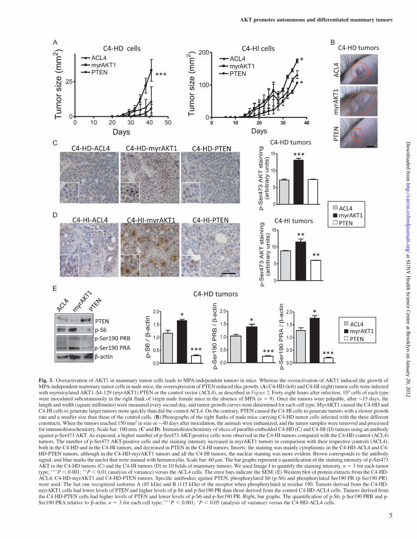
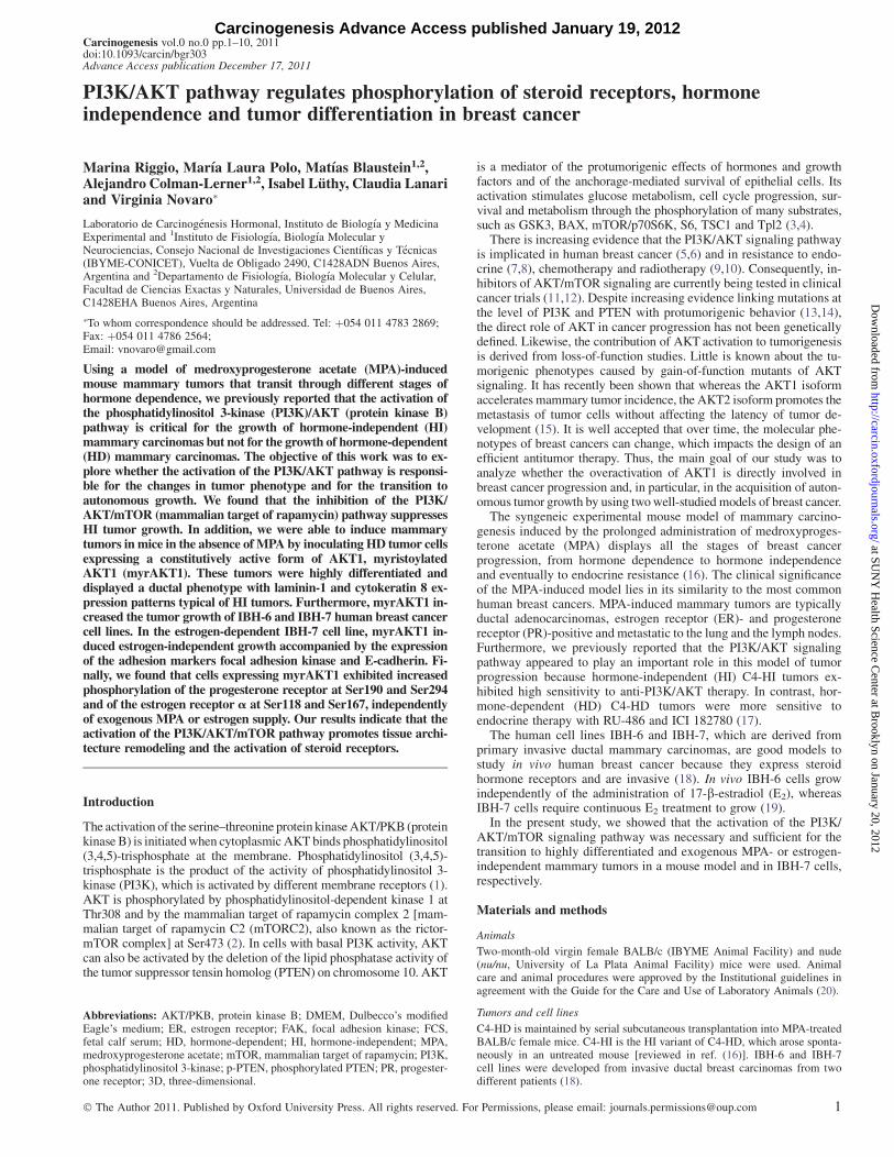
Fig. 3. Overactivation of AKT1 in mammary tumor cells leads to MPA-independent tumors in mice. Whereas the overactivation of AKT1 induced the growth ofMPA-independent mammary tumor cells in nude mice, the overexpression of PTEN reduced this growth. (A) C4-HD (left) and C4-HI (right) tumor cells were infectedwith myristoylated AKT1-D4-129 (myrAKT1) PTEN or the control vector (ACL4), as described in Figure 2. Forty-eight hours after infection, 103 cells of each typewere inoculated subcutaneously in the right flank of virgin nude female mice in the absence of MPA (n 5 9). Once the tumors were palpable, after �15 days, thelength and width (square millimeter) were measured every second day, and tumor growth curves were determined for each cell type. MyrAKT1 caused the C4-HD andC4-HI cells to generate larger tumors more quickly than did the control ACL4. On the contrary, PTEN caused the C4-HI cells to generate tumors with a slower growthrate and a smaller size than those of the control cells. (B) Photographs of the right flanks of nude mice carrying C4-HD tumor cells infected with the three differentconstructs. When the tumors reached 150 mm2 in size or�40 days after inoculation, the animals were euthanized, and the tumor samples were removed and processedfor immunohistochemistry. Scale bar: 100 mm. (C and D). Immunohistochemistry of slices of paraffin-embedded C4-HD (C) and C4-HI (D) tumors using an antibodyagainst p-Ser473 AKT. As expected, a higher number of p-Ser473 AKT-positive cells were observed in the C4-HI tumors compared with the C4-HD control (ACL4)tumors. The number of p-Ser473 AKT-positive cells and the staining intensity increased in myrAKT1 tumors in comparison with their respective controls (ACL4),both in the C4-HD and in the C4-HI tumors, and decreased in PTEN in the C4-HI tumors. Inserts: the staining was mainly cytoplasmic in the C4-HD-ACL4 and C4-HD-PTEN tumors, although in the C4-HD-myrAKT1 tumors and all the C4-HI tumors, the nuclear staining was more evident. Brown corresponds to the antibodysignal, and blue marks the nuclei that were stained with hematoxylin. Scale bar: 60 lm. The bar graphs represent a quantification of the staining intensity of p-Ser473AKT in the C4-HD tumors (C) and the C4-HI tumors (D) in 10 fields of mammary tumors. We used Image J to quantify the staining intensity. n5 3 for each tumortype; ���P, 0.001; ��P, 0.01 (analysis of variance) versus the ACL4 cells. The error bars indicate the SEM. (E) Western blot of protein extracts from the C4-HD-ACL4, C4-HD-myrAKT1 and C4-HD-PTEN tumors. Specific antibodies against PTEN, phosphorylated S6 (p-S6) and phosphorylated Ser190 PR (p-Ser190 PR)were used. The last one recognized isoforms A (85 kDa) and B (115 kDa) of the receptor when phosphorylated at residue 190. Tumors derived from the C4-HD-myrAKT1 cells had lower levels of PTEN and higher levels of p-S6 and p-Ser190 PR than those derived from the control C4-HD-ACL4 cells. Tumors derived fromthe C4-HD-PTEN cells had higher levels of PTEN and lower levels of p-S6 and p-Ser190 PR. Right, bar graphs. The quantification of p-S6, p-Ser190 PRB and p-Ser190 PRA relative to b-actin. n 5 3 for each cell type; ���P , 0.001; �P , 0.05 (analysis of variance) versus the C4-HD-ACL4 cells.
AKT promotes autonomous and differentiated mammary tumors
5
at SUN
Y H
ealth Science Center at B
rooklyn on January 20, 2012http://carcin.oxfordjournals.org/
Dow
nloaded from
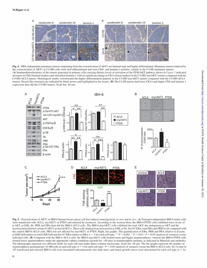
Fig. 4. MPA-independent mammary tumors originating from the overactivation of AKT1 are luminal-type and highly differentiated. Mammary tumors induced bythe overactivation of AKT1 in C4-HD cells were well differentiated and were CK8- and laminin-1-positive, similar to the C4-HI mammary tumors.(A) Immunohistochemistry of the tumors generated in primary cells carrying distinct levels of activation of the PI3K/AKT pathway shown in Figure 3 indicatedincreases in CK8 (luminal marker) and interstitial laminin-1 with no significant change in CK14 (basal marker) in the C4-HD-myrAKT1 tumors compared with theC4-HD-ACL4 tumors. Histological studies corroborated the higher differentiation patterns in the C4-HD-myrAKT1 tumors compared with the C4-HD-ACL4tumors. Ductal-like structures are indicated by black arrows and highlighted in the inserts. (B) The C4-HI tumors had lower CK14 and higher CK8 and laminin-1expression than did the C4-HD tumors. Scale bar: 60 lm.
Fig. 5. Overactivation of AKT1 in IBH-6 human breast cancer cell line induces tumorigenicity in vitro and in vivo. (A) Estrogen-independent IBH-6 tumor cellswere transfected with ACL4, myrAKT1 or PTEN and selected by neomycin. According to the western blots, the IBH-6-PTEN cells exhibited lower levels ofp-AKT, p-GSK, S6, PRB and ERa than did the IBH-6-ACL4 cells. The IBH-6-myrAKT1 cells exhibited the total AKT, the endogenous p-AKT and themyristoylated deleted variant of AKT1 (p-myrDAKT1). These cells displayed an increased in p-GSK, p-S6, Ser167 ERa, total ERa and PRB levels compared withthe control IBH-6-ACL4 cells. PRA was not affected by myrAKT1 or PTEN. Right, bar graphs. The quantification of ERa, PRB and PRA relative to b-actin,p-GSK3a/b relative to total GSK3a/b and Ser167 ERa relative to ERa. n5 3 for each cell type; ���P, 0.001; ��P, 0.01; �P, 0.05 (analysis of variance) versusindicated cells. (B) Compared with the IBH-6-ACL4 cells, the IBH-6-myrAKT1 cells formed more and bigger mammospheres, whereas the IBH-6-PTEN cellsformed fewer mammospheres under the appropriate culture conditions (growth for �60 days in mammosphere medium, as indicated in Materials and methods).The photographs represent two different fields for each cell type under phase contrast microscopy. Scale bar: 60 lm. The bar graphs represent the number ofmammospheres generated per 10 000 cells in each cell type. n5 3 for each cell type; �P, 0.05 (analysis of variance) versus the IBH-6-ACL4 cells. (C) A total of106 transfected and selected IBH-6 cells were inoculated subcutaneously into nude mice, and tumor growth curves were determined for each cell type (n 5 6).
M.Riggio et al.
6
at SUN
Y H
ealth Science Center at B
rooklyn on January 20, 2012http://carcin.oxfordjournals.org/
Dow
nloaded from
To evaluate if the overactivation of AKT1 induced cancer stem/progenitor cell properties, we used a mammosphere assay. In thisassay, the number of mammospheres reflects the ability of self-renewal of cancer cells in an undifferentiated state, based on theirability to proliferate in suspension as non-adherent cell popu-lations (23). In this condition, we found that the IBH-6-myrAKT1cells formed more mammospheres than did the IBH-6-ACL4 cells(Figure 5B). The inhibition of PI3K/AKT signaling in the IBH-6-PTEN cells suppressed mammosphere formation.
To determine if an increase in the levels of AKT activation resultedin a detectable in vivo tumorigenic effect in estrogen-independentIBH-6 cells, we inoculated nude mice with IBH-6 cells expressingACL4, myrAKT1 or PTEN. Tumor growth curves were determinedfor each cell type. The IBH-6-myrAKT1 tumors grew faster than didthe control IBH-6-ACL4 or IBH-PTEN tumors (P , 0.001;Figure 5C). No differences were observed in tumor growth betweenthe control cells and the IBH-PTEN cells. The incidence of tumors,�100%, was not affected by any construct (data not shown), and thelatency and slope curve (representing the pattern of tumor growth)were more similar for the ACL4 than for the naive IBH-6 cells (19). Inaddition, no differences in spindle cell morphology were observedbetween the different IBH-6-transfected cells, although the expressionof the adhesion protein FAK was reduced by myrAKT1 and increasedby PTEN (Figure 5D). Furthermore, no histological differences wereobserved between the induced tumors (Figure 5E). In summary, theoveractivation of AKT1 enriched the tumorigenic progenitor cellsin vitro and in vivo in IBH-6 cells, with no critical changes in cellmorphology.
We then analyzed the effects of the PI3K/AKT pathway onestrogen-dependent IBH-7 cells. Surprisingly, IBH-7 cells thatoverexpressed PTEN did not survive more than a few days inculture. The IBH-7-myrAKT1 cells exhibited the myristoylatedvariant of AKT1 (p-myrDAKT1) and increases in p-S6, p-GSK3a/b, p-Ser294 PRB, p-Ser118 ERa, p-Ser167 ERa and totalERa (Figure 6A). This last result indicated that exogenousestrogen-independent activation of ERa was mainly through aninduction of ERa expression.
The overactivation of AKT was corroborated by immunofluores-cence studies, in which high nuclear p-AKT staining was observed(Figure 6B). In contrast to the findings for the IBH-6 cells, the over-activation of AKT1 was associated with a change in cell morphologyin the IBH-7 cells. As shown in Figure 6B, the IBH-7-myrAKT1 cellsexhibited a rounded morphology. This was associated with increasedlevels of FAK and E-cadherin (Figure 6B). In some IBH-7-myrAKT1cell clusters, phosphorylated FAK (p-FAK) was localized to the ad-hesive borders of the cells, suggesting a rudimentary polarizationpattern.
We then evaluated the in vivo effects of the overactivation of AKT1on IBH-7 tumorigenicity and hormone independence. We inoculatednude mice with IBH-7 cells expressing ACL4 or myrAKT1. Thepatterns of tumor growth (the latency and slope curve) were similarfor the ACL4 cells (Figure 6C) than for the naive IBH-7 cells (19).Whereas the IBH-7-ACL4 cells required E2 to grow, as was the casewith the naıve IBH-7 cells, the IBH-7-myrAKT1 cells could surpris-ingly generate tumors even in the absence of E2. However, the sizes ofthe IBH-7-myrAKT1 tumors were still smaller than those generatedby the IBH-7-ACL4 or IBH-7-myrAKT1 cells in the E2-treated mice.This result indicates that although gaining AKT1 function may bypasshormone dependence, other pathways that are also triggered by E2
treatment may collaborate with AKT signaling. Finally, the incidenceof lung metastasis was not affected by the expression of myrAKT1 inthe IBH-7 cells (data not shown).
Together, these results indicate that the overactivation of AKT1 wassufficient to increase tumor growth and to induce a critical alterationin the cancer phenotype. Depending on the cell type, this alterationalso led to changes in cell adhesion and to a conversion to ligand-independent cell growth.
Discussion
Our data show that the overactivation of AKT1 was sufficient to in-duce HI breast cancer growth, which suggests that upon the adaptationto hormone deprivation, breast cancer cells rely heavily on PI3K/AKTand its downstream targets mTOR, S6 and GSK3, which regulate theactivation of steroid receptors and the tissue architecture of mammarycarcinomas without affecting the metastatic potential of the tumorcells. Thus, our results support AKT1 as a critical driver for HI tumorgrowth and provide a strong rationale for the clinical use of specificPI3K/AKT/mTOR kinase inhibitors in steroid receptor-positivebreast cancer. A potential candidate to initiate this activation is thetumor microenvironment because, as described previously (28),carcinoma-associated fibroblasts contribute to HI tumor growth.We postulate that under still unknown circumstances, the tumormicroenvironment become ‘activated’ and signals to the epithelialcompartment through the activation of different protein kinases, suchas the PI3K/AKT pathway. Once the PI3K/AKT/GSK pathway isactivated, the mechanisms of tissue remodeling and phosphorylationof steroid receptors are switched on and the tumor phenotype starts tochange.
It is also interesting to note that the effects of the overactivation ofAKT1 were cell line specific. Whereas the IBH-7-myrAKT1 cellsunderwent a notable change in cell morphology in vitro, no changeswere observed in the IBH-6-myrAKT1 cells, indicating that evenwhen the final result was increased tumor growth in both cell lines,the mechanisms underlying this effect may have been dependent onother signaling pathways that regulate cell proliferation. One possibleexplanation is that in IBH-7 cells basal levels of p-Ser473 AKT werehigher than in IBH-6 cells (data not shown) suggesting that IBH-7cells were more dependent on the PI3K/AKT pathway than IBH-6cells.
There is extensive literature on the role of PI3K/AKT in the regu-lation of mammary gland differentiation at different levels (30–32).Consistent with our data showing that constitutively active AKT1induced laminin-1 and collagen-IV deposition around C4-HD tumorcells. Li et al. (33) found that AKT induced the synthesis anddeposition of the same proteins in differentiating C2 myoblast- andinsulin-induced Chinese hamster ovary T-cell cultures. With respectto the increase in the luminal marker CK8 induced by AKT1, similardata have been reported in MCF-7 cells (34). In contrast, AKT2 hasbeen reported to upregulate CK18 and vimentin expression in severalepithelial carcinoma cell lines (35). Our data suggest that activatedAKT1, as opposed to the effect of progestins (which reduce the ex-pression of luminal cytokeratins) (36), is involved in the positiveregulation of CK8 and in basement membrane formation, which areessential for epithelial cell differentiation.
We also showed that the overactivation of AKT1 increased the phos-phorylation of FAK in the IBH-7 cells. FAK not only serves as a critical
MyrAKT1 caused the IBH-6 cells to grow more quickly than the control ACL4 cells. PTEN did not affect the tumor growth of the IBH-6 cells. ���P , 0.001(analysis of variance) versus the IBH-6-ACL4 cells. (D) Immunofluorescence assays using Texas Red to stain for the adhesion protein FAK in IBH-6 cells inculture. FAK staining was reduced in the myrAKT1 cells and was increased in the PTEN cells. All the IBH-6 transfected cells presented a spindle-likemorphology. Some nuclei of the transfected cells fluoresced green, indicating green fluorescent protein expression. Scale bar: 50 lm. The bar graphs representa quantification of the staining intensity of FAK in 20 IBH-6 transfected cells in culture. We used Image J to quantify the staining intensity. n5 3 for each cell type;��P , 0.01 (analysis of variance) versus the ACL4 cells. The error bars indicate the SEM. (E) All tumors that originated from the IBH-6-transfected cells weresarcomatoid and poorly differentiated, similar to the tumors that originated from the naive IBH-6 cells (control). Scale bar: 50 lm.
AKT promotes autonomous and differentiated mammary tumors
7
at SUN
Y H
ealth Science Center at B
rooklyn on January 20, 2012http://carcin.oxfordjournals.org/
Dow
nloaded from

component of integrin signaling but is also required for cell spreading,polarity and chemotactic invasion. Although FAK is typically consid-ered to be upstream of AKT, it has been reported that extracellularpressure stimulates cancer cell adhesion and tumor differentiation viaAKT-dependent FAK phosphorylation, suggesting a positive regulatoryloop between FAK and AKT (37). Most studies have consideredE-cadherin as a tumor suppressor gene, and the decreased expressionof this protein has been associated with tumor progression. Moreover,AKT has been shown to repress the expression of E-cadherin in severalhuman tumor cell lines (38). However, our results show that the over-activation of AKT1 increased E-cadherin expression in IBH-7 cellswithout affecting the invasive behavior of those cells.
We hypothesize that most of the effects described here for AKT1 inC4-HD and IBH-7 cells are related to the transition to a HI stage ofgrowth. However, the same effects are not necessarily expected duringthe conversion to a more invasive phenotype or to increased cell pro-liferation in all tumor models. Consistent with this theory, we foundintriguingly that in estrogen-independent IBH-6 cells, AKT1 down-regulated FAK expression and upregulated the ability of these cells toform mammospheres.
Along the same lines, Blanco-Aparicio et al. (39) demonstrated thatmyrAKT1 expression in the mammary glands of transgenic mice in-creased the susceptibility to the induction of ER-positive mammary
adenocarcinomas by 9,10-dimethyl-1,2-benzanthracene. The authorsfound that whereas 9,10-dimethyl-1,2-benzanthracene-treated wild-type mice exhibited mostly sarcomatous mammary tumors, AKTtransgenic mice treated with 9,10-dimethyl-1,2-benzanthracene pri-marily developed adenocarcinomas. Similarly, others have found thatfemale mice coexpressing activated AKT1 and ErbB-2 developedmammary tumors faster than did the activated ErbB-2 strain alone(40). Furthermore, bitransgenic mice exhibited lower levels of inva-siveness, fewer metastatic lesions and more differentiated phenotypes.Hutchinson et al. (40) concluded that the overexpression of AKT byitself was unable to induce mammary tumors; rather, it needed anotheroncogenic event that would eventually synergize with the overexpres-sion of AKT. Our findings are consistent with the hypothesis thatAKT1 participates in cancer progression. In C4-HD and IBH-7 cells,the AKT1 pathway replaced the need for an exogenous hormonesupply by inducing morphological cell changes that may have ex-ploited ligand-independent hormone receptor pathways. However,in IBH-6 cells that already grew without a hormone supply, the mech-anism related to increased tumor growth may have been different,favoring the expansion of subpopulations with stem cell-like proper-ties. The role of the AKT/GSK3 pathway in enriching the mammarycancer stem/progenitor cell compartment has been demonstratedin vitro and in vivo by others (41).
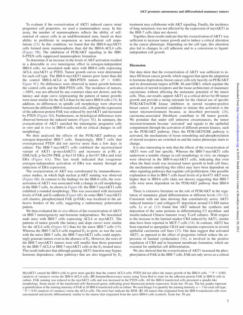
Fig. 6. Overactivation of AKT1 in IBH-7 human breast cancer cell line induces a more adhesive phenotype and an estrogen-independent tumor growth. Estrogen-dependent IBH-7 tumor cells were transfected with ACL4, myrAKT1 or PTEN and selected by neomycin. The IBH-7-PTEN cells only survived a few days inculture. (A) According to the western blotting results, the IBH-7-myrAKT1 cells exhibited total AKT, endogenous p-AKT and the myristoylated deleted variantof AKT1 (p-myrDAKT1). These cells exhibited higher levels of p-S6, p-GSK3a/b, p-Ser294 PRB, p-Ser118 ERa, p-Ser167 ERa and total ERa than did the IBH-7-ACL4 cells. The bar graphs displayed the quantification of p-S6 relative to S6, p-Ser294 PRB and ERa relative to b-actin and p-Ser118 ERa and p-Ser167 ERarelative to total ERa. n5 3 for each cell type; ��P, 0.01 (analysis of variance) versus the IBH-7-ACL4 cells. (B) Top. Photographs of the IBH-7-ACL4 and IBH-7-myrAKT1 cells in culture. The IBH-7-ACL4 cells exhibited the typical polygonal shape morphology, whereas the IBH-7-myrAKT1 cells exhibited a morerounded morphology. Immunofluorescence assays using Texas Red to stain for p-Ser473 AKT, total AKT, the adhesion proteins FAK and E-cadherin in IBH-7cells in culture. Some nuclei of transfected cells fluoresced green for green fluorescent protein. IBH-7-myrAKT1 cells displayed increased staining for p-Ser473AKT, AKT, p-FAK, FAK and E-cadherin. In some IBH-7-myrAKT1 cell clusters, p-FAK was localized to the adhesive border of the cells. Scale bar: 25 lm. (C) Atotal of 106 transfected and selected IBH-7 cells expressing ACL4 or myrAKT1 were inoculated subcutaneously into nude mice, and tumor growth curveswere determined for each cell type (n 5 6). MyrAKT1 caused the IBH-7 cells to grow more quickly than the control ACL4 cells. To grow IBH-7-ACL4 cells invivo, the cells were mixed with 100 ll of Matrigel and a subcutaneous pellet of 0.5 mg E2 was administrated 1 week before cell inoculation when indicated (þE2).The IBH-7-myrAKT1 cells generated tumors in the absence of the E2 pellet. Different letters indicate significant differences between cell types with a versus b:P , 0.05; a versus c: P , 0.001; b versus c: P , 0.01 (analysis of variance).
M.Riggio et al.
8
at SUN
Y H
ealth Science Center at B
rooklyn on January 20, 2012http://carcin.oxfordjournals.org/
Dow
nloaded from
Our results showed that the overactivation of AKT1 led to exoge-nous MPA- or estrogen-independent activation of the PR and ERathrough an increase in the total expression level of the steroid recep-tors. These findings are promising because there is extensive literatureregarding the regulation of the phosphorylation of ER by AKT andERK (42–44). According to http://www.hprd.org/PhosphoMotif_find-er, Ser167 of ERa is a consensus phosphorylation site for AKT andother seven kinases; thus, it might be a direct target of AKT in IBH-6and IBH-7 cells. Ser118 of ERa is a known consensus phosphoryla-tion site for ERK and it could be a target of other three kinases, GSK3among them. On the other hand, little is known about the regulation ofthe phosphorylation of PR (45) or steroid receptors expression (17) byprotein kinases. Miller et al. (46) showed that PTEN loss, whichoveractivates AKT, reduced ER, PRA and PRB expression but in-creased transcriptional activity of ER in MCF-7 cells. However, inT47D cells, PTEN loss reduced both expression and transcriptionalactivity of ER and PR. Such findings, even contrary to our results,support the idea that the final effect of the overactivation of the PI3K/AKT pathway is cell line specific. Thus, ligand-independent activa-tion of ER or PR through kinase activity deregulation may underliethe mechanisms that contribute to acquired HI growth and, ultimately,endocrine resistance.
Miller et al. (47) demonstrated that PI3K and mTOR inhibitionprevented the emergence of hormone-resistant breast cancer cells,suggesting that PI3K is required for the adaptation of ER-positivecells to hormone deprivation. Our findings suggest that acquiredautonomous growth may be abrogated by combination therapiestargeting both steroid receptors and protein kinases. Preclinical trialsin animal models of breast tumor progression, such as those used here,will be critical in the design of better therapies targeting PI3K/AKT/mTOR for the maximal prevention of HI cell growth. Furthermore, earlyintervention with combined endocrine and PI3K-directed therapiescould limit the resistance to antiestrogens and antiprogestins in ER/PR-positive mammary tumors and could ultimately circumvent theoccurrence of endocrine resistance when the remaining tumor cellsswitch to a complementary intracellular signal.
Supplementary material
Supplementary and Figures 1 and 2 can be found at http://carcin.oxfordjournals.org/.
Funding
Sciences and Technology Secretary (SECyT) (PICT 07 00939);National Council for Scientific and Technical Research (CONICET)(PIP 2010–2012 N� 692); SALES Foundation Argentina. The fundershad no role in study design, data collection and analysis, decision topublish or preparation of the manuscript.
Acknowledgements
We are very grateful to Pablo Do Campo and Bruno Luna for excellenttechnical assistance. We also thank Dr Richard Roth (Stanford School ofMedicine, CA) for kindly provided myristoylated AKT1 plasmid; to Dr KeithYamada (NIH, Bethesda, MD) for PTEN plasmid; to Laboratorios Craveri,Argentina for MPA and to Dr Silvio Gutkind (NIDCR/NIH) for sharingselected reagents.
Conflict of Interest Statement: None declared.
References
1.Feng,J. et al. (2004) Identification of a PKB/Akt hydrophobic motif Ser-473 kinase as DNA-dependent protein kinase. J. Biol. Chem., 279, 41189–41196.
2.Sarbassov,D.D. et al. (2005) Phosphorylation and regulation of Akt/PKBby the rictor-mTOR complex. Science, 307, 1098–1101.
3.Carracedo,A. et al. (2008) The PTEN-PI3K pathway: of feedbacks andcross-talks. Oncogene, 27, 5527–5541.
4.Wickenden,J.A. et al. (2010) Key signalling nodes in mammary glanddevelopment and cancer. Signalling downstream of PI3 kinase in mammaryepithelium: a play in 3 Akts. Breast Cancer Res., 12, 202
5.Perez-Tenorio,G. et al. (2002) Activation of AKT/PKB in breast cancerpredicts a worse outcome among endocrine treated patients. Br. J. Cancer,86, 540–545.
6.Wu,Y. et al. (2008) Clinical significance of Akt and HER2/neu overexpres-sion in African-American and Latina women with breast cancer. BreastCancer Res., 10, R3
7.Tokunaga,E. et al. (2006) The association between Akt activation andresistance to hormone therapy in metastatic breast cancer. Eur. J. Cancer,42, 629–635.
8.Beeram,M. et al. (2007) Akt-induced endocrine therapy resistance isreversed by inhibition of mTOR signaling. Ann. Oncol., 18, 1323–1328.
9.Knuefermann,C. et al. (2003) HER2/PI-3K/Akt activation leads to a multi-drug resistance in human breast adenocarcinoma cells. Oncogene, 22,3205–3212.
10.Stal,O. et al. (2003) Akt kinases in breast cancer and the results of adjuvanttherapy. Breast Cancer Res., 5, R37–R44.
11.Di Cosimo,S. et al. (2008) Targeted therapies in breast cancer: where arewe now? Eur. J. Cancer, 44, 2781–2790.
12.Alvarez,R.H. et al. (2010) Emerging targeted therapies for breast cancer.J. Clin. Oncol., 28, 3366–3379.
13.Tokunaga,E. et al. (2007) Coexistence of the loss of heterozygosity at thePTEN locus and HER2 overexpression enhances the Akt activity thusleading to a negative progesterone receptor expression in breast carcinoma.Breast Cancer Res. Treat., 101, 249–257.
14.Mills,G.B. et al. (2001) The role of genetic abnormalities of PTEN and thephosphatidylinositol 3-kinase pathway in breast and ovarian tumorigenesis,prognosis, and therapy. Semin. Oncol., 28, 125–141.
15.Dillon,R.L. et al. (2010) Distinct biological roles for the akt family inmammary tumor progression. Cancer Res., 70, 4260–4264.
16.Lanari,C. et al. (2009) The MPA mouse breast cancer model: evidence fora role of progesterone receptors in breast cancer. Endocr. Relat. Cancer, 16,333–350.
17.Polo,M.L. et al. (2010) Responsiveness to PI3K and MEK inhibitors inbreast cancer. Use of a 3D culture system to study pathways related tohormone independence in mice. PLoS One, 5, e10786
18.Vazquez,S.M. et al. (2004) Three novel hormone-responsive cell linesderived from primary human breast carcinomas: functional characterization.J. Cell. Physiol., 199, 460–469.
19.Bruzzone,A. et al. (2009) Novel human breast cancer cell lines IBH-4,IBH-6, and IBH-7 growing in nude mice. J. Cell. Physiol., 219, 477–484.
20. Institute of Laboratory Animal Resources, C.o.L.S.N.R.C. (1996) Guidefor the Care and Use of Laboratory Animals. National Academy Press,Washington, DC
21.Lanari,C. et al. (2001) Five novel hormone-responsive cell lines derivedfrom murine mammary ductal carcinomas: in vivo and in vitro effects ofestrogens and progestins. Cancer Res., 61, 293–302.
22.Lee,G.Y. et al. (2007) Three-dimensional culture models of normal andmalignant breast epithelial cells. Nat. Methods, 4, 359–365.
23.Dontu,G. et al. (2003) In vitro propagation and transcriptional profiling ofhuman mammary stem/progenitor cells. Genes Dev., 17, 1253–1270.
24.Kohn,A.D. et al. (1996) Akt, a pleckstrin homology domain containingkinase, is activated primarily by phosphorylation. J. Biol. Chem., 271,21920–21926.
25.Tamura,M. et al. (1998) Inhibition of cell migration, spreading, and focaladhesions by tumor suppressor PTEN. Science, 280, 1614–1617.
26.Soneoka,Y. et al. (1995) A transient three-plasmid expression systemfor the production of high titer retroviral vectors. Nucleic Acids Res., 23,628–633.
27.Kordon,E. et al. (1990) Hormone dependence of a mouse mammary tumorline induced in vivo by medroxyprogesterone acetate. Breast Cancer Res.Treat., 17, 33–43.
28.Giulianelli,S. et al. (2008) Carcinoma-associated fibroblasts activateprogesterone receptors and induce hormone independent mammarytumor growth: a role for the FGF-2/FGFR-2 axis. Int. J. Cancer, 123,2518–2531.
29.Soldati,R. et al. (2009) Inhibition of mammary tumor growth by estrogens:is there a specific role for estrogen receptors alpha and beta? Breast CancerRes. Treat., 123, 709–724.
30.Li,G. et al. (2002) Conditional loss of PTEN leads to precociousdevelopment and neoplasia in the mammary gland. Development, 129,4159–4170.
AKT promotes autonomous and differentiated mammary tumors
9
at SUN
Y H
ealth Science Center at B
rooklyn on January 20, 2012http://carcin.oxfordjournals.org/
Dow
nloaded from
31. Irie,H.Y. et al. (2005) Distinct roles of Akt1 and Akt2 in regulatingcell migration and epithelial-mesenchymal transition. J. Cell Biol., 171,1023–1034.
32.Maroulakou,I.G. et al. (2008) Distinct roles of the three Akt isoforms inlactogenic differentiation and involution. J. Cell. Physiol., 217, 468–477.
33.Li,X. et al. (2001) Akt/PKB regulates laminin and collagen IV isotypes ofthe basement membrane. Proc. Natl Acad. Sci. USA, 98, 14416–14421.
34.Vandermoere,F. et al. (2007) Proteomics exploration reveals that actin isa signaling target of the kinase Akt. Mol. Cell. Proteomics, 6, 114–124.
35.Fortier,A.M. et al. (2010) Akt isoforms regulate intermediate filament pro-tein levels in epithelial carcinoma cells. FEBS Lett., 584, 984–988.
36.Sartorius,C.A. et al. (2005) Progestins initiate a luminal to myoepithelialswitch in estrogen-dependent human breast tumors without altering growth.Cancer Res., 65, 9779–9788.
37.Wang,S. et al. (2011) Akt directly regulates focal adhesion kinase throughassociation and serine phosphorylation: implication for pressure-inducedcolon cancer metastasis. Am. J. Physiol. Cell Physiol., 300, C657–C670.
38. Julien,S. et al. (2007) Activation of NF-kappaB by Akt upregulates Snailexpression and induces epithelium mesenchyme transition. Oncogene, 26,7445–7456.
39.Blanco-Aparicio,C. et al. (2007) Mice expressing myrAKT1 in the mam-mary gland develop carcinogen-induced ER-positive mammary tumors thatmimic human breast cancer. Carcinogenesis, 28, 584–594.
40.Hutchinson,J.N. et al. (2004) Activation of Akt-1 (PKB-alpha) can accel-erate ErbB-2-mediated mammary tumorigenesis but suppresses tumorinvasion. Cancer Res., 64, 3171–3178.
41.Korkaya,H. et al. (2009) Regulation of mammary stem/progenitor cells byPTEN/Akt/beta-catenin signaling. PLoS Biol., 7, e1000121
42.Campbell,R.A. et al. (2001) Phosphatidylinositol 3-kinase/AKT-mediatedactivation of estrogen receptor alpha: a new model for anti-estrogenresistance. J. Biol. Chem., 276, 9817–9824.
43.Sun,M. et al. (2001) Phosphatidylinositol-3-OH Kinase (PI3K)/AKT2,activated in breast cancer, regulates and is induced by estrogen receptoralpha (ERalpha) via interaction between ERalpha and PI3K. Cancer Res.,61, 5985–5991.
44.Massarweh,S. et al. (2008) Tamoxifen resistance in breast tumors is drivenby growth factor receptor signaling with repression of classic estrogenreceptor genomic function. Cancer Res., 68, 826–833.
45.Vicent,G.P. et al. (2006) Chromatin remodeling and control of cell pro-liferation by progestins via cross talk of progesterone receptor with theestrogen receptors and kinase signaling pathways. Ann. N. Y. Acad. Sci.,1089, 59–72.
46.Miller,T.W. et al. (2009) Loss of phosphatase and tensin homologue deletedon chromosome 10 engages ErbB3 and insulin-like growth factor-I receptorsignaling to promote antiestrogen resistance in breast cancer. Cancer Res.,69, 4192–4201.
47.Miller,T.W. et al. (2010) Hyperactivation of phosphatidylinositol-3 kinasepromotes escape from hormone dependence in estrogen receptor-positivehuman breast cancer. J. Clin. Invest., 120, 2406–2413.
Received August 25, 2011; revised December 3, 2011;accepted December 11, 2011
M.Riggio et al.
10
at SUN
Y H
ealth Science Center at B
rooklyn on January 20, 2012http://carcin.oxfordjournals.org/
Dow
nloaded from