T cell homeostatic proliferation elicits effective antitumor autoimmunity
Rs
SVVa
b
c
d
e
f
I
a
ARR1A
KMPSA
med
n4
1d
Pharmacological Research 65 (2012) 275– 284
Contents lists available at SciVerse ScienceDirect
Pharmacological Research
jo ur n al hom epage: www.elsev ier .com/ locate /yphrs
osuvastatin elicits KDR-dependent vasculogenic response of human placentaltem cells through PI3K/AKT pathway
ilvia Cantonia,b,c, Claudia Cavallini a,b, Francesca Bianchia,b, Francesca Bonavitaa,b,alentina Vaccari a,b, Elena Olivia,b,c, Irene Frascari a,b, Riccardo Tassinari a,b, Sabrina Valentec,incenzo Lionettid,e,f, Carlo Venturaa,b,∗
Laboratory of Molecular Biology and Stem Cell Engineering, National Institute of Biostructures and Biosystems, Bologna, ItalyCardiovascular Department, S. Orsola – Malpighi Hospital, University of Bologna, Bologna, ItalyDepartment of Anesthesiological and Surgical Sciences, University of Bologna, ItalyLaboratory of Medical Science, Institute of Life Sciences, Scuola Superiore Sant’ Anna, Pisa, ItalyFondazione CNR-Regione Toscana “G. Monasterio”, Pisa, ItalyUnit of Molecular and Translational Medicine, Laboratory of Molecular Biology and Stem Cell Engineering, National Institute of Biostructures and Biosystems, University of Bologna,taly
r t i c l e i n f o
rticle history:eceived 4 August 2011eceived in revised form2 December 2011ccepted 12 December 2011
eywords:esenchymal stem cell
aracrine effectstatinngiogenesis
a b s t r a c t
The growth and plasticity of engrafted human mesenchymal stem cells is regulated by external stim-uli. Rosuvastatin (RSV) promotes myocardial neovascularization and limits myocardial remodeling inpatients with chronic heart failure (CHF). While these non-lipid benefits may in part depend on the acti-vation of stem cells, experimental evidence that RSV directly elicits vasculogenic differentiation of humanmesenchymal stem cells is still lacking.
We assessed whether RSV may drive a gene program of vascular commitment and the secretion oftrophic mediators with antiapoptotic, angiogenic and antifibrotic activities in human mesenchymal stemcells from full-term placentas (FMhMSCs).
With real-time RT-PCR, immunofluorescence, chemiluminescence, Western blot analysis, and in vitrovasculogenesis assays, we show that RSV enhanced expression of vascular endothelial growth factor(VEGF), kinase insert domain receptor (KDR), encoding a major VEGF receptor, hepatocyte growth factor(HGF), and platelet-derived growth factor-BB (PDGF-BB) in a time- and dose-dependent manner. GATA-4and Nkx-2.5 transcription was not affected. RSV enhanced capillary-like formation in vitro, but capillary-embedded FMhMSCs lacked endothelial marker expression, suggesting a role of pericyte-like elementsin tube formation. In HUVEC/FMhMSC cocultures, RSV increases PDGFR� expression in FMhMSCs, andenhanced capillary density and organizational efficiency, promoting a long-lasting survival of tubular
networks. RSV also activated PI3K–Akt pathway; the vasculogenic effects of the statin were abrogatedfollowing PI3K inhibition by LY294002.In conclusion, RSV-induced increase in capillary formation was dependent on VEGF and KDR. RSVpromotes the activation of paracrine signals for vascular commitment of FMhMSCs through PI3K–Aktpathway. This observation may pave the way to the use of RSV as a pharmacological enhancer of stemcell potential for cardiovascular cell therapy.
Abbreviations: RSV, rosuvastatin; CHF, chronic heart failure; FMhMSCs, humanesenchymal stem cells from fetal membranes of term placenta; VEGF, vascular
ndothelial growth factor; KDR, kinase insert domain receptor; PDGF-BB, platelet-erived growth factor-BB; vWF, von Willebrand factor.∗ Corresponding author at: Laboratory of Molecular Biology and Stem Cell Engi-eering, National Institute of Biostructures and Biosystems, Strada Maggiore 42,0125 Bologna, Italy. Tel.: +39 051340339; fax: +39 051340339.
E-mail addresses: [email protected], [email protected] (C. Ventura).
043-6618/$ – see front matter © 2011 Elsevier Ltd. All rights reserved.oi:10.1016/j.phrs.2011.12.004
© 2011 Elsevier Ltd. All rights reserved.
1. Introduction
Statins have been shown to elicit a host of non-cholesterol-lowering responses, including reduced systemic inflammation [1],enhanced angiogenesis [2], and bone marrow cell (BMC) mobiliza-tion [3]. Rosuvastatin (RSV), a relatively new molecule, preventedprogressive left ventricular remodeling and promoted neoangio-
genesis in failing heart of patients [4,5] and animal models [6].This outcome was partially ascribed to normalization of cardiaccytokine expression, including TNF-�, and increase in circulat-ing BMCs [6] and endothelial progenitor cells [7], an event that
2 gical R
mc
ttaiodfcpttpbtaipfft
m(pg
2
2
Ao
2
ridDaat1cm3ad
lft3
2
fv1T
76 S. Cantoni et al. / Pharmacolo
ay potentially account for myocardial neovascularization and res-ue.
Whether RSV may directly commit human adult stem cellsoward a cardiovascular fate and/or enhance the paracrine secre-ion of trophic mediators with angiogenic, antiapoptotic andntifibrotic properties remains to be established. Addressing thisssue may be of particular biomedical relevance. In fact, only poorr negligible myocardial rescue has been obtained so far in ran-omized clinical trials with simple stem cell transplantation inailing heart [8]. Conversely, Strauer et al. [9] demonstrated thatell therapy improves ventricular performance and prognosis inatients with chronic heart failure when stem cells were adminis-ered in addition to standard therapeutic regimes. It is conceivablehat pharmacological agents modulate the magnitude of stem cellotential. Ex vivo preconditioning with synthetic molecules har-oring differentiating and paracrine “logics” remarkably enhancedhe cardiovascular commitment in both mouse embryonic [10]nd human stem cells [11,12], improving their rescuing propertiesn vivo in animal models of heart failure [11,13,14]. These findingsrompt further studies to assess whether drugs already availableor conventional cardiovascular therapy (i.e. RSV) may also be usedor stem cell preconditioning to enhance the potential for a cellherapy.
Here, we investigated whether exposure to RSV of humanesenchymal stem cells from fetal membranes of term placenta
FMhMSCs) may activate the transcription of vascular lineage-romoting genes, and whether RSV may drive the secretion ofrowth factors enhancing vasculogenesis in vitro.
. Methods
.1. Rosuvastatin preparation
Rosuvastatin (RSV) Ca2+ – salt was kindly provided bystraZeneca and it was dissolved in DMSO at the final concentrationf 50 mM.
.2. Cell isolation
Term placenta obtained from caesarian sections were rapidlyinsed in PBS containing penicillin and streptomycin and usedmmediately. Pieces from fetal membranes were minced andigested for 10 min in DMEM with 0.25% trypsin–EDTA, 10 U/mlNaseI and 0.1% collagenase. Tissues were pipetted vigorously upnd down avoiding foam for 5 min; larger pieces of tissue werellowed to settle under gravity for 5 min. Each supernatant wasransferred to a fresh tube, neutralized with FBS, then spun at000 × g for 10 min. Each pellet was resuspended in 5 ml of DMEMontaining 20% FBS, 10 U/ml penicillin and 100 �g/ml strepto-ycin. FMhMSCs were seeded into culture flasks and grown at
7 ◦C in 5% CO2. Non-adherent cells were removed after 1 weeknd medium (with 10% FBS) was changed subsequently every 4ays.
Human umbilical vascular endothelial cells (HUVECs) were iso-ated in our laboratory with the method described by Davis [15]rom human umbilical cord obtained during elective caesarian sec-ions. The cells collected were grown in EGM-2 medium (Lonza) at7 ◦C in 5% CO2.
.3. Flow cytometry analysis (FACS) and cellular phenotyping
To assess the mesenchymal origin of fibroblast-like cells isolated
rom fetal membranes we performed FACS analysis. Cells were har-ested by treatment with 0.08% trypsin–EDTA and incubated with�g/106 cells conjugated antibodies for 40 min at 4 ◦C in the dark.he antibodies used were: anti-CD105-Alexa Fluor 488 (Chemicon),
esearch 65 (2012) 275– 284
anti-CD73-PE (BD), anti-CD29-PE-Cy5 (BD), anti-CD90-R-PE (BD),anti-CD166-R-PE (BD), anti-CD14-APC (BD), anti-CD34-FITC (BD),anti-CD44-FITC (BD), and anti-CD45-Per-CP (BD). After washing,cells were analyzed on a flow cytometer (FACSCalibur, Becton Dick-inson, San Jose, CA, USA) by collecting 10,000 events and the dataanalyzed using the Cell Quest Software (Becton Dickinson).
Adipogenic (Chemicon), osteogenic (Chemicon) and chon-drogenic (Lonza) differentiations were performed to test themultipotency of FMhMSCs, following the manufacturer’s instruc-tions.
For HUVEC characterization, anti-CD31 (Santa Cruz) was usedas primary antibody and fluorescent conjugated goat anti-mouse(MP Biomedicals) as secondary antibody. Cells were analyzed on aflow cytometer.
2.4. Cell treatment and viability
Drug sensitivity was estimated by the MTT method, essentiallyas described by the manufacturer’s instructions (SIGMA). FMhMSCs(7500 cells/cm2) were seeded into 24-well plates and then treatedwith different concentrations of RSV (0.1–75 �M) for 1, 2, 3 and 6days. Untreated cells and DMSO treated cells were used as control.Data were collected by reading at 570 nm with a multi-well platereader (Dynex Technology).
Cell viability was determined by the trypan blue dye exclusiontest. The cells were treated with RSV (0.1–75 �M). After 1, 2, 3 and6 days, both attached and floating cells were harvested and theywere counted by using a hemocytometer.
To assess whether RSV, in range between 0.1 and 10 �M, belowthe toxic concentration, may coax cells into a “cardiovascular deci-sion”, we performed comparative analyses of the expression oftargeted cardiogenic and angiogenic genes and immunocytochem-ical assessment of lineage-restricted markers, as detailed below.
2.5. Gene expression
Real-time RT-PCR was used to assess the time- and dose-dependent effect of RSV on the gene expression of vascularendothelial growth factor (VEGF), KDR, hepatocyte growth factor(HGF), GATA-4, Nkx-2.5 and Akt. Primer sequences were: humanGAPDH reverse: 5′ TGTGGTCATGAGTCCTTCCA 3′, forward: 5′ CAGC-CTCAAGATCATCAGCA 3′; human VEGF reverse: 5′ ACACAGGATG-GCTTGAAGATG 3′, forward: 5′ AGAAGGAGGAGGGCAGAATC 3′;human HGF reverse: 5′ ACTCCAGGGCTGACATTTGAT 3′, forward: 5′
ATTTGGCCATGAATTTGACCT 3′; human KDR reverse: 5′ GAGCTCTG-GCTACTGGTGATG 3′, forward: 5′ CTGCAAATTTGGAAACCTGTC 3′;human GATA-4 reverse: 5′ TAGCCTTGTGGGGAGAGCTT 3′, forward:5′ TGGCCTGTCATCTCACTACG 3′; human Nkx-2.5 reverse: 5′ GCG-CACAGCTCTTTCTTTTC 3′, forward: 5′ CAAGTGTGCGTCTGCCTTT 3′;human Akt: reverse: 5′ CTTAATGTGCCCGTCCTTGT 3′, forward: 5′
TCTATGGCGCTGAGATTGTG 3′.Data were normalized using GAPDH as an index of cDNA content
after reverse transcription. Amplification included initial denatura-tion at 95 ◦C for 10 min, 50 cycles of denaturation at 95 ◦C for 10 s,annealing at 59–63 ◦C for 6–10 s, and extension at 72 ◦C for 10 s,performed at a temperature transition rate of 20 ◦C/s. Fluorescencewas measured at the end of each extension step. Specificity of theproduct was determined by a melting curve analysis, conductedafter completion of the cycling process with the aid of a tempera-ture ramp (from 55 to 95 ◦C at 0.1 ◦C/s) and continuous fluorescencemonitoring.
Samples were run in duplicate, and the average threshold cycle
(Ct) value was used for calculations. Relative quantification ofmRNA expression was calculated with the comparative Ct methodusing the “Delta–delta method” for comparing relative expressionresults between treatments in Real-time RT-PCR [16].
S. Cantoni et al. / Pharmacological Research 65 (2012) 275– 284 277
Fig. 1. Treatment of FMhMSCs with RSV and MTT proliferation assay. (A) FMhMSCs (7500 cells/cm2) were seeded into 75 cm2 flasks and treated after 24 h with differentconcentrations of RSV (0.1–1–10 �M) for 1, 3 and 6 days (100×, original magnification). (B) FMhMSCs (7500 cells/cm2) were seeded into 24-well plates and then treated withd ed by rD trl); u
2
VBtp1
bB
ifferent concentrations of RSV (0.1–75 �M) for 1, 2, 3 and 6 days. Data were collectunnett’s Multiple Comparison Test, *significantly different from untreated cells (C
.6. Effect of rosuvastatin on growth factor production
We assessed whether RSV (0.1–10 �M) acts on the secretion ofEGF, HGF and platelet-derived growth factor-BB (PDGF-BB) usingio-Plex Pro Human Cytokine 3-Plex assay from Bio-Rad, followinghe manufacturer’s instructions. Cell culture supernatants and totalrotein content were collected at different times of treatment (16 h,
day, 3 and 6 days).PDGF-BB production in HUVECs cocultured with FMhMSCs has
een evaluated using Bio-Plex Pro Human Cytokine Plex assay fromio-Rad, following the manufacturer’s instructions. FMhMSCs and
eading at 570 nm with a multi-well plate reader (n = 4). Statistical analysis: ANOVAnpaired t-test, #significantly different from 5 to 10 �M treated cells; P < 0.05.
HUVECs have been cocultured using cell culture insert with 0.4 �mpore diameter (BD). Total proteins were extracted 8 and 24 h afterthe treatment.
All the samples were assayed at least in duplicate and normal-ized with total protein content.
2.7. In vitro vasculogenesis
Analysis of capillary-like tube formation was performedusing Cultrex® Basement Membrane Extract (BME, Thema). Cellswere seeded in gel-precoated wells and cultured in the absence or
278 S. Cantoni et al. / Pharmacological Research 65 (2012) 275– 284
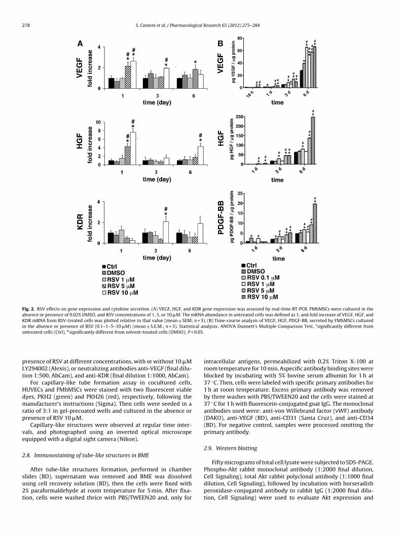
Fig. 2. RSV effects on gene expression and cytokine secretion. (A) VEGF, HGF, and KDR gene expression was assessed by real-time RT-PCR. FMhMSCs were cultured in theabsence or presence of 0.02% DMSO, and RSV concentrations of 1, 5, or 10 �M. The mRNA abundance in untreated cells was defined as 1, and fold increase of VEGF, HGF, andKDR mRNA from RSV-treated cells was plotted relative to that value (mean ± SEM; n = 3). (B) Time-course analysis of VEGF, HGF, PDGF-BB, secreted by FMhMSCs culturedin the absence or presence of RSV (0.1–1–5–10 �M) (mean ± S.E.M.; n = 3). Statistical analysis: ANOVA Dunnett’s Multiple Comparison Test, *significantly different fromu #
0.05.
pLt
Hdmrp
ve
2
su2t
ntreated cells (Ctrl), significantly different from solvent-treated cells (DMSO); P <
resence of RSV at different concentrations, with or without 10 �MY294002 (Alexis), or neutralizing antibodies anti-VEGF (final dilu-ion 1:500, AbCam), and anti-KDR (final dilution 1:1000, AbCam).
For capillary-like tube formation assay in cocultured cells,UVECs and FMhMSCs were stained with two fluorescent viableyes, PKH2 (green) and PKH26 (red), respectively, following theanufacturer’s instructions (Sigma). Then cells were seeded in a
atio of 3:1 in gel-precoated wells and cultured in the absence orresence of RSV 10 �M.
Capillary-like structures were observed at regular time inter-als, and photographed using an inverted optical microscopequipped with a digital sight camera (Nikon).
.8. Immunostaining of tube-like structures in BME
After tube-like structures formation, performed in chamber
lides (BD), supernatant was removed and BME was dissolvedsing cell recovery solution (BD), then the cells were fixed with% paraformaldehyde at room temperature for 5 min. After fixa-ion, cells were washed thrice with PBS/TWEEN20 and, only forintracellular antigens, permeabilized with 0.2% Triton X-100 atroom temperature for 10 min. Aspecific antibody binding sites wereblocked by incubating with 5% bovine serum albumin for 1 h at37 ◦C. Then, cells were labeled with specific primary antibodies for1 h at room temperature. Excess primary antibody was removedby three washes with PBS/TWEEN20 and the cells were stained at37 ◦C for 1 h with fluorescein-conjugated goat IgG. The monoclonalantibodies used were: anti-von Willebrand factor (vWF) antibody(DAKO), anti-VEGF (BD), anti-CD31 (Santa Cruz), and anti-CD34(BD). For negative control, samples were processed omitting theprimary antibody.
2.9. Western blotting
Fifty micrograms of total cell lysate were subjected to SDS-PAGE.Phospho-Akt rabbit monoclonal antibody (1:2000 final dilution,
Cell Signaling), total Akt rabbit polyclonal antibody (1:1000 finaldilution, Cell Signaling), followed by incubation with horseradishperoxidase-conjugated antibody to rabbit IgG (1:2000 final dilu-tion, Cell Signaling) were used to evaluate Akt expression and
S. Cantoni et al. / Pharmacological Research 65 (2012) 275– 284 279
Fig. 3. RSV promotes FMhMSC in vitro vasculogenesis through a VEGF-dependent mechanism. (A) Tube-like formation was assessed in semisolid medium in FMhMSCs culturedfor 24 h in the absence or presence of 10 �M RSV, and in cells exposed to anti-VEGF or anti-KDR (final dilution 1:500 and 1:1000, respectively) neutralizing antibodies in theabsence or presence of 10 �M RSV. All images are representative of three experiments with similar results (40×, original magnification). (B) Quantification and morphologicalassessment of tube-like structures in semisolid medium (A) were performed by NIS-Elements D Nikon software (version 3.06). The parameters branches per field, the totalc = 3).
( d cellst s VEG
aHHpt
dfa
bu
2
vda
apillary length, and the average capillary length were expressed as mean ± S.E.M. (nreduction) from RSV treatment; §significantly different (reduction) from untreatereated cells were stained with anti-VEGF specific antibody. RSV treatment increase
ctivation. The expression PDGFR� in FMhMSCs cocultured withUVECs has been evaluated using Western blotting. FMhMSCs andUVECs have been cocultured using cell culture inserts with 0.4 �more diameter (BD). Total proteins were extracted 8 and 24 h afterhe treatment.
PDGFR� (1:1000 final dilution, AbCam) and tubulin (1:2000 finalilution, Cell Signaling) rabbit polyclonal antibodies were used,ollowed by incubation with horseradish peroxidase-conjugatedntibody to rabbit IgG (Cell Signaling).
Antigen–antibody complexes were visualized by ECL Westernlotting detection reagents (GE Healthcare), according to the man-facturer’s instructions.
.10. Statistical analysis
The statistical analysis was performed using GraphPad Prismer. 5. Data were evaluated by using a two-tailed, unpaired Stu-ent’s t-test, and ANOVA Dunnett’s Multiple Comparison Test,ssuming a P value less than 0.05 as the limit of significance.
*Significantly different (increase) from untreated cells (Ctr); #significantly different (Ctr); P < 0.05. (C) After tube-like structures formation, untreated and RSV 10 �MF production during vasculogenesis (100×, original magnification).
3. Results
3.1. Cell treatment
Nontoxic effect was observed following FMhMSC exposure toRSV ranging from 0.1 to 10 �M throughout 6 days of treatment(Fig. 1).
3.2. Effect of rosuvastatin on gene activation and growth factorsecretion
RSV increased the gene expression of VEGF, HGF, and KDR,encoding a major VEGF receptor in a time- and dose-dependentfashion (Fig. 2A). The stimulatory effect peaked at a concentrationof 10 �M, reaching a maximum after 1-day exposure for both VEGFand HGF transcription, and after 3-day treatment in the case of
KDR. RSV did not affect the gene expression of GATA-4 and Nkx-2.5, two cardiac lineage promoting genes, nor did it change Akttranscription (data not shown). RSV time- and dose-dependentlyincreased PDGF-BB and HGF release in the culture medium (Fig. 2B).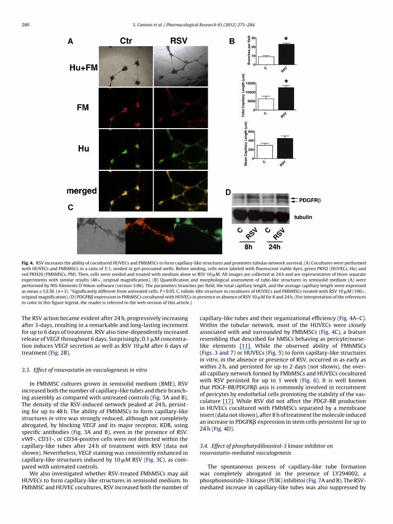
280 S. Cantoni et al. / Pharmacological Research 65 (2012) 275– 284
Fig. 4. RSV increases the ability of cocultured HUVECs and FMhMSCs to form capillary-like structures and promotes tubular network survival. (A) Cocultures were performedwith HUVECs and FMhMSCs in a ratio of 3:1, seeded in gel-precoated wells. Before seeding, cells were labeled with fluorescent viable dyes, green PKH2 (HUVECs, Hu) andred PKH26 (FMhMSCs, FM). Then, cells were seeded and treated with medium alone or RSV 10 �M. All images are collected at 24 h and are representative of three separateexperiments with similar results (40×, original magnification). (B) Quantification and morphological assessment of tube-like structures in semisolid medium (A) wereperformed by NIS-Elements D Nikon software (version 3.06). The parameters branches per field, the total capillary length, and the average capillary length were expresseda ule-liko s in pt .)
Tafrtt
3
iiTisasvcscp
HF
s mean ± S.E.M. (n = 3). *Significantly different from untreated cells. P < 0.05. C, tubriginal magnification). (D) PDGFR� expression in FMhMSCs cocultured with HUVECo color in this figure legend, the reader is referred to the web version of this article
he RSV action became evident after 24 h, progressively increasingfter 3-days, resulting in a remarkable and long-lasting incrementor up to 6 days of treatment. RSV also time-dependently increasedelease of VEGF throughout 6 days. Surprisingly, 0.1 �M concentra-ion induces VEGF secretion as well as RSV 10 �M after 6 days ofreatment (Fig. 2B).
.3. Effect of rosuvastatin on vasculogenesis in vitro
In FMhMSC cultures grown in semisolid medium (BME), RSVncreased both the number of capillary-like tubes and their branch-ng assembly as compared with untreated controls (Fig. 3A and B).he density of the RSV-induced network peaked at 24 h, persist-ng for up to 48 h. The ability of FMhMSCs to form capillary-liketructures in vitro was strongly reduced, although not completelybrogated, by blocking VEGF and its major receptor, KDR, usingpecific antibodies (Fig. 3A and B), even in the presence of RSV.WF-, CD31-, or CD34-positive cells were not detected within theapillary-like tubes after 24 h of treatment with RSV (data nothown). Nevertheless, VEGF staining was consistently enhanced inapillary-like structures induced by 10 �M RSV (Fig. 3C), as com-
ared with untreated controls.We also investigated whether RSV-treated FMhMSCs may aidUVECs to form capillary-like structures in semisolid medium. InMhMSC and HUVEC cocultures, RSV increased both the number of
e structure in cocultures of HUVECs and FMhMSCs treated with RSV 10 �M (100×,resence or absence of RSV 10 �M for 8 and 24 h. (For interpretation of the references
capillary-like tubes and their organizational efficiency (Fig. 4A–C).Within the tubular network, most of the HUVECs were closelyassociated with and surrounded by FMhMSCs (Fig. 4C), a featureresembling that described for hMSCs behaving as pericyte/nurse-like elements [11]. While the observed ability of FMhMSCs(Figs. 3 and 7) or HUVECs (Fig. 5) to form capillary-like structuresin vitro, in the absence or presence of RSV, occurred in as early aswithin 2 h, and persisted for up to 2 days (not shown), the over-all capillary network formed by FMhMSCs and HUVECs coculturedwith RSV persisted for up to 1 week (Fig. 6). It is well knownthat PDGF-BB/PDGFR� axis is commonly involved in recruitmentof pericytes by endothelial cells promoting the stability of the vas-culature [17]. While RSV did not affect the PDGF-BB productionin HUVECs cocultured with FMhMSCs separated by a membraneinsert (data not shown), after 8 h of treatment the molecule inducedan increase in PDGFR� expression in stem cells persistent for up to24 h (Fig. 4D).
3.4. Effect of phosphatydilinositol-3 kinase inhibitor onrosuvastatin-mediated vasculogenesis
The spontaneous process of capillary-like tube formationwas completely abrogated in the presence of LY294002, aphosphoinositide-3 kinase (PI3K) inhibitor (Fig. 7A and B). The RSV-mediated increase in capillary-like tubes was also suppressed by

S. Cantoni et al. / Pharmacological Research 65 (2012) 275– 284 281
Fig. 5. RSV increases the ability of HUVECs to form capillary-like structures. (A–D) Before seeding, HUVECs were labeled with fluorescent viable dye green PKH2. Then,cells were seeded and treated with medium alone (A and B) or RSV 10 �M (C and D). Phase contrast (A and C) and fluorescent images (B and D) are representative of threeexperiments with similar results at 24 h (40×, original magnification). (E) Quantification and morphological assessment of tube-like structures in semisolid medium werep es pem differi
tpip
4
vitafhwghdawcssoc
erformed by NIS-Elements D Nikon software (version 3.06). The parameters branchean ± S.E.M. (n = 3). *Significantly different from untreated cells (C), #significantly
n this figure legend, the reader is referred to the web version of this article.)
he same inhibitor (Fig. 7A and B). RSV rapidly primed Akt phos-horylation within 30 min of treatment. Inhibition of PI3K, which
s placed upstream to Akt signaling, suppressed RSV-mediated Akthosphorylation (Fig. 7C).
. Discussion
We provide evidence that RSV may behave as an effective acti-ator of stem cell-mediated angiogenic potential. The RSV-inducedncrease in VEGF and HGF gene expression and release is notewor-hy, since both factors modulate pro-angiogenic, pro-survival andntifibrotic responses in vitro and in vivo [11,18–21]. These solubleactors are involved in limiting the ischemic remodeling in infarctedearts in a paracrine manner [22]. However, it is still debatedhether common drugs may strictly modulate the paracrine angio-
enic response in a receptor-dependent manner. VEGF binds withigh affinity to tyrosine kinase receptors Flt-1 and KDR, activatingistinct signal transduction pathways [23]. Among them, KDR plays
major role in the VEGF mediated angiogenesis [24]. Thus, a defectithin the VEGF–KDR system could result in impaired physiologic
oronary angiogenesis in ischemic myocardium [25]. Previous
tudies suggested that the activation of autocrine VEGF loop inelected microenvironment contributes to optimal differentiationf stem cells in a receptor-dependent manner [26] and rescues stemell survival [27]. Therefore, it is conceivable that the RSV exposurer field, the total capillary length, and the average capillary length were expressed asent from treated cells (RSV); P < 0.05. (For interpretation of the references to color
of FMhMSCs may change the microenvironment and increase theKDR transcription, which primes a paracrine/autocrine circuitryenhancing capillary-like formation during stem cell differentiation.Thus, KDR activation should be the main target of statin-elicitedangiogenic response. This intriguing hypothesis is supported byour findings: (i) under RSV treatment, cells embedded withincapillary tubes continued to express VEGF, and (ii) a neutralizingVEGF antibody significantly reduced RSV-induced capillarogen-esis. However, VEGF antagonism failed to completely abrogatethe paracrine angiogenic response by stem cells, suggesting theinvolvement of other factors. Our findings in part support previousstudies showing that KDR is activated in a VEGF-independent fash-ion by other soluble factors (e.g. cytokines) [28,29] and strengthenthe needs of further dissecting the complexity of signals formingfunctionally mature vessels from stem cells. In fact, it is wellknown that VEGF mainly primes endothelial differentiation, but itis still not well defined which mechanisms underline the stem cell-derived mature vasculogenesis. In our study, persistence of VEGFexpression within stem cells in the absence of endothelial markershighlights the notion that VEGF is an early signal and FMhMSCsmay have been committed only in part along an endothelial fateby VEGF, rather acting as pericyte-like elements supporting tube
formation, growth and functional maturity. This is further inferredby the observation that: (i) RSV also increased capillary-like den-sity in cocultures of HUVECs and FMhMSCs, (ii) stem cells closely
282 S. Cantoni et al. / Pharmacological Research 65 (2012) 275– 284
Fig. 6. The capillary network formed by FMhMSCs and HUVECs cocultured with RSV persisted up to one week. (A) Cocultures were performed with HUVECs and FMhMSCsin a ratio of 3:1, seeded in gel-precoated wells and treated with medium alone or RSV 10 �M. Before seeding, cells were labeled with fluorescent viable dyes, green PKH2(HUVECs, HU) and red PKH26 (FMhMSCs, FM). All images are representative of three experiments with similar results at 7 days (40×, original magnification). (B) Quantificationand morphological assessment of tube-like structures in semisolid medium were performed by NIS-Elements D Nikon software (version 3.06). The parameters branchesp as mi to the
attetidp
ssl[a
wdf
tp
er field, the total capillary length, and the average capillary length were expressednterpretation of the references to color in this figure legend, the reader is referred
ssociated with and gathered around HUVECs along the assembledubes, and (iii) the tubular network formed by RSV-exposed cocul-ures survived significantly longer and with higher organizationalfficiency than that elicited by statin-treated HUVECs. To this end,he ability of RSV to enhance stem cell expression of PDGFR�n HUVEC/FMhMSC cocultures may be worthy of consideration,ue to the major role of PDGFBB/PDGFR� axis in supportingericyte/endothelial cell interplay and vasculature stability [17].
Recent study showed that activation of the PI3K/Akt-dependentignal pathway is involved in vasculogenesis of mouse embryonictem cells [30]. Moreover, statins promote the activity of endothe-ial nitric oxide synthase, which is a hallmark of mature vasculature31], through a PI3K/Akt-dependent pathway in endothelial cells in
dose-dependent manner [32].We showed in vitro that the RSV-dependent angiogenesis
as directly accompanied by the activation of a PI3K/Akt-ependent signal pathway in FMhMSCs in a time-dependent
ashion.FMhMSC exposure to RSV resulted in Akt phosphoryla-ion, an event associated with signaling networks ensuringroper vessel growth and assembly [2]. Recent studies showed
ean ± S.E.M. (n = 3). *Significantly different from untreated cells (Ctr); P < 0.05. (For web version of this article.)
that Akt phosphorylation is augmented during the endothelialdifferentiation [33] and its inhibition significantly impairs theendothelial differentiation of mesenchymal stem cells [34]. It iswell established that PI3K is the critical activator of Akt duringangiogenesis in both physiological and pathological conditions[35], and this pathway is well preserved in some progenitorcells [36]. Previous study demonstrated that HMG-CoA reduc-tase inhibitors increase the number of differentiated endothelialprogenitor cells (EPCs), which is dose-dependently limited by PI3K-inhibition [37]. This study assessed that statins can increase thenumber of EPCs and improve angiogenic response through mod-ulation of PI3K/Akt pathway, yet it was not clear whether thePI3K/Akt activation following statin exposure was critical for angio-genic response also in the absence of VEGF mediation. Moreover,the previous studies did not investigate the role of PI3K/Akt path-way during statin exposure of more undifferentiated cells such asFMhMSCs.
In our investigation, the block of spontaneous FMhMSC-derivedvasculogenesis by a PI3K inhibitor demonstrates that Akt is aconserved player in the vasculogenic potential of human mes-enchymal stem cells. The finding that inhibition of PI3K, an

S. Cantoni et al. / Pharmacological Research 65 (2012) 275– 284 283
Fig. 7. RSV stimulates Akt phosphorylation in FMhMSCs. (A) Tube-like formation was assessed in FMhMSCs cultured in semisolid medium after 2 h of treatment withmedium alone, 10 �M LY294002 (LY), 10 �M RSV, 10 �M LY294002 (LY) + 10 �M RSV (40×, original magnification). (B) Quantification and morphological assessment oftube-like structures in semisolid medium were performed by NIS-Elements D Nikon software (version 3.06). The parameters branches per field, the total capillary length,a antly§ orylai times
uAaaaa
rmtKtcKti
C
nd the average capillary length were expressed as mean ± S.E.M. (n = 3). *Significsignificantly different (reduction) from untreated cells (C); P < 0.05. (C) Akt phosphn the absence or presence of 10 �M RSV or 10 �M LY294002 (LY), for the indicated
pstream Akt activator, abrogated both rosuvastatin-inducedkt patterning and in vitro capillarogenesis, indicates that thebility of statin to enhance vascular commitment in FMhMSCs islso related to PI3K-dependent activation of Akt. Further studiesre on the way to deeply dissect the mechanistic bases of RSVction.
In conclusion, the results of the present study demonstrate thatosuvastatin can modulate in vitro the vasculogenesis by humanesenchymal stem cells also in a VEGF-independent manner. In
his way, the PI3K/Akt pathway appears to play a major role forDR-induced increase in angiogenic response following exposure
o statin. The augmentation of vasculogenic response by pharma-ological modulation of the signaling pathways, which regulateDR-activation during FMhMSCs differentiation, may be a novel
herapeutic concept to improve neovascularization in a VEGF-ndependent manner.
onflict of interest
The authors declare no potential conflicts of interest.
different from untreated cells (C); #significantly different (reduction) from RSV;tion was assessed by immunoblot analysis in cell extracts from FMhMSCs cultured. All images are representative of three experiments with similar results.
Acknowledgments
This work was supported by AstraZeneca (C.V.), and partly by:Regione Emilia Romagna, Programma di Ricerca Regione – Uni-versità 2007/2009, Area 1b “Medicina rigenerativa”, Italy (C.V.);Ministero della Salute, Italy, Programma per la Ricerca Sanitaria:attività di Ricerca sulle Cellule Staminali (C.V.); Fondazione LuisaFanti-Melloni, Bologna, Italy (C.V.); Fondazione Cardinale GiacomoLercaro, Bologna, Italy; Fondazione Fornasini, Poggio Renatico,Italy; Tavola Valdese, Rome, Italy (C.V).
References
[1] Liao JK. Effects of statins on 3-hydroxy-3-methylglutaryl coenzyme a reduc-tase inhibition beyond lowdensity lipoprotein cholesterol. Am J Cardiol2005;96:24F–33F.
[2] Kureishi Y, Luo Z, Shiojima I, Bialik A, Fulton D, Lefer DJ, et al. The HMG-CoAreductase inhibitor simvastatin activates the protein kinase Akt and promotesangiogenesis in normocholesterolemic animals. Nat Med 2000;6:1004–10.
[3] Llevadot J, Murasawa S, Kureishi Y, Uchida S, Masuda H, Kawamoto A, et al.HMG-CoA reductase inhibitor mobilizes bone marrow-derived endothelialprogenitor cells. J Clin Invest 2001;108:399–405.
[4] Erbs S, Beck EB, Linke A, Adams V, Gielen S, Kränkel N, et al. High-dose rosu-vastatin in chronic heart failure promotes vasculogenesis, corrects endothelial

2 gical R
[
[
[
[
[
[[
[
[
[
[
[
[
[
[
[
[
[
[
[
[
[
[
[
[
[
[
tional effects on endothelial progenitor cell biology: focus on PI3K/AKT/eNOSpathway. Int J Cardiol 2010;144(October (3)):350–66.
[37] Dimmeler S, Aicher A, Vasa M, Mildner-Rihm C, Adler K, Tiemann M, et al. HMG-
84 S. Cantoni et al. / Pharmacolo
function, and improves cardiac remodeling – results from a randomized,double-blind, and placebo-controlled study. Int J Cardiol 2011;146(January(1)):56–63.
[5] Cleland JG, McMurray JJ, Kjekshus J, Cornel JH, Dunselman P, Fonseca C,et al. Plasma concentration of amino-terminal pro-brain natriuretic peptidein chronic heart failure: prediction of cardiovascular events and interactionwith the effects of rosuvastatin: a report from CORONA (Controlled Rosuvas-tatin Multinational Trial in Heart Failure). J Am Coll Cardiol 2009;54(November(20)):1850–9.
[6] Zacà V, Rastogi S, Imai M, Wang M, Sharov VG, Jiang A, et al. Chronic monother-apy with rosuvastatin prevents progressive left ventricular dysfunction andremodeling in dogs with heart failure. J Am Coll Cardiol 2007;50:551–7.
[7] Tousoulis D, Andreou I, Tsiatas M, Miliou A, Tentolouris C, Siasos G, et al. Effectsof rosuvastatin and allopurinol on circulating endothelial progenitor cells inpatients with congestive heart failure: the impact of inflammatory process andoxidative stress. Atherosclerosis 2011;214(January (1)):151–7.
[8] Diederichsen AC, Møller JE, Thayssen P, Junker AB, Videbaek L, Saekmose SG,et al. Effect of repeated intracoronary injection of bone marrow cells in patientswith ischaemic heart failure the Danish stem cell study – congestive heartfailure trial (DanCell-CHF). Eur J Heart Fail 2008;10(July (7)):661–7.
[9] Strauer BE, Yousef M, Schannwell CM. The acute and long-term effects of intra-coronary stem cell transplantation in 191 patients with chronic heARt failure:the STAR-heart study. Eur J Heart Fail 2010;12(July (7)):721–9.
10] Ventura C, Maioli M, Asara Y, Santoni D, Scarlata I, Cantoni S, et al. Butyricand retinoic mixed ester of hyaluronan. A novel differentiating glycoconjugateaffording a high throughput of cardiogenesis in embryonic stem cells. J BiolChem 2004;279:23574–9.
11] Ventura C, Cantoni S, Bianchi F, Lionetti V, Cavallini C, Scarlata I, et al. Hyaluro-nan mixed esters of butyric and retinoic acid drive cardiac and endothelial fatein term placenta human mesenchymal stem cells and enhance cardiac repairin infarcted rat hearts. J Biol Chem 2007;282:14243–52.
12] Shinmura D, Togashi I, Miyoshi S, Nishiyama N, Hida N, Tsuji H, et al. Pretreat-ment of human mesenchymal stem cells with pioglitazone improved efficiencyof cardiomyogenic transdifferentiation and improved cardiac function. StemCells 2011;29(2):357–66.
13] Simioniuc A, Campan M, Lionetti V, Marinelli M, Aquaro GD, Cavallini C, et al.Placental stem cells pre-treated with a hyaluronan mixed ester of butyric andretinoic acid to cure infarcted pig hearts: a multimodal study. Cardiovasc Res2011;90(3):546–56.
14] Zemani F, Silvestre JS, Fauvel-Lafeve F, Bruel A, Vilar J, Bieche I, et al. Exvivo priming of endothelial progenitor cells with SDF-1 before transplanta-tion could increase their proangiogenic potential. Arterioscler Thromb VascBiol 2008;28(April (4)):644–50.
15] Crampton SP, Davis J, Hughes CC. J Vis Exp 2007;(3):183.16] Pfaffl MW. A new mathematical model for relative quantification in real time
PCR. Nucleic Acids Res 2001;29:e45.17] Stratman AN, Schwindt AE, Malotte KM, Davis GE. Endothelial-derived PDGF-
BB and HB-EGF coordinately regulate pericyte recruitment during vasculogenictube assembly and stabilization. Blood 2010;116(22):4720–30.
18] Zentilin L, Puligadda U, Lionetti V, Zacchigna S, Collesi C, Pattarini L, et al.VEGFR-1 activation by VEGF-B induces compensatory hypertrophy and pre-serves cardiac function after myocardial infarction. FASEB J 2010;24(May(5)):1467–78.
19] Siltanen A, Kitabayashi K, Lakkisto P, Mäkelä J, Pätilä T, Ono M, et al. hHGFoverexpression in myoblast sheets enhances their angiogenic potential in rat
chronic heart failure. PLoS One 2011;6(April (4)):e19161.20] Rastogi S, Guerrero M, Wang M, Ilsar I, Sabbah MS, Gupta RC, et al. Myocardialtransfection with naked DNA plasmid encoding hepatocyte growth factorprevents the progression of heart failure in dogs. Am J Physiol Heart CircPhysiol 2011;300(4):H1501–9.
esearch 65 (2012) 275– 284
21] Kinnaird T, Stabile E, Burnett MS, Lee CW, Barr S, Fuchs S, et al. Marrow-derived stromal cells express genes encoding a broad spectrum of arteriogeniccytokines and promote in vitro and in vivo arteriogenesis through paracrinemechanisms. Circ Res 2004;94:678–85.
22] Lionetti V, Bianchi G, Recchia FA, Ventura C. Control of autocrine and paracrinemyocardial signals: an emerging therapeutic strategy in heart failure. HeartFail Rev 2010;15(November (6)):531–42.
23] Waltenberger J, Claesson-Welsh L, Siegbahn A, Shibuya M, Heldin C. Differ-ent signal transduction properties of KDR and Flt1, two receptors for vascularendothelial growth factor. J Biol Chem 1994;269:26988–95.
24] Ferrara N. Role of vascular endothelial growth factor in regulation of physio-logical angiogenesis. Am J Physiol Cell Physiol 2001;280:C1358–66.
25] Maruyama K, Mori Y, Murasawa S, Masaki H, Takahashi N, Tsutusmi Y, et al.Interleukin-1 beta upregulates cardiac expression of vascular endothelialgrowth factor and its receptor KDR/flk-1 via activation of protein tyrosinekinases. J Mol Cell Cardiol 1999;31(March (3)):607–17.
26] Casella I, Feccia T, Chelucci C, Samoggia P, Castelli G, Guerriero R, et al.Autocrine-paracrine VEGF loops potentiate the maturation of megakaryocyticprecursors through Flt1 receptor. Blood 2003;101(February (4)):1316–23.
27] Gerber HP, Malik AK, Solar GP, Sherman D, Liang XH, Meng G, et al. VEGFregulates haematopoietic stem cell survival by an internal autocrine loop mech-anism. Nature 2002;417(Jun (6892)):954–8.
28] Petreaca ML, Yao M, Liu Y, Defea K, Martins-Green M. Transactivation ofvascular endothelial growth factor receptor-2 by interleukin-8 (IL-8/CXCL8)is required for IL-8/CXCL8-induced endothelial permeability. Mol Biol Cell2007;18(December (12)):5014–23.
29] Yao JS, Zhai W, Young WL, Yang GY. Interleukin-6 triggers human cerebralendothelial cells proliferation and migration: the role for KDR and MMP-9.Biochem Biophys Res Commun 2006;342(April (4)):1396–404.
30] Bekhite MM, Finkensieper A, Binas S, Müller J, Wetzker R, Figulla HR, et al.VEGF-mediated PI3K class IA and PKC signaling in cardiomyogenesis andvasculogenesis of mouse embryonic stem cells. J Cell Sci 2011;124(June (Pt11)):1819–30.
31] Benest AV, Stone OA, Miller WH, Glover CP, Uney JB, Baker AH, et al. Arte-riolar genesis and angiogenesis induced by endothelial nitric oxide synthaseoverexpression results in a mature vasculature. Arterioscler Thromb Vasc Biol2008;28(August (8)):1462–8.
32] Wang J, Tokoro T, Matsui K, Higa S, Kitajima I. Pitavastatin at low dose activatesendothelial nitric oxide synthase through PI3K–AKT pathway in endothelialcells. Life Sci 2005;76(March (19)):2257–68.
33] Ando H, Nakanishi K, Shibata M, Hasegawa K, Yao K, Miyaji H. Benidipine, adihydropyridine-Ca2+ channel blocker, increases the endothelial differentia-tion of endothelial progenitor cells in vitro. Hypertens Res 2006;29(December(12)):1047–54.
34] Chu L, Hao H, Luo M, Huang Y, Chen Z, Lu T, et al. Ox-LDL modifies the behaviorof bone marrow stem cells and impairs their endothelial differentiation viainhibition of Akt phosphorylation. J Cell Mol Med 2009;(October) [Epub aheadof print].
35] Aoyagi T, Matsui T. Phosphoinositide-3 kinase signaling in cardiac hypertrophyand heart failure. Curr Pharm Des 2011;(June) [Epub ahead of print].
36] Everaert BR, Van Craenenbroeck EM, Hoymans VY, Haine SE, Van Nassauw L,Conraads VM, et al. Current perspective of pathophysiological and interven-
CoA reductase inhibitors (statins) increase endothelial progenitor cells via thePI 3-kinase/Akt pathway. J Clin Invest 2001;108(August (3)):391–7.
Copyright © 2022 FDOKUMEN