Steroid Receptor Regulation of Epidermal Growth Factor Signaling through Src in Breast and Prostate...
10
2005;65:10585-10593. Cancer Res Antimo Migliaccio, Marina Di Domenico, Gabriella Castoria, et al. Steroid Antagonist Action Signaling through Src in Breast and Prostate Cancer Cells: Steroid Receptor Regulation of Epidermal Growth Factor Updated version http://cancerres.aacrjournals.org/content/65/22/10585 Access the most recent version of this article at: Cited Articles http://cancerres.aacrjournals.org/content/65/22/10585.full.html#ref-list-1 This article cites by 28 articles, 12 of which you can access for free at: Citing articles http://cancerres.aacrjournals.org/content/65/22/10585.full.html#related-urls This article has been cited by 14 HighWire-hosted articles. Access the articles at: E-mail alerts related to this article or journal. Sign up to receive free email-alerts Subscriptions Reprints and . [email protected] Department at To order reprints of this article or to subscribe to the journal, contact the AACR Publications Permissions . [email protected] Department at To request permission to re-use all or part of this article, contact the AACR Publications Research. on October 13, 2014. © 2005 American Association for Cancer cancerres.aacrjournals.org Downloaded from Research. on October 13, 2014. © 2005 American Association for Cancer cancerres.aacrjournals.org Downloaded from
Transcript of Steroid Receptor Regulation of Epidermal Growth Factor Signaling through Src in Breast and Prostate...

2005;65:10585-10593. Cancer Res Antimo Migliaccio, Marina Di Domenico, Gabriella Castoria, et al. Steroid Antagonist ActionSignaling through Src in Breast and Prostate Cancer Cells: Steroid Receptor Regulation of Epidermal Growth Factor
Updated version
http://cancerres.aacrjournals.org/content/65/22/10585
Access the most recent version of this article at:
Cited Articles
http://cancerres.aacrjournals.org/content/65/22/10585.full.html#ref-list-1
This article cites by 28 articles, 12 of which you can access for free at:
Citing articles
http://cancerres.aacrjournals.org/content/65/22/10585.full.html#related-urls
This article has been cited by 14 HighWire-hosted articles. Access the articles at:
E-mail alerts related to this article or journal.Sign up to receive free email-alerts
Subscriptions
Reprints and
To order reprints of this article or to subscribe to the journal, contact the AACR Publications
Permissions
To request permission to re-use all or part of this article, contact the AACR Publications
Research. on October 13, 2014. © 2005 American Association for Cancercancerres.aacrjournals.org Downloaded from
Research. on October 13, 2014. © 2005 American Association for Cancercancerres.aacrjournals.org Downloaded from
Steroid Receptor Regulation of Epidermal Growth Factor
Signaling through Src in Breast and Prostate
Cancer Cells: Steroid Antagonist Action
Antimo Migliaccio, Marina Di Domenico, Gabriella Castoria, Merlin Nanayakkara,Maria Lombardi, Antonietta de Falco, Antonio Bilancio, Lilian Varricchio,Alessandra Ciociola, and Ferdinando Auricchio
Dipartimento di Patologia Generale, Facolta di Medicina e Chirurgia, II Universita di Napoli, Naples, Italy
Abstract
Under conditions of short-term hormone deprivation, epider-mal growth factor (EGF) induces DNA synthesis, cytoskeletalchanges, and Src activation in MCF-7 and LNCaP cells. Theseeffects are drastically inhibited by pure estradiol or androgenantagonists, implicating a role of the steroid receptors inthese findings. Interestingly, EGF triggers rapid associationof Src with androgen receptor (AR) and estradiol receptorA (ERA) in MCF-7 cells or ERB in LNCaP cells. Here, we showthat, through EGF receptor (EGFR) and erb-B2, EGF inducestyrosine phosphorylation of ER preassociated with AR,thereby triggering the assembly of ER/AR with Src andEGFR. Remarkably, experiments in Cos cells show that thiscomplex stimulates EGF-triggered EGFR tyrosine phosphory-lation. In turn, estradiol and androgen antagonists, throughthe Src-associated receptors, prevent Src activation by EGFand heavily reduce EGFR tyrosine phosphorylation and thesubsequent multiple effects, including DNA synthesis andcytoskeletal changes in MCF-7 cells. In addition, knockdown ofERa or AR gene by small interfering RNA (siRNA) almostabolishes EGFR tyrosine phosphorylation and DNA synthesisin EGF-treated MCF-7 cells. The present findings reveal thatsteroid receptors have a key role in EGF signaling. EGFRtyrosine phosphorylation, depending on Src, is a part of thismechanism. Understanding of EGF-triggered growth andinvasiveness of mammary and prostate cancer cells expressingsteroid receptors is enhanced by this report, which revealsnovel aspects of steroid receptor action. (Cancer Res 2005;65(22): 10585-93)
Introduction
Growth factors and steroid receptors control breast cancerprogression. In the absence of estradiol, epidermal growth factor(EGF) activity converges on the estradiol receptor (ER) in humanmammary cancer-derived cells, as well as in uterus, therebytriggering DNA synthesis and cell proliferation. EGF can alsoactivate genes regulated by estrogen-responsive elements (1–3). Inaddition, growth factors directly activate androgen receptor (AR)in androgen-deprived prostate cancer cells (4). An increase in
uterine weight and proliferation of the uterine epithelial cellsfollows EGF or insulin-like growth factor-I (IGF-I) treatment ofovariectomized mice. Interestingly, these effects are not observedin ERa knockout mice, indicating that ERa is required in thiscase (3).Tyrosine kinases are involved in human breast cancer develop-
ment and EGF receptor (EGFR) is amplified in 10% to 35% ofhuman breast carcinomas. This event is correlated with a poordisease prognosis (5). Elevated Src activity has been found in breasttumor specimens and cell lines (6). Moreover, both EGFR and Srcare overexpressed in a subset of human breast tumors and a wealthof evidence indicates physical and functional associations betweenEGFR and Src (reviewed in ref. 7). Expression of a dominant-negative form of Src in murine fibroblasts interferes with EGF-induced mitogenesis and cytoskeletal changes (8). In addition, Srchas been implicated in the regulation of EGFR endocytosis (9).Therefore, Src seems to be a component of the signaling path-way elicited by EGF.MCF-7 and LNCaP cells are hormone-responsive cells derived
from human mammary and prostate cancers, respectively. Theyexpress AR and either ERa (MCF-7) or ERh (LNCaP) and are widelyused to analyze the effect of sex-steroid hormones and the cross-talk between growth factors and steroid hormones. We previouslyreported that in MCF-7 and LNCaP cells, ER and AR, once activatedby steroid hormones, stimulate a mitogenic signaling networkknown to be engaged by growth factors (10, 11). In this report, weattempted to shed light on the cross-talk between EGFR andsteroid receptors and to determine the effect of the steroidantagonists on mammary and prostate cancer cells in the absenceof steroid hormones. We now observe that in the two humancancer-derived cell lines, under conditions of short-term hormonedeprivation, specific steroid antagonists suppress EGF-inducedDNA synthesis, cytoskeletal changes, and Src activation. Ouranalysis of the effect of these antagonists revealed unexpectedfeatures in the cross-talk between EGFR and steroid receptors. EGFinduced ER/AR/Src complex in MCF-7 and LNCaP cells. A smallpercentage of the total AR, ERa, or ERh was found to be associatedin these cells under basal conditions. These preassociatedreceptors, together with ER tyrosine phosphorylation elicited byEGF, play a major role in the heterodimer steroid receptorinteraction with Src and the regulation of Src-dependent action.We suggest that once Src-associated receptors are occupied bysteroid antagonists, they interfere with the EGF-induced activeAR/ER/Src complex, thereby silencing their effects on Src. One ofthese, and probably the most unexpected, is EGFR tyrosinephosphorylation. Remarkably, ERa and AR knockdown by smallinterfering RNA (siRNA) heavily reduces EGFR tyrosine phosphor-ylation as well as DNA synthesis stimulated by EGF in MCF-7 cells.
Note: A. Migliaccio and M. Di Domenico contributed equally to this work.Requests for reprints: Ferdinando Auricchio, Dipartimento di Patologia Generale,
Facolta di Medicina e Chirurgia, II Universita di Napoli, Via L. De Crecchio, 7,80138 Naples, Italy. Phone: 39-081-5665676; Fax: 39-081-291327; E-mail: [email protected].
I2005 American Association for Cancer Research.doi:10.1158/0008-5472.CAN-05-0912
www.aacrjournals.org 10585 Cancer Res 2005; 65: (22). November 15, 2005
Research Article
Research. on October 13, 2014. © 2005 American Association for Cancercancerres.aacrjournals.org Downloaded from
The role of the steroid receptors in EGF signaling is furtherstressed by experiments in Cos cells showing that EGFR tyrosinephosphorylation triggered by EGF is strongly up-regulated bytransient expression of ERa and AR, provided that the AR/ER/Srccomplex can be assembled.
Materials and Methods
Constructs and small interfering RNA. cDNAs encoding hAR, wild-
type hER (HEGO), HEG241, and the point-mutated (HEG537F) forms ofhERa were cloned into pSG5 expression vector as described previously
(11–14). The wild-type and the kinase-inactive form of Src (Lys259 changed
to methionine) were cloned into pSG5 as reported (15). Smart pools ofdouble-stranded siRNA against AR as well as nonspecific siRNAs were
obtained from Dharmacon Tech (Lafayette, CO). The stealth RNA
interference (RNAi) against ERa and the stealth RNAi-negative control
were from Invitrogen (Carslbad, CA). The negative control siRNA AlexaFluor 488 was from Qiagen (Valencia, CA). Specific and nonspecific siRNA
were used according to the instructions of the manufacturer.
Cell culture and transfection techniques. Human mammary cancer-
derived MCF-7 and MDA-MB231 cells were grown and made quiescent asreported elsewhere (16). Human prostate cancer-derived LNCaP and Cos
cells were grown and made quiescent as previously reported (10, 11). When
indicated, Cos cells were made quiescent by serum starvation (17).Quiescent cells were transfected by Superfect (Qiagen) using 2 Ag of
purified plasmids. Twenty-four hours later, transfected cells were left
unstimulated or stimulated with the indicated compounds. For the siRNA
studies, MCF-7 cells at 70% confluence were transfected in completemedium with LipofectAMINE 2000 Reagent (Invitrogen) according to the
instructions of the manufacturer. RNAi against ERa was transfected for
48 hours, and siRNA against AR was transfected for 24 hours. Cells
were then made quiescent and left unstimulated or stimulated with theindicated compounds.
DNA synthesis analysis and cytoskeletal changes. For BrdUrd
incorporation analysis, quiescent cells on coverslips were left unstimulated
or stimulated for 24 hours with the indicated compounds. After a 6-hourpulse with 100 Amol/L BrdUrd (Sigma, St. Louis, MO), BrdUrd incorporation
was analyzed as described (16) and nuclei were stained with Hoechst 33258
(Sigma). For the siRNA studies, the cells were cotransfected with negativecontrol siRNA Alexa Fluor 488 (at 15 nmol/L) to help identify transfected
cells and BrdUrd incorporation was analyzed as reported (16) using Alexa
Fluor 594–conjugated mouse monoclonal anti-BrdUrd antibody (Molecular
Probes, Eugene, OR). For cytoskeletal changes, cells on coverslips weremade quiescent by serum starvation (0.1% serum) for 16 hours. They were
then left unstimulated or stimulated for 20 minutes with the indicated
compounds. F-actin was visualized using either Texas red or FITC-labeled
phalloidin as reported (17). Finally, coverslips were mounted in Mowiol(Calbiochem, Darmstadt, Germany). Images were generated with a DMLB
fluorescent microscope (Leica, Wetzlar, Germany) equipped with �40 and�100 lens and processed with IM1000 software (Leica).
Immunoprecipitation, kinase, and Ras assays. Lysates were prepared
as described previously (10) and protein concentration was measured with a
Bio-Rad protein assay kit (Bio-Rad, Hercules, CA). Equal amounts of cell
lysates (2 mg protein/mL) were used for immunoprecipitation and kinaseassay of Src and extracellular signal-regulated kinase-2 (Erk-2; ref. 10).
ERa was immunoprecipitated using the anti-ER rabbit polyclonal antibody
(HC-20, Santa Cruz, Santa Cruz, CA) and AR was immunoprecipitated using
the anti-AR rabbit polyclonal antibody (N-20, Santa Cruz). EGFR wasimmunoprecipitated using the anti-EGFR rabbit polyclonal antibodies
(UBI, Charlottesville, VA). The Ras activation assay kit (UBI) was used for
Raf-RBD pullout.Protein-protein interaction assay. Recombinant proteins were pro-
duced as previously described (11). Coupled in vitro transcription/
translation reactions were used to produce 35S-labeled proteins in rabbit
reticulocyte lysates (Promega, Madison, VA) and protein-protein interactionassays were done according to the same report (11). Eluted proteins were
resolved by SDS-PAGE and protein bands were revealed by fluorography.
Electrophoresis and immunoblotting. The electrophoresis andimmunoblotting procedures were done as described elsewhere (10). Src
was revealed using the mouse monoclonal anti-Src antibody (clone 327;
Calbiochem), EGFR was visualized using anti-EGF receptor rabbit
polyclonal antibody (UBI), and Ras was detected using anti-pan Rasantibody (UBI). P-tyrosine proteins were detected using the mouse
monoclonal antiphosphotyrosine antibody (clone 2G4; UBI). The rabbit
polyclonal anti-AR antibodies (either C-19 or N-20, Santa Cruz) were used
to reveal AR. ERa was immunoblotted using the rabbit polyclonal anti-ER(HC-20, Santa Cruz) antibody. Modulator of nongenomic activity of estrogen
receptor (MNAR) was detected using rabbit polyclonal anti-PELP1/MNAR
antibody (Novus Biologicals, Littleton, CO). Immunoreactive proteins
were revealed with the ECL detection system (Amersham Biosciences,Bucks, United Kingdom).
Results
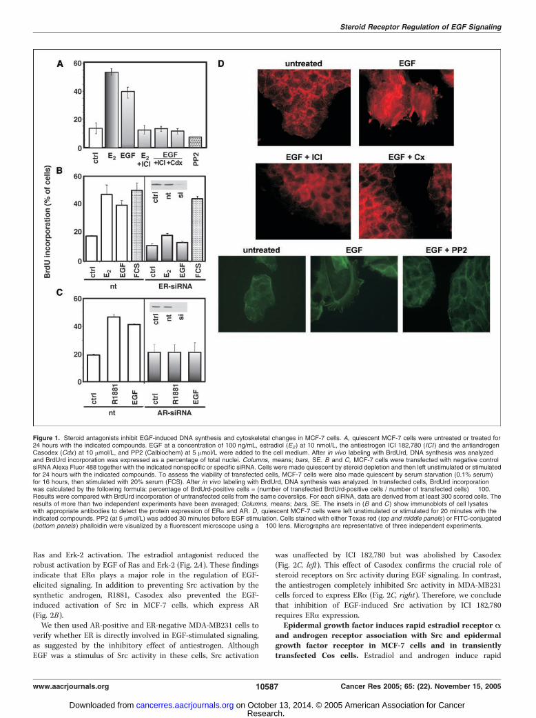
Epidermal growth factor–stimulated DNA synthesis andcytoskeletal changes in MCF-7 cells are regulated by steroidreceptors and Src. EGF (100 ng/mL) stimulated the S-phase entryof MCF-7 cells maintained in phenol red–free medium supple-mented with charcoal-treated serum (Fig. 1A). This response wassimilar in intensity to that of the same cells treated with 10 nmol/Lestradiol (Fig. 1A). In accordance with a previous report (1), theeffect of EGF was abolished by 10 Amol/L ICI 182,780, a pureantiestrogen. The same antagonist showed a similar effect at a100-fold lower concentration (not shown). Because estradiol andandrogen stimulate cross-talk between ERa and AR in MCF-7 cells(11), we verified the effect of a pure antiandrogen, Casodex, on theeffect of EGF. Interestingly, Casodex also (10 Amol/L) abolished thegrowth hormone effect (Fig. 1A). This antagonist also inhibitedthe G1-S transition at a concentration of 0.1 Amol/L (not shown).Effects similar to those of the two steroid antagonists wereobserved in the presence of the Src kinase family inhibitor, PP2(Fig. 1A), as well as in cells transiently transfected with siRNAsilencing ERa or AR (Fig. 1B and C).Altogether, these findings indicate that steroid receptors and Src
play a key role in EGF-triggered DNA synthesis.EGF evokes dramatic morphologic changes and stress fiber
breakdown in a number of cell types (18). Here, we show that EGFat a concentration of 100 ng/mL rapidly induces fan-likemembrane protrusions and ruffles in MCF-7 cells. Also, in thiscase, both steroid antagonists prevented EGF-induced cytoskeletalchanges (Fig. 1D). Src activity is also involved in these responses asindicated by the PP2 inhibitory effect on EGF-induced cytoskeletalchanges (Fig. 1D). Findings in Fig. 1 show that the two steroidreceptors and Src have a key role in the EGF-elicited effects inMCF-7 cells.Epidermal growth factor–triggered Src activation in MCF-7
cells is inhibited by steroid antagonists. The effect of EGFon DNA synthesis and membrane ruffling is inhibited by steroidantagonists and Src inhibitor, PP2. Src family tyrosine kinases areinvolved in signaling of different growth factor receptors includingEGFR. They can promote initiation of signaling pathways requiredfor DNA synthesis and actin cytoskeleton rearrangements (19).Therefore, we evaluated the effect of 10 Amol/L ICI 182,780 onEGF-stimulated Src activity. The activity was measured as enolasephosphorylation by the immunoprecipitated Src. We used controlantibodies to verify that Src immunoprecipitation was specificand that the samples contained equal amounts of Src (Fig. 2A).EGF activated Src, whereas ICI 182,780 prevented this activation.Because Src acts upstream of Ras and the Ras-dependent kinasecascade, we evaluated the effect of ICI 182,780 on EGF-induced
Cancer Research
Cancer Res 2005; 65: (22). November 15, 2005 10586 www.aacrjournals.org
Research. on October 13, 2014. © 2005 American Association for Cancercancerres.aacrjournals.org Downloaded from
Ras and Erk-2 activation. The estradiol antagonist reduced therobust activation by EGF of Ras and Erk-2 (Fig. 2A). These findingsindicate that ERa plays a major role in the regulation of EGF-elicited signaling. In addition to preventing Src activation by thesynthetic androgen, R1881, Casodex also prevented the EGF-induced activation of Src in MCF-7 cells, which express AR(Fig. 2B).We then used AR-positive and ER-negative MDA-MB231 cells to
verify whether ER is directly involved in EGF-stimulated signaling,as suggested by the inhibitory effect of antiestrogen. AlthoughEGF was a stimulus of Src activity in these cells, Src activation
was unaffected by ICI 182,780 but was abolished by Casodex(Fig. 2C, left). This effect of Casodex confirms the crucial role ofsteroid receptors on Src activity during EGF signaling. In contrast,the antiestrogen completely inhibited Src activity in MDA-MB231cells forced to express ERa (Fig. 2C, right). Therefore, we concludethat inhibition of EGF-induced Src activation by ICI 182,780requires ERa expression.Epidermal growth factor induces rapid estradiol receptor A
and androgen receptor association with Src and epidermalgrowth factor receptor in MCF-7 cells and in transientlytransfected Cos cells. Estradiol and androgen induce rapid
Figure 1. Steroid antagonists inhibit EGF-induced DNA synthesis and cytoskeletal changes in MCF-7 cells. A, quiescent MCF-7 cells were untreated or treated for24 hours with the indicated compounds. EGF at a concentration of 100 ng/mL, estradiol (E2) at 10 nmol/L, the antiestrogen ICI 182,780 (ICI ) and the antiandrogenCasodex (Cdx ) at 10 Amol/L, and PP2 (Calbiochem) at 5 Amol/L were added to the cell medium. After in vivo labeling with BrdUrd, DNA synthesis was analyzedand BrdUrd incorporation was expressed as a percentage of total nuclei. Columns, means; bars, SE. B and C, MCF-7 cells were transfected with negative controlsiRNA Alexa Fluor 488 together with the indicated nonspecific or specific siRNA. Cells were made quiescent by steroid depletion and then left unstimulated or stimulatedfor 24 hours with the indicated compounds. To assess the viability of transfected cells, MCF-7 cells were also made quiescent by serum starvation (0.1% serum)for 16 hours, then stimulated with 20% serum (FCS). After in vivo labeling with BrdUrd, DNA synthesis was analyzed. In transfected cells, BrdUrd incorporationwas calculated by the following formula: percentage of BrdUrd-positive cells = (number of transfected BrdUrd-positive cells / number of transfected cells) � 100.Results were compared with BrdUrd incorporation of untransfected cells from the same coverslips. For each siRNA, data are derived from at least 300 scored cells. Theresults of more than two independent experiments have been averaged; Columns, means; bars, SE. The insets in (B and C) show immunoblots of cell lysateswith appropriate antibodies to detect the protein expression of ERa and AR. D, quiescent MCF-7 cells were left unstimulated or stimulated for 20 minutes with theindicated compounds. PP2 (at 5 Amol/L) was added 30 minutes before EGF stimulation. Cells stained with either Texas red (top and middle panels ) or FITC-conjugated(bottom panels ) phalloidin were visualized by a fluorescent microscope using a �100 lens. Micrographs are representative of three independent experiments.
Steroid Receptor Regulation of EGF Signaling
www.aacrjournals.org 10587 Cancer Res 2005; 65: (22). November 15, 2005
Research. on October 13, 2014. © 2005 American Association for Cancercancerres.aacrjournals.org Downloaded from

association of Src with AR and ERh or ERa in LNCaP and MCF-7cells, respectively (11). Phosphotyrosine 537 of ERa directlyinteracts with the Src-SH2 domain and is required for ER/Srcassociation (11). Here, we show that, simultaneous to Srcactivation, EGF induces or increases the association of ERa andAR with Src in MCF-7 cells (Fig. 2D and 3C , respectively).We next investigated the role of EGFR-tyrosine kinase and
erb-B2 in Src/ERa/AR association. A selective inhibitor of EGFR-tyrosine kinase, Iressa (ZD 1839), blocked EGF-elicited Src activity(Fig. 2D). It also prevented the association of Src with ER and AR(Fig. 2D and 3C , respectively). Preincubation of MCF-7 cells withthe anti-erb-B2 antibody herceptin (Trastuzumab) produced thesame effects [i.e., inhibition of Src activity (Fig. 2D) and reductionof association of Src with ERa and AR (Fig. 2D and 3C ,respectively)]. The growth factor induced tyrosine phosphorylationof ERa (Fig. 3A and B).The parallel inhibition of the steroid receptor/Src complex
and ER tyrosine phosphorylation by Iressa (Fig. 3A) and herceptin(Fig. 3B) suggests that these two events are causally related. Toaddress this point, we transiently transfected Cos cells with hARand hERa (HEG0) cDNAs. The HEG537 cDNA mutant, lacking theonly phosphorylatable tyrosine (20), was also expressed instead
of wild-type ERa (Fig. 3D, left). Cells were then stimulated withEGF and lysates were immunoprecipitated with anti-Src antibody.Immunoblotting revealed that the samples contained equalamounts of Src (Fig. 3D, right, top). As shown in Fig. 3D (right),EGF was a potent inducer of the Src/AR/ERa complex in cellsexpressing wild-type ERa. The association with Src of the mutantwas very weak and that of AR was absent, which shows thatERa tyrosine phosphorylation in position 537 is required for thecoexpressed receptors/Src complex assembly.When MCF-7 cells were stimulated by EGF for 5 minutes,
association of the EGFR with ER, AR, Src, and MNAR wasdetectable. It has been reported that MNAR modulates theestradiol-dependent ER interaction with Src (21). Its associationin the EGFR/ER/AR/Src complex triggered by EGF supports theview that this protein interacts with activated ER. In the absence ofthe growth factor, no association of the EGFR with these proteinswas observed (Fig. 4A, left).In addition, in Cos cells transiently expressing hERa or its
mutant HE241G, EGF induces association of its own receptor notonly with the wild-type hERa but also with the HEG241 mutant(Fig. 4A, right). Because the HE241G mutant does not bind DNA,lacks the nuclear localization signal (14), and permanently resides
Figure 2. Inhibition by steroid antagonists and Iressa (ZD 1839) or herceptin (Trastuzumab) of EGF-induced Src activation in MCF-7 cells. ERa requirement forthe antiestrogen effect. A and B, quiescent MCF-7 cells were left unstimulated or stimulated for 5 minutes with the indicated compounds. The antiestrogen ICI 182,780and the antiandrogen Casodex were used at a concentration of 10 Amol/L, EGF at 100 ng/mL, and R1881 at 10 nmol/L. Lysate proteins were immunoprecipitatedwith either anti-Src (anti-src ab) or control (Ctrl ab ) antibodies. The immunoprecipitates were either blotted with anti-Src antibody (top panels in A and B ) or analyzedfor Src activity using enolase (en ) as a substrate (top panels in A and B ). A, middle, Ras pulldown assay of MCF-7 cell lysates. A, bottom, Erk-2 activity assaydone using myelin basic protein (MBP ) as a substrate. C, quiescent MDA-MB231 cells were left unstimulated or stimulated for 5 minutes with the indicated compounds.Src activity was assayed in untransfected (MDA ) or hERa-transfected (MDA-HEG0 ) cells. D, MCF-7 cells, pretreated with 0.5 Amol/L Iressa (ZD ) or 10 Ag herceptin(Her ), were stimulated with EGF for 5 minutes. Lysate proteins were immunoprecipitated with either control or anti-Src antibodies. Immunocomplexes were eitherblotted with antibodies against the indicated proteins (Src or ERa) or submitted to Src kinase assay.
Cancer Research
Cancer Res 2005; 65: (22). November 15, 2005 10588 www.aacrjournals.org
Research. on October 13, 2014. © 2005 American Association for Cancercancerres.aacrjournals.org Downloaded from

outside the nucleus of MCF-7 cells,1 ERa associated with EGFRmust be in the extranuclear compartment of cells. This finding is inagreementwith the involvement in the EGF-triggered EGFR/ER/AR/Src complex of Src, which is a classic cytoplasmic protein, and therole of steroid receptors in the EGF-triggered cytoskeletal changes,which are expressed independently of the cell nucleus (22).Androgen receptor and estradiol receptor A are associated
in MCF-7 cells. An association between AR and ERa but not Srcwas observed in unstimulated MCF-7 cells (not shown). To evaluatethe proportion of AR and ERa in the complex, we coimmunopre-cipitated the receptors from MCF cell lysates with anti-ERa oranti-AR antibodies. Supernatants were collected for furtheranalysis, and the immunocomplexes were eluted. Lysates, break-throughs, and immunocomplexes were probed by Western blotswith anti-AR or anti-ERa antibodies (Fig. 4B). The band intensitieswere measured and the percentage of associated proteins wascalculated as described in Fig. 4 caption. About 8% of the total ARand ERa were associated. Interestingly, similar results wereobserved with LNCaP cells using immunoprecipitating anti-ERhinstead of anti-ERa antibodies (not shown).We then conducted pulldown experiments with glutathione
S-transferase (GST) fusion protein constructs to determine whetherthe association between the two receptors was direct (Fig. 4C). GST-agarose, GST-HEG0 (HEG0 is the entire hERa), GST-HEG14 (HEG14is the carboxyl-terminal half of hERa containing the hormonebinding domain), and GST-HEG15 (HEG15 is the amino-terminalhalf of hERa) were incubated with the [35S]methionine-labeled hAR.Association of AR with GST-HEG14 was comparable with the low,nonspecific interaction of ARwith GST-agarose, whereas there was amuch stronger association with the entire receptor (GST-HEG0) andthe amino-terminal half of ERa, GST-HEG15. These experimentsreveal a direct interaction between the ERa amino-terminal half andAR in unstimulated cells.Epidermal growth factor–stimulated epidermal growth
factor receptor tyrosine phosphorylation in MCF-7 cells isregulated by steroid receptors and Src. The foregoing experi-ments show that EGFR regulates ligand-independent steroidreceptors and Src activity. We conducted experiments to determinewhether steroid receptors in turn regulate the EGFR. To thisend, quiescent MCF-7 cells were stimulated or not with EGFalone or in the presence of 10 Amol/L ICI 182,780 or 10 Amol/LCasodex. Cell lysates were then immunoprecipitated with anti-EGFR antibody and the immunocomplexes probed with thesame antibody or with antiphosphotyrosine antibodies (Fig. 5A).As expected, Western blot analysis showed that EGF stimulatedphosphorylation of its own receptor. Remarkably, EGFR phospho-rylation was strongly reduced when the cells were stimulated bythe growth factor in the presence of antihormones. This findingsuggests a novel, steroid-independent regulatory role of steroidreceptors on EGFR. This conclusion is further confirmed by thestrong inhibitory effect on EGFR phosphorylation detected afterthe knockdown of ERa or AR gene in MCF-7 cells (Fig. 5B).We then conducted experiments to verify whether Src was
involved in EGFR phosphorylation in MCF-7 cells. In the experi-ment shown in Fig. 5C , cells were transfected with the wild-typeSrc or with a kinase-inactive Src and then stimulated with EGF.The growth factor induced rapid EGFR tyrosine phosphorylationin cells expressing wild-type Src. This phosphorylation was much
Figure 3. Role of phosphorylation on ERa tyrosine 537 in the EGF-elicitedER/AR/Src association in MCF-7 and Cos cells. A to D, quiescent MCF-7 cellswere untreated or treated with EGF (100 ng/mL) for 5 minutes in the absenceor presence of the indicated compounds. Iressa and herceptin were used at0.5 Amol/L and 10 Ag/mL, respectively. The antiestrogen was used at 10 Amol/L.A and B, lysates were immunoprecipitated with either anti-ER (anti-ER ab) orcontrol antibodies. C, lysates were immunoprecipitated with either anti-Src orcontrol antibodies. Immunoprecipitated proteins were blotted with the antibodiesagainst the indicated proteins or antiphosphotyrosine (pY ) antibodies.D, Cos cells were transfected with the indicated plasmids and then leftunstimulated or stimulated for 5 minutes with 100 ng/mL EGF. Lysates wereimmunoprecipitated with anti-Src antibodies and immunocomplexes wereblotted with antibodies against the indicated proteins (right). Left, expressionof steroid receptors evaluated by immunoblots with appropriate antibodies.HEG0 is the hERa, 537 is the hERa carrying the Y537F mutation, and ARis the hAR.1 In preparation.
Steroid Receptor Regulation of EGF Signaling
www.aacrjournals.org 10589 Cancer Res 2005; 65: (22). November 15, 2005
Research. on October 13, 2014. © 2005 American Association for Cancercancerres.aacrjournals.org Downloaded from

weaker in cells expressing kinase-inactive Src. All together, theseresults are consistent with the view that Src kinase activity plays akey role in the EGF-dependent EGFR phosphorylation in MCF-7cells and suggest that this activity is under the control of the Src-associated steroid receptors. This possibility is strongly corrobo-rated by the experiments in Fig. 5D . Cos cells were transientlycotransfected with hAR (AR) and the wild-type hERa (HEG0) or itsmutant HEG537 (537), which lacks the property of interacting withSrc in Cos cells stimulated by EGF (see Fig. 3D). EGF stronglystimulated EGFR tyrosine phosphorylation in Cos cells expressingHEGO, whereas a much weaker stimulation was detected in cellsexpressing HEG537. This shows that the ER/AR/Src complex hasa key role in EGFR tyrosine phosphorylation. Remarkably, com-parison of EGFR tyrosine phosphorylation in cells transfected with
the empty vector or ERa- and AR-expressing plasmids shows thatin the presence of the two steroid receptors a much stronger EGFRphosphorylation is triggered by EGF (Fig. 5D).Epidermal growth factor–elicited effects are inhibited by
steroid antagonists in LNCaP cells. In LNCaP cells, like in MCF-7cells, EGF induced DNA synthesis. This response was similar tothat evoked in the same cells by the synthetic androgen R1881.Antiandrogen and antiestrogen abolished this stimulation (Fig. 6A).The antagonists also prevented EGF-induced cytoskeleton changes(Fig. 6B) and the growth factor–stimulated Src activation (Fig. 6C).EGF induced association of ERh and AR with Src. This associationwas inhibited by steroid antagonists (Fig. 6C). These findingsslightly indicate that mammary cancer cells and prostate cancercells are similarly affected by EGF and steroid antagonists.
Discussion
Steroid receptors can be activated in the absence of theircognate ligands. Dopamine indeed causes nuclear translocationof the chicken progesterone receptor (23). EGF enhances nuclearlocalization of ERa, induces a nuclear form of ERa, and stimulatesDNA synthesis in mouse uterus (2). Remarkably, such a stimulationrequires the expression of ERa (3). Heregulin induces tyrosinephosphorylation of ERa in MCF-7 cells, interaction of ERa withestrogen responsive element, and progesterone receptor induction(24). EGF stimulates serine and tyrosine phosphorylation of ERaand its coimmunoprecipitation with EGFR (25). Androgen receptoris transcriptionally activated in a ligand-independent manner byIGF-I, EGF, and keratinocyte growth factor in human prostatetumor cell lines (26). HER-2/neu activates the AR pathway in theabsence of ligand and increases AR responses in the presence oflow levels of androgen in prostate cancer cells (4). From these andmany other reports, it seems that cross-talk between growthfactors and steroid receptors, which occurs at multiple levels, isinvolved in cancer progression and in endocrine resistance (27, 28).Here, we report evidence of a novel interplay between the
EGF-activated EGFR, its partner erb-B2, and steroidal receptors. Inthis regard, it is noteworthy that an EGFR/erb-B2 heterocomplex hasbeen identified (29) and an association between erb-B2 and Src hasbeen detected in human breast cancer cell lines (see references inref. 7). The results concerning EGFR tyrosine phosphorylationwarrant particular attention. In MCF-7 cells, the EGF-stimulatedphosphorylation of EGFR was greatly reduced by either ER and ARantagonists or knockdown by siRNA of each of the two steroid recep-tors. The role of the two steroid receptors in this phosphorylation isfurther underlined by the observation that in Cos cells, EGFR tyrosinephosphorylation is greatly increased by expression of ERa and AR.Present findings suggest that in MCF-7 and LNCaP cells steroid
receptors regulate EGFR receptor phosphorylation through Src.Such a hypothesis is supported by the EGF-triggered association ofEGFR with ERa, AR, and Src. In addition, this phosphorylation isalmost abolished in Cos cells by expression of the ERa mutantHEG537, which lacks the ability of interaction with Src. Altogether,the present findings indicate that the ERa/AR/Src association isresponsible for the Src-dependent EGFR tyrosine phosphorylation.As a part of the described regulatory mechanism, EGF treatmentof hormone-responsive cells led to ER tyrosine phosphorylationand triggered strong association of Src with AR and ERa or ERhin MCF-7 and LNCaP cells, respectively. The crucial role of Src inEGF signaling is supported by the inhibition of EGF-triggered DNAsynthesis and cytoskeletal changes observed in MCF-7 cells treatedwith the Src kinase family inhibitor, PP2, and the great reduction
Figure 4. Protein association in MCF-7 cells and in vitro. A, left, quiescentMCF-7 cells were untreated or treated with EGF (100 ng/mL) for 5 minutes.Lysates were incubated with control or anti-EGFR antibody (Ab ). Theimmunoprecipitates were blotted with antibodies raised against the indicatedproteins. Right, Cos cells were transfected with wild-type hERa (HEG0) or itsmutant (HEG241) and were then stimulated with 100 ng EGF for 5 minutes.Lysates were immunoprecipitated and blotted as in (A, left). B, unstimulatedMCF-7 cells were used. Lysates were immunoprecipitated with either anti-ERa
or anti-AR antibodies. Loaded samples (Input ), breakthroughs (BT ), andimmunocomplexes (El ) were blotted with antibodies against the indicatedproteins. A NIH 1.61 image program showed that 8% of the total AR and ER wereassociated under basal conditions. C, GST-agarose (GST ), GST-HEGO,GST-HEG14, and GST-HEG15 were incubated with 35S-labeled AR. Elutedproteins were subjected to SDS-PAGE and revealed by fluorography. HEG15and HEG14 are the amino-terminal half and the carboxyl-terminal half ofHEGO (hERa), respectively.
Cancer Research
Cancer Res 2005; 65: (22). November 15, 2005 10590 www.aacrjournals.org
Research. on October 13, 2014. © 2005 American Association for Cancercancerres.aacrjournals.org Downloaded from

of EGFR tyrosine phosphorylation in MCF-7 cells transfected witha dominant-negative form of Src.Based on these results, one may envisage a new model of cross-
talk between steroid receptors and EGFR with a central role ofthe physical and functional interactions between EGFR, steroidalreceptors, and Src. The EGF-activated Src, which is associated withER/AR dimer, acts strongly on EGFR phosphorylation. Conversely,when ER and/or AR are locked in an inactive conformation (i.e., byhormone antagonists or when the steroid receptor levels are down-regulated by siRNA), their action on Src and EGFR is missing orheavily impaired and EGF-induced EGFR tyrosine phosphorylationis minimal. Interestingly, in MCF-7 cells, steroid antagonists andsilencing of steroid receptor genes abolished the EGF-elicited DNAsynthesis, thus indicating that such an effect requires steroidreceptors. Similarly, ERa is required for EGF-triggered DNAsynthesis in uterine epithelial cells (3).Our report indicates that steroidal receptors play a dominant
role in regulating the functions of hormone-dependent cells, evenin the absence of steroids, and that ER and AR represent check-points of EGF signaling.The present findings call for additional comments. The complex-
ity of the described cross-talk between EGFand the steroid receptor/
Src complex is underlined by the observation that steroid receptorsalso control, through Src, the EGF-elicited cytoskeleton changes, anongenomic effect in breast and prostate cancer cells. Association ofAR with ER in MCF-7 and LNCaP cells under basal conditionsrepresents another novel and, in our opinion, important aspect ofthe cross-talk between the two receptors previously analyzed (11).This study also reveals other aspects of the molecular assembly thatgovern nongenomic steroid receptor action. In the ER/AR/Srccomplex triggered by estradiol or androgen in MCF7, LNCaP, andT47D cells (11), phosphotyrosine in position 537 of ERa is crucial forthe hormone-induced association of ERa with Src-SH2 and con-sequent Src activation and mitogenesis (11). The same phospho-tyrosine residue is required for the association of ERa with Srctriggered by EGF. Indeed, treatment of MCF-7 cells with Iressa orherceptin blocked EGF-stimulated tyrosine phosphorylation ofERa and the ERa/AR association with Src. It is worth mentioning,in this regard, that ERa in MCF-7 cells has a single phosphotyro-sine residue in position 537 (30). Furthermore, the failure of EGFto induce the ER/AR/Src association in Cos cells cotransfected withAR and HEG537 cDNA shows that the ER phosphotyrosine inposition 537 is the residue involved in the EGF-induced receptorassociation with Src.
Figure 5. EGFR tyrosine phosphorylationtriggered by EGF is regulated by the ER/AR/Srccomplex. A, quiescent MCF-7 cells were untreatedor treated for 5 minutes with the indicatedcompounds. EGF at a concentration of 100 ng/mL,the antiestrogen ICI 182,780, and the antiandrogenCasodex at a concentration of 10 Amol/L wereadded to the cell medium. Cell lysates wereimmunoprecipitated with control or anti-EGFRantibodies (anti-EGFR ab ). Immunoprecipitateswere either blotted with anti-EGFR antibodies ormonoclonal antiphosphotyrosine antibodies.B, MCF-7 cells were transfected with control,nontargeting siRNA (nt ), and ER targeting siRNA(siER ) or AR-targeting siRNA (siAR ), thenstimulated with 100 ng EGF for 5 minutes. Celllysates were immunoprecipitated and blotted like in(A). C, MCF-7 cells were transfected with theindicated plasmids and were then left unstimulatedor stimulated with 100 ng/mL EGF. Lysates wereeither analyzed for the expression of wild-type Srcand kinase-inactive Src by immunoblots with theappropriate antibody (right ) or immunoprecipitatedwith control or anti-EGFR antibodies (left). Theimmunocomplexes were then blotted withanti-EGFR or antiphosphotyrosine antibodies(left). D, Cos cells were transfected with theindicated plasmids and then made quiescent byserum starvation. The cells were left unstimulatedor stimulated for 5 minutes with 100 ng/mL EGF.Lysates were immunoprecipitated with anti-EGFRantibody. The immunocomplexes were thenblotted with anti-EGFR or antiphosphotyrosineantibodies (left ). Coexpression of steroid receptors(wild-type hERa or AR or hERa-537 mutant) wasverified by immunoblots with the appropriateantibodies (right ).
Steroid Receptor Regulation of EGF Signaling
www.aacrjournals.org 10591 Cancer Res 2005; 65: (22). November 15, 2005
Research. on October 13, 2014. © 2005 American Association for Cancercancerres.aacrjournals.org Downloaded from

On the basis of previous and present findings, the ER/AR/Srcassociation is crucial for proliferation triggered by steroidhormones (11) or EGF in hormone-responsive cells. This is animportant point because a large number of mammary and pros-tate cancers respond to steroid hormones and growth factors.
Acknowledgments
Received 3/18/2005; revised 7/14/2005; accepted 9/2/2005.Grant support: Associazione Italiana per la Ricerca sul Cancro, Ministero
dell’Universita e della Ricerca Scientifica (Cofinanziamenti Ministero dell’Universitaa
e Ricerca Scientifica a Tecnologica 2003 and 2004; Fondo per gli Investimenti dellaRicerca di Base 2001), Ministero della Sanita (Programmi Speciali; Lgs. 229/99), and aFondazione Italiana per la Ricerca sul Cancro fellowship (M. Lombardi).
The costs of publication of this article were defrayed in part by the payment of pagecharges. This article must therefore be hereby marked advertisement in accordancewith 18 U.S.C. Section 1734 solely to indicate this fact.
We thank P. Chambon (Institut de genetique et de Biologie Moleculaireet Cellulaire, CNRS, Jeekirch, Strasbourge, France) for the HEG0-expressing plasmid,F. Ciardiello and G. Tortora (Dipartimento di Internistica Clinica e Sperimentale,II Universita’ degli Studi di Napoli e Dipartimento di Endocrinologia Molecolare,Universita’ Federico II, Napoli, Italy) for the kind gift of Iressa and Herceptin, Zeneca(Milan, Italy) for antiestrogen ICI 182,780 and Casodex, Jean Gilder for text editing,Flavia Vitale for the technical assistance, and Ester Calogero for collaborating in someof the presented experiments.
Figure 6. Steroid antagonist inhibition of the effects elicited by EGF in LNCaP cells. Quiescent LNCaP cells were used. A, cells were untreated or treated for 24 hourswith the indicated compounds. The synthetic androgen R1881 was used at 10 nmol/L. After in vivo labeling with BrdUrd, DNA synthesis was analyzed andBrdUrd incorporation was expressed as a percentage of total nuclei. Columns, means; bars, SE. B, cells stained with Texas red phalloidin were visualized witha fluorescent microscope using a �40 lens. Micrographs are representative of three independent experiments. C, the cells were left unstimulated or stimulated for5 minutes with the indicated compounds. Lysates were immunoprecipitated with either anti-Src or control antibodies. Immunoprecipitates were either blotted withantibodies against the indicated proteins or assayed for Src kinase activity using enolase as a substrate (bottom ).
References1. Vignon F, Bouton MM, Rochefort H. Antiestrogensinhibit the mitogenic effect of growth factors on breastcancer cells in the total absence of estrogens. BiochemBiophys Res Commun 1987;146:1502–8.
2. Ignar-Trowbridge DM, Nelson KG, Bidwell MC, et al.Coupling of dual signaling pathways: epidermal growthfactor action involves the estrogen receptor. Proc NatlAcad Sci U S A 1992;89:4658–62.
3. Hewitt S, Harrell JC, Korach KS. Lessons in estrogenbiology from knockout and transgenic animals. AnnuRev Physiol 2005;67:285–308.
4. Craft N, Shostak Y, Carey M, Sawyers CL. A mech-anism for hormone-independent prostate cancerthrough modulation of androgen receptor signal-ing by the HER-2/neu tyrosine kinase. Nat Med 1999;5:280–5.
5. Slamon DJ, Clark GM, Wong SG, Levin WJ, Ullrich A,McGuire WL. Human breast cancer: correlation of
relapse and survival with amplification of the HER/neu oncogene. Science 1987;235:177–82.
6. Ottenhoff-Kalff AE, Rijksen G, van Beurden EA,Hennipman A, Michels AA, Staal GE. Characterization ofprotein kinases from human breast cancer:involvement ofthe c-Src oncogene product. Cancer Res 1992;52:4773–8.
7. Ishizwar R, Parsons SJ. c-Src and cooperating partnersin human cancer. Cancer Cell 2004;6:209–14.
8. Roche S, Koegl M, Barone MV, Roussel MF, CourtneidgeSA. DNA synthesis induced by some but not all growth
Cancer Research
Cancer Res 2005; 65: (22). November 15, 2005 10592 www.aacrjournals.org
Research. on October 13, 2014. © 2005 American Association for Cancercancerres.aacrjournals.org Downloaded from

factors requires Src family protein tyrosine kinases. MolCell Biol 1995;15:1102–9.
9. Wilde A, Beattie EC, Lem L, et al. EGF receptorsignaling stimulates src kinase phosphorylation ofclathrin, influencing clathrin redistribution and EGFuptake. Cell 1999;96:677–87.
10. Migliaccio A, Di Domenico M, Castoria G, et al.Tyrosine kinase/p21ras/MAP-kinase pathway activationby estradiol-receptor complex in MCF-7 cells. EMBO J1996;15:1292–300.
11. Migliaccio A, Castoria G, Di Domenico M, et al.Steroid-induced androgen receptor-oestradiol receptorh-Src complex triggers prostate cancer cell proliferation.EMBO J 2000;19:5406–17.
12. Tora L, Mullick A, Metzger D, Ponglikitmongkol M,Park I, Chambon P. The cloned human oestrogenreceptor contains a mutation which alters its hormonebinding properties. EMBO J 1989;8:1981–6.
13. Chang CS, Kokontis J, Liao ST. Molecular cloning ofhuman and rat complementary DNA encoding andro-gen receptors. Science 1988;240:324–6.
14. Ylikomi T, Bocquel MT, Berry M, Gronemeyer H,Chambon P. Cooperation of proto-signals for nuclearaccumulation of estrogen and progesterone receptors.EMBO J 1992;11:3681–94.
15. Barone MV, Courtneidge SA. Myc but not Fos rescueof PDGF signalling block caused by kinase-inactive Src.Nature 1996;378:509–12.
16. Castoria G, Barone MV, Di Domenico M, et al. Non-transcriptional action of estrogen and progestin triggersDNA synthesis. EMBO J 1999;18:2500–10.
17. Castoria G, Lombardi, Barone MV, et al. Androgen-stimulated DNA synthesis and cytoskeletal changes infibroblasts by a nontranscriptional receptor action.J Cell Biol 2003;161:547–56.
18. Lu Z, Jiang G, Blume-Jensen P, Hunter T. Epidermalgrowth factor-induced tumor cell invasion and metas-tasis initiated by dephosphorylation and downregula-tion of focal adhesion kinase. Mol Cell Biol 2001;21:4016–31.
19. Bromann AP, Korkaya H, Courtneige SA. Theinterplay between Src family kinases and receptortyrosine kinases. Oncogene 2004;23:7957–68.
20. Castoria G, Migliaccio A, Green S, Di Domenico M,Chambon P, Auricchio F. Properties of a purifiedestradiol-dependent calf uterus tyrosine kinase. Bio-chemistry 1993;32:1740–50.
21. Wong CW, McNally C, Nickbarg E, Komm BS,Cheskis BJ. Estrogen receptor-interacting protein thatmodulates its nongenomic activity—cross talk withSrc/Erk phosphorylation cascade. Proc Natl Acad SciU S A 2002;99:14783–8.
22. Beug H, Chaviez M, Jockcusch BM, Graf T. Differen-tial expression of Rous sarcoma virus-specific trans-formation parameters in enucleated cells. Cell 1978;14:843–56.
23. Power RF, Mani SK, Codina J, Conneely OM, O’MalleyBW. Dopaminergic and ligand-independent activationof steroid hormone receptors. Science 1991;254:1636–9.
24. Pietras RJ, Arboleda J, Reese DM, et al. HER-2tyrosine kinase pathway targets estrogen receptor andpromotes hormone-independent growth in humanbreast cancer cells. Oncogene 1995;10:2435–46.
25. Marquez DC, Lee J, Lin T, Pietras RJ. Epidermalgrowth factor receptor and tyrosine phosphorylation ofestrogen receptor. Endocrine 2001;16:73–81.
26. Hobisch CZ, Cronauer MV, Trapman J, Hittmayir A,Bartsch G, Klocker H. Androgen receptor activation inprostatic tumor cell lines by insulin-like growth factor-I,keratinocyte growth factor and epidermal growth factor.Cancer Res 1994;54:5474–8.
27. Schiff R, Suleiman A, Shou MJ, et al. Cross talkbetween estrogen receptor and growth factor pathwaysas a molecular target for overcome endocrine resistance.Clin Cancer Res 2004;10:331–6.
28. Levin ER. Bidirectional signaling between the estro-gen receptor and the epidermal growth factor receptor.Mol Endocrinol 2003;17:309–17.
29. Schlessinger J. Cell signaling by receptor tyrosinekinases. Cell 2000;103:211–25.
30. Arnold SF, Obourn JD, Jaffe H, Notides AC. Phos-phorylation of the human estrogen receptor on tyrosine537 in vivo and by src family tyrosine kinases in vitro .Mol Endocrinol 1995;9:24–33.
Steroid Receptor Regulation of EGF Signaling
www.aacrjournals.org 10593 Cancer Res 2005; 65: (22). November 15, 2005
Research. on October 13, 2014. © 2005 American Association for Cancercancerres.aacrjournals.org Downloaded from