Growth patterns of Candida albicans in relation to radicular dentin
M A J O R A R T I C L E
Protection Against Epithelial Damage DuringCandida albicans Infection Is Mediated by PI3K/Akt and Mammalian Target of RapamycinSignaling
David L. Moyes,1 Chengguo Shen,1,c Celia Murciano,1,a Manohursingh Runglall,1,b Jonathan P. Richardson,1
Matthew Arno,2 Estibaliz Aldecoa-Otalora,2 and Julian R. Naglik1
1Department of Oral Immunology, King’s College London Dental Institute, and 2Genomics Centre, King’s College London, United Kingdom
Background. The ability of epithelial cells (ECs) to discriminate between commensal and pathogenic microbes isessential for healthy living. Key to these interactions are mucosal epithelial responses to pathogen-induced damage.
Methods. Using reconstituted oral epithelium, we assessed epithelial gene transcriptional responses to Candida al-bicans infection by microarray. Signal pathway activation was monitored by Western blotting and transcription factorenzyme-linked immunosorbent assay, and the role of these pathways in C. albicans–induced damage protection wasdetermined using chemical inhibitors.
Results. Transcript profiling demonstrated early upregulation of epithelial genes involved in immune responses.Many of these genes constituted components of signaling pathways, but only NF-κB, MAPK, and PI3K/Akt pathwayswere functionally activated. We demonstrate that PI3K/Akt signaling is independent of NF-κB and MAPK signalingand plays a key role in epithelial immune activation and damage protection via mammalian target of rapamycin(mTOR) activation.
Conclusions. PI3K/Akt/mTOR signaling may play a critical role in protecting epithelial cells from damage duringmucosal fungal infections independent of NF-κB or MAPK signaling.
Keywords. Akt; Candida albicans; epithelial; inflammation; fungal; PI3 kinase; damage; MAPK; c-Fos; microar-ray; mTOR.
The ability of mucosal surfaces to discriminate com-mensal from pathogenic organisms is critical in
maintaining health. A distinguishing feature ofmucosal pathogens over commensals is their ability tocause epithelial damage [1] and apart from activatingimmune responses, an essential function of epithelialcells (ECs) is damage limitation.
Innate immune response activation is mediated bypattern recognition receptors (PRRs) via intracellularsignal pathways, including nuclear factor kappa B (NF-κB), phosphoinositide 3 kinase (PI3K), mitogen-activatedproteinkinase (MAPK)pathways [2, 3],nuclearfactor of activated T cells (NFAT), and interferon regu-latory factors (IRFs) [3, 4]. Notably, in myeloid cells,PI3K signaling is associated with suppression of PRR-mediated responses [2, 5]. However, the role of thePI3K pathway in epithelial PRR responses is unclear.Likewise, the epithelial intracellular signaling pathwaysassociated with protecting ECs against microbial-induced damage are poorly understood. Studies usingbacteria show protective responses mediated via p38
Received 17 July 2013; accepted 3 December 2013; electronically published 19December 2013.
cPresent affiliation: Bioinformatics Unit, Source Bioscience, Nottingham, NG86PX, United Kingdom
aPresent affiliation: Department of Microbiology, Faculty of Biology, University ofValencia, Burjassot, Valencia, Spain.
bPresent affiliation: Biomedical Research Centre, MRC Centre for Transplanta-tion, Guy’s & St Thomas’ NHS Foundation Trust, London, United Kingdom.
Presented in part: 15th International Congress of Mucosal Immunology, 2011.Abstract F.43; 16th International Congress of Mucosal Immunology, 2013; andSociety for General Microbiology Autumn Conference 2013.
Correspondence: David Moyes, Department of Oral Immunology, Hodgkin Build-ing, King’s College London Dental Institute, King’s College London, London SE11UL, UK ([email protected]).
The Journal of Infectious Diseases 2014;209:1816–26© The Author 2013. Published by Oxford University Press on behalf of the InfectiousDiseases Society of America. This is an Open Access article distributed under theterms of the Creative Commons Attribution License (http://creativecommons.org/licenses/by/3.0/), which permits unrestricted reuse, distribution, and reproduction inany medium, provided the original work is properly cited.DOI: 10.1093/infdis/jit824
1816 • JID 2014:209 (1 June) • Moyes et al
by guest on August 21, 2016
http://jid.oxfordjournals.org/D
ownloaded from
signaling [6], and NF-κB and MAPK signaling are associatedwith inflammation-induced cell death [7, 8]. However, host sig-naling pathways associated with protecting ECs against fungal-induced damage remain unidentified.
The opportunistic human fungal pathogen Candida albicansis a normal microbiota constituent in approximately 50% of in-dividuals but causes mucosal diseases with significant morbidi-ty in immunocompromised hosts [9]. Most host–fungalinteraction studies have been performed using myeloid cells,but ECs can also play an active role [10–14]. Understandinghow ECs interact with C. albicans is of paramount importancein determining how ECs respond to fungal-induced damage.Earlier work characterized the global transcriptional changes inC. albicans cells during infection of oral ECs [15]; however, thetranscriptional profile of ECs during C. albicans infection hasonly recently been addressed [16].
Using targeted proteomics, we previously identified thatCandida species activate MAPK and NF-κB signaling in oraland vaginal ECs [11, 17–19]. We discovered that yeast/hyphaldiscrimination was dependent on MAPK-p38 and constitutedactivation of proinflammatory cytokines via the c-Fos tran-scription factor [11, 17]. Furthermore, this EC activation mech-anism results in neutrophil-dependent, epithelial Toll-likereceptor (TLR4)–mediated protection of mucosal surfacesagainst C. albicans infection [10].
In this study, we used gene expression profiling to determinewhich signaling pathways are activated by C. albicans infectionof oral epithelium. We discovered that in addition to MAPKand NF-κB, PI3K/Akt signaling is also activated by C. albicans.Although PI3K/Akt signaling is not associated with EC dis-crimination of C. albicans morphology, it is involved in regula-tion of granulocyte macrophage colony-stimulating factor(GM-CSF) and granulocyte colony-stimulating factor (G-CSF)secretion independent of MAPK or NF-κB signaling. Crucially,PI3K/Akt signaling activates damage protection cellular pro-cesses in response to C. albicans.
MATERIALS ANDMETHODS
Cell Lines and ReagentsAll monolayer experiments were performed using the TR146buccal epithelial carcinoma cell line. Cells were cultured in Dul-becco’s modified Eagle medium 10%/fetal calf serum 1% peni-cillin/streptomycin. Cells were serum starved overnight andinfections performed in serum-free conditions. Reconstitutedhuman oral epithelium (ROE) created using TR146 cells waspurchased from SkinEthic Laboratories and used as previouslydescribed [10]. Wortmannin, LY294002, Ku-63794, andSB203580 were from Calbiochem. All inhibitors were dissolvedin dimethyl sulfoxide (DMSO) with equivalent quantities ofDMSO used as vehicle controls. Antibodies to phospho-c-Jun,phospho-MKP1 phospho-Akt, phospho-PDK1, phospho-IκBα,
phospho-GSK3β, phospho-IRF3, phospho-STAT3, and c-Foswere purchased from Cell Signaling Technologies (NewEngland Biolabs). Antibody to α-actin was purchased fromMillipore. The C. albicans SC5314 strain was used in all experi-ments [11, 17].
RNA Isolation and AnalysisRNA was isolated using the GenElute total mammalian RNAminiprep kit (Sigma). Genomic DNA contamination wasremoved using the Turbo DNase free kit (Ambion). For micro-array analysis, RNA was amplified using the MessageAmpPremier RNA Amplification Kit (Ambion), then hybridizedonto U133a 2.0 gene chips (Affymetrix) after fragmentation.Chips were scanned (Affymetrix GeneChip Scanner 3000) andchecked using Affymetrix Command Console (AGCC) soft-ware suite. These data was statistically analyzed using PartekGenomics Suite (version 6.4). Gene Ontology analysis was per-formed using MetaCore (version 2.4, GeneGo Inc). Real-timereverse transcription polymerase chain reaction (qRT-PCR)analysis was carried out using primers listed in Table 1, primersand probes previously described for TLR5 [20], or assay-on-demand set for YWHAZ (Applied Biosystems). Reactions wereperformed using a Rotogene 6000 (Qiagen) for 45 cycles of95°C for 5 seconds and 60°C for 20 seconds. Data were analyzedusing the 2 standard curve method.
ImmunoblottingCells were lysed as previously described [11] using RIPA lysisbuffer containing protease (Sigma) and phosphatase inhibitorcocktails (Perbio). Protein content was assayed using a bicin-choninic acid protein assay (Perbio) and 15 µg was separatedon 12% sodium dodecyl sulfate polyacrylamide gel electropho-resis gels, transferred to polyvinylidene fluoride (GE Health-care), probed with primary and secondary antibodies, and thendeveloped using an Enhanced Chemiluminescent substrate(Millipore) before being exposed to photographic film (GRILtd).
Transcription Factor AnalysisNuclear proteins were isolated from cells using a nuclearprotein extraction kit (Active Motif ). Protein levels wereassayed as above and 5 µg was used in a TransAM enzyme-linked immunosorbent assay (Active Motif, Belgium).
Cytokine DeterminationCytokine levels were determined using the Fluorokine microbe-ad assay system (R&D Systems) and measured on a Bioplex-200 machine (Bio-Rad).
Epithelial Cell Damage DeterminationEC damage was determined by measuring lactate dehydroge-nase (LDH) activity in cell-culture supernatant using theCytotox 96 nonradioactive cytotoxicity assay (Promega).
Epithelial Protection Against C. albicans • JID 2014:209 (1 June) • 1817
by guest on August 21, 2016
http://jid.oxfordjournals.org/D
ownloaded from
Statistical AnalysisData were analyzed using the 2-tailed t test. In all cases, P < .05was taken to be significant.
RESULTS
Microarray Gene Expression Analysis of C. albicans–InfectedReconstituted Human EpitheliumThe transcriptional response of C. albicans during EC interac-tions has been categorized into 3 phases: early attachment phase(1–3 hours), intermediate invasion phase (3–12 hours), and lateinfection phase (12–24 hours) [15]. Here, we determined ECtranscriptional responses during the intermediate (6 hours) andlate (24 hours) phases in the C. albicans–ROE model using mi-croarrays. Whole-genome expression of 3 independent ROEstreated for 6 hours or 24 hours with 107 colony-forming units/mL of C. albicans yeast cells (epithelial cells induce a rapidswitch of yeast to hyphal growth by 1–2 hours [21]) or phos-phate-buffered saline (PBS) was analyzed. The pattern of genetranscription revealed clear differences between the 2 differenttime points relative to PBS controls (Supplementary Figure 1A).A total of 266 genes were significantly (P < .001) altered at least2-fold at 6 hours and 2887 genes at 24 hours, with 233 genesaltered at both time points (Supplementary Figure 1B). At 6hours postinfection, 205 genes were up-regulated and 62 genesdown-regulated, whereas at 24 hours postinfection, 1126 geneswere up-regulated and 1782 genes down-regulated (Supplemen-tary Figure 1B). At both time points, 183 genes were up-regulat-ed and 44 genes down-regulated (Supplementary Figure 1B).Multiple cytokine, signaling, and inflammatory genes were up-regulated at both time points (Table 2). Analysis of cell damageshowed significant increases in LDH levels only after 12 hours(Supplementary Figure 2), indicating that the majority ofdamage occurs during the late phase of infection.
Intermediate gene up-regulation is associated with detectionof fungal infection, whereas the later time point contains genesinvolved in prolonged EC responses to infection. At bothtime points, MAPK-induced transcription factors (TFs) andphosphatases, NF-κB signaling, other signaling pathway TFs,
and cytokines demonstrated changes (Table 2). Up- or down-regulation of selected genes previously implicated as important/involved in inflammatory processes or EC responses to C. albi-cans was confirmed by qRT-PCR (Supplementary Figure 3).
Among the most highly up-regulated genes at both timepoints were MAPK phosphatases dusp1 (MKP1), dusp5 (HVH3),and dusp6 (MKP3), with increased expression of other genes in-volved in MAPK regulation (sprouty-2 and -4 and tribbles-1), aswell as MAPK TFs (Fos and Jun family members) (Table 2).Other signaling and TF genes showed increased expression at 6hours, including egr1, egr3, and socs1. tlr5 and dectin-1 (clec7a)were down-regulated whereas ptx3 and galectin-3 were up-regulated and TLR-associated adapters and regulators (trif, a20,and itch) showed up-regulation. We observed increased expres-sion of cytokine genes in common with our previous studiesdemonstrating increased cytokine protein expression [11].
Gene Ontology Analysis of C. albicans–Infected ReconstitutedHuman EpitheliumAt 6 hours, several pathways showed significant enrichment ingene expression (Figure 1A), including immune responsepathways (interleukin [IL] 17 signaling [−log P value = 6.6], IL-1signaling [5.1], Macrophage migration Inhibitory Factor (MIF)-mediated glucocorticoid regulation [4.61], and Triggering Recep-tor Expressed on Myeloid cells-1 (TREM-1) signaling [4]). Thereis also significant enrichment in other pathways (ErbB family sig-naling [4.7], epidermal growth factor receptor [EGFR] signaling[3.6], and extracellular matrix remodeling [4.7]). Analysis of Meta-Core Process Networks enrichment (Figure 1B) provides furtherevidence of immune activation, with Th17-derived cytokines(6.5), ERBB-family signaling (5.3), MIF signaling (3.6), and innateinflammatory response (3.6) among the 10 most enriched process-es. Several cell survival processes show enrichment (negative regu-lation of cell proliferation [4.4], antiapoptosis mediated byexternal signals via PI3K/Akt [4.3]). These data suggest that ECsmount an immediate and robust response to C. albicans infection.
The responses 24 hours postinfection are more heterogeneous,reflecting the variety of stimuli affecting ECs at this time point.The most enriched MetaCore pathways include those involved in
Table 1. Primers Used for Quantitative Polymerase Chain Reaction Validation
Gene Sense Antisense
mmp1 5′–ACTCTGGAGTAATGTCACACCT–3′ 5′–GTTGGTCCACCTTTCATCTTCA–3′mmp10 5′–CCCACTCTACAACTCATTCACAG–3′ 5′–TCAGATCCCGAAGGAACAGAT–3′timp-1 5′ - GGGTTCCAAGCCTTAGGGG–3′ 5′–TTCCAGCAATGAGAAACTCCTC3′ -cox2 5′ - ATATGTTCTCCTGCCTACTGGAA–3′ 5′–GCCCTTCACGTTATTGCAGATG–3′dusp1 5′ - GGCCCCGAGAACAGACAAA–3′ 5′–GTGCCCACTTCCATGACCAT–3′dusp6 5′ - ACACCCCTCCTTGCTGGAAT–3′ 5′–CACACACAAAGAAAGCAGCCC–3′dusp5 5′ - GCGACCCACCTACACTACAAA–3′ 5′–CTTCATAAGGTAAGCCATGCAGA–3′β-def4 5′ - GGTGGTATAGGCGATCCTGTT–3′ 5′–AGGGCAAAAGACTGGATGACA–3′c-fos 5′–GGGCAAGGTGGAACAGTTATC–3′ 5′–CCGCTTGGAGTGTATCAGTCA–3′
1818 • JID 2014:209 (1 June) • Moyes et al
by guest on August 21, 2016
http://jid.oxfordjournals.org/D
ownloaded from
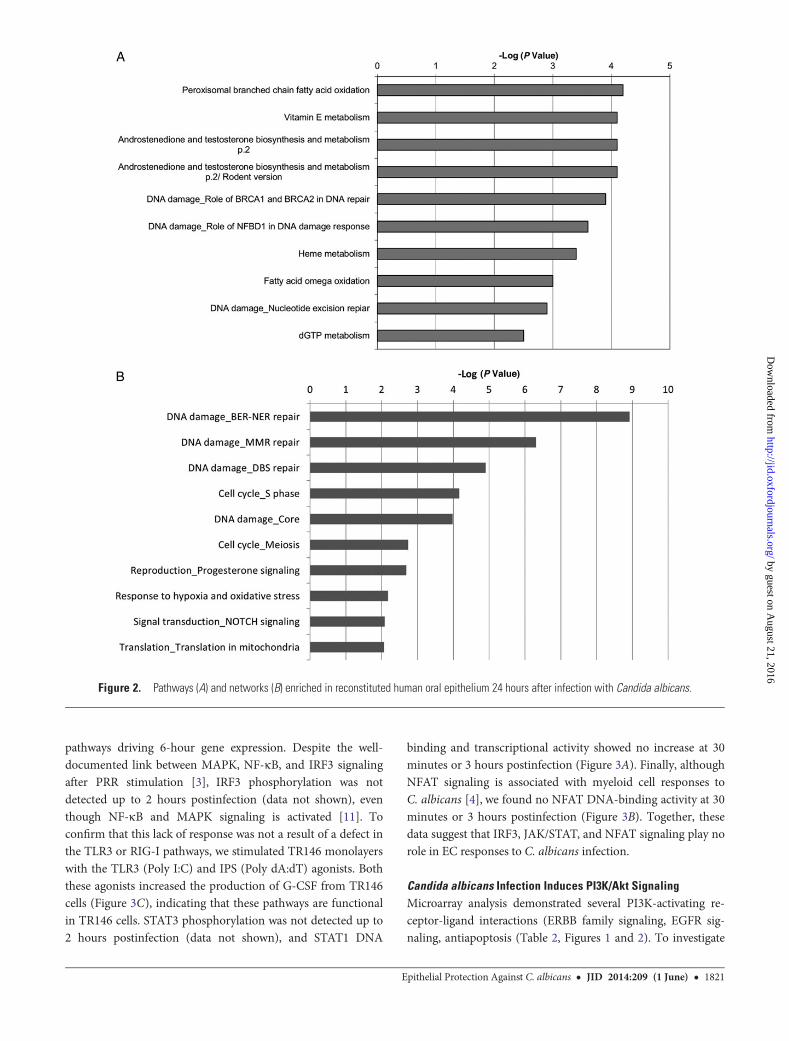
metabolism (Figure 2A), and analysis of enriched MetaCoreProcess Networks identifies several DNA damage-associatedprocesses (Base Excision Repair-Nucleotide Excision Repair(BER-NER) repair [8.9], DNA Mismatch Repair (MMR) repair[6.3], DBS repair [4.9], and core processes [3.9]) (Figure 2B).Furthermore, there is enrichment of networks involved in cellcycle (DNA production (S phase [4.2]), likely linked withDNA repair processes. Along with enrichment in mitochondrialtranslation genes (translation in mitochondria [2.1]), these mayrepresent a continuation of earlier apoptotic processes. Other en-riched processes suggest changes in intercellular communication(NOTCH signaling [2.1]) and stress responses (response tohypoxia and oxidative stress [2.2]). Together, these data suggesta general response to an invasive, damage-inducing pathogen.
Table 2. Selected Genes Showing Significant Up-regulation inReconstituted Human Oral Epithelium 6 Hours and 24 Hours AfterInfection With Candida albicans
Gene
6 Hours Postinfection 24 Hours Postinfection
P ValueFold
Change P ValueFold
Change
Transcription factors
FOS (c-Fos) 5.61 × 10−03 3.5 ncEGR3 7.03 × 10−04 4.3 2.75 × 10−05 32.4
FOSL1(Fra1)
7.88 × 10−05 7 1.05 × 10−05 12.9
ATF3 5.81 × 10−04 4.2 5.56 × 10−05 8.4
ELK3 nc 1.47 × 10−05 5.4
EGR1 4.61 × 10−06 4 3.38 × 10−04 5.3JUN nc 4.54 × 10−05 4.0
FOSB nc 5.35 × 10−04 3.2
CREB1 nc 8.26 × 10−04 2.7MAPKmodulators
DUSP1(MKP1)
1.95 × 10−04 4.7 2.27 × 10−05 7.6
DUSP5(HVH3)
2.6 × 10−04 5.6 1.19 × 10−04 7.0
DUSP6(MKP3)
9.14 × 10−05 8.8 8.86 × 10−04 6.8
SPRY2 2.93 × 10−04 4.4 1.33 × 10−04 5.9
TRIB1 1.9 × 10−04 4.8 5.38 × 10−05 4.7
SPRY4 4.72 × 10−03 3 9.49 × 10−05 4.7Signaling molecules
INPP4B 1.42 × 10−05 10.9
ITPKC 6.15 × 10−04 4 ncGADD45A nc 9.62 × 10−05 5.4
SOCS1 7.91 × 10−04 2.6 7.11 × 10−05 5.0
TNFAIP3 5.79 × 10−03 3.2 1.03 × 10−04 4.7PITPNC1 nc 4.28 × 10−04 4.0
NFKBIA nc 8.71 × 10−06 3.0
NFKBIE nc 5.66 × 10−05 3.0NFAT5 nc 3.6 × 10−04 3.0
SOCS3 nc 6.98 × 10−05 2.8
BCL10 nc 1.9 × 10−04 2.5TRIF nc 4.49 × 10−05 2.4
IL-1 family cytokines
IL-1F9 3.8 × 10−04 8.1 9.59 × 10−05 34.7IL-1α 2.16 × 10−03 6.6 1.11 × 10−04 10.0
IL-1β 1.63 × 10−03 3.6 1.16 × 10−04 7.9
IL-1F5 nc 1.53 × 10−04 7.0IL-1ra nc 8.51 × 10−04 6.1
IL-6 family cytokines
CLCF-1 1.32 × 10−05 6 2.09 × 10−05 12.7LIF 2.66 × 10−04 3.9 6.33 × 10−05 7.1
IL-6 1.53 × 10−03 3 5.07 × 10−05 5.6
Other cytokines/chemokinesIL-8 5.12 × 10−05 6.5 3.45 × 10−04 24.2
GM-CSF 1.67 × 10−06 24.2
HBEGF 1.32 × 10−05 6.4 8.11 × 10−06 11.3CCL20 8.13 × 10−04 6.8 5.40 × 10−08 10.6
Table 2 continued.
Gene
6 Hours Postinfection 24 Hours Postinfection
P ValueFold
Change P ValueFold
Change
IL-11 6.48 × 10−03 2.3 2.5 × 10−04 6.3
IL-24 nc 2.80 × 10−05 4.3Surface receptors
CEACAM6 nc 2.60 × 10−06 42.1
CEACAM1 nc 1.07 × 10−05 15.6PTX3 5.83 × 10−03 5.3 5.15 × 10−05 11.2
ICAM1 6.65 × 10−03 3.5 1.05 × 10−05 5.6
TLR5 nc 2.4 × 10−03 −8.1CLEC7A nc 3.13 × 10−04 −9.2
Proteases/inhibitors
SERPINB2 8.20 × 10−05 7.6 1.06 × 10−04 42.3MMP10 4.27 × 10−04 3.5 4.30 × 10−06 28.1
MMP1 1.02 × 10−03 5.7 1.79 × 10−06 17.7
SERPINB1 nc 6.1 × 10−04 7.9TIMP1 nc 3.54 × 10−05 3.9
Apoptosis
BCL2A1 8.51 × 10−03 3.3 6.4 × 10−04 20.0BCL2L1 nc 4.4 × 10−04 2.2
CASP8 nc 6.89 × 10−04 −2.1BCLAF1 nc 1.51 × 10−04 −3.7
Antimicrobial peptides
DEFB4 nc 1.69 × 10−04 30.8
S100P nc 1.36 × 10−06 6.4S100A12 nc 9.85 × 10−04 3.2
Interferon-stimulated genes
ISG20 nc 6.54 × 10−05 4.9PRKRIR nc 6.87 × 10−04 −2.1IFI16 nc 1.34 × 10−05 −2.2IFIT3 nc 2.25 × 10−03 −2.4IFIT5 nc 2.87 × 10−05 −5.2IFIT1 nc 3.43 × 10−04 −3.15
Abbreviations: GM-CSF, granulocyte macrophage colony-stimulating factor; IL,interleukin; nc, no change.
Epithelial Protection Against C. albicans • JID 2014:209 (1 June) • 1819
by guest on August 21, 2016
http://jid.oxfordjournals.org/D
ownloaded from
Candida albicans Does Not Induce IRF-3, NFAT, or STAT1Signaling in Epithelial Cells
As well as NF-κB and MAPK signaling [11], the microarraydata indicated increased IRF3 and Janus kinase/signal trans-ducer and activator of transcription (JAK/STAT)–regulatedgenes, as well as increases in NFAT expression (Table 2).
Therefore, we determined whether these pathways and theirdownstream TFs were functionally activated (phosphorylated)in response to C. albicans at early time points (optimum 2hours [11]) in monolayer TR146 ECs, which comprise the ROEmodel. TR146 monolayers were used to maximize the signal,and times up to 2 hours were optimum to identify signal
Figure 1. Pathways (A) and networks (B) enriched in reconstituted human oral epithelium 6 hours after infection with Candida albicans.
1820 • JID 2014:209 (1 June) • Moyes et al
by guest on August 21, 2016
http://jid.oxfordjournals.org/D
ownloaded from
pathways driving 6-hour gene expression. Despite the well-documented link between MAPK, NF-κB, and IRF3 signalingafter PRR stimulation [3], IRF3 phosphorylation was notdetected up to 2 hours postinfection (data not shown), eventhough NF-κB and MAPK signaling is activated [11]. Toconfirm that this lack of response was not a result of a defect inthe TLR3 or RIG-I pathways, we stimulated TR146 monolayerswith the TLR3 (Poly I:C) and IPS (Poly dA:dT) agonists. Boththese agonists increased the production of G-CSF from TR146cells (Figure 3C), indicating that these pathways are functionalin TR146 cells. STAT3 phosphorylation was not detected up to2 hours postinfection (data not shown), and STAT1 DNA
binding and transcriptional activity showed no increase at 30minutes or 3 hours postinfection (Figure 3A). Finally, althoughNFAT signaling is associated with myeloid cell responses toC. albicans [4], we found no NFAT DNA-binding activity at 30minutes or 3 hours postinfection (Figure 3B). Together, thesedata suggest that IRF3, JAK/STAT, and NFAT signaling play norole in EC responses to C. albicans infection.
Candida albicans Infection Induces PI3K/Akt SignalingMicroarray analysis demonstrated several PI3K-activating re-ceptor-ligand interactions (ERBB family signaling, EGFR sig-naling, antiapoptosis (Table 2, Figures 1 and 2). To investigate
Figure 2. Pathways (A) and networks (B) enriched in reconstituted human oral epithelium 24 hours after infection with Candida albicans.
Epithelial Protection Against C. albicans • JID 2014:209 (1 June) • 1821
by guest on August 21, 2016
http://jid.oxfordjournals.org/D
ownloaded from
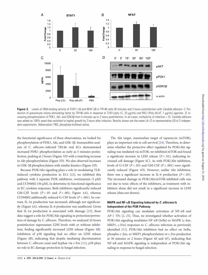
the functional significance of these observations, we looked forphosphorylation of PDK1, Akt, and GSK-3β. Immunoblot anal-ysis of C. albicans–infected TR146 oral ECs demonstratedincreased PDK1 phosphorylation as early as 5 minutes postin-fection, peaking at 2 hours (Figure 3D) with a matching increasein Akt phosphorylation (Figure 3D). We also observed increasesin GSK-3β phosphorylation with similar kinetics (Figure 3D).
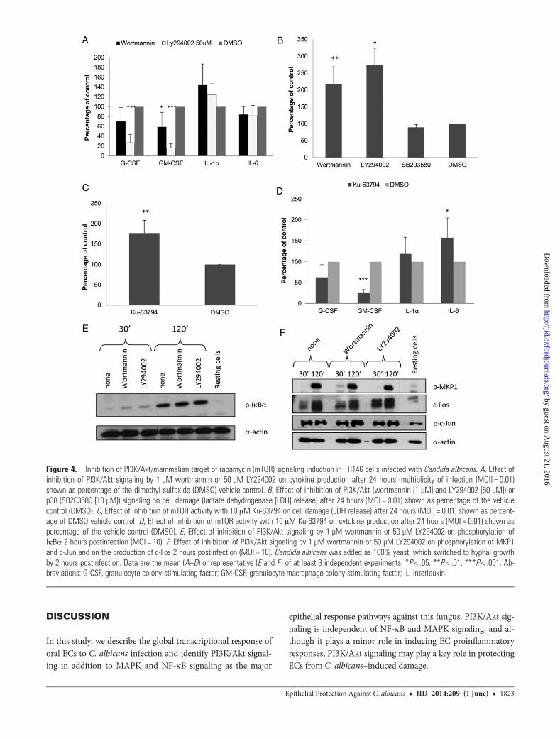
Because PI3K/Akt signaling plays a role in modulating TLR-induced cytokine production in ECs [22], we inhibited thispathway with 2 separate PI3K inhibitors, wortmannin (1 µM)and LY294002 (50 µM), to determine its functional significancein EC cytokine responses. Both inhibitors significantly reducedGM-CSF levels (P < .05 and P < .001; Figure 4A), whereasLY294002 additionally reduced G-CSF levels (P < .001). In con-trast, IL-1α production was increased, although not significan-tly (Figure 4A), whereas IL-6 production was unaffected. Giventhat IL-1α production is associated with damage [23], thesedata suggest a role for PI3K/Akt signaling in protection/preven-tion of damage by C. albicans. Therefore, we analyzed 24 hourspostinfection supernatant LDH levels with or without inhibi-tion, finding significantly increased LDH release (Figure 4B).Inhibition of p38 signaling had no effect on LDH release(Figure 4B), indicating that despite mediating discriminationbetween C. albicans yeast and hyphae via c-Fos [11], p38 playsno role in EC damage protection in fungal infection.
The Akt target, mammalian target of rapamycin (mTOR),plays an important role in cell survival [24]. Therefore, to deter-mine whether the protective effect regulated by PI3K/Akt sig-naling was mediated via mTOR, we inhibited mTOR and founda significant increase in LDH release (P < .01), indicating in-creased cell damage (Figure 4C). As with PI3K/Akt inhibition,levels of G-CSF (P < .05) and GM-CSF (P < .001) were signifi-cantly reduced (Figure 4D). However, unlike Akt inhibition,there was a significant increase in IL-6 production (P < .05).The increased damage in PI3K/Akt/mTOR-inhibited cells wasnot due to toxic effects of the inhibitors, as treatment with in-hibitors alone did not result in a significant increase in LDHrelease (data not shown).
MAPK and NF-κB Signaling Induced by C. albicans IsIndependent of the PI3K PathwayPI3K/Akt signaling can modulate activation of NF-κB andAP-1 TFs [2, 25]. Thus, we investigated whether activation ofPI3K/Akt signaling modulates NF-κB (IκBa) or MAPK (c-Jun,MKP1, c-Fos) responses to C. albicans infection as previouslyidentified [11]. PI3K/Akt inhibition had no effect on IκBα,phospho-c-Jun, or MKP1 phosphorylation or c-Fos productionat 30 minutes or 2 hours (Figure 4E and 4F), indicating thatNF-κB and MAPK signaling is independent of PI3K/Akt sig-naling in response to fungal infection.
Figure 3. Levels of DNA-binding activity of STAT1 (A) and NFAT (B) in TR146 cells 30 minutes and 3 hours postinfection with Candida albicans. C, Pro-duction of granulocyte colony-stimulating factor by TR146 cells in response to TLR3 (poly I:C, 25 μg/mL) and RIG-I (Poly dA:dT, 1 μg/mL) agonists. D, In-creasing phosphorylation of PDK1, Akt, and GSK3β from 5 minutes up to 2 hours postinfection. In all cases, multiplicity of infection = 10. Candida albicanswas added as 100% yeast that switched to hyphal growth by 2 hours after infection. Results shown are the mean (A–C) or representative (D) of 3 indepen-dent experiments. Abbreviation: PBS, phosphate-buffered saline.
1822 • JID 2014:209 (1 June) • Moyes et al
by guest on August 21, 2016
http://jid.oxfordjournals.org/D
ownloaded from
DISCUSSION
In this study, we describe the global transcriptional response oforal ECs to C. albicans infection and identify PI3K/Akt signal-ing in addition to MAPK and NF-κB signaling as the major
epithelial response pathways against this fungus. PI3K/Akt sig-naling is independent of NF-κB and MAPK signaling, and al-though it plays a minor role in inducing EC proinflammatoryresponses, PI3K/Akt signaling may play a key role in protectingECs from C. albicans–induced damage.
Figure 4. Inhibition of PI3K/Akt/mammalian target of rapamycin (mTOR) signaling induction in TR146 cells infected with Candida albicans. A, Effect ofinhibition of PI3K/Akt signaling by 1 µM wortmannin or 50 µM LY294002 on cytokine production after 24 hours (multiplicity of infection [MOI] = 0.01)shown as percentage of the dimethyl sulfoxide (DMSO) vehicle control. B, Effect of inhibition of PI3K/Akt (wortmannin [1 µM] and LY294002 [50 µM]) orp38 (SB203580 [10 µM]) signaling on cell damage (lactate dehydrogenase [LDH] release) after 24 hours (MOI = 0.01) shown as percentage of the vehiclecontrol (DMSO). C, Effect of inhibition of mTOR activity with 10 μM Ku-63794 on cell damage (LDH release) after 24 hours (MOI] = 0.01) shown as percent-age of DMSO vehicle control. D, Effect of inhibition of mTOR activity with 10 μM Ku-63794 on cytokine production after 24 hours (MOI = 0.01) shown aspercentage of the vehicle control (DMSO). E, Effect of inhibition of PI3K/Akt signaling by 1 µM wortmannin or 50 µM LY294002 on phosphorylation ofIκBα 2 hours postinfection (MOI = 10). F, Effect of inhibition of PI3K/Akt signaling by 1 µM wortmannin or 50 µM LY294002 on phosphorylation of MKP1and c-Jun and on the production of c-Fos 2 hours postinfection (MOI = 10). Candida albicans was added as 100% yeast, which switched to hyphal growthby 2 hours postinfection. Data are the mean (A–D) or representative (E and F ) of at least 3 independent experiments. *P < .05, **P < .01, ***P < .001. Ab-breviations: G-CSF, granulocyte colony-stimulating factor; GM-CSF, granulocyte macrophage colony-stimulating factor; IL, interleukin.
Epithelial Protection Against C. albicans • JID 2014:209 (1 June) • 1823
by guest on August 21, 2016
http://jid.oxfordjournals.org/D
ownloaded from
The transcriptome of C. albicans–infected ROE reveals inter-esting findings. At 6 hours postinfection, several enrichedimmune response pathways were related to cytokine and otherreceptor signaling pathways but, notably, neither TLR or otherconventional fungal PRR receptor pathways were enriched,supporting our previous findings implying no role for TLRs orother conventional PRRs in early recognition of C. albicans byECs [11]. In contrast, ERBB family signaling showed significantenrichment, and previous studies support a role for EGFR-ERBB1 signaling during C. albicans infections [26, 27]. Al-though the TREM-1 signaling pathway was enriched, we foundno increase in the PRR TREM-1 expression. Thus, this enrich-ment possibly represents an increase in PRR signaling-associatedgene expression. Enrichment of amphoterin and IL-1 signalingpathways suggests involvement of damage-associated molecularpatterns in EC responses to C. albicans. This is expected giventhe stress/damage resulting from tissue invasion. Together,these pathways probably form the core EC response to fungalinvasion and damage. The increase in the protease inhibi-tor SerpinB2 is interesting but should be interpreted withcaution as a role has yet to be identified in host infectionresponses [13].
The 24-hour pathways and networks show enrichment incell survival, DNA repair, or metabolism, reflecting differingEC priorities during late infection, predominantly targeting ap-optosis and cell growth. Along with increases in tissue remodel-ing genes, this suggests a general damage repair mechanism,maintaining mucosal barrier integrity. The late appearance ofapoptosis genes suggests that apoptotic mechanisms areinduced at later stages of infection, as previously reported forC. albicans [28, 29] and bacterial EC infection [30].
In the intermediate infection stage (6 hours), NF-κB–induced genes were up-regulated, which was expected giventhis pathway’s role in EC antifungal responses [11]. Althoughno MAPK proteins showed increased expression, we observedincreases in expression of MKP1 (dusp1) and other members ofthis family—HVH3 (dusp5) and MKP3 (dusp6). As withMKP1, these proteins negatively regulate MAPK signaling, butwith different specificity, acting to dephosphorylate ERK1/2[31]. Given their high degree of up-regulation, these phospha-tases potentially form the core MAPK-regulatory responsemechanism during C. albicans infection. Other up-regulatedMAPK regulators include 2 Sprouty genes, spry2 and spry4, in-volved in regulating ERK1/2 signaling [7, 32], and trib1 control-ling activation of MAPKs by MAP2Ks [33] and involved inantifungal responses of Caenorhabditis elegans [34].
In addition to MAPK regulatory factors, we observed up-regulation of several AP-1 proteins, persisting throughoutinfection, confirming a central role for AP-1 in EC antifungalresponses [11]. In particular, ATF3, a transcriptional repressormodulating responses to infection [35], is upregulated early,possibly as part of a negative feedback loop. Alternatively,
ATF3 may work in conjunction with other TFs, allowing forshifts in gene expression profile during infection.
Along with MAPK and NF-κB pathways, other signalingpathways and TFs show upregulation after C. albicans infection,most notably EGR1 and EGR3. These proteins are associatedwith myeloid cell responses to C. albicans [4, 36], playing a rolein NFAT signaling via dectin-1, although their role in fungalEC responses is unknown. Elevation of EGR1 and EGR3despite a lack of signaling via dectin-1, Syk [11], or NFAT (thisstudy) implies that EGR gene activation in ECs occurs via dif-ferent pathways, potentially via ERK1/2 [13, 31]. Other genesassociated with PRR regulation and immune response signalsare upregulated, particularly those involved in ubiquitin signal-ing, including A20 and ITCH [37].
The observed discrepancy between microarrays and function-al studies reflects the difference between RNA expression andsubsequent protein activation, with many signaling pathwaysshowing crossover in target genes. Microarray data represent anexpression snapshot, whereas signal activation data from 0–3hours postinfection represent EC responses to C. albicans ratherthan subsequent secreted components. Despite the microarrayevidence, only PI3K/Akt signaling via PDK1/Akt appears to befunctionally activated alongside MAPK and NF-κB, with no evi-dence for activation of IRF, STATs, or NFAT. Required fordectin-1 signaling in myeloid cells [4, 36], the lack of NFAT sig-naling confirms our previous findings that dectin-1 is not in-volved in EC recognition of C. albicans [11]. Induction of PI3K/Akt pathway activity in ECs was expected, given the wide rangeof stimuli and receptors reported to activate this pathway [38]and the demonstrated role of PI3K/Akt signaling in monocytesduring fungal immunity [39, 40], TLR-mediated pathogen detec-tion [13], and cytokine secretion [22, 41]. Here, we demonstratePI3K/Akt/mTOR signaling is important in regulating G-CSFand GM-CSF production from ECs. Notably, these cytokines areassociated with mucosal healing, having been used to treatdamaged mucosa [42, 43]. In contrast, IL-1α production in-creased when PI3K/Akt/mTOR signaling was inhibited, correlat-ing with increased EC damage, implying that PI3K/Akt/mTORsignaling may play a role in protecting ECs from damage induc-tion during C. albicans infection. Given that PI3K/Akt signalingalso protects macrophages against fungal-induced killing [40],our findings indicate that PI3K/Akt signaling may represent acommon mechanism by which host cells protect against patho-gen-induced damage. Furthermore, as p38 inhibition did notaffect damage, EC damage protection is probably independentof the p38/c-Fos hyphal recognition response we previously re-ported [11]. Evidence from the MetaCore ontology analysis ofmicroarray data in this study suggests that this protection maybe induced by inhibiting apoptosis in infected cells, given theenrichment in PI3K-induced antiapoptosis pathways.
PI3K/Akt signaling may be involved in other EC responsemechanisms connected to damage, for example, fungal invasion.
1824 • JID 2014:209 (1 June) • Moyes et al
by guest on August 21, 2016
http://jid.oxfordjournals.org/D
ownloaded from
Candida albicans–induced endocytosis in ECs is clathrin-dependent, and PI3K/Akt signaling is thought to be crucial forthis process [44] and for microbe endocytosis [45]. Interesting-ly, although PI3K/Akt signaling appears to initiate EC damageprotection/repair mechanisms, in contrast to myeloid cell PRRligation [25, 46, 47], it has no effect on NF-κB or MAPK signal-ing. Consequently, the roles of PI3K/Akt signaling probablydiffer between myeloid and ECs.
It is important to note that this work was carried out usingthe TR146 carcinoma cell line and thus may not accuratelyreflect findings in a normal host. However, this cell line hasbeen used extensively in C. albicans infection studies of bothmonolayers and organotypic models [11, 18, 19, 21, 48–50] andhas been found to give comparable data to those obtained frompatient biopsies [11].
Our data suggest that the role of PI3K/Akt signaling in hyphaldiscrimination is minimal but may constitute a major damageprotection response in ECs. We therefore propose a modelwhereby (1) p38/c-Fos signaling identifies C. albicans hyphae[11, 17], (2) p38/c-Fos and NF-κB drive cytokine production [11,17], (3) ERK1/2 signaling (via MKPs) regulates p38/c-Fos activa-tion [11, 17], and (4) PI3K/Akt/mTOR signaling induces cellprotection responses (this study). Together, these EC signalingpathways act in concert to recognize potential fungal threats, ini-tiating protective cellular responses limiting mucosal fungal in-fections. This study identifies PI3K/Akt/mTOR signaling, a keycomponent of this mechanism, as potentially an importanttarget for future antifungal therapy development.
Supplementary Data
Supplementary materials are available at The Journal of Infectious Diseasesonline (http://jid.oxfordjournals.org/). Supplementary materials consist ofdata provided by the author that are published to benefit the reader. Theposted materials are not copyedited. The contents of all supplementary dataare the sole responsibility of the authors. Questions or messages regardingerrors should be addressed to the author.
Notes
Acknowledgments. We thank Stephen Challacombe, Bernhard Hube,and Duncan Wilson for many interesting discussions about Candida patho-genicity.Financial support. This work was supported by the Medical Research
Council (MR-J008303-1) and Biotechnology and Biological Sciences Re-search Council (BB-J016411-1). Funding to pay the Open Access publica-tion charges for this article was provided by the Medical Research Council(MR-J008303-1).Potential conflicts of interest. All authors: No reported conflicts.All authors have submitted the ICMJE Form for Disclosure of Potential
Conflicts of Interest. Conflicts that the editors consider relevant to thecontent of the manuscript have been disclosed.
References
1. Koropatnick TA, Engle JT, Apicella MA, Stabb EV, Goldman WE,McFall-Ngai MJ. Microbial factor-mediated development in a host-bacterial mutualism. Science 2004; 306:1186–8.
2. Hazeki K, Nigorikawa K, Hazeki O. Role of phosphoinositide 3-kinasein innate immunity. Biol Pharm Bull 2007; 30:1617–23.
3. Lee MS, Kim YJ. Signaling pathways downstream of pattern-recogni-tion receptors and their cross talk. Annu Rev Biochem 2007; 76:447–80.
4. Goodridge HS, Simmons RM, Underhill DM. Dectin-1 stimulation byCandida albicans yeast or zymosan triggers NFAT activation in macro-phages and dendritic cells. J Immunol 2007; 178:3107–15.
5. Fukao T, Koyasu S. PI3K and negative regulation of TLR signaling.Trends Immunol 2003; 24:358–63.
6. Husmann M, Dersch K, Bobkiewicz W, Beckmann E, Veerachato G,Bhakdi S. Differential role of p38 mitogen activated protein kinase forcellular recovery from attack by pore-forming S. aureus alpha-toxin orstreptolysin O. Biochem Biophys Res Commun 2006; 344:1128–34.
7. Ather JL, Alcorn JF, Brown AL, et al. Distinct functions of airway epi-thelial nuclear factor-kappaB activity regulate nitrogen dioxide-induced acute lung injury. Am J Respir Cell Mol Biol 2010; 43:443–51.
8. Eckmann L, Nebelsiek T, Fingerle AA, et al. Opposing functions ofIKKbeta during acute and chronic intestinal inflammation. Proc NatlAcad Sci U S A 2008; 105:15058–63.
9. Odds F. Candida and candidosis. 2nd ed. Bailliere Tindall, 1988.10. Schaller M, Zakikhany K, Naglik JR, Weindl G, Hube B. Models of oral
and vaginal candidiasis based on in vitro reconstituted human epithe-lia. Nat Protoc 2006; 1:2767–73.
11. Moyes DL, Runglall M, Murciano C, et al. A biphasic innate immuneMAPK response discriminates between the yeast and hyphal forms ofCandida albicans in epithelial cells. Cell Host Microbe 2010; 8:225–35.
12. Naglik JR, Moyes DL, Wachtler B, Hube B. Candida albicans interac-tions with epithelial cells and mucosal immunity. Microbes Infect 2011;13:963–76.
13. Joung SM, Park ZY, Rani S, Takeuchi O, Akira S, Lee JY. Akt contrib-utes to activation of the TRIF-dependent signaling pathways of TLRsby interacting with TANK-binding kinase 1. J Immunol 2011; 186:499–507.
14. Tomalka J, Ganesan S, Azodi E, et al. A novel role for the NLRC4 in-flammasome in mucosal defenses against the fungal pathogen Candidaalbicans. PLoS Pathog 2011; 7:e1002379.
15. Zakikhany K, Naglik JR, Schmidt-Westhausen A, Holland G, SchallerMHube B. In vivo transcript profiling of Candida albicans identifies agene essential for interepithelial dissemination. Cell Microbiol 2007; 9:2938–54.
16. Ikuta T, Bhawal UK, Tsushima K, Aoki A, Kuboyama N, Abiko Y. Iden-tification by DNA microarray of genes involved in Candida albicans-treated gingival epithelial cells. J Oral Pathol Med 2012; 41:769–78.
17. Moyes DL, Murciano C, Runglall M, Islam A, Thavaraj S, Naglik JR.Candida albicans yeast and hyphae are discriminated by MAPK signal-ling in vaginal epithelial cells. PLoS One 2011; 6:e26580.
18. Murciano C, Moyes DL, Runglall M, et al. Candida albicans cell wallglycosylation may be indirectly required for activation of epithelial cellproinflammatory responses. Infect Immun 2011; 79:4902–11.
19. Murciano C, Moyes DL, Runglall M, et al. Evaluation of the role ofCandida albicans agglutinin-like sequence (Als) proteins in human oralepithelial cell interactions. PLoS One 2012; 7:e33362.
20. Zarember KA, Godowski PJ. Tissue expression of human Toll-like re-ceptors and differential regulation of Toll-like receptor mRNAs in leu-kocytes in response to microbes, their products, and cytokines. JImmunol 2002; 168:554–61.
21. Dalle F, Wachtler B, L’Ollivier C, et al. Cellular interactions of Candidaalbicans with human oral epithelial cells and enterocytes. Cell Micro-biol 2010; 12:248–71.
22. Rhee SH, Kim H, Moyer MP, Pothoulakis C. Role of MyD88 in phos-phatidylinositol 3-kinase activation by flagellin/Toll-like receptor 5 en-gagement in colonic epithelial cells. J Biol Chem 2006; 281:18560–8.
23. Bianchi ME. DAMPs, PAMPs and alarmins: all we need to know aboutdanger. J Leukoc Biol 2007; 81:1–5.
24. Laplante M, Sabatini DM. mTOR signaling at a glance. J Cell Sci 2009;122:3589–94.
Epithelial Protection Against C. albicans • JID 2014:209 (1 June) • 1825
by guest on August 21, 2016
http://jid.oxfordjournals.org/D
ownloaded from
25. Manna SK, Aggarwal BB. Wortmannin inhibits activation of nucleartranscription factors NF-kappaB and activated protein-1 induced by li-popolysaccharide and phorbol ester. FEBS Lett 2000; 473:113–8.
26. Zhu W, Phan QT, Boontheung P, Solis NV, Loo JA, Filler SG. EGFRand HER2 receptor kinase signaling mediate epithelial cell invasion byCandida albicans during oropharyngeal infection. Proc Natl Acad SciU S A 2012; 109:14194–9.
27. Pahl R, Brunke G, Steubesand N, et al. IL-1beta and ADAM17 arecentral regulators of beta-defensin expression in Candida esophagitis.Am J Physiol Gastrointest Liver Physiol 2011; 300:G547–53.
28. Villar CC, Chukwuedum Aniemeke J, Zhao XR, Huynh-Ba G. Induc-tion of apoptosis in oral epithelial cells by Candida albicans. Mol OralMicrobiol 2012; 27:436–48.
29. Wagener J, Weindl G, de Groot PW, et al. Glycosylation of Candida al-bicans cell wall proteins is critical for induction of innate immune re-sponses and apoptosis of epithelial cells. PLoS One 2012; 7:e50518.
30. Kim JM, Eckmann L, Savidge TC, Lowe DC, Witthoft T, Kagnoff MF.Apoptosis of human intestinal epithelial cells after bacterial invasion. JClin Invest 1998; 102:1815–23.
31. Liu W, Tundwal K, Liang Q, et al. Establishment of extracellular signal-regulated kinase 1/2 bistability and sustained activation throughSprouty 2 and its relevance for epithelial function. Mol Cell Biol 2010;30:1783–99.
32. Bundschu K, Walter U, Schuh K. The VASP-Spred-Sprouty domainpuzzle. J Biol Chem 2006; 281:36477–81.
33. Kiss-Toth E, Bagstaff SM, Sung HY, et al. Human tribbles, a proteinfamily controlling mitogen-activated protein kinase cascades. J BiolChem 2004; 279:42703–8.
34. Ziegler K, Kurz CL, Cypowyj S, et al. Antifungal innate immunity in C.elegans: PKCdelta links G protein signaling and a conserved p38MAPK cascade. Cell Host Microbe 2009; 5:341–52.
35. Litvak V, Ramsey SA, Rust AG, et al. Function of C/EBPdelta in a regu-latory circuit that discriminates between transient and persistent TLR4-induced signals. Nat Immunol 2009; 10:437–43.
36. Greenblatt MB, Aliprantis A, Hu B, Glimcher LH. Calcineurin regulatesinnate antifungal immunity in neutrophils. J Exp Med 2010; 207:923–31.
37. Shembade N, Harhaj NS, Parvatiyar K, et al. The E3 ligase Itch negativelyregulates inflammatory signaling pathways by controlling the function ofthe ubiquitin-editing enzyme A20. Nat Immunol 2008; 9:254–62.
38. Hawkins PT, Anderson KE, Davidson K, Stephens LR. Signallingthrough class I PI3Ks in mammalian cells. Biochem Soc Trans 2006; 34:647–62.
39. Gabrielli E, Pericolini E, Cenci E, et al. Antibody complementarity-determining regions (CDRs): a bridge between adaptive and innate im-munity. PLoS One 2009; 4:e8187.
40. Volling K, Thywissen A, Brakhage AA, Saluz HP. Phagocytosis ofmelanized Aspergillus conidia by macrophages exerts cytoprotectiveeffects by sustained PI3K/Akt signalling. Cell Microbiol 2011;13:1130–48.
41. Martin M, Schifferle RE, Cuesta N, Vogel SN, Katz J, Michalek SM.Role of the phosphatidylinositol 3 kinase-Akt pathway in the regulationof IL-10 and IL-12 by Porphyromonas gingivalis lipopolysaccharide. JImmunol 2003; 171:717–25.
42. Bernasconi E, Favre L, Maillard MH, et al. Granulocyte-macrophagecolony-stimulating factor elicits bone marrow-derived cells thatpromote efficient colonic mucosal healing. Inflamm Bowel Dis 2010;16:428–41.
43. Krishnan K, Arnone B, Buchman A. Intestinal growth factors: potentialuse in the treatment of inflammatory bowel disease and their role inmucosal healing. Inflamm Bowel Dis 2011; 17:410–22.
44. Araki N, Johnson MT, Swanson JA. A role for phosphoinositide 3-kinase in the completion of macropinocytosis and phagocytosis bymacrophages. J Cell Biol 1996; 135:1249–60.
45. Agarwal V, Hammerschmidt S. Cdc42 and the phosphatidylinositol3-kinase-Akt pathway are essential for PspC-mediated internalizationof pneumococci by respiratory epithelial cells. J Biol Chem 2009; 284:19427–36.
46. Aksoy E, Vanden Berghe W, Detienne S, et al. Inhibition of phosphoi-nositide 3-kinase enhances TRIF-dependent NF-kappa B activationand IFN-beta synthesis downstream of Toll-like receptor 3 and 4. Eur JImmunol 2005; 35:2200–9.
47. Guha M, Mackman N. The phosphatidylinositol 3-kinase-Akt pathwaylimits lipopolysaccharide activation of signaling pathways and expres-sion of inflammatory mediators in human monocytic cells. J Biol Chem2002; 277:32124–32.
48. Moyes DL, Murciano C, Runglall M, Kohli A, Islam A, Naglik JR. Acti-vation of MAPK/c-Fos induced responses in oral epithelial cells is spe-cific to Candida albicans and Candida dubliniensis hyphae. MedMicrobiol Immunol 2012; 201:93–101.
49. Weindl G, Naglik JR, Kaesler S, et al. Human epithelial cells establishdirect antifungal defense through TLR4-mediated signaling. J ClinInvest 2007; 117:3664–72.
50. Mayer FL, Wilson D, Jacobsen ID, Miramon P, Grosse K, Hube B. Thenovel Candida albicans transporter Dur31 Is a multi-stage pathogenici-ty factor. PLoS Pathog 2012; 8:e1002592.
1826 • JID 2014:209 (1 June) • Moyes et al
by guest on August 21, 2016
http://jid.oxfordjournals.org/D
ownloaded from
Copyright © 2022 FDOKUMEN