Interaction between Candida albicans and Pseudomonas ...
165
UCC Library and UCC researchers have made this item openly available. Please let us know how this has helped you. Thanks! Title Interaction between Candida albicans and Pseudomonas aeruginosa Author(s) Konstantinidou, Nina Publication date 2016 Original citation Konstantinidou, N. 2016. Interaction between Candida albicans and Pseudomonas aeruginosa. PhD Thesis, University College Cork. Type of publication Doctoral thesis Rights © 2016, Nina Konstantinidou. http://creativecommons.org/licenses/by-nc-nd/3.0/ Embargo information No embargo required Item downloaded from http://hdl.handle.net/10468/3015 Downloaded on 2022-07-21T13:18:41Z
-
Upload
khangminh22 -
Category
Documents
-
view
1 -
download
0
Transcript of Interaction between Candida albicans and Pseudomonas ...
UCC Library and UCC researchers have made this item openly available.Please let us know how this has helped you. Thanks!
Title Interaction between Candida albicans and Pseudomonas aeruginosa
Author(s) Konstantinidou, Nina
Publication date 2016
Original citation Konstantinidou, N. 2016. Interaction between Candida albicans andPseudomonas aeruginosa. PhD Thesis, University College Cork.
Type of publication Doctoral thesis
Rights © 2016, Nina Konstantinidou.http://creativecommons.org/licenses/by-nc-nd/3.0/
Embargo information No embargo required
Item downloadedfrom
http://hdl.handle.net/10468/3015
Downloaded on 2022-07-21T13:18:41Z
Interaction between Candida albicans and Pseudomonas aeruginosa
A Thesis presented to the
National University of Ireland
for the Degree of
Doctor of Philosophy
By
Nina Konstantinidou, BSc, MSc
School of Microbiology
University College Cork
Cork, April 2016
Head of School: Prof. Gerald F. Fitzgerald
Supervisor: Dr. John P. Morrissey

ii
For my daughter, Arlene (Arli), and my family
...
Thank you for everything, especially for your patience
...

iii
Declaration
I, the undersigned Nina Konstantinidou, declare that I have not obtained a degree from the
University College Cork, National University of Ireland, Cork, or elsewhere on the basis of
this Ph.D. Thesis. The results presented in this thesis were derived from the experiments
undertaken by myself in the laboratories of the University College Cork.
_____________________________________
Νίνα Κωνσταντινίδου
iv
Publications
Peer reviewed publication
The results from the Chapters 2 and 3 were published in FEMS Yeast Research:
Konstantinidou N & Morrissey JP (2015) Co-occurrence of filamentation defects
and impaired biofilms in Candida albicans protein kinase mutants. FEMS Yeast Res
15: 1–10. pii fov092 doi: 10.1093/femsyr/fov092.
Public access database
A TFbsST (Transcription Factor binding site Search Tool) database was published online
under the following link:
http://bioinfo.ucc.ie/TFbsST/
Chapter 4 is in preparation for submission in the Database Issue of Nucleic Acid Research
(Oxford Journals).
General scientific communication
Parts of the Chapter 1 were published in the following articles:
Konstantinidou N (2014) Decoding bug chatter to fight infections. The Boolean 4:
51-55.
Konstantinidou N (2014) Membership Q & A. Microbiology Today Magazine 41: 82-
83.
v
Table of Contents
Publications .................................................................................................................. iv
Peer reviewed publication ......................................................................................... iv
Public access database ............................................................................................... iv
General scientific communication .............................................................................. iv
Table of Contents ........................................................................................................... v
List of Figures .............................................................................................................. viii
List of Tables ................................................................................................................. ix
List of Abbreviations ...................................................................................................... x
Abstract ....................................................................................................................... xii
Summary .................................................................................................................... xiii
Chapter 1 ............................................................................. 1 1. General introduction .................................................................................................. 2
1.1 Candida albicans virulence .................................................................................... 2
1.2 Drug resistance strategies of Candida albicans ....................................................... 3
1.3 Candida albicans morphology and morphogenesis ................................................. 4
1.4 Biology of yeast, pseudohyphae and hyphae ......................................................... 6
1.4.1 Yeast ........................................................................................................................ 6
1.4.2 Pseudohyphae ......................................................................................................... 6
1.4.3 Hyphae .................................................................................................................... 7
1.5 Candida albicans biofilms ...................................................................................... 8
1.6 Interaction between Candida albicans and Pseudomonas aeruginosa .................. 10
1.7 Quorum Sensing ................................................................................................. 12
1.7.1 Pseudomonas aeruginosa quorum sensing molecules ......................................... 13
1.7.2 Candida albicans quorum sensing molecules ....................................................... 13
Chapter 2 ........................................................................... 16 Abstract ....................................................................................................................... 17
2. Co-occurrence of filamentation defects and impaired biofilms in Candida albicans protein kinase mutants ................................................................................................ 18
2.1 Introduction ....................................................................................................... 18
2.2 Materials and Methods ....................................................................................... 20
2.2.1 Yeast strains and growth conditions ..................................................................... 20
2.2.2 PCR ........................................................................................................................ 20
2.2.3 Biofilm assay ......................................................................................................... 21
vi
2.2.4 Morphological analyses ........................................................................................ 21
2.2.5 Bioinformatics analyses ........................................................................................ 22
2.3 Results................................................................................................................ 25
2.3.1 Identification of the protein kinases involved in Candida albicans biofilm formation ....................................................................................................................... 25
2.3.2 Identification of the protein kinases involved in Candida albicans filamentous growth ............................................................................................................................ 34
2.3.3 STRING network analyses ..................................................................................... 39
2.4 Discussion ........................................................................................................... 42
2.5 Appendix ............................................................................................................ 44
Chapter 3 ........................................................................... 46 Abstract ....................................................................................................................... 47
3. Communication between Candida albicans and Pseudomonas aeruginosa ................ 48
3.1 Introduction ....................................................................................................... 48
3.2 Materials and Methods ....................................................................................... 50
3.2.1 Yeast strains and growth conditions ..................................................................... 50
3.2.2 PCR ........................................................................................................................ 50
3.2.3 Bacterial strains and preparation of supernatants ............................................... 51
3.2.4 Biofilm assay ......................................................................................................... 51
3.2.5 Morphological analyses ........................................................................................ 52
3.2.6 Bioinformatics analyses ........................................................................................ 52
3.3 Results................................................................................................................ 54
3.3.1 STRING network analyses ..................................................................................... 54
3.3.2 Bacterial supernatants inhibit biofilm formation of the ’Good’ biofilm formers . 56
3.3.3 ΔQS bacterial supernatants inhibit biofilm formation of the good biofilm formers without affecting their morphology .............................................................................. 60
3.4 Discussion ........................................................................................................... 62
3.5 Appendix ............................................................................................................ 63
Chapter 4 ........................................................................... 68 Abstract ....................................................................................................................... 69
4. TFbsST: Transcription Factor binding site Search Tool ................................................ 70
4.1 Introduction ....................................................................................................... 70
4.2 Materials and Methods ....................................................................................... 73
4.2.1 General approach.................................................................................................. 73
4.2.2 Transcription factor binding site database creation ............................................. 75
4.2.3 Python algorithm design ....................................................................................... 77
vii
4.2.4 TFbsST website development ............................................................................... 78
4.2.5 GO annotation ....................................................................................................... 78
4.2.6 Candida albicans and Candida parapsilosis promoter analysis ............................ 78
4.3 Results................................................................................................................ 80
4.3.1 Transcription factor binding site database ........................................................... 80
4.3.2 TFbsST software .................................................................................................... 81
4.3.3 Overrepresented and underrepresented transcription factors in Candida albicans gene promoter regions .................................................................................................. 83
4.3.4 RBT and ALS are important for interaction between Candida albicans and Pseudomonas aeruginosa .............................................................................................. 83
4.3.5 Overrepresented TFs in the RBT and ALS family gene promoters ........................ 88
4.3.6 Efg1p binding sites in Candida albicans and Candida parapsilosis promoter regions ............................................................................................................................ 92
4.4 Discussion ........................................................................................................... 94
4.5 Appendix ............................................................................................................ 97
Chapter 5 ......................................................................... 108 5. General discussion .................................................................................................. 109
5.1 Introduction ..................................................................................................... 109
5.2 Research result summary .................................................................................. 109
5.3 Candida albicans interaction with bacteria ........................................................ 110
5.4 Pseudomonas aeruginosa interaction with fungi ................................................ 111
5.5 In vivo models for the investigation of fungal biofilms ....................................... 111
5.5.1 In vivo models for the investigation of bacterial biofilms ................................... 113
5.6 Future directions for the progression of this project ........................................... 113
5.6.1 Candida albicans biofilm studies using ‘Poor’ biofilm former protein kinase mutants ........................................................................................................................ 113
5.6.2 Candida albicans – Pseudomonas aeruginosa interaction studies in vivo.......... 114
5.6.3 In vitro validation of preditied in silico analyses ................................................. 115
5.6.4 Updating and maintenance of the TFbsST database .......................................... 115
Acknowledgments ...................................................................................................... 116
Bibliography .............................................................................................................. 119
Appendix ................................................................................................................... 150
viii
List of Figures
Figure Description
Chapter 1
1 Some examples of yeast infection 2 Schematic of C. albicans biofilm development 3 Morphological forms of wild-type C. albicans DAY286 grown in filament-inducing
medium at 37oC 4 The structure of HSL (N-acyl-L-homoserine lactone) network in Vibrio fischeri 5 Quorum sensing (QS) molecule biosynthesis pathways in C. albicans
Chapter 2
1 Workflow for the categorisation of C. albicans protein kinase mutants based on their biofilm formation ability
2 Biofilm formation of the ‘Poor’ biofilm former mutants 3 Biofilm formation of the ‘Moderate’ and ‘Good’ biofilm formers 4 Morphology of the ‘Poor’ biofilm formers reveals their defect in the switch from
yeast to filamentous growth 5 Morphology of the ‘Very poor’ biofilm formers reveals their defect in the switch
from yeast to filamentous growth 6 The ‘Poor’ biofilm former mutants are defective in the switch from yeast to
filamentous growth 7 Predicted STRING interaction networks 8 Examples of molecular validation of the ‘Very poor’ biofilm former mutants with
strain-specific PCR
Chapter 3
1 Predicted STRING interaction networks 2 Biofilm formation of representatives of major MAPK pathways in the presence of
P. aeruginosa supernatants 3 Biofilm formation of the ‘Good’ biofilm formers after application of the
Pseudomonas supernatants derived from the wild-type, PAO1, and a mutant, ΔQS 4 Morphology of representatives of major MAPK pathways in the presence of P.
aeruginosa supernatants
Chapter 4
1 Workflow for the analyses of C. albicans gene promoter regions (from -1000 bp to +1 bp)
2 Screenshots illustrating some features of the TFbsST database 3 Localization of C. albicans proteins encoded by the upregulated (green bars) and
downregulated (red bars) genes in response to P. aeruginosa supernatants 4 Promoter region (from -1000 bp to +1 bp) analyses of the RBT and ALS gene
families across the TFbsST database 5 C. albicans and C. parapsilosis RBT gene promoter analyses in the TFbsST database 6 Efg1p binding sites across the C. albicans and C. parapsilosis RBT family gene
promoters
ix
List of Tables
Table Description
Chapter 1
1 Candida infection manifestations at different body sites 2 Chemicals that impair biofilm development in Candida
Chapter 2
1 Protein kinase mutants used in this study 2 Primers used in this study 3 Classes of C. albicans protein kinase mutants based on their biofilm development 4 KEGG pathways from the STRING database 5 Functional partners predicted by the STRING database
Chapter 3
1 Primers used in this study 2 KEGG pathways from the STRING database 3 Functional partners predicted by the STRING database 4 PK mutants of C. albicans less affected by the ΔQS supernatants 5 CGD Descriptions of C. albicans PK mutants
Chapter 4
1 Candida transcription factor binding site consensus sequences 2 Promoter region (from -1000 bp to +1 bp) analyses of C. albicans genes that
were impaired after application of P. aeruginosa QS molecules 3 C. albicans gene subsets coding for cell wall/surface proteins 4 Promoter region (from -1000 bp to +1 bp) analyses of C. albicans genes 5 Promoter region (from -1000 bp to +1 bp) analyses of C. albicans genes 6 Gene ontology (GO) annotations of the upregulated gene set in response to the
supernatants derived from 4 strains of Pseudomonas, PAO1, CF144, CF177 and ΔQS
7 Gene ontology (GO) annotations of the downregulated gene set in response to the supernatants derived from 4 strains of Pseudomonas, PAO1, CF144, CF177 and ΔQS
8 Gene ontology (GO) annotations of the upregulated gene set in response to HSL-containing Pseudomonas supernatants
9 Gene ontology (GO) annotations of the downregulated gene set in response to HSL-containing Pseudomonas supernatants
x
List of Abbreviations
Abbreviation Term
% Percentage 3O-C12-HSL N-(3-oxododecanoyl)-L-Homoserine Lactone A600 Absorbency at 600 nm wave length λ (Lambda) AIDS Acquired Immune Deficiency Syndrome Arg Arginine bp Base pair C4-HSL N-butyryl-L-Homoserine Lactone cAMP Cyclic Adenosine Monophosphate CF Cystic Fibrosis CGD Candida Genome Database CDR Candida Drug Resistance genes DBD DNA-Binding Domain DBD database DNA-Binding Domain database of predicted transcription factors dH2O Distilled water DNA Deoxyribonucleic acid DRE Drug Responsive Element ECM Extracellular Matrix EDTA Ethylene-Diamine-Tetraacetic Acid EMSA Electrophoretic Mobility Shift Assay g Gram GO Gene Ontology h Hour HEA Higher Education Authority HHQ 2-Heptyl-4-Quinolone (PQS precursor) His Histidine HSL N-acyl-L-Homoserine Lactone HIV Human Immunodeficiency Virus HTML5 Hyper Text Markup Language 5 ID Identifier KEGG Kyoto Encyclopaedia of Genes and Genomes LB Luria-Bertani or Lysogeny Broth Leu Leucine LHS Left Hand-Side LSM Laser Scanning Microscope MAPK Mitogen-Activated Protein Kinase Mb Mega base MDR Multidrug Resistance genes Min Minute mL Millilitre mySQL My Structured Query Language n Number oC Degree Celsius PCR Polymerase Chain Reaction pH Power of Hydrogen PK Protein Kinase PQS Pseudomonas Quinolone Signal QS Quorum Sensing RAS/cAMP/PKA Rat Sarcoma/cyclic Adenosine Monophosphate/Protein Kinase A
xi
RNA Ribonucleic acid rpm Revolutions or Rotation Per Minute S Supplementary s/n Supernatant sec Second STRING Search Tool for the Retrieval of Interacting Genes/Proteins TAD Trans-Activating Domain TF Transcription Factor TFbsST Transcription Factor binding site Search Tool Tm Melting temperature Tn7-UAU1 Tn7 unit Transposon (transposable element) -ura3Δ3’-ARG4-ura3Δ5 Ura Uracil v Version XTT 2,3-bis-(2-methoxy-4-nitro-5-sulfophenyl)-2H-tetrazolium-5-
carboxanilide YNB Yeast Nitrogen Base YNBNP Yeast Nitrogen Base N-acetyl-D-glucosamine Phosphate YPD Yeast extract Peptone Dextrose (rich yeast medium) WBC White Blood Cells Δ (Delta) Deletion (when used before a gene name) μg μicrograms μL μicrolitre μm μicrometer
xii
Abstract
Fungal pathogen Candida albicans causes serious nosocomial infections in patients, in part,
due to formation of drug-resistant biofilms. Protein kinases (PK) and transcription factors
(TF) mediate signal transduction and transcription of proteins involved in biofilm
development. To discover biofilm-related PKs, a collection of 63 C. albicans PK mutants was
screened twice independently with microtiter plate-based biofilm assay (XTT). Thirty-eight
(60%) mutants showed different degrees of biofilm impairment with the poor biofilm
formers additionally possessing filamentation defects. Most of these genes were already
known to encode proteins associated with Candida morphology and biofilms but VPS15,
PKH3, PGA43, IME2 and CEX1, were firstly associated with both processes in this study.
Previous studies of Holcombe et al. (2010) had shown that bacterial pathogen,
Pseudomonas aeruginosa can impair C. albicans filamentation and biofilm development. To
investigate their interaction, the good biofilm former PK mutants of C. albicans were
assessed for their response to P. aeruginosa supernatants derived from two strains, wild-
type PAO1 and homoserine lactone (HSL)-free mutant ΔQS, without finding any non-
responsive mutants. This suggested that none of the PKs in this study was implicated in
Candida-Pseudomonas signaling.
To screen promoter sequences for overrepresented TFs across C. albicans gene sets
significantly up/downregulated in presence of bacterial supernatants from Holcombe et al.
(2010) study, TFbsST database was created online. The TFbsST database integrates
experimentally verified TFs of Candida to analyse promoter sequences for TF binding sites.
In silico studies predicted that Efg1p was overrepresented in C. albicans and C. parapsilosis
RBT family genes.
xiii
Summary
C. albicans is a serious human pathogen partially due to its drug resistant biofilm
development that depends on the environmental signals sensed by the cell receptors. The
PKs and TFs regulate signal transduction and gene transcription process respectively.
The primary aim of this research was to discover the most important PKs for C. albicans
biofilm development. Indeed, we identified 38 PK mutants with different degrees of biofilm
impairment but some of them were already linked with biofilms. Nearly half of these
mutants were classified as ‘Poor’ biofilm formers possessing additional filamentation
defects. The novel findings included 5 genes, VPS15, PKH3, PGA43, IME2 and CEX1, not
previously associated with either filamentation or biofilm formation. All these 5 genes
seem to participate in processes that are important for biofilm formation, validating our
approach. For example, even though VPS15 encodes a kinase involved in vacuolar protein
sorting, other members of the Vps protein family, Vps1p and Vps34p, were reported to
significantly contribute in Candida biofilm formation. Vps15p and Vps34p form a complex
on the vacuole/golgi membrane indicating that a proper fungal cell development requires
correct protein sorting mechanisms. PKH3 encodes a kinase that may be involved in Protein
Kinase C (PKC) activity, which is important for drug resistance of C. albicans. Additionally,
the pkh3 mutant is reported to display significantly decreased cell-substrate adherence,
what prevents biofilm formation. Interestingly, Pkh3p was clustered with the vacuolar
proteins Vps15p and Vps34p in the network, probably indicating that the defect in this
mutant is also related to vacuole protein sorting mechanisms. PGA43 encodes a Glycosyl-
Phosphatidyl-Inositol (GPI)-anchored protein with unknown function but other Gpi family
proteins, Pga59p and Pga62p, are known to be required for cell wall integrity, which is
mandatory for filamentation. Involvement of C. albicans Ime2p in morphological
development is not surprising since its Saccharomyces cerevisiae orthologue is essential for
pseudohyphal growth independent of the MAPK filamentation cascade. The function of C.
albicans Cex1p is unknown but in S. cerevisiae it is exported from the nucleus via tRNA
binding.
Apart from vacuolar process, our data showed that the PKA pathway, ribosome biogenesis
and some aspects of the cell cycle are also required for efficient filamentation and biofilm
development. For example, mutants that were disrupted in C. albicans PKA genes, BCY1,
TPK1 and TPK2, and the ribosome biogenesis genes, CKA2, CKB1 and CKB2, exhibited
impaired biofilms under our assay conditions. Bcy1p is a regulatory subunit of PKA, which is
xiv
prominent for C. albicans morphogenesis and, Tpk1p and Tpk2p are well-documented to be
involved in filamentation. The ribosome biogenesis genes were also reported to display
elevated transcription in biofilm forming cells. Upon these findings, it was interesting to see
which proteins were not absolutely required for biofilm formation. Surprisingly, none of
the individual PKs in MAPK (Mitogen-Activated Protein Kinase) cascades was essential,
indicating that MAPK mutants are compensated by parallel MAPK pathways in the cell.
The second goal of this project was to investigate the interaction between C. albicans and
P. aeruginosa. Previous studies had shown that Pseudomonas supernatants could impair
both the yeast-hyphal transition and biofilm development in Candida. Thus, the ‘Good’
biofilm former mutants of C. albicans were assessed for their response to P. aeruginosa
supernatants without finding any non-responsive mutants . This result suggested that none
of the PKs in this study was implicated in Candida signal transduction response to
Pseudomonas signals. However, the comprehensive analysis of the mutants in presence of
supernatants derived from N-acyl-L-Homoserine Lactone (HSL)-positive and HSL-negative
strains of P. aeruginosa showed 2 distinct effects on Candida: HSL-dependent impairment
of morphology and HSL-independent impairment of biofilms.
To further investigate the TF regulation of Candida genes that were altered in presence of
P. aeruginosa secreted chemicals, we created a TFbsST database (http://bioinfo.ucc.ie/
TFbsST/). Candida TF library with experimentally validated motifs and Python scripts were
integrated to develop a user-friendly application for the analysis of gene promoter regions.
The TFbsST database includes TFs of C. albicans and closely related Candida species such as
C. parapsilosis, C. dubliniensis as well as evolutionary distinct C. glabrata. Initially, we
annotated the differentially expressed genes of C. albicans that were up/downregulated in
response to P. aeruginosa supernatants and shortlisted the genes coding for cell
wall/surface proteins including members of RBT and ALS families. Using TFbsST database,
we showed that several members of both gene families possessed Efg1p binding sites in
their promoters enhancing the importance of Efg1p in the yeast to hyphae switch. The
presence of Efg1p binding motifs in C. parapsilosis RBT family gene promoters further
supported its regulatory role across the Candida spices.
Conclusively, our approach, bioinformatics tools and data generated from this study seed
into the existing models of C. albicans and increase our understanding of its cellular
mechanisms.

1
Chapter 1
General Introduction
Parts of this chapter were published in The Boolean (Konstantinidou N (2014) Decoding bug
chatter to fight infections. Boolean 4: 51-55) and Microbiology Today (Konstantinidou N
(2014) Membership Q & A. MT magazine 41: 82-83).

2
A B C E
1. General introduction
1.1 Candida albicans virulence
C. albicans lives in healthy human host as a commensal colonising the mucosal microflora
of urogenital and gastrointestinal tracts as well as oral cavity. However, C. albicans
becomes pathogen after the overgrowth of the communities that can cause infectious
diseases by bypassing the hosts defence system. C. albicans can overcome the macrophage
innate immunity barrier with the metabolic changes mediated by the members of the Ato
protein family (Danhof and Lorenz 2015) and the excretion of farnesol that stimulates
macrophage chemokinesis (Hargarten et al. 2015). Candida infections range from life-
threatening invasive candidiasis (candidaemia) to superficial mucosal infections known as
‘thrush’. Other conditions caused by Candida include oral, mucosal and dermatological
candidiasis; lung, hepatic (liver) and renal (kidney) abscess; pyelonephritis, vulvovaginitis
and candiduria; as well as osteomyelitis (bone marrow), nail and eye infections (reviewed
by Gulati and Nobile 2016) (Figure 1 and Table 1).
Figure 1| Some examples of yeast infection. A| Oral infection in children, B| Nail infection,
C| Skin infection, D| Plaque due to dental appliances and E| Biofilms on catheters causing
bloodstream infections. The images were taken in this study with the patients’ consent
under the confidentiality policy of the Irish Health Care Board.
The main groups of patients vulnerable to Candida infections include immunocompromised
populations, due to cancer treatment (with chemotherapy), organ transplant (with
immunosuppressants) and HIV (with immunodeficiency virus). Candida profits from the
dysfunction of the immune system to dominate and infect the patients with immune
D
3
disorders. Another group involves the cystic fibrosis (CF) patients with depleted defence
bacteria due to the widespread use of antibiotics that promotes growth of C. albicans
communities. Along with the pathogens (usually Pseudomonas aeruginosa), the antibiotics
also eradicate the commensal bacteria that activate hypoxia-inducible factor-1α (HIF-1α)
and the antimicrobial peptide LL-37 of a host preventing host response to C. albicans
infections (Fan et al. 2015). Additionally, enhanced growth of Candida colonies was
observed in the cortisone administered patient groups (Seligmann 1953).
C. albicans is the 4th common pathogen that causes infections in nosocomial patients and
accounts for 30% mortality rate mainly because of invasive bloodstream infections. C.
albicans pathogenesis is enhanced with its virulence factors such as adhesins (biomolecules
that facilitate host recognition and cell adhearance), aspartyl proteases and phospholipases
(secreted molecules), morphogenesis (reversible transition forms between unicellular yeast
and filamentous hyphae) and biofilms (complex community structures protected with
extracellular matrix). The bloodstream infections are seeded by the C. albicans biofilms that
are developed on implanted medical devices and catheters. Drug resistant biofilms covered
by the extracellular matrix (ECM), protect Candida communities from antifungals and
antibodies of the immune system making Candida infections practically untreatable.
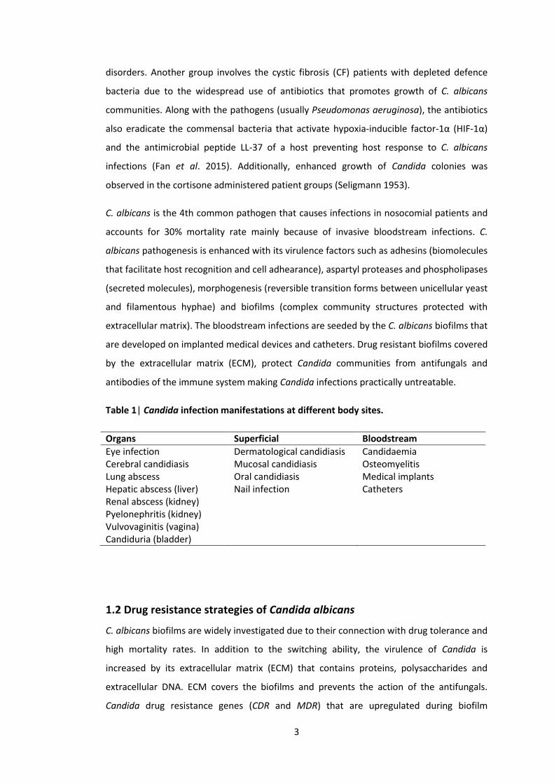
Table 1| Candida infection manifestations at different body sites.
Organs Superficial Bloodstream
Eye infection Dermatological candidiasis Candidaemia Cerebral candidiasis Mucosal candidiasis Osteomyelitis Lung abscess Oral candidiasis Medical implants Hepatic abscess (liver) Nail infection Catheters Renal abscess (kidney) Pyelonephritis (kidney) Vulvovaginitis (vagina) Candiduria (bladder)
1.2 Drug resistance strategies of Candida albicans
C. albicans biofilms are widely investigated due to their connection with drug tolerance and
high mortality rates. In addition to the switching ability, the virulence of Candida is
increased by its extracellular matrix (ECM) that contains proteins, polysaccharides and
extracellular DNA. ECM covers the biofilms and prevents the action of the antifungals.
Candida drug resistance genes (CDR and MDR) that are upregulated during biofilm

4
formation, encode for 2 types of efflux pumps, ABC transporters (CDR) and MFS (major)
facilitators (MDR) further increasing drug tolerance of Candida (Ramage et al. 2002). C.
albicans communities in the biofilms (only) also contain persister cells, variants of normal
cells that are usually in a dormant state, surviving the high doses of antifungals (LaFleur et
al. 2006; Lewis 2012). Due to the latter, the commercially available antifungals (azoles,
polyenes and echinocandins) are often ineffective against Candida biofilms. Azoles inhibit
lanosterol, polyenes target ergosterol of cell membrane and weaken it, and echinocandins
inhibit the synthesis of glucans in the fungal cell wall. Ineffectiveness of these drugs directs
research towards alternative therapeutic strategies. These strategies were reviewed by
Nett (2014) and include targeting extracellular matrix (ECM) and quorum sensing (QS) (Nett
2014). The recent insights in the field of biofilms and drug resistance have also highlighted
the role of combination therapy that includes the use of the antifungals and the natural
compounds derived from the plants (e.g. menthol, nerol), fungi (e.g. penicillin) and bacteria
(e.g. Pseudomonas phenazines and QS molecules) (Kerr et al. 1999; Hogan and Kolter 2002;
Hogan et al. 2004; McAlester et al. 2008; Deveau and Hogan 2011). This approach reduces
the probability for resistance development which is the main issue for the fungal infection
treatments.
1.3 Candida albicans morphology and morphogenesis
The human pathogen, C. albicans, is a polymorphic fungus with a complex life cycle. In
order to adapt to new environments it develops a full repertoire of distinct morphological
forms including budding yeast (blastospore), intermediate pseudohyphae (Sudbery et al.
2004), filamentous hyphae, mycelium with secondary blastospores and biofilms with
extracellular matrix (ECM) (recently reviewed in detail by Nobile and Johnson 2015). The
ploidy plasticity of C. albicans allows rapid adaptation to the stressful conditions (Berman
2016). Different cell types vary in terms of polarization degree, septum position and
nucleus movement. However, yeast, pseudohyphae and hyphae possess a single nucleus in
each cell before mitosis. Uhl et al. (2003) identified 146 genes that are involved in switch
between yeast and filamentous growth. The morphological switch between these forms
represents a crucial factor for C. albicans virulence (Calderone and Fonzi 2001) (Figure 2).
Apart from yeast to hyphae switch, the white round yeast cells can also be transformed
into the elongated opaque cells reversibly and proliferate by mating projection (Slutsky et
al. 1987; Rikkerink et al. 1988; Magee and Magee 1997; Molero et al. 1998). Ssn6p plays an
important role in white-opaque switching (Hernday et al. 2016). C. albicans gray phenotype
5
was recently described by Tao et al. 2014. Additionally, C. albicans has the ability to form
chlamydospores with thicker cell wall in response to the nutrient or environmental stress
(Fabry et al. 2003). Chlamydospores are larger than blastospores and possess thicker cell
wall. In order to develop the chlamydospores, C. albicans mainly requires 6 genes (ISW2,
MDS3, RIM13, RIM101, SCH9 and SUV3) (Nobile et al. 2003) and a MAPK Hog1 (Alonso-
Monge et al. 2003). However, C. albicans biofilms represent the most intriguing topic that
has received enhanced scientific focus because they are responsible for virulence and drug
resistance that lead to increased morbidity and enormous economic expenditure (Brajtburg
et al. 1981; Lamfon et al. 2004).
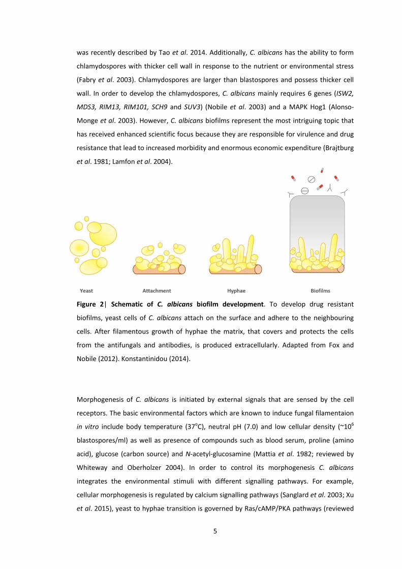
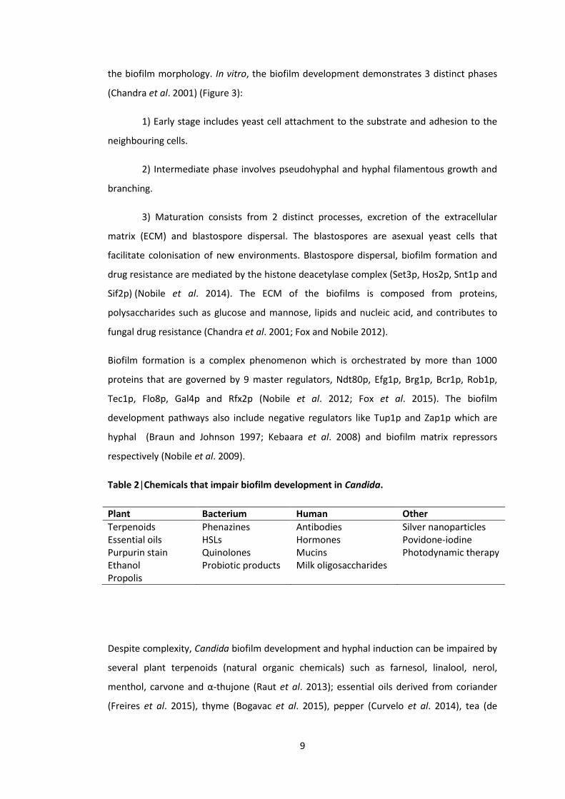
Figure 2| Schematic of C. albicans biofilm development. To develop drug resistant
biofilms, yeast cells of C. albicans attach on the surface and adhere to the neighbouring
cells. After filamentous growth of hyphae the matrix, that covers and protects the cells
from the antifungals and antibodies, is produced extracellularly. Adapted from Fox and
Nobile (2012). Konstantinidou (2014).
Morphogenesis of C. albicans is initiated by external signals that are sensed by the cell
receptors. The basic environmental factors which are known to induce fungal filamentaion
in vitro include body temperature (37oC), neutral pH (7.0) and low cellular density (~106
blastospores/ml) as well as presence of compounds such as blood serum, proline (amino
acid), glucose (carbon source) and N-acetyl-glucosamine (Mattia et al. 1982; reviewed by
Whiteway and Oberholzer 2004). In order to control its morphogenesis C. albicans
integrates the environmental stimuli with different signalling pathways. For example,
cellular morphogenesis is regulated by calcium signalling pathways (Sanglard et al. 2003; Xu
et al. 2015), yeast to hyphae transition is governed by Ras/cAMP/PKA pathways (reviewed
6
by Hogan and Sundstrom 2009), hyphal development is coordinated by adenylyl cyclase
(cAMP) pathways (Rocha et al. 2001) and pseudohyphal induction is orchestrated by MAPK
pathways (reviewed by Srinivasa et al. 2012). These pathways involve some key
transcriptional factors such as Efg1p, Tup1p, Ssn6p, Nrg1p, Brg1p and Cph1p that play an
important role in the signal transduction. For instance, inactivation of the transcription
factors Cph1p (MAPK pathway) or Efg1p (Ras/cAMP/PKA pathway) can inhibit hyphal
growth (Lo et al. 1997). These transcription factors collaborate with the histone
deacetylases for the morphological transition of C. albicans (reviewed by Kim et al. 2015).
Deacetylases are the enzymes that can remove acetyl groups from the amino acids of a
histone allowing DNA to wrap tighter around the histone.
1.4 Biology of yeast, pseudohyphae and hyphae
Early publications have broadly noted the pleiotropic pathogenicity of C. albicans. Candida
causes denture stomatitis (Lilienthal 1955), asthma (Huguenin-Dumittan and Girard 1972),
vaginitis (Banner 1974), endocarditis (Calderone et al. 1978), septicaemia (Rosin 1974) and
infects burn wounds (Albano and Schmitt 1973). The different morphological forms of
C. albicans induce distinct T helper (Th) cell responses during adaptive immunity providing
tissue-specific protection (Kashem et al. 2015). The Th17 and Th1 cell responses provide
protection against cutaneous and systemic infections respectively. For instance, the yeast
form of Candida drives Th17 cell response while a filamentous Candida induces Th1 cell
response (Kashem et al. 2015). C. albicans morphological forms including yeast,
pseudohyphae and hyphae, play a determinant role in fungal virulence (Figure 3).
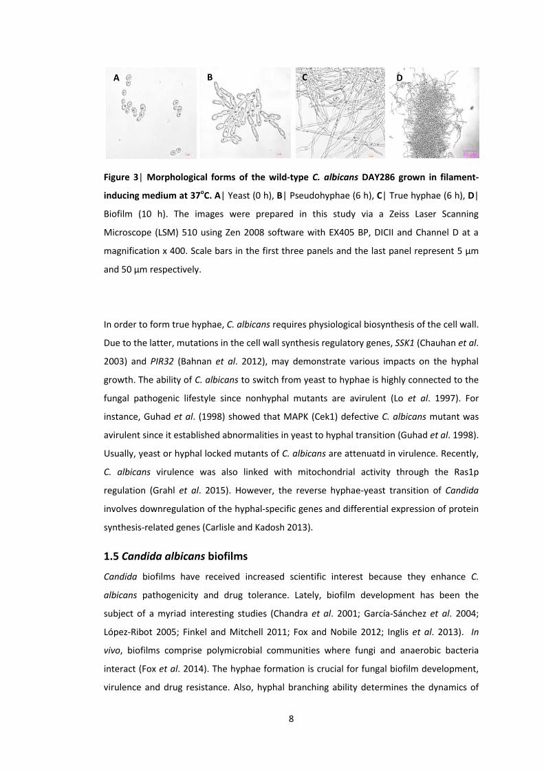
1.4.1 Yeast
The simplest form of C. albicans is a round unicellular yeast (blastospore) formed
vegetatively (asexual cell division) via the budding process. Budding cell selection is a
temperature dependent phenomenon. Under propitious conditions septin rings signal
nuclei to divide across the mother bud neck by asymmetric budding (Sudbery 2001). Fungal
isotropic growth is characterised by actin polarization. After cytokinesis, the smaller
daughter cell disassociates from the mother cell and enters the next cell cycle once it
reaches the threshold size (Figure 3A). A sexual mating and a white-opaque switching can
be regulated through the pH of the environment (Sun et al. 2015).
1.4.2 Pseudohyphae
The defining characteristic of C. albicans pseudohyphal cells is their ellipsoid shape. They
bud in an unipolar manner although septin rings appear before the budding process like in
7
the yeast cells (Sudbery 2001). However, pseudohyphal cells stay in G2 phase longer than
the yeast cells. During the polarized pseudohyphal growth, each cell remains attached to
another but they are separated by the septa, forming the pseudofilamentous pattern. The
new cells enter the next cell cycle in a more synchronized manner than the yeast cells. The
elongated pseudohyphal cells form chains and rough colonies after cytokinesis (Figure 3B).
The pseudohyphae-associated genes are expressed at low levels and represent a small
subset of the hypha-related genes (Carlisle and Kadosh 2013).
1.4.3 Hyphae
C. albicans filamentous growth is regulated independently from its cell cycle (Hazan et al.
2002). The yeast-hyphal transition is driven by the extended duration of filament-specific
gene expression (Carlisle and Kadosh 2013). C. albicans true hyphal cells possess
considerable elongation, parallel walls and extreme degrees of polarity (Figure 3C).
Polarized growth mode implicates polarisome elements as well as septins, tag/bud site
components, Cdc42 module (cell division control protein) and actin-myosin system
(Whiteway and Oberholzer 2004). The hyphal cell nucleus divides in the elongated germ
tube. Expansion of the filaments is initiated with the asynchronous cell cycle since solely
the apical cells start dividing whereas the subapical cells remain in the primary cell phase
G1 (Barelle et al. 2003). Transcriptional regulator Ash1, which controls filamentous growth,
is widely associated with the asymmetric cell division as it is found in the nucleus of apical
but not of subapical cells (Inglis and Johnson 2002). Asymmetric vacuolar inheritance also
appears to be vital in the hyphae formation (Barelle et al. 2003). The true filaments
develop via germ tube elongation process and filamentous cells display no distinct
constrictions (Berman and Sudbery 2002). Hyphal formation is correlated with the
bioactivity of small GTPases (Cdc42/Cdc24 (Ushinsky et al. 2002; Bassilana et al. 2003)),
myosins (molecular motors Myo3/5 (Oberholzer et al. 2002)) and PKs (Hsl1p (Umeyama et
al. 2005) and Cek1p (Csank et al. 1998)) that are likely to interact with the actin network.
8
*
Figure 3| Morphological forms of the wild-type C. albicans DAY286 grown in filament-
inducing medium at 37oC. A| Yeast (0 h), B| Pseudohyphae (6 h), C| True hyphae (6 h), D|
Biofilm (10 h). The images were prepared in this study via a Zeiss Laser Scanning
Microscope (LSM) 510 using Zen 2008 software with EX405 BP, DICII and Channel D at a
magnification x 400. Scale bars in the first three panels and the last panel represent 5 μm
and 50 μm respectively.
In order to form true hyphae, C. albicans requires physiological biosynthesis of the cell wall.
Due to the latter, mutations in the cell wall synthesis regulatory genes, SSK1 (Chauhan et al.
2003) and PIR32 (Bahnan et al. 2012), may demonstrate various impacts on the hyphal
growth. The ability of C. albicans to switch from yeast to hyphae is highly connected to the
fungal pathogenic lifestyle since nonhyphal mutants are avirulent (Lo et al. 1997). For
instance, Guhad et al. (1998) showed that MAPK (Cek1) defective C. albicans mutant was
avirulent since it established abnormalities in yeast to hyphal transition (Guhad et al. 1998).
Usually, yeast or hyphal locked mutants of C. albicans are attenuatd in virulence. Recently,
C. albicans virulence was also linked with mitochondrial activity through the Ras1p
regulation (Grahl et al. 2015). However, the reverse hyphae-yeast transition of Candida
involves downregulation of the hyphal-specific genes and differential expression of protein
synthesis-related genes (Carlisle and Kadosh 2013).
1.5 Candida albicans biofilms
Candida biofilms have received increased scientific interest because they enhance C.
albicans pathogenicity and drug tolerance. Lately, biofilm development has been the
subject of a myriad interesting studies (Chandra et al. 2001; García-Sánchez et al. 2004;
López-Ribot 2005; Finkel and Mitchell 2011; Fox and Nobile 2012; Inglis et al. 2013). In
vivo, biofilms comprise polymicrobial communities where fungi and anaerobic bacteria
interact (Fox et al. 2014). The hyphae formation is crucial for fungal biofilm development,
virulence and drug resistance. Also, hyphal branching ability determines the dynamics of
A
D
B C D
9
the biofilm morphology. In vitro, the biofilm development demonstrates 3 distinct phases
(Chandra et al. 2001) (Figure 3):
1) Early stage includes yeast cell attachment to the substrate and adhesion to the
neighbouring cells.
2) Intermediate phase involves pseudohyphal and hyphal filamentous growth and
branching.
3) Maturation consists from 2 distinct processes, excretion of the extracellular
matrix (ECM) and blastospore dispersal. The blastospores are asexual yeast cells that
facilitate colonisation of new environments. Blastospore dispersal, biofilm formation and
drug resistance are mediated by the histone deacetylase complex (Set3p, Hos2p, Snt1p and
Sif2p) (Nobile et al. 2014). The ECM of the biofilms is composed from proteins,
polysaccharides such as glucose and mannose, lipids and nucleic acid, and contributes to
fungal drug resistance (Chandra et al. 2001; Fox and Nobile 2012).
Biofilm formation is a complex phenomenon which is orchestrated by more than 1000
proteins that are governed by 9 master regulators, Ndt80p, Efg1p, Brg1p, Bcr1p, Rob1p,
Tec1p, Flo8p, Gal4p and Rfx2p (Nobile et al. 2012; Fox et al. 2015). The biofilm
development pathways also include negative regulators like Tup1p and Zap1p which are
hyphal (Braun and Johnson 1997; Kebaara et al. 2008) and biofilm matrix repressors
respectively (Nobile et al. 2009).
Table 2|Chemicals that impair biofilm development in Candida.
Plant Bacterium Human Other
Terpenoids Phenazines Antibodies Silver nanoparticles Essential oils HSLs Hormones Povidone-iodine Purpurin stain Quinolones Mucins Photodynamic therapy Ethanol Probiotic products Milk oligosaccharides Propolis
Despite complexity, Candida biofilm development and hyphal induction can be impaired by
several plant terpenoids (natural organic chemicals) such as farnesol, linalool, nerol,
menthol, carvone and α-thujone (Raut et al. 2013); essential oils derived from coriander
(Freires et al. 2015), thyme (Bogavac et al. 2015), pepper (Curvelo et al. 2014), tea (de
10
Campos Rasteiro et al. 2014), cinnamon (Pires et al. 2011), lemon (Oliveira et al. 2014) and
carrot (Alves-Silva et al. 2016); as well as with propolis (Freires et al. 2016). The biofilms are
additionally affected by exogenous human hormones including progesterone and
oestradiol (Kinsman et al. 1988; Zhao et al. 1995), the mucins covering the epithelial cells
(Kavanaugh et al. 2014) and the oligosaccharides contained in the human milk (Gonia et al.
2015). Also, biofilms are modulated by application of purpurin (Tsang et al. 2012), ethanol
(Peters et al. 2013) and photodynamic treatment (Sousa et al. 2016). Photodynamic
therapy was also effective against Candida in the murine model in vivo (Fabio et al. 2016).
Recently, the use of povidone-iodine ointment (Hoekstra et al. 2016) and silver
nanoparticals (drug delivery system) were also proposed as one of the effective strategies
for the treatment of C. albicans infections (Qasim et al. 2015; Wang and Xie 2015). The
probiotic supplements containing Bacillus subtilis were found to be effective against oral
infections caused by Candida species (Zhao et al. 2016). Apart from these factors, C.
albicans morphology and biofilm formation is also influenced by some Gram-negative
bacteria including P. aeruginosa (Peleg et al. 2010; Holcombe et al. 2011). P. aeruginosa
phenazines (parent substance of stains/dyes including safranin), pyocyanin, phenazine
methosulfate and phenazine-1-carboxylate, affect C. albicans biofilm development and
metabolism (Gibson et al. 2009; Morales et al. 2013) (Table 2).
1.6 Interaction between Candida albicans and Pseudomonas aeruginosa
In clinical settings, the majority of the infections are polymicrobial. Multiple pathogens
including yeast, bacteria and viruses, can colonize and infect the same niche. For example,
C. albicans is often coisolated with an opportunistic human pathogen bacterium P.
aeruginosa. C. albicans and P. aeruginosa infections are difficult to treat since both can
form biofilms resisting the antifungal and antibiotic treatments respectively (Kojic and
Darouiche 2004). P. aeruginosa is one of the best studied bacterium found in the plethora
of niches due to its extremely adaptable abilities. P. aeruginosa can colonise kidneys and
urinary as well as gastrointestinal tract of susceptible individuals causing inflammation and
sepsis. Additionally, P. aeruginosa is the main cause of morbidity in populations with cystic
fibrosis (CF) (Govan and Deretic 1996; Chambers et al. 2005; Leclair and Hogan 2010) and
AIDS (Mendelson et al. 1994). Authors of independent studies have established the
importance of quorum sensing (QS) system for P. aeruginosa fitness (Heurlier et al. 2006),
virulence (Smith and Iglewski 2003) and inter-kingdom signalling (Shiner et al. 2005).

11
A wide variety of investigations suggest that C. albicans and P. aeruginosa can coexist and
interact in both natural and clinical settings (Hogan and Kolter 2002; Nseir et al. 2007;
Gibson et al. 2009). C. albicans and P. aeruginosa were coisolated from serious burn
wounds (Gupta et al. 2005) and the lungs of the CF patients (Martin et al. 1993; Bakare et
al. 2003). CF patients usually demonstrate imbalance in the microbial flora as a result of
chronic use of antibiotics that leads to candidiasis (Burns et al. 1999). C. albicans and P.
aeruginosa have an antagonistic interaction. In vitro analysis established that C. albicans
biofilm formation and metabolism can be influenced by P. aeruginosa phenazines (Gibson
et al. 2009; Morales et al. 2013). For instance, P. aeruginosa phenazines enhance ethanol
production in C. albicans to stimulate biofilm formation in Pseudomonas (Chen et al. 2014).
Another phenazine, methosulphate (PMS), can kill Candida within its biofilms (Morales et
al. 2010; Morales et al. 2013). Several studies have also shown that Pseudomonas can
inhibit Candida biofilm development in vitro (Holcombe et al. 2010; Bandara et al. 2010a;
Bandara et al. 2010b; Reen et al. 2011; Bandara et al. 2013). Our previous studies further
confirmed that this biofilm inhibition is N-acyl-L-Homoserine Lactone (HSL)-independent
(Holcombe et al. 2010; Konstantinidou and Morrissey 2015). Collectively, these data
suggest that Candida biofilm development and metabolism are intimately related with each
other (Lindsay et al. 2014).
Interaction between C. albicans and P. aeruginosa is mainly based on signalling. Several
studies suggest that Pseudomonas QS molecules are responsible for the signal-mediated
communication (Hogan and Kolter 2002; McAlester et al. 2008; Deveau and Hogan 2011).
Pseudomonas QS molecules that are known to modulate Candida – Pseudomonas
interaction include two types of HSLs, N-(3-oxododecanoyl)-L-homoserine lactone (3O-C12-
HSL) (Hogan et al. 2004; McAlester et al. 2008) and N-butyryl-L-homoserine lactone (C4-
HSL) (Smith and Iglewski 2003), as well as HHQ (2-heptyl-4-quinolone) (Reen et al. 2011). P.
aeruginosa can inhibit morphological switch of C. albicans from yeast to hyphae using
these secreted chemicals. It was reported that Pseudomonas HSLs can inhibit the switch of
Candida from yeast to filamentous growth (Hogan et al. 2004). However, QS molecule of C.
albicans, farnesol, can limit the virulence of P. aeruginosa by blocking the production of
Pseudomonas QS molecules and pyocyanin (Kerr et al. 1999; Cugini et al. 2007) and
affecting motility (McAlester et al. 2008). P. aeruginosa pyocyanin and 1-hydroxyphenazine
can prevent the growth of C. albicans (Kerr et al. 1999).
12
Other factors that affect the interaction between C. albicans and P. aeruginosa include iron
availability (Purschke et al. 2012), bacterial cell wall lipopolysaccharides (LPS) (Bandara et
al. 2010a; Bandara et al. 2013) and extracellular DNA (Sapaar et al. 2014).
1.7 Quorum Sensing
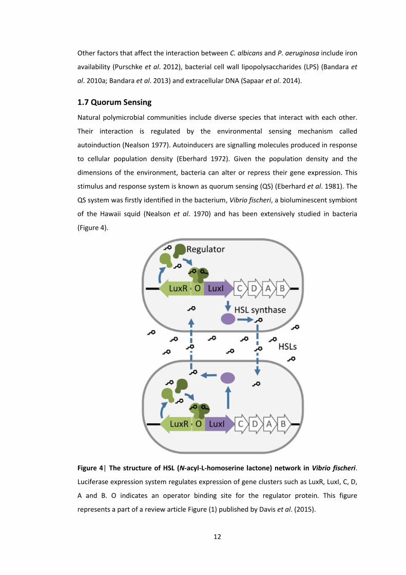
Natural polymicrobial communities include diverse species that interact with each other.
Their interaction is regulated by the environmental sensing mechanism called
autoinduction (Nealson 1977). Autoinducers are signalling molecules produced in response
to cellular population density (Eberhard 1972). Given the population density and the
dimensions of the environment, bacteria can alter or repress their gene expression. This
stimulus and response system is known as quorum sensing (QS) (Eberhard et al. 1981). The
QS system was firstly identified in the bacterium, Vibrio fischeri, a bioluminescent symbiont
of the Hawaii squid (Nealson et al. 1970) and has been extensively studied in bacteria
(Figure 4).
Figure 4| The structure of HSL (N-acyl-L-homoserine lactone) network in Vibrio fischeri.
Luciferase expression system regulates expression of gene clusters such as LuxR, LuxI, C, D,
A and B. O indicates an operator binding site for the regulator protein. This figure
represents a part of a review article Figure (1) published by Davis et al. (2015).
13
Inter-kingdom communication across the prokaryotic and eukaryotic organisms is based on
the QS molecules. For paradigm, a bacterial HSL (N-acyl-L-homoserine lactone) can regulate
the gene expression in eukaryotes. The eukaryotes recognise and respond to the bacterial
HSLs because their chemical structure considerably resembles eukaryotic hormones. This
phenomenon is known as “global sensing” (Shiner et al. 2005; Hartmann and Schikora
2012).
1.7.1 Pseudomonas aeruginosa quorum sensing molecules
P. aeruginosa possesses a well-studied QS system (Pesci et al. 1997; Smith and Iglewski
2003; Bjarnsholt et al. 2010). The two known QS networks in P. aeruginosa are Las and Rhl
(Pesci et al. 1997). Both systems employ a transcriptional activator and an autoinducer
synthase to control gene expression (Pearson et al. 1997). Pseudomonas QS network
regulates semantic functions such as virulence, protein secretion, swarming motility, 4-
quinolone signalling as well as production of secondary metabolites, exoenzymes and
exotoxins (Diggle et al. 2008).
Pseudomonas supernatants are rich in signalling molecules. PQS and its precursor HHQ (2-
heptyl-4-quinolone) play an important role in Pseudomonas signalling. P. aeruginosa
secrets the HSLs, N-(3-oxododecanoyl)-L-homoserine lactone (3O-C12-HSL) and N-butyryl-L-
homoserine lactone (C4-HSL), that signal and regulate C. albicans behaviour. P. aerugionsa
also produces phenazines such as phenazine methosulfate, phenazine-1-carboxylate and
pyocyanin that can impair C. albicans biofilm development and metabolism (Gibson et al.
2009; Morales et al. 2013). Recently, coumarin (fragrance oil) was shown to inhibit P.
aeruginosa phenazine production, biofilm development and swarming motility (Gutiérrez-
Barranquero et al. 2015).
1.7.2 Candida albicans quorum sensing molecules
To promote biofilm formation and pathogenesis C. albicans also produces signalling
molecules (Kruppa 2009). C. albicans QS molecules control the initiation of hyphae
development via the protein degradation mediated with Ubr1 (Lu et al. 2014). C. albicans
excretes among others two well-established QS molecules, farnesol and tyrosol (Figure 5).
Farnesol is a water insoluble organic alcohol present in essential oils and used in
perfumery. Farnesol represents a natural pesticide and insect pheromone. Additionally,
farnesol is reported to possess antitumor (Joo and Jetten 2009) and antibacterial properties
(Kromidas et al. 2006). For example, farnesol alters cell morphology and disrupts cell
14
membrane integrity of a bacterium Acinetobacter baumannii ultimately leading to the
biofilm impairment (Kostoulias et al. 2015).
C. albicans produces farnesol in order to control its cell density (Figure 5A). Farnesol is
secreted by solely the white cells of C. albicans. The principal biological function of farnesol
is an inhibition of C. albicans filamentation (Hornby et al. 2001) by affecting cell amino acid
incorporation (Braun 2005). Farnesol causes inflammation response by activation of the
human innate immune cells (neutrophils and monocytes) simultaneously suppressing
cellular adaptive immunity, differentiation of monocytes into immature dendritic cells
(Leonhardt et al. 2015). Farnesol participates in complex signal transduction pathways of
yeast to hyphae transition. Its mechanism of action involves activation of Ras/cAMP/PKA
pathways (Davis-Hanna et al. 2008) and of a hyphal repressor, Tup1p (Kebaara et al. 2008),
as well as of a Chk1p kinase (Kruppa et al. 2004). Farnesol has the ability to prevent biofilm
formation in its late phase but cannot affect already existing hyphae (Hornby et al. 2001).
Moreover, farnesol elevates the expression of genes regulating hyphal formation, drug
resistance, cell wall maintenance and heat shock protein production (Cao et al. 2005).
Westwater et al. (2005) also proposed a possible link between farnesol and the oxidative
stress resistance. Collectively these data indicate that farnesol affects the dynamics of C.
albicans morphogenesis (Martins et al. 2007).
Another QS molecule of C. albicans, tyrosol, represents a natural antioxidant derived from
an aromatic phenethyl alcohol (Figure 5B). White wine and olive oil that are known to
possess cardioprotective properties are rich in tyrosol (Samuel et al. 2008; Miró-Casas et al.
2003). Chen et al. (2004) showed that tyrosol is an autoregulatory QS molecule that delays
fungal growth and stimulates germ tube formation in the diluted population of C. albicans
(Chen et al. 2004). After more investigation it became evident that tyrosol also affects
intermediate and early stages of the hyphae formation (Alem et al. 2006). In synopsis, C.
albicans QS molecules, farnesol and tyrosol, have antagonistic function on filamentation.
15
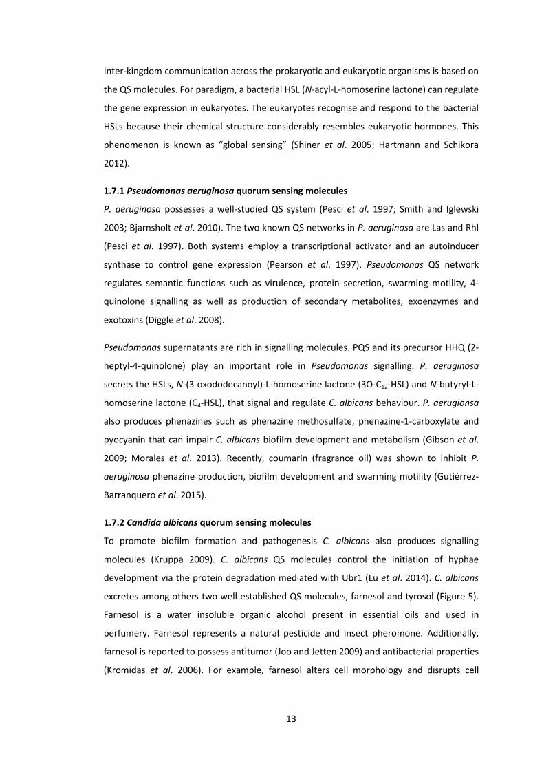
A
Figure 5| Quorum sensing (QS) molecule biosynthesis pathways in C. albicans. The
Enzymes shown in bold indicate experimental evidence for their enzymatic activity. A|
Biosynthesis pathway of farnesol. Farnesol is produced from farnesyl pyrophosphate, an
intermediate in the ergosterol biosynthesis pathway and inhibits C. albicans filamentation
and biofilm formation. B| Biosynthesis pathway of tyrosol. Both, constitutive (Aro8p) and
inducible (Aro9p) aromatic aminotransferases can catalyze the initial reaction in tyrosine
degradation. Tyrosol stimulates growth under dilute culture conditions and has a protective
effect against human phagocytic cells. Tyrosol production is enhanced in biofilms compared
to planktonic culture. Figures were obtained from Candida Genome database (CGD)
(Arnaud et al. 2005).
B

16
Chapter 2
Co-occurrence of filamentation defects and impaired biofilms in
Candida albicans protein kinase mutants
Nina Konstantinidou and John Patrick Morrissey
School of Microbiology, University College Cork, Cork, T12YN60, Ireland
Key words: Candida albicans; protein kinase; biofilms; filamentation; yeast – hyphae.
Subtitle: Candida albicans morphology and biofilms
This chapter was published as part of publication by Konstantinidou N & Morrissey JP
(2015) Co-occurrence of filamentation defects and impaired biofilms in Candida albicans
protein kinase mutants. FEMS Yeast Res 15: 1–10. pii fov092 doi: 10.1093/femsyr/fov092.
Here it is represented with some figure rearrangements.
17
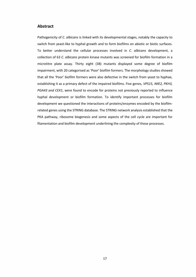
Abstract
Pathogenicity of C. albicans is linked with its developmental stages, notably the capacity to
switch from yeast-like to hyphal growth and to form biofilms on abiotic or biotic surfaces.
To better understand the cellular processes involved in C. albicans development, a
collection of 63 C. albicans protein kinase mutants was screened for biofilm formation in a
microtitre plate assay. Thirty eight (38) mutants displayed some degree of biofilm
impairment, with 20 categorised as ‘Poor’ biofilm formers. The morphology studies showed
that all the ‘Poor’ biofilm formers were also defective in the switch from yeast to hyphae,
establishing it as a primary defect of the impaired biofilms. Five genes, VPS15, IME2, PKH3,
PGA43 and CEX1, were found to encode for proteins not previously reported to influence
hyphal development or biofilm formation. To identify important processes for biofilm
development we questioned the interactions of proteins/enzymes encoded by the biofilm-
related genes using the STRING database. The STRING network analysis established that the
PKA pathway, ribosome biogenesis and some aspects of the cell cycle are important for
filamentation and biofilm development underlining the complexity of these processes.
18
2. Co-occurrence of filamentation defects and impaired biofilms in
Candida albicans protein kinase mutants
2.1 Introduction
C. albicans has emerged as a serious nosocomial pathogen in part due to formation of drug
resistant biofilms on indwelling medical devices such as urinary and vascular catheters.
Hence, fungal biofilms have received a significant interest during the last decade (Chandra
et al. 2001; López-Ribot 2005; Inglis et al. 2013). Biofilm development represents a complex
phenomenon and in vitro it demonstrates three distinct phases. The early stage includes
yeast cell attachment to the substrate as well as adhesion to the neighbouring cells. The
mechanisms governing yeast attachment are important since they determine subsequent
biofilm development. The intermediate phase involves pseudohyphal and hyphal
filamentous growth. The maturation phase consists from two distinct processes, excretion
of extracellular matrix (ECM) and blastospore dispersal (Chandra et al. 2001; Fox and
Nobile 2012). This complex phenomenon is orchestrated by more than 1000 proteins
including PKs, which are governed by 9 master regulators, Ndt80p, Efg1p, Brg1p, Bcr1p,
Rob1p Tec1p, Flo8p, Gal4p and Rfx2p, that regulate signal transduction pathways at the
genetic level (Nobile et al. 2012; Fox et al. 2015).
Biofilm development is regulated by diverse pathways but some well-characterised
pathways are involved in the yeast to hyphae transition, Ras/cAMP/PKA (reviewed by
Hogan and Sundstrom 2009); pseudohyphal induction, MAPK (reviewed by Srinivasa et al.
2012); and morphogenesis, calcium signalling pathways (Sanglard et al. 2003). For instance,
inactivation of the transcription factors Cph1p (MAPK pathway) or Efg1p (Ras/cAMP/PKA
pathway) can inhibit hyphal growth and biofilm development (Lo et al. 1997). Also, it was
demonstrated that signalling via adenylyl cyclase is essential for hyphal development since
cells defective in Cdc35p were unable to develop filaments (Rocha et al. 2001). The basic
environmental factors characterized to induce fungal filamentation in vitro include body
temperature (37oC), neutral pH (7.0), and low cellular density (~106 blastospores/ml), as
well as presence of compounds such as serum (blood plasma without clotting factors),
proline, glucose and N-acetyl-D-glucosamine (Mattia et al. 1982; Whiteway and Oberholzer
2004).
Protein phosphorylation is important for signal transduction processes with sequential
activation of proteins often mediated by PKs. In this study, we took advantage of a set of PK

19
mutants to ask which PKs are required for biofilm formation in C. albicans. This collection
has been the subject to many different screens showing that particular PK genes were
responsible for cell wall regulation (Blankenship et al. 2010), cell-substrate attachment
(Fanning et al. 2013), cell morphology (Blankenship et al. 2010), propolis-induced cell death
(de Castro et al. 2013) and cell metabolism (Morales et al. 2013).
20
2.2 Materials and Methods
2.2.1 Yeast strains and growth conditions
The wild-type C. albicans strains used in this study were BWP17
(ura3Δ::λimm434/ura3Δ::λimm434 his1::hisG/his1::hisG arg4::hisG/arg4::hisG) (Wilson et
al. 1999) and DAY286 (ura3::λimm434 his1::hisG pARG4::URA3::arg4::hisG) (Davis et al.
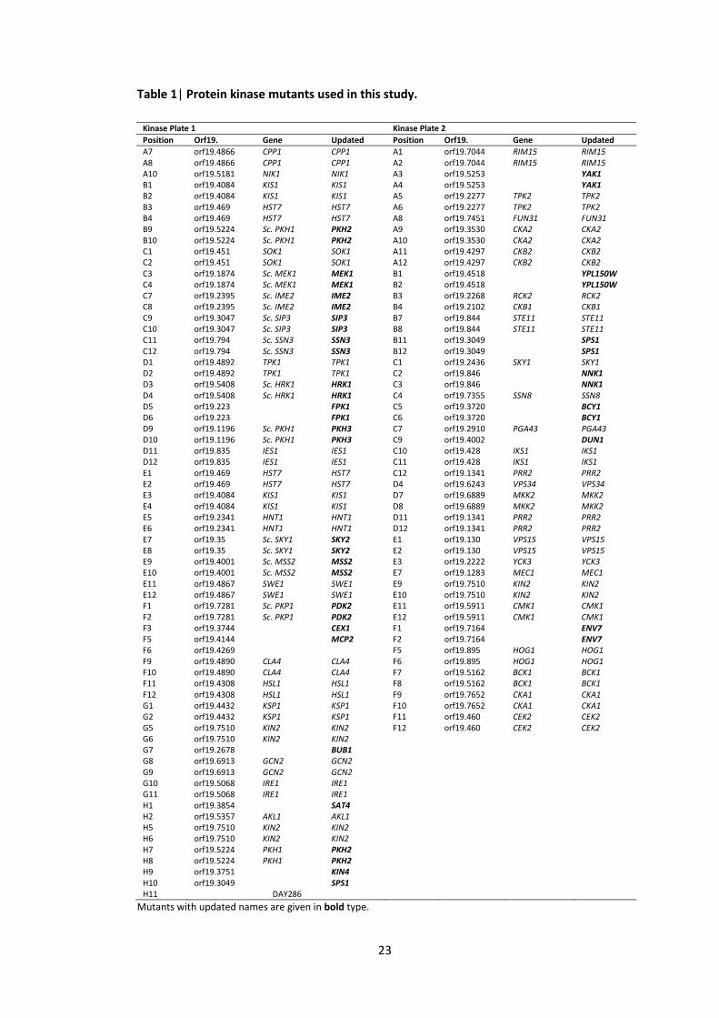
2002). The PK mutants of C. albicans are listed in Table 1. This kinase set was created by
Aaron Mitchell’s group (Blankenship et al. 2010) and obtained from the Fungal Genetics
Stock Center (www.fgsc.net/candida/FGSCcandidaresources.htm). PK homozygous
insertion mutants were created in BWP17 parental strain via Tn7-UAU1 cassette
(Blankenship et al. 2010). The majority of the PK-coding genes were represented by double
independent mutant strains but in some cases only a single mutant was available. In total,
we examined 63 genes using 45 independent duplicate mutants and 18 single mutants. The
initial FGSC collection was larger and included mutants deleted in ~80 PK/PK-related genes.
Our reduced set comprised the mutants that, after delivery and culturing, we were able to
verify by strain-specific PCR (see below).
Yeast strains were routinely cultured in standard rich medium containing 1% yeast extract,
2% peptone and 2% glucose (YPD). For biofilm and morphological analyses, the yeast
strains were pre-grown in non-filament-inducing medium YNB (yeast nitrogen base), as
described by McAlester et al. (2008) and Holcombe et al. (2010) with some adjustments.
Briefly, filter-sterilized YNB salts without amino acids (Difco 291940) were supplemented
with 0.2% glucose, 0.1% maltose and 0.16% filter-sterilized synthetic amino acid drop-out
Leu– (Kaiser Formedium DSCK052). For the induction of hyphal growth the strains were
transferred to filament-inducing medium, YNBNP, which consisted from YNB supplemented
with 2.5 mM N-acetyl-D-glucosamine (Sigma A8625) and 25 mM phosphate (sodium) buffer
(pH 7).
2.2.2 PCR
DNA of wild-type C. albicans and PK mutants was extracted according to Hoffman (2001)
protocol. The primers were designed with SnapGene (www.snapgene.com) and their
sequences are listed in the Table 2. Strain-specific PCR was carried out using primers
flanking the gene insertion sites listed in the Supplementary Table S1 of Blankenship et al.
(2010) publication. Absence of the band indicated a mutant disrupted in the gene amplified
by the corresponding primers (Appendix 2.5). All the PCR reactions were carried out
21
utilising a GoTaq Green Master Mix (Promega) according to the manufacturers’
instructions.
2.2.3 Biofilm assay
A microtiter plate-based biofilm assay (XTT reductase salts) that measured metabolically
active cells, was carried out as described by Ramage et al. (2001) and Holcombe et al.
(2010). Briefly, the yeast strains were pre-grown in non-filament-inducing medium (YNB)
overnight (30oC) and diluted into filament-inducing medium (YNBNP) to A6000.05. The
diluted cultures (100 μL) were incubated for 1 h (37°C) in flat-bottomed 96-well
polystyrene plates. After incubation, the attached cells were washed with fresh YNBNP
medium twice, by inverting the plates carefully, to eliminate non-adhered cells. In order to
induce biofilm formation the plates were incubated in the dark statically for 24 h (37°C).
The next day, the biofilms developed on the bottom of the wells were washed with fresh
YNBNP by careful pipetting. The XTT-menadione solution (100 μL), prepared as below, was
added to the overnight cultures and incubated in the dark for 2 h (37oC). Lastly, the dyed
supernatants (80 μL) were transferred to a clean plate for the quantification at a
wavelength of A490 nm.
For the preparation of the XTT solutions, 10 mM menadione (Sigma M5625) was dissolved
in pure acetone and added (10 μL) to the XTT solution. The latter was prepared by
dissolving 0.015 g of XTT powder (Sigma X4626) in 30 mL sterile dH2O and filtered with 0.2
μm pore size filter.
To increase the statistical power of our experiments we carried out 2 independent screens
and tested the mutants in triplicate with eight technical replicates (n = 24). Biofilms of the
control, wild-type C. albicans DAY286, ranged from approximately A4901 to A4901.5. Thus,
the mutants with biofilms above A4901 and below A4900.5 were assigned to the ‘Good’ and
‘Poor’ biofilm formers respectively. The remaining mutants were ‘Moderate’ biofilm
formers. A distribution of each sample was assessed and the coincidence of the median and
the mean indicated a Gaussian (normal) distribution of our data. All the statistical analysis
of the biofilm assays were carried out in R Statistical Software (R Development Core Team
2013).
2.2.4 Morphological analyses
Morphological assays were carried out as described previously by Hogan et al. (2004) and
McAlester et al. (2008). Briefly, the yeast cultures were pre-grown overnight in YNB broth
22
(30oC) and diluted into YNBNP (A6001.0) to assay their capacity to switch from yeast to
filamentous growth. Before (0 h) and after 6 h and 10 h incubation (37oC), the morphology
of the mutants was examined microscopically. The images were captured digitally at a
magnification x 400 via Zen2008 software with EX405 BP and Channel D by Zeiss Laser
Scanning Microscope (LSM) 510.
2.2.5 Bioinformatics analyses
The protein sequences of C. albicans PKs were retrieved from the CGD (Candida Genome
Database) (Arnaud et al. 2005), the analysis of the protein associations were carried out
using the STRING v9.1 database (Search Tool for the Retrieval of Interacting
Genes/Proteins) (Jensen et al. 2009), the GOs (Gene Ontologies) were obtained with the
Blast2GO annotations (Conesa et al. 2005) and pathway analysis were carried out using
KEGG pathway database (Kyoto Encyclopaedia of Genes and Genomes) (Kanehisa and Goto
2000).
23
Table 1| Protein kinase mutants used in this study.
Kinase Plate 1 Kinase Plate 2
Position Orf19. Gene Updated Position Orf19. Gene Updated
A7 orf19.4866 CPP1 CPP1 A1 orf19.7044 RIM15 RIM15 A8 orf19.4866 CPP1 CPP1 A2 orf19.7044 RIM15 RIM15 A10 orf19.5181 NIK1 NIK1 A3 orf19.5253 YAK1 B1 orf19.4084 KIS1 KIS1 A4 orf19.5253 YAK1 B2 orf19.4084 KIS1 KIS1 A5 orf19.2277 TPK2 TPK2 B3 orf19.469 HST7 HST7 A6 orf19.2277 TPK2 TPK2 B4 orf19.469 HST7 HST7 A8 orf19.7451 FUN31 FUN31 B9 orf19.5224 Sc. PKH1 PKH2 A9 orf19.3530 CKA2 CKA2 B10 orf19.5224 Sc. PKH1 PKH2 A10 orf19.3530 CKA2 CKA2 C1 orf19.451 SOK1 SOK1 A11 orf19.4297 CKB2 CKB2 C2 orf19.451 SOK1 SOK1 A12 orf19.4297 CKB2 CKB2 C3 orf19.1874 Sc. MEK1 MEK1 B1 orf19.4518 YPL150W C4 orf19.1874 Sc. MEK1 MEK1 B2 orf19.4518 YPL150W C7 orf19.2395 Sc. IME2 IME2 B3 orf19.2268 RCK2 RCK2 C8 orf19.2395 Sc. IME2 IME2 B4 orf19.2102 CKB1 CKB1 C9 orf19.3047 Sc. SIP3 SIP3 B7 orf19.844 STE11 STE11 C10 orf19.3047 Sc. SIP3 SIP3 B8 orf19.844 STE11 STE11 C11 orf19.794 Sc. SSN3 SSN3 B11 orf19.3049 SPS1 C12 orf19.794 Sc. SSN3 SSN3 B12 orf19.3049 SPS1 D1 orf19.4892 TPK1 TPK1 C1 orf19.2436 SKY1 SKY1 D2 orf19.4892 TPK1 TPK1 C2 orf19.846 NNK1 D3 orf19.5408 Sc. HRK1 HRK1 C3 orf19.846 NNK1 D4 orf19.5408 Sc. HRK1 HRK1 C4 orf19.7355 SSN8 SSN8 D5 orf19.223 FPK1 C5 orf19.3720 BCY1 D6 orf19.223 FPK1 C6 orf19.3720 BCY1 D9 orf19.1196 Sc. PKH1 PKH3 C7 orf19.2910 PGA43 PGA43 D10 orf19.1196 Sc. PKH1 PKH3 C9 orf19.4002 DUN1 D11 orf19.835 IES1 IES1 C10 orf19.428 IKS1 IKS1 D12 orf19.835 IES1 IES1 C11 orf19.428 IKS1 IKS1 E1 orf19.469 HST7 HST7 C12 orf19.1341 PRR2 PRR2 E2 orf19.469 HST7 HST7 D4 orf19.6243 VPS34 VPS34 E3 orf19.4084 KIS1 KIS1 D7 orf19.6889 MKK2 MKK2 E4 orf19.4084 KIS1 KIS1 D8 orf19.6889 MKK2 MKK2 E5 orf19.2341 HNT1 HNT1 D11 orf19.1341 PRR2 PRR2 E6 orf19.2341 HNT1 HNT1 D12 orf19.1341 PRR2 PRR2 E7 orf19.35 Sc. SKY1 SKY2 E1 orf19.130 VPS15 VPS15 E8 orf19.35 Sc. SKY1 SKY2 E2 orf19.130 VPS15 VPS15 E9 orf19.4001 Sc. MSS2 MSS2 E3 orf19.2222 YCK3 YCK3 E10 orf19.4001 Sc. MSS2 MSS2 E7 orf19.1283 MEC1 MEC1 E11 orf19.4867 SWE1 SWE1 E9 orf19.7510 KIN2 KIN2 E12 orf19.4867 SWE1 SWE1 E10 orf19.7510 KIN2 KIN2 F1 orf19.7281 Sc. PKP1 PDK2 E11 orf19.5911 CMK1 CMK1 F2 orf19.7281 Sc. PKP1 PDK2 E12 orf19.5911 CMK1 CMK1 F3 orf19.3744 CEX1 F1 orf19.7164 ENV7 F5 orf19.4144 MCP2 F2 orf19.7164 ENV7 F6 orf19.4269 F5 orf19.895 HOG1 HOG1 F9 orf19.4890 CLA4 CLA4 F6 orf19.895 HOG1 HOG1 F10 orf19.4890 CLA4 CLA4 F7 orf19.5162 BCK1 BCK1 F11 orf19.4308 HSL1 HSL1 F8 orf19.5162 BCK1 BCK1 F12 orf19.4308 HSL1 HSL1 F9 orf19.7652 CKA1 CKA1 G1 orf19.4432 KSP1 KSP1 F10 orf19.7652 CKA1 CKA1 G2 orf19.4432 KSP1 KSP1 F11 orf19.460 CEK2 CEK2 G5 orf19.7510 KIN2 KIN2 F12 orf19.460 CEK2 CEK2 G6 orf19.7510 KIN2 KIN2
G7 orf19.2678 BUB1 G8 orf19.6913 GCN2 GCN2 G9 orf19.6913 GCN2 GCN2 G10 orf19.5068 IRE1 IRE1 G11 orf19.5068 IRE1 IRE1 H1 orf19.3854 SAT4 H2 orf19.5357 AKL1 AKL1 H5 orf19.7510 KIN2 KIN2 H6 orf19.7510 KIN2 KIN2 H7 orf19.5224 PKH1 PKH2 H8 orf19.5224 PKH1 PKH2 H9 orf19.3751 KIN4 H10 orf19.3049 SPS1 H11 DAY286 Mutants with updated names are given in bold type.
24
Table 2| Primers used in this study.
Forward Sequence (3’ – 5’) Reverse Sequence (3’ – 5’)
BUD32_F ATGACAGATCACCTAATTGCTAAAGTAC BUD32_R TCAACCCAACATACTTCTTTTTCTTCC
CKA2_F AGTTTTCCAAGGTGTCAATGTTTT CKA2_R TTGAAAAATGGATGTTCCATTGCC
CLA4_F CCTTCATCTCAACAACAGCAACAA CLA4_R TCCTTTTGTTTAACCACTTCAGGT
CTK3_F TTCACACAGGAAACAGCTATGACCATGATTACGCCAAGCTcatcacattggtcgtcctggaaatc
CTK3_R TCGACCATATGGGAGAGCTCCCAACGCGTTGGATGCATAGctattgatgaagcaaactacgagtatgtgaac
GIN4_F CGTTTGGATAAAGCTGGATTGGC GIN4_R GGAACTTTGGATTTTGGTCTTTGCC
IME2_F AAGTGCAACTATTTCCATCGTGAC IME2_R CTTGTAGCTTTCATTCCCAGAACT
KIN3_F ATGTCGATTATCGATGAATATGAATC KIN3_R TTATCGGTACTTACTTATATACTCAAACT
KIS1_F TGCTCAGTCCAAAATCTACAAATC KIS1_R CATTGCTTTCATCATCATGGTATC
KIS1_FF TGAATCAGCAACAGCATTCACAAT KIS1_RR ATTCAACACAACGTGGTTTGGAAT
MSS2_F TCAAATGCAACGAAAGCGACTATT MSS2_R TCCTGAACTTGATGAAATTTCCCA
NIK1_F GGTTACCTCGGAGTATGGATCCG NIK1_R GAATAGAATGATGGACCAAAACCAACGG
orf19.3744_F CCTCCTAAGATGTCAGCGTCCG orf19.3744_R GTTGATAGTGTTTCTTGACGTCCTGGG
PGA43_F GCCCTAGCACGAATTATTGATCCAG PGA43_R GGCTTGACATTGTGGATACTTCCG
PKH3_123_F GAACATCTACAGAACTTATCTATCCAGCC PKH3_123_R GGAATATGATCCTTCTCCTATTTTCGC
PKH3_749_F CCGGAATTACTTAAGCACAATATATGCG PKH3_749_R CCACCTTGATGACATGATATGTGGG
PRK1_1145_F CACCTCTAAACCAAAGACAGATCCG PRK1_1145_R CCCTGAGAATATATTCTTGGTGTATTGCC
PRK1_485_F GTATCAGGTGACTATAGGTGTGGCC PRK1_485_R GGTGGTAAATAATTTACCGACGAGCC
PTK2_F CAATGGATATGTTGTTTGACGACCC PTK2_R GAATGTACCTCTTCTAGATGGCGC
RIO2_F ATGCACCCAAAAAAAAAAAAAAAGAAG RIO2_R CTATTCATCGAGTATATAATTTCCTAGCT
SAT4_F CCTTCCCCTTCTAATGGAACTACCG SAT4_R CAGTAGGGGTATTGACAGAAGTCGG
SSN3_F AATGTTGGGATATCTCAACCATCA SSN3_R GGAATTGGTTTAAAATCAGGATGC
SSN8_F CCTCCTCATACTATAGCGGTGGC SSN8_R CTTGACCAAGAACTTGAGTTTCTTGGG
VPS15_F TAAACATCAATACCTGCAACAGCA VPS15_R TACCACCGTCATTCTTTGTCTCAA
VPS34_F GCTTTTTGAGGAAATTAGCAGTTG VPS34_R CGGAAATTGGACTAGTAGCCAATA

25
2.3 Results
2.3.1 Identification of the protein kinases involved in Candida albicans biofilm formation
To define the genetic control of biofilm development in C. albicans, a collection of 63
homozygous insertion mutants disrupted in PK and PK-related genes (Blankenship et al.
2010) was screened twice independently for altered biofilm formation. A pipeline was
developed to carry out the screen and to classify the mutants into 5 classes based on their
biofilm formation (Figure 1). A 96-well polystyrene microtitre plate-based biofilm assay
(XTT reductase) was used to represent the abiotic surface of indwelling medical devices
that serve as a convenient substrate for Candida biofilm development in vivo. The
variability in the XTT assay illustrated the importance of the two independent screens and
of a large sample size (n = 24), which enhanced the statistical power of the results. The
biofilm assay revealed that 38 mutants (>60%) had reduced biofilm development and were
classified as ‘Poor’ (sub-classes ‘Very poor’ and ‘Poor’) or ‘Moderate’ biofilm formers. Data
for the biofilm formation of the mutants classified as ‘Poor’ and ‘Moderate’ are shown in
Figures 2 (A, B) and 3 (A) respectively. The biofilm formation levels of the remaining 25
mutants were not significantly different from that of the control (DAY286), hence they
were classified as ‘Good’ (sub-classes ’Good’ and ‘Very good’) biofilm formers (Figure 3 (B
and C); Table 3). Our results were consistent with the published literature, since more than
60% (23/38) of biofilm-defective mutants had deletions in the genes previously known to
be required for biofilm formation and/or filamentation (see footnotes at the end of Table
3). Fifteen mutants (5 poor and 10 moderate) represented genes in C. albicans that were
not previously directly implicated in filamentation or biofilm formation (Table 3). The CGD
descriptions of all the C. albicans PK mutants used in this study can be found in the
Appendix 3.5 (Chapter 3).
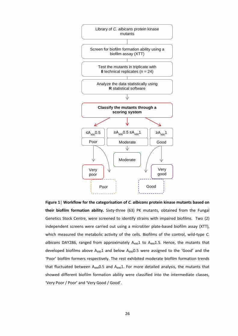
26
Figure 1| Workflow for the categorisation of C. albicans protein kinase mutants based on
their biofilm formation ability. Sixty-three (63) PK mutants, obtained from the Fungal
Genetics Stock Centre, were screened to identify strains with impaired biofilms. Two (2)
independent screens were carried out using a microtiter plate-based biofilm assay (XTT),
which measured the metabolic activity of the cells. Biofilms of the control, wild-type C.
albicans DAY286, ranged from approximately A4901 to A4901.5. Hence, the mutants that
developed biofilms above A4901 and below A4900.5 were assigned to the ‘Good’ and the
‘Poor’ biofilm formers respectively. The rest exhibited moderate biofilm formation trends
that fluctuated between A4900.5 and A4901. For more detailed analysis, the mutants that
showed different biofilm formation ability were classified into the intermediate classes,
‘Very Poor / Poor’ and ‘Very Good / Good’.
Library of C. albicans protein kinase mutants
Screen for biofilm formation ability using a biofilm assay (XTT)
Test the mutants in triplicate with 8 technical replicates (n = 24)
Analyze the data statistically using R statistical software
Classify the mutants through a scoring system
≤A490
0.5 ≥A490
1 ≥A490
0.5 ≤A490
1
Poor Moderate Good
Very poor
Very good
Moderate
Poor Good
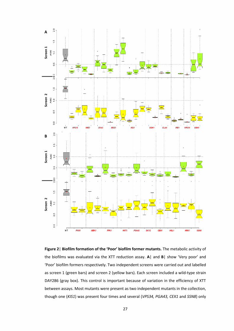
27
Scre
en
1
Scre
en
2
A
Scre
en
1
Scre
en
2
B
Figure 2| Biofilm formation of the ‘Poor’ biofilm former mutants. The metabolic activity of
the biofilms was evaluated via the XTT reduction assay. A| and B| show ‘Very poor’ and
‘Poor’ biofilm formers respectively. Two independent screens were carried out and labelled
as screen 1 (green bars) and screen 2 (yellow bars). Each screen included a wild-type strain
DAY286 (gray box). This control is important because of variation in the efficiency of XTT
between assays. Most mutants were present as two independent mutants in the collection,
though one (KIS1) was present four times and several (VPS34, PGA43, CEX1 and SSN8) only

28
once. Sample size equals to 24 observations (n = 24). 50% of the observations fall into the
box. The bottom and the top of the box correspond to the 1st and the 3rd quartiles
respectively. The notch shows the median confidence interval and the middle line inside
the box indicates the median. The upper and the lower limits demonstrate the maximum
and the minimum observations. The red dot shows the mean and the white circle an
outlier. Horizontal dashed line illustrates a threshold defined in Figure 1.
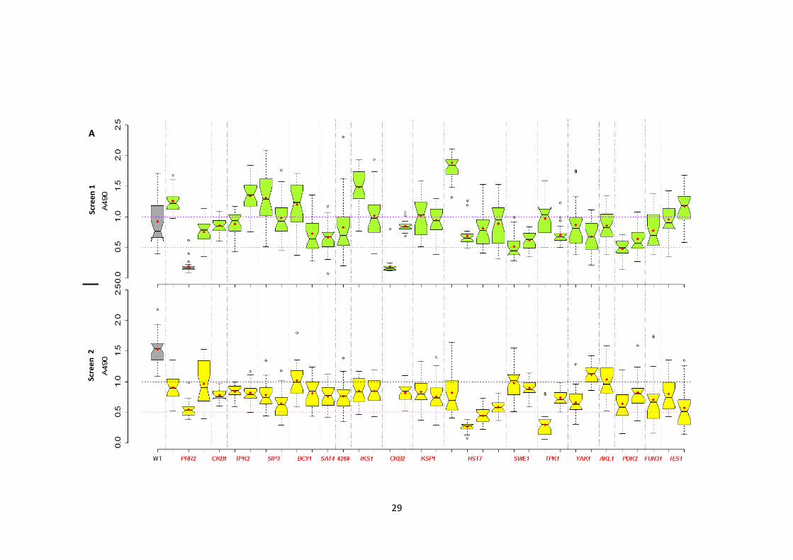
29
Scre
en
2
A
Scre
en
1
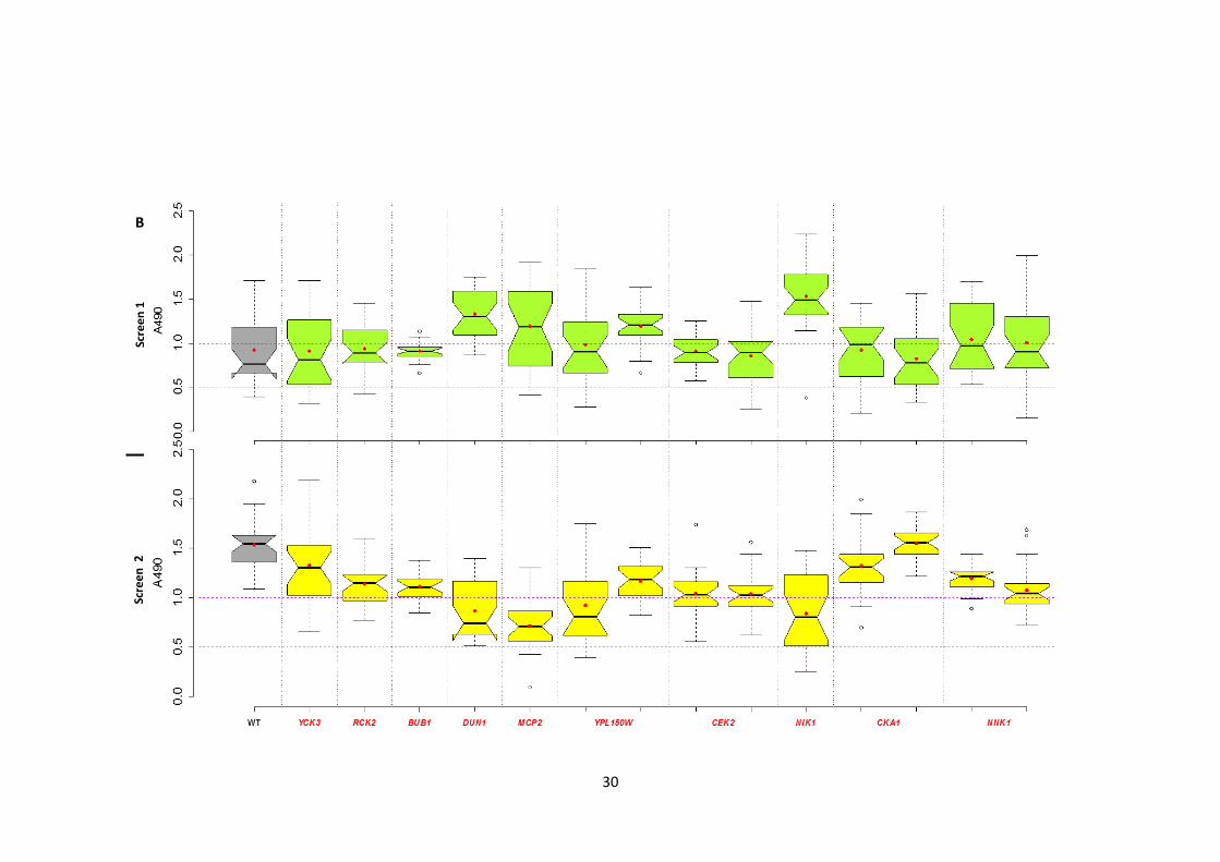
30
Scre
en
2
B
Scre
en
1
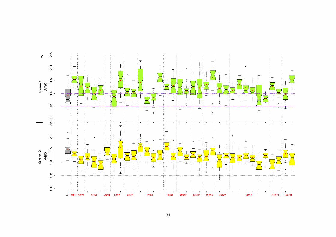
31
Scre
en
2
Scre
en
1
C

32
Figure 3| Biofilm formation of the ‘Moderate’ and ‘Good’ biofilm formers. The metabolic
activity of the biofilms was evaluated via the XTT reduction assay. A|, B| and C| show the
‘Moderate’, ‘Good’ and ‘Very good’ biofilm formers respectively. Two independent screens were
carried out and the results are presented as follows: top panel, green boxes – screen 1; bottom
panel, yellow boxplots – screen 2. Each screen included a wild-type C. albicans DAY286 (gray box).
This control is important because of variation in the efficiency of XTT between assays. Most
mutants were presented as two independent mutants in the collection, though some were
presented several times (e.g. PKH2, KIN2) and others (e.g. MEC1, SKY1) only once. Sample size
equals to 24 observations (n = 24). 50% of the observations fall into the box. The bottom and the
top of the box correspond to the 1st and the 3rd quartiles respectively. The notch shows the
median confidence interval and the middle line inside the box indicates the median. The upper
and the lower limits demonstrate the maximum and the minimum observations. The red dot
shows the mean and the white circle an outlier. Horizontal dashed line illustrates a threshold
defined in Figure 1.
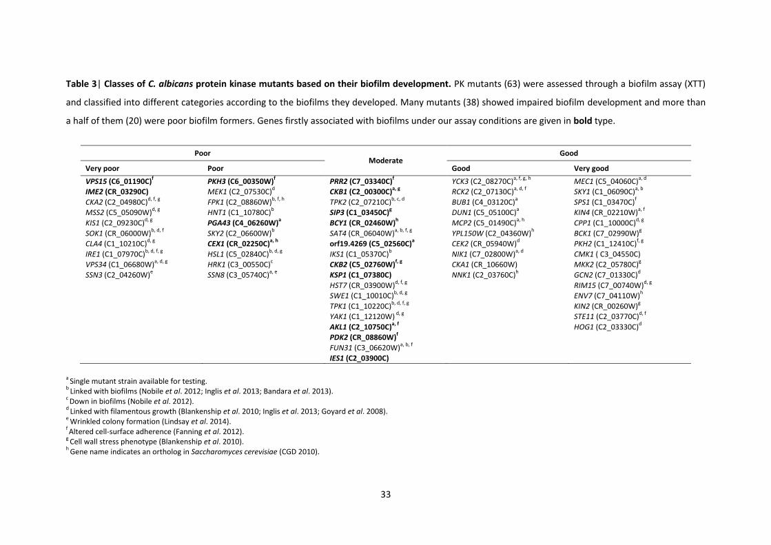
33
Table 3| Classes of C. albicans protein kinase mutants based on their biofilm development. PK mutants (63) were assessed through a biofilm assay (XTT)
and classified into different categories according to the biofilms they developed. Many mutants (38) showed impaired biofilm development and more than
a half of them (20) were poor biofilm formers. Genes firstly associated with biofilms under our assay conditions are given in bold type.
a Single mutant strain available for testing.
b Linked with biofilms (Nobile et al. 2012; Inglis et al. 2013; Bandara et al. 2013).
c Down in biofilms (Nobile et al. 2012).
d Linked with filamentous growth (Blankenship et al. 2010; Inglis et al. 2013; Goyard et al. 2008).
e Wrinkled colony formation (Lindsay et al. 2014).
f Altered cell-surface adherence (Fanning et al. 2012).
g Cell wall stress phenotype (Blankenship et al. 2010).
h Gene name indicates an ortholog in Saccharomyces cerevisiae (CGD 2010).
Poor Moderate
Good
Very poor Poor Good Very good
VPS15 (C6_01190C)f PKH3 (C6_00350W)
f PRR2 (C7_03340C)
f YCK3 (C2_08270C)
a, f, g, h MEC1 (C5_04060C)
a, d
IME2 (CR_03290C) MEK1 (C2_07530C)d CKB1 (C2_00300C)
a, g RCK2 (C2_07130C)
a, d, f SKY1 (C1_06090C)
a, b
CKA2 (C2_04980C)d, f, g
FPK1 (C2_08860W)b, f, h
TPK2 (C2_07210C)b, c, d
BUB1 (C4_03120C)a SPS1 (C1_03470C)
f
MSS2 (C5_05090W)d, g
HNT1 (C1_10780C)b SIP3 (C1_03450C)
g DUN1 (C5_05100C)
a KIN4 (CR_02210W)
a, f
KIS1 (C2_09230C)d, g
PGA43 (C4_06260W)a BCY1 (CR_02460W)
h MCP2 (C5_01490C)
a, h CPP1 (C1_10000C)
d, g
SOK1 (CR_06000W)b, d, f
SKY2 (C2_06600W)b SAT4 (CR_06040W)
a, b, f, g YPL150W (C2_04360W)
h BCK1 (C7_02990W)
g
CLA4 (C1_10210C)d, g
CEX1 (CR_02250C)a, h
orf19.4269 (C5_02560C)a
CEK2 (CR_05940W)d PKH2 (C1_12410C)
f, g
IRE1 (C1_07970C)b, d, f, g
HSL1 (C5_02840C)b, d, g
IKS1 (C1_05370C)b NIK1 (C7_02800W)
a, d CMK1 ( C3_04550C)
VPS34 (C1_06680W)a, d, g
HRK1 (C3_00550C)c CKB2 (C5_02760W)
f, g CKA1 (CR_10660W) MKK2 (C2_05780C)
g
SSN3 (C2_04260W)e SSN8 (C3_05740C)
a, e KSP1 (C1_07380C) NNK1 (C2_03760C)
h GCN2 (C7_01330C)
d
HST7 (CR_03900W)d, f, g
RIM15 (C7_00740W)d, g
SWE1 (C1_10010C)
b, d, g ENV7 (C7_04110W)
h
TPK1 (C1_10220C)
b, d, f, g KIN2 (CR_00260W)
g
YAK1 (C1_12120W) d, g
STE11 (C2_03770C)d, f
AKL1 (C2_10750C)
a, f HOG1 (C2_03330C)
d
PDK2 (CR_08860W)
f
FUN31 (C3_06620W)
a, b, f
IES1 (C2_03900C)
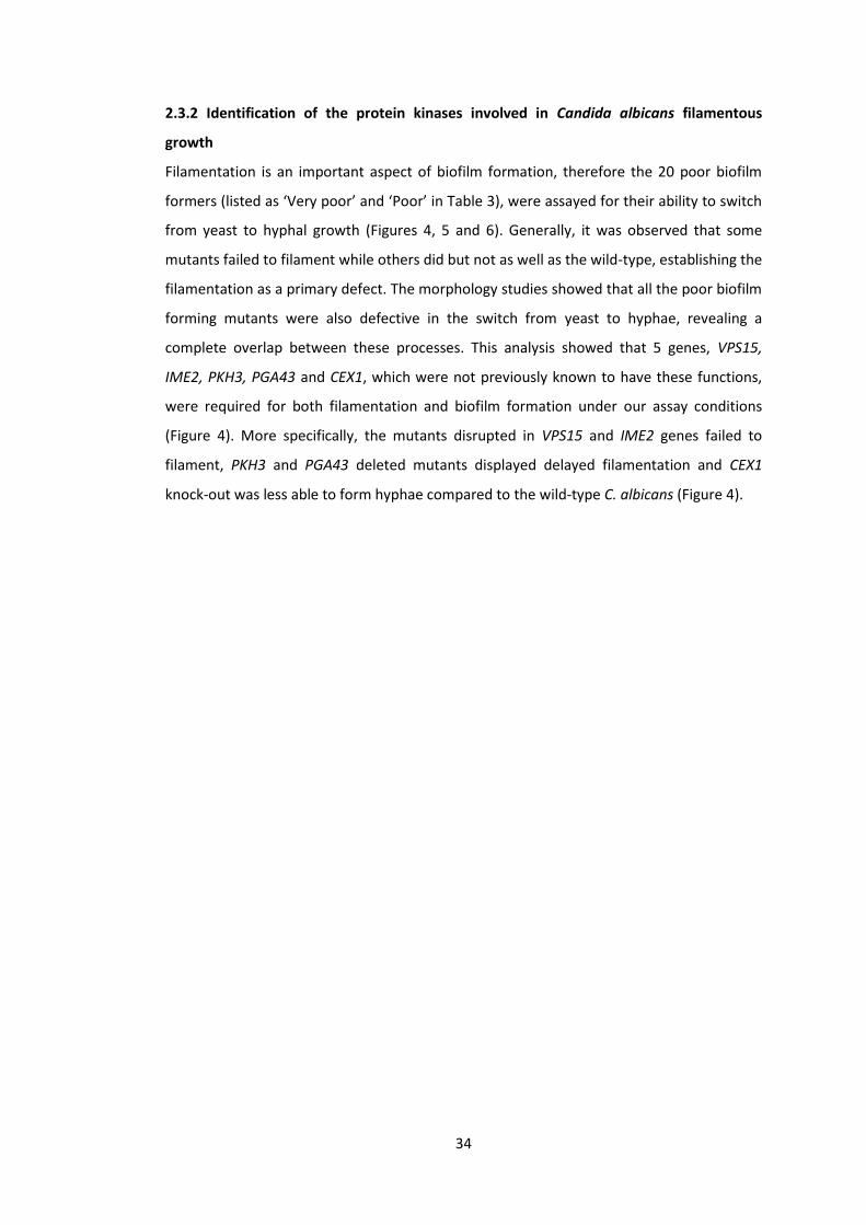
34
2.3.2 Identification of the protein kinases involved in Candida albicans filamentous
growth
Filamentation is an important aspect of biofilm formation, therefore the 20 poor biofilm
formers (listed as ‘Very poor’ and ‘Poor’ in Table 3), were assayed for their ability to switch
from yeast to hyphal growth (Figures 4, 5 and 6). Generally, it was observed that some
mutants failed to filament while others did but not as well as the wild-type, establishing the
filamentation as a primary defect. The morphology studies showed that all the poor biofilm
forming mutants were also defective in the switch from yeast to hyphae, revealing a
complete overlap between these processes. This analysis showed that 5 genes, VPS15,
IME2, PKH3, PGA43 and CEX1, which were not previously known to have these functions,
were required for both filamentation and biofilm formation under our assay conditions
(Figure 4). More specifically, the mutants disrupted in VPS15 and IME2 genes failed to
filament, PKH3 and PGA43 deleted mutants displayed delayed filamentation and CEX1
knock-out was less able to form hyphae compared to the wild-type C. albicans (Figure 4).
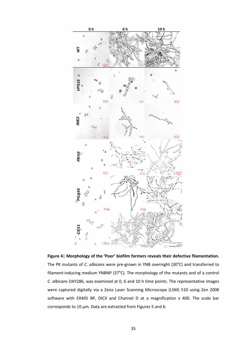
35
Figure 4| Morphology of the ‘Poor’ biofilm formers reveals their defective filamentation.
The PK mutants of C. albicans were pre-grown in YNB overnight (30oC) and transferred to
filament-inducing medium YNBNP (37oC). The morphology of the mutants and of a control
C. albicans DAY286, was examined at 0, 6 and 10 h time points. The representative images
were captured digitally via a Zeiss Laser Scanning Microscope (LSM) 510 using Zen 2008
software with EX405 BP, DICII and Channel D at a magnification x 400. The scale bar
corresponds to 10 μm. Data are extracted from Figures 5 and 6.
WT
0 h 6 h 10 h
VP
S1
5
IME
2
PK
H3
P
GA
43
C
EX
1
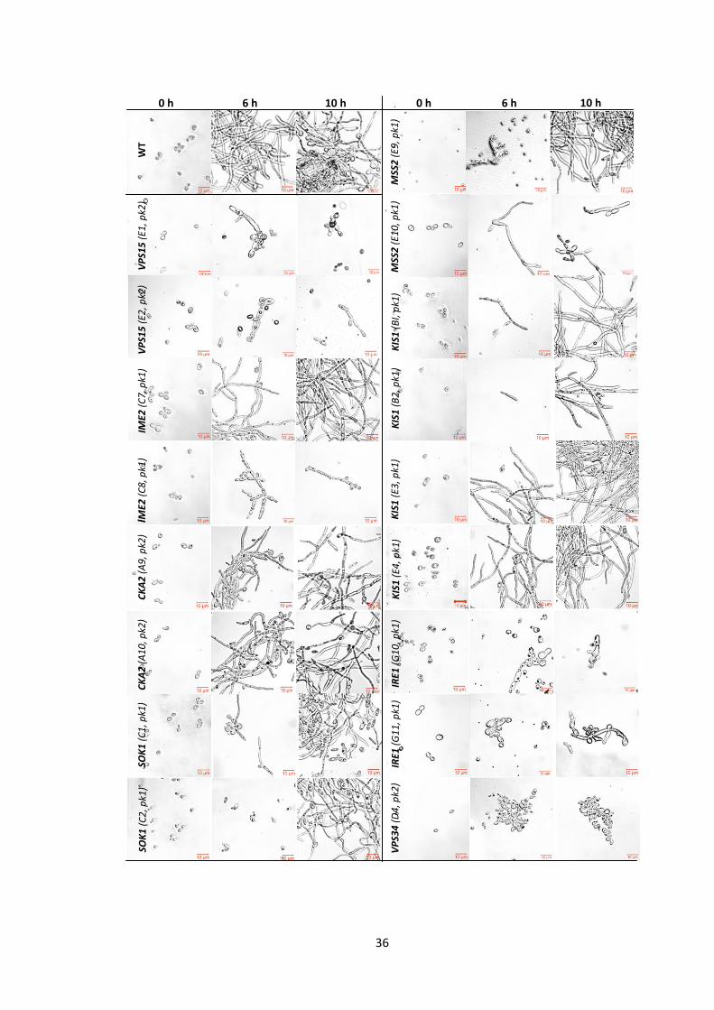
36
VP
S15
(E1
, pk2
) W
T
MSS
2 (
E9, p
k1)
0 h 6 h 10 h
VP
S15
(E2
, pk2
)
MSS
2 (
E10,
pk1
)
IME2
(C
7, p
k1)
KIS
1 (
BI,
pk1
)
IME2
(C
8, p
k1)
KIS
1 (
B2
, pk1
) K
IS1
(E3
, pk1
)
CK
A2
(A
9, p
k2)
10 h 6 h 0 h
CK
A2
(A
10
, pk2
)
KIS
1 (
E4, p
k1)
SOK
1 (
C1
, pk1
)
IRE1
(G
10
, pk1
) IR
E1 (
G1
1, p
k1)
SOK
1 (
C2
, pk1
)
VP
S34
(D
4, p
k2)
37
Figure 5| Morphology of the ‘Very poor’ biofilm formers reveals their defect in the switch
from yeast to filamentous growth. The PK mutants of C. albicans were pre-grown in YNB
overnight (30oC) and transferred to filament-inducing medium YNBNP (37oC). The
morphology of the mutants and of a control C. albicans DAY286, was examined at 0, 6 and
10 h time points. The position of the mutant on the PK plate is indicated in parentheses.
The representative images were captured digitally via a Zeiss Laser Scanning Microscope
(LSM) 510 using Zen 2008 software with EX405 BP, DICII and Channel D at a magnification x
400. The scale bar corresponds to 10 μm.
WT
0 h 6 h 10 h 10 h 6 h 0 h
CLA
4 (
F9, p
k1)
SSN
3 (
C1
1, p
k1)
CLA
4 (
F10
, pk1
)
SSN
3 (
C1
2, p
k1)
WT
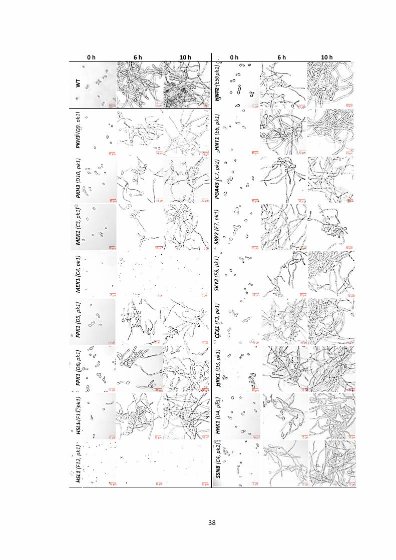
38
WT
0 h 6 h 10 h 10 h 6 h 0 h
PK
H3
(D
9, p
k1)
HN
T1 (
E5, p
k1)
PK
H3
(D
10
, pk1
)
HN
T1 (
E6, p
k1)
MEK
1 (
C3
, pk1
)
PG
A4
3 (
C7
, pk2
)
MEK
1 (
C4
, pk1
)
SKY
2 (
E7, p
k1)
FPK
1 (
D5
, pk1
)
SKY
2 (
E8, p
k1)
FPK
1 (
D6
, pk1
)
CEX
1 (
F3, p
k1)
HSL
1 (
F11
, pk1
)
, pk1
)
HR
K1
(D
3, p
k1)
, pk1
)
HR
K1
(D
4, p
k1)
, pk1
)
HSL
1 (
F12
, pk1
)
, pk1
)
SSN
8 (
C4
, pk2
)
, pk1
)
39
Figure 6| The ‘Poor’ biofilm former mutants are defective in the switch from yeast to
filamentous growth. The PK mutants of C. albicans were pre-grown in YNB overnight (30oC)
and transferred into filament-inducing media YNBNP (37oC). C. albicans DAY286 was used
as a control. The position of the mutant on the PK plate is indicated in parentheses. The
morphology of the mutants was examined at three time-points, 0 h, 6 h and 10 h, utilizing
Zeiss Laser Scanning Microscope (LSM) 510. The representative images were captured
digitally using Zen 2008 software with EX405 BP, DICII and Channel D at a magnification x
400. Scale bar corresponds to 10 μm.
2.3.3 STRING network analyses
To identify important processes for biofilm development we questioned the interactions of
proteins/enzymes encoded by the 38 biofilm-related genes using a STRING database
(Figure 7). STRING is a powerful database that quantitatively integrates experimental data,
computational prediction methods and published literature to produce an interaction
network (Jensen et al. 2009). Protein sequences were retrieved from CGD
(www.candidagenome.org) and entered into the STRING network generation program
(www.string-db.org). The resulted networks, which were analysed using a K-means option,
showed that biofilm-related proteins were assigned into three main clusters (Figure 7). The
38 proteins were also analysed for their function in the KEGG annotated pathways (Table
4). The proteins of the clusters mostly shared similarity in function or occurrence in the
same pathway. A list of the putative functional partners can be found in the Table 5.
Combined, these analyses revealed three main clusters: the first (on the left) corresponded
to the genes of the Protein Kinase A (PKA) pathway; the second (centre) to the elements of
cell cycle; and the third (right) to vacuolar function (Figure 7). Some genes (11) did not
show any associations in the STRING analysis (see the bottom of Figure 7).

40
Figure 7| Predicted STRING interaction networks. In order to produce an interaction network, the STRING database quantitatively integrates experimental
data, computational prediction methods and published literature. The spheres and edges represent PKs and known/predicted interactions respectively.
Stronger blue lines indicate the connections with higher confidence. Protein kinase encoding genes are boxed and the asterisk (*) indicates a functional
partner predicted by the STRING database. A network shows the interaction of the 37 biofilm-related proteins/genes and their functional partners (SKY2
was not present in the STRING database).
Poor biofilm formers
Very poor biofilm formers
Moderate biofilm formers
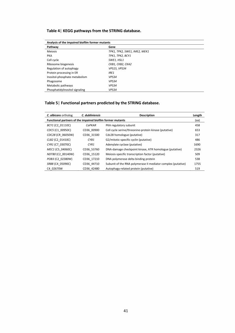
41
Table 4| KEGG pathways from the STRING database.
Analysis of the impaired biofilm former mutants
Pathway Gene
Meiosis TPK1, TPK2, SWE1, IME2, MEK1
PKA TPK1, TPK2, BCY1
Cell cycle SWE1, HSL1
Ribosome biogenesis CKB1, CKB2, CKA2
Regulation of autophagy VPS15, VPS34
Protein processing in ER IRE1
Inositol-phosphate metabolism VPS34
Phagosome VPS34
Metabolic pathways VPS34
Phosphatidylinositol signaling VPS34
Table 5| Functional partners predicted by the STRING database.
C. albicans ortholog C. dubliniensis Description Length
Functional partners of the impaired biofilm former mutants (aa)
BCY1 (C2_01110C) CaPKAR PKA regulatory subunit 458
CDC5 (C1_00950C) CD36_00900 Cell cycle serine/threonine-protein kinase (putative) 653
CDC28 (CR_06050W) CD36_31500 Cdc28 homologue (putative) 317
CLB2 (C2_01410C) CYB1 G2/mitotic-specific cyclin (putative) 486
CYR1 (C7_03070C) CYR1 Adenylate cyclase (putative) 1690
MEC1 (C5_04060C) CD36_53760 DNA-damage checkpoint kinase, ATR homologue (putative) 2326
NDT80 (C2_00140W) CD36_15120 Meiosis-specific transcription factor (putative) 509
POB3 (C2_02380W) CD36_17210 DNA polymerase delta binding protein 538
SRB8 (C4_05090C) CD36_44710 Subunit of the RNA polymerase II mediator complex (putative) 1755
C4_02670W CD36_42480 Autophagy-related protein (putative) 519
42
2.4 Discussion
Biofilm formation on the surface of catheters is a leading cause of C. albicans infections and
drug resistance in hospitals. The aim of our study was to establish whether particular PKs
were essential for biofilm formation in C. albicans as this might reveal novel processes and
pathways that could serve as drug targets. We identified 38 PK mutants that showed some
degree of biofilm impairment, but some of them were already linked with biofilms. Because
there is an established link between filamentation and biofilm formation, we assessed the
capacity of the 20 mutants that were severely impaired in biofilms to switch from yeast to
hyphal growth and found a complete coincidence of switching and biofilm formation. Thus,
although processes other than filamentation can be required for biofilm formation, either
our assay was biased for mutants with filamentation defects or none of the protein kinases
in our screens is involved in these other processes. There were, however, some novel
findings and five strains were mutated in genes (VPS15, PKH3, PGA43, IME2 and CEX1) not
previously directly associated with either filamentation or biofilm formation. It is important
to note that other studies used different types of filamentation/biofilm assays (and media),
which may explain minor variance between studies.
VPS15 encodes a kinase involved in vacuolar protein sorting and although this gene was not
previously linked to biofilms, other members of the Vps family, Vps1p and Vps34p (also
identified in our screens), were reported to significantly contribute in filamentation and
biofilm formation in Candida (Bernardo et al. 2008). Furthermore, the S. cerevisiae vps15
mutant also forms impaired biofilms (Vandenbosch et al. 2013). Vps15p and Vps34p form a
complex on the vacuole/Golgi membrane, thus these data indicate that correct protein
sorting is required for proper fungal development, though this is likely to be an indirect
rather than a direct effect. PKH3, which encodes a kinase that may be involved in Protein
Kinase C (PKC) activity, was not previously described as being involved in morphological
development but the mutant is reported to display significantly decreased cell-substrate
adherence (Fanning et al. 2012), an important early step for biofilm development (Finkel
and Mitchell 2011). Interestingly, the network analysis showed that Pkh3p is associated
with the vacuolar proteins Vps15p and Vps4p, perhaps indicating that the defect in this
mutant is also related to protein sorting in the vacuole. PGA43 encodes a Glycosyl-
Phosphatidyl-Inositol (GPI)-anchored protein and thus is likely to be present at the cell
surface. Its function is not known but other also Gpi family proteins, for example Pga59p
and Pga62p, are required for cell wall integrity (Moreno-Ruiz et al. 2009). There is very
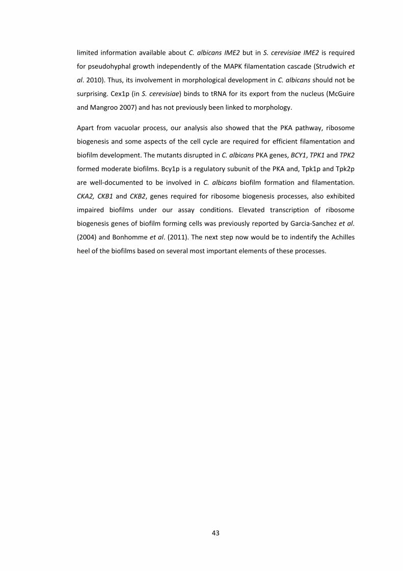
43
limited information available about C. albicans IME2 but in S. cerevisiae IME2 is required
for pseudohyphal growth independently of the MAPK filamentation cascade (Strudwich et
al. 2010). Thus, its involvement in morphological development in C. albicans should not be
surprising. Cex1p (in S. cerevisiae) binds to tRNA for its export from the nucleus (McGuire
and Mangroo 2007) and has not previously been linked to morphology.
Apart from vacuolar process, our analysis also showed that the PKA pathway, ribosome
biogenesis and some aspects of the cell cycle are required for efficient filamentation and
biofilm development. The mutants disrupted in C. albicans PKA genes, BCY1, TPK1 and TPK2
formed moderate biofilms. Bcy1p is a regulatory subunit of the PKA and, Tpk1p and Tpk2p
are well-documented to be involved in C. albicans biofilm formation and filamentation.
CKA2, CKB1 and CKB2, genes required for ribosome biogenesis processes, also exhibited
impaired biofilms under our assay conditions. Elevated transcription of ribosome
biogenesis genes of biofilm forming cells was previously reported by Garcia-Sanchez et al.
(2004) and Bonhomme et al. (2011). The next step now would be to indentify the Achilles
heel of the biofilms based on several most important elements of these processes.
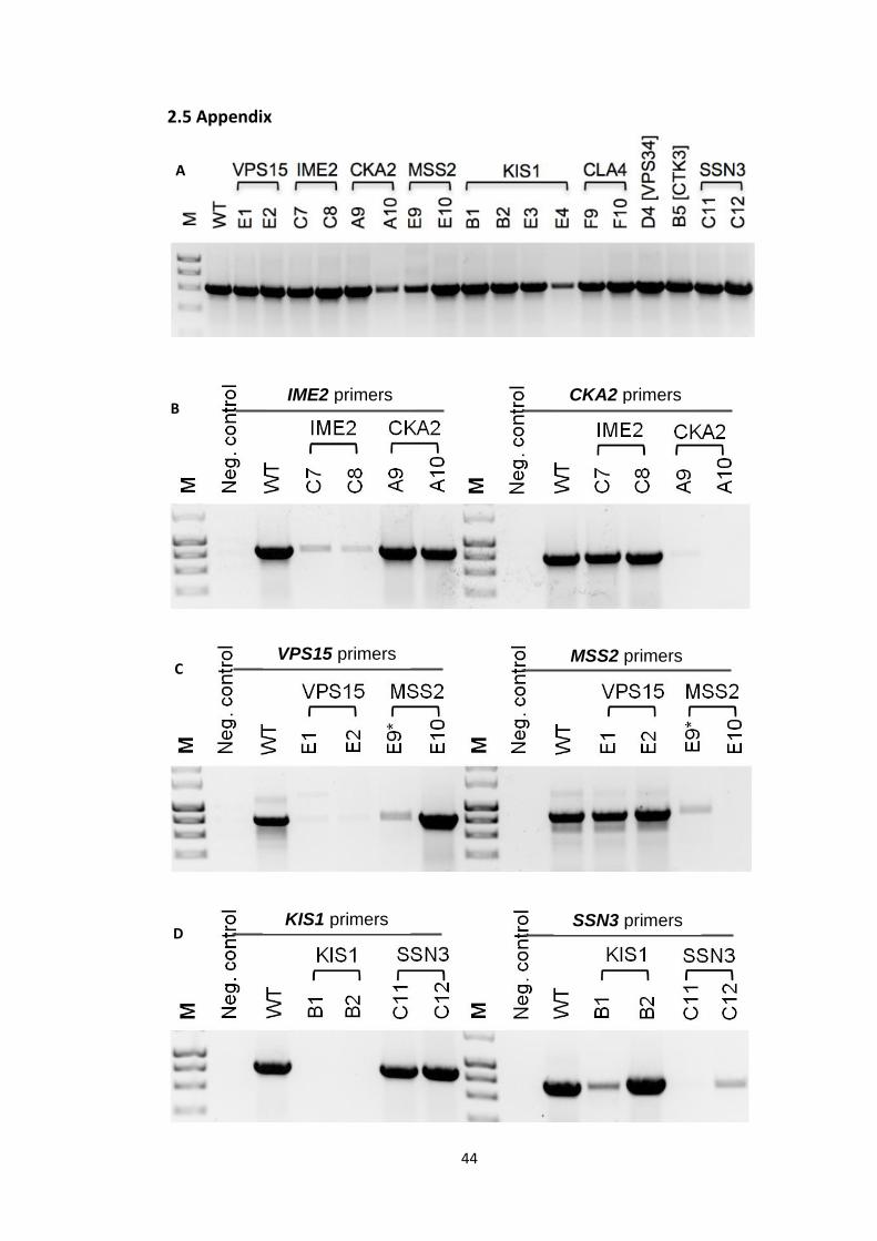
44
A
B
C
D
A
2.5 Appendix
IME2 primers CKA2 primers
VPS15 primers MSS2 primers
KIS1 primers SSN3 primers
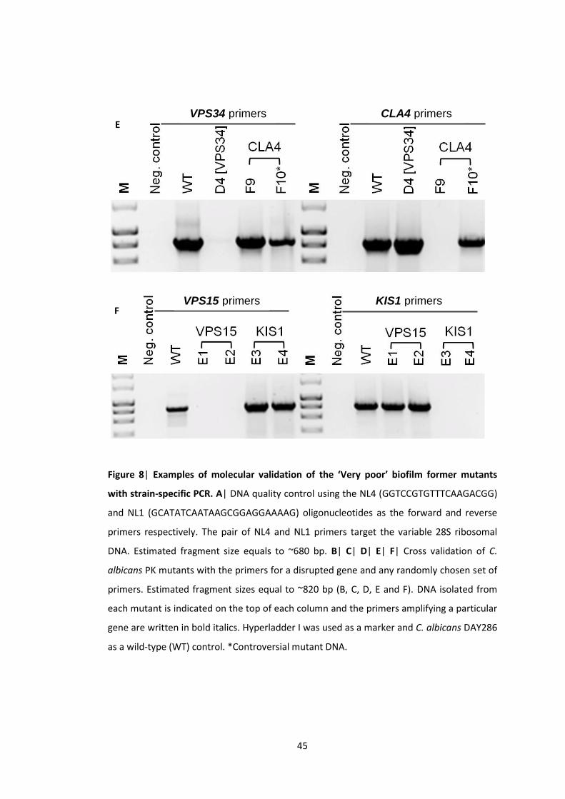
45
E
F
Figure 8| Examples of molecular validation of the ‘Very poor’ biofilm former mutants
with strain-specific PCR. A| DNA quality control using the NL4 (GGTCCGTGTTTCAAGACGG)
and NL1 (GCATATCAATAAGCGGAGGAAAAG) oligonucleotides as the forward and reverse
primers respectively. The pair of NL4 and NL1 primers target the variable 28S ribosomal
DNA. Estimated fragment size equals to ~680 bp. B| C| D| E| F| Cross validation of C.
albicans PK mutants with the primers for a disrupted gene and any randomly chosen set of
primers. Estimated fragment sizes equal to ~820 bp (B, C, D, E and F). DNA isolated from
each mutant is indicated on the top of each column and the primers amplifying a particular
gene are written in bold italics. Hyperladder I was used as a marker and C. albicans DAY286
as a wild-type (WT) control. *Controversial mutant DNA.
VPS34 primers CLA4 primers
VPS15 primers KIS1 primers
46
Chapter 3
Communication between Candida albicans and
Pseudomonas aeruginosa
Nina Konstantinidou and John Patrick Morrissey
School of Microbiology, University College Cork, Cork, T12YN60, Ireland
Key words: Candida albicans; protein kinase; biofilms; filamentation; yeast - hyphae;
Pseudomonas aeruginosa.
Subtitle: Candida albicans morphology and interaction with
Pseudomonas aerugionsa
This chapter was published as part of publication by Konstantinidou N & Morrissey JP
(2015) Co-occurrence of filamentation defects and impaired biofilms in Candida albicans
protein kinase mutants. FEMS Yeast Res 15: 1–10. pii fov092 doi: 10.1093/femsyr/fov092.
Here it is represented with some figure rearrangements.
47
Abstract
C. albicans biofilm development is a complex phenomenon that mainly implicates
processes such as adhesion, filamentation supported by intensive cell wall biosynthesis,
excretion of the extracellular matrix (ECM) and dispersal of blastospores. The filamentation
is largely governed by the MAPK pathways but our analysis of the ‘Good’ biofilm former PK
mutants showed that the individual components of these pathways are not required for
robust biofilm formation, indicating functional redundancy. The ‘Good’ biofilm formers
were established after the analyses of their biofilms with a biofilm assay (XTT) and with the
assessment of their clusters in the STRING networks. These mutants were further
investigated for their response to bacterial supernatants, since C. albicans biofilm
impairment with P. aeruginosa supernatants, was well-documented by the previous
publications of McAlester et al. (2008) and Holcombe et al. (2010). These supernatants
contained QS (quorum sensing) molecules derived from 2 distinct strains of P. aeruginosa
the wild-type, PAO1, and a homoserine lactone-free (HSL) mutant, ΔQS. The biofilm assay
showed that P. aeruginosa supernatants inhibited biofilm formation in all mutants,
regardless of the presence of HSLs. To clarify an effect of the HSLs on Candida morphology
and filamentation we conducted a morphology assay. This assay suggested that the
morphology of Candida was affected only by the HSL-containing supernatants that were
derived from the wild-type P. aeruginosa, PAO1. This confirms the distinct HSL-dependent
inhibition of filamentation and the HSL-independent impairment of biofilm development by
P. aeruginosa supernatants.
48
3. Communication between Candida albicans and Pseudomonas
aeruginosa
3.1 Introduction
Apart from the established environmental factors, C. albicans morphology and biofilm
formation is also influenced by some environmental microorganisms such as the Gram-
negative bacterium P. aeruginosa (reviewed by Peleg et al. 2010 and Holcombe et al.
2011). P. aeruginosa is an opportunistic human pathogen capable of causing inflammation
and sepsis by colonizing kidneys, urinary and gastrointestinal tract of the susceptible
individuals. Clinical observations suggest that C. albicans and P. aeruginosa populations can
coexist and influence each other (Bauernfeind et al. 1987; Kerr 1994; Nseir et al. 2007;
Chotirmall et al. 2010). Notably, Candida and Pseudomonas were coisolated from the lungs
of Cystic Fibrosis (CF) patients (Martin et al. 1993; Leclair and Hogan 2010), serious burn
wounds (Gupta et al. 2005) and the urinary catheters (Falleiros de Pádua et al. 2008). In
vitro analysis established that C. albicans biofilm formation and metabolism can be
influenced by P. aeruginosa phenazines (Gibson et al. 2009; Morales et al. 2013). For
example, P. aeruginosa phenazines enhance C. albicans ethanol production which
stimulates Pseudomonas biofilm formation (Chen et al. 2014). Another phenazine,
methosulphate (PMS), can kill Candida within its biofilms (Morales et al. 2010; Morales et
al. 2013). Several studies have also shown that Pseudomonas can inhibit Candida biofilm
development in vitro (Holcombe et al. 2010; Bandara et al. 2010a; Bandara et al. 2010b;
Reen et al. 2011; Bandara et al. 2013). These studies collectively suggest that Candida
biofilm development and metabolism are closely related (Lindsay et al. 2014).
Communication between C. albicans and P. aeruginosa is based on signalling. P. aeruginosa
can inhibit the C. albicans morphological switch from yeast to hyphae using secreted
chemicals. Several studies suggest that Pseudomonas QS (quorum sensing) molecules are
responsible for the signal-mediated communication between the two species (Hogan and
Kolter 2002; McAlester et al. 2008; Deveau and Hogan 2011). It was reported that HSLs can
inhibit the switch of Candida from yeast to filamentous growth (Hogan et al. 2004).
However, C. albicans QS molecule, farnesol, can limit the virulence of P. aeruginosa by
blocking the production of Pseudomonas QS molecules and pyocyanin (Cugini et al. 2007)
and affecting motility (McAlester et al. 2008). P. aeruginosa pyocyanin and 1-
hydroxyphenazine can prevent the growth of C. albicans (Kerr et al. 1999). We further

49
investigated whether deletion of the PK-encoding genes disrupted in the ‘Good’ biofilm
former mutants affected the signal-mediated interaction between P. aeruginosa and C.
albicans.
50
3.2 Materials and Methods
3.2.1 Yeast strains and growth conditions
The wild-type C. albicans strains used in this study were SC5314 (Gillum et al. 1984),
BWP17 (ura3Δ::λimm434/ura3Δ::λimm434 his1::hisG/his1::hisG arg4::hisG/arg4::hisG)
(Wilson et al. 1999) and DAY286 (ura3::λimm434 his1::hisG pARG4::URA3::arg4::hisG)
(Davis et al. 2002). The PK mutants of C. albicans are listed in Table 1 (Chapter 2). This
kinase set was created by Aaron Mitchell’s group (Blankenship et al. 2010) and obtained
from the Fungal Genetics Stock Centre (www.fgsc.net/candida/FGSCcandida
resources.htm). PK homozygous insertion mutants were created in BWP17 parental strain
via Tn7-UAU1 cassette (Blankenship et al. 2010). The CGD descriptions of the C. albicans PK
mutants can be found in the Appendix 3.5 (Table 5). The majority of the PK-coding genes
were represented by double independent mutant strains but in some cases only a single
mutant was available. In total, we examined 63 genes using 45 independent duplicate
mutants and 18 single mutants. The initial FGSC collection was larger and included mutants
deleted in ~80 PK/PK-related genes. Our reduced set comprised the mutants that, after
delivery and culturing, we were able to verify by strain-specific PCR using primers flanking
the gene insertion sites (see PCR below).
Yeast strains were routinely cultured in standard rich medium containing 1% yeast extract,
2% peptone and 2% glucose (YPD). For biofilm and morphological analyses, the yeast
strains were pre-grown in non-filament-inducing medium YNB (yeast nitrogen base), as
described by McAlester et al. (2008) and Holcombe et al. (2010) with some adjustments.
Briefly, filter-sterilized YNB salts without amino acids (Difco 291940) were supplemented
with 0.2% glucose, 0.1% maltose and 0.16% filter-sterilized synthetic amino acid drop-out
Leu– (Kaiser Formedium DSCK052). For the induction of hyphal growth the strains were
transferred to filament-inducing medium, YNBNP, which consisted from YNB supplemented
with 2.5 mM N-acetyl-D-glucosamine (Sigma A8625) and 25 mM phosphate (sodium) buffer
(pH 7).
3.2.2 PCR
DNA of wild-type C. albicans and PK mutants was extracted according to Hoffman (2001)
protocol. The primers were designed with SnapGene (www.snapgene.com) and evaluated
via Primer-BLAST (www.ncbi.nlm.nih.gov). Strain-specific PCR was carried out using primers
flanking the gene insertion sites listed in the Supplementary Table S1 of Blankenship et al.
(2010) publication. Absence of the band indicated a mutant disrupted in the gene amplified
51
by the corresponding primers. The primer sequences are listed in the Table 1. All the PCR
reactions were carried out utilising a GoTaq Green Master Mix (Promega) according to the
manufacturers’ instructions.
3.2.3 Bacterial strains and preparation of supernatants
The P. aeruginosa strains used in this study were a wild-type PAO1 (Holloway and Morgan
1986) and a HSL-negative mutant, ΔQS (ΔlasRI::Gm ΔrhlRI::Tc) (Beatson et al. 2002).
Pseudomonas supernatants were prepared as described by McAlester et al. (2008). Briefly,
after overnight growth of Pseudomonas in LB broth (37oC) the culture was inoculated into
100 mL of fresh LB and further grown until it reached an approximate absorbance of
A6001.2. To harvest the cells, the cultures were centrifuged twice (15 min, 5000 rpm). The
resulting supernatants were filter-sterilized with 0.2 μm pore size filter, lyophilized and
used immediately or stored at –80oC. Before utilization, the supernatants were re-
suspended to yield concentration of 2x from a 20x stock in sterile dH2O. Sterile LB broth
was used as a control after it was prepared in the same manner as the above supernatants.
3.2.4 Biofilm assay
A microtiter plate-based biofilm assay (XTT reductase salts) was carried out as described by
Ramage et al. (2001) and Holcombe et al. (2010). Briefly, the yeast strains were pre-grown
in non-filament-inducing medium (YNB) overnight (30oC) and diluted into filament-inducing
medium (YNBNP) to A6000.05. The diluted cultures (100 μL) were incubated for 1 h (37°C) in
flat-bottomed 96-well polystyrene plates. After incubation, the attached cells were washed
with fresh YNBNP medium twice, by inverting the plates carefully, to eliminate non-
adhered cells. Where indicated, YNBNP (100 μL) with the Pseudomonas supernatant (2x)
and LB (2x) was added to the sample and the control cultures respectively. In order to
induce biofilm formation the plates were incubated in the dark statically for 24 h (37°C).
The next day, the biofilms developed on the bottom of the plates were washed with fresh
YNBNP by careful pipetting. The XTT-menadione solution (100 μL), prepared as below, was
added to the overnight cultures and incubated in the dark for 2 h (37oC). Lastly, the dyed
supernatants (80 μL) were transferred to a clean plate for the quantification at a
wavelength of A490 nm.
Metabolic activity of the cells was measured with a XTT (reductase salts) assay. For the
preparation of the XTT solutions, 10 mM menadione (Sigma M5625) was dissolved in pure
acetone and added (10 μL) to the XTT solution. The latter was prepared by dissolving 0.01 g
of XTT powder (Sigma X4626) in 20 mL sterile dH2O and filtered with 0.2 μm pore size filter.

52
To increase the statistical power of our experiments we tested the mutants in triplicate
with eight technical replicates (n = 24). Biofilms of the control, wild-type C. albicans
DAY286, ranged from approximately A4901 to A4901.5. Thus, the mutants with biofilms
above A4901 and below A4900.5 were assigned to the ‘Good’ and ‘Poor’ biofilm formers
respectively. The remaining mutants were ‘Moderate’ biofilm formers. A distribution of
each sample was assessed and the coincidence of the median and the mean indicated a
Gaussian (normal) distribution of our data. All the statistical analysis of the biofilm assays
were carried out in R Statistical Software (R Development Core Team 2013).
3.2.5 Morphological analyses
Morphological assays were carried out as described previously by Hogan et al. (2004) and
McAlester et al. (2008). Briefly, the yeast cultures were pre-grown overnight in YNB broth
(30oC) and diluted into YNBNP (A6001.0) to assay their capacity to switch from yeast to
filamentous growth. Before (0 h) and after 6 h and 10 h incubation (37oC), the morphology
of the mutants was examined microscopically. Where indicated, before incubation for 6 h
(37oC), the sample and the control cultures were treated with the bacterial supernatants
(2x) and LB (2x) respectively. Since the previous experiments of McAlester et al. (2008) and
Holcombe et al. (2010) had shown that the morphology of C. albicans SC5314 was impaired
by the Pseudomonas supernatants, SC5314 was used as an additional control. The images
were captured digitally at a magnification x 400 via Zen2008 software with EX405 BP and
Channel D by Zeiss Laser Scanning Microscope (LSM) 510.
3.2.6 Bioinformatics analyses
The protein sequences of C. albicans PKs were retrieved from the CGD (Candida Genome
Database) (Arnaud et al. 2005), the analysis of the protein associations were carried out
using the STRING v9.1 database (Search Tool for the Retrieval of Interacting
Genes/Proteins) (Jensen et al. 2009), the GOs (Gene Ontologies) were obtained with the
Blast2GO annotations (Conesa et al. 2005) and pathway analysis were carried out using
KEGG pathway database (Kyoto Encyclopaedia of Genes and Genomes) (Kanehisa and Goto
2000).
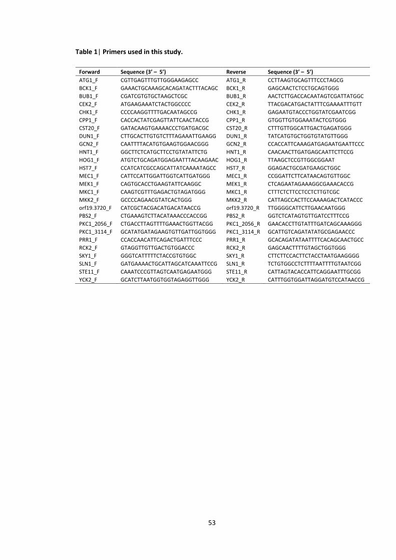
53
Table 1| Primers used in this study.
Forward Sequence (3’ – 5’) Reverse Sequence (3’ – 5’)
ATG1_F CGTTGAGTTTGTTGGGAAGAGCC ATG1_R CCTTAAGTGCAGTTTCCCTAGCG
BCK1_F GAAACTGCAAAGCACAGATACTTTACAGC BCK1_R GAGCAACTCTCCTGCAGTGGG
BUB1_F CGATCGTGTGCTAAGCTCGC BUB1_R AACTCTTGACCACAATAGTCGATTATGGC
CEK2_F ATGAAGAAATCTACTGGCCCC CEK2_R TTACGACATGACTATTTCGAAAATTTGTT
CHK1_F CCCCAAGGTTTTGACAATAGCCG CHK1_R GAGAATGTACCCTGGTATCGAATCGG
CPP1_F CACCACTATCGAGTTATTCAACTACCG CPP1_R GTGGTTGTGGAAATACTCGTGGG
CST20_F GATACAAGTGAAAACCCTGATGACGC CST20_R CTTTGTTGGCATTGACTGAGATGGG
DUN1_F CTTGCACTTGTGTCTTTAGAAATTGAAGG DUN1_R TATCATGTGCTGGTGTATGTTGGG
GCN2_F CAATTTTACATGTGAAGTGGAACGGG GCN2_R CCACCATTCAAAGATGAGAATGAATTCCC
HNT1_F GGCTTCTCATGCTTCCTGTATATTCTG HNT1_R CAACAACTTGATGAGCAATTCTTCCG
HOG1_F ATGTCTGCAGATGGAGAATTTACAAGAAC HOG1_R TTAAGCTCCGTTGGCGGAAT
HST7_F CCATCATCGCCAGCATTATCAAAATAGCC HST7_R GGAGACTGCGATGAAGCTGGC
MEC1_F CATTCCATTGGATTGGTCATTGATGGG MEC1_R CCGGATTCTTCATAACAGTGTTGGC
MEK1_F CAGTGCACCTGAAGTATTCAAGGC MEK1_R CTCAGAATAGAAAGGCGAAACACCG
MKC1_F CAAGTCGTTTGAGACTGTAGATGGG MKC1_R CTTTCTCTTCCTCCTCTTGTCGC
MKK2_F GCCCCAGAACGTATCACTGGG MKK2_R CATTAGCCACTTCCAAAAGACTCATACCC
orf19.3720_F CATCGCTACGACATGACATAACCG orf19.3720_R TTGGGGCATTCTTGAACAATGGG
PBS2_F CTGAAAGTCTTACATAAACCCACCGG PBS2_R GGTCTCATAGTGTTGATCCTTTCCG
PKC1_2056_F CTGACCTTAGTTTTGAAACTGGTTACGG PKC1_2056_R GAACACCTTGTATTTGATCAGCAAAGGG
PKC1_3114_F GCATATGATAGAAGTGTTGATTGGTGGG PKC1_3114_R GCATTGTCAGATATATGCGAGAACCC
PRR1_F CCACCAACATTCAGACTGATTTCCC PRR1_R GCACAGATATAATTTTCACAGCAACTGCC
RCK2_F GTAGGTTGTTGACTGTGGACCC RCK2_R GAGCAACTTTTGTAGCTGGTGGG
SKY1_F GGGTCATTTTTCTACCGTGTGGC SKY1_R CTTCTTCCACTTCTACCTAATGAAGGGG
SLN1_F GATGAAAACTGCATTAGCATCAAATTCCG SLN1_R TCTGTGGCCTCTTTTAATTTTGTAATCGG
STE11_F CAAATCCCGTTAGTCAATGAGAATGGG STE11_R CATTAGTACACCATTCAGGAATTTGCGG
YCK2_F GCATCTTAATGGTGGTAGAGGTTGGG YCK2_R CATTTGGTGGATTAGGATGTCCATAACCG
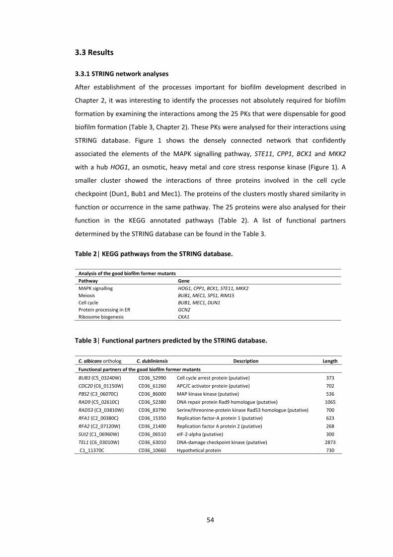
54
3.3 Results
3.3.1 STRING network analyses
After establishment of the processes important for biofilm development described in
Chapter 2, it was interesting to identify the processes not absolutely required for biofilm
formation by examining the interactions among the 25 PKs that were dispensable for good
biofilm formation (Table 3, Chapter 2). These PKs were analysed for their interactions using
STRING database. Figure 1 shows the densely connected network that confidently
associated the elements of the MAPK signalling pathway, STE11, CPP1, BCK1 and MKK2
with a hub HOG1, an osmotic, heavy metal and core stress response kinase (Figure 1). A
smaller cluster showed the interactions of three proteins involved in the cell cycle
checkpoint (Dun1, Bub1 and Mec1). The proteins of the clusters mostly shared similarity in
function or occurrence in the same pathway. The 25 proteins were also analysed for their
function in the KEGG annotated pathways (Table 2). A list of functional partners
determined by the STRING database can be found in the Table 3.
Table 2| KEGG pathways from the STRING database.
Analysis of the good biofilm former mutants
Pathway Gene
MAPK signalling HOG1, CPP1, BCK1, STE11, MKK2
Meiosis BUB1, MEC1, SPS1, RIM15
Cell cycle BUB1, MEC1, DUN1
Protein processing in ER GCN2
Ribosome biogenesis CKA1
Table 3| Functional partners predicted by the STRING database.
C. albicans ortholog C. dubliniensis Description Length
Functional partners of the good biofilm former mutants
BUB3 (C5_03240W) CD36_52990 Cell cycle arrest protein (putative) 373
CDC20 (C6_01150W) CD36_61260 APC/C activator protein (putative) 702
PBS2 (C3_06070C) CD36_86000 MAP kinase kinase (putative) 536
RAD9 (C5_02610C) CD36_52380 DNA repair protein Rad9 homologue (putative) 1065
RAD53 (C3_03810W) CD36_83790 Serine/threonine-protein kinase Rad53 homologue (putative) 700
RFA1 (C2_00380C) CD36_15350 Replication factor-A protein 1 (putative) 623
RFA2 (C2_07120W) CD36_21400 Replication factor A protein 2 (putative) 268
SUI2 (C1_06960W) CD36_06510 eIF-2-alpha (putative) 300
TEL1 (C6_03010W) CD36_63010 DNA-damage checkpoint kinase (putative) 2873
C1_11370C CD36_10660 Hypothetical protein 730

55
Figure 1| Predicted STRING interaction networks. In order to produce an interaction network, the STRING database quantitatively integrates experimental
data, computational prediction methods and published literature. The spheres and edges represent PKs and known/predicted interactions respectively.
Stronger blue lines indicate the connections with higher confidence. Protein kinase encoding genes are boxed and the asterisk (*) indicates a functional
partner predicted by the STRING database. A network shows the interaction of the genes/proteins not individually required for biofilm formation.
Very good biofilm formers
Good biofilm formers
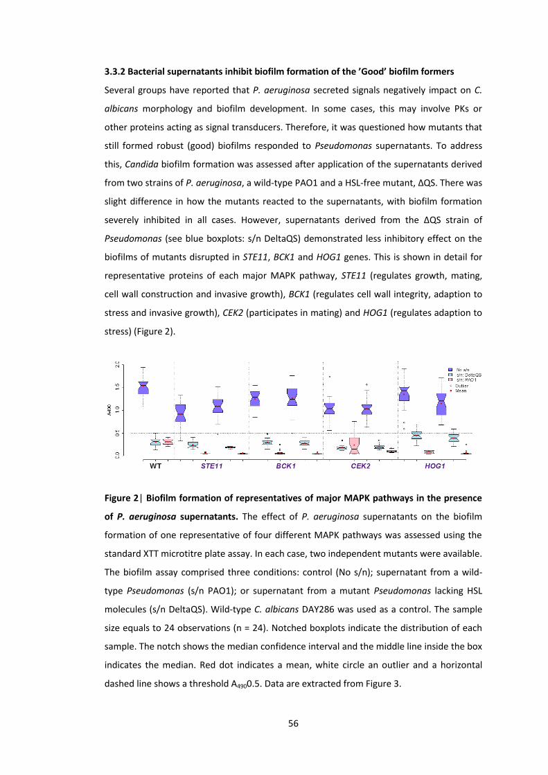
56
3.3.2 Bacterial supernatants inhibit biofilm formation of the ’Good’ biofilm formers
Several groups have reported that P. aeruginosa secreted signals negatively impact on C.
albicans morphology and biofilm development. In some cases, this may involve PKs or
other proteins acting as signal transducers. Therefore, it was questioned how mutants that
still formed robust (good) biofilms responded to Pseudomonas supernatants. To address
this, Candida biofilm formation was assessed after application of the supernatants derived
from two strains of P. aeruginosa, a wild-type PAO1 and a HSL-free mutant, ΔQS. There was
slight difference in how the mutants reacted to the supernatants, with biofilm formation
severely inhibited in all cases. However, supernatants derived from the ΔQS strain of
Pseudomonas (see blue boxplots: s/n DeltaQS) demonstrated less inhibitory effect on the
biofilms of mutants disrupted in STE11, BCK1 and HOG1 genes. This is shown in detail for
representative proteins of each major MAPK pathway, STE11 (regulates growth, mating,
cell wall construction and invasive growth), BCK1 (regulates cell wall integrity, adaption to
stress and invasive growth), CEK2 (participates in mating) and HOG1 (regulates adaption to
stress) (Figure 2).
Figure 2| Biofilm formation of representatives of major MAPK pathways in the presence
of P. aeruginosa supernatants. The effect of P. aeruginosa supernatants on the biofilm
formation of one representative of four different MAPK pathways was assessed using the
standard XTT microtitre plate assay. In each case, two independent mutants were available.
The biofilm assay comprised three conditions: control (No s/n); supernatant from a wild-
type Pseudomonas (s/n PAO1); or supernatant from a mutant Pseudomonas lacking HSL
molecules (s/n DeltaQS). Wild-type C. albicans DAY286 was used as a control. The sample
size equals to 24 observations (n = 24). Notched boxplots indicate the distribution of each
sample. The notch shows the median confidence interval and the middle line inside the box
indicates the median. Red dot indicates a mean, white circle an outlier and a horizontal
dashed line shows a threshold A4900.5. Data are extracted from Figure 3.
57
Biofilm formation of all the good biofilm former mutants with and without application of
bacterial supernatants is demonstrated in Figure 3. This figure shows that the biofilms of
some C. albicans PK mutants were less affected by the supernatants derived from a ΔQS
strain of Pseudomonas lacking HSL coding gene. These mutants are listed in the Table 4.
Table 4| C. albicans PK mutants less affected by the ΔQS supernatants. Data are extracted
from Figure 3.
Mutants less affected by the ΔQS supernatants
‘Good’ biofilm formers ‘Very good’ biofilm formers
YCK3 MEC1
DUN1 SKY1
YPL150W BCK1
CKA1 CMK1
NNK1 MKK2
RIM15
ENV7
STE11
HOG1
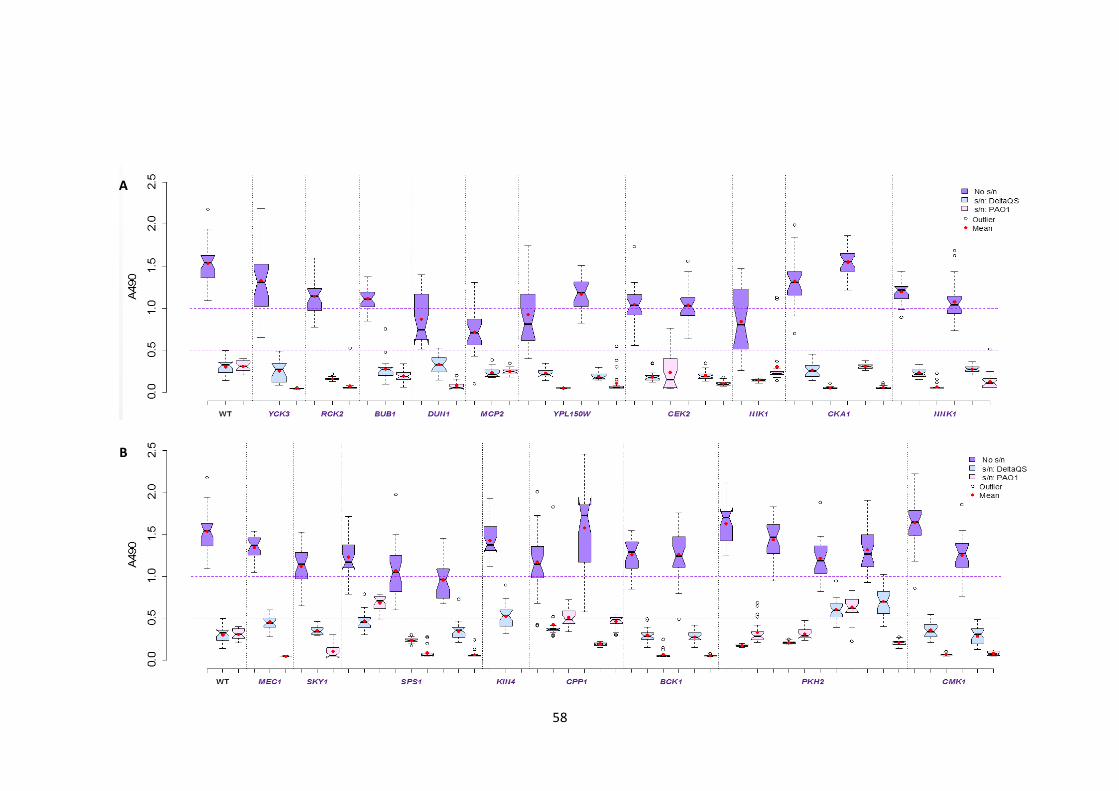
58
B
A
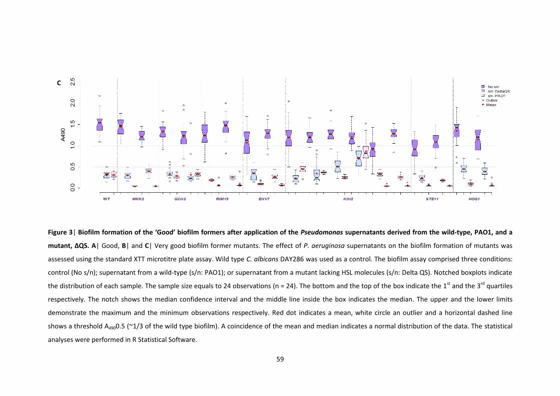
59
Figure 3| Biofilm formation of the ‘Good’ biofilm formers after application of the Pseudomonas supernatants derived from the wild-type, PAO1, and a
mutant, ΔQS. A| Good, B| and C| Very good biofilm former mutants. The effect of P. aeruginosa supernatants on the biofilm formation of mutants was
assessed using the standard XTT microtitre plate assay. Wild type C. albicans DAY286 was used as a control. The biofilm assay comprised three conditions:
control (No s/n); supernatant from a wild-type (s/n: PAO1); or supernatant from a mutant lacking HSL molecules (s/n: Delta QS). Notched boxplots indicate
the distribution of each sample. The sample size equals to 24 observations (n = 24). The bottom and the top of the box indicate the 1st and the 3rd quartiles
respectively. The notch shows the median confidence interval and the middle line inside the box indicates the median. The upper and the lower limits
demonstrate the maximum and the minimum observations respectively. Red dot indicates a mean, white circle an outlier and a horizontal dashed line
shows a threshold A4900.5 (~1/3 of the wild type biofilm). A coincidence of the mean and median indicates a normal distribution of the data. The statistical
analyses were performed in R Statistical Software.
C

60
3.3.3 ΔQS bacterial supernatants inhibit biofilm formation of the good biofilm formers
without affecting their morphology
Since the biofilms are linked to yeast morphology, we also assessed the mutants’ ability to
switch from yeast to hyphal growth in the absence or presence of bacterial supernatants
(Figure 4). As previously reported by McAlester et al. (2008) and Holcombe et al. (2010),
supernatants from the wild-type P. aeruginosa prevented yeast switching to hyphal
growth. This was the case for both wild-type C. albicans and the mutants tested. In
contrast, all the ΔQS supernatant-treated mutants maintained a yeast to hyphae switch
(with the possible exception of CEK2) consistent with other reports of HSL-dependent and
HSL-independent effects.
Figure 4| Morphology of representatives of major MAPK pathways in the presence of P.
aeruginosa supernatants. The effect of P. aeruginosa supernatants on morphological
development of one representative of four different MAPK pathways was assessed using
the standard yeast to hyphae transition assay. Two wild-type C. albicans strains, DAY286
and SC5314, and four mutants were tested for filamentation. The morphology of each
WT
(DA
Y28
6)
No s/n PAO1 s/n ΔQS s/n
WT
(SC
531
4)
No s/n PAO1 s/n ΔQS s/n
BC
K1
(F7
, pk2
)
HO
G1
(F5
pk2
)
STE1
1 (
B8
, pk2
) ST
E11
(B
7, p
k2)
CEK
2 (
F12,
pk2
) C
EK2
(F1
1, p
k2)
BC
K1
(F8
, pk2
)
HO
G1
(F6
, pk2
)

61
strain was examined after 6 h incubation (37oC) in filament-inducing medium (YNBNP). The
treatments were: control (No s/n); supernatant from a wild-type P. aeruginosa strain (PAO1
s/n); or supernatant from a P. aeruginosa mutant lacking HSL molecules (ΔQS s/n). The
scale bar corresponds to 10 μm.

62
3.4 Discussion
Biofilm formation in C. albicans is mediated by diverse processes such as adherence and
hyphal growth. After highlighting the additional processes that were important for robust
biofilm development (Chapter 2), it was also interesting to see which proteins were not
absolutely required for biofilm formation and, in this regard, the finding that none of the
individual PKs in MAP kinase cascades is required appears surprising. This result can be
explained by the redundancy and cross-talk that is built into these pathways and thus is
likely that individual mutants are compensated by parallel processes in the cell. Network
analysis also suggested that impairing aspects of the cell cycle has no impact on these
functions and the presence of cell cycle processes in both the ‘Poor’ and the ‘Good’
categories reflects the complexity of these processes in the cell.
Previous studies of McAlester et al. (2008) and Holcombe et al. (2010) showed that both
the yeast-hyphal transition and Candida biofilms were impaired by Pseudomonas
supernatants. In the case of morphology, QS molecules (HSLs) are known to play a role by
targeting the Ras/cAMP/PKA pathway (reviewed by Hogan and Sundstrom 2009), whereas
quinolones may be involved in the biofilm effect, through an unknown mechanism (Reen et
al. 2011). We assessed whether any of the mutants that formed good biofilms failed to
respond to P. aeruginosa supernatants without finding any obvious positives. Furthermore,
the remaining PK mutants were also subject to this screen, with the same findings (data not
shown). However, mutants were briefly differentiated into HSL-dependent and HSL-
independent but none of the protein kinases was strongly related with signal transduction
response in Candida to Pseudomonas signals. Initially, CEK2 stood out as this mutant failed
to filament in the presence of HSL-negative supernatant but close inspection of the control
conditions indicated that this is an underlying morphological defect in this mutant (Figure
4). In fact, it is somewhat surprising that the mutant is able to form biofilms given the
pseudohyphal nature of the strain (Figure 2 and 4).
The comprehensive analysis of the response of wild-type and 63 mutants to supernatants
from wild-type and QS- (HSL-negative) strains of P. aeruginosa unequivocally separates the
two effects on C. albicans: HSL-dependent impairment of morphology and HSL-
independent impairment of biofilms. It remains to be determined whether the latter effect
is fully explained by the action of quinolones (natural antimicrobial signalling molecules) or
whether it arises from a combination of effects, for examples, quinolones, LPS
(lipopolysaccharides) and other molecules.
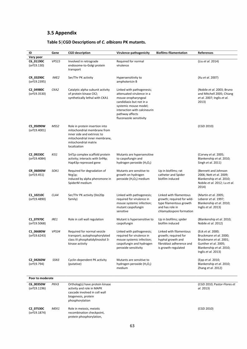
63
3.5 Appendix
Table 5|CGD Descriptions of C. albicans PK mutants.
ID Gene CGD description Virulence-pathogenicity Biofilms-filamentation References
Very poor
C6_01190C (orf19.130)
VPS15 Involved in retrograde endosome-to-Golgi protein transport
Required for normal virulence
(Liu et al. 2014)
CR_03290C (orf19.2395)
IME2 Ser/Thr PK activity Hypersensitivity to amphotericin B
(Xu et al. 2007)
C2_04980C (orf19.3530)
CKA2 Catalytic alpha-subunit activity of protein kinase CK2; synthetically lethal with CKA1
Linked with pathogenesis; attenuated virulence in a mouse oropharyngeal candidiasis but not in a systemic mouse model; interaction with calcineurin pathway affects fluconazole sensitivity
(Nobile et al. 2003; Bruno and Mitchell 2005; Chiang et al. 2007; Inglis et al. 2013)
C5_05090W (orf19.4001)
MSS2 Role in protein insertion into mitochondrial membrane from inner side and extrinsic to mitochondrial inner membrane, mitochondrial matrix localization
(CGD 2010)
C2_09230C (orf19.4084)
KIS1 Snf1p complex scaffold protein activity; interacts with Snf4p; Hap43p-repressed gene
Mutants are hypersensitive to caspofungin and hydrogen peroxide (H2O2)
(Corvey et al. 2005; Blankenship et al. 2010; Singh et al. 2011)
CR_06000W (orf19.451)
SOK1 Required for degradation of Nrg1p; induced by alpha pheromone in SpiderM medium
Mutants are sensitive to growth on hydrogen peroxide (H2O2) medium
Up in biofilms; rat catheter and Spider biofilm induced
(Bennett and Johnson 2006; Nett et al. 2009; Blankenship et al. 2010; Nobile et al. 2012; Lu et al. 2014)
C1_10210C (orf19.4890)
CLA4 Ser/Thr PK activity (Ste20p family)
Linked with pathogenesis; required for virulence in mouse systemic infection; mutant caspofungin sensitive
Linked with filamentous growth; required for wild-type filamentous growth and has role in chlamydospore formation
(Martin et al. 2005; Leberer et al. 1997; Blankenship et al. 2010; Inglis et al. 2013)
C1_07970C (orf19.5068)
IRE1 Role in cell wall regulation Mutant is hypersensitive to caspofungin
Up in biofilms; spider biofilm induced
(Blankenship et al. 2010; Nobile et al. 2012)
C1_06680W (orf19.6243)
VPS34 Required for normal vesicle transport; autophosphorylated class III phosphatidylinositol 3-kinase activity
Linked with pathogenesis; required for virulence in mouse systemic infection; caspofungin and hydrogen peroxide sensitivity
Linked with filamentous growth; required for hyphal growth and fibroblast adherence and is growth-regulated
(Eck et al. 2000; Bruckmann et al. 2000; Bruckmann et al. 2001; Gunther et al. 2005; Blankenship et al. 2010; Inglis et al. 2013)
C2_04260W (orf19.794)
SSN3 Cyclin-dependent PK activity (putative)
Mutants are sensitive to hydrogen peroxide (H2O2) medium
(Epp et al. 2010; Blankenship et al. 2010; Zhang et al. 2012)
Poor to moderate
C6_00350W (orf19.1196)
PKH3 Ortholog(s) have protein kinase activity and role in MAPK cascade involved in cell wall biogenesis, protein phosphorylation
(CGD 2010; Pastor-Flores et al. 2013)
C2_07530C (orf19.1874)
MEK1 Role in meiosis, meiotic recombination checkpoint, protein phosphorylation,
(CGD 2010)
64
regulation of linear element assembly and cytosol, linear element localization
C2_08860W (orf19.223)
FPK1 Hap43-repressed; possibly an essential gene, disruptants not obtained by UAU1 method
Induced by prostaglandins Up in biofilms; flow model biofilm induced; Spider biofilm induced
(Levitin and Whiteway 2007; Mitchell 2009; Bonhomme et al. 2011; Singh et al. 2011; Nobile et al. 2012)
C1_10780C (orf19.2341)
HNT1 Similar activity with protein kinase C inhibitor-I; protein level decreases in stationary phase cultures
(Kusch et al. 2008)
C4_06260W (orf19.2910)
PGA43 Putative GPI-anchored protein activity (posttranslational modification)
(De Groot et al. 2003)
C2_06600W (orf19.35)
SKY2 Predicted kinase activity Up in biofilms; rat catheter, flow model, spider biofilm induced
(Nett et al. 2009; Bonhomme et al. 2011; Nobile et al. 2012)
CR_02250C (orf19.3744)
CEX1 Ortholog(s) have tRNA binding activity, role in tRNA export from nucleus and cytoplasm, nuclear pore localization
(CGD 2010)
C5_02840C (orf19.4308)
HSL1 Involved in determination of morphology during the cell cycle of both yeast-form and hyphal cells via regulation of Swe1p and Cdc28p
Required for full virulence and kidney colonization in mouse systemic infection
Linked with filamentation and biofilms
(Wightman et al. 2004; Umeyama et al. 2005; Inglis et al. 2013)
C3_00550C (orf19.5408)
HRK1 Predicted role in cellular ion homeostasis
Down in biofilms; spider biofilm repressed
(Blankenship et al. 2010; Nobile et al. 2012)
C3_05740C (orf19.7355)
SSN8 Ortholog of S. cerevisiae Ssn8; a component of RNA polymerase II holoenzyme
Mutants are viable and are sensitive to hydrogen peroxide medium
(Nobile et al. 2003; Blankenship et al. 2010; Zhang et al. 2012)
Moderate
C7_03340C (orf19.1341)
PRR2 Ser/Thr PK activity (putative) Mutation confers resistance to 5-fluorocytosine (5-FC)
(Xu et al. 2007)
C2_00300C (orf19.2102)
CKB1 Regulation of casein kinase II (β subunit)
Null mutants are hypersensitive to caspofungin and hydrogen peroxide (H2O2) medium
(Zelada et al. 2003; Blankenship et al. 2010; Bruno and Mitchell 2005)
C2_07210C (orf19.2277)
TPK2 Controls morphogenesis and stress response; cAMP-dependent PK catalytic subunit; isoform of Tpk1
Linked with pathogenesis; needed for epithelial cell damage, engulfment and oral (not systemic) virulence in mice
Linked with filamentous growth and biofilms; down in biofilms; rat catheter, spider biofilm repressed
(Sonneborn et al. 2000; Bockmuhl et al. 2001; Cloutier et al. 2003; Park et al. 2005; Nett et al. 2009; Nobile et al. 2012; Inglis et al. 2013)
C1_03450C (orf19.3047)
SIP3 Protein kinase-related protein activity
Required for normal sensitivity to caspofungin
(Blankenship et al. 2010)
CR_02460W (orf19.3720)
BCY1 Ortholog(s) have endo-1,4-beta-xylanase activity and SCF ubiquitin ligase complex, cytoplasm localization
(CGD 2010)
CR_06040W (orf19.3854)
SAT4 Clade-associated gene expression
Amphotericin B induced Up in biofilms; spider biofilm induced
(Liu et al. 2005; MacCallum et al. 2009; Nobile et al. 2012)
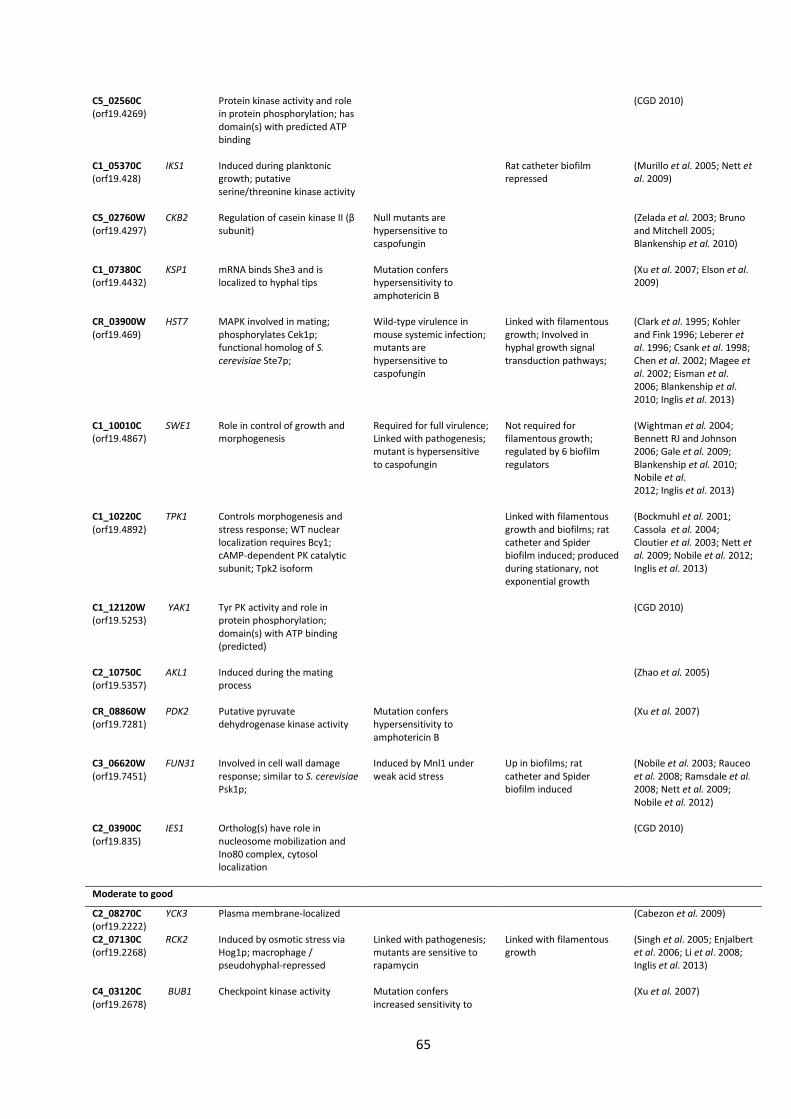
65
C5_02560C (orf19.4269)
Protein kinase activity and role in protein phosphorylation; has domain(s) with predicted ATP binding
(CGD 2010)
C1_05370C (orf19.428)
IKS1 Induced during planktonic growth; putative serine/threonine kinase activity
Rat catheter biofilm repressed
(Murillo et al. 2005; Nett et al. 2009)
C5_02760W (orf19.4297)
CKB2 Regulation of casein kinase II (β subunit)
Null mutants are hypersensitive to caspofungin
(Zelada et al. 2003; Bruno and Mitchell 2005; Blankenship et al. 2010)
C1_07380C (orf19.4432)
KSP1 mRNA binds She3 and is localized to hyphal tips
Mutation confers hypersensitivity to amphotericin B
(Xu et al. 2007; Elson et al. 2009)
CR_03900W (orf19.469)
HST7 MAPK involved in mating; phosphorylates Cek1p; functional homolog of S. cerevisiae Ste7p;
Wild-type virulence in mouse systemic infection; mutants are hypersensitive to caspofungin
Linked with filamentous growth; Involved in hyphal growth signal transduction pathways;
(Clark et al. 1995; Kohler and Fink 1996; Leberer et al. 1996; Csank et al. 1998; Chen et al. 2002; Magee et al. 2002; Eisman et al. 2006; Blankenship et al. 2010; Inglis et al. 2013)
C1_10010C (orf19.4867)
SWE1 Role in control of growth and morphogenesis
Required for full virulence; Linked with pathogenesis; mutant is hypersensitive to caspofungin
Not required for filamentous growth; regulated by 6 biofilm regulators
(Wightman et al. 2004; Bennett RJ and Johnson 2006; Gale et al. 2009; Blankenship et al. 2010; Nobile et al. 2012; Inglis et al. 2013)
C1_10220C (orf19.4892)
TPK1 Controls morphogenesis and stress response; WT nuclear localization requires Bcy1; cAMP-dependent PK catalytic subunit; Tpk2 isoform
Linked with filamentous growth and biofilms; rat catheter and Spider biofilm induced; produced during stationary, not exponential growth
(Bockmuhl et al. 2001; Cassola et al. 2004; Cloutier et al. 2003; Nett et al. 2009; Nobile et al. 2012; Inglis et al. 2013)
C1_12120W (orf19.5253)
YAK1 Tyr PK activity and role in protein phosphorylation; domain(s) with ATP binding (predicted)
(CGD 2010)
C2_10750C (orf19.5357)
AKL1 Induced during the mating process
(Zhao et al. 2005)
CR_08860W (orf19.7281)
PDK2 Putative pyruvate dehydrogenase kinase activity
Mutation confers hypersensitivity to amphotericin B
(Xu et al. 2007)
C3_06620W (orf19.7451)
FUN31 Involved in cell wall damage response; similar to S. cerevisiae Psk1p;
Induced by Mnl1 under weak acid stress
Up in biofilms; rat catheter and Spider biofilm induced
(Nobile et al. 2003; Rauceo et al. 2008; Ramsdale et al. 2008; Nett et al. 2009; Nobile et al. 2012)
C2_03900C (orf19.835)
IES1 Ortholog(s) have role in nucleosome mobilization and Ino80 complex, cytosol localization
(CGD 2010)
Moderate to good
C2_08270C (orf19.2222)
YCK3 Plasma membrane-localized (Cabezon et al. 2009)
C2_07130C (orf19.2268)
RCK2 Induced by osmotic stress via Hog1p; macrophage / pseudohyphal-repressed
Linked with pathogenesis; mutants are sensitive to rapamycin
Linked with filamentous growth
(Singh et al. 2005; Enjalbert et al. 2006; Li et al. 2008; Inglis et al. 2013)
C4_03120C (orf19.2678)
BUB1 Checkpoint kinase activity Mutation confers increased sensitivity to
(Xu et al. 2007)
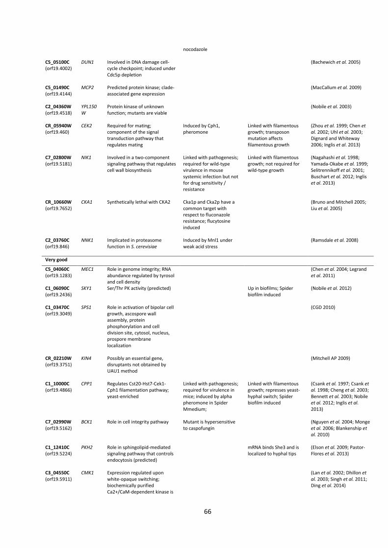
66
nocodazole
C5_05100C (orf19.4002)
DUN1 Involved in DNA damage cell-cycle checkpoint; induced under Cdc5p depletion
(Bachewich et al. 2005)
C5_01490C (orf19.4144)
MCP2 Predicted protein kinase; clade-associated gene expression
(MacCallum et al. 2009)
C2_04360W (orf19.4518)
YPL150W
Protein kinase of unknown function; mutants are viable
(Nobile et al. 2003)
CR_05940W (orf19.460)
CEK2 Required for mating; component of the signal transduction pathway that regulates mating
Induced by Cph1, pheromone
Linked with filamentous growth; transposon mutation affects filamentous growth
(Zhou et al. 1999; Chen et al. 2002; Uhl et al. 2003; Dignard and Whiteway 2006; Inglis et al. 2013)
C7_02800W (orf19.5181)
NIK1 Involved in a two-component signaling pathway that regulates cell wall biosynthesis
Linked with pathogenesis; required for wild-type virulence in mouse systemic infection but not for drug sensitivity / resistance
Linked with filamentous growth; not required for wild-type growth
(Nagahashi et al. 1998; Yamada-Okabe et al. 1999; Selitrennikoff et al. 2001; Buschart et al. 2012; Inglis et al. 2013)
CR_10660W (orf19.7652)
CKA1 Synthetically lethal with CKA2 Cka1p and Cka2p have a common target with respect to fluconazole resistance; flucytosine induced
(Bruno and Mitchell 2005; Liu et al. 2005)
C2_03760C (orf19.846)
NNK1 Implicated in proteasome function in S. cerevisiae
Induced by Mnl1 under weak acid stress
(Ramsdale et al. 2008)
Very good
C5_04060C (orf19.1283)
MEC1 Role in genome integrity; RNA abundance regulated by tyrosol and cell density
(Chen et al. 2004; Legrand et al. 2011)
C1_06090C (orf19.2436)
SKY1 Ser/Thr PK activity (predicted) Up in biofilms; Spider biofilm induced
(Nobile et al. 2012)
C1_03470C (orf19.3049)
SPS1 Role in activation of bipolar cell growth, ascospore wall assembly, protein phosphorylation and cell division site, cytosol, nucleus, prospore membrane localization
(CGD 2010)
CR_02210W (orf19.3751)
KIN4 Possibly an essential gene, disruptants not obtained by UAU1 method
(Mitchell AP 2009)
C1_10000C (orf19.4866)
CPP1 Regulates Cst20-Hst7-Cek1-Cph1 filamentation pathway; yeast-enriched
Linked with pathogenesis; required for virulence in mice; induced by alpha pheromone in Spider Mmedium;
Linked with filamentous growth; represses yeast-hyphal switch; Spider biofilm induced
(Csank et al. 1997; Csank et al. 1998; Cheng et al. 2003; Bennett et al. 2003; Nobile et al. 2012; Inglis et al. 2013)
C7_02990W (orf19.5162)
BCK1 Role in cell integrity pathway Mutant is hypersensitive to caspofungin
(Nguyen et al. 2004; Monge et al. 2006; Blankenship et al. 2010)
C1_12410C (orf19.5224)
PKH2 Role in sphingolipid-mediated signaling pathway that controls endocytosis (predicted)
mRNA binds She3 and is localized to hyphal tips
(Elson et al. 2009; Pastor-Flores et al. 2013)
C3_04550C (orf19.5911)
CMK1 Expression regulated upon white-opaque switching; biochemically purified Ca2+/CaM-dependent kinase is
(Lan et al. 2002; Dhillon et al. 2003; Singh et al. 2011; Ding et al. 2014)
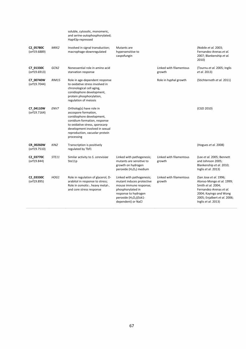
67
soluble, cytosolic, monomeric, and serine-autophosphorylated; Hap43p-repressed
C2_05780C (orf19.6889)
MKK2 Involved in signal transduction; macrophage-downregulated
Mutants are hypersensitive to caspofungin
(Nobile et al. 2003; Fernandez-Arenas et al. 2007; Blankenship et al. 2010)
C7_01330C (orf19.6913)
GCN2 Nonessential role in amino acid starvation response
Linked with filamentous growth
(Tournu et al. 2005; Inglis et al. 2013)
C7_00740W (orf19.7044)
RIM15 Role in age-dependent response to oxidative stress involved in chronological cell aging, conidiophore development, protein phosphorylation, regulation of meiosis
Role in hyphal growth (Stichternoth et al. 2011)
C7_04110W (orf19.7164)
ENV7 Ortholog(s) have role in ascospore formation, conidiophore development, conidium formation, response to oxidative stress, sporocarp development involved in sexual reproduction, vacuolar protein processing
(CGD 2010)
CR_00260W (orf19.7510)
KIN2 Transcription is positively regulated by Tbf1
(Hogues et al. 2008)
C2_03770C (orf19.844)
STE11 Similar activity to S. cerevisiae Ste11p
Linked with pathogenesis; mutants are sensitive to growth on hydrogen peroxide (H2O2) medium
Linked with filamentous growth
(Lee et al. 2005; Bennett and Johnson 2005; Blankenship et al. 2010; Inglis et al. 2013)
C2_03330C (orf19.895)
HOG1 Role in regulation of glycerol, D-arabitol in response to stress; Role in osmotic-, heavy metal-, and core stress response
Linked with pathogenesis; mutant induces protective mouse immune response; phosphorylated in response to hydrogen peroxide (H2O2)(Ssk1-dependent) or NaCl
Linked with filamentous growth
(San Jose et al. 1996; Alonso-Monge et al. 1999; Smith et al. 2004; Fernandez-Arenas et al. 2004; Kayingo and Wong 2005; Enjalbert et al. 2006; Inglis et al. 2013)
68
Chapter 4
TFbsST: Transcription Factor binding site Search Tool
Nina Konstantinidou and John Patrick Morrissey
School of Microbiology, University College Cork, Cork, T12YN60, Ireland
Key words: Candida albicans; transcription factor binding site motifs; biofilms;
Pseudomonas aeruginosa; promoter regions; TFbsST database.
Subtitle: In silico analysis of Candida albicans promoter sequences
This chapter is in preparation for submission in the Database Issue of Nucleic Acid Research
(Oxford Journals). The TFbsST database is published online under the following link:
http://bioinfo.ucc.ie/TFbsST/.
69
Abstract
C. albicans is a serious pathogen that features high drug resistant properties partially due
to its biofilms. The biofilm development depends on the environmental stimuli/signals that
are sensed by the fungal cell receptors. These signals are transmitted by the protein kinases
(PK) and the gene transcription is regulated by the transcription factors (TF). Using C.
albicans TF binding site library and Python algorithms, we created a TFbsST database
(Transcription Factor binding site Search Tool), a user-friendly application for in silico
analyses of Candida promoters. TFbsST database contains TF binding sites of C. albicans
and related Candida species such as C. parapsilosis, C. dubliniensis and C. glabrata.
Additionally, TFbsST performs TF frequency (outputs a table with %) and TF localization
analysis (outputs graphs and tables). These features render the TFbsST software as a useful
tool for in silico analysis of Candida gene promoter sequences.
Previous studies of Holcombe et al. (2010) had shown that P. aeruginosa secreted signals
can affect C. albicans biofilms. To investigate the regulation of Candida genes that were
altered in response to P. aeruginosa secreted chemicals, we utilised a TFbsST application.
The up-to-date gene ontology (GO) annotation indicated that the genes coding for the cell
wall/surface proteins, important for biofilm formation, contained members of the biofilm-
related gene families, RBT and ALS. To identify the TFs that were overrepresented in these
gene families, we carried out TF frequency analyses. These analyses showed that Efg1p was
overrepresented in both RBT and ALS families. To question whether Efg1p could regulate
the RBT family genes across other Candida species, C. parapsilosis RBT family gene
promoters were screened against C. parapsilosis Efg1p using a Motif search option of the
TFbsST application. Efg1p binding sites were predicted to regulate some C. parapsilosis RBT
genes indicating that Efg1p is an important TF across the different Candida species.
70
4. TFbsST: Transcription Factor binding site Search Tool
4.1 Introduction
A serious human pathogen, C. albicans, can cause nosocomial infections partially due to its
ability to form drug resistant biofilms on indwelling medical devices. In these clinical
settings, C. albicans is frequently coisolated with P. aeruginosa. P. aeruginosa is the main
cause of morbidity in populations with cystic fibrosis (CF) (Govan and Deretic 1996;
Chambers et al. 2005; Leclair and Hogan 2010) and AIDS (Mendelson et al. 1994). Studies
from our laboratory (Holcombe et al. 2010; Reen et al. 2011;) and independent
investigations (Bandara et al. 2010a; Bandara et al. 2010b; Bandara et al. 2013) have shown
that Pseudomonas can inhibit Candida biofilm development in vitro. C. albicans biofilm
development is a complex phenomenon orchestrated by more than 1000 genes that are
governed by 9 master transcription regulators, Efg1p, Ndt80p, Brg1p, Bcr1p, Rob1p, Tec1p,
Flo8p, Gal4p and Rfx2p (Nobile et al. 2012; Fox et al. 2015).
Transcription factors (TF) regulate expression of target genes during transcription. TFs
contain DNA binding domains that facilitate recognition and binding of TFs to specific small
DNA sequences across the promoter region (1000 bp upstream) of target genes. A
collection of TF DNA binding site data facilitates computational analysis of the gene
promoter regions when using appropriate software. A comprehensive TF motif database is
useful for in silico analysis of transcriptional networks and gene regulation studies. We
additionally used our TF binding site database for computational analysis of biofilm
regulation in C. albicans as well as for the investigation of interaction between C. albicans
and P. aeruginosa.
Given the rapid progress of bioinformatics tools, many databases were developed to
analyse the promoter regions of genes in S. cerevisiae or other model organisms:
YeTFaSCo (Yeast Transcription Factor Specificity Compendium) focuses on S. cerevisiae
TF binding sites that are evaluated for quality using several metrics. These specificities
are given in Position Frequency (PFM) or Position Weight Matrix (PWM) formats.
YeTFaSCo is mainly used to find the position of potential binding sites of a sequence
and for inspection of precomputed genome-wide binding sites (http://yetfasco.ccbr.
utoronto.ca) (de Boer and Hughes 2011).
71
UniPROBE (Universal PBM Resource for Oligonucleotide Binding Evaluation) database
hosts S. cerevisiae data generated by protein binding microarray (PBM) technology.
The UniPROBE database provides comprehensive data on the preferences of proteins
for any DNA sequence variants of length K (K-mer). For more complete analyses, the
UniPROBE website can output weight matrix (PWM) and graphical logo of the K-mer
sequence (http://the_brain.bwh.harvard.edu/ uniprobe) (Newburger and Bulyk 2009).
YEASTRACT DISCOVERER provides tools for de novo identification of binding site
consensus sequences from a set of gene promoter regions. DISCOVERER contains
MUSA (Motif finding using an UnSupervised Approach) algorithms that return the list
of motifs ordered by their p-value. In order to extract these motifs, specification of the
parameters such as box lengths and distances between boxes is not required from the
user (http://www.yeastract.com/formfindregulators.php) (Teixeira et al. 2006).
MYBS (Mining Yeast Binding Sites) integrates both experimentally verified data and
predicted position weight matrixes (PWMs) from 11 different databases. To search for
motifs in the promoters of the input genes, MYBS uses ChIP-chip data and
phylogenetic footprinting as its main filters and considers 7 species including C.
glabrata. For the identification of potential regulatory associations between two TFs
and their combinatorial regulation, MYBS enables the visualization of potential
regulators and target gene sets for each TF pair (http://bits.iis.sinica.edu.tw) (Tsai et
al. 2007).
YPA (Yeast Promoter Atlas) compiles promoter features of S. cerevisiae. YPA integrates
various resources (including promoter sequences, TATA boxes, TF binding sites,
nucleosome occupancy, DNA bendability, TF-TF interaction and gene expression data)
to provide a comprehensive view of gene promoter region
(http://ypa.csbb.ntu.edu.tw) (Chang et al. 2011).
JASPAR CORE database contains a curated set of profiles, derived from published
collections of experimentally defined TF binding sites for eukaryotes. JASPAR CORE
Fungi mostly contains S. cerevisiae TF binding site sequence logos
(http://jaspar.genereg.net) (Sandelin et al. 2004).
TRANSFAC® provides experimentally-proven eukaryotic TF motif consensus sequences
and a list of TF-regulated genes. TRANSCompel contains eukaryotic TF sets that are
experimentally proven to interact in a synergistic or antagonistic manner
(http://www.gene-regulation.com/pub/databases.html) (Matys et al. 2006).
72
The MacIsaac collection contains TF motifs from solely ChIP-chip data (MacIsaac et al.
2006).
To our knowledge there is no bioinformatics tool available for C. albicans promoter region
analyses. Hence we generated a C. albicans TF binding site database and created Python
algorithms to develop publically available software TFbsST (Transcription Factor binding
site Search Tool) for the analyses of Candida promoter regions. TFbsST is the first
bioinformatics tool for in silico promoter analyses of Candida species including C. albicans,
C. dubliniensis, C. parapsilosis and C. glabrata (http://bioinfo.ucc.ie/TFbsST/).
To investigate the regulation of Candida genes that were altered in the presence of
bacterial supernatants, we screened the C. albicans gene sets that were upregulated or
downregulated in response to Pseudomonas supernatants (McAlester et al. 2008) across
the TFbsST database. After gene ontology (GO) analysis with Blast2Go, the proteins
encoded by these genes were grouped according to their localization in the cell. Cell wall
proteins are important for biofilm formation. To question their regulation, the promoter
regions of the genes coding for the proteins localized around the cell wall were investigated
in more detail. The overrepresented TFs were established after analyses of different gene
sets and gene families.
73
4.2 Materials and Methods
4.2.1 General approach
In order to generate a comprehensive TFbsST database for C. albicans promoter region
analyses in silico, we developed a research strategy shown in Figure 1. This workflow
represents a modified ‘Waterfall’ software development model where the project
requirements were based on the analyses of DNA sequences in gene promoters. Briefly, to
populate a TF database with the corresponding DNA binding site motifs, the relevant
literature was reviewed and experimentally validated motifs were selected for the
database. Python scripts were designed for promoter analysis and TF database was used as
a source of DNA binding site motifs. These files served as the basis for the creation of the
TFbsST application. After software design we implemented the testing and verification
process that involved prediction of TF frequencies across the C. albicans genome and
analyses of cell wall protein coding gene families.
To analyse the 4 gene sets, the expression of which was impaired in response to bacterial
supernatants according to Holcombe et al. (2010), we retrieved their protein sequences
from the Candida Genome Database (CGD) (Arnaud et al. 2005) and annotated with the
Blast2Go annotation tool (Conesa et al. 2005). Blast2Go uses input protein sequences to
assign the proteins into 3 classes according to their molecular function (F), biological
process they are involved in (P) and the cellular component (C) where they are localized in
the cell. Thus, we defined smaller subsets based on the localisation of the proteins that
these differentially expressed genes encoded in the cell. In parallel, as a control group of
genes, we downloaded a file with all C. albicans genes plus 1000 bp upstream and 1000 bp
downstream regions from the CGD and retrieved 1000 bp upstream regions using Python
scripts for further TF binding site frequency predictions (%). In silico comparative analyses
of C. albicans and C. parapsilosis RBT family gene promoter regions against Efg1p via the
TFbsST database validated our computational methodology.
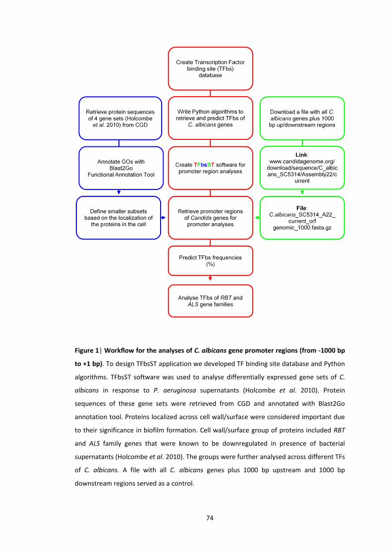
74
Figure 1| Workflow for the analyses of C. albicans gene promoter regions (from -1000 bp
to +1 bp). To design TFbsST application we developed TF binding site database and Python
algorithms. TFbsST software was used to analyse differentially expressed gene sets of C.
albicans in response to P. aeruginosa supernatants (Holcombe et al. 2010). Protein
sequences of these gene sets were retrieved from CGD and annotated with Blast2Go
annotation tool. Proteins localized across cell wall/surface were considered important due
to their significance in biofilm formation. Cell wall/surface group of proteins included RBT
and ALS family genes that were known to be downregulated in presence of bacterial
supernatants (Holcombe et al. 2010). The groups were further analysed across different TFs
of C. albicans. A file with all C. albicans genes plus 1000 bp upstream and 1000 bp
downstream regions served as a control.
75
4.2.2 Transcription factor binding site database creation
For reliable computational prediction, the ideal TF binding site library would contain TFs
associated to a single most accurate DNA binding site motif. Since the minimum number of
features increases the prediction power of the results in computational biology, we
optimized the TF database relating a single accurate motif to each TF (except from Cwt1p,
Msn4p and Srr1p). As a result, our database includes an optimized set of TF binding site
motifs that increase the reliability of the predictions. However, the source of each binding
site differs for each TF. Hence, it was difficult to compare the TF binding site data. Thus, to
select an optimum motif for each TF we took several approaches that lead to the creation
of the curated TF binding site library (Table 1).
The TF binding site database was based on publically available experimental data related to
C. albicans TFs and their binding site motifs (Table 1). To facilitate comparative analysis
among Candida species we also searched for indicative TF binding sites of C. parapsilosis, C.
dubliniensis and C. glabrata. To evaluate the TF binding sites for quality, we applied several
approaches. Firstly, a direct search was performed to find DNA binding site motifs of the
TFs from the Homann et al. (2009) TF mutant collection. Secondly, a general search of the
literature was performed to assign binding sites to the corresponding TF, looking for any TF
binding sites of C. albicans and indicative TF binding sites of C. parapsilosis, C. dubliniensis
and C. glabrata. Next, a reference-directed approach was taken to identify the most
relevant motifs. For this we prioritised the motifs that were found by the reliable methods,
for paradigm ChIP-Chip analysis, and were published in established journals. To optimise
our database, we also took a time-directed approach considering the most recently
identified binding sites. Some TF binding sites of C. albicans and S. cerevisiae display
similarity in their sequence. Finally, we compared C. albicans TF binding sites with that of S.
cerevisiae for similarities in YeTFaSCo (de Boer and Hughes 2011). YeTFaSCo contains a S.
cerevisiae TF collection of specificities that are evaluated for quality with several metrics.
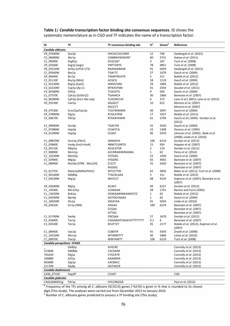
76
Table 1| Candida transcription factor binding site consensus sequences. ID shows the systematic nomenclature as in CGD and TF indicates the name of a transcription factor.
a Frequency of the TFs among all C. albicans (SC3514) genes (~6219) is given in % that is rounded to its closest
digit (This study). The analyses were carried out from December 2015 to January 2016. b Number of C. albicans genes predicted to possess a TF binding site (This study).
ID TF TF consensus binding site %a Genesb Reference
Candida albicans
CR_07440W Ace2p HHCACCACCWM 12 746 Swidergall et al. (2015) C3_06000W Ahr1p CGNBWVWWVNT 60 3731 Askew et al. (2011) C1_09090C Arg81p KCGCGST 3 187 Tuch et al. (2008) CR_02560C Asg1p (asgs) YMTTGKYS 78 4851 Tuch et al. (2008) CR_02510W Azf1p (orf19.173) RAADAARAAR 91 5659 Swidergall et al. (2015) C2_05640W Bas1p TGACTC 27 1679 Gasch et al. (2004) CR_06440C Bcr1p TAMATRCATR 5 311 Nobile et al. (2012) C2_01110C Bcy1p (Mcb) ACGCG 18 1119 Gasch et al. (2004) C1_05140W Brg1p (Gat2) SMGGTAM 29 1804 Nobile et al. (2012) C3_02220W Cap1p (Ap-1) MTKASTMA 41 2550 Goudot et al. (2011) C4_06580W Cbf1p TCACGTG 9 560 Gasch et al. (2004) C1_07370C Cph1p (ScSte12) TGAAACA 30 1866 Banerjee et al. (2007) C6_00280W Cph2p (Sre1-like seq) YCACMCCAY 6 373 Lane et al.( 2001); Lane et al. (2015) CR_05530C Cwt1p AGGGCT 10 622 Moreno et al. (2007) AGCCCT Moreno et al. (2007) CR_07530C Ecm22p/Upc2p TCGTWWWW 49 3047 Gasch et al. (2004) CR_07890W Efg1p RTGCATRW 17 1057 Nobile et al. (2012) C3_00670C Fkh2p RTAAAYAWW 61 3794 Gasch et al.( 2004); Gordan et al.
(2012) C2_09940W Gcn4p TGACTM 55 3420 Gasch et al. (2004) C1_07680W Hap2p CCAATCA 22 1368 Ozsarac et al. (1995) C4_01390W Hap3p CCAAT 96 5970 Johnson et al. (2005); Baek et al.
(2008); Linde et al. (2010) C1_09870W Hcm1p (Fkh2) WMAAYA 100 6219 Gordan et al. (2012) C2_03840C Ino4p (Ino2+Ino4) NBWTCASRTG 15 933 Hoppen et al. (2007) C3_04110C Mbp1p ACGCGTSR 2 124 Gordan et al. (2012) C7_00890C Mcm1p ACCRRAWWRGGMA 1 62 Perez et al. (2014) C2_10230W Met32p TGTGGC 23 1430 Gasch et al. (2004) C5_02940C Mig1p SYGGRG 65 4042 Banerjee et al. (2007) C1_08940C Msn4p (STRE - Msn2/4) CCCCT 55 3420 Banerjee et al. (2007) AGGGG Banerjee et al. (2007) C5_01755C Matα1p(MtlALPHA1) WTCCTTW 62 3856 Baker et al. (2011); Tuch et al. (2008) C2_00140W Ndt80p TTACACAAA 5 311 Nobile et al. (2012) C7_04230W Nrg1p MVCCCT 68 4229 Argimon et al. (2007); Banerjee et al.
(2007) CR_02640W Rfg1p ACAAT 99 6157 Gordan et al. (2012) C1_14340C Rim101p CCAAGAA 28 1741 Ramon and Fonzi (2003) C1_13620W Rob1p GGWAAWNWAWWTCC 1 62 Nobile et al. (2012) C1_04330W Rpn4p GGTGGCAAAA 1 62 Gasch et al. (2004) C1_10020W Sfu1p WGATAA 91 5659 Linde et al. (2010) CR_05610C Srr1p (SRR) AAGAA 100 6219 Banerjee et al. (2007) CCGAA Banerjee et al. (2007) ATTGG Banerjee et al. (2007) C1_01790W Swi4p CRCGAA 27 1679 Gordan et al. (2012) C5_01840C Tac1p CGGAWATCGGATATTTTTTTT 0.1 6 Banerjee et al. (2007) C3_04530C Tec1p RCATTCY 35 2177 Nobile et al. (2012); Argimon et al.
(2007) C1_08460C Upc2p CGBDTR 91 5659 Znaidi et al. (2008) C1_10150W Wor1p WTARRSTTT 30 1866 Lohse et al. (2010) C7_00970C Yox1p WWYAWTT 100 6219 Tuch et al. (2008)
Candida parapsilosis CPAR2
_ Dal82p AYGCRC Connolly et al. (2013) 213640 Ndt80p CACAAAR Connolly et al. (2013) 701620 Efg1p CTGCATR Connolly et al. (2013) 100880 Azf1p AAAARDA Connolly et al. (2013) 403080 Zap1p CACBACC Connolly et al. (2013) 211740 Stp4p GGTAGCR Connolly et al. (2013) Candida dubliniensis
Cd36_07150 Hap2P CCAAT CGD
Candida glabrata
CAGL0A00451g Pdr1p HYCCRKGGR Paul et al. (2014)
77
4.2.3 Python algorithm design
To analyze the promoter regions (1000 bp upstream) of Candida genes, Python algorithms
were designed using mainly ‘Bio’ and ‘Bio.Seq’ libraries as well as ‘Seq’, ‘SeqIO’, ‘sys’ and
‘re’ modules of Python 3.2 and 2.7 versions (www.python.org). Two similar scripts were
designed to answer two different biological questions. The first script predicted the
frequency (%) of the input genes possessing a selected TF. However, for more detailed
analysis the second script was created using the first as a template. The second script
identified a DNA binding site motif, sequence ID where this binding site was present, the
exact start position of the binding site, as well as a total number of the binding sites in each
sequence.
For both algorithms an input file had a FASTA format with ‘>’ sign followed by a gene ID,
and a DNA sequence starting on the next line. The input file was treated as a dictionary
with the IDs and their sequences defined as the keys and values respectively. Another
dictionary was required for the TF database where a TF was defined as the key and its
corresponding binding sites as a string of values. To decode the motifs written with
ambiguous DNA code an incorporated combinatorial program was designed and called
from the main script.
To output the percentage of sequences possessing a specific TF binding site, we followed
simple mathematical logic. To obtain TF information, its copy was made and the value of
each TF (key) was zeroed. Next, to define a sequence line and the reverse complementary
DNA strand, the sequences and TFs were looped in the file creating a sequence counter. To
check whether a specific binding site matched with any sequence, a ‘match’ statement was
used in combination with ‘for’ and ‘in’ statements. When the motif was found in the
sequence the count was increased by 1. To output the percentage count, the final
statement was iterated through the TF count data.
To retrieve the detailed information about the binding site motif, the sequence ID where
this motif was present, the exact start position of the motif and a total number of binding
sites in each sequence, a similar logic was followed with minor changes. The above script
that outputted the percentage data was modified and the output was differentiated
towards the final ‘print’ statement. Namely, when more than 0 binding sites were found in
the sequence, the program outputted the sequence, TF, binding site motif as well as the
start position and a total number of the binding sites for each DNA strand.
78
4.2.4 TFbsST website development
To avail comprehensive in silico analyses of Candida promoter regions we went beyond the
Python scripts and created the TFbsST database (Transcription Factor binding site Search
Tool). TFbsST was based on the TF binding site database (Table 1) and Python scripts (see
above) for promoter region analyses following a ‘Waterfall’ model of software
development. The Waterfall model includes 5 elements: requirements, design,
implementation, verification and maintenance. To develop the TFbsST software we mainly
used HTML5 (Hyper Text Markup Language 5), mySQL (Structured Query Language) and
JavaScript programming languages that incorporated different text files. These text files
stored data for TF binding site motifs, Candida species and references related to each
motif. This flexible design simplified the upgrading and maintenance processes of the
TFbsST website. TFbsST logos were designed using the free online software
(www.logomakr.com).
4.2.5 GO annotation
To analyze Candida genes that were altered in expression in response to bacterial
supernatants, we annotated their Gene Ontologies (GOs). More specifically, the protein
sequences of C. albicans genes upregulated or downregulated in presence of P. aeruginosa
supernatants, were retrieved from CGD and annotated with the Blast2Go functional
annotation tool (Conesa et al. 2005). Blast2Go uses input protein sequences to assign the
proteins into 3 classes according to their molecular function (F), biological process they are
involved in (P) and the cellular component (C) where they are localized in the cell. Cellular
component (C) annotation was used to analyze the localization of proteins whose gene
expression was impaired after application of bacterial supernatants. The corresponding
graphs were performed using Prism V6 for Mac (www.graphpad.com) or R Statistical
Software (R development core team).
4.2.6 Candida albicans and Candida parapsilosis promoter analysis
Cell wall proteins are important for biofilm formation. In order to validate our
computational method, we analysed the promoter regions of the RBT genes coding for the
cell wall proteins in two Candida species, C. albicans and C. parapsilosis. All RBT family
genes with their promoter regions were retrieved and stored in a single FASTA file for each
Candida species. To extract and store the promoter regions from each file, we used Python
scripts. RBT family gene promoter regions were analysed against Efg1p binding sites of the
corresponding Candida species via the TFbsST database. The screening option of the TFbsST

79
database that availed analytical analysis of the specified promoters produced the graphical
output of the TF binding site positions in addition to other details. These details included
the ID of the promoter sequence, TF, TF binding site motif and its start position as well as
the total number of binding sites in each promoter. To design the TF binding site logos
corresponding to each set of RBT family gene promoter, we used a Weblogo designer tool
(http://weblogo.berkeley.edu/logo.cgi) incorporated into the TFbsST database.
80
4.3 Results
4.3.1 Transcription factor binding site database
To analyse C. albicans promoter regions a detailed plan was designed as shown in the
Figure 1. Initially, to develop a TFbsST application, a TF binding site database was generated
from the published experimentally defined motifs. Python algorithms were designed
following a modified ‘Waterfall’ software development strategy. In order to analyse C.
albicans 4 gene sets of Holcombe et al. (2010), with upregulated or downregulated gene
expression in response to bacterial supernatants, we retrieved their protein sequences
from CGD and annotated their GOs using the Blast2Go annotation. Thus, we defined
smaller protein subsets based on the localisation of the proteins that these differentially
expressed genes encoded in the cell. As a control group of genes, we downloaded a file
with all C. albicans genes with 1000 bp up/downstream regions (CGD) and retrieved their
promoters using Python scripts. To evaluate the TFbsST database we predicted the TF
frequencies (%) in C. albicans genome and carried out promoter analyses of the cell wall
protein coding family genes, RBT and ALS. Comparative analyses of C. albicans and C.
parapsilosis RBT family gene promoters against Efg1p validated our approach.
TF binding site database was based on the published literature (Table 1). In total, we found
binding site motifs for 41 TFs and 1 TF complex (Ino2p+Ino4p) in C. albicans. To facilitate
comparative analysis among Candida species we also searched for indicative TF binding
sites of C. parapsilosis, C. dubliniensis and C. glabrata. TFbsST contains 4 tables storing TF
binding site data that are tagged with the corresponding Candida species (Table 1). For
more precise presentation of the TFs, their updated systematic nomenclature was included
in the first column of the Table 1. The second column displays a name of each TF and the
third consensus binding site motif written with IUPAC nucleotide code (for abbreviations
see www.genome.jp/kegg/catalog/codes1). To better understand the distribution of the
TFs in C. albicans genome, we calculated TF frequencies (fourth column) and the
approximate number of genes (fifth column) regulated by each TF. The final column refers
to the relevant publication from where the TF binding site motif was retrieved (Table 1).
The preliminarily generated TF binding site database was further optimised to create a TF
binding site library with a single binding site corresponding to each TF. However, in some
cases multiple motifs were associated with one TF, for example, Cwt1p, Msn4p and Srr1p.
Several motifs possessed flanking or internal bases with low information content. Since a
81
motif is not improved by removing these bases (de Boer and Hughes 2011), most binding
site motifs were left in their original form in our database.
4.3.2 TFbsST software
TFbsST was based on the TF binding site database (Table 1) and the Python algorithms (see
Materials and methods) that were designed for promoter analyses. The Python algorithms
use the TF database as a source of motifs for DNA sequence analysis. The home page of the
TFbsST website displays the basic information about the software and its function (Figure
2A). A left hand-side (LHS) menu bar was created to facilitate rapid navigation within the
website. The LHS bar contains links to access DNA sequence analysis tool (Scan) (Figure 2B),
downloadable table of the TF motif database (Motifs), downloadable list of publications
where each motif was found (References) (also see Table 1) and an option allowing
addition of a new TF binding site (Add Motif). The TFbsST database can also be used to
access 7 similar databases listed in the ‘Databases’ section of the LHS bar (Figure 2C). The
last features of the LHS menu include the information about the authors (Authors) and
contacts (Contact us) (Figure 2).
The main function of the TFbsST application is to analyse the DNA sequences across all the
TFs available in its database. A DNA sequence analysis tool located under the ‘Scan’ link
leads to the next page where a desired Candida species can be selected. The TFbsST
website can be used to analyze promoter regions of C. albicans and related species
including C. parapsilosis, C. dubliniensis and C. glabrata. Each Candida species is linked to a
different database table availing more information about C. albicans (Table 1). The
sequence scanner of the TFbsST website outputs 2 different results based on the users’
request. The first option outputs a table with the percentage (%) of the user-defined
sequences possessing a selected TF or all the available TFs of the database. To calculate the
frequency (%), a number of genes with predicted TF is divided by the total number of input
genes and multiplied by 100. The second option outputs a graphical display of the precise
binding site position(s) of a selected TF among other details. These details are tabulated
and include the ID and strand of input promoter sequence where any binding site is found,
binding site motif and the corresponding TF.
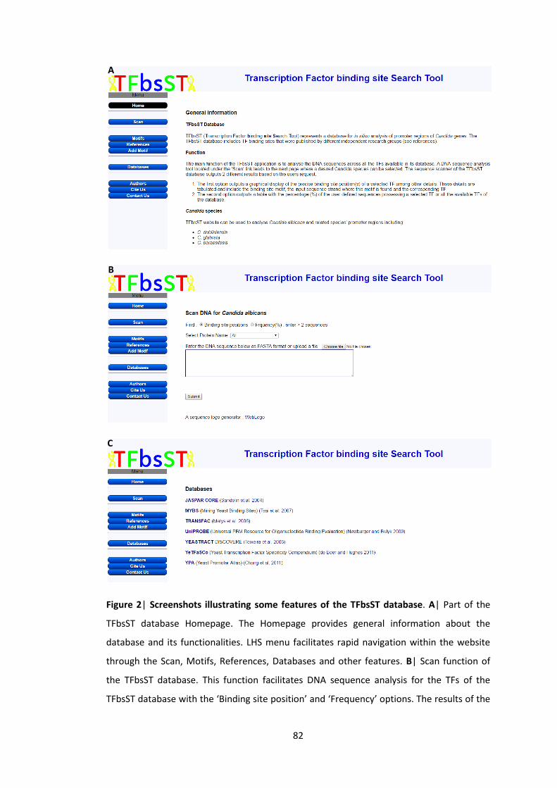
82
Figure 2| Screenshots illustrating some features of the TFbsST database. A| Part of the
TFbsST database Homepage. The Homepage provides general information about the
database and its functionalities. LHS menu facilitates rapid navigation within the website
through the Scan, Motifs, References, Databases and other features. B| Scan function of
the TFbsST database. This function facilitates DNA sequence analysis for the TFs of the
TFbsST database with the ‘Binding site position’ and ‘Frequency’ options. The results of the
A
B
C

83
‘Binding site position’ analyses are summarised in graphs and tables, the ‘Frequency’
analyses are organised in tables. C| Databases listed in the TFbsST database. The TFbsST
database provides an access to the 7 TF databases that mainly focus on S. cerevisiae.
4.3.3 Overrepresented and underrepresented transcription factors in Candida albicans
gene promoter regions
To analyze C. albicans promoter regions we used TFbsST application and Python programs.
The Python scripts that analysed the TF frequencies and predicted the TF binding sites were
incorporated into the TFbsST software. However, some Python algorithms were
additionally designed to retrieve gene promoter regions (-1000 bp to +1 bp). In silico
genome-wide frequency analyses of C. albicans promoters against TFbsST database
predicted that 3 TFs Hcm1p, Srr1p and Yox1p possessed ubiquitously distributed binding
sites across all its 6219 genes (100%) (Table 1). However, these TFs are unlikely to regulate
the whole C. albicans genome in vivo, hence more work is required to optimise their
binding site motifs. The DNA binding sites of 5 TFs, Tac1p, Rob1p, Rpn4p, Mcm1p and
Mbp1p, were predicted to appear sporadically (1% - 2%) in C. albicans promoters occupying
about 100 genes. Such a low occurrence frequency indicates that the above TFs regulate
specific group of genes (Table 1).
4.3.4 RBT and ALS are important for interaction between Candida albicans and
Pseudomonas aeruginosa
To better understand the regulation of genes whose expression was impaired in response
to bacterial supernatants we carried out in silico promoter analyses via the TFbsST
database. Previous studies of Holcombe et al. (2010) and McAlester et al. (2008) had
established differentially expressed genes of C. albicans in response to P. aeruginosa
supernatants. In Holcombe et al. (2010) study, C. albicans cultures were grown in presence
of 4 different types of Pseudomonas supernatants (wild-type PAO1, clinical isolates CF144,
CF177 and HSL-free mutant PAO1ΔQS) as well as in presence of supernatants derived from
HSL-producing Pseudomonas strains. Gene expression profiles of Candida indicated that
some genes were significantly upregulated or downregulated in the above conditions
(threshold ≥2 fold) (Holcombe et al. 2010). Promoter regions of these gene sets were
subject to TF frequency analysis (Table 2). The results of computational investigation
revealed that Efg1p and Mbp1p were overrepresented in the downregulated sets and
Ndt80p in the upregulated sets of the genes treated with all 4 types of bacterial
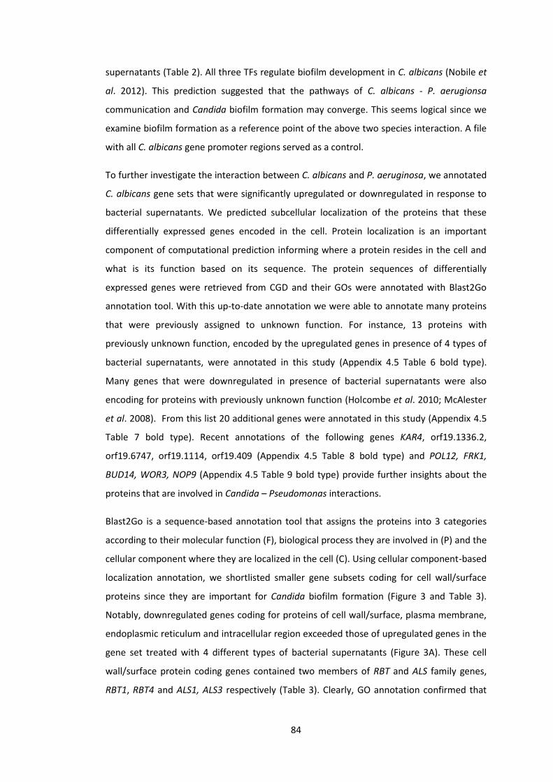
84
supernatants (Table 2). All three TFs regulate biofilm development in C. albicans (Nobile et
al. 2012). This prediction suggested that the pathways of C. albicans - P. aerugionsa
communication and Candida biofilm formation may converge. This seems logical since we
examine biofilm formation as a reference point of the above two species interaction. A file
with all C. albicans gene promoter regions served as a control.
To further investigate the interaction between C. albicans and P. aeruginosa, we annotated
C. albicans gene sets that were significantly upregulated or downregulated in response to
bacterial supernatants. We predicted subcellular localization of the proteins that these
differentially expressed genes encoded in the cell. Protein localization is an important
component of computational prediction informing where a protein resides in the cell and
what is its function based on its sequence. The protein sequences of differentially
expressed genes were retrieved from CGD and their GOs were annotated with Blast2Go
annotation tool. With this up-to-date annotation we were able to annotate many proteins
that were previously assigned to unknown function. For instance, 13 proteins with
previously unknown function, encoded by the upregulated genes in presence of 4 types of
bacterial supernatants, were annotated in this study (Appendix 4.5 Table 6 bold type).
Many genes that were downregulated in presence of bacterial supernatants were also
encoding for proteins with previously unknown function (Holcombe et al. 2010; McAlester
et al. 2008). From this list 20 additional genes were annotated in this study (Appendix 4.5
Table 7 bold type). Recent annotations of the following genes KAR4, orf19.1336.2,
orf19.6747, orf19.1114, orf19.409 (Appendix 4.5 Table 8 bold type) and POL12, FRK1,
BUD14, WOR3, NOP9 (Appendix 4.5 Table 9 bold type) provide further insights about the
proteins that are involved in Candida – Pseudomonas interactions.
Blast2Go is a sequence-based annotation tool that assigns the proteins into 3 categories
according to their molecular function (F), biological process they are involved in (P) and the
cellular component where they are localized in the cell (C). Using cellular component-based
localization annotation, we shortlisted smaller gene subsets coding for cell wall/surface
proteins since they are important for Candida biofilm formation (Figure 3 and Table 3).
Notably, downregulated genes coding for proteins of cell wall/surface, plasma membrane,
endoplasmic reticulum and intracellular region exceeded those of upregulated genes in the
gene set treated with 4 different types of bacterial supernatants (Figure 3A). These cell
wall/surface protein coding genes contained two members of RBT and ALS family genes,
RBT1, RBT4 and ALS1, ALS3 respectively (Table 3). Clearly, GO annotation confirmed that

85
RBT and ALS gene families are important for interaction between C. albicans and P.
aerugionsa.
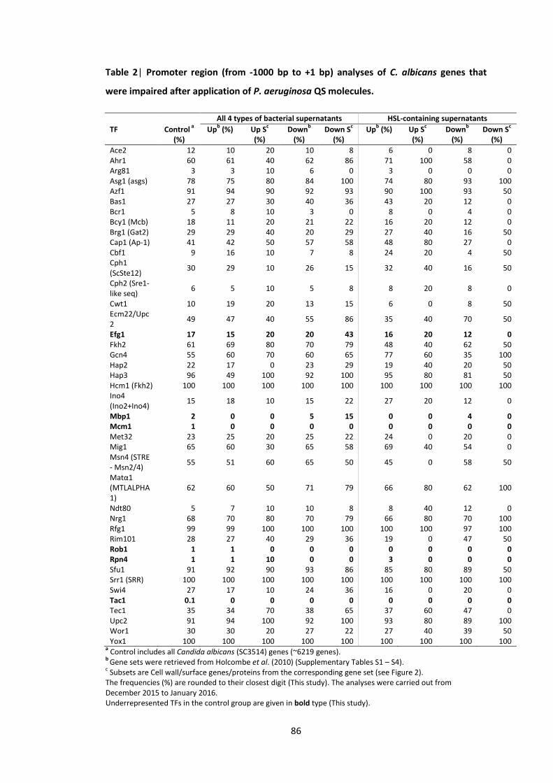
86
Table 2| Promoter region (from -1000 bp to +1 bp) analyses of C. albicans genes that
were impaired after application of P. aeruginosa QS molecules.
All 4 types of bacterial supernatants HSL-containing supernatants
TF Control a
(%)
Upb (%) Up S
c
(%) Down
b
(%) Down S
c
(%)
Upb (%) Up S
c
(%) Down
b
(%) Down S
c
(%)
Ace2 12 10 20 10 8 6 0 8 0 Ahr1 60 61 40 62 86 71 100 58 0 Arg81 3 3 10 6 0 3 0 0 0 Asg1 (asgs) 78 75 80 84 100 74 80 93 100 Azf1 91 94 90 92 93 90 100 93 50 Bas1 27 27 30 40 36 43 20 12 0 Bcr1 5 8 10 3 0 8 0 4 0 Bcy1 (Mcb) 18 11 20 21 22 16 20 12 0 Brg1 (Gat2) 29 29 40 20 29 27 40 16 50 Cap1 (Ap-1) 41 42 50 57 58 48 80 27 0 Cbf1 9 16 10 7 8 24 20 4 50 Cph1 (ScSte12)
30 29 10 26 15 32 40 16 50
Cph2 (Sre1-like seq)
6 5 10 5 8 8 20 8 0
Cwt1 10 19 20 13 15 6 0 8 50 Ecm22/Upc2
49 47 40 55 86 35 40 70 50
Efg1 17 15 20 20 43 16 20 12 0 Fkh2 61 69 80 70 79 48 40 62 50 Gcn4 55 60 70 60 65 77 60 35 100 Hap2 22 17 0 23 29 19 40 20 50 Hap3 96 49 100 92 100 95 80 81 50 Hcm1 (Fkh2) 100 100 100 100 100 100 100 100 100 Ino4 (Ino2+Ino4)
15 18 10 15 22 27 20 12 0
Mbp1 2 0 0 5 15 0 0 4 0 Mcm1 1 0 0 0 0 0 0 0 0 Met32 23 25 20 25 22 24 0 20 0 Mig1 65 60 30 65 58 69 40 54 0 Msn4 (STRE - Msn2/4)
55 51 60 65 50 45 0 58 50
Matα1 (MTLALPHA1)
62 60 50 71 79 66 80 62 100
Ndt80 5 7 10 10 8 8 40 12 0 Nrg1 68 70 80 70 79 66 80 70 100 Rfg1 99 99 100 100 100 100 100 97 100 Rim101 28 27 40 29 36 19 0 47 50 Rob1 1 1 0 0 0 0 0 0 0 Rpn4 1 1 10 0 0 3 0 0 0 Sfu1 91 92 90 93 86 85 80 89 50 Srr1 (SRR) 100 100 100 100 100 100 100 100 100 Swi4 27 17 10 24 36 16 0 20 0 Tac1 0.1 0 0 0 0 0 0 0 0 Tec1 35 34 70 38 65 37 60 47 0 Upc2 91 94 100 92 100 93 80 89 100 Wor1 30 30 20 27 22 27 40 39 50 Yox1 100 100 100 100 100 100 100 100 100
a Control includes all Candida albicans (SC3514) genes (~6219 genes).
b Gene sets were retrieved from Holcombe et al. (2010) (Supplementary Tables S1 – S4).
c Subsets are Cell wall/surface genes/proteins from the corresponding gene set (see Figure 2).
The frequencies (%) are rounded to their closest digit (This study). The analyses were carried out from December 2015 to January 2016. Underrepresented TFs in the control group are given in bold type (This study).

87
Figure 3| Localization of C. albicans proteins encoded by the upregulated (green bars)
and downregulated (red bars) genes in response to P. aeruginosa supernatants. A| Genes
with altered expression in response to supernatants derived from 4 strains of P. aeruginosa
(wild-type PAO1, clinical isolates CF144, CF177 and HSL-free PAO1ΔQS). B| Genes with
altered expression in response to supernatants derived from HSL-producing P. aeruginosa
strains. Image was produced using scientific statistics software GraphPad Prism v 6 for Mac.
Table 3| C. albicans gene subsets coding for cell wall/surface proteins. These subsets
were either upregulated or downregulated in response to the supernatants derived from 4
strains of Pseudomonas, PAO1, CF144, CF177, PAO1ΔQS and HSL-containing supernatants
(data were extracted from Figure 3).
4 types of bacterial supernatants HSL-containing supernatants
Upregulated Downregulated Upregulated Downregulated
ID Gene ID Gene ID Gene ID Gene
CR_10110W CHT3 C4_02410C AHP1 C6_03700W ALS1 C4_03570W HWP1 C2_08490W DSE1 CR_07070C ALS3 C4_01160W CRD2 C2_01380W PLB4.5 C5_02080C HSP12 C4_03470C ECE1 C5_02790C GAP1 C4_06720W NOP1 C5_02460C ECM331 C6_02010C GPD2 C2_08870C PIR1 C6_00440C FET34 C4_04080C PGA31 C6_02070C RPL23A C1_14130W FTR1 C2_08040C RPS10 CR_10100C INO1 C6_00650C RPS13 C3_01360C IRO1 C5_04110W SCW11 CR_10790W MAL2 C2_08590W YWP1 C4_00200C MET15 C4_03520C RBT1 C1_07030C RBT4 C2_00680C SOD5 C7_00260C YLR001C
0 10 20 30 40 50
Not yet annotated
Glyoxysome
Endosome
Microtubule
Peroxisome
Autophagosome
Fungal-type vacuole
Mating projection tip
Polysome
43S/48S preinitiation complex
Box C/D snoRNP complex
Small-subunit processome
Ribosome
Intracellular region
Endoplasmic reticulum
Golgi apparatus
Viral nucleocapsid
Vacuole
Mitochondrion
Cell wall/surface
Plasma membrane
Cytoplasm
Nucleus
UpregulatedDownregulated
Number of proteins annotated to localization
0 10 20
Not yet annotated
Bud
Intracellular part
Fatty acid synthase complex
α DNApol:primase complex
Exosome (RNase complex)
Preribosome
Trans-Golgi network
Exocyst
Vacuole
Nucleus
Cytoplasm
Cell wall/surface
Mitochondrion
Plasma membrane
UpregulatedDownregulated
Number of proteins annotated to localization
A B
88
4.3.5 Overrepresented TFs in the RBT and ALS family gene promoters
Previous studies of Holcombe et al. (2010) had shown that RBT1 and RBT4 were
downregulated in presence of bacterial supernatants. From Holcombe et al. (2010) dataset
RBT1 and RBT4, along with ALS1 and ALS3 (although not downregulated), were also found
among our shortlisted genes coding for cell wall proteins (Table 3). Hence, it was
interesting to explore which TFs could possibly regulate expression of these genes. To
predict TF distribution frequencies across C. albicans RBT and ALS family gene promoters
we retrieved their gene sequences plus 1000 bp up/downstream regions from CGD. The
promoters of RBT family genes, including RBT1, RBT2, RBT4, RBT5, RBT6, RBT7 and RBT8,
were retrieved with Python scripts and analysed for TF frequencies in the TFbsST software.
The promoter regions of ALS family genes, ALS1, ALS2, ALS3 (CDC24), ALS3, ALS4, ALS5,
ALS6, ALS7 and ALS9, were analysed in the same way using a separate FASTA file. Figure 4
shows predicted TF ratios in the promoters of the RBT and ALS family genes (Figure 4). RBT
family possessed 7 overrepresented TFs, Mcm1p, Mbp1p, Bcr1p, Efg1p, Ndt80p, Cwt1p and
Cph2p, determined by the 2-fold threshold (red horizontal line). Only 2 TFs, Rpn4p and
Efg1p, were overrepresented in the ALS family. Notably, Efg1p was overrepresented in both
gene families (Figure 4).
More detailed analyses showed that 3 out of 9 master regulators, Efg1p, Bcr1p and Ndt80p,
that govern biofilm development in C. albicans (Nobile et al. 2012; Fox et al. 2015),
possessed binding sites across promoter regions of 5 different genes in the RBT family
(Table 4). Table 4 shows that RBT1, RBT4 and RBT6 are mainly regulated by Efg1p, RBT2 is
additionally regulated by Bcr1p and RBT5 solely by Bcr1p and Ndt80p. DNA binding sites of
Efg1p were also found in 4 ALS family genes, ALS1, ALS3, ALS4 and ALS9 (Table 5). Thus,
Efg1p appeared as a protagonist of gene regulation in both gene families, RBT and ALS.
Other overrepresented TFs in the RBT family were Cph2p and Cwt1p. Cph2p promotes
hyphal growth by directly regulating Tec1p to induce hyphal-specific genes (Lane et al.
2001) and Cwt1p regulates cell wall integrity (Moreno et al. 2003). These results partially
confirmed our approach since RBT family genes are strongly expressed during hyphal
growth but they are dramatically downregulated in presence of Pseudomonas supernatants
under yeast growth conditions (McAlester et al. 2008; Holcombe et al. 2010).
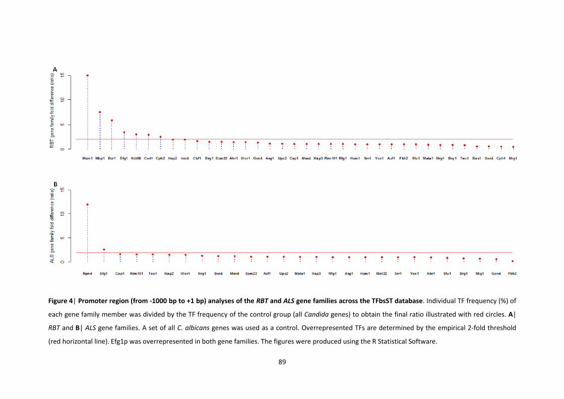
89
Figure 4| Promoter region (from -1000 bp to +1 bp) analyses of the RBT and ALS gene families across the TFbsST database. Individual TF frequency (%) of
each gene family member was divided by the TF frequency of the control group (all Candida genes) to obtain the final ratio illustrated with red circles. A|
RBT and B| ALS gene families. A set of all C. albicans genes was used as a control. Overrepresented TFs are determined by the empirical 2-fold threshold
(red horizontal line). Efg1p was overrepresented in both gene families. The figures were produced using the R Statistical Software.
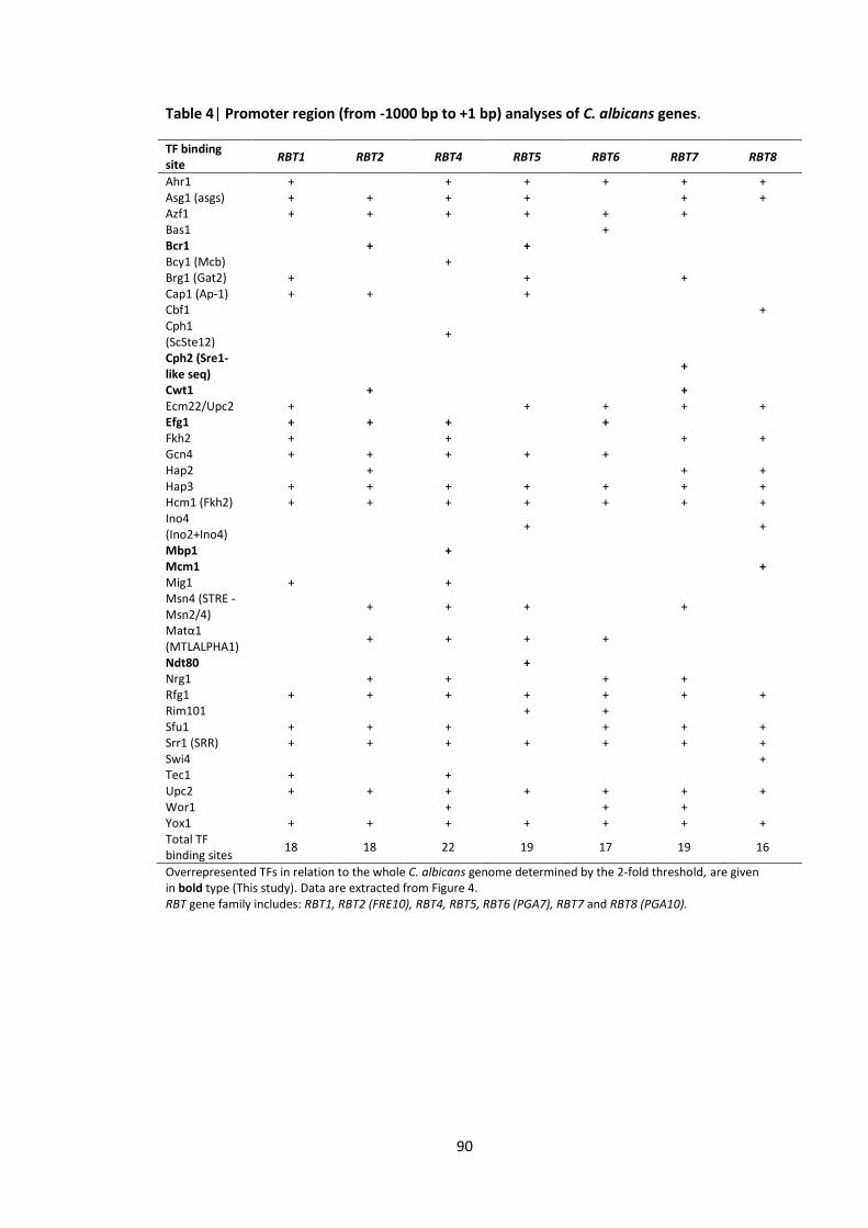
90
Table 4| Promoter region (from -1000 bp to +1 bp) analyses of C. albicans genes.
Overrepresented TFs in relation to the whole C. albicans genome determined by the 2-fold threshold, are given in bold type (This study). Data are extracted from Figure 4. RBT gene family includes: RBT1, RBT2 (FRE10), RBT4, RBT5, RBT6 (PGA7), RBT7 and RBT8 (PGA10).
TF binding site
RBT1 RBT2 RBT4 RBT5 RBT6 RBT7 RBT8
Ahr1 + + + + + + Asg1 (asgs) + + + + + + Azf1 + + + + + + Bas1 + Bcr1 + + Bcy1 (Mcb) + Brg1 (Gat2) + + + Cap1 (Ap-1) + + + Cbf1 + Cph1 (ScSte12)
+
Cph2 (Sre1-like seq)
+
Cwt1 + + Ecm22/Upc2 + + + + + Efg1 + + + + Fkh2 + + + + Gcn4 + + + + + Hap2 + + + Hap3 + + + + + + + Hcm1 (Fkh2) + + + + + + + Ino4 (Ino2+Ino4)
+ +
Mbp1 + Mcm1 + Mig1 + + Msn4 (STRE - Msn2/4)
+ + + +
Matα1 (MTLALPHA1)
+ + + +
Ndt80 + Nrg1 + + + + Rfg1 + + + + + + + Rim101 + + Sfu1 + + + + + + Srr1 (SRR) + + + + + + + Swi4 + Tec1 + + Upc2 + + + + + + + Wor1 + + + Yox1 + + + + + + + Total TF binding sites
18 18 22 19 17 19 16
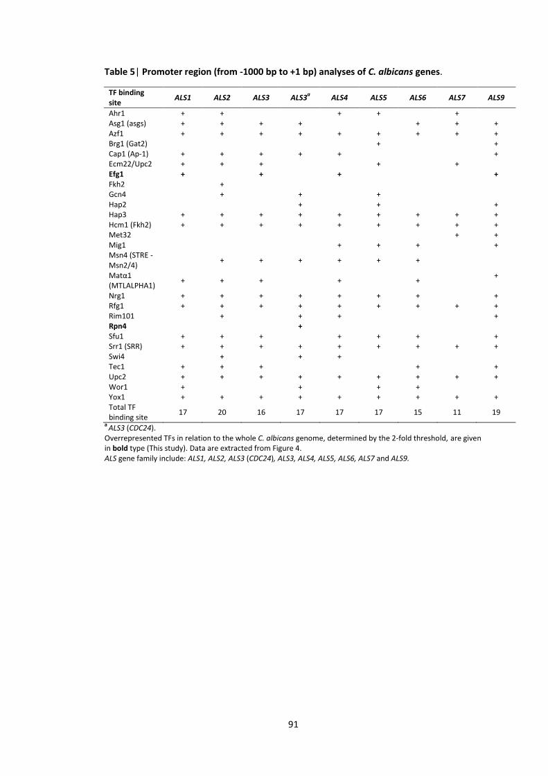
91
Table 5| Promoter region (from -1000 bp to +1 bp) analyses of C. albicans genes.
TF binding site
ALS1 ALS2 ALS3 ALS3a ALS4 ALS5 ALS6 ALS7 ALS9
Ahr1 + + + + + Asg1 (asgs) + + + + + + + Azf1 + + + + + + + + + Brg1 (Gat2) + + Cap1 (Ap-1) + + + + + + Ecm22/Upc2 + + + + + Efg1 + + + + Fkh2 + Gcn4 + + + Hap2 + + + Hap3 + + + + + + + + + Hcm1 (Fkh2) + + + + + + + + + Met32 + + Mig1 + + + + Msn4 (STRE - Msn2/4)
+ + + + + +
Matα1 (MTLALPHA1)
+ + + + + +
Nrg1 + + + + + + + + Rfg1 + + + + + + + + + Rim101 + + + + Rpn4 + Sfu1 + + + + + + + Srr1 (SRR) + + + + + + + + + Swi4 + + + Tec1 + + + + + Upc2 + + + + + + + + + Wor1 + + + + Yox1 + + + + + + + + + Total TF binding site
17 20 16 17 17 17 15 11 19
a ALS3 (CDC24).
Overrepresented TFs in relation to the whole C. albicans genome, determined by the 2-fold threshold, are given in bold type (This study). Data are extracted from Figure 4. ALS gene family include: ALS1, ALS2, ALS3 (CDC24), ALS3, ALS4, ALS5, ALS6, ALS7 and ALS9.
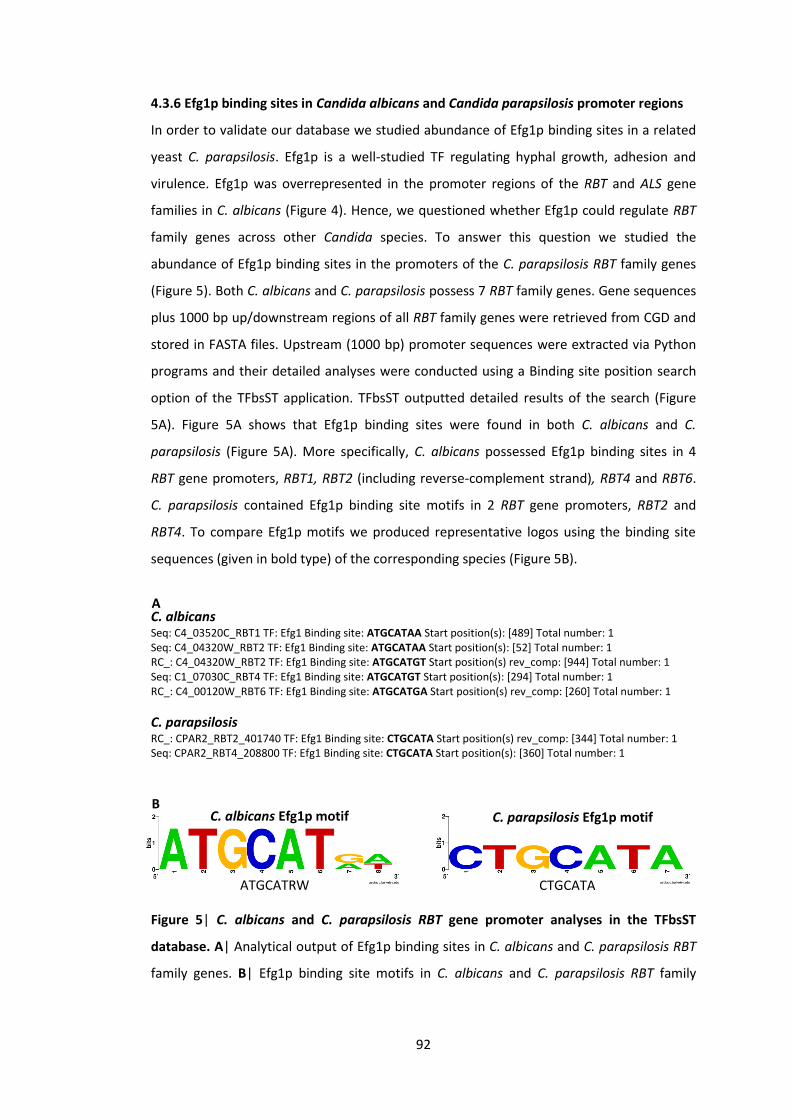
92
4.3.6 Efg1p binding sites in Candida albicans and Candida parapsilosis promoter regions
In order to validate our database we studied abundance of Efg1p binding sites in a related
yeast C. parapsilosis. Efg1p is a well-studied TF regulating hyphal growth, adhesion and
virulence. Efg1p was overrepresented in the promoter regions of the RBT and ALS gene
families in C. albicans (Figure 4). Hence, we questioned whether Efg1p could regulate RBT
family genes across other Candida species. To answer this question we studied the
abundance of Efg1p binding sites in the promoters of the C. parapsilosis RBT family genes
(Figure 5). Both C. albicans and C. parapsilosis possess 7 RBT family genes. Gene sequences
plus 1000 bp up/downstream regions of all RBT family genes were retrieved from CGD and
stored in FASTA files. Upstream (1000 bp) promoter sequences were extracted via Python
programs and their detailed analyses were conducted using a Binding site position search
option of the TFbsST application. TFbsST outputted detailed results of the search (Figure
5A). Figure 5A shows that Efg1p binding sites were found in both C. albicans and C.
parapsilosis (Figure 5A). More specifically, C. albicans possessed Efg1p binding sites in 4
RBT gene promoters, RBT1, RBT2 (including reverse-complement strand), RBT4 and RBT6.
C. parapsilosis contained Efg1p binding site motifs in 2 RBT gene promoters, RBT2 and
RBT4. To compare Efg1p motifs we produced representative logos using the binding site
sequences (given in bold type) of the corresponding species (Figure 5B).
C. albicans Seq: C4_03520C_RBT1 TF: Efg1 Binding site: ATGCATAA Start position(s): [489] Total number: 1 Seq: C4_04320W_RBT2 TF: Efg1 Binding site: ATGCATAA Start position(s): [52] Total number: 1 RC_: C4_04320W_RBT2 TF: Efg1 Binding site: ATGCATGT Start position(s) rev_comp: [944] Total number: 1 Seq: C1_07030C_RBT4 TF: Efg1 Binding site: ATGCATGT Start position(s): [294] Total number: 1 RC_: C4_00120W_RBT6 TF: Efg1 Binding site: ATGCATGA Start position(s) rev_comp: [260] Total number: 1
C. parapsilosis RC_: CPAR2_RBT2_401740 TF: Efg1 Binding site: CTGCATA Start position(s) rev_comp: [344] Total number: 1 Seq: CPAR2_RBT4_208800 TF: Efg1 Binding site: CTGCATA Start position(s): [360] Total number: 1
Figure 5| C. albicans and C. parapsilosis RBT gene promoter analyses in the TFbsST
database. A| Analytical output of Efg1p binding sites in C. albicans and C. parapsilosis RBT
family genes. B| Efg1p binding site motifs in C. albicans and C. parapsilosis RBT family
A
B C. albicans Efg1p motif C. parapsilosis Efg1p motif
ATGCATRW CTGCATA
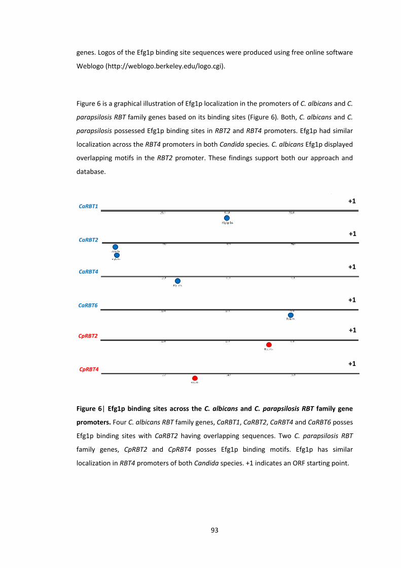
93
genes. Logos of the Efg1p binding site sequences were produced using free online software
Weblogo (http://weblogo.berkeley.edu/logo.cgi).
Figure 6 is a graphical illustration of Efg1p localization in the promoters of C. albicans and C.
parapsilosis RBT family genes based on its binding sites (Figure 6). Both, C. albicans and C.
parapsilosis possessed Efg1p binding sites in RBT2 and RBT4 promoters. Efg1p had similar
localization across the RBT4 promoters in both Candida species. C. albicans Efg1p displayed
overlapping motifs in the RBT2 promoter. These findings support both our approach and
database.
Figure 6| Efg1p binding sites across the C. albicans and C. parapsilosis RBT family gene
promoters. Four C. albicans RBT family genes, CaRBT1, CaRBT2, CaRBT4 and CaRBT6 posses
Efg1p binding sites with CaRBT2 having overlapping sequences. Two C. parapsilosis RBT
family genes, CpRBT2 and CpRBT4 posses Efg1p binding motifs. Efg1p has similar
localization in RBT4 promoters of both Candida species. +1 indicates an ORF starting point.
CaRBT1 +1
CaRBT2 +1
CaRBT4 +1
CaRBT6 +1
CpRBT2 +1
CpRBT4 +1
94
4.4 Discussion
C. albicans biofilms cause serious infections to nosocomial patients in hospitals. C. albicans
biofilm-related genes are governed by 9 transcriptional regulators (Nobile et al. 2012; Fox
et al. 2015). According to the DBD transcription factor prediction database, C. albicans
possesses more than 500 putative TFs (www.transcriptionfactor.org). However, the binding
site motifs of many TFs are unknown. Additionally, a limited number of bioinformatics tools
for the analyses of C. albicans promoter sequences makes the in silico investigation even
harder. Due to the latter, a novel bioinformatics tool, TFbsST database, was established to
compile C. albicans TF binding site data. TFbsST software can be used to screen Candida
gene promoter sequences for these TFs.
The vast majority of yeast databases are generated for a model yeast S. cerevisiae
(UniPROBE, YEASTRACT, MYBS, YPA, JASPAR, TRANSFAC® and YeTFaSCo). To design the
TFbsST application these databases were carefully reviewed and YeTFaSCo was used as the
basis of our database. Nevertheless, there are significant differences between the
YeTFaSCo and TFbsST databases. The main difference is that TFbsST includes TF motifs for
C. albicans. Another important feature of the TFbsST database is that it also contains other
Candida species such as C. parapsilosis, C. dubliniensis and C. glabrata. Additionally, the
TFbsST database can perform TF frequency analysis (outputs a table with %) along with the
TF localization analysis (outputs graphs and tables). Therefore, TFbsST is a user-friendly and
dynamic website that can be used for more complete gene regulation analyses in Candida.
However, a list of rational criteria for further evaluation of the motifs and database tools
was generated to optimise the TFbsST database. To improve in silico prediction power a
sophisticated TF motif scoring system based on their establishment method (e.g. ChIP-chip
or gene expression) is planned to be incorporated in the TFbsST database. In addition, GO
annotation and inter-study agreement could provide further details and relevant scores to
the TF binding sites. Clearly, browsing TFbsST reveals that limited piece of information is
available for the Candida species other than C. albicans. Due to the latter, easily modifiable
text files were integrated into the TFbsST website. This flexible structure facilitates rapid
enrichment of TF, motif and related organism lists.
The TFbsST database was used to analyse TF frequencies in C. albicans genome. These in
silico analyses showed several TF motifs that were ubiquitous in nearly all C. albicans genes
(Hcm1p, Srr1p and Yox1p) and others that were significantly rare (Tac1p, Rob1p, Rpn4p,
95
Mcm1p and Mbp1p). Hcm1p is a forkhead TF that plays an important role in fungal
morphogenesis (Bensen et al. 2002) and iron homeostasis (Singh et al. 2011). Srr1p is a 2-
component response regulator also required for morphogenesis (Desai et al. 2011) and
H2O2 (hydrogen peroxide) resistance (Bruce et al. 2011). Yox1p is a putative transcriptional
repressor (Tuch et al. 2008) that peaks at G1/S phase of cell cycle (Cote et al. 2009).
Despite their important role, it is unlikely that these TFs regulate the expression of all genes
in C. albicans. Thus, more research is required to reveal the most accurate DNA binding site
motifs of these TFs. Closer look at some TFs predicted to regulate small number of genes
revealed that these TFs are parts of a larger protein complex. For example, Mbp1p and
Tac1p represent part of MBF complex (Cote et al. 2009) and DRE (drug responsive element)
element respectively. Other TFs in this group regulate different functions in the cell. For
instance, Rob1p regulates biofilm development (Liu et al. 2005; Nett et al. 2009) and a C2H2
(Acetylene) TF, Rpn4p, regulates proteasome synthesis (Gasch et al. 2004; Enjalbert et al.
2006). Mcm1p is a protein with unknown function (CGD). This data suggest that more
research is required to elucidate the exact role of these TFs.
Mcm1p and Mbp1p were predicted to be overrepresented in RBT family genes. However, a
closer inspection of Mcm1p and Mbp1p in the RBT family indicated that binding site motifs
of these TFs were present in a single RBT gene. Overrepresentation was due to their low
frequency in C. albicans genome. The remaining overrepresented TFs in RBT genes,
predicted on the basis of their binding sites, regulate biofilm formation in C. albicans.
Bcr1p, Efg1p, Ndt80p are master regulators (Nobile et al. 2012), Cph2p regulates
filamentous growth (Lane et al. 2001) and Cwt1p governs cell wall integrity (Moreno et al.
2003). Efg1p and Rpn4p were overrepresented in the ALS gene family. However, binding
sites of Rpn4p were present only in a single ALS gene (ALS9). Efg1p though was predicted to
be present in some genes of both gene families, RBT and ALS. These results suggest that
Efg1p can regulate important gene families that shape biofilm development in C. albicans.
Efg1p binding sites were also found in the C. parapsilosis RBT family genes. This finding
suggests that Efg1p can regulate RBT genes across different species of Candida. To
experimentally validate these in silico analyses, gene expression experiments of Candida
EFG1 mutants grown in YNBNP (filament-inducing medium) can be conducted quantifying
RBT genes expression with RTqPCR.
To analyse the effect of bacterial supernatants on Candida, differentially expressed genes
of C. albicans, defined by Holcombe et al. (2010) that were significantly upregulated or

96
downregulated in response to P. aeruginosa supernatants, were annotated with Blast2Go.
The up-to-date annotation of these genes elucidated the localization of many proteins that
were not previously annotated. These data provide new insights about the proteins that
are involved in Candida – Pseudomonas interactions. The question now is whether these
genes are regulated by a specific transcription regulator or if they are governed by different
TFs.

97
4.5 Appendix
Table 6| Gene ontology (GO) annotations of upregulated gene set in response to the
supernatants derived from 4 strains of Pseudomonas, PAO1, CF144, CF177 and ΔQS. This
Holcombe et al. (2010) dataset was re-analysed here and up-to-date GO annotations were
obtained with Blast2Go. This table is summarised in the Figure 3A (green bars).
ID Gene GO – cellular component
Plasma membrane
C4_01100C AGP2 Integral component of plasma membrane C3_03070W AMF1 Integral component of membrane C3_04070C CDR11 Integral component of membrane C2_06020W CNT Integral component of plasma membrane CR_09370W ELF1 Plasma membrane C7_02910W ENA21 Integral component of membrane C4_03700W FNX1 Integral component of membrane C4_05430C GAP5 Integral component of plasma membrane C6_00330C GNP1 Integral component of plasma membrane C4_06760W GUT2 Plasma membrane C6_03790C HGT10 Integral component of plasma membrane C1_13130C HIP1 Integral component of plasma membrane C1_09680W MTS1 Plasma membrane C4_01940W PHO89 Integral component of plasma membrane C1_09210C SGE11 Integral component of membrane C6_03840C SNQ2 Plasma membrane; Integral component of membrane C2_09900C TIM23 Plasma membrane CR_04200W YDJ1 TRC complex C2_08590W YWP1 Anchored component of membrane
Cytoplasm
C1_07710C ADE4 Cytoplasm C2_01270W CHA1 Cytoplasm C5_03640W DPH51 Cytosol C2_05100C ERF1 Cytoplasmic stress granule; Cytosol C6_02500C GCV1 Cytoplasm C1_08400C GCV2 Cytosol C1_10450W GLY1 Cytosol C1_09490C GUA1 Cytoplasm C2_06390C IMH3 Cytoplasm C6_02230W LSG1 Cytosolic large ribosomal subunit C4_04720W MTD1 Cytosol CR_06720W NMD3 Cytosolic large ribosomal subunit C5_00260W PRS1 Cytosol C1_03350C RLI1 Cytosolic ribosome C1_11040W RPL29 Cytosolic large ribosomal subunit C1_06890C RPL34B Cytosolic large ribosomal subunit C1_11360W RPL37B Cytosolic large ribosomal subunit C3_04680W RPP2B Cytosol C6_00650C RPS13 Cytosolic small ribosomal subunit C3_04670C RPS15 Cytosolic small ribosomal subunit C1_01370C RPS21B Cytosolic small ribosomal subunit C3_00090W RPS24 Cytosolic small ribosomal subunit CR_07630C RPS27 Cytosol C7_00710W RPS28B Cytosolic small ribosomal subunit C3_04860W SFP1 Cytoplasm C2_03220C STP4 Cytoplasm C5_02490C TIF5 Cytosolic small ribosomal subunit C5_04570C URA7 Cytoplasm CR_04200W YDJ1 Perinuclear region of cytoplasm
98
Nucleus
C3_05160C DBP10 Nucleolus CR_02530W DBP2 Nucleus C1_10030W DBP3 Nucleolus C5_03640W DPH51 Nucleus CR_09370W ELF1 Nucleus C1_04130W ERB1 Nucleoplasm; PeBoW complex C2_05100C ERF1 Nucleus C3_06850W FCR1 Nucleus C5_04750C HAS1 Nucleolus; Nuclear envelope C2_08000C KRE30 Nucleus; Nucleoid C6_02770W MRT4 Nucleoplasm; Nucleolus C4_03030C NAN1 rDNA heterochromatin; RENT complex; t-UTP complex C2_00140W NDT80 Nuclear chromatin CR_05520W NOC2 Nucleolus; Noc1p-Noc2p complex; Noc2p-Noc3p complex C6_03640W NOG2 Nucleolus; Nucleoplasm C4_06720W NOP1 Nuclear chromosome; Cajal body C7_04230W NRG1 Nucleus C6_00920W orf19.93 Nucleus C5_00260W PRS1 Nucleus C2_07450C RCL1 Nucleolus C1_03350C RLI1 Nucleus C7_00570W RPA135 DNA-directed RNA polymerase I complex C3_00090W RPS24 Nucleolus C1_12680W RRB1 Nucleolus C2_08480W RRP8 Nucleolus C3_04860W SFP1 Nucleus C3_04380C SNU13 Spliceosomal complex; U4/U6 x U5 tri-snRNP complex C2_03220C STP4 Nucleus C1_02790W TIF34 Eukaryotic translation initiation factor 3 complex C3_02130W UTP4 t-UTP complex
Ribosome
C3_05160C DBP10 Preribosome, large subunit precursor C1_10030W DBP3 Preribosome, large subunit precursor C2_08490W DSE1 Pwp2p-containing subcomplex of 90S preribosome C1_04130W ERB1 Preribosome, large subunit precursor C5_04750C HAS1 Preribosome, large subunit precursor C2_08000C KRE30 Ribosome C6_02770W MRT4 Ribosome; Preribosome, large subunit precursor C4_03030C NAN1 90S preribosome CR_04360C NHP2 Ribosome C6_03640W NOG2 Preribosome, large subunit precursor C4_06720W NOP1 Ribosome; 90S preribosome C6_00370C NOP5 90S preribosome C5_00260W PRS1 Ribose phosphate diphosphokinase complex C1_03350C RLI1 Preribosome, large subunit precursor C2_06810C RPL11 Ribosome C6_02070C RPL23A Ribosome C4_04900W RPL30 Ribosome C5_04590C RPL43A Ribosome C3_04680W RPP2B Ribosome C2_08040C RPS10 Ribosome C6_00650C RPS13 90S preribosome C7_00960W RPS18 Small ribosomal subunit C1_06460C RPS22A Ribosome CR_07630C RPS27 Ribosome CR_09950C SIK1 90S preribosome C1_02790W TIF34 Multi-eIF complex C5_02490C TIF5 Multi-eIF complex C3_02130W UTP4 90S preribosome C3_05160C DBP10 Preribosome, large subunit precursor
Mitochondrion
C2_08100W CPD1 Mitochondrion
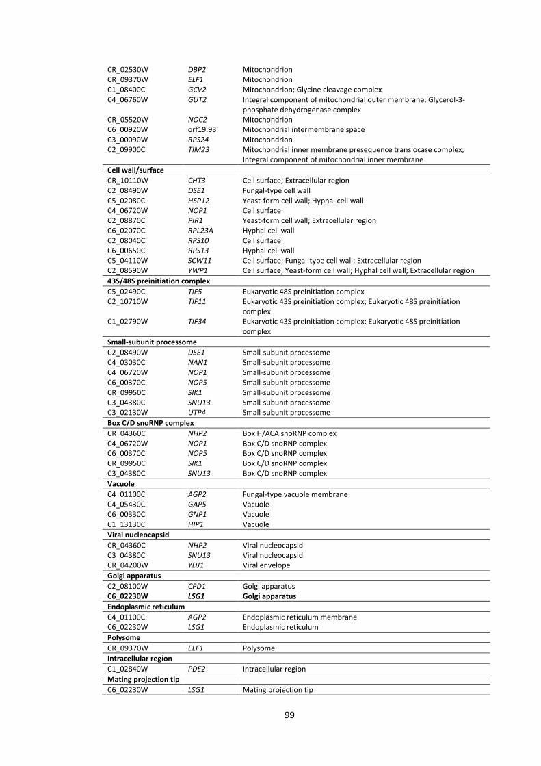
99
CR_02530W DBP2 Mitochondrion CR_09370W ELF1 Mitochondrion C1_08400C GCV2 Mitochondrion; Glycine cleavage complex C4_06760W GUT2 Integral component of mitochondrial outer membrane; Glycerol-3-
phosphate dehydrogenase complex CR_05520W NOC2 Mitochondrion C6_00920W orf19.93 Mitochondrial intermembrane space C3_00090W RPS24 Mitochondrion C2_09900C TIM23 Mitochondrial inner membrane presequence translocase complex;
Integral component of mitochondrial inner membrane
Cell wall/surface
CR_10110W CHT3 Cell surface; Extracellular region C2_08490W DSE1 Fungal-type cell wall C5_02080C HSP12 Yeast-form cell wall; Hyphal cell wall C4_06720W NOP1 Cell surface C2_08870C PIR1 Yeast-form cell wall; Extracellular region C6_02070C RPL23A Hyphal cell wall C2_08040C RPS10 Cell surface C6_00650C RPS13 Hyphal cell wall C5_04110W SCW11 Cell surface; Fungal-type cell wall; Extracellular region C2_08590W YWP1 Cell surface; Yeast-form cell wall; Hyphal cell wall; Extracellular region
43S/48S preinitiation complex
C5_02490C TIF5 Eukaryotic 48S preinitiation complex C2_10710W TIF11 Eukaryotic 43S preinitiation complex; Eukaryotic 48S preinitiation
complex C1_02790W TIF34 Eukaryotic 43S preinitiation complex; Eukaryotic 48S preinitiation
complex
Small-subunit processome
C2_08490W DSE1 Small-subunit processome C4_03030C NAN1 Small-subunit processome C4_06720W NOP1 Small-subunit processome C6_00370C NOP5 Small-subunit processome CR_09950C SIK1 Small-subunit processome C3_04380C SNU13 Small-subunit processome C3_02130W UTP4 Small-subunit processome
Box C/D snoRNP complex
CR_04360C NHP2 Box H/ACA snoRNP complex C4_06720W NOP1 Box C/D snoRNP complex C6_00370C NOP5 Box C/D snoRNP complex CR_09950C SIK1 Box C/D snoRNP complex C3_04380C SNU13 Box C/D snoRNP complex
Vacuole
C4_01100C AGP2 Fungal-type vacuole membrane C4_05430C GAP5 Vacuole C6_00330C GNP1 Vacuole C1_13130C HIP1 Vacuole
Viral nucleocapsid
CR_04360C NHP2 Viral nucleocapsid C3_04380C SNU13 Viral nucleocapsid CR_04200W YDJ1 Viral envelope
Golgi apparatus
C2_08100W CPD1 Golgi apparatus C6_02230W LSG1 Golgi apparatus
Endoplasmic reticulum
C4_01100C AGP2 Endoplasmic reticulum membrane C6_02230W LSG1 Endoplasmic reticulum
Polysome
CR_09370W ELF1 Polysome
Intracellular region
C1_02840W PDE2 Intracellular region
Mating projection tip
C6_02230W LSG1 Mating projection tip
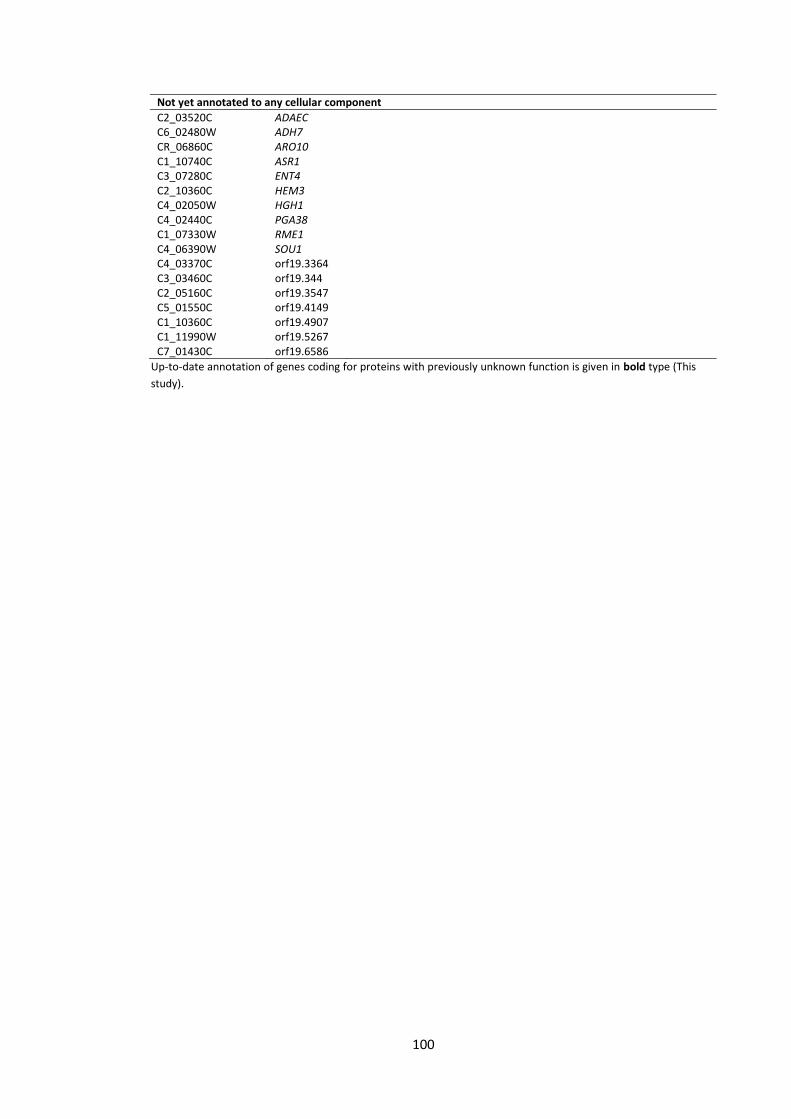
100
Not yet annotated to any cellular component
C2_03520C ADAEC C6_02480W ADH7 CR_06860C ARO10 C1_10740C ASR1 C3_07280C ENT4 C2_10360C HEM3 C4_02050W HGH1 C4_02440C PGA38 C1_07330W RME1 C4_06390W SOU1 C4_03370C orf19.3364 C3_03460C orf19.344 C2_05160C orf19.3547 C5_01550C orf19.4149 C1_10360C orf19.4907 C1_11990W orf19.5267 C7_01430C orf19.6586
Up-to-date annotation of genes coding for proteins with previously unknown function is given in bold type (This
study).
101
Table 7| Gene ontology (GO) annotations of the downregulated gene set in response to
the supernatants derived from 4 strains of Pseudomonas, PAO1, CF144, CF177 and ΔQS.
This Holcombe et al. (2010) dataset was re-analysed here and up-to-date GO annotations
were obtained with Blast2Go. This table is summarised in the Figure 3A (red bars).
ID Gene GO – cellular component
Plasma membrane
CR_07070C ALS3 Plasma membrane; Anchored component of membrane CR_02910W APG13 Extrinsic component of membrane C6_04210C ATM1 Integral component of membrane C1_05700W AUT7 Extrinsic component of membrane C1_06520C BPH1 Extrinsic component of membrane C2_03320W CHK1 Membrane C5_02460C ECM331 Plasma membrane; Anchored component of membrane C1_08590C ERG1 Plasma membrane; Integral component of membrane; Lipid particle C6_00440C FET34 Plasma membrane C1_14130W FTR1 High-affinity iron permease complex C3_05580C GAP2 Integral component of plasma membrane C7_00280W HGT12 Integral component of plasma membrane C1_02110C HGT2 Integral component of plasma membrane C2_04940C ITR1 Integral component of plasma membrane C2_07580W MAE1 Integral component of membrane C5_04930C MAL31 Integral component of plasma membrane C4_00430W MEP2 Integral component of plasma membrane C3_00650W NGT1 Plasma membrane; Integral component of membrane CR_02240C OPT2 Plasma membrane CR_02900W ScOPT2 Plasma membrane CR_02490W OPT4 Plasma membrane C4_03520C RBT1 Anchored component of membrane CR_06660W SEO1 Integral component of membrane C2_00680C SOD5 Anchored component of membrane C2_06010W SPO72 Extrinsic component of membrane C2_02860W SUR2 Integral component of membrane C5_03060C TNA1 Plasma membrane; Integral component of membrane C3_06710W VHC1 Integral component of vacuolar membrane CR_03270W VHT1 Integral component of plasma membrane
Cytoplasm
C4_02410C AHP1 Cytosol C4_05560C ARO9 Cytoplasm C1_05700W AUT7 Cytosol CR_10360C CTM1 Cytosol C1_04660W DUR1,2 Cytoplasm C6_03340C GLC3 Cytoplasm C4_02990C GST2 Cytoplasm CR_10100C INO1 Cytoplasm C1_12010C KIP4 Cytoplasm CR_04480C LAP3 Cytoplasm C7_00400W LEU2 Cytosol C4_05320W LYS1 Cytoplasm C3_06590W LYS9 Cytosol CR_10790W MAL2 Cytoplasm C3_02950C MET13 Cytosol C4_00200C MET15 Cytoplasm C4_00150C PEX5 Cytosol CR_02820W PGM2 Cytosol C2_07570W RNR22 Cytosol; Ribonucleoside-diphosphate reductase complex C2_00680C SOD5 Cytoplasm CR_05720W TPS1 Alpha,alpha-trehalose-phosphate synthase complex (UDP-forming) CR_09010C YHR112C Cytoplasm
102
Nucleus
C4_05560C ARO9 Nucleus C1_05700W AUT7 Nucleus C2_09940W GCN4 Nucleoplasm; Nuclear chromatin; Transcription factor complex C2_06650C PRP31 U4/U6 x U5 tri-snRNP complex C4_02030W RFX2 Nucleus C5_00980W TRY3 Nucleus C1_06280C UME6 Nucleus CR_09930W YDR124W Nucleus CR_09010C YHR112C Nucleus
Cell wall/surface
C4_02410C AHP1 Yeast-form cell wall; Hyphal cell wall CR_07070C ALS3 Yeast-form cell wall; Hyphal cell wall; Cell surface C4_03470C ECE1 Hyphal cell wall C5_02460C ECM331 Fungal-type cell wall; Cell surface C6_00440C FET34 Cell surface C1_14130W FTR1 Cell surface CR_10100C INO1 Hyphal cell wall; Cell surface C3_01360C IRO1 Cell C4_00200C MET15 Hyphal cell wall C4_03520C RBT1 Cell wall C2_00680C SOD5 Yeast-form cell wall; Hyphal cell wall; Cell surface
Extracellular region
CR_07070C ALS3 Extracellular region C5_02460C ECM331 Extracellular region CR_10790W MAL2 Extracellular region C4_03520C RBT1 Extracellular region C1_07030C RBT4 Extracellular region C2_00680C SOD5 Extracellular region C7_00260C YLR001C Extracellular space
Mitochondrion
C6_04210C ATM1 Mitochondrial inner membrane C4_04620C CTN2 Mitochondrion CR_01400W LYS12 Mitochondrion C3_02950C MET13 Mitochondrion C2_08390W orf19.1433 Mitochondrion C7_00260C YLR001C Mitochondrion
Vacuole
C4_02360W AMS1 Fungal-type vacuole membrane C1_05700W AUT7 Fungal-type vacuole membrane C6_00440C FET34 Fungal-type vacuole membrane C2_06880C PRB1 Fungal-type vacuole C7_00260C YLR001C Fungal-type vacuole membrane
Endoplasmic reticulum
C1_06520C BPH1 Endomembrane system C1_08590C ERG1 Endoplasmic reticulum membrane C4_06810C SLY1 Endoplasmic reticulum; ER to Golgi transport vesicle C2_02860W SUR2 Endoplasmic reticulum membrane
Intracellular
C1_06520C BPH1 Intracellular C4_06480C CEK1 Intracellular C2_03320W CHK1 Intracellular C5_00450C IFG3 Intracellular CR_07190W RGD2 Intracellular
Autophagosome
CR_02910W APG13 Pre-autophagosomal structure membrane C1_05700W AUT7 Autophagosome membrane; Pre-autophagosom; Cvt vesicle membrane C2_06010W SPO72 Pre-autophagosomal structure
Peroxisome
C4_04620C CTN2 Peroxisome C3_05360C LON1 Peroxisomal matrix C4_00150C PEX5 Peroxisomal membrane
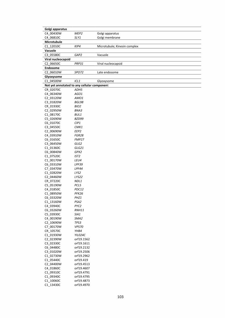
103
Golgi apparatus
C4_00430W MEP2 Golgi apparatus C4_06810C SLY1 Golgi membrane
Microtubule
C1_12010C KIP4 Microtubule; Kinesin complex
Vacuole
C3_05580C GAP2 Vacuole
Viral nucleocapsid
C2_06650C PRP31 Viral nucleocapsid
Endosome
C2_06010W SPO72 Late endosome
Glyoxysome
C1_04500W ICL1 Glyoxysome
Not yet annotated to any cellular component
CR_02070C ADH5 C4_06340W AGO1
C2_03120W AMO1 C3_01820W BGL98 CR_01930C BIO2 C2_02950W BNA3 C1_08170C BUL1 C5_02690W BZD99 C6_01070C CIP1 C3_04550C CMK1 C2_00690W EEP2 C4_03910W FGR28
C6_01650C FMP27 C3_06450W GLG2
C1_01360C GLG21 C6_00840W GPX2 C1_07520C IST2
C1_00170W LEU4 C6_03310W LPF39 C7_03470W LPF44 C1_02820W LYS2
C2_04460W LYS22 CR_07220C NDL1 C5_05190W PCL5
C4_01850C PDC12 C1_08950W PFK26 C6_03320W PHZ1 C1_13160W PSA2 C4_03940C PYC2 C6_03260W RNH11
C5_03930C SIA1 C4_00190W SMA2 C2_10690W TPS3 C7_00170W VPS70 CR_10570C YHB4 C1_01930W YIL024C C2_02390W orf19.1562 C3_02330C orf19.1611 C6_04480C orf19.2132 C3_01020W orf19.2506 C1_02730W orf19.2962 C1_05440C orf19.419 C2_04400W orf19.4513 C4_01860C orf19.4607 C1_09310C orf19.4791 C1_09340C orf19.4795 C1_10060C orf19.4873 C1_13430C orf19.4970

104
Annotation of genes coding for proteins with previously unknown function is given in bold type (This study).
C7_03280C orf19.5125 CR_06570C orf19.915
105
Table 8| Gene ontology (GO) annotations of the upregulated gene set in response to the
HSL-containing Pseudomonas supernatants. This Holcombe et al. (2010) dataset was re-
analysed here and up-to-date GO annotations were obtained with Blast2Go. This table is
summarised in the Figure 3B (green bars).
ID Gene GO – cellular component
Plasma membrane
C6_03700W ALS1 Plasma membrane; Anchored component of membrane C1_09150W AOX2 Plasma membrane; Integral component of membrane C4_04310W COX11 Plasma membrane C4_01160W CRD2 Integral component of membrane C2_02280W FMP39 Plasma membrane; Integral component of membrane C5_02790C GAP1 Integral component of plasma membrane C5_03500W GAP6 Integral component of plasma membrane C2_02610C HGT20 Integral component of plasma membrane C3_02310W MEP1 Integral component of plasma membrane C1_11870W MUP1 Integral component of plasma membrane CM_00310W NAD2 Integral component of membrane C5_03800W orf19.1114 Plasma membrane; Integral component of mitochondrial inner
membrane C1_08610C orf19.409 Plasma membrane; Integral component of membrane C4_04080C PGA31 Anchored component of membrane C3_03800W PTR22 Plasma membrane; Integral component of membrane C4_02890C QCR9 Plasma membrane
C2_06470W RTA2 Plasma membrane; Integral component of membrane; Membrane raft
C2_06460W RTA3 Integral component of membrane CR_09170C SSU1 Plasma membrane; Integral component of membrane
Mitochondrion
C1_09150W AOX2 Mitochondrion; Respiratory chain C7_03380W CMC2 Mitochondrial intermembrane space
C4_04310W COX11 Mitochondrial inner membrane; Mitochondrial intermembrane space; Mitochondrial ribosome
C2_01180W COX17 Mitochondrial intermembrane space C2_02280W FMP39 Mitochondrial membrane C2_10240W GPD1 Glycerol-3-phosphate dehydrogenase complex C6_02010C GPD2 Glycerol-3-phosphate dehydrogenase complex CM_00310W NAD2 Mitochondrial respiratory chain complex I C5_03800W orf19.1114 Mitochondrial respiratory chain supercomplex C4_02890C QCR9 Mitochondrial respiratory chain complex III
Cell wall/surface
C6_03700W ALS1 Yeast-form cell wall; Hyphal cell wall; Cell surface C4_01160W CRD2 Yeast-form cell wall C5_02790C GAP1 Fungal-type cell wall; Cell surface C6_02010C GPD2 Cell surface C4_04080C PGA31 Yeast-form cell wall; Cell surface
Cytoplasm
CR_06950C ATX1 Cytosol CR_02330C KAR4 Cytoplasm C3_07430W orf19.6747 Cytosol C7_02810W PRX1 Cytoplasm
Nucleus
CR_06950C ATX1 Nucleus C7_02810W PRX1 Nucleus C7_03380W orf19.1336.2 Nucleus
Vacuole
C5_02790C GAP1 Vacuole C5_03500W GAP6 Vacuole
Extracellular region
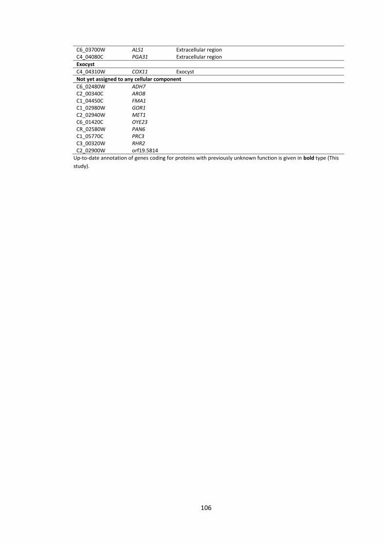
106
C6_03700W ALS1 Extracellular region C4_04080C PGA31 Extracellular region
Exocyst
C4_04310W COX11 Exocyst
Not yet assigned to any cellular component
C6_02480W ADH7 C2_00340C ARO8 C1_04450C FMA1 C1_02980W GOR1 C2_02940W MET1 C6_01420C OYE23 CR_02580W PAN6 C1_05770C PRC3 C3_00320W RHR2 C2_02900W orf19.5814 Up-to-date annotation of genes coding for proteins with previously unknown function is given in bold type (This
study).
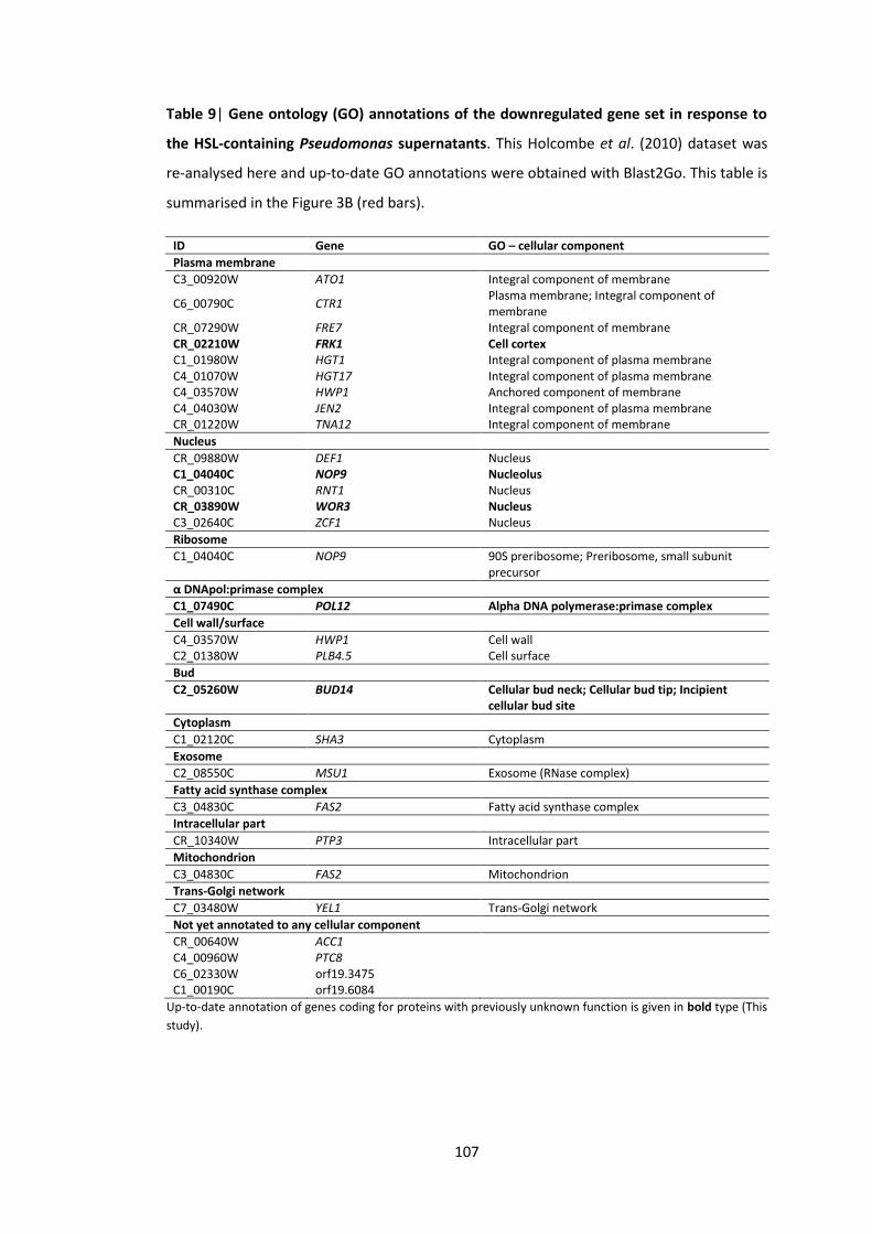
107
Table 9| Gene ontology (GO) annotations of the downregulated gene set in response to
the HSL-containing Pseudomonas supernatants. This Holcombe et al. (2010) dataset was
re-analysed here and up-to-date GO annotations were obtained with Blast2Go. This table is
summarised in the Figure 3B (red bars).
ID Gene GO – cellular component
Plasma membrane
C3_00920W ATO1 Integral component of membrane
C6_00790C CTR1 Plasma membrane; Integral component of membrane
CR_07290W FRE7 Integral component of membrane CR_02210W FRK1 Cell cortex C1_01980W HGT1 Integral component of plasma membrane C4_01070W HGT17 Integral component of plasma membrane C4_03570W HWP1 Anchored component of membrane C4_04030W JEN2 Integral component of plasma membrane CR_01220W TNA12 Integral component of membrane
Nucleus
CR_09880W DEF1 Nucleus C1_04040C NOP9 Nucleolus CR_00310C RNT1 Nucleus CR_03890W WOR3 Nucleus C3_02640C ZCF1 Nucleus
Ribosome
C1_04040C NOP9 90S preribosome; Preribosome, small subunit precursor
α DNApol:primase complex
C1_07490C POL12 Alpha DNA polymerase:primase complex
Cell wall/surface
C4_03570W HWP1 Cell wall C2_01380W PLB4.5 Cell surface
Bud
C2_05260W BUD14 Cellular bud neck; Cellular bud tip; Incipient cellular bud site
Cytoplasm
C1_02120C SHA3 Cytoplasm
Exosome
C2_08550C MSU1 Exosome (RNase complex)
Fatty acid synthase complex
C3_04830C FAS2 Fatty acid synthase complex
Intracellular part
CR_10340W PTP3 Intracellular part
Mitochondrion
C3_04830C FAS2 Mitochondrion
Trans-Golgi network
C7_03480W YEL1 Trans-Golgi network
Not yet annotated to any cellular component
CR_00640W ACC1 C4_00960W PTC8 C6_02330W orf19.3475 C1_00190C orf19.6084 Up-to-date annotation of genes coding for proteins with previously unknown function is given in bold type (This
study).

108
Chapter 5
General Discussion
109
5. General discussion
5.1 Introduction
In the final part, the main findings of this research are briefly summarised and integrated
with the existing knowledge to describe the main conclusions. A detailed experimental plan
and database update strategies are outlined for the progression of this project. Also, new
insights in C. albicans biofilms and interspecies interaction are highlighted indicating future
directions of the field. This chapter concludes with the recommendation for investigation of
C. albicans biofilms rather than of the planktonic cells, focus on the polymicrobial
communities instead of monocultures and suggestion of translational studies from in vitro
to in vivo models.
5.2 Research result summary
Protein kinases (PK) and transcription factors (TF) mediate signal transduction and
transcription of proteins involved in C. albicans biofilm development. We described 5
additional PKs, VPS15, PKH3, PGA43, IME2 and CEX1, that could contribute to the efficient
filamentation and robust biofilm development. To identify the ‘Poor’ biofilm former
mutants we used different biofilm and morphology assays from those who had previously
screened this PK collection (Blankenship et al. 2010; Fanning et al. 2013; de Castro et al.
2013; Morales et al. 2013). Filamentation is essential for biofilm development but our
screens highlighted additional processes, such as vascular biosynthesis and ribosome
biogenesis, that were also important for C. albicans biofilms. The discovery that the
individual elements of the MAPK pathway were not essential for Candida biofilms was
unexpected suggesting that in order to filament the MAPK mutants may exploit the
functionally overlapping MAPK pathways. However, consistent with earlier publications of
Holcombe et al. (2010) and McAlester et al. (2008), P. aeruginosa supernatants were
shown to have 2 distinct effects on C. albicans: HSL-independent biofilm impairment and
HSL-dependent filamentation inhibition. To further investigate TF-regulated genes in
Candida we created a TFbsST database and found that one of the master biofilm regulators,
Efg1p, is also implicated in yeast-hyphae switch. Efg1p binding site elements were
additionally predicted to be conserved in C. parapsilosis hyphae-related genes (RBT).
Currently, the TFbsST database is limited to some Candida species TFs but these findings
increase our knowledge of Candida biofilms and inter-species interactions adding novel
bioinformatics tools to answer interesting questions of, for example, Candida gene
regulation mechanisms.
110
5.3 Candida albicans interaction with bacteria
This research was focused on the investigation of C. albicans – P. aeruginosa interaction
but C. albicans is also known to communicate with Gram-positive bacteria in addition to P.
aeruginosa, which is Gram-negative, and increase the health burden. Polymicrobial
infections are hard to treat since they include various pathogens such as bacteria, yeast and
viruses, requiring different treatments. These pathogens possess synergistic or antagonistic
behaviour affecting the dynamics of their communities and the outcome of the therapies.
The Candida-bacterial interaction can promote or prevent the infections in the human-
host. For example, a bacterium Streptococcus gordonii promotes the adherence of C.
albicans cells, filamentous growth of hyphae and C. albicans biofilm formation on the
epithelial cells of the oral cavity (reviewed by Morales and Hogan 2010). S. gordonii
attaches to the epithelial cell surface with its polypeptides promoting the adherence of C.
albicans. In return, the presence of C. albicans decreases the oxygen to the preferred levels
by S. gordonii. This leads to the increased growth of S. gordonii communities (reviewed by
Morales and Hogan 2010). These conditions in the oral cavity favour the development of
yeast-bacterial biofilms that are difficult to treat with the existing therapies.
The synergetic behaviour of C. albicans and another Gram-positive bacterium,
Staphylococcus aureus, also ultimately leads to robust biofilm development, significantly
increasing mortality rates in mice (Shirtliff et al. 2009; Harriott and Noverr 2009). In these
mixed biofilms, C. albicans prostaglandin E2 (hormone-like fatty acid) stimulates the
growth of S. aureus (Krause et al. 2015). Urinary tract infections are enhanced by the
cooperation of C. albicans and Escherichia coli (Gram-negative), that increases the
attachment ability of C. albicans on the surface of the epithelial cells (Levison and Pitsakis
1987). The lipopolysaccharides of E. coli further enhance the mortality rates of C. albicans-
infected mice (Akagawa et al. 1995).
However, the human host can benefit from the antagonistic behaviour of C. albicans and
Lactobacillus. Different Lactobacillus species that are normal residents of the intestinal and
female reproductive tracts can prevent adherence of C. albicans on the surface of the
epithelial cells (reviewed by Morales and Hogan 2010). They secrete surlactins that prevent
the adherence of C. albicans and compete for the attachment on the cell surface. These
data reflect the complexity of mixed infection and the inter-kingdom interaction. Hence,
more emphasis should be given to the studies of the polymicrobial communities.

111
5.4 Pseudomonas aeruginosa interaction with fungi
In this study we looked at the effects of P. aeruginosa on C. albicans but P. aeruginosa was
also shown to affect other fungi. Conditions like CF and burn wounds involve polymicrobial
infections with mixed bacterial and yeast communities. For example, the lungs of the CF
patients are colonised by bacteria (mainly P. aeruginosa) and yeast including C. albicans,
different Aspergillus and Scedosporium species and Exophiala dermatitidis (reviewed by
Pihet et al. 2009). The study of the mixed cocultures indicated that P. aeruginosa can kill
Aspergillus fumigatus conidia (Manavathu et al. 2014) and inhibit the growth of
Scedosporium aurantiacum (Kaur et al. 2015). P. aeruginosa extracellular molecules can
also inhibit A. fumigatus biofilm formation (Mowat et al. 2010) and its phenazines can
manipulate A. fumigatus iron homeostasis (Briard et al. 2015). In contrast, P. aeruginosa
volatiles stimulate the growth of A. fumigatus colonies on minimal media (Briard et al.
2016). The interaction between P. aeruginosa and E. dermatitidis (black yeast) remains
unclear. These interactions highlight the importance of in vivo models that can shed more
light to the mixed infections.
5.5 In vivo models for the investigation of fungal biofilms
We investigated the C. albicans biofilms and inter-kingdom cross-talk highlighting several
aspects that are important for the biofilm formation in vitro. The majority of the
researchers study fungal biofilms and yeast-bacterial interaction in vitro. The wealth of
knowledge generated from the in vitro analyses can be employed to clarify the microbial
interaction and biofilm development in vivo. Plethora of in vivo models is utilised for the
investigation of fungal biofilms that significantly contribute to the knowledge of biofilm
physiology and microbial interaction with the host. The in vivo biofilm development is
significantly affected by the host factors including the host antibodies, nutrient availability,
flow of the liquids (e.g. blood, urine) and the substrate properties. Although biofilms are
known to be developed by several medically important fungi, Aspergillus, Pneumocystis,
Blastomyces, Zygomycetes, Trichosporon, Cryptococcus, Fusarium and Malassezia, Candida
biofilms are the mostly studied since C. albicans is a model pathogen for the investigation
of the fungal biofilm infections (Reviewed by Nett and Andes 2016).
Many in vivo models are developed for the study of the Candida biofilm-related infections
but the most popular is a central venous catheter model that is adopted for utilisation in a
mouse (Lazzell et al. 2009), a rat (Andes et al. 2004) and a rabbit (Schinabeck et al. 2004).
This model involves the insertion of a vascular catheter employing the surgical procedures
112
and allows the assessment of antifungal influence on the biofilm growth following systemic
administration of a drug. These models significantly contributed into the establishment of
the multi-drug resistance phenomenon in vivo directing the research towards the search
for more effective therapies. Another, subcutaneous implant model in a mouse (Zumbuehl
et al. 2007) and a rat (Ricicova et al. 2010), was designed using amphogel, hydrogel and
amphotericin B-containing disks, inoculated with C. albicans and implanted
subcuntaneously. Employing this model, Zumbuehl et al. (2007) showed that the hydrogel
of this antifungal is suitable for the prevention of device-related infections due to its long-
term efficiency.
To investigate denture infections that are common within the denture-wearers, a rat
denture stomatitis model with a novel intraoral system was developed by Johnson et al.
(2012). The previous Macacairus monkey models contributed into the knowledge of host
response to denture biofilms but the less costly rat models were found to be more useful
for the drug efficacy studies. Additional to the animal models, a human dental plaque
model is used for more realistic studies of the oral communities. To study the oral flora,
disks made of different materials are inserted in the removable intrabuccal splints of
volunteers and after removal of the discs bearing oral microbial communities are analysed
by the researchers (Rimondini et al. 1997; Scarano et al. 2004).
To study oropharyngeal and vaginal candidiasis the mouse oral (Dongari-Bagtzoglou et al.
2009) and vaginal mucosal models were developed respectively (Harriott et al. 2010).
These models revealed the complexity of the oral biofilms that involved bacteria,
neutrophils of host innate immune system (WBC) and keratin that protects epithelial cells
from damage/stress (Dongari-Bagtzoglou et al. 2009). A vaginal candidiasis model however,
showed that vaginal biofilms and those developed on abiotic surfaces share regulation
factors (Harriott et al. 2010).
The clinical scenario of candiduria was characterised using a mouse urinary catheter model
(Wang and Fries 2011). This model employs an insertion of a catheter with a guide wire
into the bladder through the urethra of a female mouse. To investigate the candiduria and
other catheter-related infections, this animal is infected with C. albicans intravesicularly.
The occurred infection is rapidly detectable persisting up to a month and closely resembles
the Candida biofilm formation environment in the patients (Wang and Fries 2011). Thus,
this model can also be used for C. albicans biofilm-related studies in the future.
113
Clearly, animal models of biofilm-associated infections are beneficial for pathogenesis and
drug discovery studies. However, more in vivo models are required to study the biofilms of
other clinically relevant niches, for instance of (burn) wounds.
5.5.1 In vivo models for the investigation of bacterial biofilms
This project was mainly focused on fungal biofilms but bacterial biofilms also cause serious
infections. Diverse in vivo models are developed to mimic the bacterial biofilm infections
addressing important therapeutic questions of bacterial infections. In vivo biofilm infection
models include the rat and mouse CF models for the investigation of the lung microbiome
(Keiser and Engelhardt 2011), a murine cystitis (bladder infection) model for the study of
bacteria inoculated in the bladder (Justice et al. 2004), an excisional wound model (mice)
for the S. aureus infection in the cut wounds (Akiyama et al. 1996) and a rabbit model for
the infective endocarditis where a high bacterial concentration is injected intravenously
(Xiong et al. 2006).
In this research, fungal biofilms formed on abiotic surface were explored in great detail but
bacterial biofilms can also be found on abiotic surfaces. The in vivo device-related infection
models are common for the investigation of the bacterial biofilms developed on medical
devises. The most popular are the rat vascular catheter models that were developed with
the utilisation of the plastic catheter inserted in the vein of the rat (Ebert et al. 2011).
Additionally, many urinary catheter models were described in mice, rats and rabbits in
order to study the diverse aspects of the catheter-related infections. For example, using the
urethral infection model Cirioni et al. (2011) examined the effects of the antibacterials on
the infections caused by P. aeruginosa (Cirioni et al. 2011) and Allison et al. (2011)
described the eradication of the bacterial persister cells via aminoglycoside therapy that
inhibits protein synthesis (Allison et al. 2011). The orthopaedic and dental implant models
also contribute into the understanding of the bacterial biofilms. To investigate the fungal
biofilms, these models can be modified by incorporating the aspects of fungal infection and
biofilms.
5.6 Future directions for the progression of this project
5.6.1 Candida albicans biofilm studies using ‘Poor’ biofilm former protein kinase mutants
Based on our discussion, the future research should be directed towards the detailed
investigation of C. albicans biofilms in addition to its planktonic cells. The results of this
Ph.D. project provide useful lead towards different directions for further research. Many
questions arise in relation to the PKs that were firstly assigned to C. albicans biofilm
114
formation and filamentation in this study (Chapter 2). Hence, for the progression of this
work, the mutants disrupted in these PK coding genes can be studied in detail. More
specifically, 5 genes VPS15, PKH3, PGA43, IME2 and CEX1 disrupted in the mutants firstly
associated with poor biofilm formation and impaired filamentous growth in this study can
be followed up for further clarification of their role in C. albicans (Chapter 2). Vps15p,
involved in vacuolar protein sorting and Pkh3p, required for PKC activity and cell-substrate
adherence (Fanning et al. 2012), were clustered together in the protein-protein interaction
networks (Chapter 2). To clarify their association, vacuolar morphology of the mutants
disrupted in the genes coding for these proteins can be examined microscopically and their
cell wall proteome can be determined with the spectrometry. Pga43p is probably required
for cell wall integrity (Moreno-Ruiz et al. 2009) and can additionally be included in these
studies. Apart from their role in C. albicans biofilm formation and filamentation, IME2 and
CEX1 are not yet confidently assigned to any specific function. However, mutant disrupted
in IME2 is hypersensitive to amphotericin B (Xu et al. 2007) and CEX1 is largely unexplored.
Hence, their sensitivity to diverse antifungals can be determined using azoles, polyenes and
echinocardins. In order to clarify the reason behind the poor biofilm and filamentation
phenotypes of these mutants, their stress response under different osmotic and oxidative
stress conditions can be explored in addition to the virulence in animal models in vivo. This
knowledge will enable to design more effective antifungal therapies.
5.6.2 Candida albicans – Pseudomonas aeruginosa interaction studies in vivo
As the most infections are polymicrobial, the future studies should definitely focus on both
physical and signal-mediated interaction between the microbes of the polymicrobial
communities. The in vivo animal models including 2 pathogens are highly desirable because
they can highlight more details of the infections (Lindsay and Hogan 2014). For example,
our evidence suggests that in vitro, P. aeruginosa can significantly affect C. albicans and
filamentous growth by its secreted molecules (Chapter 3). In vivo studies of their behaviour
in animal models coinfected with both C. albicans and P. aeruginosa could further clarify
the communication of these pathogens. To explore other elements involved in their
communication the host needs to be included in the future experiments. The ultimate goal
is to exploit this knowledge and devise virulence-limiting methods in order to benefit the
infected patients.
115
5.6.3 In vitro validation of predicted in silico analyses
In silico studies of a master transcriptional regulator Efg1p, predicting its important role in
yeast-hyphae switch as well as in C. albicans – P. aeruginosa interaction (Chapter 4), can be
validated via in vitro analyses. To validate the in silico analysis of Efg1p across the RBT
family gene promoter sequences (Chapter 4), RBT gene expression analysis can be
conducted in vitro. Gene expression of RBT1, RBT2, RBT4 and RBT6 can be quantified in the
EFG1 mutant grown at 37oC in YNBNP (filament-inducing medium) with RTqPCR including
EFG1 mutant grown at 30oC in YNB (non-filament-inducing medium) as a control. The
expression of the RBT genes is expected to be decreased in the EFG1 mutant grown at 37oC
in YNBNP since Holcombe et al. (2010) have shown that both RBT1 and RBT4 were
upregulated during filamentation of wild-type C. albicans and severely downregulated in
the yeast conditions.
5.6.4 Updating and maintenance of the TFbsST database
A TFbsST database, developed here (Chapter 4), represents an important bioinformatics
tool for in silico analysis of C. albicans promoter sequences and should be regularly
updated in the framework of the future projects. To ensure operational efficiency, the
TFbsST database should be updated regularly by populating the existing lists with more
functionalities, TFs, experimentally tested motifs and Candida/non-Candida species. One of
the functionalities that can be considered is an additional option for the user-defined motif
that can be used to screen the promoter sequences. Constantly increasing lists of verified
TFs and their motifs are good source of data and will need to be captured in the next
versions of the TFbsST database. To accelerate Candida research, emerging pathogenic
Candida species including C. tropicalis and C. krusei, need to be added to the TFbsST
database list of organisms. An addition of an industrial yeast Kluyveromyces marxianus will
make the TFbsST database useful bioinformatics tool for the remaining students in Dr.
Morrissey’s laboratory, who currently study different aspects of K. marxianus.

116
Acknowledgments
I would like to honestly thank every person, institution and organisation that contributed
into the successful completion of this project. I will start with my supervisor, Dr. John
Morrissey, who gave me the opportunity to work in his laboratory. I worked on a very
interesting topic, otherwise it would be difficult to spend so much time on XTT biofilm
assays (2 screens of ~200 mutants) and PCR validations (with a published primer that did
not work, for me), but I mastered important lab techniques. These 3 years of research were
additionally challenging with the compulsory modules (>70 credits), demonstrations and
invigilations, not to mention maternity. However, John was considerably flexible with my
timetable, probably because he had noticed my hard-working attitude. He was right, in
order to complete this project in 3 years I planned ahead systematically, worked overtime
and during the weekends and holidays. All this time, John was very supportive in everything
from the confirmation of an expensive reagent order (e.g. XTT powder) to stimulation with
the spicy biological questions to think of for the Viva. Our frequent meetings additionally
accelerated the progress of this work. I appreciated his honest feedback on my critical
thinking : ) and the fact that he replied to my late night e-mails even when he was on
holidays, saving a lot of my time! I have learned from John an optimistic way of thinking,
seeing the things from different optical angles, which was impressive! He was very patient
waiting for initiatives and good research ideas that I only managed on my last year of the
Ph.D. probably because during the previous 2 years I was practically sleepless (due to the
baby at night and research – by day) literally experiencing a ‘brain repression’ : ). However,
my ‘Eureka’ moment came with the TFbsST website and I really enjoyed the last part of this
research. Lastly, I am very thankful to John for the 3 month extension of the stipend that
allowed my daughter and me to escape from the financial difficulties.
Many thanks to my Ph.D. course coordinators, Dr. Kelly Dean and Dr. Maryanne Donovan,
who arranged MolCell modules, workshops, some conferences and the related
travel/accommodation. I express my great gratitude to our School Manager Ms. Hilda
Bohane for her support in regards to the paper work throughout my Ph.D. and Ms. Aine
Murphy for reading parts of this Thesis. Thanks to Prof. Aaron Mitchell for helpful
comments and advice in molecular validation of the PK mutants using a cross validation
approach and the Higher Education Authority (HEA) for funding our project under the
PRTLI5 Structured Ph.D. programme “Molecular and Cellular Mechanisms underlying
Inflammatory Processes”. I would also like to thank my internal and external examiners, Dr.
117
David Clarke and Dr. Donna MacCallum respectively, for reading my Thesis and for
objective evaluation of this work.
Next, I want to thank all the staff and students of the School of Microbiology, UCC, who
directly or indirectly affected this research with small hints that made a huge difference. I
will start with the members of the Lab 441, Javier Varela and Loughlin Gethins. Javier
provided his molecular lights and protocols generously after he came to the lab, right in the
start of my molecular experiments. He significantly helped during the initiation of
molecular experiments including DNA and RNA isolation, PCR, RTqPCR, transformations and
preparation of the competent cells. I also thank him for the initial Blast2Go annotations, for
the retrieval of protein sequences and for listening to my problems patiently. Javier stands
out with his good and friendly nature (he is Latin!) arranging the social events of the lab
that I rarely managed to attend due to the parental responsibilities. Loughlin was a quiet
lab mate, he made me realize that I need to label the distinct sets of PCR tubes differently
in case they are mixed by the colleagues. Also, I want to thank Dr. Jerry Reen (BIOMERIT –
Lab 442) for his help during the first period of my Ph.D. research. Jerry had contributed in
the clarification of bacterial supernatant collection and dilution processes, lent me the
multichannel pipette that I used for all the XTT experiments and had expressed a genuine
interest in this research. Eoin Burke gave me the tour around the School of Microbiology
for the initial orientation and introduced me to culture media preparation procedures.
Karen McCarthy (Lab 403) thanks for your help in the initial setting up and operation of the
96-well plate reader. Maire O’Dwyer and John O’Callaghan always ordered the different
reagents ‘ASAP’. Also, I would like to express my gratitude to Paddy O’Reilly for excellent
technical support in lyophilisation of bacterial supernatants, general discussions and his
smiles. I was surprised that he remembered my name from the first time I was introduced
to him! Dan Walsh used to solve general IT and printing issues!
Additionally, I want to thank my colleagues who later became friends, for their friendship
and psychological support. I deeply appreciate the useful suggestions of Dr. Klaudia Sikora,
Dr. Panos Lehouritis, Dr. Liam Loftus and Mr. John McSweeney in relation to our first paper.
Also, many thanks to Dr. Sol Schvartzman for her valuable help in Python scripts and Emily
Power for setting up the TFbsST website. Ian O’Neill, Alicia Campion, Ioanna Tzani and
Stephanie Davies thank you for being friendly course mates. Ian additionally showed the
functionalities of the Confocal/Laser Scanning Microscope, which was quite complex. Also, I
118
thank all the members of Prof. Paul O’Toole’s Labs 338-339 for being excellent lunch
companions.
Finally, I dedicate this Thesis to my family members who generously provided their moral
support, material funds and obvious respect to my choices trough out all the period of my
studies. I am very, very touched with the attitude of my parents towards my Ph.D. research
and with their invaluable help in raising my child. I could not imagine how patient you can
be, minding Arli all day, until late, during the weekends and holiday, while I was away for a
conference and when I just needed to sleep. I wish all the girls with children could have this
kind of support so as they can pursue a better career. I thank my brother, Nick, for being a
good uncle for Arli and for setting up a Python environment for my in silico analysis. My
sister, Natalie, thanks for trying to teach Chemical Engineering to Arli and for feeding her
with healthy food during the holidays. Stratos, always gave tough critiques and partially
covered our holidays. Finally, my warmest thanks go to my lovely daughter, Arli, for being
the strongest and sweetest motivation!
. . .
LΓLΓLΓLΓLΓLΓLΓLΓLΓLΙ
119
Bibliography
Akagawa G, Abe S & Yamaguchi H (1995) Mortality of Candida albicans-infected mice is
facilitated by superinfection of Escherichia coli or administration of its lipopolysaccharide. J
Infect Dis 171: 1539-1544.
Akiyama H, Kanzaki H, Tada J & Arata J (1996) Staphylococcus aureus infection on cut
wounds in the mouse skin: experimental Staphylococcal botryomycosis. J Dermatol Sci 11:
234-238.
Albano MM & Schmitt JA (1973) Pathogenicity in mice of strains of Candida albicans
(Robin) Berk. isolated from burn patients. Mycopathol Mycol Appl 49: 283-288.
Alem MA, Oteef MD, Flowers TH & Douglas LJ (2006) Production of tyrosol by Candida
albicans biofilms and its role in quorum sensing and biofilm development. Eukaryot Cell 5:
1770-1779.
Allison KR, Brynildsen MP & Collins JJ (2011) Metabolite-enabled eradication of bacterial
persisters by aminoglycosides. Nature 473: 216-220.
Alonso-Monge R, Navarro-Garcia F, Roman E, Negredo AI, Eisman B, Nombela C & Pla J
(2003) The Hog1 mitogen-activated protein kinase is essential in the oxidative stress
response and chlamydospore formation in Candida albicans. Eukaryot Cell 2: 351-361.
Alonso-Monge R, Navarro-Garcia F, Molero G, Diez-Orejas R, Gustin M, Pla J, Sanchez M &
Nombela C (1999) Role of the mitogen-activated protein kinase Hog1p in morphogenesis
and virulence of Candida albicans. J Bacteriol 181: 3058-3068.
Alves-Silva JM, Zuzarte M, Goncalves MJ, Cavaleiro C, Cruz MT, Cardoso SM & Salgueiro L
(2016) New claims for wild carrot (Daucus carota subsp. carota) essential oil. Evid Based
Complement Alternat Med 2016: 9045196 doi: 10.1155/2016/9045196.
Andes D, Nett J, Oschel P, Albrecht R, Marchillo K & Pitula A (2004) Development and
characterization of an in vivo central venous catheter Candida albicans biofilm model.
Infect Immun 72: 6023-6031.
Argimon S, Wishart JA, Leng R, Macaskill S, Mavor A, Alexandris T, Nicholls S, Knight AW,
Enjalbert B, Walmsley R, Odds FC, Gow NA & Brown AJ (2007) Developmental regulation of
http://www.ncbi.nlm.nih.gov/pubmed/?term=Macaskill%20S%5BAuthor%5D&cauthor=true&cauthor_uid=17277173
120
an adhesin gene during cellular morphogenesis in the fungal pathogen Candida albicans.
Eukaryot Cell 6: 682-692.
Arnaud MB, Costanzo MC, Skrzypek MS, Binkley G, Lane C, Miyasato SR & Sherlock G (2005)
The Candida Genome Database (CGD), a community resource for Candida albicans gene
and protein information. Nucleic Acids Res 33: D358-363.
Askew C, Sellam A, Epp E, Mallick J, Hogues H, Mullick A, Nantel A & Whiteway M (2011)
The zinc cluster transcription factor Ahr1p directs Mcm1p regulation of Candida albicans
adhesion. Mol Microbiol 79: 940-953.
Bachewich C, Nantel A & Whiteway M (2005) Cell cycle arrest during S or M phase
generates polarized growth via distinct signals in Candida albicans. Mol Microbiol 57: 942-
959.
Baek YU, Li M & Davis DA (2008) Candida albicans ferric reductases are differentially
regulated in response to distinct forms of iron limitation by the Rim101 and CBF
transcription factors. Eukaryot Cell 7: 1168-1179.
Bahnan W, Koussa J, Younes S, Abi Rizk M, Khalil B, El Sitt S, Hanna S, El-Sibai M & Khalaf RA
(2012) Deletion of the Candida albicans PIR32 results in increased virulence, stress
response, and upregulation of cell wall chitin deposition. Mycopathologia 174: 107-119.
Baker KL & Coe LM (1993) Growing up with a chronic condition: transition to young
adulthood for the individual with cystic fibrosis. Holist Nurs Pract 8: 8-15.
Baker CR, Tuch BB & Johnson AD (2011) Extensive DNA-binding specificity divergence of a
conserved transcription regulator. Proc Natl Acad Sci USA 108: 7493-7498.
Bandara HM, Lam OL, Watt RM, Jin LJ & Samaranayake LP (2010a) Bacterial
lipopolysaccharides variably modulate in vitro biofilm formation of Candida species. J Med
Microbiol 59: 1225-1234.
Bandara HM, Yau JY, Watt RM, Jin LJ & Samaranayake LP (2010b) Pseudomonas aeruginosa
inhibits in-vitro Candida biofilm development. BMC Microbiol 10: 125.
Bandara HM, BP KC, Watt RM, Jin LJ & Samaranayake LP (2013) Pseudomonas aeruginosa
lipopolysaccharide inhibits Candida albicans hyphae formation and alters gene expression
during biofilm development. Mol Oral Microbiol 28: 54-69.
121
Banerjee D, Martin N, Nandi S, Shukla S, Dominguez A, Mukhopadhyay G & Prasad R (2007)
A genome-wide steroid response study of the major human fungal pathogen Candida
albicans. Mycopathologia 164: 1-17.
Banner EA (1974) Vaginitis. Med Clin North Am 58: 759-768.
Barelle CJ, Bohula EA, Kron SJ, Wessels D, Soll DR, Schafer A, Brown AJ & Gow NA (2003)
Asynchronous cell cycle and asymmetric vacuolar inheritance in true hyphae of Candida
albicans. Eukaryot Cell 2: 398-410.
Bassilana M, Blyth J & Arkowitz RA (2003) Cdc24, the GDP-GTP exchange factor for Cdc42,
is required for invasive hyphal growth of Candida albicans. Eukaryot Cell 2: 9-18.
Bauernfeind A, Bertele RM, Harms K, Horl G, Jungwirth R, Petermuller C, Przyklenk B &
Weisslein-Pfister C (1987) Qualitative and quantitative microbiological analysis of sputa of
102 patients with cystic fibrosis. Infection 15: 270-277.
Beatson SA, Whitchurch CB, Semmler AB & Mattick JS (2002) Quorum sensing is not
required for twitching motility in Pseudomonas aeruginosa. J Bacteriol 184: 3598-3604.
Bennett RJ & Johnson AD (2005) Mating in Candida albicans and the search for a sexual
cycle. Annu Rev Microbiol 59: 233-255.
Bennett RJ & Johnson AD (2006) The role of nutrient regulation and the Gpa2 protein in the
mating pheromone response of C. albicans. Mol Microbiol 62: 100-119.
Bennett RJ, Uhl MA, Miller MG & Johnson AD (2003) Identification and characterization of a
Candida albicans mating pheromone. Mol Cell Biol 23: 8189-8201.
Bensen ES, Filler SG & Berman J (2002) A forkhead transcription factor is important for true
hyphal as well as yeast morphogenesis in Candida albicans. Eukaryot Cell 1: 787-798.
Berman J (2016) Ploidy plasticity: a rapid and reversible strategy for adaptation to stress.
FEMS Yeast Res doi: 10.1093/femsyr/fow020.
Berman J & Sudbery PE (2002) Candida albicans: a molecular revolution built on lessons
from budding yeast. Nat Rev Genet 3: 918-930.
Bernardo SM, Khalique Z, Kot J, Jones JK & Lee SA (2008) Candida albicans VPS1 contributes
to protease secretion, filamentation, and biofilm formation. Fungal Genet Biol 45: 861-877.
122
Bjarnsholt T, Jensen PO, Jakobsen TH, Phipps R, Nielsen AK, Rybtke MT, Tolker-Nielsen T,
Givskov M, Høiby N & Ciofu O (2010) Quorum sensing and virulence of Pseudomonas
aeruginosa during lung infection of cystic fibrosis patients. PLoS One 5: e10115.
Blankenship JR, Fanning S, Hamaker JJ & Mitchell AP (2010) An extensive circuitry for cell
wall regulation in Candida albicans. PLoS Pathog 6: e1000752.
Bockmuhl DP, Krishnamurthy S, Gerads M, Sonneborn A & Ernst JF (2001) Distinct and
redundant roles of the two protein kinase A isoforms Tpk1p and Tpk2p in morphogenesis
and growth of Candida albicans. Mol Microbiol 42: 1243-1257.
Bogavac M, Karaman M, Janjusevic L, Sudji J, Radovanovic B, Novakovic Z, Simeunovic J &
Bozin B (2015) Alternative treatment of vaginal infections - in vitro antimicrobial and toxic
effects of Coriandrum sativum L. and Thymus vulgaris L. essential oils. J Appl Microbiol 119:
697-710.
Bonhomme J, Chauvel M, Goyard S, Roux P, Rossignol T & d'Enfert C (2011) Contribution of
the glycolytic flux and hypoxia adaptation to efficient biofilm formation by Candida
albicans. Mol Microbiol 80: 995-1013.
Brajtburg J, Elberg S, Medoff G & Kobayashi GS (1981) Increase in colony-forming units of
Candida albicans after treatment with polyene antibiotics. Antimicrob Agents Chemother
19: 199-200.
Braun PC (2005) The effect of farnesol on amino acid incorporation by a wild-type and cell-
wall variant strain of Candida albicans. Can J Microbiol 51: 715-718.
Braun BR & Johnson AD (1997) Control of filament formation in Candida albicans by the
transcriptional repressor TUP1. Science 277: 105-109.
Briard B, Heddergott C & Latge JP (2016) Volatile compounds emitted by Pseudomonas
aeruginosa stimulate growth of the fungal pathogen Aspergillus fumigatus. MBio doi:
10.1128/mBio.00219-16.
Briard B, Bomme P, Lechner BE, Mislin GL, Lair V, Prevost MC, Latge JP, Haas H & Beauvais
A (2015) Pseudomonas aeruginosa manipulates redox and iron homeostasis of its
microbiota partner Aspergillus fumigatus via phenazines. Sci Rep 5: 8220.
123
Bruce CR, Smith DA, Rodgers D, da Silva Dantas A, MacCallum DM, Morgan BA & Quinn J
(2011) Identification of a novel response regulator, Crr1, that is required for hydrogen
peroxide resistance in Candida albicans. PLoS One 6: e27979.
Bruckmann A, Kunkel W, Hartl A, Wetzker R & Eck R (2000) A phosphatidylinositol 3-kinase
of Candida albicans influences adhesion, filamentous growth and virulence. Microbiology
146: 2755-2764.
Bruno VM & Mitchell AP (2005) Regulation of azole drug susceptibility by Candida albicans
protein kinase CK2. Mol Microbiol 56: 559-573.
Burns JL, Van Dalfsen JM, Shawar RM, Otto KL, Garber RL, Quan JM, Montgomery AB,
Albers GM, Ramsey BW & Smith AL (1999) Effect of chronic intermittent administration of
inhaled tobramycin on respiratory microbial flora in patients with cystic fibrosis. J Infect Dis
179: 1190-1196.
Buschart A, Gremmer K, El-Mowafy M, van den Heuvel J, Mueller PP & Bilitewski U (2012) A
novel functional assay for fungal histidine kinases group III reveals the role of HAMP
domains for fungicide sensitivity. J Biotechnol 157: 268-277.
Cabezon V, Llama-Palacios A, Nombela C, Monteoliva L & Gil C (2009) Analysis of Candida
albicans plasma membrane proteome. Proteomics 9: 4770-4786.
Calderone RA & Fonzi WA (2001) Virulence factors of Candida albicans. Trends Microbiol 9:
327-335.
Calderone RA, Rotondo MF & Sande MA (1978) Candida albicans endocarditis:
ultrastructural studies of vegetation formation. Infect Immun 20: 279-289.
Cao YY, Cao YB, Xu Z, Ying K, Li Y, Xie Y, Zhu ZY, Chen WS & Jiang YY (2005) cDNA microarray
analysis of differential gene expression in Candida albicans biofilm exposed to farnesol.
Antimicrob Agents Chemother 49: 584-589.
Carlisle PL & Kadosh D (2013) A genome-wide transcriptional analysis of morphology
determination in Candida albicans. Mol Biol Cell 24: 246-260.
Cassola A, Parrot M, Silberstein S, Magee BB, Passeron S, Giasson L & Cantore ML (2004)
Candida albicans lacking the gene encoding the regulatory subunit of protein kinase A
displays a defect in hyphal formation and an altered localization of the catalytic subunit.
Eukaryot Cell 3: 190-199.
124
CGD (2010) Description lines for gene products, based on orthologs and predicted
Gene Ontology (GO) annotations.
Chambers D, Scott F, Bangur R, Davies R, Lim A, Walters S, Smith G, Pitt T, Stableforth D &
Honeybourne D (2005) Factors associated with infection by Pseudomonas aeruginosa in
adult cystic fibrosis. Eur Respir J 26: 651-656.
Chandra J, Kuhn DM, Mukherjee PK, Hoyer LL, McCormick T & Ghannoum MA (2001)
Biofilm formation by the fungal pathogen Candida albicans: development, architecture,
and drug resistance. J Bacteriol 183: 5385-5394.
Chang DT, Huang CY, Wu CY & Wu WS (2011) YPA: an integrated repository of promoter
features in Saccharomyces cerevisiae. Nucleic Acids Res 39: D647-652.
Chauhan N, Inglis D, Roman E, Pla J, Li D, Calera JA & Calderone R (2003) Candida albicans
response regulator gene SSK1 regulates a subset of genes whose functions are associated
with cell wall biosynthesis and adaptation to oxidative stress. Eukaryot Cell 2: 1018-1024.
Chen J, Chen J, Lane S & Liu H (2002) A conserved mitogen-activated protein kinase
pathway is required for mating in Candida albicans. Mol Microbiol 46: 1335-1344.
Chen H, Fujita M, Feng Q, Clardy J & Fink GR (2004) Tyrosol is a quorum-sensing molecule in
Candida albicans. Proc Natl Acad Sci USA 101: 5048-5052.
Chen AI, Dolben EF, Okegbe C, Harty CE, Golub Y, Thao S, Ha DG, Willger SD, O'Toole GA,
Harwood CS, Dietrich LE & Hogan DA (2014) Candida albicans ethanol stimulates
Pseudomonas aeruginosa WspR-controlled biofilm formation as part of a cyclic relationship
involving phenazines. PLoS Pathog 10: e1004480.
Cheng S, Clancy CJ, Checkley MA, Handfield M, Hillman JD, Progulske-Fox A, Lewin AS, Fidel
PL & Nguyen MH (2003) Identification of Candida albicans genes induced during thrush
offers insight into pathogenesis. Mol Microbiol 48: 1275-1288.
Chiang LY, Sheppard DC, Bruno VM, Mitchell AP, Edwards JE Jr & Filler SG (2007) Candida
albicans protein kinase CK2 governs virulence during oropharyngeal candidiasis. Cell
Microbiol 9: 233-245.
Chotirmall SH, Greene CM & McElvaney NG (2010) Candida species in cystic fibrosis: A road
less travelled. Med Mycol 48 Suppl 1: S114-124.
125
Cirioni O, Ghiselli R, Silvestri C, Minardi D, Gabrielli E, Orlando F, Rimini M, Brescini L,
Muzzonigro G, Guerrieri M & Giacometti A (2011) Effect of the combination of
clarithromycin and amikacin on Pseudomonas aeruginosa biofilm in an animal model of
ureteral stent infection. J Antimicrob Chemother 66: 1318-1323.
Clark KL, Feldmann PJ, Dignard D, Larocque R, Brown AJ, Lee MG, Thomas DY & Whiteway
M (1995) Constitutive activation of the Saccharomyces cerevisiae mating response pathway
by a MAP kinase kinase from Candida albicans. Mol Gen Genet 249: 609-621.
Cloutier M, Castilla R, Bolduc N, Zelada A, Martineau P, Bouillon M, Magee BB, Passeron S,
Giasson L & Cantore ML (2003) The two isoforms of the cAMP-dependent protein kinase
catalytic subunit are involved in the control of dimorphism in the human fungal pathogen
Candida albicans. Fungal Genet Biol 38: 133-141.
Conesa A, Gotz S, Garcia-Gomez JM, Terol J, Talon M & Robles M (2005) Blast2GO: a
universal tool for annotation, visualization and analysis in functional genomics research.
Bioinformatics 21: 3674-3676.
Connolly LA, Riccombeni A, Grozer Z, Holland LM, Lynch DB, Andes DR, Gacser A & Butler G
(2013) The APSES transcription factor Efg1 is a global regulator that controls
morphogenesis and biofilm formation in Candida parapsilosis. Mol Microbiol 90: 36-53.
Corvey C, Koetter P, Beckhaus T, Hack J, Hofmann S, Hampel M, Stein T, Karas M & Entian
KD (2005) Carbon source-dependent assembly of the Snf1p kinase complex in Candida
albicans. J Biol Chem 280: 25323-25330.
Cote P, Hogues H & Whiteway M (2009) Transcriptional analysis of the Candida albicans cell
cycle. Mol Biol Cell 20: 3363-3373.
Csank C, Makris C, Meloche S, Schroppel K, Rollinghoff M, Dignard D, Thomas DY &
Whiteway M (1997) Derepressed hyphal growth and reduced virulence in a VH1 family-
related protein phosphatase mutant of the human pathogen Candida albicans. Mol Biol
Cell 8: 2539-2551.
Csank C, Schroppel K, Leberer E, Harcus D, Mohamed O, Meloche S, Thomas DY &
Whiteway M (1998) Roles of the Candida albicans mitogen-activated protein kinase
homolog, Cek1p, in hyphal development and systemic candidiasis. Infect Immun 66: 2713-
2721.
http://www.ncbi.nlm.nih.gov/pubmed/?term=Gabrielli%20E%5BAuthor%5D&cauthor=true&cauthor_uid=21406436
126
Cugini C, Calfee MW, Farrow JM, 3rd, Morales DK, Pesci EC & Hogan DA (2007) Farnesol, a
common sesquiterpene, inhibits PQS production in Pseudomonas aeruginosa. Mol
Microbiol 65: 896-906.
Curvelo JA, Marques AM, Barreto AL, Romanos MT, Portela MB, Kaplan MA & Soares RM
(2014) A novel nerolidol-rich essential oil from Piper claussenianum modulates Candida
albicans biofilm. J Med Microbiol 63: 697-702.
Danhof HA & Lorenz MC (2015) The Candida albicans ATO gene family promotes
neutralization of the macrophage phagolysosome. Infect Immun 83: 4416-4426.
Davis DA, Bruno VM, Loza L, Filler SG & Mitchell AP (2002) Candida albicans Mds3p, a
conserved regulator of pH responses and virulence identified through insertional
mutagenesis. Genetics 162: 1573-1581.
Davis RM, Muller RY & Haynes KA (2015) Can the natural diversity of quorum-sensing
advance synthetic biology? Fron Bioeng Biotechnol. 3: 30.
Davis-Hanna A, Piispanen AE, Stateva LI & Hogan DA (2008) Farnesol and dodecanol effects
on the Candida albicans Ras1-cAMP signalling pathway and the regulation of
morphogenesis. Mol Microbiol 67: 47-62.
de Boer CG & Hughes TR (2012) YeTFaSCo: a database of evaluated yeast transcription
factor sequence specificities. Nucleic Acids Res 40: D169-179.
de Campos Rasteiro VM, da Costa AC, Araújo CF, de Barros PP, Rossoni RD1, Anbinder AL,
Jorge AO & Junqueira JC (2014) Essential oil of Melaleuca alternifolia for the treatment of
oral candidiasis induced in an immunosuppressed mouse model. BMC Complement Altern
Med 15: 489.
de Castro PA, Bom VL, Brown NA, de Almeida RS, Ramalho LN, Savoldi M, Goldman MH,
Berretta AA & Goldman GH (2013) Identification of the cell targets important for propolis-
induced cell death in Candida albicans. Fungal Genet Biol 60: 74-86.
De Groot PW, Hellingwerf KJ & Klis FM (2003) Genome-wide identification of fungal GPI
proteins. Yeast 20: 781-796.
Desai C, Mavrianos J & Chauhan N (2011) Candida albicans SRR1, a putative two-
component response regulator gene, is required for stress adaptation, morphogenesis, and
virulence. Eukaryot Cell 10: 1370-1374.
127
Deveau A & Hogan DA (2011) Linking quorum sensing regulation and biofilm formation by
Candida albicans. Methods Mol Biol 692: 219-233.
Dhillon NK, Sharma S & Khuller GK (2003) Biochemical characterization of Ca2+/calmodulin
dependent protein kinase from Candida albicans. Mol Cell Biochem 252: 183-191.
Diggle SP, West SA, Gardner A & Griffin AS (2008) Communication in bacteria (Chapter 2).
In sociobiology of communication: an interdisciplinary perspective (Hughes D & Ettorre PD
editors) Oxford University Press. UK. DettoreBook 11-31.
Dignard D & Whiteway M (2006) SST2, a regulator of G-protein signaling for the Candida
albicans mating response pathway. Eukaryot Cell 5: 192-202.
Ding X, Yu Q, Zhang B, Xu N, Jia C, Dong Y, Chen Y, Xing L & Li M (2014) The type II
Ca2+/calmodulin-dependent protein kinases are involved in the regulation of cell wall
integrity and oxidative stress response in Candida albicans. Biochem Biophys Res Commun
446: 1073-1078.
Dongari-Bagtzoglou A, Kashleva H, Dwivedi P, Diaz P & Vasilakos J (2009) Characterization
of mucosal Candida albicans biofilms. PLoS One 4: e7967.
Eberhard A (1972) Inhibition and activation of bacterial luciferase synthesis. J Bacteriol 109:
1101-1105.
Eberhard A, Burlingame AL, Eberhard C, Kenyon GL, Nealson KH & Oppenheimer NJ (1981)
Structural identification of autoinducer of Photobacterium fischeri luciferase. Biochemistry
20: 2444-2449.
Ebert T, Smith S, Pancari G, Wu X, Zorman J, Clark D, Cook J, Burns C, Antonello JM, Cope L,
Nagy E, Meinke A & McNeely T (2011) Development of a rat central venous catheter model
for evaluation of vaccines to prevent Staphylococcus epidermidis and Staphylococcus
aureus early biofilms. Hum Vaccin 7: 630-638.
Eck R, Bruckmann A, Wetzker R & Kunkel W (2000) A phosphatidylinositol 3-kinase of
Candida albicans: molecular cloning and characterization. Yeast 16: 933-944.
Eisman B, Alonso-Monge R, Roman E, Arana D, Nombela C & Pla J (2006) The Cek1 and
Hog1 mitogen-activated protein kinases play complementary roles in cell wall biogenesis
and chlamydospore formation in the fungal pathogen Candida albicans. Eukaryot Cell 5:
347-358.
128
Elson SL, Noble SM, Solis NV, Filler SG & Johnson AD (2009) An RNA transport system in
Candida albicans regulates hyphal morphology and invasive growth. PLoS Genet 5:
e1000664.
Enjalbert B, Smith DA, Cornell MJ, Alam I, Nicholls S, Brown AJ & Quinn J (2006) Role of the
Hog1 stress-activated protein kinase in the global transcriptional response to stress in the
fungal pathogen Candida albicans. Mol Biol Cell 17: 1018-1032.
Epp E, Vanier G, Harcus D, Lee AY, Jansen G, Hallett M, Sheppard DC, Thomas DY, Munro
CA, Mullick A & Whiteway M (2010) Reverse genetics in Candida albicans predicts ARF
cycling is essential for drug resistance and virulence. PLoS Pathog 6: e1000753.
Fabry W, Schmid EN, Schraps M & Ansorg R (2003) Isolation and purification of
chlamydospores of Candida albicans. Med Mycol 41: 53-58.
Falleiros de Pádua RA, Norman Negri MF, Svidzinski AE, Nakamura CV & Svidzinski TI (2008)
Adherence of Pseudomonas aeruginosa and Candida albicans to urinary catheters. Rev
Iberoam Micol 25: 173-175.
Fan D, Coughlin LA, Neubauer MM, Kim J, Kim MS, Zhan X, Simms-Waldrip TR, Xie Y,
Hooper LV & Koh AY (2015) Activation of HIF-1alpha and LL-37 by commensal bacteria
inhibits Candida albicans colonization. Nat Med 21: 808-814.
Fanning S, Xu W, Beaurepaire C, Suhan JP, Nantel A & Mitchell AP (2012) Functional control
of the Candida albicans cell wall by catalytic protein kinase A subunit Tpk1. Mol Microbiol
86: 284-302.
Fernandez-Arenas E, Molero G, Nombela C, Diez-Orejas R & Gil C (2004) Low virulent
strains of Candida albicans: unravelling the antigens for a future vaccine. Proteomics 4:
3007-3020.
Fernandez-Arenas E, Cabezon V, Bermejo C, Arroyo J, Nombela C, Diez-Orejas R & Gil C
(2007) Integrated proteomics and genomics strategies bring new insight into Candida
albicans response upon macrophage interaction. Mol Cell Proteomics 6: 460-478.
Finkel JS & Mitchell AP (2011) Genetic control of Candida albicans biofilm development.
Nat Rev Microbiol 9: 109-118.
Fox EP & Nobile CJ (2012) A sticky situation: untangling the transcriptional network
controlling biofilm development in Candida albicans. Transcription 3: 315-322.
129
Fox EP, Cowley ES, Nobile CJ, Hartooni N, Newman DK & Johnson AD (2014) Anaerobic
bacteria grow within Candida albicans biofilms and induce biofilm formation in suspension
cultures. Curr Biol 24: 2411-2416.
Fox EP, Bui CK, Nett JE, Hartooni N, Mui MC, Andes DR, Nobile CJ & Johnson AD (2015) An
expanded regulatory network temporally controls Candida albicans biofilm formation. Mol
Microbiol 96: 1226-1239.
Freires IA, Queiroz VC, Furletti VF, Ikegaki M, de Alencar SM, Duarte MC & Rosalen PL
(2016) Chemical composition and antifungal potential of Brazilian propolis against Candida
spp. J Mycol Med 26: 122-32.
Freires IA, Bueno-Silva B, Galvao LC, Duarte MC, Sartoratto A, Figueira GM, de Alencar SM
& Rosalen PL (2015) The effect of essential oils and bioactive fractions on Streptococcus
mutans and Candida albicans biofilms: a confocal analysis. Evid Based Complement
Alternat Med 2015: 871316.
Gale CA, Leonard MD, Finley KR, Christensen L, McClellan M, Abbey D, Kurischko C, Bensen
E, Tzafrir I, Kauffman S, Becker J & Berman J (2009) SLA2 mutations cause SWE1-mediated
cell cycle phenotypes in Candida albicans and Saccharomyces cerevisiae. Microbiology 155:
3847-3859.
Garcia-Sanchez S, Aubert S, Iraqui I, Janbon G, Ghigo JM & d'Enfert C (2004) Candida
albicans biofilms: a developmental state associated with specific and stable gene
expression patterns. Eukaryot Cell 3: 536-545.
Gasch AP, Moses AM, Chiang DY, Fraser HB, Berardini M & Eisen MB (2004) Conservation
and evolution of cis-regulatory systems in ascomycete fungi. PLoS Biol 2: e398.
Gibson J, Sood A & Hogan DA (2009) Pseudomonas aeruginosa-Candida albicans
interactions: localization and fungal toxicity of a phenazine derivative. Appl Environ
Microbiol 75: 504-513.
Gillum AM, Tsay EY & Kirsch DR (1984) Isolation of the Candida albicans gene for orotidine-
5'-phosphate decarboxylase by complementation of S. cerevisiae ura3 and E. coli pyrF
mutations. Mol Gen Genet 198: 179-182.
http://www.ncbi.nlm.nih.gov/pubmed/?term=McClellan%20M%5BAuthor%5D&cauthor=true&cauthor_uid=19778960
130
Gonia S, Tuepker M, Heisel T, Autran C, Bode L & Gale CA (2015) Human milk
oligosaccharides inhibit Candida albicans invasion of human premature intestinal epithelial
cells. J Nutr 145: 1992-1998.
Gordan R, Pyne S & Bulyk ML (2012) Identification of cell cycle-regulated, putative hyphal
genes in Candida albicans. Pac Symp Biocomput 299-310.
Goudot C, Etchebest C, Devaux F & Lelandais G (2011) The reconstruction of condition-
specific transcriptional modules provides new insights in the evolution of yeast AP-1
proteins. PLoS One 6: e20924.
Govan JR & Deretic V (1996) Microbial pathogenesis in cystic fibrosis: mucoid Pseudomonas
aeruginosa and Burkholderia cepacia. Microbiol Rev 60: 539-574.
Goyard S, Knechtle P, Chauvel M, Mallet A, Prévost MC, Proux C, Coppée JY, Schwarz P,
Dromer F, Park H, Filler SG, Janbon G & d'Enfert C (2008) The Yak1 kinase is involved in the
initiation and maintenance of hyphal growth in Candida albicans. Mol Biol Cell 19: 2251-
2266.
Grahl N, Demers EG, Lindsay AK, Harty CE, Willger SD, Piispanen AE & Hogan DA (2015)
Mitochondrial Activity and Cyr1 Are Key Regulators of Ras1 Activation of C. albicans
Virulence Pathways. PLoS Pathog 11: e1005133.
Guhad FA, Jensen HE, Aalbaek B, Csank C, Mohamed O, Harcus D, Thomas DY, Whiteway M
& Hau J (1998) Mitogen-activated protein kinase-defective Candida albicans is avirulent in a
novel model of localized murine candidiasis. FEMS Microbiol Lett 166: 135-139.
Gulati M & Nobile CJ (2016) Candida albicans biofilms: development, regulation, and
molecular mechanisms. Microbes Infect 18: 310-321.
Gunther J, Nguyen M, Hartl A, Kunkel W, Zipfel PF & Eck R (2005) Generation and functional
in vivo characterization of a lipid kinase defective phosphatidylinositol 3-kinase Vps34p of
Candida albicans. Microbiology 151: 81-89.
Gupta N, Haque A, Mukhopadhyay G, Narayan RP & Prasad R (2005) Interactions between
bacteria and Candida in the burn wound. Burns 31: 375-378.
Gutierrez-Barranquero JA, Reen FJ, McCarthy RR & O'Gara F (2015) Deciphering the role of
coumarin as a novel quorum sensing inhibitor suppressing virulence phenotypes in
bacterial pathogens. Appl Microbiol Biotechnol 99: 3303-3316.
131
Hargarten JC, Moore TC, Petro TM, Nickerson KW & Atkin AL (2015) Candida albicans
Quorum Sensing Molecules Stimulate Mouse Macrophage Migration. Infect Immun 83:
3857-3864.
Harriott MM & Noverr MC (2009) Candida albicans and Staphylococcus aureus form
polymicrobial biofilms: effects on antimicrobial resistance. Antimicrob Agents Chemother
53: 3914-3922.
Harriott MM, Lilly EA, Rodriguez TE, Fidel PL Jr & Noverr MC (2010) Candida albicans forms
biofilms on the vaginal mucosa. Microbiology 156: 3635-3644.
Hartmann A & Schikora A (2012) Quorum sensing of bacteria and trans-kingdom
interactions of N-acyl homoserine lactones with eukaryotes. J Chem Ecol 38: 704-713.
Hazan I, Sepulveda-Becerra M & Liu H (2002) Hyphal elongation is regulated independently
of cell cycle in Candida albicans. Mol Biol Cell 13: 134-145.
Hernday AD, Lohse MB, Nobile CJ, Noiman L, Laksana CN & Johnson AD (2016) Ssn6 defines
a new level of regulation of white-opaque switching in Candida albicans and is required for
the stochasticity of the switch. MBio 7: e01565-15.
Heurlier K, Denervaud V & Haas D (2006) Impact of quorum sensing on fitness of
Pseudomonas aeruginosa. Int J Med Microbiol 296: 93-102.
Hoekstra MJ, Westgate SJ & Mueller S (2016) Povidone-iodine ointment demonstrates in
vitro efficacy against biofilm formation. Int Wound J doi: 10.1111/iwj.12578.
Hoffman (2001) Preparation of yeast DNA. Curr Protoc Mol Biol 13: 11.
Hogan DA & Kolter R (2002) Pseudomonas-Candida interactions: an ecological role for
virulence factors. Science 296: 2229-2232.
Hogan DA & Sundstrom P (2009) The Ras/cAMP/PKA signaling pathway and virulence in
Candida albicans. Future Microbiol 4: 1263-1270.
Hogan DA, Vik A & Kolter R (2004) A Pseudomonas aeruginosa quorum-sensing molecule
influences Candida albicans morphology. Mol Microbiol 54: 1212-1223.
Hogues H, Lavoie H, Sellam A, Mangos M, Roemer T, Purisima E, Nantel A & Whiteway M
(2008) Transcription factor substitution during the evolution of fungal ribosome regulation.
Mol Cell 29: 552-562.
132
Holcombe LJ, O'Gara F & Morrissey JP (2011) Implications of interspecies signaling for
virulence of bacterial and fungal pathogens. Future Microbiol 6: 799-817.
Holcombe LJ, McAlester G, Munro CA, Enjalbert B, Brown AJ, Gow NA, Ding C, Butler G,
O'Gara F & Morrissey JP (2010) Pseudomonas aeruginosa secreted factors impair biofilm
development in Candida albicans. Microbiology 156: 1476-1486.
Holloway BW & Morgan AF (1986) Genome organization in Pseudomonas. Annu Rev
Microbiol 40: 79-105.
Homann OR, Dea J, Noble SM & Johnson AD (2009) A phenotypic profile of the Candida
albicans regulatory network. PLoS Genet 5: e1000783.
Hoppen J, Dietz M, Warsow G, Rohde R & Schuller HJ (2007) Ribosomal protein genes in the
yeast Candida albicans may be activated by a heterodimeric transcription factor related to
Ino2 and Ino4 from S. cerevisiae. Mol Genet Genomics 278: 317-330.
Hornby JM, Jensen EC, Lisec AD, Tasto JJ, Jahnke B, Shoemaker R, Dussault P & Nickerson
KW (2001) Quorum sensing in the dimorphic fungus Candida albicans is mediated by
farnesol. Appl Environ Microbiol 67: 2982-2992.
Huguenin-Dumittan SA & Girard JP (1972) [Asthma caused by Candida albicans. Diagnosis,
immunology, new chemotherapeutic treatment]. Schweiz Med Wochenschr 102: 1471-
1478.
Inglis DO & Johnson AD (2002) Ash1 protein, an asymmetrically localized transcriptional
regulator, controls filamentous growth and virulence of Candida albicans. Mol Cell Biol 22:
8669-8680.
Inglis DO, Skrzypek MS, Arnaud MB, Binkley J, Shah P, Wymore F & Sherlock G (2013)
Improved gene ontology annotation for biofilm formation, filamentous growth, and
phenotypic switching in Candida albicans. Eukaryot Cell 12: 101-108.
Jensen LJ, Kuhn M, Stark M, Chaffron S, Creevey C, Muller J, Doerks T, Julien P, Roth
A, Simonovic M, Bork P & von Mering C (2009) STRING 8--a global view on proteins and
their functional interactions in 630 organisms. Nucleic Acids Res 37: D412-416.
Johnson DC, Cano KE, Kroger EC & McNabb DS (2005) Novel regulatory function for the
CCAAT-binding factor in Candida albicans. Eukaryot Cell 4: 1662-1676.
133
Johnson CC, Yu A, Lee H, Fidel PL, Jr. & Noverr MC (2012) Development of a contemporary
animal model of Candida albicans-associated denture stomatitis using a novel intraoral
denture system. Infect Immun 80: 1736-1743.
Joo JH & Jetten AM (2010) Molecular mechanisms involved in farnesol-induced apoptosis.
Cancer Lett 287: 123-135.
Justice SS, Hung C, Theriot JA, Fletcher DA, Anderson GG, Footer MJ & Hultgren SJ (2004)
Differentiation and developmental pathways of uropathogenic Escherichia coli in urinary
tract pathogenesis. Proc Natl Acad Sci U S A 101: 1333-1338.
Kanehisa M & Goto S (2000) KEGG: kyoto encyclopedia of genes and genomes. Nucleic
Acids Res 28: 27-30.
Kashem SW, Igyarto BZ, Gerami-Nejad M, Kumamoto Y, Mohammed J, Jarrett
E, Drummond RA, Zurawski SM, Zurawski G, Berman J, Iwasaki A, Brown GD & Kaplan DH
(2015) Candida albicans morphology and dendritic cell subsets determine T helper cell
differentiation. Immunity 42: 356-366.
Kaur J, Pethani BP, Kumar S, Kim M, Sunna A, Kautto L, Penesyan A, Paulsen IT &
Nevalainen H (2015) Pseudomonas aeruginosa inhibits the growth of Scedosporium
aurantiacum, an opportunistic fungal pathogen isolated from the lungs of cystic fibrosis
patients. Front Microbiol 6: 866.
Kavanaugh NL, Zhang AQ, Nobile CJ, Johnson AD & Ribbeck K (2014) Mucins suppress
virulence traits of Candida albicans. MBio 5: e01911.
Kayingo G & Wong B (2005) The MAP kinase Hog1p differentially regulates stress-induced
production and accumulation of glycerol and D-arabitol in Candida albicans. Microbiology
151: 2987-2999.
Kebaara BW, Langford ML, Navarathna DH, Dumitru R, Nickerson KW & Atkin AL (2008)
Candida albicans Tup1 is involved in farnesol-mediated inhibition of filamentous-growth
induction. Eukaryot Cell 7: 980-987.
Keiser NW & Engelhardt JF (2011) New animal models of cystic fibrosis: what are they
teaching us? Curr Opin Pulm Med 17: 478-483.
Kerr JR (1994) Suppression of fungal growth exhibited by Pseudomonas aeruginosa. J Clin
Microbiol 32: 525-527.
http://www.ncbi.nlm.nih.gov/pubmed/?term=Drummond%20RA%5BAuthor%5D&cauthor=true&cauthor_uid=25680275
134
Kerr JR, Taylor GW, Rutman A, Hoiby N, Cole PJ & Wilson R (1999) Pseudomonas aeruginosa
pyocyanin and 1-hydroxyphenazine inhibit fungal growth. J Clin Pathol 52: 385-387.
Kim J, Lee JE & Lee JS (2015) Histone deacetylase-mediated morphological transition in
Candida albicans. J Microbiol 53: 805-811.
Kinsman OS, Pitblado K & Coulson CJ (1988) Effect of mammalian steroid hormones and
luteinizing hormone on the germination of Candida albicans and implications for vaginal
candidosis. Mycoses 31: 617-626.
Kohler JR & Fink GR (1996) Candida albicans strains heterozygous and homozygous for
mutations in mitogen-activated protein kinase signaling components have defects in hyphal
development. Proc Natl Acad Sci USA 93: 13223-13228.
Kojic EM & Darouiche RO (2004) Candida infections of medical devices. Clin Microbiol Rev
17: 255-267.
Konstantinidou N (2014) Decoding bug chatter to fight infections. The Boolean 4: 51-55.
Konstantinidou N & Morrissey JP (2015) Co-occurrence of filamentation defects and
impaired biofilms in Candida albicans protein kinase mutants. FEMS Yeast Res 15: 1–10. pii
fov092 doi: 10.1093/femsyr/fov092.
Kostoulias X, Murray GL, Cerqueira GM, Kong JB, Bantun F, Mylonakis E, Khoo CA & Peleg
AY (2015) Impact of a cross-kingdom signaling molecule of Candida albicans on
Acinetobacter baumannii physiology. Antimicrob Agents Chemother 60: 161-167.
Krause J, Geginat G & Tammer I (2015) Prostaglandin E2 from Candida albicans stimulates
the growth of Staphylococcus aureus in mixed biofilms. PLoS One 10: e0135404.
Kromidas L, Perrier E, Flanagan J, Rivero R & Bonnet I (2006) Release of antimicrobial
actives from microcapsules by the action of axillary bacteria. Int J Cosmet Sci 28: 103-108.
Kruppa M (2009) Quorum sensing and Candida albicans. Mycoses 52: 1-10.
Kruppa M, Krom BP, Chauhan N, Bambach AV, Cihlar RL & Calderone RA (2004) The two-
component signal transduction protein Chk1p regulates quorum sensing in Candida
albicans. Eukaryot Cell 3: 1062-1065.
135
Kusch H, Engelmann S, Bode R, Albrecht D, Morschhauser J & Hecker M (2008) A proteomic
view of Candida albicans yeast cell metabolism in exponential and stationary growth
phases. Int J Med Microbiol 298: 291-318.
LaFayette SL, Collins C, Zaas AK, Schell WA, Betancourt-Quiroz M, Gunatilaka AA, Perfect JR
& Cowen LE (2010) PKC signaling regulates drug resistance of the fungal pathogen Candida
albicans via circuitry comprised of Mkc1, calcineurin, and Hsp90. PLoS Pathog 6: e1001069.
LaFleur MD, Kumamoto CA & Lewis K (2006) Candida albicans biofilms produce antifungal-
tolerant persister cells. Antimicrob Agents Chemother 50: 3839-3846.
Lamfon H, Porter SR, McCullough M & Pratten J (2004) Susceptibility of Candida albicans
biofilms grown in a constant depth film fermentor to chlorhexidine, fluconazole and
miconazole: a longitudinal study. J Antimicrob Chemother 53: 383-385.
Lan CY, Newport G, Murillo LA, Jones T, Scherer S, Davis RW & Agabian N (2002) Metabolic
specialization associated with phenotypic switching in Candida albicans. Proc Natl Acad Sci
USA 99: 14907-14912.
Lane S, Zhou S, Pan T, Dai Q & Liu H (2001) The basic helix-loop-helix transcription factor
Cph2 regulates hyphal development in Candida albicans partly via TEC1. Mol Cell Biol 21:
6418-6428.
Lane S, Di Lena P, Tormanen K, Baldi P & Liu H (2015) Function and Regulation of Cph2 in
Candida albicans. Eukaryot Cell 14: 1114-1126.
Lazzell AL, Chaturvedi AK, Pierce CG, Prasad D, Uppuluri P & Lopez-Ribot JL (2009)
Treatment and prevention of Candida albicans biofilms with caspofungin in a novel central
venous catheter murine model of candidiasis. J Antimicrob Chemother 64: 567-570.
Leberer E, Ziegelbauer K, Schmidt A, Harcus D, Dignard D, Ash J, Johnson L & Thomas DY
(1997) Virulence and hyphal formation of Candida albicans require the Ste20p-like protein
kinase CaCla4p. Curr Biol 7: 539-546.
Leberer E, Harcus D, Broadbent ID, Clark KL, Dignard D, Ziegelbauer K, Schmidt A, Gow NA,
Brown AJ & Thomas DY (1996) Signal transduction through homologs of the Ste20p and
Ste7p protein kinases can trigger hyphal formation in the pathogenic fungus Candida
albicans. Proc Natl Acad Sci USA 93: 13217-13222.
136
Leclair LW & Hogan DA (2010) Mixed bacterial-fungal infections in the CF respiratory tract.
Med Mycol 48 Suppl 1: S125-132.
Lee KH, Jun S, Hur HS, Ryu JJ & Kim J (2005) Candida albicans protein analysis during hyphal
differentiation using an integrative HA-tagging method. Biochem Biophys Res Commun
337: 784-790.
Legrand M, Chan CL, Jauert PA & Kirkpatrick DT (2011) The contribution of the S-phase
checkpoint genes MEC1 and SGS1 to genome stability maintenance in Candida albicans.
Fungal Genet Biol 48: 823-830.
Leonhardt I, Spielberg S, Weber M, Albrecht-Eckardt D, Bläss M, Claus R, Barz D, Scherlach
K, Hertweck C, Löffler J, Hünniger K & Kurzai O (2015) The fungal quorum-sensing molecule
farnesol activates innate immune cells but suppresses cellular adaptive immunity. MBio 6:
e00143.
Levison ME & Pitsakis PG (1987) Susceptibility to experimental Candida albicans urinary
tract infection in the rat. J Infect Dis 155: 841-846.
Levitin A & Whiteway M (2007) The effect of prostaglandin E2 on transcriptional responses
of Candida albicans. Microbiol Res 162: 201-210.
Lewis K (2012) Persister cells: molecular mechanisms related to antibiotic tolerance. Handb
Exp Pharmacol 121-133.
Li X, Huang X, Zhao J, Zhao J, Wei Y & Jiang L (2008) The MAP kinase-activated protein
kinase Rck2p plays a role in rapamycin sensitivity in Saccharomyces cerevisiae and Candida
albicans. FEMS Yeast Res 8: 715-724.
Lilienthal B (1955) The pathogenicity of Candida albicans isolated from the mouth. Oral
Surg Oral Med Oral Pathol 8: 1214-1217.
Linde J, Wilson D, Hube B & Guthke R (2010) Regulatory network modelling of iron
acquisition by a fungal pathogen in contact with epithelial cells. BMC Syst Biol 4: 148.
Lindsay AK, Morales DK, Liu Z, Grahl N, Zhang A, Willger SD, Myers LC & Hogan DA (2014)
Analysis of Candida albicans mutants defective in the Cdk8 module of mediator reveal links
between metabolism and biofilm formation. PLoS Genet 10: e1004567.
http://www.ncbi.nlm.nih.gov/pubmed/?term=Scherlach%20K%5BAuthor%5D&cauthor=true&cauthor_uid=25784697
137
Liu TT, Lee RE, Barker KS, Lee RE, Wei L, Homayouni R & Rogers PD (2005) Genome-wide
expression profiling of the response to azole, polyene, echinocandin, and pyrimidine
antifungal agents in Candida albicans. Antimicrob Agents Chemother 49: 2226-2236.
Liu Y, Solis NV, Heilmann CJ, Phan QT, Mitchell AP, Klis FM & Filler SG (2014) Role of
retrograde trafficking in stress response, host cell interactions, and virulence of Candida
albicans. Eukaryot Cell 13: 279-287.
Lo HJ, Kohler JR, DiDomenico B, Loebenberg D, Cacciapuoti A & Fink GR (1997)
Nonfilamentous C. albicans mutants are avirulent. Cell 90: 939-949.
Lohse MB, Zordan RE, Cain CW & Johnson AD (2010) Distinct class of DNA-binding domains
is exemplified by a master regulator of phenotypic switching in Candida albicans. Proc Natl
Acad Sci USA 107: 14105-14110.
Lopez-Ribot JL (2005) Candida albicans biofilms: more than filamentation. Curr Biol 15:
R453-455.
Lu Y, Su C, Unoje O & Liu H (2014) Quorum sensing controls hyphal initiation in Candida
albicans through Ubr1-mediated protein degradation. Proc Natl Acad Sci USA 111: 1975-
1980.
MacCallum DM, Castillo L, Nather K, Munro CA, Brown AJ, Gow NA & Odds FC (2009)
Property differences among the four major Candida albicans strain clades. Eukaryot Cell 8:
373-387.
MacIsaac KD, Wang T, Gordon DB, Gifford DK, Stormo GD & Fraenkel E (2006) An improved
map of conserved regulatory sites for Saccharomyces cerevisiae. BMC Bioinformatics 7:
113.
Magee BB & Magee PT (1997) WO-2, a stable aneuploid derivative of Candida albicans
strain WO-1, can switch from white to opaque and form hyphae. Microbiology 143: 289-
295.
Magee BB, Legrand M, Alarco AM, Raymond M & Magee PT (2002) Many of the genes
required for mating in Saccharomyces cerevisiae are also required for mating in Candida
albicans. Mol Microbiol 46: 1345-1351.
138
Manavathu EK, Vager DL & Vazquez JA (2014) Development and antimicrobial susceptibility
studies of in vitro monomicrobial and polymicrobial biofilm models with Aspergillus
fumigatus and Pseudomonas aeruginosa. BMC Microbiol 14: 53.
Martin SW, Douglas LM & Konopka JB (2005) Cell cycle dynamics and quorum sensing in
Candida albicans chlamydospores are distinct from budding and hyphal growth. Eukaryot
Cell 4: 1191-1202.
Martin DW, Schurr MJ, Mudd MH, Govan JR, Holloway BW & Deretic V (1993) Mechanism
of conversion to mucoidy in Pseudomonas aeruginosa infecting cystic fibrosis patients. Proc
Natl Acad Sci USA 90: 8377-8381.
Martins M, Henriques M, Azeredo J, Rocha SM, Coimbra MA & Oliveira R (2007)
Morphogenesis control in Candida albicans and Candida dubliniensis through signaling
molecules produced by planktonic and biofilm cells. Eukaryot Cell 6: 2429-2436.
Mattia E, Carruba G, Angiolella L & Cassone A (1982) Induction of germ tube formation by
N-acetyl-D-glucosamine in Candida albicans: uptake of inducer and germinative response. J
Bacteriol 152: 555-562.
Matys V, Kel-Margoulis OV, Fricke E, Liebich I, Land S, Barre-Dirrie A, Reuter I, Chekmenev
D, Krull M, Hornischer K, Voss N, Stegmaier P, Lewicki-Potapov B, Saxel H, Kel AE &
Wingender E (2006) TRANSFAC and its module TRANSCompel: transcriptional gene
regulation in eukaryotes. Nucleic Acids Res 34: D108-110.
McAlester G, O'Gara F & Morrissey JP (2008) Signal-mediated interactions between
Pseudomonas aeruginosa and Candida albicans. J Med Microbiol 57: 563-569.
McGuire AT & Mangroo D (2007) Cex1p is a novel cytoplasmic component of the
Saccharomyces cerevisiae nuclear tRNA export machinery. EMBO J 26: 288-300.
Mendelson MH, Gurtman A, Szabo S, Neibart E, Meyers BR, Policar M, Cheung TW,
Lillienfeld D, Hammer G, Reddy S, et al. (1994) Pseudomonas aeruginosa bacteremia in
patients with AIDS. Clin Infect Dis 18: 886-895.
Miro-Casas E, Covas MI, Fito M, Farre-Albadalejo M, Marrugat J & de la Torre R (2003)
Tyrosol and hydroxytyrosol are absorbed from moderate and sustained doses of virgin olive
oil in humans. Eur J Clin Nutr 57: 186-190.
http://www.ncbi.nlm.nih.gov/pubmed/?term=Chekmenev%20D%5BAuthor%5D&cauthor=true&cauthor_uid=16381825
http://www.ncbi.nlm.nih.gov/pubmed/?term=Chekmenev%20D%5BAuthor%5D&cauthor=true&cauthor_uid=16381825
http://www.ncbi.nlm.nih.gov/pubmed/?term=Stegmaier%20P%5BAuthor%5D&cauthor=true&cauthor_uid=16381825
139
Mitchell AP (2009) Possibly essential genes; no disruptants obtained by UAU1 method
using the BWP17 strain. (CGD paper) Unpublished personal communication to CGD.
Molero G, Diez-Orejas R, Navarro-Garcia F, Monteoliva L, Pla J, Gil C, Sanchez-Perez M &
Nombela C (1998) Candida albicans: genetics, dimorphism and pathogenicity. Int Microbiol
1: 95-106.
Monge RA, Roman E, Nombela C & Pla J (2006) The MAP kinase signal transduction
network in Candida albicans. Microbiology 152: 905-912.
Morales DK, Jacobs NJ, Rajamani S, Krishnamurthy M, Cubillos-Ruiz JR & Hogan DA (2010)
Antifungal mechanisms by which a novel Pseudomonas aeruginosa phenazine toxin kills
Candida albicans in biofilms. Mol Microbiol 78: 1379-1392.
Morales DK, Grahl N, Okegbe C, Dietrich LE, Jacobs NJ & Hogan DA (2013) Control of
Candida albicans metabolism and biofilm formation by Pseudomonas aeruginosa
phenazines. MBio 4: e00526-00512.
Morales DK & Hogan DA (2010) Candida albicans interactions with bacteria in the context
of human health and disease. PloS Pathog 6: e1000886.
Moreno I, Castillo L, Sentandreu R & Valentin E (2007) Global transcriptional profiling of
Candida albicans cwt1 null mutant. Yeast 24: 357-370.
Moreno I, Pedreno Y, Maicas S, Sentandreu R, Herrero E & Valentin E (2003)
Characterization of a Candida albicans gene encoding a putative transcriptional factor
required for cell wall integrity. FEMS Microbiol Lett 226: 159-167.
Moreno-Ruiz E, Ortu G, de Groot PW, Cottier F, Loussert C, Prevost MC, de Koster C, Klis
FM, Goyard S & d'Enfert C (2009) The GPI-modified proteins Pga59 and Pga62 of Candida
albicans are required for cell wall integrity. Microbiology 155: 2004-2020.
Mowat E, Rajendran R, Williams C, McCulloch E, Jones B, Lang S & Ramage G (2010)
Pseudomonas aeruginosa and their small diffusible extracellular molecules inhibit
Aspergillus fumigatus biofilm formation. FEMS Microbiol Lett 313: 96-102.
Murillo LA, Newport G, Lan CY, Habelitz S, Dungan J & Agabian NM (2005) Genome-wide
transcription profiling of the early phase of biofilm formation by Candida albicans. Eukaryot
Cell 4: 1562-1573.
140
Nagahashi S, Mio T, Ono N, Yamada-Okabe T, Arisawa M, Bussey H & Yamada-Okabe H
(1998) Isolation of CaSLN1 and CaNIK1, the genes for osmosensing histidine kinase
homologues, from the pathogenic fungus Candida albicans. Microbiology 144 ( Pt 2): 425-
432.
Nealson KH (1977) Autoinduction of bacterial luciferase. Occurrence, mechanism and
significance. Arch Microbiol 112: 73-79.
Nealson KH, Platt T & Hastings JW (1970) Cellular control of the synthesis and activity of the
bacterial luminescent system. J Bacteriol 104: 313-322.
Nett M (2014) Genome mining: concept and strategies for natural product discovery. Prog
Chem Org Nat Prod 99: 199-245.
Nett JE & Andes DR (2016) Antifungal agents: spectrum of activity, pharmacology and
clinical indications. Infect Dis Clin North Am 30: 51-83.
Nett JE, Lepak AJ, Marchillo K & Andes DR (2009) Time course global gene expression
analysis of an in vivo Candida biofilm. J Infect Dis 200: 307-313.
Newburger DE & Bulyk ML (2009) UniPROBE: an online database of protein binding
microarray data on protein-DNA interactions. Nucleic Acids Res 37: D77-82.
Nguyen MH, Cheng S & Clancy CJ (2004) Assessment of Candida albicans genes expressed
during infections as a tool to understand pathogenesis. Med Mycol 42: 293-304.
Nobile CJ & Johnson AD (2015) Candida albicans biofilms and human disease. Annu Rev
Microbiol 69: 71-92.
Nobile CJ, Bruno VM, Richard ML, Davis DA & Mitchell AP (2003) Genetic control of
chlamydospore formation in Candida albicans. Microbiology 149: 3629-3637.
Nobile CJ, Fox EP, Hartooni N, Mitchell KF, Hnisz D, Andes DR, Kuchler K & Johnson AD
(2014) A histone deacetylase complex mediates biofilm dispersal and drug resistance in
Candida albicans. MBio 5: e01201-01214.
Nobile CJ, Nett JE, Hernday AD, Homann OR, Deneault JS, Nantel A, Andes DR, Johnson AD
& Mitchell AP (2009) Biofilm matrix regulation by Candida albicans Zap1. PLoS Biol 7:
e1000133.
141
Nobile CJ, Fox EP, Nett JE, Sorrells TR, Mitrovich QM, Hernday AD, Tuch BB, Andes DR &
Johnson AD (2012) A recently evolved transcriptional network controls biofilm
development in Candida albicans. Cell 148: 126-138.
Nseir S, Jozefowicz E, Cavestri B, Sendid B, Di Pompeo C, Dewavrin F, Favory R, Roussel-
Delvallez M & Durocher A (2007) Impact of antifungal treatment on Candida-Pseudomonas
interaction: a preliminary retrospective case-control study. Intensive Care Med 33: 137-
142.
Oberholzer U, Marcil A, Leberer E, Thomas DY & Whiteway M (2002) Myosin I is required
for hyphae formation in Candida albicans. Eukaryot Cell 1: 213-228.
Oliveira SA, Zambrana JR, Iorio FB, Pereira CA & Jorge AO (2014) The antimicrobial effects
of Citrus limonum and Citrus aurantium essential oils on multi-species biofilms. Braz Oral
Res 28: 22-27.
Ozsarac N, Bhattacharyya M, Dawes IW & Clancy MJ (1995) The SPR3 gene encodes a
sporulation-specific homologue of the yeast CDC3/10/11/12 family of bud neck
microfilaments and is regulated by ABFI. Gene 164: 157-162.
Park H, Myers CL, Sheppard DC, Phan QT, Sanchez AA, E Edwards J & Filler SG (2005) Role of
the fungal Ras-protein kinase A pathway in governing epithelial cell interactions during
oropharyngeal candidiasis. Cell Microbiol 7: 499-510.
Pastor-Flores D, Schulze JO, Bahi A, Giacometti R, Ferrer-Dalmau J, Passeron S, Engel M,
Suss E, Casamayor A & Biondi RM (2013) PIF-pocket as a target for C. albicans Pkh selective
inhibitors. ACS Chem Biol 8: 2283-2292.
Paul S, Bair TB & Moye-Rowley WS (2014) Identification of genomic binding sites for
Candida glabrata Pdr1 transcription factor in wild-type and rho0 cells. Antimicrob Agents
Chemother 58: 6904-6912.
Pearson JP, Pesci EC & Iglewski BH (1997) Roles of Pseudomonas aeruginosa las and rhl
quorum-sensing systems in control of elastase and rhamnolipid biosynthesis genes. J
Bacteriol 179: 5756-5767.
Peleg AY, Hogan DA & Mylonakis E (2010) Medically important bacterial-fungal
interactions. Nat Rev Microbiol 8: 340-349.

142
Pérez JC, Fordyce PM, Lohse MB, Hanson-Smith V, DeRisi JL & Johnson AD (1014) How
duplicated transcription regulators can diversify to govern the expression of
nonoverlapping sets of genes. Genes Dev 28: 1272-1277.
Pesci EC, Pearson JP, Seed PC & Iglewski BH (1997) Regulation of las and rhl quorum
sensing in Pseudomonas aeruginosa. J Bacteriol 179: 3127-3132.
Peters BM, Ward RM, Rane HS, Lee SA & Noverr MC (2013) Efficacy of ethanol against
Candida albicans and Staphylococcus aureus polymicrobial biofilms. Antimicrob Agents
Chemother 57: 74-82.
Pihet M, Carrere J, Cimon B, Chabasse D, Delhaes L, Symoens F & Bouchara JP (2009)
Occurrence and relevance of filamentous fungi in respiratory secretions of patients with
cystic fibrosis--a review. Med Mycol 47: 387-397.
Pires RH, Montanari LB, Martins CH, Zaia JE, Almeida AM, Matsumoto MT & Mendes-
Giannini MJ (2011) Anticandidal efficacy of cinnamon oil against planktonic and biofilm
cultures of Candida parapsilosis and Candida orthopsilosis. Mycopathologia 172: 453-464.
Purschke FG, Hiller E, Trick I & Rupp S (2012) Flexible survival strategies of Pseudomonas
aeruginosa in biofilms result in increased fitness compared with Candida albicans. Mol Cell
Proteomics 11: 1652-1669.
Qasim M, Singh BR, Naqvi AH, Paik P & Das D (2015) Silver nanoparticles embedded
mesoporous SiO(2) nanosphere: an effective anticandidal agent against Candida albicans
077. Nanotechnology 26: 285102.
Ramage G, Vande Walle K, Wickes BL & Lopez-Ribot JL (2001) Standardized method for in
vitro antifungal susceptibility testing of Candida albicans biofilms. Antimicrob Agents
Chemother 45: 2475-2479.
Ramage G, Bachmann S, Patterson TF, Wickes BL & Lopez-Ribot JL (2002) Investigation of
multidrug efflux pumps in relation to fluconazole resistance in Candida albicans biofilms. J
Antimicrob Chemother 49: 973-980.
Ramon AM & Fonzi WA (2003) Diverged binding specificity of Rim101p, the Candida
albicans ortholog of PacC. Eukaryot Cell 2: 718-728.
143
Ramsdale M, Selway L, Stead D, Walker J, Yin Z, Nicholls SM, Crowe J, Sheils EM & Brown AJ
(2008) MNL1 regulates weak acid-induced stress responses of the fungal pathogen Candida
albicans. Mol Biol Cell 19: 4393-4403.
Rauceo JM, Blankenship JR, Fanning S, Hamaker JJ, Deneault JS, Smith FJ, Nantel A &
Mitchell AP (2008) Regulation of the Candida albicans cell wall damage response by
transcription factor Sko1 and PAS kinase Psk1. Mol Biol Cell 19: 2741-2751.
Raut JS, Shinde RB, Chauhan NM & Karuppayil SM (2013) Terpenoids of plant origin inhibit
morphogenesis, adhesion, and biofilm formation by Candida albicans. Biofouling 29: 87-96.
Reen FJ, Mooij MJ, Holcombe LJ, McSweeney CM, McGlacken GP, Morrissey JP & O'Gara F
(2011) The Pseudomonas quinolone signal (PQS), and its precursor HHQ, modulate
interspecies and interkingdom behaviour. FEMS Microbiol Ecol 77: 413-428.
Ricicova M, Kucharikova S, Tournu H, Hendrix J, Bujdakova H, Van Eldere J, Lagrou K & Van
Dijck P (2010) Candida albicans biofilm formation in a new in vivo rat model. Microbiology
156: 909-919.
Rikkerink EH, Magee BB & Magee PT (1988) Opaque-white phenotype transition: a
programmed morphological transition in Candida albicans. J Bacteriol 170: 895-899.
Rimondini L, Fare S, Brambilla E, Felloni A, Consonni C, Brossa F & Carrassi A (1997) The
effect of surface roughness on early in vivo plaque colonization on titanium. J Periodontol
68: 556-562.
Rocha CR, Schroppel K, Harcus D, Marcil A, Dignard D, Taylor BN, Thomas DY, Whiteway M
& Leberer E (2001) Signaling through adenylyl cyclase is essential for hyphal growth and
virulence in the pathogenic fungus Candida albicans. Mol Biol Cell 12: 3631-3643.
Rosin H (1974) Septicemia due to Candida albicans. Pathogenesis, diagnosis and
chemotherapy. Dtsch Med Wochenschr 99: 2514, 2526-2530.
Samuel SM, Thirunavukkarasu M, Penumathsa SV, Paul D & Maulik N (2008)
Akt/FOXO3a/SIRT1-mediated cardioprotection by n-tyrosol against ischemic stress in rat in
vivo model of myocardial infarction: switching gears toward survival and longevity. J Agric
Food Chem 56: 9692-9698.
144
San José C, Monge RA, Pérez-Díaz R, Pla J & Nombela C (1996) The mitogen-activated
protein kinase homolog HOG1 gene controls glycerol accumulation in the pathogenic
fungus Candida albicans. J Bacteriol 178: 5850-5852.
Sandelin A, Alkema W, Engstrom P, Wasserman WW & Lenhard B (2004) JASPAR: an open-
access database for eukaryotic transcription factor binding profiles. Nucleic Acids Res 32:
D91-94.
Sanglard D, Ischer F, Marchetti O, Entenza J & Bille J (2003) Calcineurin A of Candida
albicans: involvement in antifungal tolerance, cell morphogenesis and virulence. Mol
Microbiol 48: 959-976.
Sapaar B, Nur A, Hirota K, Yumoto H, Murakami K, Amoh T, Matsuo T, Ichikawa T & Miyake
Y (2014) Effects of extracellular DNA from Candida albicans and pneumonia-related
pathogens on Candida biofilm formation and hyphal transformation. J Appl Microbiol 116:
1531-1542.
Scarano A, Piattelli M, Caputi S, Favero GA & Piattelli A (2004) Bacterial adhesion on
commercially pure titanium and zirconium oxide disks: an in vivo human study. J
Periodontol 75: 292-296.
Schinabeck MK, Long LA, Hossain MA, Chandra J, Mukherjee PK, Mohamed S & Ghannoum
MA (2004) Rabbit model of Candida albicans biofilm infection: liposomal amphotericin B
antifungal lock therapy. Antimicrob Agents Chemother 48: 1727-1732.
Seligmann E (1953) Virulence enhancement of Candida albicans by antibiotics and
cortisone. Proc Soc Exp Biol Med 83: 778-781.
Selitrennikoff CP, Alex L, Miller TK, Clemons KV, Simon MI & Stevens DA (2001) COS-l, a
putative two-component histidine kinase of Candida albicans, is an in vivo virulence factor.
Med Mycol 39: 69-74.
Shapiro RS, Uppuluri P, Zaas AK, Collins C, Senn H, Perfect JR, Heitman J & Cowen LE (2009)
Hsp90 orchestrates temperature-dependent Candida albicans morphogenesis via Ras1-PKA
signaling. Curr Biol 19: 621-629.
Shiner EK, Rumbaugh KP & Williams SC (2005) Inter-kingdom signaling: deciphering the
language of acyl homoserine lactones. FEMS Microbiol Rev 29: 935-947.
145
Shirtliff ME, Peters BM & Jabra-Rizk MA (2009) Cross-kingdom interactions: Candida
albicans and bacteria. FEMS Microbiol Lett 299: 1-8.
Singh V, Sinha I & Sadhale PP (2005) Global analysis of altered gene expression during
morphogenesis of Candida albicans in vitro. Biochem Biophys Res Commun 334: 1149-
1158.
Singh RP, Prasad HK, Sinha I, Agarwal N & Natarajan K (2011) Cap2-HAP complex is a critical
transcriptional regulator that has dual but contrasting roles in regulation of iron
homeostasis in Candida albicans. J Biol Chem 286: 25154-25170.
Slutsky B, Staebell M, Anderson J, Risen L, Pfaller M & Soll DR (1987) White-opaque
transition: a second high-frequency switching system in Candida albicans. J Bacteriol 169:
189-197.
Smith RS & Iglewski BH (2003) Pseudomonas aeruginosa quorum sensing as a potential
antimicrobial target. J Clin Invest 112: 1460-1465.
Smith DA, Nicholls S, Morgan BA, Brown AJ & Quinn J (2004) A conserved stress-activated
protein kinase regulates a core stress response in the human pathogen Candida albicans.
Mol Biol Cell 15: 4179-4190.
Sonneborn A, Bockmuhl DP, Gerads M, Kurpanek K, Sanglard D & Ernst JF (2000) Protein
kinase A encoded by TPK2 regulates dimorphism of Candida albicans. Mol Microbiol 35:
386-396.
Sousa AS, Prates RA, Santi ME, Lopes RG, Bussadori SK, Ferreira LR & Deana AM (2016)
Photodynamic inactivation of Candida albicans biofilm: influence of the radiant energy and
photosensitizer charge. Photodiagnosis Photodyn Ther 14: 111-114.
Srinivasa K, Kim J, Yee S, Kim W & Choi W (2012) A MAP kinase pathway is implicated in the
pseudohyphal induction by hydrogen peroxide in Candica albicans. Mol Cells 33: 183-193.
Stichternoth C, Fraund A, Setiadi E, Giasson L, Vecchiarelli A & Ernst JF (2011) Sch9 kinase
integrates hypoxia and CO2 sensing to suppress hyphal morphogenesis in Candida albicans.
Eukaryot Cell 10: 502-511.
Strudwick N, Brown M, Parmar VM & Schroder M (2010) Ime1 and Ime2 are required for
pseudohyphal growth of Saccharomyces cerevisiae on nonfermentable carbon sources. Mol
Cell Biol 30: 5514-5530.
146
Sudbery PE (2001) The germ tubes of Candida albicans hyphae and pseudohyphae show
different patterns of septin ring localization. Mol Microbiol 41: 19-31.
Sudbery P, Gow N & Berman J (2004) The distinct morphogenic states of Candida albicans.
Trends Microbiol 12: 317-324.
Sun Y, Cao C, Jia W, Tao L, Guan G & Huang G (2015) pH regulates white-opaque switching
and sexual mating in Candida albicans. Eukaryot Cell 14: 1127-1134.
Swidergall M, van Wijlick L & Ernst JF (2015) Signaling domains of mucin Msb2 in Candida
albicans. Eukaryot Cell 14: 359-370.
Tao L, Du H, Guan G, Dai Y, Nobile CJ, Liang W, Cao C, Zhang Q, Zhong J & Hung (2014)
Discovery of a "white-gray-opaque" tristable phenotypic switching system in candida
albicans: roles of non-genetic diversity in host adaptation. PLoS Biol 12: e1001830.
Team R Development Core Team (2013) R: A language and environment for statistical
computing. R Foundation for Statistical Computing, Vienna, Austria.
Teixeira MC, Monteiro P, Jain P, Tenreiro S, Fernandes AR, Mira NP, Alenquer M, Freitas AT,
Oliveira AL & Sa-Correia I (2006) The YEASTRACT database: a tool for the analysis of
transcription regulatory associations in Saccharomyces cerevisiae. Nucleic Acids Res 34:
D446-451.
Tournu H, Tripathi G, Bertram G, Macaskill S, Mavor A, Walker L, Odds FC, Gow NA & Brown
AJ (2005) Global role of the protein kinase Gcn2 in the human pathogen Candida albicans.
Eukaryot Cell 4: 1687-1696.
Tsai HK, Chou MY, Shih CH, Huang GT, Chang TH & Li WH (2007) MYBS: a comprehensive
web server for mining transcription factor binding sites in yeast. Nucleic Acids Res 35:
W221-226.
Tsang PW, Bandara HM & Fong WP (2012) Purpurin suppresses Candida albicans biofilm
formation and hyphal development. PLoS One 7: e50866.
Tuch BB, Galgoczy DJ, Hernday AD, Li H & Johnson AD (2008) The evolution of
combinatorial gene regulation in fungi. PLoS Biol 6: e38.
147
Uhl MA, Biery M, Craig N & Johnson AD (2003) Haploinsufficiency-based large-scale
forward genetic analysis of filamentous growth in the diploid human fungal pathogen C.
albicans. EMBO J 22: 2668-2678.
Umeyama T, Kaneko A, Nagai Y, Hanaoka N, Tanabe K, Takano Y, Niimi M & Uehara Y
(2005) Candida albicans protein kinase CaHsl1p regulates cell elongation and virulence. Mol
Microbiol 55: 381-395.
Ushinsky SC, Harcus D, Ash J, Dignard D, Marcil A, Morchhauser J, Thomas DY, Whiteway M
& Leberer E (2002) CDC42 is required for polarized growth in human pathogen Candida
albicans. Eukaryot Cell 1: 95-104.
Vandenbosch D, De Canck E, Dhondt I, Rigole P, Nelis HJ & Coenye T (2013) Genomewide
screening for genes involved in biofilm formation and miconazole susceptibility in
Saccharomyces cerevisiae. FEMS Yeast Res 13: 720-730.
Wang X & Fries BC (2011) A murine model for catheter-associated candiduria. J Med
Microbiol 60: 1523-1529.
Wang H & Xie B (2015) Study on how nanosilver-based inorganic antibacterial agent
functions on biofilm formation of Candida albicans, inside the oral cavity. Artif Cells
Nanomed Biotechnol 1-5.
Westwater C, Balish E & Schofield DA (2005) Candida albicans-conditioned medium
protects yeast cells from oxidative stress: a possible link between quorum sensing and
oxidative stress resistance. Eukaryot Cell 4: 1654-1661.
Whiteway M & Oberholzer U (2004) Candida morphogenesis and host-pathogen
interactions. Curr Opin Microbiol 7: 350-357.
Wightman R, Bates S, Amornrrattanapan P & Sudbery P (2004) In Candida albicans, the
Nim1 kinases Gin4 and Hsl1 negatively regulate pseudohyphae formation and Gin4 also
controls septin organization. J Cell Biol 164: 581-591.
Wilson RB, Davis D & Mitchell AP (1999) Rapid hypothesis testing with Candida albicans
through gene disruption with short homology regions. J Bacteriol 181: 1868-1874.
Xiong YQ, Willard J, Yeaman MR, Cheung AL & Bayer AS (2006) Regulation of
Staphylococcus aureus alpha-toxin gene (hla) expression by agr, sarA, and sae in vitro and in
experimental infective endocarditis. J Infect Dis 194: 1267-1275.
148
Xu D, Cheng J, Cao C, Wang L & Jiang L (2015) Genetic interactions between Rch1 and the
high-affinity calcium influx system Cch1/Mid1/Ecm7 in the regulation of calcium
homeostasis, drug tolerance, hyphal development and virulence in Candida albicans. FEMS
Yeast Res 15: pii fov079. doi: 10.1093/femsyr/fov079.
Xu D, Jiang B, Ketela T, Lemieux S, Veillette K, Martel N, Davison J, Sillaots S, Trosok S,
Bachewich C, Bussey H, Youngman P & Roemer T (2007) Genome-wide fitness test and
mechanism-of-action studies of inhibitory compounds in Candida albicans. PLoS Pathog 3:
e92.
Yamada-Okabe T, Mio T, Ono N, Kashima Y, Matsui M, Arisawa M & Yamada-Okabe H
(1999) Roles of three histidine kinase genes in hyphal development and virulence of the
pathogenic fungus Candida albicans. J Bacteriol 181: 7243-7247.
Zelada A, De Souza FS, Walz K, Giasson L & Passeron S (2003) cDNA cloning, biochemical
and phylogenetic characterization of beta- and beta'-subunits of Candida albicans protein
kinase CK2. Yeast 20: 471-478.
Zhang A, Petrov KO, Hyun ER, Liu Z, Gerber SA & Myers LC (2012) The Tlo proteins are
stoichiometric components of Candida albicans mediator anchored via the Med3 subunit.
Eukaryot Cell 11: 874-884.
Zhao X, Malloy PJ, Ardies CM & Feldman D (1995) Oestrogen-binding protein in Candida
albicans: antibody development and cellular localization by electron
immunocytochemistry. Microbiology 141: 2685-2692.
Zhao R, Daniels KJ, Lockhart SR, Yeater KM, Hoyer LL & Soll DR (2005) Unique aspects of
gene expression during Candida albicans mating and possible G(1) dependency. Eukaryot
Cell 4: 1175-1190.
Zhao C, Lv X, Fu J, He C, Hua H & Yan Z (2016) In vitro inhibitory activity of probiotic
products against oral Candida species. J Appl Microbiol 121: 254-62.
Zhou S, Wang Q, Chen J & Chen JY (1999) Molecular cloning of MAPK gene family using
synthetic oligonucleotide probe. Chinese journal of biochemistry & biophysics 31: 537-544.
Znaidi S, Weber S, Al-Abdin OZ, Bomme P, Saidane S, Drouin S, Lemieux S, De Deken X,
Robert F & Raymond M (2008) Genomewide location analysis of Candida albicans Upc2p, a
regulator of sterol metabolism and azole drug resistance. Eukaryot Cell 7: 836-847.
http://www.ncbi.nlm.nih.gov/pubmed/?term=Veillette%20K%5BAuthor%5D&cauthor=true&cauthor_uid=17604452

149
Zumbuehl A, Ferreira L, Kuhn D, Astashkina A, Long L, Yeo Y, Iaconis T, Ghannoum M, Fink
GR, Langer R & Kohane DS (2007) Antifungal hydrogels. Proc Natl Acad Sci USA 104: 12994-
12998.
150
Appendix
Konstantinidou N & Morrissey JP (2015) Co-occurrence of filamentation defects
and impaired biofilms in Candida albicans protein kinase mutants. FEMS Yeast Res
15: 1–10. pii fov092 doi: 10.1093/femsyr/fov092.
Konstantinidou N (2014) Decoding bug chatter to fight infections. The Boolean 4:
51-55.
Konstantinidou N (2014) Membership Q & A. Microbiology Today Magazine 41: 82-
83.