Role of the Mitogen-Activated Protein Kinase Hog1p in Morphogenesis and Virulence of Candida...
11
JOURNAL OF BACTERIOLOGY, 0021-9193/99/$04.0010 May 1999, p. 3058–3068 Vol. 181, No. 10 Copyright © 1999, American Society for Microbiology. All Rights Reserved. Role of the Mitogen-Activated Protein Kinase Hog1p in Morphogenesis and Virulence of Candida albicans R. ALONSO-MONGE, F. NAVARRO-GARCI ´ A, G. MOLERO, R. DIEZ-OREJAS, M. GUSTIN,² J. PLA,* M. SA ´ NCHEZ, AND C. NOMBELA Departamento de Microbiologı ´a II, Facultad de Farmacia, Universidad Complutense de Madrid, E-28040 Madrid, Spain Received 9 December 1998/Accepted 11 March 1999 The relevance of the mitogen-activated protein (MAP) kinase Hog1p in Candida albicans was addressed through the characterization of C. albicans strains without a functional HOG1 gene. Analysis of the phenotype of hog1 mutants under osmostressing conditions revealed that this mutant displays a set of morphological alterations as the result of a failure to complete the final stages of cytokinesis, with parallel defects in the budding pattern. Even under permissive conditions, hog1 mutants displayed a different susceptibility to some compounds such as nikkomycin Z or Congo red, which interfere with cell wall functionality. In addition, the hog1 mutant displayed a colony morphology different from that of the wild-type strain on some media which promote morphological transitions in C. albicans. We show that C. albicans hog1 mutants are derepressed in the serum-induced hyphal formation and, consistently with this behavior, that HOG1 overexpression in Saccha- romyces cerevisiae represses the pseudodimorphic transition. Most interestingly, deletion of HOG1 resulted in a drastic increase in the mean survival time of systemically infected mice, supporting a role for this MAP kinase pathway in virulence of pathogenic fungi. This finding has potential implications in antifungal therapy. Fungi, like all living organisms, must be able to respond to changes in environmental conditions and hence develop a re- sponse which enables their adaptation to the new physiological situation. Signal transduction pathways serve as a molecular mechanism to accomplish this cellular response. In Saccharo- myces cerevisiae, a model eukaryotic cell system, some of these pathways involve members of the MAP kinase family (from mitogen-activated protein kinase), a set of enzymes performing essential functions in cell physiology first discovered in mam- malian cells but later shown to be also present in lower eu- karyotes (3, 12). Among these, the high-osmolarity glycerol (HOG) response pathway (6) allows adaptation to high-osmo- larity conditions and seems to be especially important in terms of ecological adaptation. This latter route is triggered in re- sponse to high external osmolarity (i.e., low water activity) and results in the accumulation of glycerol as an intracellular com- patible solute in S. cerevisiae. Several elements of this cascade have been identified in recent years (see reference 3 for a recent review). The initial triggering events in the cascade are initiated by at least two different pathways (43): the first one involves Sln1p (58), Ypd1 (65), Ssk1, and Ssk2p/Ssk22p kinases (44), while the second involves Sho1p, a putative membrane protein able to interact with, and activate, the Pbs2p MAP kinase kinase via Ste11p (43, 64) and possibly Ste20p/Ste50p (57). Both signals converge at the Pbs2p level, which in turn phosphorylates and activates Hog1p, which mediates the intra- cellular accumulation of osmolytes such as glycerol (4). This response also involves reorganizations of the cytoskeleton (11) and, presumably, cell wall modifications, as suggested by the involvement of PBS2 in b-(1,6)-glucan assembly (27, 35). Therefore, the HOG pathway participates in a pleiotropic re- sponse that enables a correct and rapid adaptation to osmotic stress. Functionally homologue-related cascades have been found in other fungal systems such as the fission yeast Schizo- saccharomyces pombe. Interestingly, in this organism, this route not only is restricted to osmoadaptation but also links cell cycle control and sexual development (70, 71). Candida albicans is a pathogenic yeast of great clinical in- terest in view of the increasing incidence of the infection that it causes in immunocompromised individuals (55). In addition, its ability to switch between a yeast-like and a hyphal form of growth has long been suspected to play a role in virulence (13, 30, 56). C. albicans has therefore been chosen as a model of pathogenic dimorphic fungi, although its diploid nature and lack of a sexual cycle have hampered molecular genetic studies, which have relied mostly on S. cerevisiae, a nonpathogenic yeast, as a host organism (62, 68). Knowledge of signal trans- duction pathways in pathogenic fungi is essential not only to understand their mechanisms of adaptation to a complex and changing environment such as the human body (and, therefore, their virulence) but also as a way to identify potential novel targets in antifungal therapy. In C. albicans, some genes ho- mologous to the mating or pseudohyphal pathway genes (32, 36, 46, 72, 73, 77) or PKC1 pathway (53, 60) have been iden- tified in recent years. We have previously described the isola- tion of the C. albicans gene homologue of the S. cerevisiae HOG1 gene (designated HOG1 Ca , previously) and shown its involvement in osmoadaptation by increasing the internal glyc- erol content upon osmotic stress (67). In the present work, we characterize the phenotype of C. albicans hog1 mutants, show- ing their defects in the last stages of cytokinesis and cell wall biogenesis and repositioning certain elements of the budding machinery after osmotic stress. In addition, we show that the HOG pathway represses the serum-induced yeast-to-hypha transition in C. albicans and describe its role as a major deter- minant of virulence. These results suggest an additional role for this MAP kinase pathway in pathogenic fungi. * Corresponding author. Mailing address: Departamento de Micro- biologı ´a II, Facultad de Farmacia, Universidad Complutense de Ma- drid, Plaza de Ramo ´n y Cajal s/n, E-28040 Madrid, Spain. Phone: 34 91 3941617. Fax: 34 91 3941745. E-mail: [email protected]. ² Present address: Department of Biochemistry & Cell Biology, Rice University, Houston, TX 77005-1892. 3058 on December 10, 2014 by guest http://jb.asm.org/ Downloaded from
Transcript of Role of the Mitogen-Activated Protein Kinase Hog1p in Morphogenesis and Virulence of Candida...
JOURNAL OF BACTERIOLOGY,0021-9193/99/$04.0010
May 1999, p. 3058–3068 Vol. 181, No. 10
Copyright © 1999, American Society for Microbiology. All Rights Reserved.
Role of the Mitogen-Activated Protein Kinase Hog1p inMorphogenesis and Virulence of Candida albicans
R. ALONSO-MONGE, F. NAVARRO-GARCIA, G. MOLERO, R. DIEZ-OREJAS, M. GUSTIN,†J. PLA,* M. SANCHEZ, AND C. NOMBELA
Departamento de Microbiologıa II, Facultad de Farmacia, UniversidadComplutense de Madrid, E-28040 Madrid, Spain
Received 9 December 1998/Accepted 11 March 1999
The relevance of the mitogen-activated protein (MAP) kinase Hog1p in Candida albicans was addressedthrough the characterization of C. albicans strains without a functional HOG1 gene. Analysis of the phenotypeof hog1 mutants under osmostressing conditions revealed that this mutant displays a set of morphologicalalterations as the result of a failure to complete the final stages of cytokinesis, with parallel defects in thebudding pattern. Even under permissive conditions, hog1 mutants displayed a different susceptibility to somecompounds such as nikkomycin Z or Congo red, which interfere with cell wall functionality. In addition, thehog1 mutant displayed a colony morphology different from that of the wild-type strain on some media whichpromote morphological transitions in C. albicans. We show that C. albicans hog1 mutants are derepressed in theserum-induced hyphal formation and, consistently with this behavior, that HOG1 overexpression in Saccha-romyces cerevisiae represses the pseudodimorphic transition. Most interestingly, deletion of HOG1 resulted ina drastic increase in the mean survival time of systemically infected mice, supporting a role for this MAPkinase pathway in virulence of pathogenic fungi. This finding has potential implications in antifungal therapy.
Fungi, like all living organisms, must be able to respond tochanges in environmental conditions and hence develop a re-sponse which enables their adaptation to the new physiologicalsituation. Signal transduction pathways serve as a molecularmechanism to accomplish this cellular response. In Saccharo-myces cerevisiae, a model eukaryotic cell system, some of thesepathways involve members of the MAP kinase family (frommitogen-activated protein kinase), a set of enzymes performingessential functions in cell physiology first discovered in mam-malian cells but later shown to be also present in lower eu-karyotes (3, 12). Among these, the high-osmolarity glycerol(HOG) response pathway (6) allows adaptation to high-osmo-larity conditions and seems to be especially important in termsof ecological adaptation. This latter route is triggered in re-sponse to high external osmolarity (i.e., low water activity) andresults in the accumulation of glycerol as an intracellular com-patible solute in S. cerevisiae. Several elements of this cascadehave been identified in recent years (see reference 3 for arecent review). The initial triggering events in the cascade areinitiated by at least two different pathways (43): the first oneinvolves Sln1p (58), Ypd1 (65), Ssk1, and Ssk2p/Ssk22p kinases(44), while the second involves Sho1p, a putative membraneprotein able to interact with, and activate, the Pbs2p MAPkinase kinase via Ste11p (43, 64) and possibly Ste20p/Ste50p(57). Both signals converge at the Pbs2p level, which in turnphosphorylates and activates Hog1p, which mediates the intra-cellular accumulation of osmolytes such as glycerol (4). Thisresponse also involves reorganizations of the cytoskeleton (11)and, presumably, cell wall modifications, as suggested by theinvolvement of PBS2 in b-(1,6)-glucan assembly (27, 35).
Therefore, the HOG pathway participates in a pleiotropic re-sponse that enables a correct and rapid adaptation to osmoticstress. Functionally homologue-related cascades have beenfound in other fungal systems such as the fission yeast Schizo-saccharomyces pombe. Interestingly, in this organism, this routenot only is restricted to osmoadaptation but also links cell cyclecontrol and sexual development (70, 71).
Candida albicans is a pathogenic yeast of great clinical in-terest in view of the increasing incidence of the infection thatit causes in immunocompromised individuals (55). In addition,its ability to switch between a yeast-like and a hyphal form ofgrowth has long been suspected to play a role in virulence (13,30, 56). C. albicans has therefore been chosen as a model ofpathogenic dimorphic fungi, although its diploid nature andlack of a sexual cycle have hampered molecular genetic studies,which have relied mostly on S. cerevisiae, a nonpathogenicyeast, as a host organism (62, 68). Knowledge of signal trans-duction pathways in pathogenic fungi is essential not only tounderstand their mechanisms of adaptation to a complex andchanging environment such as the human body (and, therefore,their virulence) but also as a way to identify potential noveltargets in antifungal therapy. In C. albicans, some genes ho-mologous to the mating or pseudohyphal pathway genes (32,36, 46, 72, 73, 77) or PKC1 pathway (53, 60) have been iden-tified in recent years. We have previously described the isola-tion of the C. albicans gene homologue of the S. cerevisiaeHOG1 gene (designated HOG1Ca, previously) and shown itsinvolvement in osmoadaptation by increasing the internal glyc-erol content upon osmotic stress (67). In the present work, wecharacterize the phenotype of C. albicans hog1 mutants, show-ing their defects in the last stages of cytokinesis and cell wallbiogenesis and repositioning certain elements of the buddingmachinery after osmotic stress. In addition, we show that theHOG pathway represses the serum-induced yeast-to-hyphatransition in C. albicans and describe its role as a major deter-minant of virulence. These results suggest an additional rolefor this MAP kinase pathway in pathogenic fungi.
* Corresponding author. Mailing address: Departamento de Micro-biologıa II, Facultad de Farmacia, Universidad Complutense de Ma-drid, Plaza de Ramon y Cajal s/n, E-28040 Madrid, Spain. Phone: 34 913941617. Fax: 34 91 3941745. E-mail: [email protected].
† Present address: Department of Biochemistry & Cell Biology, RiceUniversity, Houston, TX 77005-1892.
3058
on Decem
ber 10, 2014 by guesthttp://jb.asm
.org/D
ownloaded from
MATERIALS AND METHODS
Strains and growth conditions. The yeast and bacterial strains used in thisstudy are listed in Table 1. For clarity and unless otherwise stated, the designa-tion hog1 will always indicate the homozygous hog1/hog1 Ura1 strain (strainCNC13). Although the genotypes of the strains were confirmed by Southern blotanalyses, a control strain integrating the HOG1 gene at the LEU2 locus wasconstructed by homologous recombination using the restriction endonucleaseKpnI, thus obtaining strain CNC15-10. Strain CNCH1 was obtained by integrat-ing a p34H derivative (constructed by inserting a 1.59-kbp SspI-SspI fragmentfrom YEP-HISX [63] into the HindII site of p34H [76]) containing the HIS1marker at the HIS1 locus in the genome of CNC15-10 by using NruI as therestriction enzyme. Yeast strains were grown at 37°C (unless otherwise stated) inYED medium (1% yeast extract, 2% glucose) or SD minimal medium (2%glucose, 0.67% yeast nitrogen base without amino acids) with the appropriateauxotrophic requirements (50 mg/ml). The ability of cells to undergo the yeast-to-hypha transition was tested by using Lee’s medium at pH 6.7 (38), SD mediumplus 10% (vol/vol) fetal bovine serum, fetal bovine serum, Spider medium (1%mannitol, 1% nutrient broth, 0.2% K2HPO4, 1.35% agar) (39), SLAD medium(21), or YED medium plus fetal bovine serum at 1, 5, 10, and 20% as well aswhole serum. To check the behavior of C. albicans strains with respect todimorphic transition, cells were inoculated at 105 cells/ml in prewarmed liquidmedium. Growth in liquid medium was estimated as the absorbance at 600 nm(A600) or dry weight; in this case, 10 ml of the culture was filtered with a0.45-mm-pore-size filter (Millipore) and dried at 42°C until a stable weight hadbeen attained. Time lapse photography was performed, with images taken atdefined intervals with cells deposited onto a thermostabilized chamber at 37°Ccontaining solid yeast extract-peptone-dextrose (YEPD) medium supplementedwith 0.75 M NaCl.
Molecular biology procedures and plasmid constructions. Standard molecularbiology procedures were used (2). C. albicans was transformed as describedpreviously (31). The plasmid YEp352 (a URA3 2mm-derived vector),pHOG1c24.2 (the C. albicans HOG1 gene in YEp352), and pJB30 (the S. cer-evisiae HOG1 gene in a 2mm-derived vector) have been described previously (25,67). The pRM-HOG1 plasmid, an episomic plasmid carrying the C. albicansHOG1 gene and the 59 regulatory regions, was obtained by inserting a HindIIIfragment from pHOG1c24.2 into the SmaI site of pRM1 (63).
Confocal microscopy, flow cytometry, and fluorescent staining methods. Cellsgrown in YED medium plus 1 M NaCl were washed twice with 0.2 M NaCl andstained with primuline (Sigma, St. Louis, Mo.) at 50 ng/ml (final concentration)for 30 min at 37°C or calcofluor white (Bayer) at 4 ng/ml (final concentration) for5 min at room temperature. Cells were briefly sonicated before the fluorescencewas quantified with a Bio-Rad Bryte HS flow cytometer (for calcofluor white) ora Becton Dickinson (San Jose, Calif.) FACScan flow cytometer (for primuline).The same procedure was used for visualization of chitin under a Bio-Rad MRC1000 confocal microscope. For analyses of DNA content, exponentially growingcells were transferred to YED medium supplemented or not with 1 M NaCl at37°C. Aliquots were removed at defined intervals, collected by low-speed cen-trifugation, washed with phosphate-buffered saline (PBS), and resuspended incold 70% ethanol for 1 min. They were then washed twice with PBS, resuspendedin 500 ml of PBS containing 1 mg of RNase/ml, and incubated for 30 min at 37°C.Cells were briefly sonicated, washed twice with PBS, and stained with propidiumiodide (Sigma) at a final concentration of 0.005% in PBS, and the DNA content
was analyzed with a flow cytometer. Cell viability was assessed by staining thecells with propidium iodide at 0.005% in PBS.
Electron microscopy. Cells growing in YED medium at 37°C were transferredto YED medium supplemented with 1 M NaCl, and samples were taken atdifferent times. Cells for scanning electron microscopy were prepared as de-scribed previously (75, 78) and visualized with a JEOL JSM-6400 microscope.Transmission electron microscopy samples were obtained as described previously(49) and embedded in Epon 812. Eighty-nanometer-thick sections were observedthrough a Zeiss 902 microscope from the Centro de Microscopıa ElectronicaLuis Bru (Universidad Complutense de Madrid [UCM]).
Antifungal assays. MICs were determined by the microdilution method in96-well plates as described elsewhere (51, 52) by using SD medium withouturidine. Drop tests were used to check susceptibility to Congo red and calcofluorwhite. They were performed by spotting 105, 104, 103, and 102 cells (in a 10-mlvolume) onto YEPD solid medium plus Congo red or calcofluor white at 100,120, and 150 mg/ml and incubated for 24 h at 24, 30, and 37°C for S. cerevisiae orat 30, 37, and 42°C for C. albicans.
Chitinase activity assays. Chitinase assays were carried out as described be-fore (34). Hydrolysis of the substrate was determined after 1 h of incubation at30°C. Units of activity are defined as nanomoles of 4-methylumbelliferone (thefluorescent product of hydrolysis of the substrate by chitinase) released per hour.
Virulence assays. Virulence assays were performed essentially as describedpreviously (17).
RESULTS
Structural alterations in C. albicans hog1 mutants. To char-acterize the influence of high solute concentrations on thestructure and morphology of C. albicans and the role of theHOG pathway in C. albicans under these conditions, we per-formed a detailed characterization of the alterations of wild-type and hog1 mutants. For this purpose, exponentially grow-ing cells in YED medium were diluted and transferred tohyperosmolar conditions (1 M NaCl) and both the opticaldensity (OD) and dry weight were measured at regular inter-vals. This shift to higher osmolarity caused both wild-type andhog1 cultures to arrest growth (approximately 3 h for the wildtype and 5 h for the mutants). Both wild-type and hog1 strainsresumed growth, with subsequent doubling times of 2 h for thewild type and 5 h for the hog1 mutants (Fig. 1A). Mutant cellsachieved a lower final OD (5.5 versus 10.1) or mass (4.5 versus8.0 mg/ml) after 48 h of growth and entry into stationary phase.These results indicate that the high osmolarity-induced growtharrest is transient and that despite being osmosensitive andfailing to accumulate glycerol intracellularly (67), hog1 mutantsare still able to grow in mass.
Cell viability was determined by both flow cytometry quan-
TABLE 1. Strains used in this study
Microorganism Strain Genotype Source
E. coli DH5aF9 K-12 D(lacZYA-argF)U169 supE44 thi1 recA1 endA1 hsdR17 gyrA relA1 (f80lacZDM15) F9 23
S. cerevisiae L5366 MATa/MATa ura3D52/ura3D52 33YPH499 MATa ura3 leu2 his3 trp1 lys2 ade2 M. GustinJBY10 MATa ura3 leu2 his3 trp1 lys2 ade2 hog1-D1::TRP1 M. GustinW303 MATa ura3 leu2 his3 trp1 ade2 can1 M. GustinJ134 MATa ura3 leu2 his3 trp1 ade2 can1 hog1::LEU2 M. Gustin
C. albicans SC5314 Wild type 20CAl4 ura3D::imm434/ura3D::imm434 18RM100 ura3D::imm434/ura3D::imm434 his1D::hisG/his1D::hisG-URA3-hisG 54RM1000 ura3D::imm434/ura3D::imm434 his1D::hisG/his1D::hisG 54CNC11 ura3D::imm434/ura3D::imm434 his1D::hisG/his1D::hisG HOG1/hog1::hisG-URA3-hisG 67CNC12 ura3D::imm434/ura3D::imm434 his1D::hisG/his1D::hisG HOG1/hog1::hisG 67CNC13 ura3D::imm434/ura3D::imm434 his1D::hisG/his1D::hisG hog1::hisG-URA3-hisG/hog1::hisG 67CNC15 ura3D::imm434/ura3D::imm434 his1D::hisG/his1D::hisG hog1::hisG/hog1::hisG This workCNC15-10 ura3D::imm434/ura3D::imm434 his1D::hisG/his1D::hisG hog1::hisG/hog1::hisG
LEU2/leu2::HOG1 URA3This work
CNCH1 ura3D::imm434/ura3D::imm434 his1D::hisG/HIS1 hog1::hisG/hog1::hisG LEU2/leu2::HOG1URA3
This work
VOL. 181, 1999 ROLE OF Hog1p IN C. ALBICANS MORPHOGENESIS 3059
on Decem
ber 10, 2014 by guesthttp://jb.asm
.org/D
ownloaded from
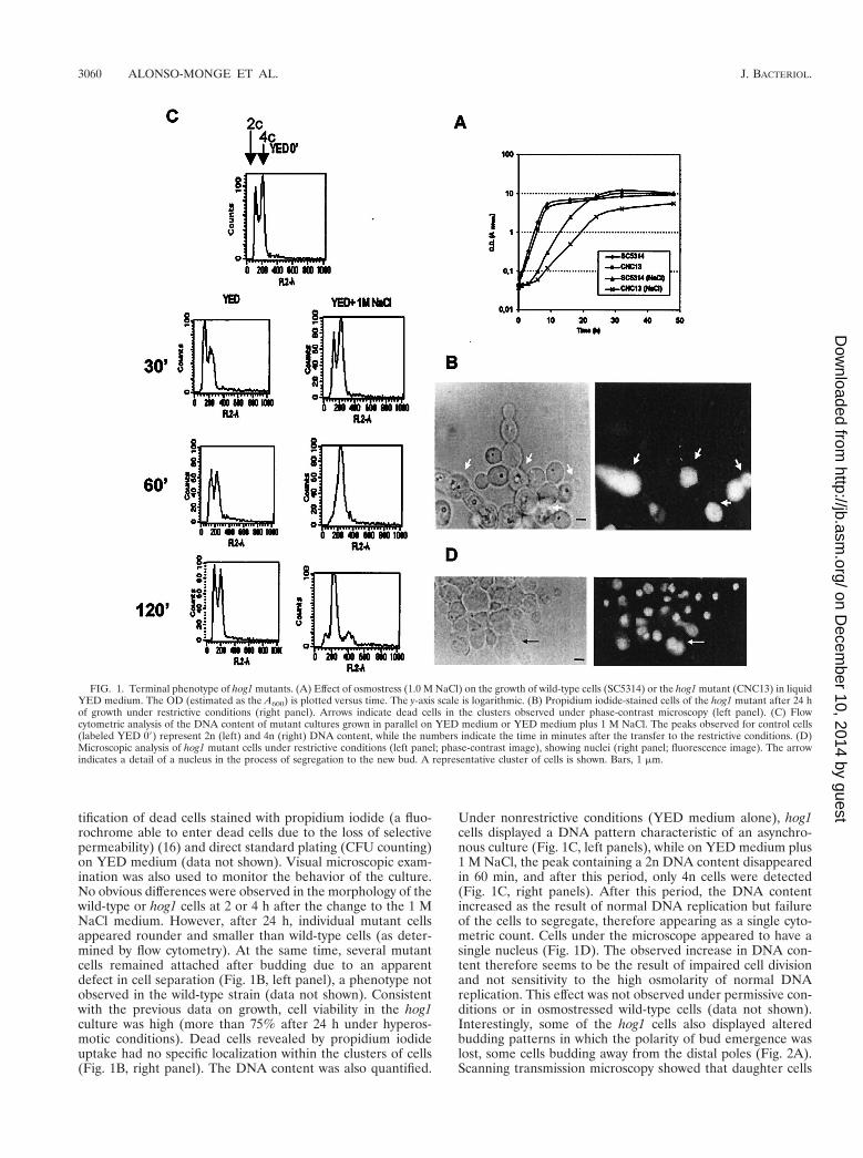
tification of dead cells stained with propidium iodide (a fluo-rochrome able to enter dead cells due to the loss of selectivepermeability) (16) and direct standard plating (CFU counting)on YED medium (data not shown). Visual microscopic exam-ination was also used to monitor the behavior of the culture.No obvious differences were observed in the morphology of thewild-type or hog1 cells at 2 or 4 h after the change to the 1 MNaCl medium. However, after 24 h, individual mutant cellsappeared rounder and smaller than wild-type cells (as deter-mined by flow cytometry). At the same time, several mutantcells remained attached after budding due to an apparentdefect in cell separation (Fig. 1B, left panel), a phenotype notobserved in the wild-type strain (data not shown). Consistentwith the previous data on growth, cell viability in the hog1culture was high (more than 75% after 24 h under hyperos-motic conditions). Dead cells revealed by propidium iodideuptake had no specific localization within the clusters of cells(Fig. 1B, right panel). The DNA content was also quantified.
Under nonrestrictive conditions (YED medium alone), hog1cells displayed a DNA pattern characteristic of an asynchro-nous culture (Fig. 1C, left panels), while on YED medium plus1 M NaCl, the peak containing a 2n DNA content disappearedin 60 min, and after this period, only 4n cells were detected(Fig. 1C, right panels). After this period, the DNA contentincreased as the result of normal DNA replication but failureof the cells to segregate, therefore appearing as a single cyto-metric count. Cells under the microscope appeared to have asingle nucleus (Fig. 1D). The observed increase in DNA con-tent therefore seems to be the result of impaired cell divisionand not sensitivity to the high osmolarity of normal DNAreplication. This effect was not observed under permissive con-ditions or in osmostressed wild-type cells (data not shown).Interestingly, some of the hog1 cells also displayed alteredbudding patterns in which the polarity of bud emergence waslost, some cells budding away from the distal poles (Fig. 2A).Scanning transmission microscopy showed that daughter cells
FIG. 1. Terminal phenotype of hog1 mutants. (A) Effect of osmostress (1.0 M NaCl) on the growth of wild-type cells (SC5314) or the hog1 mutant (CNC13) in liquidYED medium. The OD (estimated as the A600) is plotted versus time. The y-axis scale is logarithmic. (B) Propidium iodide-stained cells of the hog1 mutant after 24 hof growth under restrictive conditions (right panel). Arrows indicate dead cells in the clusters observed under phase-contrast microscopy (left panel). (C) Flowcytometric analysis of the DNA content of mutant cultures grown in parallel on YED medium or YED medium plus 1 M NaCl. The peaks observed for control cells(labeled YED 09) represent 2n (left) and 4n (right) DNA content, while the numbers indicate the time in minutes after the transfer to the restrictive conditions. (D)Microscopic analysis of hog1 mutant cells under restrictive conditions (left panel; phase-contrast image), showing nuclei (right panel; fluorescence image). The arrowindicates a detail of a nucleus in the process of segregation to the new bud. A representative cluster of cells is shown. Bars, 1 mm.
3060 ALONSO-MONGE ET AL. J. BACTERIOL.
on Decem
ber 10, 2014 by guesthttp://jb.asm
.org/D
ownloaded from
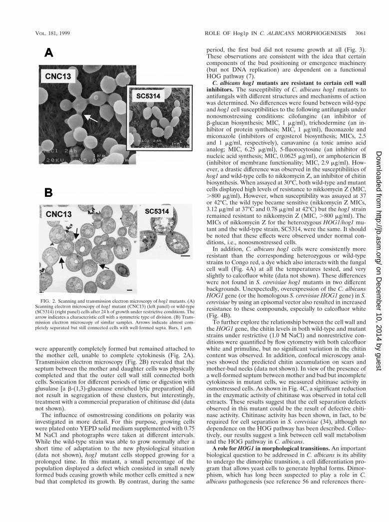
were apparently completely formed but remained attached tothe mother cell, unable to complete cytokinesis (Fig. 2A).Transmission electron microscopy (Fig. 2B) revealed that theseptum between the mother and daughter cells was physicallycompleted and that the outer cell wall still connected bothcells. Sonication for different periods of time or digestion withglusulase [a b-(1,3)-glucanase enriched lytic preparation] didnot result in segregation of these clusters, but interestingly,treatment with a commercial preparation of chitinase did (datanot shown).
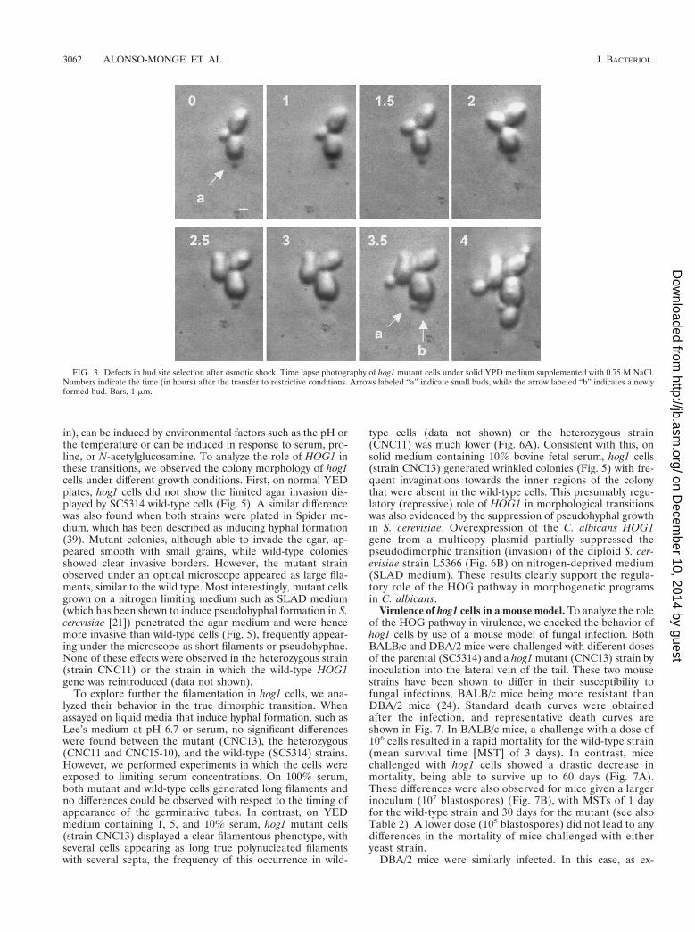
The influence of osmostressing conditions on polarity wasinvestigated in more detail. For this purpose, growing cellswere plated onto YEPD solid medium supplemented with 0.75M NaCl and photographs were taken at different intervals.While the wild-type strain was able to grow normally after ashort time of adaptation to the new physiological situation(data not shown), hog1 mutant cells stopped growing for aprolonged time. In this mutant, a small percentage of thepopulation displayed a defect which consisted in small newlyformed buds ceasing growth while mother cells emitted a newbud that completed its growth. By contrast, during the same
period, the first bud did not resume growth at all (Fig. 3).These observations are consistent with the idea that certaincomponents of the bud positioning or emergence machinery(but not DNA replication) are dependent on a functionalHOG pathway (7).
C. albicans hog1 mutants are resistant to certain cell wallinhibitors. The susceptibility of C. albicans hog1 mutants toantifungals with different structures and mechanisms of actionwas determined. No differences were found between wild-typeand hog1 cell susceptibilities to the following antifungals undernonosmostressing conditions: cilofungine (an inhibitor ofb-glucan biosynthesis; MIC, 1 mg/ml), trichodermine (an in-hibitor of protein synthesis; MIC, 1 mg/ml), fluconazole andmiconazole (inhibitors of ergosterol biosynthesis; MICs, 2.5and 1 mg/ml, respectively), canavanine (a toxic amino acidanalog; MIC, 6.25 mg/ml), 5-fluorocytosine (an inhibitor ofnucleic acid synthesis; MIC, 0.0625 mg/ml), or amphotericin B(inhibitor of membrane functionality; MIC, 2.9 mg/ml). How-ever, a drastic difference was observed in the susceptibilities ofhog1 and wild-type cells to nikkomycin Z, an inhibitor of chitinbiosynthesis. When assayed at 30°C, both wild-type and mutantcells displayed high levels of resistance to nikkomycin Z (MIC,.800 mg/ml). However, when susceptibility was assayed at 37or 42°C, the wild type became sensitive (nikkomycin Z MICs,3.12 mg/ml at 37°C and 0.78 mg/ml at 42°C) but the hog1 strainremained resistant to nikkomycin Z (MIC, .800 mg/ml). TheMICs of nikkomycin Z for the heterozygous HOG1/hog1 mu-tant and the wild-type strain, SC5314, were the same. It shouldbe noted that these effects were observed under normal con-ditions, i.e., nonosmostressed cells.
In addition, C. albicans hog1 cells were consistently moreresistant than the corresponding heterozygous or wild-typestrains to Congo red, a dye which also interacts with the fungalcell wall (Fig. 4A) at all the temperatures tested, and veryslightly to calcofluor white (data not shown). These differenceswere not found in S. cerevisiae hog1 mutants in two differentbackgrounds. Unexpectedly, overexpression of the C. albicansHOG1 gene (or the homologous S. cerevisiae HOG1 gene) in S.cerevisiae by using an episomal vector also resulted in increasedresistance to these compounds, especially to calcofluor white(Fig. 4B).
To further explore the relationship between the cell wall andthe HOG1 gene, the chitin levels in both wild-type and mutantstrains under restrictive (1.0 M NaCl) and nonrestrictive con-ditions were quantified by flow cytometry with both calcofluorwhite and primuline, but no significant variation in the chitincontent was observed. In addition, confocal microscopy anal-yses showed the predicted chitin accumulation on scars andmother-bud necks (data not shown). In view of the presence ofa well-formed septum between mother and bud but incompletecytokinesis in mutant cells, we measured chitinase activity inosmostressed cells. As shown in Fig. 4C, a significant reductionin the enzymatic activity of chitinase was observed in total cellextracts. These results suggest that the cell separation defectsobserved in this mutant could be the result of defective chiti-nase activity. Chitinase activity has been shown, in fact, to berequired for cell separation in S. cerevisiae (34), although nodependence on the HOG pathway has been described. Collec-tively, our results suggest a link between cell wall metabolismand the HOG pathway in C. albicans.
A role for HOG1 in morphological transitions. An importantbiological question to be addressed in C. albicans is its abilityto undergo the dimorphic transition, a cell differentiation pro-gram that allows yeast cells to generate hyphal forms. Dimor-phism, which has long been suspected to play a role in C.albicans pathogenesis (see reference 56 and references there-
FIG. 2. Scanning and transmission electron microscopy of hog1 mutants. (A)Scanning electron microscopy of hog1 mutant (CNC13) (left panel) or wild-type(SC5314) (right panel) cells after 24 h of growth under restrictive conditions. Thearrow indicates a characteristic cell with a symmetric type of division. (B) Trans-mission electron microscopy of similar samples. Arrows indicate almost com-pletely separated but still connected cells with well-formed septa. Bars, 1 mm.
VOL. 181, 1999 ROLE OF Hog1p IN C. ALBICANS MORPHOGENESIS 3061
on Decem
ber 10, 2014 by guesthttp://jb.asm
.org/D
ownloaded from
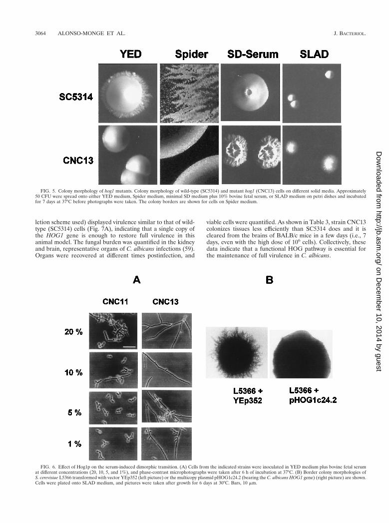
in), can be induced by environmental factors such as the pH orthe temperature or can be induced in response to serum, pro-line, or N-acetylglucosamine. To analyze the role of HOG1 inthese transitions, we observed the colony morphology of hog1cells under different growth conditions. First, on normal YEDplates, hog1 cells did not show the limited agar invasion dis-played by SC5314 wild-type cells (Fig. 5). A similar differencewas also found when both strains were plated in Spider me-dium, which has been described as inducing hyphal formation(39). Mutant colonies, although able to invade the agar, ap-peared smooth with small grains, while wild-type coloniesshowed clear invasive borders. However, the mutant strainobserved under an optical microscope appeared as large fila-ments, similar to the wild type. Most interestingly, mutant cellsgrown on a nitrogen limiting medium such as SLAD medium(which has been shown to induce pseudohyphal formation in S.cerevisiae [21]) penetrated the agar medium and were hencemore invasive than wild-type cells (Fig. 5), frequently appear-ing under the microscope as short filaments or pseudohyphae.None of these effects were observed in the heterozygous strain(strain CNC11) or the strain in which the wild-type HOG1gene was reintroduced (data not shown).
To explore further the filamentation in hog1 cells, we ana-lyzed their behavior in the true dimorphic transition. Whenassayed on liquid media that induce hyphal formation, such asLee’s medium at pH 6.7 or serum, no significant differenceswere found between the mutant (CNC13), the heterozygous(CNC11 and CNC15-10), and the wild-type (SC5314) strains.However, we performed experiments in which the cells wereexposed to limiting serum concentrations. On 100% serum,both mutant and wild-type cells generated long filaments andno differences could be observed with respect to the timing ofappearance of the germinative tubes. In contrast, on YEDmedium containing 1, 5, and 10% serum, hog1 mutant cells(strain CNC13) displayed a clear filamentous phenotype, withseveral cells appearing as long true polynucleated filamentswith several septa, the frequency of this occurrence in wild-
type cells (data not shown) or the heterozygous strain(CNC11) was much lower (Fig. 6A). Consistent with this, onsolid medium containing 10% bovine fetal serum, hog1 cells(strain CNC13) generated wrinkled colonies (Fig. 5) with fre-quent invaginations towards the inner regions of the colonythat were absent in the wild-type cells. This presumably regu-latory (repressive) role of HOG1 in morphological transitionswas also evidenced by the suppression of pseudohyphal growthin S. cerevisiae. Overexpression of the C. albicans HOG1gene from a multicopy plasmid partially suppressed thepseudodimorphic transition (invasion) of the diploid S. cer-evisiae strain L5366 (Fig. 6B) on nitrogen-deprived medium(SLAD medium). These results clearly support the regula-tory role of the HOG pathway in morphogenetic programsin C. albicans.
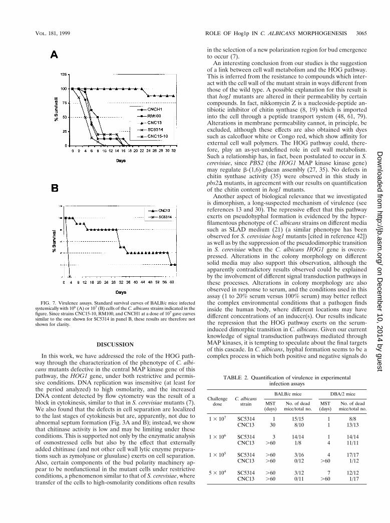
Virulence of hog1 cells in a mouse model. To analyze the roleof the HOG pathway in virulence, we checked the behavior ofhog1 cells by use of a mouse model of fungal infection. BothBALB/c and DBA/2 mice were challenged with different dosesof the parental (SC5314) and a hog1 mutant (CNC13) strain byinoculation into the lateral vein of the tail. These two mousestrains have been shown to differ in their susceptibility tofungal infections, BALB/c mice being more resistant thanDBA/2 mice (24). Standard death curves were obtainedafter the infection, and representative death curves areshown in Fig. 7. In BALB/c mice, a challenge with a dose of106 cells resulted in a rapid mortality for the wild-type strain(mean survival time [MST] of 3 days). In contrast, micechallenged with hog1 cells showed a drastic decrease inmortality, being able to survive up to 60 days (Fig. 7A).These differences were also observed for mice given a largerinoculum (107 blastospores) (Fig. 7B), with MSTs of 1 dayfor the wild-type strain and 30 days for the mutant (see alsoTable 2). A lower dose (105 blastospores) did not lead to anydifferences in the mortality of mice challenged with eitheryeast strain.
DBA/2 mice were similarly infected. In this case, as ex-
FIG. 3. Defects in bud site selection after osmotic shock. Time lapse photography of hog1 mutant cells under solid YPD medium supplemented with 0.75 M NaCl.Numbers indicate the time (in hours) after the transfer to restrictive conditions. Arrows labeled “a” indicate small buds, while the arrow labeled “b” indicates a newlyformed bud. Bars, 1 mm.
3062 ALONSO-MONGE ET AL. J. BACTERIOL.
on Decem
ber 10, 2014 by guesthttp://jb.asm
.org/D
ownloaded from

pected, differences at the 107 cell dose were not observed dueto the increased susceptibility of DBA/2 mice to C. albicansinfections, both strains producing a high mortality. However,differences in MST were observed for the mutant and wild typeat a challenge dose of 106 cells (Table 2), and the use of thismouse strain allowed us to detect differences between both C.albicans strains when a small inoculum dose (105) was used(Table 2). In all these experiments, ura3 auxotrophic strainswere avoided due to the effect that this nutritional requirement
(especially in certain genetic backgrounds) has on C. albicansvirulence (29, 69). However, since the hog1 knockout strainswere obtained in a his1 background (RM1000), it was con-firmed that HIS1 did not play a role in the observed reductionin virulence, as shown for the control strains RM100 andCNCH1 (Fig. 7A). In addition, the hog1 heterozygous strainCNC15-10 (a strain obtained through the reintroduction of theHOG1 gene in the genome of strain CNC15 [see Materials andMethods] to serve as an internal control of the knockout de-
FIG. 4. Antifungal susceptibility and cell wall architecture. (A) Different amounts of cells (indicated at the top of the rows) from the indicated C. albicans strainswere spotted onto YEPD medium (as a control) or YEPD medium supplemented with Congo red at 150 mg/ml and incubated at 37°C. (B) Experiments similar to thosedescribed for panel A were done with the S. cerevisiae strains indicated; cells were spotted onto YEPD medium supplemented with a 150-mg/ml final concentration ofcalcofluor white and incubated at 30°C for 24 h. (C) Chitinase activity in cell extracts after 24 and 48 h of growth under nonrestrictive (1 M NaCl) (left panel) andrestrictive (right panel) conditions. Units of activity (UA) (see Material and Methods) per milligram of dried extract are given in the y axis. Data are the mean valueof two independent experiments.
VOL. 181, 1999 ROLE OF Hog1p IN C. ALBICANS MORPHOGENESIS 3063
on Decem
ber 10, 2014 by guesthttp://jb.asm
.org/D
ownloaded from
letion scheme used) displayed virulence similar to that of wild-type (SC5314) cells (Fig. 7A), indicating that a single copy ofthe HOG1 gene is enough to restore full virulence in thisanimal model. The fungal burden was quantified in the kidneyand brain, representative organs of C. albicans infections (59).Organs were recovered at different times postinfection, and
viable cells were quantified. As shown in Table 3, strain CNC13colonizes tissues less efficiently than SC5314 does and it iscleared from the brains of BALB/c mice in a few days (i.e., 7days, even with the high dose of 106 cells). Collectively, thesedata indicate that a functional HOG pathway is essential forthe maintenance of full virulence in C. albicans.
FIG. 5. Colony morphology of hog1 mutants. Colony morphology of wild-type (SC5314) and mutant hog1 (CNC13) cells on different solid media. Approximately50 CFU were spread onto either YED medium, Spider medium, minimal SD medium plus 10% bovine fetal serum, or SLAD medium on petri dishes and incubatedfor 7 days at 37°C before photographs were taken. The colony borders are shown for cells on Spider medium.
FIG. 6. Effect of Hog1p on the serum-induced dimorphic transition. (A) Cells from the indicated strains were inoculated in YED medium plus bovine fetal serumat different concentrations (20, 10, 5, and 1%), and phase-contrast microphotographs were taken after 6 h of incubation at 37°C. (B) Border colony morphologies ofS. cerevisiae L5366 transformed with vector YEp352 (left picture) or the multicopy plasmid pHOG1c24.2 (bearing the C. albicans HOG1 gene) (right picture) are shown.Cells were plated onto SLAD medium, and pictures were taken after growth for 6 days at 30°C. Bars, 10 mm.
3064 ALONSO-MONGE ET AL. J. BACTERIOL.
on Decem
ber 10, 2014 by guesthttp://jb.asm
.org/D
ownloaded from
DISCUSSION
In this work, we have addressed the role of the HOG path-way through the characterization of the phenotype of C. albi-cans mutants defective in the central MAP kinase gene of thispathway, the HOG1 gene, under both restrictive and permis-sive conditions. DNA replication was insensitive (at least forthe period analyzed) to high osmolarity, and the increasedDNA content detected by flow cytometry was the result of ablock in cytokinesis, similar to that in S. cerevisiae mutants (7).We also found that the defects in cell separation are localizedto the last stages of cytokinesis but are, apparently, not due toabnormal septum formation (Fig. 3A and B); instead, we showthat chitinase activity is low and may be limiting under theseconditions. This is supported not only by the enzymatic analysisof osmostressed cells but also by the effect that externallyadded chitinase (and not other cell wall lytic enzyme prepara-tions such as zymolyase or glusulase) exerts on cell separation.Also, certain components of the bud polarity machinery ap-pear to be nonfunctional in the mutant cells under restrictiveconditions, a phenomenon similar to that of S. cerevisiae, wheretransfer of the cells to high-osmolarity conditions often results
in the selection of a new polarization region for bud emergenceto occur (7).
An interesting conclusion from our studies is the suggestionof a link between cell wall metabolism and the HOG pathway.This is inferred from the resistance to compounds which inter-act with the cell wall of the mutant strain in ways different fromthose of the wild type. A possible explanation for this result isthat hog1 mutants are altered in their permeability by certaincompounds. In fact, nikkomycin Z is a nucleoside-peptide an-tibiotic inhibitor of chitin synthase (8, 19) which is importedinto the cell through a peptide transport system (48, 61, 79).Alterations in membrane permeability cannot, in principle, beexcluded, although these effects are also obtained with dyessuch as calcofluor white or Congo red, which show affinity forexternal cell wall polymers. The HOG pathway could, there-fore, play an as-yet-undefined role in cell wall metabolism.Such a relationship has, in fact, been postulated to occur in S.cerevisiae, since PBS2 (the HOG1 MAP kinase kinase gene)may regulate b-(1,6)-glucan assembly (27, 35). No defects inchitin synthase activity (35) were observed in this study inpbs2D mutants, in agreement with our results on quantificationof the chitin content in hog1 mutants.
Another aspect of biological relevance that we investigatedis dimorphism, a long-suspected mechanism of virulence (seereferences 13 and 30). The repressive effect that this pathwayexerts on pseudohyphal formation is evidenced by the hyper-filamentous phenotype of C. albicans strains on different mediasuch as SLAD medium (21) (a similar phenotype has beenobserved for S. cerevisiae hog1 mutants [cited in reference 42])as well as by the suppression of the pseudodimorphic transitionin S. cerevisiae when the C. albicans HOG1 gene is overex-pressed. Alterations in the colony morphology on differentsolid media may also support this observation, although theapparently contradictory results observed could be explainedby the involvement of different signal transduction pathways inthese processes. Alterations in colony morphology are alsoobserved in response to serum, and the conditions used in thisassay (1 to 20% serum versus 100% serum) may better reflectthe complex environmental conditions that a pathogen findsinside the human body, where different locations may havedifferent concentrations of an inducer(s). Our results indicatethe repression that the HOG pathway exerts on the serum-induced dimorphic transition in C. albicans. Given our currentknowledge of signal transduction pathways mediated throughMAP kinases, it is tempting to speculate about the final targetsof this cascade. In C. albicans, hyphal formation seems to be acomplex process in which both positive and negative signals do
FIG. 7. Virulence assays. Standard survival curves of BALB/c mice infectedsystemically with 106 (A) or 107 (B) cells of the C. albicans strains indicated in thefigure. Since strains CNC15-10, RM100, and CNCH1 at a dose of 107 gave curvessimilar to the one shown for SC5314 in panel B, these results are therefore notshown for clarity.
TABLE 2. Quantification of virulence in experimentalinfection assays
Challengedose
C. albicansstrain
BALB/c mice DBA/2 mice
MST(days)
No. of deadmice/total no.
MST(days)
No. of deadmice/total no.
1 3 107 SC5314 1 15/15 1 8/8CNC13 30 8/10 1 13/13
1 3 106 SC5314 3 14/14 1 14/14CNC13 .60 1/8 4 11/11
1 3 105 SC5314 .60 3/16 4 17/17CNC13 .60 0/12 .60 1/12
5 3 104 SC5314 .60 3/12 7 12/12CNC13 .60 0/11 .60 1/17
VOL. 181, 1999 ROLE OF Hog1p IN C. ALBICANS MORPHOGENESIS 3065
on Decem
ber 10, 2014 by guesthttp://jb.asm
.org/D
ownloaded from

play a role (see reference 45 for an elegant model). For exam-ple, the HOG pathway could be involved in the repression ofthe pathway that leads to a pseudofilamentous or filamentousgrowth pathway in C. albicans, interacting with those elementsof the mating-hyphal pathway presumably involved in dimor-phic transition. In fact, in S. cerevisiae, elements of the matingpathway are used for pseudofilamentous growth (32, 36, 40),and it has been shown that the HOG pathway represses theactivity of the mating pathway in S. cerevisiae (22). Further-more, recent studies in S. cerevisiae (57) reveal that HOG1prevents cross talking between both the mating and HOGpathways. The repressive role of the HOG pathway in C. albi-cans hyphal formation could be its involvement in the activa-tion of RBF1, a transcription factor whose deletion generateshyphal forms (26), or, alternatively, a putative SSN6-TUP1complex in C. albicans. In fact, it has been recently shown thatthe S. cerevisiae Ssn6-Tup1 repressor complex (28) plays a rolein the repression of different osmolarity-inducible genes in S.cerevisiae (some of which are HOG1 dependent) and that ssn6or tup1 mutants partially suppress the characteristic osmoticsensitivity associated with hog1 mutants (47). More interest-ingly, the C. albicans TUP1 gene has been shown to play a rolein hyphal formation in C. albicans since deletion of this generesults in a gene dosage-dependent filamentous growth (5). Apossible explanation for our results would be the HOG1-de-pendent expression or activation of a DNA binding proteinable to recruit the Ssn6-Tup1 complex for the repression ofspecific hyphal genes. In any case, these similarities must beanalyzed carefully because of the pleiotropic role of transcrip-
tion factors like TUP1 in fungal cell physiology and the factthat phenotypes associated with S. cerevisiae and C. albicansmutants may clearly diverge, as occurs with tup1 mutants (5).
It is noteworthy that other elements of this pathway havebeen found to play a role in hyphal development. For example,deletion of the nik-11 gene, a Neurospora crassa homologue ofthe SLN1 gene (58), results, in addition to osmotic sensitivity,in restricted mycelial growth, the loss of conidiophore devel-opment, and in aberrant hyphal structures under restrictiveconditions (1). Recently, two-component C. albicans kinasegene homologues of SLN1 have been identified (10, 50, 74)and effects on the efficiency of the transition process have beenobserved (74).
Our results also demonstrate that the HOG pathway plays amajor role in C. albicans virulence. The genes involved invirulence currently identified are functionally diverse, probablyreflecting the character of a commensal opportunistic patho-gen instead of a primary pathogen of C. albicans. Dimorphismprovides a morphological switch that has been related to cer-tain features undoubtedly related to pathogenicity, such asadhesion, escape from phagocytic cells, and invasion (9, 30, 56,66). It is therefore not surprising that strains defective in hy-phal formation (under certain conditions) should display areduced virulence, as has been shown for some signal trans-duction protein kinases (15, 36, 37). However, although therole that hyphal formation must play in virulence is evident,our results clearly demonstrate that this trait is not enough forvirulence, since a functional (and even enhanced) in vitro hy-phal development does not necessarily correlate with virulence
TABLE 3. Virulence in a systemic mouse model of experimental infection in BALB/c and DBA/2 mice
Challengedose Strain Timea
Fungal burden (log CFU)b in:
BALB/c mice DBA/2 mice
Kidney Brain Kidney Brain
1 3 107 SC5314 1 6.03 6 0.84 4.47 6 0.13 NTc NT
CNC13 1 4.39 6 0.24 3.24 6 0.2 5.27 6 0.21 3.98 6 0.13
1 3 106 SC5314 1 5.93 6 0.18 4.04 6 0.22 6.02 6 0.92 3.81 6 0.16
CNC13 1 3.26 6 0.24 1.89 6 0.31* 4.43 6 0.09 3.16 6 0.137 3.94 6 1.13 *
60 5.11 6 0.98** *
1 3 105 SC5314 1 3.61 6 0.10 3.34 6 0.36 5.17 6 0.76 3.70 6 0.197 4.10 6 0.84 *
60 4.32 6 1.51* *
CNC13 1 2.53 6 0.21 1.78 6 0.00** 3.76 6 0.08 2.35 6 0.187 * * 3.60 6 0.98* 3.34 6 0.00**
60 4.98 6 0.00*** * 4.35 6 0.35** *
5 3 104 SC5314 7 6.45 6 0.56 4.53 6 0.5060 4.87 6 0.96* *
CNC13 1 * * 3.71 6 0.73 3.66 6 0.00**7 * * * *
60 * * 3.92 6 0.00** *
a Day postchallenge (up to MST) when mice were killed by cervical dislocation for CFU quantification.b Only log CFU values that are statistically significantly different at P 5 0.003 (kidney) and P , 0.0001 (brain) are shown (analysis of variance test with C. albicans
strain; covariates were time, mice, and dose). *, some or all mice cleared infection; **, more than 50% mice cleared infection.c NT, not tested.
3066 ALONSO-MONGE ET AL. J. BACTERIOL.
on Decem
ber 10, 2014 by guesthttp://jb.asm
.org/D
ownloaded from

in this animal model, as suggested recently (14). It should beemphasized that both the dose of cells used in the virulenceexperiments and the length of the period in which mice in-fected were followed up indicate the complete avirulence ofthe mutant strain, in comparison with the standard defined inother recent studies (14, 41), and suggest that the HOG path-way participates in other as-yet-unraveled cellular processeswhich are essential for virulence. Although it is difficult todefine what this role is at this stage, it is tempting to speculatethat Hog1p behaves like a general stress kinase, similar to theS. pombe homologue, and that this cellular response is essen-tial for the successful establishment of an infection in the host.In conclusion, we show in this work that the pathway(s) con-trolling osmotic sensitivity in C. albicans also plays a role indifferentiation programs and virulence in this pathogenic fun-gus, a result which identifies this route of primary importancein the search for novel antifungal targets.
ACKNOWLEDGMENTS
We thank Alistair J. P. Brown for generously providing strain L5366.Calcofluor was a generous gift from Bayer. The excellent assistance ofA. Vazquez and A. Alvarez from the Centro de Citometrıa de Flujo yMicroscopıa Confocal of the UCM and M. J. Asensio Vela is acknowl-edged. Electron microscopy was carried out at the Centro de Mi-croscopıa Electronica “Luis Bru” of the UCM. We also thank M.Molina for critical reading of the manuscript.
R. Alonso Monge is recipient of a fellowship from the ComunidadAutonoma de Madrid. This work was supported by FIS grant SAF96-1540 and by grant FISS97/0047-01.
REFERENCES1. Alex, L. A., K. A. Borkovich, and M. I. Simon. 1996. Hyphal development in
Neurospora crassa: involvement of a two-component histidine kinase. Proc.Natl. Acad. Sci. USA 93:3416–3421.
2. Ausubel, F. M., R. E. Kingston, R. Brent, et al. (ed.). 1993. Current protocolsin molecular biology. Wiley Interscience, New York, N.Y.
3. Banuett, F. 1998. Signalling in the yeasts: an informational cascade with linksto the filamentous fungi. Microbiol. Mol. Biol. Rev. 62:249–274.
4. Blomberg, A., and L. Adler. 1992. Physiology of osmotolerance in fungi. Adv.Microb. Physiol. 33:145–212.
5. Braun, B. R., and A. D. Johnson. 1997. Control of filament formation inCandida albicans by the transcriptional repressor TUP1. Science 277:105–109.
6. Brewster, J. L., T. de Valoir, N. D. Dwyer, E. Winter, and M. C. Gustin. 1993.An osmosensing signal transduction pathway in yeast. Science 259:1760–1763.
7. Brewster, J. L., and M. C. Gustin. 1994. Positioning of cell growth anddivision after osmotic stress requires a MAP kinase pathway. Yeast 10:425–439.
8. Cabib, E. 1991. Differential inhibition of chitin synthetases 1 and 2 fromSaccharomyces cerevisiae by polyoxin D and nikkomycins. Antimicrob.Agents Chemother. 35:170–173.
9. Calderone, R. A. 1993. Recognition between Candida albicans and host cells.Trends Microbiol. 1:55–58.
10. Calera, J. A., G. H. Choi, and R. A. Calderone. 1998. Identification of aputative histidine kinase two-component phosphorelay gene (CaHK1) inCandida albicans. Yeast 14:665–674.
11. Chowdhury, S., K. W. Smith, and M. C. Gustin. 1992. Osmotic stress and theyeast cytoskeleton: phenotype-specific suppression of an actin mutation.J. Cell Biol. 118:561–571.
12. Cid, V. J., A. Duran, F. del Rey, M. P. Snyder, C. Nombela, and M. Sanchez.1995. Molecular basis of cell integrity and morphogenesis in Saccharomycescerevisiae. Microbiol. Rev. 59:345–386.
13. Corner, B. E., and P. T. Magee. 1997. Candida pathogenesis: unravelling thethreads of infection. Curr. Biol. 7:R691–R694.
14. Csank, C., C. Makris, S. Meloche, K. Schroppel, M. Rollinghoff, D. Dignard,D. Y. Thomas, and M. Whiteway. 1997. Derepressed hyphal growth andreduced virulence in a VH1 family-related protein phosphatase mutant ofthe human pathogen Candida albicans. Mol. Biol. Cell 8:2539–2551.
15. Csank, C., K. Schroppel, E. Leberer, D. Harcus, O. Mohamed, S. Meloche,D. Y. Thomas, and M. Whiteway. 1998. Roles of the Candida albicansmitogen-activated protein kinase homolog, Cek1p, in hyphal developmentand systemic candidiasis. Infect. Immun. 66:2713–2721.
16. de la Fuente, J. M., A. Alvarez, C. Nombela, and M. Sanchez. 1992. Flowcytometric analysis of Saccharomyces cerevisiae autolytic mutants and proto-plasts. Yeast 8:39–45.
17. Dıez-Orejas, R., G. Molero, F. Navarro-Garcıa, J. Pla, C. Nombela, and M.Sanchez-Perez. 1997. Reduced virulence of Candida albicans MKC1 mu-tants: a role for a mitogen-activated protein kinase in pathogenesis. Infect.Immun. 65:833–837.
18. Fonzi, W. A., and M. Y. Irwin. 1993. Isogenic strain construction and genemapping in Candida albicans. Genetics 134:717–728.
19. Gaughran, J. P., M. H. Lai, D. R. Kirsch, and S. J. Silverman. 1994.Nikkomycin Z is a specific inhibitor of Saccharomyces cerevisiae chitin syn-thase isozyme Chs3 in vitro and in vivo. J. Bacteriol. 176:5857–5860.
20. Gillum, A. M., E. Y. H. Tsay, and D. R. Kirsch. 1984. Isolation of theCandida albicans gene for orotidine-59-phosphate decarboxylase by comple-mentation of S. cerevisiae ura3 and E. coli pyrF mutations. Mol. Gen. Genet.198:179–182.
21. Gimeno, C. J., P. O. Ljungdahl, C. A. Styles, and G. R. Fink. 1992. Unipolarcell divisions in the yeast S. cerevisiae lead to filamentous growth: regulationby starvation and RAS. Cell 68:1077–1090.
22. Hall, J. P., V. Cherkasova, E. A. Elion, M. C. Gustin, and E. Winter. 1996.The osmoregulatory pathway represses mating pathway activity in Saccha-romyces cerevisiae: isolation of a FUS3 mutant that is insensitive to therepression mechanism. Mol. Cell. Biol. 16:6715–6723.
23. Hanahan, D. 1988. Techniques for transformation of E. coli, p. 109–135. InD. M. Glover (ed.), DNA cloning. IRL Press, Oxford, United Kingdom.
24. Hector, R. F., J. E. Domer, and E. W. Carrow. 1982. Immune responses toCandida albicans in genetically distinct mice. Infect. Immun. 38:1020–1028.
25. Hill, J. E., A. M. Myers, T. J. Koerner, and A. Tzagoloff. 1986. Yeast/E. colishuttle vectors with multiple unique restriction sites. Yeast 2:163–167.
26. Ishii, N., M. Yamamoto, F. Yoshihara, M. Arisawa, and Y. Aoki. 1997.Biochemical and genetic characterization of Rbf1p, a putative transcriptionfactor of Candida albicans. Microbiology 143:429–435.
27. Jiang, B., A. F. J. Ram, J. Sheraton, F. M. Klis, and H. Bussey. 1995.Regulation of cell wall beta-glucan assembly: PTC1 negatively affects PBS2action in a pathway that includes modulation of EXG1 transcription. Mol.Gen. Genet. 248:260–269.
28. Keleher, C. A., M. J. Redd, J. Schultz, M. Carlson, and A. D. Johnson. 1992.Ssn6-Tup1 is a general repressor of transcription in yeast. Cell 68:709–719.
29. Kirsch, D. R., and R. R. Whitney. 1991. Pathogenicity of Candida albicansauxotrophic mutants in experimental infections. Infect. Immun. 59:3297–3300.
30. Kobayashi, G. S., and J. E. Cutler. 1998. Candida albicans hyphal formationand virulence: is there a clearly defined role? Trends Microbiol. 6:92–94.
31. Kohler, G. A., T. C. White, and N. Agabian. 1997. Overexpression of a clonedIMP dehydrogenase gene of Candida albicans confers resistance to thespecific inhibitor mycophenolic acid. J. Bacteriol. 179:2331–2338.
32. Kohler, J., and G. R. Fink. 1996. Candida albicans strains heterozygous andhomozygous for mutations in mitogen-activated protein kinase signalingcomponents have defects in hyphal development. Proc. Natl. Acad. Sci. USA93:13223–13228.
33. Kron, S. J., C. A. Styles, and G. R. Fink. 1994. Symmetric cell division inpseudohyphae of the yeast Saccharomyces cerevisiae. Mol. Biol. Cell 5:1003–1022.
34. Kuranda, M. J., and P. W. Robbins. 1991. Chitinase is required for cellseparation during growth of Saccharomyces cerevisiae. J. Biol. Chem. 266:19758–19767.
35. Lai, M. H., S. J. Silverman, J. P. Gaughran, and D. R. Kirsch. 1997. Multiplecopies of PBS2, MHP1 or LRE1 produce glucanase resistance and other cellwall effects in Saccharomyces cerevisiae. Yeast 13:199–213.
36. Leberer, E., D. Harcus, I. D. Broadbent, K. L. Clark, D. Dignard, K.Ziegelbauer, A. Schmidt, N. A. R. Gow, A. J. P. Brown, and D. Y. Thomas.1996. Signal transduction through homologs of the Ste20p and Ste7p proteinkinases can trigger hyphal formation in the pathogenic fungus Candidaalbicans. Proc. Natl. Acad. Sci. USA 93:13217–13222.
37. Leberer, E., K. Ziegelbauer, A. Schmidt, D. Harcus, D. Dignard, J. Ash, L.Johnson, and D. Y. Thomas. 1997. Virulence and hyphal formation of Can-dida albicans require the Ste20p-like protein kinase CaCla4p. Curr. Biol.7:539–546.
38. Lee, K. L., H. R. Buckley, and C. C. Campbell. 1975. An amino acid liquidsynthetic medium for the development of mycelial and yeast forms of Can-dida albicans. J. Med. Vet. Mycol. 13:148–153.
39. Liu, H., J. Kohler, and G. R. Fink. 1994. Suppression of hyphal formation inCandida albicans by mutation of a STE12 homolog. Science 266:1723–1726.
40. Liu, H., C. A. Styles, and G. R. Fink. 1993. Elements of the yeast pheromoneresponse pathway required for filamentous growth of diploids. Science 262:1741–1744.
41. Lo, H. J., J. R. Kohler, B. DiDomenico, D. Loebenberg, A. Cacciapuoti, andG. R. Fink. 1997. Nonfilamentous C. albicans mutants are avirulent. Cell90:939–949.
42. Madhani, H. D., C. A. Styles, and G. R. Fink. 1997. MAP kinases withdistinct inhibitory functions impart signaling specificity during yeast differ-entiation. Cell 91:673–684.
43. Maeda, T., M. Takekawa, and H. Saito. 1995. Activation of yeast PBS2MAPKK by MAPKKKs or by binding of an SH3-containing osmosensor.Science 269:554–558X.
VOL. 181, 1999 ROLE OF Hog1p IN C. ALBICANS MORPHOGENESIS 3067
on Decem
ber 10, 2014 by guesthttp://jb.asm
.org/D
ownloaded from
44. Maeda, T., S. M. Wurgler-Murphy, and H. Saito. 1994. A two-componentsystem that regulates an osmosensing MAP kinase cascade in yeast. Nature369:242–245.
45. Magee, P. T. 1998. Which came first: the hypha or the yeast? Science277:52–53.
46. Malathi, K., K. Ganesan, and A. Datta. 1994. Identification of a putativetranscription factor in Candida albicans that can complement the matingdefect of Saccharomyces cerevisiae ste12 mutants. J. Biol. Chem. 269:22945–22951.
47. Marquez, J. A., A. Pascual-Ahuir, M. Proft, and R. Serrano. 1998. TheSsn6-Tup1 repressor complex of Saccharomyces cerevisiae is involved in theosmotic induction of HOG-dependent and -independent genes. EMBO J.17:2543–2553.
48. McCarthy, P. J., P. F. Troke, and K. Gull. 1985. Mechanism of action ofnikkomycin and the peptide transport system of Candida albicans. J. Gen.Microbiol. 131:775–780.
49. Miret, J. J., A. J. Solari, P. A. Barderi, and S. H. Goldemberg. 1992. Poly-amines and cell wall organization in Saccharomyces cerevisiae. Yeast 8:1033–1041.
50. Nagahashi, S., T. Mio, N. Ono, T. Yamada-Okabe, M. Arisawa, H. Bussey,and H. Yamada-Okabe. 1998. Isolation of CaSLN1 and CaNIK1, the genesfor osmosensing histidine kinase homologues, from the pathogenic fungusCandida albicans. Microbiology 144:425–432.
51. National Committee for Clinical Laboratory Standards. 1992. Referencemethod for broth dilution antifungal susceptibility testing of yeast. Proposedstandard M27-P. National Committee for Clinical Laboratory Standards,Villanova, Pa.
52. Navarro-Garcıa, F., R. Alonso-Monge, H. Rico, J. Pla, R. Sentandreu, and C.Nombela. 1998. A role for the MAP kinase gene MKC1 in cell wall construc-tion and morphological transitions in Candida albicans. Microbiology 144:411–424.
53. Navarro-Garcıa, F., M. Sanchez, J. Pla, and C. Nombela. 1995. Functionalcharacterization of the MKC1 gene of Candida albicans, which encodes amitogen-activated protein kinase homolog related to cell integrity. Mol. Cell.Biol. 15:2197–2206.
54. Negredo, A., L. Monteoliva, C. Gil, J. Pla, and C. Nombela. 1997. Cloning,analysis and one-step disruption of the ARG5,6 gene of Candida albicans.Microbiology 143:297–302.
55. Odds, F. C. 1988. Candida and candidosis. Bailliere Tindall, London, UnitedKingdom.
56. Odds, F. C. 1994. Candida species and virulence. ASM News 60:313–318.57. O’Rourke, S. M., and I. Herskowitz. 1998. The Hog1 MAPK prevents cross
talk between the HOG and pheromone response MAPK pathways in Sac-charomyces cerevisiae. Genes Dev. 12:2874–2886.
58. Ota, I. M., and A. Varshavsky. 1993. A yeast protein similar to bacterialtwo-component regulators. Science 262:566–569.
59. Papadimitriou, J. M., and R. B. Ashman. 1986. The pathogenesis of acutesystemic candidiasis in a susceptible inbred mouse strain. J. Pathol. 150:257–265.
60. Paravicini, G., A. Mendoza, B. Antonsson, M. Cooper, C. Losberger, and M.Payton. 1996. The Candida albicans PKC1 gene encodes a protein kinase Chomolog necessary for cellular integrity but not dimorphism. Yeast 12:741–756.
61. Payne, J. W., and D. A. Shallow. 1985. Studies on drug targetting in thepathogenic fungus C. albicans: peptide transport mutants resistant to polyox-
ins, nikkomycins and bacilysin. FEMS Microbiol. Lett. 28:55–60.62. Pla, J., C. Gil, L. Monteoliva, F. Navarro-Garcıa, M. Sanchez, and C.
Nombela. 1996. Understanding Candida albicans at the molecular level.Yeast 12:1677–1702.
63. Pla, J., R. M. Perez-Dıaz, F. Navarro-Garcıa, M. Sanchez, and C. Nombela.1995. Cloning of the Candida albicans HIS1 gene by direct complementationof a C. albicans histidine auxotroph using an improved double-ARS shuttlevector. Gene 165:115–120.
64. Posas, F., and H. Saito. 1997. Osmotic activation of the HOG MAPKpathway via Ste11p MAPKKK: scaffold role of Pbs2p MAPKK. Science276:1702–1705.
65. Posas, F., S. M. Wurgler-Murphy, T. Maeda, E. A. Witten, T. C. Thai, andH. Saito. 1996. Yeast HOG1 MAP kinase cascade is regulated by a multistepphosphorelay mechanism in the SLN1-YPD1-SSK1 “two-component” osmo-sensor. Cell 86:865–875.
66. Ryley, J. F., and N. G. Ryley. 1990. Candida albicans—do mycelia matter?J. Med. Vet. Mycol. 28:225–239.
67. San Jose, C., R. Alonso, R. M. Perez-Dıaz, J. Pla, and C. Nombela. 1996. Themitogen-activated protein kinase homolog HOG1 gene controls glycerolaccumulation in the pathogenic fungus Candida albicans. J. Bacteriol. 178:5850–5852.
68. Scherer, S., and P. T. Magee. 1990. Genetics of Candida albicans. Microbiol.Rev. 54:226–241.
69. Shepherd, M. G. 1985. Pathogenicity of morphological and auxotrophicmutants of Candida albicans in experimental infections. Infect. Immun. 50:541–544.
70. Shiozaki, K., and P. Russell. 1995. Cell-cycle control linked to extracellularenvironment by MAP kinase pathway in fission yeast. Nature 378:739–743.
71. Shiozaki, K., and P. Russell. 1996. Conjugation, meiosis, and the osmoticstress response are regulated by Spc1 kinase through Atf1 transcriptionfactor in fission yeast. Genes Dev. 10:2276–2288.
72. Singh, P., K. Ganesan, K. Malathi, D. Ghosh, and A. Datta. 1994. ACPR, aSTE12 homologue from Candida albicans, is a strong inducer of pseudohy-phae in Saccharomyces cerevisiae haploids and diploids. Biochem. Biophys.Res. Commun. 205:1079–1085.
73. Singh, P., S. Ghosh, and A. Datta. 1997. A novel MAP-kinase kinase fromCandida albicans. Gene 190:99–104.
74. Srikantha, T., L. Tsai, K. Daniels, L. Enger, K. Highley, and D. R. Soll. 1998.The two-component hybrid kinase regulator caNIK1 of Candida albicans.Microbiology 144:2715–2729.
75. Tiedt, L. R., W. R. Jooste, and V. L. Hamilton-Attwell. 1987. Technique forpreserving aerial fungus structure for scanning electron microscopy. Trans.Br. Mycol. 88:420–422.
76. Tsang, T., V. Copeland, and G. T. Bowden. 1991. A set of cassette cloningvectors for rapid and versatile adaptation of restriction fragments. Biotech-niques 10:330.
77. Whiteway, M., D. Dignard, and D. Y. Thomas. 1992. Dominant negativeselection of heterologous genes: isolation of Candida albicans genes thatinterfere with Saccharomyces cerevisiae mating factor-induced cell cycle ar-rest. Proc. Natl. Acad. Sci. USA 89:9410–9414.
78. Williams, S., and C. Veldkamp. 1974. Preparation of fungi for scanningelectron microscopy. Trans. Br. Mycol. 63:409–412.
79. Yadan, J. C., M. Gonneau, P. Sarthou, and F. Le Goffic. 1984. Sensitivity tonikkomycin Z in Candida albicans: role of peptide permeases. J. Bacteriol.160:884–888.
3068 ALONSO-MONGE ET AL. J. BACTERIOL.
on Decem
ber 10, 2014 by guesthttp://jb.asm
.org/D
ownloaded from