LOXL2 induces aberrant acinar morphogenesis via ErbB2 signaling
18
RESEARCH ARTICLE Open Access LOXL2 induces aberrant acinar morphogenesis via ErbB2 signaling Joan Chang 1,2 , Monica M Nicolau 3 , Thomas R Cox 1,2 , Daniel Wetterskog 4 , John WM Martens 5 , Holly E Barker 1† and Janine T Erler 1,2*† Abstract Introduction: Lysyl oxidase-like 2 (LOXL2) is a matrix-remodeling enzyme that has been shown to play a key role in invasion and metastasis of breast carcinoma cells. However, very little is known about its role in normal tissue homeostasis. Here, we investigated the effects of LOXL2 expression in normal mammary epithelial cells to gain insight into how LOXL2 mediates cancer progression. Methods: LOXL2 was expressed in MCF10A normal human mammary epithelial cells. The 3D acinar morphogenesis of these cells was assessed, as well as the ability of the cells to form branching structures on extracellular matrix (ECM)-coated surfaces. Transwell-invasion assays were used to assess the invasive properties of the cells. Clinically relevant inhibitors of ErbB2, lapatinib and Herceptin (traztuzumab), were used to investigate the role of ErbB2 signaling in this model. A retrospective study on a previously published breast cancer patient dataset was carried out by using Disease Specific Genomic Analysis (DSGA) to investigate the correlation of LOXL2 mRNA expression level with metastasis and survival of ErbB2-positive breast cancer patients. Results: Fluorescence staining of the acini revealed increased proliferation, decreased apoptosis, and disrupted polarity, leading to abnormal lumen formation in response to LOXL2 expression in MCF10A cells. When plated onto ECM, the LOXL2-expressing cells formed branching structures and displayed increased invasion. We noted that LOXL2 induced ErbB2 activation through reactive oxygen species (ROS) production, and ErbB2 inhibition by using Herceptin or lapatinib abrogated the effects of LOXL2 on MCF10A cells. Finally, we found LOXL2 expression to be correlated with decreased overall survival and metastasis-free survival in breast cancer patients with ErbB2- positive tumors. Conclusions: These findings suggest that LOXL2 expression in normal epithelial cells can induce abnormal changes that resemble oncogenic transformation and cancer progression, and that these effects are driven by LOXL2-mediated activation of ErbB2. LOXL2 may also be a beneficial marker for breast cancer patients that could benefit most from anti-ErbB2 therapy. Introduction Lysyl oxidase-like 2 (LOXL2) is one of five members of the lysyl oxidase (LOX) family of extracellular matrix proteins and mediates the cross-linking of stromal col- lagens and elastin [1-3]. We previously showed that LOXL2 expression is clinically correlated with increased metastasis and poor survival in breast cancer patients with estrogen receptor (ER)-negative tumors [4]. Consistent with this, other studies found LOXL2 protein levels to be higher in poorly differentiated breast carci- nomas, and elevated LOXL2 mRNA was observed in invasive and metastatic breast cancer cell lines [5,6]. We showed that LOXL2 plays a critical role in breast cancer progression, and further demonstrated that genetic, che- mical, or antibody inhibition of LOXL2 significantly reduced the size and number of metastases in the lungs, liver, and bone through blocking the effects of extracel- lular LOXL2 on matrix remodeling and cell invasion [4]. In accordance with our findings, Barry-Hamilton et al. [7] demonstrated efficacy of an LOXL2-targeting anti- body in reduction of bone and soft tissue metastases * Correspondence: [email protected] † Contributed equally 1 Hypoxia and Metastasis Team, Division of Cancer Biology, The Institute of Cancer Research, 237 Fulham Road, London, UK SW3 6JB Full list of author information is available at the end of the article Chang et al. Breast Cancer Research 2013, 15:R67 http://breast-cancer-research.com/content/15/4/R67 © 2013 Chang et al.; licensee BioMed Central Ltd. This is an open access article distributed under the terms of the Creative Commons Attribution License (http://creativecommons.org/licenses/by/2.0), which permits unrestricted use, distribution, and reproduction in any medium, provided the original work is properly cited.
-
Upload
independent -
Category
Documents
-
view
0 -
download
0
Transcript of LOXL2 induces aberrant acinar morphogenesis via ErbB2 signaling

RESEARCH ARTICLE Open Access
LOXL2 induces aberrant acinar morphogenesisvia ErbB2 signalingJoan Chang1,2, Monica M Nicolau3, Thomas R Cox1,2, Daniel Wetterskog4, John WM Martens5, Holly E Barker1† andJanine T Erler1,2*†
Abstract
Introduction: Lysyl oxidase-like 2 (LOXL2) is a matrix-remodeling enzyme that has been shown to play a key rolein invasion and metastasis of breast carcinoma cells. However, very little is known about its role in normal tissuehomeostasis. Here, we investigated the effects of LOXL2 expression in normal mammary epithelial cells to gaininsight into how LOXL2 mediates cancer progression.
Methods: LOXL2 was expressed in MCF10A normal human mammary epithelial cells. The 3D acinarmorphogenesis of these cells was assessed, as well as the ability of the cells to form branching structures onextracellular matrix (ECM)-coated surfaces. Transwell-invasion assays were used to assess the invasive properties ofthe cells. Clinically relevant inhibitors of ErbB2, lapatinib and Herceptin (traztuzumab), were used to investigate therole of ErbB2 signaling in this model. A retrospective study on a previously published breast cancer patient datasetwas carried out by using Disease Specific Genomic Analysis (DSGA) to investigate the correlation of LOXL2 mRNAexpression level with metastasis and survival of ErbB2-positive breast cancer patients.
Results: Fluorescence staining of the acini revealed increased proliferation, decreased apoptosis, and disruptedpolarity, leading to abnormal lumen formation in response to LOXL2 expression in MCF10A cells. When platedonto ECM, the LOXL2-expressing cells formed branching structures and displayed increased invasion. We notedthat LOXL2 induced ErbB2 activation through reactive oxygen species (ROS) production, and ErbB2 inhibition byusing Herceptin or lapatinib abrogated the effects of LOXL2 on MCF10A cells. Finally, we found LOXL2 expressionto be correlated with decreased overall survival and metastasis-free survival in breast cancer patients with ErbB2-positive tumors.
Conclusions: These findings suggest that LOXL2 expression in normal epithelial cells can induce abnormalchanges that resemble oncogenic transformation and cancer progression, and that these effects are driven byLOXL2-mediated activation of ErbB2. LOXL2 may also be a beneficial marker for breast cancer patients that couldbenefit most from anti-ErbB2 therapy.
IntroductionLysyl oxidase-like 2 (LOXL2) is one of five members ofthe lysyl oxidase (LOX) family of extracellular matrixproteins and mediates the cross-linking of stromal col-lagens and elastin [1-3]. We previously showed thatLOXL2 expression is clinically correlated with increasedmetastasis and poor survival in breast cancer patientswith estrogen receptor (ER)-negative tumors [4].
Consistent with this, other studies found LOXL2 proteinlevels to be higher in poorly differentiated breast carci-nomas, and elevated LOXL2 mRNA was observed ininvasive and metastatic breast cancer cell lines [5,6]. Weshowed that LOXL2 plays a critical role in breast cancerprogression, and further demonstrated that genetic, che-mical, or antibody inhibition of LOXL2 significantlyreduced the size and number of metastases in the lungs,liver, and bone through blocking the effects of extracel-lular LOXL2 on matrix remodeling and cell invasion [4].In accordance with our findings, Barry-Hamilton et al.[7] demonstrated efficacy of an LOXL2-targeting anti-body in reduction of bone and soft tissue metastases
* Correspondence: [email protected]† Contributed equally1Hypoxia and Metastasis Team, Division of Cancer Biology, The Institute ofCancer Research, 237 Fulham Road, London, UK SW3 6JBFull list of author information is available at the end of the article
Chang et al. Breast Cancer Research 2013, 15:R67http://breast-cancer-research.com/content/15/4/R67
© 2013 Chang et al.; licensee BioMed Central Ltd. This is an open access article distributed under the terms of the Creative CommonsAttribution License (http://creativecommons.org/licenses/by/2.0), which permits unrestricted use, distribution, and reproduction inany medium, provided the original work is properly cited.

after intracardiac injection of human breast cancer cells[7]. This antibody is now in phase II clinical trials.Upregulation of LOXL2 is also associated with poorprognosis in patients with squamous cell, colon, andesophageal cancers [8,9]. Furthermore, LOXL2 is linkedto drug resistance in pancreatic cancer cells [10] andpromotes gastric cancer metastasis [11]. Thus, LOXL2 islikely to be an excellent drug target in many cancertypes [12].Despite the mounting evidence suggesting a critical
role for LOXL2 in metastasis, very little is known aboutits function during development or its role in normaltissue homeostasis. LOXL2 mRNA was detected at lowlevels in heart, lung, and kidney, and at high levels inthe prostate, uterus, and placenta [13]. However, LOXL2protein levels were very low in all normal adult tissues[7]. As a result, no deleterious side effects have beenobserved in response to LOXL2 inhibition [4,7].In the adult mammary gland, epithelial cells are orga-
nized into ducts and lobules. The lobules comprise mul-tiple mammary acini, and each lobule has a mammaryduct connecting the lobules to the nipple. Normal acinihave lumens formed by a single layer of polarized lumi-nal epithelial cells, surrounded by myoepithelial cells,and finally, the basement membrane. Epithelial cellsgrown on plastic do not accurately reflect the in vivomicroenvironment of the mammary gland, highlightingthe importance of studying tumorigenesis by using athree-dimensional (3D) model mimicking normal mam-mary epithelial development.Studying LOXL2 function in normal cells can provide
information about its role in cancer; thus, here we usedthe well-established MCF10A acini assay [14] as a 3Dmodel to investigate the role of LOXL2 in mammaryepithelial acini formation and to provide novel insightsinto the molecular events regulated by LOXL2.We showed that overexpression of LOXL2 induces
invasive branching structures on cells plated onto extra-cellular matrix (ECM) and increases invasiveness of thecells. LOXL2 induction also causes aberrant acini devel-opment by interfering with apoptosis, proliferation, andpolarization, and these LOXL2-mediated effects are atte-nuated when ErbB2 signaling is inhibited. In this article,we are the first to demonstrate that LOXL2 inducesphosphorylation of ErbB2 through the production ofreactive oxygen species (ROS). Moreover, we showedthat LOXL2 expression correlates with metastasis andpoor survival in ErbB2-positive breast cancer patients.
Materials and methodsCell lines and cell cultureAll cell lines used in this study were obtained fromAmerican Type Culture Collection. Human MCF10Anormal mammary epithelial cells were grown in F12/
DMEM (Invitrogen) media supplemented with 5% horseserum (HS, Invitrogen), 0.5 μg/ml hydrocortisone(Sigma), 10 μg/ml insulin (Sigma), 100 ng/ml choleratoxin (Sigma), and 20 ng/ml EGF (Peprotech). MCF10Acell lines used were cultured for no more than 20 pas-sages. Human MDA-MB-361 breast cancer cells weregrown in F12/DMEM (Invitrogen) media supplementedwith 20% fetal calf serum (Invitrogen). Human MDA-MB-231 breast cancer cells were grown in DMEM (Invi-trogen) media supplemented with 10% fetal calf serum(Invitrogen).
Generation of cell linesFor overexpression of the LOXL2 protein, the humanLOXL2 gene was amplified by polymerase chain reaction(PCR) and cloned into the pBABE retroviral vector byusing the following primers: 5’-CGC GGA TCC ATGGAG AGG CCT CTG TGC TCC CAC-3’, 3’-CGC GTCGAC TTA CTG CGG GGA CAG CTG GTT GTTTAA GAG C-5’. MCF10A cell line was infected withretroviruses expressing the pBABE vector containingLOXL2 (10A L2), or parental pBABE retroviral vector asa control (10A cont).
Quantitative real-time PCRTotal RNA was isolated from cell pellets by usingRNeasy Mini Kits (Qiagen). RNase treatment and cDNAsynthesis were done by using RNaseOUT and M-MLVSuperscript (Invitrogen), according to manufacturer’sinstructions. b-actin was used as an internal control.The following primers were used for the analysis ofLOXL2, b-actin, E-cadherin, Vimentin, SNAI1, SNAI2,a-SMA, TWIST, and N-cadherin: LOXL2 forward,5’-CTG CCA CAT AGG TGG TTC T-3’; LOXL2reverse, 5’-TGG CAT TCG TTC AGA CTC AG-3’. b-actin forward, 5’-GAG GCC CAG AGC AAG AGAGG-3’. b-actin reverse, 5’-TAC ATG GCT GGG GTGTTG AA-3’. E-cadherin forward, 5’-TGG AGG AATTCT TGC TTT GC-3’; E-cadherin reverse, 5’-CGCTCT CCT CCG AAG AAA C-3’. Vimentin forward,5’-CTT CGC CAA CTA CAT CGA CA-3’; Vimentinreverse, 5’-CGG CCA GCA GGA TCT TAT T-3’.SNAI1 forward, 5’-AGG ATC TCC AGG CTC GAAAG-3’; SNAI1 reverse, 5’-TCG GAT GTG CAT CTTGAG G-3’. SNAI2 forward, 5’-TCC GAA GCC AAATGA CAA AT-3’; SNAI2 reverse, 5’-TGT GTG TGCATA TGT GTG TGT G-3’. a-SMA forward, -5’-CCCTGA AGT ACC CGA TAG AAC A-3’; a-SMA reverse,-5’-GGC AAC ACG AAG CTC ATT G-3’. TWIST for-ward, 5’-GGG CCG GAG ACC TAG ATT-3’; TWISTreverse, 5’-TTC TGA ATT GTA TCA CAC CTT CTCA-3’. N-cadherin forward, 5’-GTG CTC AGG CTGTGG ACA TA-3’; N-cadherin reverse, 5’-CTG CACTTT GAT GAT GAA TTC TG-3’.
Chang et al. Breast Cancer Research 2013, 15:R67http://breast-cancer-research.com/content/15/4/R67
Page 2 of 18

The following reaction cycle was used: 95°C for 10minutes, followed by 40 cycles of 95°C for 15 seconds,60°C for 30 seconds, 72°C for 30 seconds, and a finalstep of 72°C for 10 minutes. The PCR was performed byusing an Applied Biosystems 7900HT Fast Real-TimePCR system (Applied Biosystems, Carlsbad, CA, USA),or Roche LightCycler480II (Roche Applied Science).
2D and 3D proliferation assaysThe 2D proliferation on plastic and 3D proliferation inMatrigel of the cells were assessed by using the CellTi-ter 96® AQueous One Solution Cell Proliferation Assay(Promega), according to the manufacturer’s instructions.Cells were plated in triplicate in 96-well plates at 2,000cells per well, either in 100 μl of media or in 50 μl ofMatrigel (BD Biosciences). All readings were normalizedto day 0 readings.
Western blottingAll cells were lysed in 1% NP40 buffer supplementedwith 10 μg/ml aprotinin (Sigma) and 10 μg/ml leupeptin(Sigma).Conditioned medium (CM) was obtained as previously
described [15]. For lysate preparation, 350,000 cells wereplated on Matrigel-coated six-well plates. After over-night incubation at 37°C, 5% CO2/20% O2, the cellswere serum-starved for 3 hours and then serum-blastedfor 15 minutes before collection for maximal signalingactivation.For drug treatments, cells were plated out as described
earlier, and 3 hours after plating, the cells were exposedto lapatinib or Herceptin at the indicated concentra-tions. Equivalent amounts of DMSO or human kappaIgG (Sigma) were added to the cells as a control forlapatinib and Herceptin, respectively. After overnightincubation with the drugs, the cells were serum-starvedfor 3 hours and then serum-blasted for 15 minutes inpresence of drug before collection.For catalase treatment with or without recombinant
human LOXL2 (rhLOXL2), the cells were plated asdescribed earlier. After overnight incubation in theabsence of treatment, the cells were serum-starved for 3hours and then subjected the following combination oftreatments for 15 minutes before collection: 200 U/mlcatalase (Sigma), 50 nM recombinant human LOXL2(rhLOXL2, R&D Systems), 200 U/ml catalase, and 50nM rhLOXL2.For rhLOXL2 treatment only, 700,000 cells were pla-
ted on polyacrylamide-crosslinked 50-mm-diameter cov-erslips that were coated with Matrigel by using apreviously published protocol [16]. The 50 nMrhLOXL2 was added onto one set of 10A control cells.After overnight incubation at 37°C, 5% CO2/20% O2, the
cells were serum-starved for 3 hours and then serum-blasted for 15 minutes before collection.Proteins were separated with NuPAGE Novex Bis-Tris
10% gels or 7% Tris-Acetate gels (Invitrogen), trans-ferred to polyvinylidene difluoride membranes (Milli-pore), and immunoblotted with antibodies specific forhuman ErbB2, pErbB2, EGFR, pEGFR, Erk1/2 (MAPK),pErk1/2 (pMAPK), Akt, and pAkt (all Cell Signaling), orhuman LOXL2 (Abcam). Hsc70 or b-actin (Abcam) wasused as a loading control.Densitometry analysis was carried out by using
ImageJ64, and intensities of bands were corrected tocontrol lanes for each individual blot. All analyses werecarried out on at least three individual repeats of theexperiment.
Acini assay10A cont and 10A L2 cells were plated on top of a thinlayer of 50-μl Matrigel in eight-well chamber slides, at adensity of 10,000 cells/ml in 400 μl of 2% Growth FactorReduced Matrigel without phenol red (BD Biosciences) inDMEM/F-12 media supplemented with 2% HS, 0.5 μg/mlhydrocortisone, 10 μg/ml insulin, 100 ng/ml choleratoxin, and 20 ng/ml EGF (Gibco/Invitrogen). The cul-tures were allowed to grow for 8, 10, and 13 days, andthen fixed with 2% paraformaldehyde (PFA, Invitrogen)in PBS at room temperature.For drug-treated cultures, lapatinib dissolved in 0.1%
DMSO or Herceptin dissolved in PBS was added to cellsat day 6 (10A cont+lap, 10A L2+lap; 10A cont+her, 10AL2+her), and 0.1% DMSO or an equivalent amount ofhuman IgG was added as a control to lapatinib and Her-ceptin, respectively (10A cont+DMSO, 10A L2+DMSO;10A cont+her, 10A L2+her). All assay media, with orwithout treatments, were changed every 3 to 4 days.Acini were permeabilized with 0.5% Triton X-100
(Invitrogen), washed thoroughly with glycine buffer(0.1 M NaCl, 0.1 M glycine in PBS), blocked in IF buffer(0.1% bovine serum albumin (BSA), 0.2% Triton X-100,0.05% Tween20, 130 mM NaCl in PBS) containing 10%goat serum (Invitrogen) and incubated with primary anti-bodies diluted in blocking buffer. Samples were thenwashed with IF buffer and incubated with appropriateAlexa-conjugated secondary antibodies (Invitrogen) in10% goat serum/IF buffer. Nuclei were counterstainedwith DAPI (Invitrogen). Slides were then mounted undercoverslips with Fluorescent Mounting Medium (Invitro-gen) and visualized with confocal microscopy (ZeissLSM710) by using a 63 × oil objective. Primary antibo-dies used were Ki-67, activated caspase-3, GM130,E-cadherin, and laminin V (all from Abcam). All photostaken were of midsections of acini. Photos presented hereare representative of the overall phenotype in each
Chang et al. Breast Cancer Research 2013, 15:R67http://breast-cancer-research.com/content/15/4/R67
Page 3 of 18

experimental condition; quantifications were carried outon at least 20 acini per experimental repeat.
Transwell invasion assayMatrigel transwell invasion assays were carried out byusing BD Bio-coat Matrigel Invasion Chambers (BDBiosciences), according to the manufacturer’s instruc-tions. Cells were serum-starved overnight before plating,and the normal culture media were added to the bottomchambers as chemoattractant. Plates were incubated atnormal culture conditions for 24 hours before noninvad-ing cells were removed from the inside of inserts, andmembranes fixed and stained. Total number of invadedcells was counted under a light microscope (Nikon Dia-phot 300).
Branching assayThe 350,000 cells were plated on Matrigel-coated six-well plates. For treated cells, lapatinib (DMSO as con-trol) or Herceptin (human kappa IgG as control) wereadded to cell cultures 3 hours after plating. Cells werethen incubated overnight and viewed under a micro-scope (Leica DM1L).
Soft agar assayTo test anchorage-independent growth in soft agar,3 × 104 cells were suspended in 2 ml of 0.4% low-melting-point agarose (Invitrogen) in culture medium and wereoverlaid over 2 ml of 0.8% low-melting-point agarose inculture medium in a six-well plate. Culture medium wasadded to the top once the agarose had solidified, andplates were incubated at 37°C, 5% CO2/21% O2. After 3weeks in culture, colonies were stained with 0.1% crystalviolet and destained with PBS. Colonies with more than20 cells were scored with a light microscope (NikonDiaphot 300).
Activity assayActivity assays using a fluorescence-based LOX familyenzymatic-activity assay measuring H2O2 (Abcam,Ab112139) were carried out according to manufacturer’sinstructions except using Matrigel as the substrate.Then 5 μl of concentrated conditioned media was addedto the reaction mixture on a thin layer of Matrigel in a96-well dish.
Patient data analysisGene-expression microarray data from a previously pub-lished NKI cohort [17] was first transformed by usingDisease Specific Genomic Analysis (DSGA) [18] to iden-tify the extent of deviation from a Healthy State Model(HSM), defined by 13 normal breast samples [18]. Thisprovided gene-expression levels in tumors that are rela-tive to the control normal tissue data. Of 295 tumors,
51 were found to express high levels of ErbB2, and aKaplan-Meier survival curve is constructed from thissubset of ErbB2-positive tumors. Shown here is theoverall and metastasis-free survival of patients withHer2/ErbB2-positive tumors (n = 51) separated into lowand high LOXL2 expression.Validation was carried out by combining another two
previously published NKI cohorts [19,20]. Samples wererun on Affymetrix U133a chips according to manufac-turer’s instructions, and data were obtained by usingMAS5 parameters with global scaling set to 600. Of 344tumors, 88 were found to express high levels of ErbB2,and Kaplan-Meier survival curves were constructedfrom this subset of ErbB2-positive tumors. Shown hereis the overall and metastasis-free survival of patientswith Her2/ErbB2-positive tumors (n = 88) separatedinto low and high LOXL2 expression.
Statistical analysisData were analysed by using the Student t test, and con-sidered significant at P ≤ 0.05. All statistical tests weretwo sided. Bar graphs represent the mean and standarderror across independent experimental repeats, and allexperiments were performed at least three independenttimes. Statistical significance representations: *P < 0.05;**P < 0.01; ***P < 0.001; and ****P < 0.0001.
Results and DiscussionExpression of LOXL2 in human MCF10A normal mammaryepithelial cellsWe first investigated the potential impact of LOXL2expression in normal breast epithelial morphogenesis.The MCF10A cell line was derived from normal humanmammary epithelium that had undergone spontaneousimmortalization without transformation, and is nottumorigenic [21]. With this normal epithelial cell line,we investigated how LOXL2 promotes tumor progres-sion. We infected MCF10A cells with pBABE retroviralvector containing a LOXL2 cDNA to generate LOXL2-expressing cells (10A L2) and generated control cells(10A cont) by infecting wild-type MCF10A cells withthe parental pBABE retroviral vector. MCF10A cells areknown to show genetic drift [22]; however, we main-tained cells at a passage lower than 20, as we notedaltered cell phenotype above passage 30, consistent witha previous report [22].Western blot analysis showed that secreted LOXL2 was
upregulated in 10A L2 cells relative to control cells, andcomparable with those in the human breast cancer cell lineMDA-MB-231 (Figure 1A). Two bands of approximately95 kDa and 65 kDa were observed, the 95-kDa band repre-senting the long form of LOXL2 that is commonlyreported in cancer cells, and the fainter 65-kDa band thatmay correspond to the proteolytically modified short form
Chang et al. Breast Cancer Research 2013, 15:R67http://breast-cancer-research.com/content/15/4/R67
Page 4 of 18
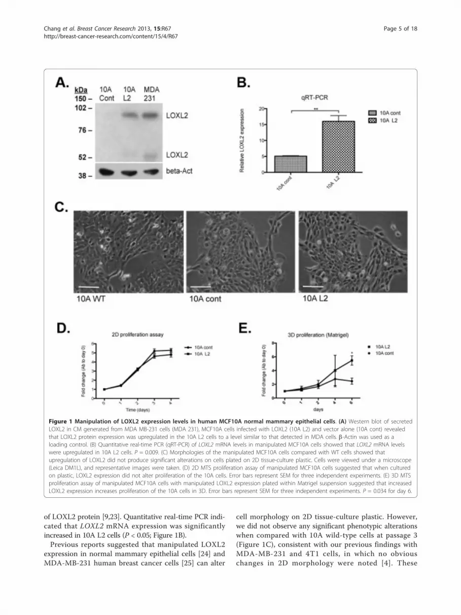
of LOXL2 protein [9,23]. Quantitative real-time PCR indi-cated that LOXL2 mRNA expression was significantlyincreased in 10A L2 cells (P < 0.05; Figure 1B).Previous reports suggested that manipulated LOXL2
expression in normal mammary epithelial cells [24] andMDA-MB-231 human breast cancer cells [25] can alter
cell morphology on 2D tissue-culture plastic. However,we did not observe any significant phenotypic alterationswhen compared with 10A wild-type cells at passage 3(Figure 1C), consistent with our previous findings withMDA-MB-231 and 4T1 cells, in which no obviouschanges in 2D morphology were noted [4]. These
Figure 1 Manipulation of LOXL2 expression levels in human MCF10A normal mammary epithelial cells. (A) Western blot of secretedLOXL2 in CM generated from MDA MB-231 cells (MDA 231), MCF10A cells infected with LOXL2 (10A L2) and vector alone (10A cont) revealedthat LOXL2 protein expression was upregulated in the 10A L2 cells to a level similar to that detected in MDA cells. b-Actin was used as aloading control. (B) Quantitative real-time PCR (qRT-PCR) of LOXL2 mRNA levels in manipulated MCF10A cells showed that LOXL2 mRNA levelswere upregulated in 10A L2 cells. P = 0.009. (C) Morphologies of the manipulated MCF10A cells compared with WT cells showed thatupregulation of LOXL2 did not produce significant alterations on cells plated on 2D tissue-culture plastic. Cells were viewed under a microscope(Leica DM1L), and representative images were taken. (D) 2D MTS proliferation assay of manipulated MCF10A cells suggested that when culturedon plastic, LOXL2 expression did not alter proliferation of the 10A cells. Error bars represent SEM for three independent experiments. (E) 3D MTSproliferation assay of manipulated MCF10A cells with manipulated LOXL2 expression plated within Matrigel suspension suggested that increasedLOXL2 expression increases proliferation of the 10A cells in 3D. Error bars represent SEM for three independent experiments. P = 0.034 for day 6.
Chang et al. Breast Cancer Research 2013, 15:R67http://breast-cancer-research.com/content/15/4/R67
Page 5 of 18

findings suggest variable effects of LOXL2 on 2D mor-phology that may be cell-line specific or sensitive tocell-culture methods.To assess the effects of LOXL2 on 2D proliferation,
we carried out MTS assays. Increased secretion ofLOXL2 did not affect 2D cell proliferation on plastic(Figure 1D). In contrast, we observed a significantincrease in proliferation of 10A L2 cells compared with10A cont cells after 6 days of culture in Matrigel sus-pension (P < 0.05; Figure 1E). Taken together, theseresults suggest that the microenvironment is importantfor the effects of LOXL2.
LOXL2 expression disrupts normal breast epithelial aciniformationThe role of LOXL2 in 3D development of normal breastepithelial cells was assessed by using the well-character-ized MCF10A acini assay [14], in which cells are cul-tured on top of a layer of recombinant basementmembrane (Matrigel) within a Matrigel suspension. The10A cont and 10A L2 cells were compared for theirability to form acini structures in Matrigel over a 13-dayperiod. In 10A cont acini, lumen formation was initiatedby apoptosis, as assessed by the expression of activatedcaspase-3, in the middle of the acinar structure at day 8,as expected. In contrast, significantly fewer central cellsin 10A L2 acinar structures expressed activated caspase-3 (P < 0.001; Figure 2A). Consistent with our finding, itwas previously reported that genetic silencing of LOXL2increased apoptosis in carcinoma tumor growth [26].During normal acini structure development, prolifera-
tion ceases after 12 days of culture when lumen forma-tion is complete [14]. In this study, we investigated theeffect of LOXL2 overexpression on proliferation throughKi67 immunofluorescence staining. After 13 days in cul-ture, control acini formed lumens and had significantlyfewer proliferating cells, whereas proliferating cells werestill present in 10A L2 acini (P < 0.001; Figure 2B). Thissustained proliferation observed may explain the largersize of the 10A L2 acini. Moreover, 10A L2 acini didnot have fully evacuated lumens (Figure 2B and 2D),similar to acini responding to oncogene activation [27].The increased proliferation was consistent with thegrowth of the cells in Matrigel (Figure 1E). These resultsstress the importance of the surrounding microenviron-ment for studying cellular processes in culture [28],especially those involved in cancer progression [15,29].Mammary epithelial polarization is an important pro-
cess during acini formation. One of the characteristicsof polarization is the redistribution of cellular organelles.The Golgi apparatus, in particular, is aligned on the api-cal side of polarized epithelial cells [30]. Immunofluores-cence staining of 10A cont acini for GM130, a Golgimarker, revealed that by day 10 of culture, the Golgi
apparatus in the outer cells of the acini was alignedtoward the lumen, forming a classic ring-like pattern, aspreviously described [31,32]. In contrast, cells in the10A L2 acini failed to polarize, as evidenced by the dis-organized GM130 staining (P < 0.001; Figure 2C). It wasrecently proposed that epithelial cell polarity may play amajor role in tumor initiation and progression [33], andthat LOXL2 decreases the expression of cell-polaritycomplexes in basal-like breast carcinoma cells [34]. Thisprovides a link between our previous report of LOXL2mediating breast cancer progression [4], and our currentreport that LOXL2 expression drives abnormal cellpolarity in normal mammary epithelial cells.As acinar lumen filling is a characteristic of oncogenic
transformation [35], we next investigated whether over-expression of LOXL2 alone can elicit oncogenic activityin the MCF10A cells. We carried out a soft agar assayfor anchorage-independent cell growth. The metastatichuman breast cancer cell line MDA-MB-231 was usedas a positive control for colony formation in soft agar.After 3 weeks in culture, neither the 10A cont nor the10A L2 cells proliferated in soft agar (SupplementaryFigure 1), suggesting that LOXL2 expression alone can-not drive complete oncogenic transformation in the nor-mal 10A cells.These findings indicate that increased LOXL2 expres-
sion in normal mammary epithelial cells sustains prolif-eration, represses apoptosis, and disrupts polarization.These are all hallmarks of cancer cells [36], suggestingthat aberrant expression of LOXL2 may drive normalmammary epithelial cells to behave like cancer cells;however, LOXL2 alone is not sufficient to drive com-plete oncogenic transformation.
LOXL2 expression activates ErbB2 signalingWe noted the similarity between the morphology ofLOXL2-expressing acini and ErbB2 activation in acini[35]. This led us to investigate whether LOXL2-mediated aberrant acini formation occurs throughErbB2 signaling. We performed Western blotting oncell lysates from cells plated on Matrigel-coated plates,and noted elevated phospho-ErbB2 in the 10A L2 cellscompared with the 10A cont cells (Figure 3A). Consis-tently, we observed elevated phospho-Akt (PKB), aknown downstream marker of ErbB2 activation [37,38],in the 10A L2 cells compared with 10A cont cells(Figure 3A). We also observed increased pErk1/2 levelsin the 10A L2 cells, suggesting that the Ras-MAPKpathway was also activated. Previous reports havedemonstrated the role of intracellular LOXL2 in cancerprogression and cell polarity [25,39], and indeed, in ourcells, the stable expression of LOXL2 increases intra-cellular LOXL2 levels in MCF10A cells (SupplementaryFigure S2A). Thus the effects of LOXL2 on acinar
Chang et al. Breast Cancer Research 2013, 15:R67http://breast-cancer-research.com/content/15/4/R67
Page 6 of 18

Figure 2 LOXL2 expression disrupts normal breast epithelial acini formation. 10A cont and 10A L2 cells were plated on top of a thin layerof 50 μl Matrigel in eight-well chamber slides in a suspension of Matrigel/culture media mix to investigate the acinar morphogenesis of thesecells. The cultures were allowed to grow for 8, 10, and 13 days, and then fixed and stained with primary antibodies, as described in the differentpanels. All data were based on at least three independent experimental repeats. Scale bar, 20 μm. (A) Immunofluorescence staining of acini withanti-activated caspase-3 antibody to detect apoptotic cells on day 8 reveals decreased apoptosis in the central cells of the L2 acini.Representative images of activated caspase-3 staining in acini for each cell line are shown. Quantification represented average percentage ±standard error of acini containing activated caspase-3-positive cells (P = 0.0007). (B) Immunofluorescence staining of acini with anti-Ki67 antibodyto detect proliferating cells on day 13 reveals that L2 acini had more proliferative cells. Representative images of Ki67-positive acini from eachcell line are shown. Quantification represented average percentage ± standard error of acini containing Ki67-positive cells. (P = 0.0007). (C)Immunofluorescence staining of acini with anti-GM130 antibody to assess cell polarity on day 10. Representative images of GM130 staining inacini for each cell line are shown. Quantification represented average percentage ± standard error of acini forming a regular ring structure, asassessed by GM130 staining (P = 0.0005; n = 80 acini for each cell line per repeat). (D) Quantification of average percentage of acini at day 13with evacuated lumens ± standard error. Acini with evacuated lumens were defined as having no more than 20% of total number of cells, aswell as Ki67-positive cells present in the center (P = 0.013).
Chang et al. Breast Cancer Research 2013, 15:R67http://breast-cancer-research.com/content/15/4/R67
Page 7 of 18

morphology and ErbB2-related signaling events may bedue to intracellular LOXL2.To distinguish the effects of extracellular LOXL2 on
these signaling events, we treated Matrigel-plated 10Acont cells with recombinant human LOXL2 (rhLOXL2).In this setting, we also observed elevated levels of phos-pho-ErbB2 (Figure 3B), indicating that extracellularLOXL2 can induce ErbB2 phosphorylation. The reduc-tion in total ErbB2 after overnight rhLOXL2 treatmentis likely the result of a feedback mechanism to compen-sate for increased signalling from the receptor due tothe presence of rhLOXL2. This reduction was notobserved in the 10A L2 cells and may thus be a result
of the cells reaching homeostasis in terms of ErbB2expression because of long-term exposure to LOXL2.We used the ErbB2-specific inhibitor Herceptin (Her)
to investigate the role of ErbB2/Her2 in mediating theobserved effects of LOXL2 expression. It was previouslyreported that when used at a concentration of 338 nM,Herceptin kills 10% of MCF7 cells, a breast epithelialcancer cell line with low ErbB2 expression levels [40].We observed no differences in the proliferation of 10Acont and L2 cells at this concentration (data notshown), consistent with the nonamplified ErbB2 statusof the MCF10A cell line; thus we proceeded with atreatment concentration of 300 nM at day 6 of culture,
Figure 3 LOXL2 expression in MCF10A cells increases phosphorylation of ErbB2. Cells were plated on a thin layer of Matrigel and serum-starved for 3 hours before being subjected to serum-blasting and subsequent lysis of cells. (A) Western blotting revealed that phospho-ErbB2was elevated in 10A L2 cells when compared with 10A cont cells, whereas total ErbB2 levels were equivalent in the two lines, suggestingincreased phosphorylation of ErbB2 in the 10A L2 cells. Densitometry analysis was calculated for pErbB2 levels relative to total ErbB2 andrevealed a significant increase in 10A L2 cells (P = 0.0241). The levels of phospho-Akt and phospho-Erk1/2 were also elevated in 10A L2 cells. (B)The 10A cont cells were subjected to 16-hour treatment with 50 nM recombinant human LOXL2 (rhLOXL2; R&D Systems) followed by serum-starvation and serum-blasting (10A cont + rhLOXL2). Western-blotting analysis showed that in 10A cont treated with rhLOXL2, phospho-ErbB2level was increased to a greater extent than 10A L2 cells when compared with sham-treated 10A cont cells. Densitometry analysis wascalculated for pErbB2 levels relative to total ErbB2 and revealed significant increase in 10A cont + rhLOXL2 cells (P = 0.0438). This suggested thatextracellular recombinant LOXL2 was capable of activating the ErbB2 receptor in 10A cells.
Chang et al. Breast Cancer Research 2013, 15:R67http://breast-cancer-research.com/content/15/4/R67
Page 8 of 18

with the equivalent amount of human IgG added ascontrols. Western blot analysis showed that phosphory-lation of ErbB2 was inhibited at this concentration (Sup-plementary Figure S2B).
Inhibition of ErbB2 restores the normal phenotype inLOXL2-expressing aciniHerceptin was added on day 6 of acini culture, withequivalent dilution of human kappa IgG added to con-trols. IgG-treated 10A L2 acini showed a reduced levelof apoptosis (Figure 4A), as observed in the untreated10A L2 acini (Figure 2A). Inhibition of ErbB2 by usingHerceptin significantly induced apoptosis in the centralcells of these acini (P < 0.05; Figure 4D), to a levelequivalent to that seen in IgG-treated 10A cont acini(Figure 4D, left panel). Apoptosis in 10A cont acini wasunaffected by Herceptin treatment (Figure 4A).Ki67 staining in 10A L2 acini treated with IgG
remained high, as observed in untreated acini, whereas10A L2 acini treated with Herceptin exhibited signifi-cantly decreased Ki67 expression (P < 0.01; Figure 4D),which was comparable to the 10A cont acini (Figure 4B).The alignment of the Golgi apparatus in 10A L2 acini
was also restored in response to Herceptin treatment,such that a classic ring-like staining pattern similar tocontrol acini was observed (Figure 4C). In contrast, theGolgi apparatus in IgG-treated 10A L2 acini retained thescrambled and disorganized staining pattern observed inuntreated 10A L2 acini. Quantification revealed that thepolarity of 10A L2 acini treated with Herceptin was com-parable to that of control acini (P < 0.005, Figure 4D),and 10A cont acini were unaffected by Herceptin treat-ment (Figure 4D, right panel). These findings suggestthat inhibition of ErbB2 blocks the effects of LOXL2 onmammary epithelial cell polarization. In this model, the10A L2 acini appear more responsive to Herceptin than10A cont acini, likely because of the constitutive highlevels of ErbB2 activation present in these cells driven byelevated levels of LOXL2, whereas 10A cont acini do nothave LOXL2-driven overactivation of ErbB2.To verify that the differences observed in the acini
after Herceptin treatment were indeed due to inhibitionof ErbB2, we used lapatinib, a dual ErbB1/ErbB2 small-molecule inhibitor, as an additional treatment. It hasbeen previously reported that in MCF10A cells, the IC50
concentration for lapatinib is 800 nM [41]. We observedno differences in the proliferation of 10A cont and L2cells at this concentration (data not shown), and aneffect on the 10A L2 cells is found only at 5 μM. Wes-tern blot analysis revealed that 5 μM was needed toinhibit ErbB2 phosphorylation in the 10A L2 cells (Sup-plementary Figure S2C). The elevated total ErbB2 levelsare likely due to a feedback mechanism used by the cellto compensate for decreased receptor signaling.
Activated caspase-3, Ki67, and GM130 stainingrevealed that 10A L2 acini reverted to a more-normalphenotype when treated with lapatinib (Figure 5Athrough C, respectively), confirming our findings withHerceptin, and showing that inhibition of ErbB2 blocksLOXL2-mediated formation of aberrant mammary acini.Interestingly, a significant decrease in Ki67 staining (P <0.05) was seen between DMSO-treated 10A cont aciniand those treated with lapatinib, suggesting that lapati-nib affects normal mammary epithelial cell proliferation(Figure 5D, middle panel). However, lapatinib has agreater efficacy on 10A L2 acini, consistent with higherlevels of phospho-ErbB2 in the 10A L2 cells. Of interest,we noted a slight decrease in LOXL2 mRNA expressionin response to ErbB2 inhibition (data not shown), con-sistent with a previous report [42], in which it wasdemonstrated that ErbB2 may regulate LOXL2 levels,suggesting the presence of a feed-forward mechanism.We showed that Herceptin inhibited ErbB2 signaling
in the 10A L2 cells, and reverted the aberrant 10A L2acinar phenotype to a more-normal phenotype, suggest-ing that the LOXL2-induced effect on acinar morpho-genesis was due to ErbB2 signaling. These results wereverified by using lapatinib. Taken together, these find-ings show that blocking ErbB2 alone is sufficient torevert the disorganized morphology of 10A L2 acini to anormal morphology similar to that of 10A cont acini.The increase of pErk1/2 in the 10A L2 cells (see Figure3A) is likely due to activation of ErbB2; it is known thatphosphorylation of ErbB2 can be coupled to the Ras-MAPK pathway [43,44].
Inhibition of ErbB2 prevents LOXL2-expressing cells fromforming branched structures on Matrigel, and LOXL2promotes invasion of normal breast epithelial cellsWhen plated on top of a thin layer of Matrigel (2D onmatrix), 10A L2 cells formed branching structures,whereas 10A cont cells remained small and round(Figure 6A, left panel). We counted branch points as aquantification of branching structures, and 10A L2 cellsshowed significantly more branch points than did 10Acont cells (P <0.001; Figure 6A, right panel). For 10Acont cells, very little branching was observed in bothhIgG- and Herceptin-treated samples, whereas Hercep-tin treatment of 10A L2 cells significantly decreased theextensive branching, and cells formed smaller and morediscrete clusters, similar to the 10A cont cells (P < 0.05;Figure 6B). The extensive branching networks observedin 10A L2 cells were also abrogated by treatment withlapatinib (P < 0.001; Figure 6C). The slight decrease in10A L2 branching as a result of DMSO treatment, aswell as the slight induction of clustering of 10A contcells under hIgG treatment, is likely due to nonspecificstress responses elicited by the cells toward these
Chang et al. Breast Cancer Research 2013, 15:R67http://breast-cancer-research.com/content/15/4/R67
Page 9 of 18

Figure 4 Treatment with the ErbB2-specific inhibitor trastuzumab (Herceptin) restores LOXL2-expressing acini phenotype to a morenormal phenotype. Acini were cultured as described in Figure 2; 300 nM Herceptin (Her) dissolved in water was added to cells at day 6 (10Acont+her; 10A L2+her), and an equivalent amount of human IgG was added as a control (10A cont+hIgG; 10A L2+hIgG). Acini were fixed,stained, quantified, and presented as described in Figure 2. All data are based on at least three independent experimental repeats. Scale bar, 20μm. (A) Immunofluorescence staining of acini with anti-activated caspase-3 antibody to detect apoptotic cells on day 8. Staining revealedincreased activation of caspase-3 in 10A L2+her acini when compared with 10A L2+hIgG acini. (B)Immunofluorescence staining of acini withanti-Ki67 antibody to detect proliferating cells on day 13. Staining revealed a decrease in proliferating cells in 10A L2+her acini when comparedwith 10A L2+hIgG acini. (C) Immunofluorescence staining of acini with anti-GM130 antibody to assess cell polarity on day 10. Staining revealedGM-130 ring-like staining effect in 10A L2+her acini, compared with the scrambled and disorganized staining observed in 10A L2+hIgG acini. (D)Quantitative analysis of activated caspase-3 staining (left panel; P = 0.0364 for 10A cont+hIgG and 10A L2+hIgG; P = 0.0189 for 10A L2 ± her),Ki67 staining (middle panel; P = 0.0268 for 10A cont+hIgG and 10A L2+hIgG; P = 0.0073 for 10A L2 ± her), and GM130-ring structures (rightpanel; P = 0.0083 for 10A cont+IgG and 10A L2+IgG; P = 0.0007 for 10A L2 ± her) in acini. These results suggest that Herceptin treatment of 10AL2 acini significantly reverted the acinar morphology to a more-normal phenotype.
Chang et al. Breast Cancer Research 2013, 15:R67http://breast-cancer-research.com/content/15/4/R67
Page 10 of 18

Figure 5 The dual ErbB1/ErbB2 inhibitor lapatinib reverts LOXL2-mediated acinar morphologic changes to a more-normal phenotype.Acini were cultured as described in Figure 2; 5 μM lapatinib (lap) dissolved in DMSO was added to cells at day 6 (10A cont+lap; 10A L2+lap),and equivalent amounts of DMSO were added as a control (10A cont+DMSO; 10A L2+DMSO). Acini were fixed, stained, and quantified asdescribed. All data are based on at least three independent experimental repeats. Scale bar, 20 μm. (A) Immunofluorescence staining of aciniwith anti-activated caspase-3 antibody to detect apoptotic cells on day 8. Staining revealed increased activation of caspase-3 in 10A L2+lap aciniwhen compared with 10A L2+DMSO acini. (B) Immunofluorescence staining of acini with anti-Ki67 antibody to detect proliferating cells on day13. Staining revealed a decrease in proliferating cells in 10A L2+lap acini when compared with 10A L2+DMSO acini. (C) Immunofluorescencestaining of acini with anti-GM130 antibody to assess cell polarity on day 10. Staining revealed GM-130 ring-like staining effect in 10A L2+lapacini, compared with the scrambled and disorganized staining observed in 10A L2+DMSO acini. (D) Quantitative analysis of activated caspase-3staining (left panel; P = 0.00047 for 10A cont+DMSO and 10A L2+DMSO; P = 0.0006 for 10A L2 ± lap), Ki67 staining (middle panel; P = 0.00032for 10A cont+DMSO and 10A L2+DMSO; P = 0.0257 for 10A cont ± lap; P = 0.00013 for 10A L2 ± lap), and GM130-ring structures (right panel. P= 0.00016 for 10A cont+DMSO and 10A L2+DMSO; P = 0.0013 for 10A L2 ± lap) in acini. These results suggest that lapatinib treatment of 10AL2 acini significantly reverted the acinar morphology to a more-normal phenotype.
Chang et al. Breast Cancer Research 2013, 15:R67http://breast-cancer-research.com/content/15/4/R67
Page 11 of 18

Figure 6 LOXL2-expressing MCF10A cells form ErbB2-dependent branching structures on matrix. (A) When plated on Matrigel-coatedplates, 10A cont cells remained small and discrete, whereas 10A L2 cells formed extensive branching structures. Quantification of branch-pointsrevealed that 10A L2 cells have significantly more branch-points (right panel; P = 0.00004). (B) Herceptin treatment of the 10A L2 cellssignificantly decreased the degree of branching of these cells to that seen for controls (right panel; P = 0.0167 and P = 0.0398, respectively). (C)Lapatinib treatment of the 10A L2 cells significantly abrogated the branching ability of these cells to that seen for controls (right panel; P =0.0007 and P = 0.0003, respectively).
Chang et al. Breast Cancer Research 2013, 15:R67http://breast-cancer-research.com/content/15/4/R67
Page 12 of 18

control treatments; however, these responses are minorin comparison to therapy treatment. These findings sug-gest that LOXL2 expression drives branching ofMCF10A cells on Matrigel, and inhibition of ErbB2abrogates this LOXL2-mediated effect.As branching structures are indicative of an invasive
phenotype [45,46], we investigated the invasive capabil-ities of these cells. Consistently, we observed that 10AL2 cells invaded more than control cells throughMatrigel in Transwell invasion assays (Figure 7A).Moreover, the invasion of 10A L2 cells was signifi-cantly reduced by treatment with either Herceptin(Figure 7B) or lapatinib (Figure 7C). These findings arealso in accordance with previous reports that ErbB2
activation induces invasion of MCF10A cells [47]. Thebranching morphology was not apparent in the 3Dacini cultures, as confirmed by immunofluorescencestaining of Laminin V, which indicated that the base-ment membrane was intact in both 10A L2 and 10Acont acini (Supplementary Figure S3A). It was pre-viously reported that only acini with ErbB2/ErbB3 dualoverexpression develop invasive structures, whereasErbB2-only overexpressing acini have a larger acinarstructure with a filled-in lumen [48]. The branchingmorphology present in 10A L2 cells in 2D culture andlack thereof in the 3D culture suggests that further sig-nals, such as ErbB3 activation reported by Aceto et al.[48], may be required for the 3D branching to occur.
Figure 7 LOXL2 promotes invasion of normal breast epithelial cells; this effect is abrogated with ErbB2 inhibition. (A) The 10A L2 cellsexhibited increased invasion through Matrigel compared with 10A cont cells in Transwell invasion assays. (B) Herceptin treatment reduced theinvasive ability of the 10A L2 cells to a level comparable to 10A cont cells. Human IgG treatment had no effect on the invasiveness of themanipulated 10A cells. (C) Lapatinib treatment of the 10A L2 cells greatly reduced the invasiveness of the cells to a level comparable to the 10Acont cells. DMSO treatment had no effect on the invasive properties of the manipulated 10A cells.
Chang et al. Breast Cancer Research 2013, 15:R67http://breast-cancer-research.com/content/15/4/R67
Page 13 of 18

The presence of branching structures, as well asincreased invasiveness when plated on top of Matrigel,suggests that the 10A L2 cells have undergone EMT[49]. Intracellular LOXL2 has previously been implicatedin mediating EMT [8,26]. Thus, we investigated themRNA levels of the EMT markers vimentin, E-cadherin,N-cadherin, SNAI1, SNAI2, Twist, and a-SMA in 10Acells plated on top of Matrigel. We found that onlyE-cadherin and SNAI1 were significantly downregulatedat the mRNA level in the 10A L2 cells (SupplementaryFigure S3B), suggesting that they may have lost some oftheir epithelial features. However, when we assessedE-cadherin expression in the 3D acinar structuresthrough immunofluorescence staining, we found thatE-cadherin protein levels were not altered between thetwo cell lines (Supplementary Figure S3C). When takentogether, these data suggest that, when plated on matrixin a 2D system, the 10A L2 cells can undergo only par-tial EMT, as no mesenchymal markers investigated herewere upregulated. Furthermore, when cultured in 3DMatrigel suspension, the 10A L2 cells are unable tomaintain this partial EMT phenotype. This suggests that
EMT does not play a role in LOXL2-induced invasive-ness in our model system.To assess the role of LOXL2 in invasion of ErbB2-
expressing cancer cells and validate our findings in asecond model, we used the noninvasive ErbB2-overex-pressing cancer cell line MDA-MB-361. Transwell-inva-sion assays revealed that these cells exhibited increasedinvasion through ECM when treated with rhLOXL2(Supplementary Figure S3D). We also carried out a4-day 3D culture of these cells, as described by Kenny etal., [50], and observed that both MDA-MB-361 controland rhLOXL2-treated cells form individual cell clusters,as previously described [50]; thus LOXL2 cannot induceinvasive “stellate” structures or branching morphologiesin 3D (data not shown), consistent with the MCF10Adata. These findings support the theory that further sig-nals are required, in addition to LOXL2 expression, for3D invasive structures to form.Taken together, these results suggest that LOXL2
expression can promote the development of a more-aggressive cancer-like phenotype in normal mammaryepithelial cells, through ErbB2 activation.
Figure 8 LOXL2 expression induces ErbB2 activation through production of 2O2. (A)Production of H2O2 on Matrigel was measured as anactivity read-out in the 10A cont and 10A L2 cells, by using a commercially available kit and following the manufacturer’s instructions (Abcam).10A L2 cells produced higher levels of H2O2, as expected, because of LOXL2 overexpression. (B) The 200 U/ml catalase (+cat) was added toremove H2O2 produced by LOXL2. Western blotting revealed that pErbB2 was decreased in 10A L2 cells treated with catalase (L2+cat) incomparison with untreated cells (L2). (C) Addition of H2O2 to 10A cont cells strongly induced ErbB2 activation. Taken together, these resultsindicate that, by removing H2O2, we can significantly abrogate ErbB2 activation in 10A L2 cells, and addition of H2O2 activated ErbB2.
Chang et al. Breast Cancer Research 2013, 15:R67http://breast-cancer-research.com/content/15/4/R67
Page 14 of 18

LOXL2 activates ErbB2 via reactive oxygen speciesproductionNext, we investigated how LOXL2 might be mediatingactivation of ErbB2. Jung et al. [51] recently demonstratedthat TIMP-1 induces aberrant acinar morphogenesis inMDCK cells [51]. As we previously showed a strong linkbetween LOXL2 and TIMP-1 in breast cancer cell inva-sion [4], we postulated that TIMP-1 may be playing a rolein the aberrant morphogenesis in our system. However,TIMP-1 protein levels were unchanged between the 10Acont and L2 lines (Supplementary S3E), suggesting thatTIMP-1 is not involved.It also was previously demonstrated that increased
matrix stiffness enhances invasiveness of ErbB2-transformed MCF10A cells [52]. We therefore investigated
whether stiffness plays a role in LOXL2-mediated ErbB2activation. However, in our model system, increasingmatrix stiffness did not increase ErbB2 activation (data notshown). Thus, activation of ErbB2 could not be attributedto regulation of TIMP1 or to altered matrix stiffness.We observed that LOXL2 activation of ErbB2 occurs
within 15 minutes of stimulation (Supplementary FigureS4A), suggesting a novel mechanism of activation invol-ving rapid receptor activation. It was previously shownthat UV can rapidly activate ErbB2 through generationof reactive oxygen species (ROS) [53,54]. As such, wehypothesized that LOXL2 may activate ErbB2 throughH2O2 produced as a byproduct of LOXL2 activity. Theactivity of LOXL2 is routinely assessed in the form ofH2O2 production [55]. Indeed, we found CM from 10AL2 to have higher production of H2O2 compared withthat from 10A cont cells (Figure 8A). The activity wasmeasured in CM added on top of Matrigel, suggestingthat Matrigel is acting as a substrate for LOXL2. Weused catalase, an enzyme that removes H2O2 [56], toinvestigate the effects of H2O2 removal on ErbB2 activa-tion. When 200 U/ml catalase was added to 10A L2cells, we observed a significant reduction in ErbB2 acti-vation (Figure 8B and Supplementary Figure S4B) with-out altering cell viability (data not shown). Weconfirmed these findings in 10A cont cells treated withrhLOXL2 for 15 minutes in the presence or absence ofcatalase (Supplementary Figure S4C). Moreover, addi-tion of H2O2 to 10A cont cells was able to induceErbB2 activation, confirming this as the likely mechan-ism of action (Figure 8C). Taken together, these resultssuggest that LOXL2 production of ROS can induceErbB2 activation.
LOXL2 expression correlates with metastasis anddecreased survival in ErbB2-overexpressing breast cancerpatientsFurther to investigate the link between LOXL2 and ErbB2and cancer progression in a clinical setting, we used a pre-viously published dataset and performed a retrospectivestudy to investigate whether LOXL2 mRNA expressionlevel is correlated with metastasis and survival of ErbB2-positive breast cancer patients [17]. We found thatpatients with ErbB2-positive tumors expressing high levelsof LOXL2 had a significantly poorer prognosis, withLOXL2 expression being significantly correlated withdecreased overall survival and decreased metastasis-freesurvival in these patients (both P < 0.05; Figure 9A, B,respectively). The relation between LOXL2, ErbB2, andmetastasis was further validated in a second patient dataset (P ≤ 0.05; Supplementary Figure S5).We previously reported that LOXL2 expression in
patients with ER- tumors also correlates with metastasisand decreased survival [4]. Thus, LOXL2 expression is
Figure 9 Elevated LOXL2 and ErbB2 are associated withinvasive cell behavior and poor patient prognosis. (A) Disease-specific genomic analysis (DSGA) was performed on gene-expression microarray data from the NKI cohort [17]. Of 295 tumors,51 were found to express high levels of ErbB2, and a Kaplan-Meiersurvival curve is constructed from this subset of ErbB2-positivetumors. Shown here is the overall survival of patients with Her2/ErbB2-positive tumors (n = 51) separated into low and high LOXL2expression. P = 0.017. (B) As in Figure 8A, showing metastasis-freesurvival. P = 0.026.
Chang et al. Breast Cancer Research 2013, 15:R67http://breast-cancer-research.com/content/15/4/R67
Page 15 of 18

associated with poor prognosis in breast cancer patientswith different subtypes of the disease. This is consistentwith our observations that LOXL2 increases invasionand metastasis through the ErbB2-dependent mechan-isms we present here, and through ErbB2-independentmechanisms reported previously [4,7].
ConclusionWe showed that, in the immortalized human mammaryepithelial cell line MCF10A increased expression ofLOXL2 interferes with normal acini development byrepressing apoptosis, prolonging proliferation, and abro-gating polarization of the cells. We further showed thatLOXL2 expression drives the formation of branchingstructures on matrix and enhances MCF10A cell inva-sion. We also demonstrated that ErbB2 is responsible formediating LOXL2 effects on cell morphology and beha-vior, and present here an exciting new mechanism bywhich LOXL2 may regulate cellular behavior throughproduction of ROS. Finally, we showed that high LOXL2expression correlates with poor prognosis in ErbB2-posi-tive breast cancer patients, demonstrating a strong linkbetween LOXL2 and ErbB2 and aggressive cell behavior.Our findings indicate that high expression of LOXL2 pro-motes tumor-like phenotype in normal mammary cellsand suggest that LOXL2 may be used as a marker toidentify patients most likely to respond to anti-ErbB2therapy. In addition, deactivation of ErbB2 may be usedas a marker of response in patients treated with anti-LOXL2 therapy. Thus, our findings may have importantclinical implications for breast cancer patients.
Additional material
Additional file 1: Supplementary Figure S1. LOXL2 expression doesnot induce oncogenic activity. The manipulated MCF10A cell lineswere compared with MDA-MB-231 breast cancer cell line for their abilityto grow in an anchorage-independent manner by using the soft agarassay. Results indicated that neither of the 10A cell lines can formcolonies in soft agar, unlike the breast cancer cell line MDA-MB-231 (P =0.00008).
Additional file 2: Supplementary Figure S2. LOXL2 expression andsignaling inhibition in MCF10A cells. (A) Western blot analysis of celllysates from 10A cont and 10A L2 cells revealed that intracellular LOXL2was also increased in the 10A L2 cells. (B) 10A L2 cells were subjected toovernight treatment with either 300 nM trastuzumab (Herceptin; L2+her)or the equivalent amount of human IgG (L2+IgG) followed by 3 hours ofserum-starvation and 15 minutes of serum-blasting. Western blot analysisof the cell lysates revealed that at 300 nM, Herceptin inhibits thephosphorylation of ErbB2 in the 10A L2 cells. (C) 10A L2 cells weresubjected to overnight treatment at the indicated dosage of lapatinib (0μM (that is, DMSO only), 1 μM, and 5 μM) followed by 3 hours of serum-starvation and 15 minutes of serum-blasting. Western blot analysisindicated that lapatinib inhibits phosphorylation of ErbB2 at only 5 μMconcentration.
Additional file 3: Supplementary Figure S3. LOXL2 does not induceinvasion or EMT in 10A acini, but increases invasiveness of MDA-MB-361 cells. TIMP-1 levels are not altered by LOXL2 expression. (A)Acini were cultured as described in Figure 2 and then fixed and stained
with laminin V (Lam-V; Millipore) for the basement membrane and Ki-67.Representative photos from three independent experimental repeats arepresented. Scale bar, 20 μm. Intact basement membrane, as evidencedby Lam-V staining, suggested that these cells do not form invasivestructures when cultured in 3D. (B) Quantitative RT-PCR of Vimentin, E-cadherin, N-cadherin, SNAI1, SNAI2, Twist, and a-SMA in manipulatedMCF10A cells showed that only E-cadherin and SNAI1 mRNA levels weredownregulated in 10A L2 cells. Error bars represent SEM of threeindependent experiments. P = 0.00005 for E-cadherin and P = 0.00039for SNAI1. (C) Acini were cultured as described in Figure 2 and then fixedand stained with E-cadherin (E-cad, Abcam). Representative photos fromthree independent experimental repeats are presented. Scale bar, 20 μm.10A L2 acini did not have decreased E-cadherin protein levels. (D) Thenoninvasive ErbB2-amplified breast cancer MDA-MB-361 cells weresubjected to Transwell invasion assays in the presence or absence of 50nM rhLOXL2. Results indicated that recombinant LOXL2 increased theinvasiveness of the noninvasive cells. P = 0.0176. (E) Western blot analysisof TIMP-1 levels in 10A cont and 10 L2 CM. Results indicate that TIMP-1levels are unchanged between the two cell lines.
Additional file 4: Supplementary Figure S4. Recombinant humanLOXL2 (rhLOXL2) rapidly activates ErbB2, and H2O2 depletioninhibits this activation. (A) The 10A cont cells were plated out, asdescribed in Figure 3. After overnight incubation, cells were serum-starved for 3 hours and stimulated with 50 nM rhLOXL2 for 15 minutes.Western blot analysis revealed that ErbB2 was activated rapidly.Densitometry analysis was calculated on pErbB2 expression relative toHsc70. (B) Densitometry analysis revealed that catalase treatmentsignificantly decreased activation of ErbB2 in 10A L2 cells (P = 0.026) andwas calculated on pErbB2 expression relative to ErbB2. (C) Catalasetreatment (cont+rh+cat) abrogated rhLOXL2-mediated ErbB2 activationin 10A cont cells (cont+rh) (right blot, P = 0.0017 for cont versus cont+rhLOXL2; P = 0.043 for cont+rhLOXL2 versus cont+rhLOXL2+cat).Densitometry analysis was calculated on pErbB2 expression relative toHsc70.
Additional file 5: Supplementary Figure S5. LOXL2 expression iscorrelated with metastasis in breast cancer patients with ErbB2+
tumors. Kaplan-Meier survival curves were constructed for patients withHer2/ErbB2-positive tumors by using MAS5 parameters, with globalscaling set to 600 [19]. Overall survival (left panel; P = 0.05) andmetastasis-free survival (right panel; P = 0.032) are shown here; n = 88patients.
AbbreviationsCM: conditioned media; DMSO: dimethyl sulfoxide; DSGA: disease-specificgenomic analysis; EGF: epidermal growth factor; ErbB2: human epidermalgrowth factor receptor 2; GM130: 130-kDa cis-Golgi matrix protein; Her:Herceptin; HSM: healthy-state model; Ki67: antigen Ki-67; Lap: lapatinib;LOXL2: lysyl oxidase-like 2; LOX: lysyl oxidase; 10A cont, MCF10A control cell;10A L2: MCF10A LOXL2 overexpressing cell; ErbB3: Receptor tyrosine-proteinkinase erbB-3; TIMP-1: tissue inhibitor of metalloproteinase-1.
Competing interestsThe authors declare that they have no competing interests.
Authors’ contributionsJC, HEB, and JTE conceived of and designed the study. JC performed the invitro experiments and drafted the manuscript. TRC, HEB, and JTE edited andfinalized the manuscript. HEB generated the two MCF10A cell lines. MMNperformed the retrospective DSGA, as well as interpretation of the data fromthe NKI patient cohort. JWM validated a second patient dataset. DWperformed and analyzed IC50 experiments for the drugs used in this study.TRC assisted with matrix studies and interpretation of the data. All authorsread and approved the final manuscript.
AcknowledgementsThe authors thank Prof. Arne Östman from the Karolinska Institute for thesuggestion to investigate ROS-mediated receptor activation. The authors alsothank Dr. Nicholas Turner from the ICR for providing the drug trastuzumab
Chang et al. Breast Cancer Research 2013, 15:R67http://breast-cancer-research.com/content/15/4/R67
Page 16 of 18

(Herceptin). This work was supported by funding from the Breast CancerCampaign (JC), Institute of Cancer Research (HEB, DW, JTE), Cancer ResearchUK (TRC, DW, JTE), AICR (HEB), and BRIC (JC, JTE, TRC). JTE is additionallysupported by a Hallas Møller Stipendum from the Novo Nordisk Foundation.JC is a PhD student registered at the Institute of Cancer Research (Universityof London).
Authors’ details1Hypoxia and Metastasis Team, Division of Cancer Biology, The Institute ofCancer Research, 237 Fulham Road, London, UK SW3 6JB. 2Biotech Research& Innovation Centre (BRIC), University of Copenhagen, Ole Maaløes Vej 5,Copenhagen 2200, Denmark. 3Department of Mathematics, StanfordUniversity, 450 Serra Mall Stanford, CA 94305, USA. 4Gene Function Team,The Breakthrough Breast Cancer Research Centre, The Institute of CancerResearch, 237 Fulham Road, London, UK SW3 6JB. 5Department of MedicalOncology, Erasmus MC Cancer Institute and Cancer Genomics Netherlands,Dr Molewaterplein 50, 3015 GE Rotterdam, The Netherlands.
Received: 17 December 2012 Revised: 24 December 2012Accepted: 23 August 2013 Published: 23 August 2013
References1. Csiszar K: Lysyl oxidases: a novel multifunctional amine oxidase family.
Prog Nucleic Acid Res Mol Biol 2001, 70:1-32.2. Kim YM, Kim EC, Kim Y: The human lysyl oxidase-like 2 protein functions
as an amine oxidase toward collagen and elastin. Mol Biol Rep 2011,38:145-149.
3. Barker HE, Cox TR, Erler JT: The rationale for targeting the LOX family incancer. Nat Rev Cancer 2012, 12:540-552.
4. Barker HE, Chang J, Cox TR, Lang G, Bird D, Nicolau M, Evans HR,Gartland A, Erler JT: LOXL2-mediated matrix remodeling in metastasisand mammary gland involution. Cancer Res 2011, 71:1561-1572.
5. Akiri G, Sabo E, Dafni H, Vadasz Z, Kartvelishvily Y, Gan N, Kessler O,Cohen T, Resnick M, Neeman M, Neufeld G: Lysyl oxidase-related protein-1 promotes tumor fibrosis and tumor progression in vivo. Cancer Res2003, 63:1657-1666.
6. Kirschmann DA, Seftor EA, Fong SF, Nieva DR, Sullivan CM, Edwards EM,Sommer P, Csiszar K, Hendrix MJ: A molecular role for lysyl oxidase inbreast cancer invasion. Cancer Res 2002, 62:4478-4483.
7. Barry-Hamilton V, Spangler R, Marshall D, McCauley S, Rodriguez HM,Oyasu M, Mikels A, Vaysberg M, Ghermazien H, Wai C, Garcia CA, Velayo AC,Jorgensen B, Biermann D, Tsai D, Green J, Zaffryar-Eliot S, Holzer A, Ogg S,Thai D, Neufeld G, Van Vlasselaer P, Smith V: Allosteric inhibition of lysyloxidase-like-2 impedes the development of a pathologicmicroenvironment. Nat Med 2010, 16:1009-1017.
8. Peinado H, Moreno-Bueno G, Hardisson D, Perez-Gomez E, Santos V,Mendiola M, de Diego JI, Nistal M, Quintanilla M, Portillo F, et al: Lysyloxidase-like 2 as a new poor prognosis marker of squamous cellcarcinomas. Cancer Res 2008, 68:4541-4550.
9. Fong SF, Dietzsch E, Fong KS, Hollosi P, Asuncion L, He Q, Parker MI,Csiszar K: Lysyl oxidase-like 2 expression is increased in colon andesophageal tumors and associated with less differentiated colon tumors.Genes Chromosomes Cancer 2007, 46:644-655.
10. Ruckert F, Joensson P, Saeger HD, Grutzmann R, Pilarsky C: Functionalanalysis of LOXL2 in pancreatic carcinoma. Int J Colorectal Dis 2010,25:303-311.
11. Peng L, Ran YL, Hu H, Yu L, Liu Q, Zhou Z, Sun YM, Sun LC, Pan J, Sun LX,et al: Secreted LOXL2 is a novel therapeutic target that promotes gastriccancer metastasis via the Src/FAK pathway. Carcinogenesis 2009,30:1660-1669.
12. Barker HE, Erler JT: The potential for LOXL2 as a target for future cancertreatment. Future Oncol 2011, 7:707-710.
13. Molnar J, Fong KS, He QP, Hayashi K, Kim Y, Fong SF, Fogelgren B,Szauter KM, Mink M, Csiszar K: Structural and functional diversity of lysyloxidase and the LOX-like proteins. Biochim Biophys Acta 2003,1647:220-224.
14. Debnath J, Muthuswamy SK, Brugge JS: Morphogenesis and oncogenesisof MCF-10A mammary epithelial acini grown in three-dimensionalbasement membrane cultures. Methods 2003, 30:256-268.
15. Erler JT, Weaver VM: Three-dimensional context regulation of metastasis.Clin Exp Metastasis 2009, 26:35-49.
16. Paszek MJ, Zahir N, Johnson KR, Lakins JN, Rozenberg GI, Gefen A, Reinhart-King CA, Margulies SS, Dembo M, Boettiger D, et al: Tensional homeostasisand the malignant phenotype. Cancer Cell 2005, 8:241-254.
17. van de Vijver MJ, He YD, van’t Veer LJ, Dai H, Hart AA, Voskuil DW, Schreiber GJ,Peterse JL, Roberts C, Marton MJ, et al: A gene-expression signature as apredictor of survival in breast cancer. N Engl J Med 2002, 347:1999-2009.
18. Nicolau M, Tibshirani R, Borresen-Dale AL, Jeffrey SS: Disease-specificgenomic analysis: identifying the signature of pathologic biology.Bioinformatics 2007, 23:957-965.
19. Wang Y, Klijn JG, Zhang Y, Sieuwerts AM, Look MP, Yang F, Talantov D,Timmermans M, Meijer-van Gelder ME, Yu J, et al: Gene-expression profilesto predict distant metastasis of lymph-node-negative primary breastcancer. Lancet 2005, 365:671-679.
20. Yu JX, Sieuwerts AM, Zhang Y, Martens JW, Smid M, Klijn JG, Wang Y,Foekens JA: Pathway analysis of gene signatures predicting metastasis ofnode-negative primary breast cancer. BMC Cancer 2007, 7:182.
21. Soule HD, Maloney TM, Wolman SR, Peterson WD Jr, Brenz R, McGrath CM,Russo J, Pauley RJ, Jones RF, Brooks SC: Isolation and characterization of aspontaneously immortalized human breast epithelial cell line, MCF-10.Cancer Res 1990, 50:6075-6086.
22. Ordinario E, Han HJ, Furuta S, Heiser LM, Jakkula LR, Rodier F, Spellman PT,Campisi J, Gray JW, Bissell MJ, et al: ATM suppresses SATB1-inducedmalignant progression in breast epithelial cells. PLoS One 2012, 7:e51786.
23. Vadasz Z, Kessler O, Akiri G, Gengrinovitch S, Kagan HM, Baruch Y, Izhak OB,Neufeld G: Abnormal deposition of collagen around hepatocytes inWilson’s disease is associated with hepatocyte specific expression oflysyl oxidase and lysyl oxidase like protein-2. J Hepatol 2005, 43:499-507.
24. Hollosi P, Yakushiji JK, Fong KS, Csiszar K, Fong SF: Lysyl oxidase-like 2promotes migration in noninvasive breast cancer cells but not in normalbreast epithelial cells. Int J Cancer 2009, 125:318-327.
25. Herranz N, Dave N, Millanes-Romero A, Morey L, Diaz VM, Lorenz-Fonfria V,Gutierrez-Gallego R, Jeronimo C, Di Croce L, Garcia de Herreros A, et al:Lysyl oxidase-like 2 deaminates lysine 4 in histone H3. Mol Cell 2012,46:369-376.
26. Peinado H, Del Carmen Iglesias-de la Cruz M, Olmeda D, Csiszar K, Fong KS,Vega S, Nieto MA, Cano A, Portillo F: A molecular role for lysyl oxidase-like 2 enzyme in snail regulation and tumor progression. EMBO J 2005,24:3446-3458.
27. Reginato MJ, Mills KR, Becker EB, Lynch DK, Bonni A, Muthuswamy SK,Brugge JS: Bim regulation of lumen formation in cultured mammaryepithelial acini is targeted by oncogenes. Mol Cell Biol 2005, 25:4591-4601.
28. Blum JL, Zeigler ME, Wicha MS: Regulation of mammary differentiation bythe extracellular matrix. Environ Health Perspect 1989, 80:71-83.
29. Cukierman E, Bassi DE: Physico-mechanical aspects of extracellular matrixinfluences on tumorigenic behaviors. Semin Cancer Biol 2010, 20:139-145.
30. Nelson WJ: Remodeling epithelial cell organization: transitions betweenfront-rear and apical-basal polarity. Cold Spring Harb Perspect Biol 2009, 1:a000513.
31. Debnath J, Brugge JS: Modelling glandular epithelial cancers in three-dimensional cultures. Nat Rev Cancer 2005, 5:675-688.
32. Liu Y, Chen N, Cui X, Zheng X, Deng L, Price S, Karantza V, Minden A: Theprotein kinase Pak4 disrupts mammary acinar architecture andpromotes mammary tumorigenesis. Oncogene 2010, 29:5883-5894.
33. Royer C, Lu X: Epithelial cell polarity: a major gatekeeper against cancer?Cell Death Differ 2011, 18:1470-1477.
34. Moreno-Bueno G, Salvador F, Martin A, Floristan A, Cuevas EP, Santos V,Montes A, Morales S, Castilla MA, Rojo-Sebastian A, et al: Lysyl oxidase-like 2(LOXL2), a new regulator of cell polarity required for metastaticdissemination of basal-like breast carcinomas. EMBO Mol Med 2011, 3:528-544.
35. Muthuswamy SK, Li D, Lelievre S, Bissell MJ, Brugge JS: ErbB2, but notErbB1, reinitiates proliferation and induces luminal repopulation inepithelial acini. Nat Cell Biol 2001, 3:785-792.
36. Hanahan D, Weinberg RA: Hallmarks of cancer: the next generation. Cell2011, 144:646-674.
37. Zhou BP, Hu MC, Miller SA, Yu Z, Xia W, Lin SY, Hung MC: HER-2/neublocks tumor necrosis factor-induced apoptosis via the Akt/NF-kappaBpathway. J Biol Chem 2000, 275:8027-8031.
38. Hermanto U, Zong CS, Wang LH: ErbB2-overexpressing human mammarycarcinoma cells display an increased requirement for thephosphatidylinositol 3-kinase signaling pathway in anchorage-independent growth. Oncogene 2001, 20:7551-7562.
Chang et al. Breast Cancer Research 2013, 15:R67http://breast-cancer-research.com/content/15/4/R67
Page 17 of 18

39. Moreno-Bueno G, Salvador F, Martin A, Floristan A, Cuevas EP, Santos V,Montes A, Morales S, Castilla MA, Rojo-Sebastian A, et al: Lysyl oxidase-like2 (LOXL2), a new regulator of cell polarity required for metastaticdissemination of basal-like breast carcinomas. EMBO Mol Med 2011,3:528-544.
40. Merlin JL, Barberi-Heyob M, Bachmann N: In vitro comparative evaluationof trastuzumab (Herceptin) combined with paclitaxel (Taxol) ordocetaxel (Taxotere) in HER2-expressing human breast cancer cell lines.Ann Oncol 2002, 13:1743-1748.
41. Strecker TE, Shen Q, Zhang Y, Hill JL, Li Y, Wang C, Kim HT, Gilmer TM,Sexton KR, Hilsenbeck SG, et al: Effect of lapatinib on the development ofestrogen receptor-negative mammary tumors in mice. J Natl Cancer Inst2009, 101:107-113.
42. Pradeep CR, Zeisel A, Kostler WJ, Lauriola M, Jacob-Hirsch J, Haibe-Kains B,Amariglio N, Ben-Chetrit N, Emde A, Solomonov I, et al: Modeling invasivebreast cancer: growth factors propel progression of HER2-positivepremalignant lesions. Oncogene 2012, 31:3569-3583.
43. Kwon YK, Bhattacharyya A, Alberta JA, Giannobile WV, Cheon K, Stiles CD,Pomeroy SL: Activation of ErbB2 during wallerian degeneration of sciaticnerve. J Neurosci 1997, 17:8293-8299.
44. Muthuswamy SK, Gilman M, Brugge JS: Controlled dimerization of ErbBreceptors provides evidence for differential signaling by homo- andheterodimers. Mol Cell Biol 1999, 19:6845-6857.
45. Erler JT, Bennewith KL, Nicolau M, Dornhofer N, Kong C, Le QT, Chi JT,Jeffrey SS, Giaccia AJ: Lysyl oxidase is essential for hypoxia-inducedmetastasis. Nature 2006, 440:1222-1226.
46. Fata JE, Werb Z, Bissell MJ: Regulation of mammary gland branchingmorphogenesis by the extracellular matrix and its remodeling enzymes.Breast Cancer Res 2004, 6:1-11.
47. Kim IY, Yong HY, Kang KW, Moon A: Overexpression of ErbB2 inducesinvasion of MCF10A human breast epithelial cells via MMP-9. Cancer Lett2009, 275:227-233.
48. Aceto N, Sausgruber N, Brinkhaus H, Gaidatzis D, Martiny-Baron G,Mazzarol G, Confalonieri S, Quarto M, Hu G, Balwierz PJ, et al: Tyrosinephosphatase SHP2 promotes breast cancer progression and maintainstumor-initiating cells via activation of key transcription factors and apositive feedback signaling loop. Nat Med 2012, 18:529-537.
49. Wendt MK, Smith JA, Schiemann WP: Transforming growth factor-beta-induced epithelial-mesenchymal transition facilitates epidermal growthfactor-dependent breast cancer progression. Oncogene 2010,29:6485-6498.
50. Kenny PA, Lee GY, Myers CA, Neve RM, Semeiks JR, Spellman PT, Lorenz K,Lee EH, Barcellos-Hoff MH, Petersen OW, et al: The morphologies of breastcancer cell lines in three-dimensional assays correlate with their profilesof gene expression. Mol Oncol 2007, 1:84-96.
51. Jung YS, Liu XW, Chirco R, Warner RB, Fridman R, Kim HR: TIMP-1 inducesan EMT-like phenotypic conversion in MDCK cells independent of itsMMP-inhibitory domain. PLoS One 2012, 7:e38773.
52. Levental KR, Yu H, Kass L, Lakins JN, Egeblad M, Erler JT, Fong SF, Csiszar K,Giaccia A, Weninger W, et al: Matrix crosslinking forces tumor progressionby enhancing integrin signaling. Cell 2009, 139:891-906.
53. Madson JG, Hansen LA: Multiple mechanisms of Erbb2 action afterultraviolet irradiation of the skin. Mol Carcinog 2007, 46:624-628.
54. Madson JG, Lynch DT, Tinkum KL, Putta SK, Hansen LA: Erbb2 regulatesinflammation and proliferation in the skin after ultraviolet irradiation.Am J Pathol 2006, 169:1402-1414.
55. Palamakumbura AH, Trackman PC: A fluorometric assay for detection oflysyl oxidase enzyme activity in biological samples. Anal Biochem 2002,300:245-251.
56. Chelikani P, Fita I, Loewen PC: Diversity of structures and propertiesamong catalases. Cell Mol Life Sci 2004, 61:192-208.
doi:10.1186/bcr3461Cite this article as: Chang et al.: LOXL2 induces aberrant acinarmorphogenesis via ErbB2 signaling. Breast Cancer Research 2013 15:R67.
Submit your next manuscript to BioMed Centraland take full advantage of:
• Convenient online submission
• Thorough peer review
• No space constraints or color figure charges
• Immediate publication on acceptance
• Inclusion in PubMed, CAS, Scopus and Google Scholar
• Research which is freely available for redistribution
Submit your manuscript at www.biomedcentral.com/submit
Chang et al. Breast Cancer Research 2013, 15:R67http://breast-cancer-research.com/content/15/4/R67
Page 18 of 18