14-3-3z Cooperates with ErbB2 to Promote Ductal Carcinoma ...
13
Cancer Cell Article 14-3-3z Cooperates with ErbB2 to Promote Ductal Carcinoma In Situ Progression to Invasive Breast Cancer by Inducing Epithelial-Mesenchymal Transition Jing Lu, 1 Hua Guo, 1 Warapen Treekitkarnmongkol, 1 Ping Li, 1 Jian Zhang, 1 Bin Shi, 1 Chen Ling, 4 Xiaoyan Zhou, 1 Tongzhen Chen, 1 Paul J. Chiao, 2 Xinhua Feng, 5 Victoria L. Seewaldt, 6 William J. Muller, 4 Aysegul Sahin, 3 Mien-Chie Hung, 1,7 and Dihua Yu 1, * 1 Department of Molecular and Cellular Oncology 2 Department of Surgical Oncology 3 Department of Pathology The University of Texas M.D. Anderson Cancer Center, Houston, TX 77030, USA 4 Molecular Oncology Group, McGill University Health Center, Montreal, Quebec, H3A 1A1, Canada 5 Department of Molecular and Cellular Biology, Baylor College of Medicine, Houston, TX 77030, USA 6 Department of Medicine, Duke University, Durham, NC 27708, USA 7 China Medical University and Hospital, Taichung, 404, Taiwan *Correspondence: [email protected] DOI 10.1016/j.ccr.2009.08.010 SUMMARY ErbB2, a metastasis-promoting oncoprotein, is overexpressed in 25% of invasive/metastatic breast cancers, but in 50%–60% of noninvasive ductal carcinomas in situ (DCIS). It has been puzzling how a subset of ErbB2-overexpressing DCIS develops into invasive breast cancer (IBC). We found that co-overexpression of 14-3-3z in ErbB2-overexpressing DCIS conferred a higher risk of progression to IBC. ErbB2 and 14-3-3z overexpression, respectively, increased cell migration and decreased cell adhesion, two prerequisites of tumor cell invasion. 14-3-3z overexpression reduced cell adhesion by activating the TGF-b/Smads pathway that led to ZFHX1B/SIP-1 upregulation, E-cadherin loss, and epithelial-mesenchymal transition. Importantly, patients whose breast tumors overexpressed both ErbB2 and 14-3-3z had higher rates of metastatic recur- rence and death than those whose tumors overexpressed only one. INTRODUCTION ErbB2 overexpression is found in approximately 25% of invasive breast cancers (IBC) and is strongly associated with poor patient survival (Slamon et al., 1989). Overexpression of ErbB2 has been demonstrated to promote breast cancer invasion and metastasis (Yu and Hung, 2000). However, ErbB2 is overexpressed in 50%– 60% of ductal carcinomas in situ (DCIS) in general and 60%– 70% of high-grade DCIS (Nofech-Mozes et al., 2005). DCIS, a precursor of IBC, consists of clonal proliferation of malignant cells within the lumen of mammary ducts, with no evidence of invasion through the basement membrane into the surrounding stroma (Burstein et al., 2004). The apparent paradox that ErbB2, the well-known metastasis-promoting oncoprotein, is more frequently overexpressed in noninvasive DCIS than in IBC has been puzzling. This stimulated debate about whether ErbB2 overexpression alone is sufficient to promote progression from noninvasive DCIS to IBC. The limited number of studies that have used patient follow-up data on invasive recurrence of primary DCIS have yielded ambiguous results. Some studies indicated that ErbB2- overexpressing DCIS had an increased risk of invasive recur- rence (Provenzano et al., 2003), while others suggested the opposite (Perin et al., 1996; Ringberg et al., 2001). Interestingly, SIGNIFICANCE More than 90% of breast cancer-related deaths are caused by metastasis not primary tumor. For effective reduction of cancer mortality, it is extremely important to predict the risk of, and to intervene in, the critical transition from noninvasive ductal carcinomas in situ (DCIS) to life-threatening invasive breast cancer (IBC). Here, we discovered that 14-3-3z overex- pression is a ‘‘second hit’’ or ‘‘risk factor’’ facilitating a subset of ErbB2-overexpressing DCIS transition into IBC and identified molecular mechanisms/pathways through which ErbB2 and 14-3-3z co-overexpression promotes invasion. This study identified biomarkers that allow selection of high-risk DCIS patients for more aggressive treatments at early stages of cancer development, while saving low-risk patients from ablative clinical procedures. Moreover, it provided promising targets for future intervention strategies to prevent DCIS progression to IBC. Cancer Cell 16, 195–207, September 8, 2009 ª2009 Elsevier Inc. 195
-
Upload
khangminh22 -
Category
Documents
-
view
3 -
download
0
Transcript of 14-3-3z Cooperates with ErbB2 to Promote Ductal Carcinoma ...

Cancer Cell
Article
14-3-3z Cooperates with ErbB2 to Promote DuctalCarcinoma In Situ Progression to Invasive BreastCancer by Inducing Epithelial-Mesenchymal TransitionJing Lu,1 Hua Guo,1 Warapen Treekitkarnmongkol,1 Ping Li,1 Jian Zhang,1 Bin Shi,1 Chen Ling,4 Xiaoyan Zhou,1
Tongzhen Chen,1 Paul J. Chiao,2 Xinhua Feng,5 Victoria L. Seewaldt,6 William J. Muller,4 Aysegul Sahin,3
Mien-Chie Hung,1,7 and Dihua Yu1,*1Department of Molecular and Cellular Oncology2Department of Surgical Oncology3Department of Pathology
The University of Texas M.D. Anderson Cancer Center, Houston, TX 77030, USA4Molecular Oncology Group, McGill University Health Center, Montreal, Quebec, H3A 1A1, Canada5Department of Molecular and Cellular Biology, Baylor College of Medicine, Houston, TX 77030, USA6Department of Medicine, Duke University, Durham, NC 27708, USA7China Medical University and Hospital, Taichung, 404, Taiwan
*Correspondence: [email protected] 10.1016/j.ccr.2009.08.010
SUMMARY
ErbB2, a metastasis-promoting oncoprotein, is overexpressed in �25% of invasive/metastatic breastcancers, but in 50%–60% of noninvasive ductal carcinomas in situ (DCIS). It has been puzzling how a subsetof ErbB2-overexpressing DCIS develops into invasive breast cancer (IBC). We found that co-overexpressionof 14-3-3z in ErbB2-overexpressing DCIS conferred a higher risk of progression to IBC. ErbB2 and 14-3-3z
overexpression, respectively, increased cell migration and decreased cell adhesion, two prerequisites oftumor cell invasion. 14-3-3z overexpression reduced cell adhesion by activating the TGF-b/Smads pathwaythat led to ZFHX1B/SIP-1 upregulation, E-cadherin loss, and epithelial-mesenchymal transition. Importantly,patients whose breast tumors overexpressed both ErbB2 and 14-3-3z had higher rates of metastatic recur-rence and death than those whose tumors overexpressed only one.
INTRODUCTION
ErbB2 overexpression is found in approximately 25% of invasive
breast cancers (IBC) and is strongly associated with poor patient
survival (Slamon et al., 1989). Overexpression of ErbB2 has been
demonstrated to promote breast cancer invasion and metastasis
(Yu and Hung, 2000). However, ErbB2 is overexpressed in 50%–
60% of ductal carcinomas in situ (DCIS) in general and 60%–
70% of high-grade DCIS (Nofech-Mozes et al., 2005). DCIS,
a precursor of IBC, consists of clonal proliferation of malignant
cells within the lumen of mammary ducts, with no evidence of
invasion through the basement membrane into the surrounding
C
stroma (Burstein et al., 2004). The apparent paradox that
ErbB2, the well-known metastasis-promoting oncoprotein, is
more frequently overexpressed in noninvasive DCIS than in
IBC has been puzzling.
This stimulated debate about whether ErbB2 overexpression
alone is sufficient to promote progression from noninvasive
DCIS to IBC. The limited number of studies that have used patient
follow-up data on invasive recurrence of primary DCIS have
yielded ambiguous results. Some studies indicated that ErbB2-
overexpressing DCIS had an increased risk of invasive recur-
rence (Provenzano et al., 2003), while others suggested the
opposite (Perin et al., 1996; Ringberg et al., 2001). Interestingly,
SIGNIFICANCE
More than 90% of breast cancer-related deaths are caused by metastasis not primary tumor. For effective reduction ofcancer mortality, it is extremely important to predict the risk of, and to intervene in, the critical transition from noninvasiveductal carcinomas in situ (DCIS) to life-threatening invasive breast cancer (IBC). Here, we discovered that 14-3-3z overex-pression is a ‘‘second hit’’ or ‘‘risk factor’’ facilitating a subset of ErbB2-overexpressing DCIS transition into IBC andidentified molecular mechanisms/pathways through which ErbB2 and 14-3-3z co-overexpression promotes invasion.This study identified biomarkers that allow selection of high-risk DCIS patients for more aggressive treatments at earlystages of cancer development, while saving low-risk patients from ablative clinical procedures. Moreover, it providedpromising targets for future intervention strategies to prevent DCIS progression to IBC.
ancer Cell 16, 195–207, September 8, 2009 ª2009 Elsevier Inc. 195
Cancer Cell
ErbB2 and 14-3-3z Cooperate in DCIS Progression
studies using 3D culture of mammary epithelial cells (MECs)
showed that ErbB2 activation in preformed, growth-arrested
mammary acini led to disruption of the well-organized acinar
structure that shared several properties with DCIS in vivo,
including uncontrolled cell proliferation, luminal filling, and no
invasion (Muthuswamy et al., 2001). Moreover, transgenic mice
expressing neu (rat homolog of human ErbB2) under its endoge-
nous promoter developed DCIS-like mammary tumors after
a long latency with rare metastasis (Andrechek et al., 2003).
These indicate that ErbB2 activation/overexpression may be
involved in DCIS formation and that ErbB2 overexpression alone
is not sufficient to drive invasion/metastasis. It was suggested
that greater ErbB2 activity or additional genetic/epigenetic
events (‘‘second hits’’) are needed for MECs to gain invasive
capability and for a subset of ErbB2-overexpressing DCIS to
transition into IBC (Muthuswamy et al., 2001). However, it
remained unclear as to what the second hits are.
The transition from a normal cell to a malignant cell is a multi-
step process, and at least six hallmark alterations in cell physi-
ology collectively drive the malignant progression (Hanahan
and Weinberg, 2000). 14-3-3 is a family of evolutionally
conserved proteins that can bind to many target proteins
involved in each of these cancer hallmark alterations (Tzivion
et al., 2006; Wilker and Yaffe, 2004). It is conceivable that dereg-
ulation of 14-3-3 may contribute to cancer development. Gener-
ally, 14-3-3 proteins are divided into two subgroups: 14-3-3s is
a tumor suppressor, whereas the other 14-3-3 isoforms may
have oncogenic functions. Increased 14-3-3z expression was
observed in several tumor types and in the early stages of breast
diseases such as DCIS (Danes et al., 2008). This raised the inter-
esting possibility that 14-3-3z overexpression might contribute
to DCIS progression to IBC.
The epithelial-mesenchymal transition (EMT) is a process
during which epithelial cells convert to a mesenchymal cell
phenotype after losing cell polarity, disassembling cell-cell adhe-
sion machinery, and subsequently acquiring cell motility (Guar-
ino, 2007). EMT promotes tumor invasion and metastasis by
facilitating escape of tumor cells from the original rigid con-
straints of the surrounding tissue architecture (Guarino, 2007).
The EMT-mediated increase in invasion/metastasis is largely
contributed by loss of E-cadherin function because E-cadherin
is essential for the maintenance of adherent junctions between
neighboring cells and thus confers physical integrity on epithelial
cells (Beavon, 2000; Guarino, 2007). E-cadherin loss has been
shown to increase cell invasion in multiple in vitro models and
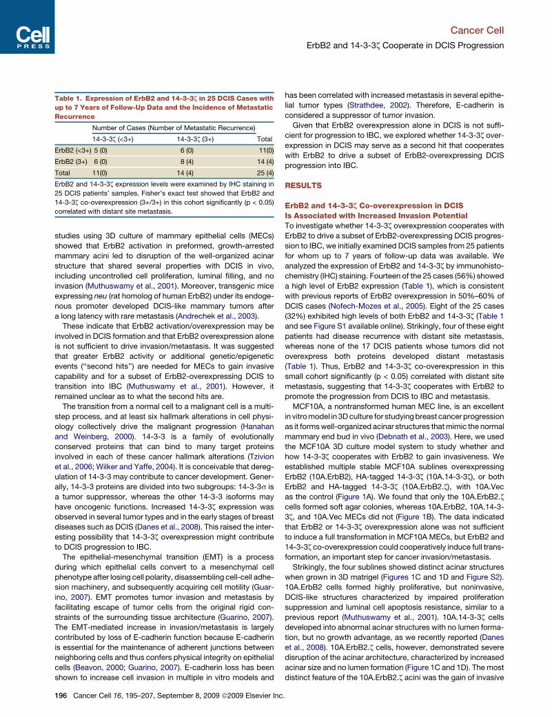
Table 1. Expression of ErbB2 and 14-3-3z in 25 DCIS Cases with
up to 7 Years of Follow-Up Data and the Incidence of Metastatic
Recurrence
Number of Cases (Number of Metastatic Recurrence)
14-3-3z (<3+) 14-3-3z (3+) Total
ErbB2 (<3+) 5 (0) 6 (0) 11(0)
ErbB2 (3+) 6 (0) 8 (4) 14 (4)
Total 11(0) 14 (4) 25 (4)
ErbB2 and 14-3-3z expression levels were examined by IHC staining in
25 DCIS patients’ samples. Fisher’s exact test showed that ErbB2 and
14-3-3z co-overexpression (3+/3+) in this cohort significantly (p < 0.05)
correlated with distant site metastasis.
196 Cancer Cell 16, 195–207, September 8, 2009 ª2009 Elsevier Inc
has been correlated with increased metastasis in several epithe-
lial tumor types (Strathdee, 2002). Therefore, E-cadherin is
considered a suppressor of tumor invasion.
Given that ErbB2 overexpression alone in DCIS is not suffi-
cient for progression to IBC, we explored whether 14-3-3z over-
expression in DCIS may serve as a second hit that cooperates
with ErbB2 to drive a subset of ErbB2-overexpressing DCIS
progression into IBC.
RESULTS
ErbB2 and 14-3-3z Co-overexpression in DCISIs Associated with Increased Invasion PotentialTo investigate whether 14-3-3z overexpression cooperates with
ErbB2 to drive a subset of ErbB2-overexpressing DCIS progres-
sion to IBC, we initially examined DCIS samples from 25 patients
for whom up to 7 years of follow-up data was available. We
analyzed the expression of ErbB2 and 14-3-3z by immunohisto-
chemistry (IHC) staining. Fourteen of the 25 cases (56%) showed
a high level of ErbB2 expression (Table 1), which is consistent
with previous reports of ErbB2 overexpression in 50%–60% of
DCIS cases (Nofech-Mozes et al., 2005). Eight of the 25 cases
(32%) exhibited high levels of both ErbB2 and 14-3-3z (Table 1
and see Figure S1 available online). Strikingly, four of these eight
patients had disease recurrence with distant site metastasis,
whereas none of the 17 DCIS patients whose tumors did not
overexpress both proteins developed distant metastasis
(Table 1). Thus, ErbB2 and 14-3-3z co-overexpression in this
small cohort significantly (p < 0.05) correlated with distant site
metastasis, suggesting that 14-3-3z cooperates with ErbB2 to
promote the progression from DCIS to IBC and metastasis.
MCF10A, a nontransformed human MEC line, is an excellent
in vitro model in 3D culture for studying breast cancer progression
as it forms well-organized acinar structures that mimic the normal
mammary end bud in vivo (Debnath et al., 2003). Here, we used
the MCF10A 3D culture model system to study whether and
how 14-3-3z cooperates with ErbB2 to gain invasiveness. We
established multiple stable MCF10A sublines overexpressing
ErbB2 (10A.ErbB2), HA-tagged 14-3-3z (10A.14-3-3z), or both
ErbB2 and HA-tagged 14-3-3z (10A.ErbB2.z), with 10A.Vec
as the control (Figure 1A). We found that only the 10A.ErbB2.z
cells formed soft agar colonies, whereas 10A.ErbB2, 10A.14-3-
3z, and 10A.Vec MECs did not (Figure 1B). The data indicated
that ErbB2 or 14-3-3z overexpression alone was not sufficient
to induce a full transformation in MCF10A MECs, but ErbB2 and
14-3-3z co-overexpression could cooperatively induce full trans-
formation, an important step for cancer invasion/metastasis.
Strikingly, the four sublines showed distinct acinar structures
when grown in 3D matrigel (Figures 1C and 1D and Figure S2).
10A.ErbB2 cells formed highly proliferative, but noninvasive,
DCIS-like structures characterized by impaired proliferation
suppression and luminal cell apoptosis resistance, similar to a
previous report (Muthuswamy et al., 2001). 10A.14-3-3z cells
developed into abnormal acinar structures with no lumen forma-
tion, but no growth advantage, as we recently reported (Danes
et al., 2008). 10A.ErbB2.z cells, however, demonstrated severe
disruption of the acinar architecture, characterized by increased
acinar size and no lumen formation (Figure 1C and 1D). The most
distinct feature of the 10A.ErbB2.z acini was the gain of invasive
.

Cancer Cell
ErbB2 and 14-3-3z Cooperate in DCIS Progression
capacity, as many cells escaped from 10A.ErbB2.z acini and
invaded the surrounding matrix (Figures 1C and 1D). An impor-
tant feature of the noninvasive DCIS is the intact basement
membrane that surrounds it, whereas invasive carcinomas are
defined by loss of basement membrane integrity (Rizki and Bis-
sell, 2004). Indeed, we observed that individual cells in
10A.ErbB2.z acini were patched by diffuse basement membrane
protein laminin V, whereas laminin V formed a continuous base-
ment membrane layer surrounding acini from 10A.ErbB2,
10A.14-3-3z, and 10A.Vec MECs (Figure 1D). Together, co-over-
expression of ErbB2 and 14-3-3z in MCF10A MECs conferred
invasiveness, whereas overexpression of ErbB2 or 14-3-3z alone
did not.
Invasion Is the Collective Effect of ErbB2-MediatedIncrease of Cell Migration and 14-3-3z-MediatedDecrease of Cell-Cell Adhesion via EMTTumor cell invasion is a multistep process, of which the key
events include increased migration, increased protease secre-
BA
ErbB2
tubulin
10A.Vec 10A.ErbB2
β-actin
14-3-3ζ10A.14-3-3ζ 10A.ErbB2.ζ
C10A.Vec 10A. ErbB2
10A.14-3-3ζ 10A.ErbB2.ζ
Laminin V/DAPI
D
10A.ErbB210A.Vec 10A.14-3-3ζ 10A.ErbB2.ζ
Figure 1. ErbB2 and 14-3-3z Co-overex-
pression Is Associated with Increased Inva-
sion Potential in an In Vitro DCIS Model
(A) Establishment of MCF10A stable cell lines
overexpressing ErbB2 alone, 14-3-3z alone, or
both ErbB2 and 14-3-3z. Multiple stable clones
were established for each subline and most exper-
iments were repeated with different clones to rule
out clonal effects. Immunoblot analysis of one
representative of each subline for the indicated
proteins is shown.
(B) Co-overexpression of ErbB2 and 14-3-3z led to
anchorage-independent growth of MCF10A cells
in soft agar assay.
(C) Distinct acinar structures of MCF10A sublines
in 3D culture. The top panel shows phase-contrast
images of acinar structures (19 days); the scale bar
represents 200 mm. The inset shows an amplified
view of individual 10A.ErbB2.z cell invading into
surrounding matrigel; the scale bar represents
100 mm.
(D) Loss of basement membrane integrity in
10A.ErbB2.z acini. MCF10A sublines were cultured
in 3D matrigel for 28 days and then stained for lam-
inin V (red) and DAPI (blue). Representative images
are shown. The scale bar represents 100 mm.
tion, and altered adhesion to allow
dissemination from primary tumor sites
(Liotta and Stetler-Stevenson, 1991). We
detected no significant difference in
matrix metalloproteinase levels among
the four MCF10A sublines (data not
shown). However, migration and wound
healing assays showed that both
10A.ErbB2 and 10A.ErbB2.z cells had
increased cell motility, whereas 10A.14-
3-3z cells had a low motility similar to
that of 10A.Vec (Figures 2A and 2B).
Thus, the increased cell motility was
largely contributed by ErbB2 overexpres-
sion, not by 14-3-3z overexpression. Multiple ErbB2 down-
stream signaling pathways can be involved in ErbB2-mediated
cell motility, including PI3K, PAK1, Rac1, and Src activation
(Feldner and Brandt, 2002). We found that Src phosphorylation
is specifically increased in the two ErbB2-overexpressing
MCF10A sublines compared to the two ErbB2 low-expressing
MCF10A sublines (Figures S3A and S3B). Moreover, treatment
with a Src kinase inhibitor (Saracatinib or AZD0530) significantly
inhibited the motility of 10A.ErbB2 and 10A.ErbB2.z cells,
whereas Rac1 and PI3K inhibitors had no significant effect
(Figure S3C).
Reduced cell-cell adhesion is another prerequisite for indi-
vidual cell invasion and EMT has been implicated in tumor inva-
sion partly by reducing cell-cell adhesion (Guarino, 2007). In
contrast to 10A.Vec and 10A.ErbB2 cells that had a cobble-
stone-like epithelial morphology in 2D culture, 10A.ErbB2.z and
10A.14-3-3z cells displayed a spindle-like shape and exhibited
a scattered distribution, indicating loss of cell-cell contact and
EMT (Figure 2C). The morphological changes during EMT are
Cancer Cell 16, 195–207, September 8, 2009 ª2009 Elsevier Inc. 197

Cancer Cell
ErbB2 and 14-3-3z Cooperate in DCIS Progression
driven by a number of molecular and cellular alterations,
including loss or decrease of epithelial cell markers (e.g., E-cad-
herin, b-catenin, a-catenin, and p120-catenin) and de novo
expression of mesenchymal markers (e.g., N-cadherin, vimentin,
and fibronectin). Certainly, we found that 10A.Vec and 10A.
ErbB2 cells expressed high levels of E-cadherin, b-catenin,
a-catenin, and p120-catenin, but minimal levels of N-cadherin
and vimentin. 10A.ErbB2.z and 10A.14-3-3z cells, however,
showed E-cadherin loss, dramatically reduced b-catenin, a-cat-
enin, and p120-catenin, and de novo expression of N-cadherin
and vimentin (Figures 2D and 2E). Similarly, 14-3-3z overexpres-
sion in HMEC-hTERT cells, immortalized by the telomerase
reverse transcriptase catalytic subunit, also led to EMT (Fig-
ure S4). Thus, 14-3-3z overexpression contributed to the loss
of cell-cell adhesion and the EMT phenotype. Together, a collec-
tive effect of ErbB2-mediated increase of cell migration and
14-3-3z-mediated decrease of cell-cell adhesion conferred
10A.ErbB2.z acini invasiveness.
l ls/fi
eld
10A.14-3-3ζ10A.Vec 10A.ErbB2 10A.ErbB2.ζ
BA
Mig
rate
d ce
l
10A. ErbB2.ζ10A. Vec 10A. ErbB2 10A.14-3-3ζC
10A.14-3-3ζ10A.ErbB2.ζ10A.Vec 10A.ErbB2
DA
PID
E-ca
dher
in/D
API
Vim
entin
/DA
E
E-cadherinErbB2
β-catenin
3D
N-cadherintubulin
β
p120-cateninα-catenin2D
Figure 2. ErbB2 Overexpression Led to
Increased Cell Migration and 14-3-3z Over-
expression Led to Decreased Cell-Cell
Adhesion as a Result of EMT
(A) Transwell migration assay of the indicated
MCF10A sublines. Cells that migrated to the
bottom of the chamber were counted in five fields
under 203 magnification. Experiments were done
three times with triplicates and error bar repre-
sents SEM.
(B) Wound healing assay of the indicated MCF10A
sublines. Wound closures were photographed at
0 and 6 hr after wounding. The scale bar repre-
sents 200 mm.
(C) The four MCF10A sublines exhibited different
morphologies in 2D culture. The scale bar repre-
sents 100 mm.
(D) IFS of EMT markers in MCF10A sublines.
E-cadherin (top, red), vimentin (bottom, red), and
DAPI (blue) are shown. The scale bar represents
100 mm.
(E) Immunoblot analysis of the indicated EMT
markers in MCF10A sublines. Expression of epithe-
lial cell markers (E-cadherin, b-catenin, a-catenin,
and p120 catenin) and mesenchymal cell marker
(N-cadherin) were examined by immunoblot
analysis in both 2D and 3D culture cell lysates.
E-Cadherin Loss, a Key Eventof EMT, Is Mediated by ZFHX1Bin 10A.ErbB2.z CellsWe next investigated how 14-3-3z over-
expression led to E-cadherin loss, a key
event of EMT resulting in decreased
cell-cell adhesion. RT-PCR analysis
showed that E-cadherin mRNA level
was dramatically lower in 10A.ErbB2.z
and 10A.14-3-3z cells than in 10A.Vec
and 10A.ErbB2 cells (Figure 3A). E-cad-
herin mRNA loss could result from hyper-
methylation of its promoter (Strathdee,
2002), but we detected no significant
differences in E-cadherin promoter methylation status among
the four MCF10A sublines (data not shown). Another major
mechanism of E-cadherin mRNA loss is direct transcriptional
repression by repressors, including snail, slug, twist, E12, E47,
ZFHX1B (also named SIP1), and deltaEF1 (Peinado et al.,
2004). These transcriptional repressors have been found to
induce EMT in vitro and their overexpression in a variety of
human tumors is associated with increased tumor invasion/
metastasis and poor prognosis. We examined the expression
levels of snail, slug, twist, E12, E47, and deltaEF1 and found
they were not significantly different among the four MCF10A
sublines (Figure 3B). Interestingly, expression of ZFHX1B was
dramatically higher in 10A.ErbB2.z and 10A.14-3-3z cells than
in 10A.Vec and 10A.ErbB2 cells at both mRNA and protein levels
(Figure 3C).
ZFHX1B is a two-handed zinc-finger protein that binds to the E
boxes in the E-cadherin proximal promoter to repress E-cadherin
transcription (Comijn et al., 2001). To examine whether the
198 Cancer Cell 16, 195–207, September 8, 2009 ª2009 Elsevier Inc.

Cancer Cell
ErbB2 and 14-3-3z Cooperate in DCIS Progression
E-cadherin loss in 10A.ErbB2.z and 10A.14-3-3z cells was due
to transcriptional repression by the upregulated ZFHX1B, we
cloned a fragment of E-cadherin promoter (containing two
consensus ZFHX1B binding motifs: CANNTG) upstream of a
luciferase reporter plasmid (pGL3.Ecad) and compared its
activity among the MCF10A sublines (Figure 3D). Indeed,
pGL3.Ecad luciferase activities were significantly repressed
(p < 0.05) in 10A.ErbB2.z and 10A.14-3-3z cells versus 10A.Vec
and 10A.ErbB2 cells (Figure 3D). Moreover, the repression of
E-cadherin promoter-driven luciferase activity was partially
relieved in 10A.ErbB2.z and 10A.14-3-3z cells when ZFHX1B
expression was inhibited by small interfering RNA (siRNA;
Figure 3E). Therefore, ZFHX1B upregulation contributed to the
transcriptional repression of E-cadherin in 10A.ErbB2.z and
10A.14-3-3z cells. In addition, examination of ZFHX1B expres-
sion in six E-cadherin-positive and four E-cadherin-negative
breast cancer cell lines showed a general correlation between
ZFHX1B expression and E-cadherin loss (Figure 3F).
ZFHX1B Is Upregulated by 14-3-3z through Upregulationof TGF-b Receptor I and Activation of TGF-b/SmadsPathwayNext, we investigated the mechanism of ZFHX1B upregulation in
10A.ErbB2.z and 10A.14-3-3z cells. Because TGF-b/Smads
pathway activation was shown to induce EMT and was also
known to be involved in ZFHX1B upregulation (Zavadil and
Bottinger, 2005), we examined whether ZFHX1B upregulation
by 14-3-3z might be due to increased TGF-b/Smads signaling.
Expression of the TbRI protein, but not RNA, was dramatically
increased in 10A.ErbB2.z and 10A.14-3-3z cells, whereas TbRII
A
B
Luciferase reporter-308 +21
E-cadherin promoterD
E-cadherin
GAPDH
E-boxE-box**
pGL3.Vec pGL3.Ecadifera
se a
ctiv
itysnailtwistslug
mm
unob
lot
Rel
ativ
e lu
c
tubulinIm
E12E47
delta EF1GAPDH
RT-P
CR
E
ZFHX1B siRNACon siRNA
ZFHX1B
10A.ErbB2.ζ 10A.14-3-3ζ+ +
+ +
C
GAPDH
10A.ErbB2.ζ10A.14-3-3ζ
ve lu
cife
rase
a c
tivity
ZFHX1B
GAPDH
ZFHX1B siRNACon siRNA + +
+ +
Rel
ativ a
Nuclear extrac
laminA/C
FZFHX1B
GAPDH
ZFHX1B
ts GAPDH
E-cadherin negative E-cadherin positive
Figure 3. EMT Phenotype in 10A.ErbB2.z
and 10A.14-3-3z Cells Were Mediated by
ZFHX1B Upregulation
(A) E-cadherin mRNA was dramatically reduced in
10A.ErbB2.z and 10A.14-3-3z cells.
(B) Expression of EMT regulators in the four
MCF10A sublines. The top panel shows immuno-
blot analysis of EMT regulators, including snail,
twist, and slug. The bottom panel shows RT-PCR
analysis of EMT regulators, including E12, E47,
and delta EF1.
(C) RT-PCR and immunoblot analysis of ZFHX1B
levels in the four MCF10A sublines. MDA-MB-
435 cells served as the positive control for ZFHX1B
protein expression.
(D) 14-3-3z overexpression led to transcriptional
repression of E-cadherin promoter activity. The
top panel shows a schematic representation of
the luciferase reporter driven by E-cadherin prox-
imal promoter containing two ZFHX1B binding
E-box motif: pGL3.Ecad. The bottom panel shows
relative luciferase activity of pGL3.Ecad in the four
MCF10A sublines. p < 0.05. Error bars indicate
SEM.
(E) Downregulation of ZFHX1B in 14-3-3z-overex-
pressing MCF10A sublines by siRNA partially
relieved suppression of E-cadherin promoter-
driven luciferase activity. As shown in the top
panel, control siRNA and ZFHX1B siRNA were
transfected into 10A.ErbB2.z cells and 10A.14-3-
3z cells. After 48 hr, downregulation of ZFHX1B
was examined by RT-PCR with GAPDH as internal
control. As shown in the bottom panel, cells were
pretransfected with ZFHX1B siRNA or control
siRNA. After 48 hr, pGL3.Ecad was cotransfected
with pRL.TK plasmid as transfection efficiency
control. Relative luciferase activity was determined
36 hr later. Error bars indicate SEM.
(F) A reverse correlation between ZFHX1B and
E-cadherin expression in breast cancer cell lines.
RNA was extracted from four E-cadherin-negative
breast cancer cell lines (BT549, Hs578T, MDA-
MB-231, and MDA-MB-435), five E-cadherin-posi-
tive breast cancer cell lines (MCF-7, BT474, T47D,
MDA-MB-361, and MDA-MB-453), and a non-
transformed breast epithelial cell line (MCF10A),
and then reverse transcribed into cDNA; this was
followed by PCR with ZFHX1B-specific primers.
GAPDH served as loading control.
Cancer Cell 16, 195–207, September 8, 2009 ª2009 Elsevier Inc. 199
Cancer Cell
ErbB2 and 14-3-3z Cooperate in DCIS Progression
protein levels were similar among the four MCF10A sublines
(Figure 4A). Consistently, we also observed increased TbRI level
in 14-3-3z-overexpressing HMEC-hTERT-HA-14-3-3z cells
accompanied by upregulation of ZFHX1B (Figure S4C). The
increased TbRI protein levels led to increased TGF-b/Smads
activation, as indicated by the increased nuclear phospho-
smad2/smad3 and total smad2/smad3 levels in 10A.ErbB2.z
and 10A.14-3-3z cells (Figure 4B). Moreover, chromatin immu-
noprecipitation (ChIP) assay detected binding of nuclear
smad3 to the ZFHX1B promoter in 10A.ErbB2.z and 10A.14-3-
3z cells, but not in 10A.Vec or 10A.ErbB2 cells (Figure 4C). These
data indicate that 14-3-3z-mediated TGF-b/Smads activation
contributed to ZFHX1B transcriptional upregulation. Indeed,
blocking 14-3-3z by siRNA reduced TbRI protein expression,
which also led to reduced ZFHX1B expression (Figure 4D and
Figure S5A).
TbRI protein level is mainly regulated by its internalization,
followed either by trafficking back to the cell membrane after
engulfed in early endosome or by ubiquitination-mediated
degradation when engulfed in lipid raft-caveolae-1 vesicles (Di
Guglielmo et al., 2003; Lin et al., 2004). To investigate the mech-
anisms of 14-3-3z-mediated TbRI protein upregulation, we first
investigated whether it is contributed by reduced TbRI ubiquitina-
tion. Indeed, ubiquitination of Myc-tagged TbRI in 10A.ErbB2.z
cells was reduced compared to 10A.ErbB2 cells when
HA-tagged ubiquitin was coexpressed (Figure 4E, left). 14-3-3z
knockdown by siRNA in 10A.ErbB2.z cells and in HeLa cells
(endogenous 14-3-3z) led to a consistent increase in TbRI ubiqui-
tination, whereas TbRI ubiquitination was inhibited when 14-3-3z
was overexpressed (Figure 4E, middle and right). Furthermore,
treatment with MG132, a proteasome inhibitor, led to greater
accumulation of TbRI in 10A.ErbB2 cells than in 10A.ErbB2.z cells
(Figure 4F), indicating a more rapid TbRI ubiquitination and
proteasome-mediated degradation in 14-3-3z low-expressing
10A.ErbB2 cells. Next, we examined whether 14-3-3z inhibited
TbRI ubiquitination and degradation by binding to TbRI. Indeed,
A B
TβRI
tubulin
tubulin
IBC
R TβRI
TβRII
Lamin C
P-smad2
P-smad3
T-smad2/3
Nuc
lear
ext
ract
GAPDH
RT-
PC
β
C
input + + + +
Con siRNA14-3-3ζ siRNA
TβRI
10A.ErbB2.ζ 10A.14-3-3ζ
B
D
14 3 3ζ
+ ++ +
Histone H1IgG
Smad3
+ + + ++ + + +
+ + + +β-actin
ZFHX1B
GAPDH
IBR
T-PC
R
14-3-3ζ
10A.ErbB2 + + - - Con siRNA pCDNA3.Vec + + - -E + + - - + + - -
A-u
bqui
tin
10A.ErbB2.ζ - - + +IgG + - + -Myc - + - +
HA
-ubq
uitin
14-3-3 ζ siRNAIgGMyc
pCDNA3.14-3-3ζ - - + +IgG + - + -Myc - + - +
HA
-ubq
uitin
E- - + ++ - + -- + - +
- - + ++ - + -- + - +
HA
Myc-TRI
H
Myc-TRI
H
Myc-TRI
F G
10A.ErbB2.ζ Hela
TβRI-full lengthTβRI-420
TβRI-370TβRI 210
H
Hela
- +IgG HA IgG HAIP
TβRI
TβRI-210TβRI-195
TβRI-full lengthTβRI 370
I- +
TβRIMG132
β-actin1.0 5.9 4.7 4.5
HA TβRI-210
TβRI-370β-actin
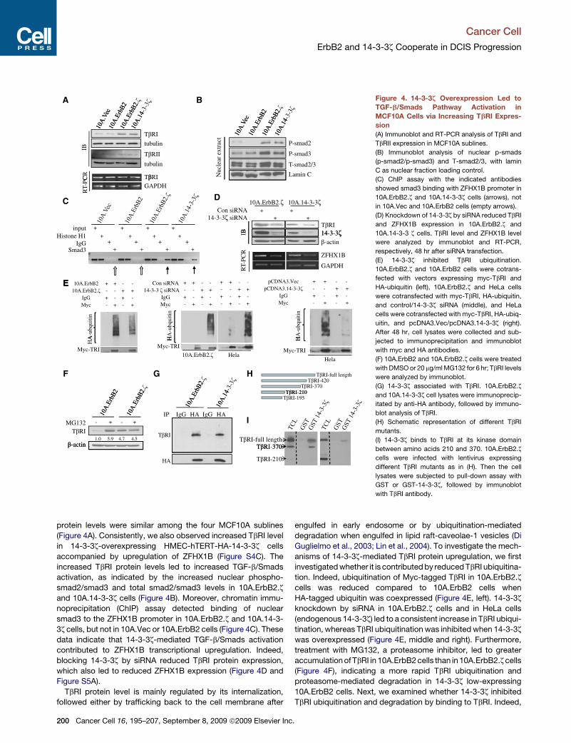
Figure 4. 14-3-3z Overexpression Led to
TGF-b/Smads Pathway Activation in
MCF10A Cells via Increasing TbRI Expres-
sion
(A) Immunoblot and RT-PCR analysis of TbRI and
TbRII expression in MCF10A sublines.
(B) Immunoblot analysis of nuclear p-smads
(p-smad2/p-smad3) and T-smad2/3, with lamin
C as nuclear fraction loading control.
(C) ChIP assay with the indicated antibodies
showed smad3 binding with ZFHX1B promoter in
10A.ErbB2.z and 10A.14-3-3z cells (arrows), not
in 10A.Vec and 10A.ErbB2 cells (empty arrows).
(D) Knockdown of 14-3-3z by siRNA reduced TbRI
and ZFHX1B expression in 10A.ErbB2.z and
10A.14-3-3 z cells. TbRI level and ZFHX1B level
were analyzed by immunoblot and RT-PCR,
respectively, 48 hr after siRNA transfection.
(E) 14-3-3z inhibited TbRI ubiquitination.
10A.ErbB2.z and 10A.ErbB2 cells were cotrans-
fected with vectors expressing myc-TbRI and
HA-ubiquitin (left), 10A.ErbB2.z and HeLa cells
were cotransfected with myc-TbRI, HA-ubiquitin,
and control/14-3-3z siRNA (middle), and HeLa
cells were cotransfected with myc-TbRI, HA-ubiq-
uitin, and pcDNA3.Vec/pcDNA3.14-3-3z (right).
After 48 hr, cell lysates were collected and sub-
jected to immunoprecipitation and immunoblot
with myc and HA antibodies.
(F) 10A.ErbB2 and 10A.ErbB2.z cells were treated
with DMSO or 20 mg/ml MG132 for 6 hr; TbRI levels
were analyzed by immunoblot.
(G) 14-3-3z associated with TbRI. 10A.ErbB2.z
and 10A.14-3-3z cell lysates were immunoprecip-
itated by anti-HA antibody, followed by immuno-
blot analysis of TbRI.
(H) Schematic representation of different TbRI
mutants.
(I) 14-3-3z binds to TbRI at its kinase domain
between amino acids 210 and 370. 10A.ErbB2.z
cells were infected with lentivirus expressing
different TbRI mutants as in (H). Then the cell
lysates were subjected to pull-down assay with
GST or GST-14-3-3z, followed by immunoblot
with TbRI antibody.
200 Cancer Cell 16, 195–207, September 8, 2009 ª2009 Elsevier Inc.

Cancer Cell
ErbB2 and 14-3-3z Cooperate in DCIS Progression
14-3-3z and TbRI coexisted in the same complex (Figure 4G) and
the binding region is between amino acid 210 and 370 in the
kinase domain of TbRI (Figures 4H and 4I). Immunofluorescence
staining (IFS) also detected diffuse staining of both 14-3-3z and
TbRI proteins both in the cytosol and on the cell membrane
(Figure S5B). The data are consistent with previous reports that
TbRI is constantly recycled between membrane and cellular
vesicles, resulting in �20% localization to the cell membrane
and �80% remaining in the cytosol (Di Guglielmo et al., 2003).
Most importantly, the binding of 14-3-3z protects TbRI from
degradation because the TbRI-210 that cannot bind to 14-3-3z
has a much shorter half-life compared to the TbRI-370 that binds
to 14-3-3z (Figure S5C). Furthermore, when 14-3-3z expression
is knocked down by siRNA, the half-life of TbRI-370 is dramati-
cally reduced, while the half-life of TbRI-210 is not affected
(Figure S5D and data not shown). These results indicated that
overexpressed 14-3-3z in 10A.ErbB2.z and 10A.14-3-3z cells
bound to TbRI and inhibited the proteasome-mediated TbRI
degradation, leading to increased TbRI protein level and TGF-b/
Smads pathway activation.
To further confirm the role of TGF-b/Smads pathway activa-
tion in the induction of the EMT phenotypes, we treated the
10A.Vec and 10A.ErbB2 cells with TGF-b1 to activate the TGF-
b/Smads pathway. The treatment induced smad3 phosphoryla-
tion, ZFHX1B upregulation, and morphological features of EMT,
with corresponding downregulation of E-cadherin and upregula-
tion of vimentin and fibronectin (Figure S6). Therefore, activation
of the TGF-b/Smads pathway was sufficient to induce EMT in
MCF10A cells.
Inhibition of TGF-b Receptor Activation PartiallyReversed EMT and Inhibited the Invasivenessof 10A.ErbB2.z AciniTo determine whether activation of the TGF-b/Smads pathway is
required for the EMT and invasive phenotype of the 10A.ErbB2.z
cells, we inhibited TGF-b/Smads pathway activation by treating
10A.ErbB2.z cells with a TGF-b receptor I/II kinase inhibitor,
LY2109761 (Melisi et al., 2008). LY2109761 treatment reduced
smad2/3 phosphorylation and total smad3, but had no significant
effect on the phosphorylation of Akt (p-Akt) or p42-MAPK (p-P42)
(Figure 5A). Interestingly, LY2109761-treated 10A.ErbB2.z cells
adhered to neighboring cells to form cell islands, indicating
improved cell-cell adhesion (Figure 5B, left). More importantly,
the invasive phenotype of 10A.ErbB2.z acini in 3D matrigel
culture was dramatically inhibited by LY2109761 treatment
compared to control treatment (Figure 5B, middle). In contrast,
LY2109761 treatment had no significant impact on acini develop-
ment and maintenance in the other MCF10A sublines (Figure S7).
Consistent with the partial reversal of EMT morphology of the
cells in 2D culture and reduced invasiveness in 3D culture, there
was increased epithelial protein expression, such as E-cadherin
and a-catenin, after LY2109761 treatment. E-cadherin was
specifically located in the membrane regions forming cell-cell
contacts, a prerequisite for adherent junction formation (Fig-
ure 5B, right). Prolonged treatment also led to decreased mesen-
chymal protein expression (Figure 5C). Collectively, these data
indicate that 14-3-3z-mediated TGF-b/Smads pathway activa-
tion plays a critical role in the EMT phenotype and gain of
invasiveness in 10A.ErbB2.z cells.
Ca
Reintroduction of E-Cadherin in 10A.ErbB2.zCells Inhibits InvasionInhibition of TGF-b/Smads pathway by LY2109761 partially
recovered E-cadherin expression that inhibited the invasion of
10A.ErbB2.z acini, indicating that E-cadherin loss was a key
event in the gain of invasiveness during EMT. To further
determine the critical role of E-cadherin loss in invasion, we
restored E-cadherin expression in the 10A.ErbB2.z cells (named
10A.ErbB2.z.Ecad) to levels similar to those in the 10A.Vec cells
(Figure 5D). The restored E-cadherin expression led to the
recovery of other epithelial proteins, such as a-catenin, b-cate-
nin, and p120-catenin, and reduced mesenchymal proteins,
such as N-cadherin and vimentin (Figure 5E). Moreover, the cells
with recovered E-cadherin expression showed a dramatic
increase in cell adhesion (Figures S8A and S8B). Importantly,
10A.ErbB2.z.Ecad cells formed acinar structures with fewer indi-
vidual cells invading into surrounding matrigel, in contrast to the
highly invasive acinar structures of 10A.ErbB2.z.Vec cells
(Figure 5F and Figure S8C). Thus, re-expression of E-cadherin
in 10A.ErbB2.z cells efficiently increased cell-cell adhesion and
inhibited, at least partially, the invasive phenotype in 3D culture.
Therefore, E-cadherin loss played a critical role in inducing inva-
siveness of 10A.ErbB2.z cells.
14-3-3z Overexpression Is Associated with High Levelsof TbRI Expression in Both Human DCIS and IBCWe have identified TbRI upregulation as a major mechanism
of 14-3-3z overexpression-induced invasiveness in MCF10A.
ErbB2.z cells. To evaluate the biological relevance of these find-
ings, we investigated whether there is a correlation between
TbRI and 14-3-3z expression in patients’ samples. Because we
did not have enough of the DCIS samples shown in Table 1
remaining for these staining, we stained 138 DCIS samples
from patients with recently diagnosed disease and 100 IBC
with clinical follow-up. We found that 14-3-3z overexpression
significantly (p < 0.05) correlated with increased TbRI levels in
both populations (Figure 6A). Moreover, IHC staining for 14-3-3z,
TbRI, ErbB2, E-cadherin, vimentin, and N-cadherin on the DCIS
samples showed that co-overexpression of 14-3-3z and TbRI
significantly (p < 0.05) correlated with (at least two) EMT marker
alterations (reduced expression of epithelial marker E-cadherin
and expression of mesenchymal markers vimentin and/or N-
cadherin; Table S1 and Figure S9). Importantly, high 14-3-3z
and TbRI expression levels plus two EMT marker alterations in
DCIS were significantly (p < 0.05) associated with high-grade
DCIS phenotype, which correlates with a higher risk of invasive
recurrence (Table S2). Representative images of multiple
markers’ expressions in a pure low-grade DCIS and in a DCIS
sample with microinvasion are shown in Figure 6B. Together,
14-3-3z overexpression in DCIS lesions correlated with TbRI up-
regulation and induced EMT that could contribute to a higher risk
of invasive recurrence.
Co-overexpression of ErbB2 and 14-3-3z Is Associatedwith Higher Metastatic Potential in Mice and IncreasedMetastatic Disease Recurrence and Death in BreastCancer PatientsThe above findings demonstrated that co-overexpression of
ErbB2 and 14-3-3z increased the invasiveness of MECs in 3D
ncer Cell 16, 195–207, September 8, 2009 ª2009 Elsevier Inc. 201

Cancer Cell
ErbB2 and 14-3-3z Cooperate in DCIS Progression
culture. To determine whether co-overexpression of ErbB2 and
14-3-3z may increase invasion/metastasis in vivo, we stably
overexpressed 14-3-3z in TM15 cells, a mouse mammary
tumor cell line from a MMTV-Cre/flox-neoNeuNT mouse that
expresses the transforming neu under an endogenous pro-
moter. We established the TM15.14-3-3z cell line with TM15.Vec
as our controls. The two sublines were injected into mammary
fat pads of nude mice to establish xenografts and mice were
monitored for metastatic lesions. Mice injected with the
TM15.14-3-3z cells definitely had more lung metastasis than
mice with TM15.Vec cells (Figure 6C). To further investigate
the impact of co-overexpression of ErbB2 and 14-3-3z on breast
cancer progression, especially metastatic disease recurrence
and death of breast cancer patients, we performed IHC analysis
to examine ErbB2 and 14-3-3z expression in 107 cases of IBC in
consecutive slides. Remarkably, 23 of the 107 patients had
breast tumors co-overexpressing both ErbB2 and 14-3-3z,
and these patients had significantly shorter overall survival
(p < 0.05) and disease-free survival (p < 0.05) than patients
whose tumors overexpressed either one or neither (Figure 6D).
In addition, in this patient cohort, multivariate analysis demon-
strated that co-overexpression of ErbB2 and 14-3-3z in breast
tumors can predict poor prognosis (Table S3). Because
a majority of these patients died of recurrent metastatic disease,
these data indicated that breast cancers overexpressing both
ErbB2 and 14-3-3z are more aggressive and have greater meta-
static potential.
DISCUSSION
14-3-3z Is a Biomarker for Patientswith ErbB2-Overexpressing DCIS Who Havea Higher Risk of Progression to IBCBoth clinical and experimental data support that ErbB2
overexpression plays a critical role in DCIS but is not sufficient
to drive progression of the noninvasive DCIS to IBC. It has
been puzzling as to what other alterations may cooperate
with ErbB2 to allow a subgroup of ErbB2-overexpressing
DCIS to progress to life-threatening invasive/metastatic breast
tumors. Here, we identified 14-3-3z as a molecule that, when
A 10A.ErbB2.ζ
TGFβ1 + + TβR inhibitor 0 1 2 5 5 10 μM
10A.ErbB2.ζ
T-smad2
p-smad2
TGFβ1TβR inhibitor
– + ++– –
p-smad3
T-smad3
TβR inhibitor 0 1 2.5 5 10 μMP-AktT-AktP-P42T-P42
tubulin
β-actin
10A.ErbB2.ζ
– +TβR inhibitor
BPhase contrast
E-cadherin/ DAPI2D 3D2D
2D3D
E-cadherinα-cateninβ-actin
E-cadherinα-cateninβ-actin
TβR
inhi
bito
r C
-
+β c
+ TβR i hibit10A.ErbB2.ζ
3D culture (day 10)
FEC
10A E bB2 ζ
E-cadherin
t iβ-catenin
– + TβR inhibitor
Vimentin
Fibronectinβ-actin
3D culture (day 10)
10A.ErbB2.ζ.Vec
E-cadherin
tubulin
10A.ErbB2.
tubulin
N-CadherinVimentin
α-cateninp120cateninβ-actin
D10A.ErbB2.ζ.Ecad
tubulin
Figure 5. Inhibition of TGF-b/Smads
Pathway Partially Reversed EMT of 14-3-
3z-Overexpressing MCF10A Cells and
Inhibited Invasion of 10A.ErbB2.z Cells
(A) LY2109761 efficiently inhibited smad2/3 phos-
phorylation in 10A.ErbB2.z cells. Cells were
treated with TGF-b1 (5 ng/ml) or TGF-b1 (5 ng/ml)
plus TbR inhibitor LY2109761 (10 mM) for 60 min
and then analyzed by immunoblotting with indi-
cated antibodies (left). Cells were treated with
increasing dose of LY2109761 and then analyzed
by immunoblotting for p-Akt, T-Akt, p-P42,
T-P42, and b-actin (right).
(B) Treatment of 10A.ErbB2.z cells with TGF-b
receptor inhibitor partially reversed the EMT
morphology in 2D culture and inhibited the inva-
siveness of acini in 3D culture. As shown in the
left panel, cells were treated with LY2109761 for
48 hr, and images were photographed under a
phase-contrast microscope. As shown in the
middle panel, 10A.ErbB2.z cells were cultured on
3D matrigel for 13 days to form invasive acinar
structures, and then treated with DMSO or
LY2109761 for 6 days. As shown in the right panel,
10A.ErbB2.z cells were treated with DMSO or
LY2109761 and subjected to IFS of E-cadherin.
The scale bar represents 100 mm. Cells lysates
were also collected after treatment and subjected
to immunoblot with E-cadherin, a-catenin, and
b-actin antibodies.
(C) Prolonged LY2109761 treatment (15 days)
reduced mesenchymal protein expression (fibro-
nectin and vimentin).
(D) E-cadherin expression was stably reintroduced
into 10A.ErbB2.z cells.
(E) E-cadherin restoration led to increased epithe-
lial protein expression (a-catenin, b-catenin, and
p120-catenin) and decreased mesenchymal
protein expression (N-cadherin and vimentin).
(F) E-cadherin restoration inhibited 10A.ErbB2.z
cell invasion. Phase-contrast microscopy of acinar
structures of 10A.ErbB2.z.Vec and 10A.ErbB2.z.
Ecad cells in 3D culture. The scale bar represents
200 mm.
202 Cancer Cell 16, 195–207, September 8, 2009 ª2009 Elsevier Inc.

Cancer Cell
ErbB2 and 14-3-3z Cooperate in DCIS Progression
co-overexpressed with ErbB2, increases the potential of DCIS
to progress to IBC.
Individual tumor cell invasion is a highly complicated process
that requires malignant cells to obtain at least both the ‘‘capa-
bility’’ (migration) and the ‘‘freedom’’ (dissemination) to escape
from the constraint of tissue structure. We found that ErbB2
overexpression alone promoted cell migration via Src activation,
but not invasion, whereas 14-3-3z overexpression alone had no
effect on cell motility but was sufficient to reduce cell-cell adhe-
sion via inducing EMT. Therefore, the increased invasive poten-
tial in cells overexpressing both the ErbB2 and the 14-3-3z
proteins is the collective effect of ErbB2-mediated increase
in migration plus 14-3-3z-mediated decrease in cell-cell adhe-
sion (Figure 7A). This finding is likely to have broader implica-
tions. Other genetic or epigenetic alterations that facilitate the
BA
TβRI(-/+)
(n=22) (n=46) (n=70)
ecim
en
DCIS with micro-invasionPure DCIS
( 18) ( 39) ( 43)
TβRI(++/+++)
-/+ ++ +++14-3-3ζ expression in DCIS
% o
f spe
ErbB
2-3
ζ
% o
f spe
cim
en
(n=18) (n=39) (n=43)
14-3
TβR
I/+ ++ +++
14-3-3ζ expression in IBC
mou
se
HA-14-3-3ζ
C
T-c
adhe
rin-/+ ++ +++
lung
met
asta
sis p
er m HA 14 3 3ζ
β-actin E-N
-cad
herin
D
No. TM15.Vec TM15.14-3-3ζ
1 val
1
3
2
1 ErbB2 <3+ and 14-3-3ζ <3+2 ErbB2 <3+ or 14 3 3ζ <3+
1
3
2
1 ErbB2 <3+ and 14-3-3ζ <3+2 ErbB2 <3+ or 14 3 3ζ <3+
Ove
rall
Surv
ival
Dis
ease
-fre
e S
urvi
v
2 ErbB2 <3+ or 14-3-3ζ <3+3 ErbB2 3+ and 14-3-3ζ 3+
2 ErbB2 <3+ or 14-3-3ζ <3+3 ErbB2 3+ and 14-3-3ζ 3+
Months Months
Figure 6. Co-overexpression of ErbB2 and
14-3-3z Promotes Invasion and Metastasis
(A) 140 DCIS (top) and 107 IBC (bottom) samples
were subjected to IHC staining for TbRI and
14-3-3z, of which 138 DCIS and 100 IBC cases
were satisfactory for analysis. Chi-square test
indicated a correlation between TbRI and 14-3-3z
expression in both cohorts (p < 0.05).
(B) Representative IHC staining for ErbB2, 14-3-
3z, TbRI, E-cadherin, and N-cadherin in a DCIS
case with microinvasion and a low-grade DCIS.
The scale bar represents 50 mm.
(C) 14-3-3z overexpression in TM15 cells
increased lung metastasis. TM15 cells were stably
transfected with HA-14-3-3z or empty vector.
TM15.Vec and TM15.14-3-3z cells were injected
into mammary fat pad of nude mice (four mice in
each group). When the primary tumors reached
the size of 150 mm3, mice were sacrificed and their
lungs underwent histological analysis to measure
metastasis (p < 0.05). Error bar indicates SEM.
Experiment was repeated twice and similar results
were observed.
(D) Co-overexpression of ErbB2 and 14-3-3z in
IBC correlated with higher rates of death and
disease recurrence. Tumor samples from 107
breast cancer patients were IHC stained for
ErbB2 and 14-3-3z. Kaplan-Meier plots of overall
survival (left) and disease-free survival (right)
according to ErbB2 and 14-3-3z expression are
shown. p < 0.05 (log-rank test).
loss/reduction of cell-cell adhesion, either
by inducing EMT, like 14-3-3z, or by other
mechanisms, may also promote the
ErbB2-overexpressing DCIS to progress
to IBC. More comprehensive investiga-
tions through unbiased analysis of both
appropriate animal models and human
patient samples will significantly advance
our understanding of the critical step in
the transition from DCIS to IBC. More
importantly, for the clinical management
of DCIS, evaluation of multiple proteins,
including ErbB2 and 14-3-3z, could facil-
itate the identification of patients at
higher risk of progressing to IBC and therefore influence the
therapeutic decision.
14-3-3z Contributes to the Increased Invasive Abilityof ErbB2-Overexpressing MECs by Inducing EMTAccumulating evidence supports the role of EMT in promoting
tumor invasion (Guarino, 2007). Pathological examination shows
that malignant cells have often detached from the tumor mass at
the periphery or at the invading front of the tumor. Moreover,
EMT has recently been associated with ‘‘cancer stem cell’’ traits,
suggesting a role for EMT in the initiation of recurrent tumors
from disseminating cancer cells (Mani et al., 2008). However,
the involvement of EMT in invasion and metastasis under a clin-
ical setting remains controversial because of the transient and
elusive nature of EMT in vivo. In this study, we detected
Cancer Cell 16, 195–207, September 8, 2009 ª2009 Elsevier Inc. 203

Cancer Cell
ErbB2 and 14-3-3z Cooperate in DCIS Progression
deregulation of EMT markers more frequently in DCIS overex-
pressing 14-3-3z and TbRI, which significantly associated with
higher grade DCIS that had a greater risk of developing invasive
recurrence. These findings strongly support the involvement of
EMT in DCIS progression toward invasive/metastatic disease.
A
rbB
2
14-3-3
E
3ζ
Proliferation MotilityApoptosis
ProliferationMotilityApoptosis
ProliferationMotilityApoptosis
DCIS
p pInvasion
p pAdhesionInvasion
p pAdhesionInvasion
IBC
B
TβRI
14-3-3ζ
TβRII
E-cadherin
Smad2/3
ZFHX-1B
EMTpppp
ppSmad2/3
Figure 7. Models Represent How Co-overexpression of ErbB2 and
14-3-3z Promotes Invasion
(A) A model of how co-overexpression of ErbB2 and 14-3-3z promotes
invasion. ErbB2 overexpression increased cell proliferation and motility;
14-3-3z overexpression decreased cell-cell adhesion via induction of EMT.
Collectively, co-overexpression of ErbB2 and 14-3-3z promoted cell invasion.
Co-overexpression of ErbB2 and 14-3-3z may promote progression from
DCIS to IBC via this mode of cooperation. [, increase; Y, decrease; �, no
significant effect.
(B) A model of 14-3-3z-mediated E-cadherin repression. 14-3-3z interacts with
and stabilizes TbRI, leading to smad2/3 phosphorylation and translocation to
the nucleus, where smads bind to ZFHX1B promoter to increase its transcrip-
tion. ZFHX1B then represses E-cadherin transcription by binding to its
promoter.
204 Cancer Cell 16, 195–207, September 8, 2009 ª2009 Elsevier In
Clearly, further studies in larger cohorts are needed and may
guide the design of strategies for intervention in the progression
from noninvasive DCIS to life-threatening IBC.
EMT-mediated invasion has been largely attributed to the loss
of E-cadherin, a tumor invasion suppressor (Beavon, 2000).
Indeed, restoration of E-cadherin expression increased cell-
cell adhesion and reduced invasion in 3D culture of the invasive
10A.ErbB2.z cells (Figure 5). A key mechanism of E-cadherin
loss downstream of 14-3-3z overexpression is ZFHX1B upregu-
lation (Figure 3). ZFHX1B, like other E-cadherin transcriptional
repressors, has been implicated in regulation of EMT during
embryogenesis (Van de Putte et al., 2003), and elevated level
of ZFHX1B mRNA has been reported to associate with metas-
tasis of ovarian (Elloul et al., 2005), gastric (Rosivatz et al.,
2002), and pancreatic tumors (Imamichi et al., 2007). Our find-
ings that ZFHX1B suppressed E-cadherin in 10A.ErbB2.z and
10A.14-3-3z cells and that high level of ZFHX1B expression
correlated with E-cadherin loss in multiple breast cancer cell
lines indicate a role for ZFHX1B in breast cancer cell invasion.
14-3-3z Overexpression Promotes TGF-b/SmadsPathway Activation14-3-3z upregulated ZFHX1B by binding to TbRI and inhibiting
the ubiquitin-proteasome pathway-mediated TbRI degradation,
resulting in increased TbRI level, which subsequently led to
TGF-b/Smads pathway activation and ZFHX1B upregulation
(Figure 7B). Interestingly, overexpression of 14-3-3z in 293T cells
has no discernable effect on ubiquitination of receptor interact-
ing protein (data not shown), which indicates that the effect of
14-3-3z on TbRI ubiquitination is selective rather than an overall
deregulation of the ubiquitination machinery. Furthermore, 14-3-3
protein binding can both positively and negatively regulate the
stability of distinct target proteins. For example, 14-3-3z has
been previously found to promote MDMX’s ubiquitination and
degradation (LeBron et al., 2006). One possible explanation for
the different effects of 14-3-3 binding is that the binding on
different target proteins may either expose or mask an additional
signaling motif that is essential for triggering the degradation
process. Further investigation is needed to elucidate the detailed
mechanism.
There are seven 14-3-3 isoforms and 14-3-3z can form heter-
odimers with other 14-3-3 isoforms. Therefore, it is possible that
overexpression of other isoforms may have an effect on TbRI
ubiquitination. Consistently, Schistosoma mansoni 14-3-33 was
found to interact with SmRK1, a divergent type I TGF-b receptor,
and positively regulated its signaling (McGonigle et al., 2001). In
contrast, despite the highly conserved sequence and tertiary
structure of 14-3-3 proteins, they appear to have distinct binding
specificity and affinity to various target proteins. For example,
14-3-3s has a unique tumor suppressor function partially by
directly binding and stabilizing p53 in response to DNA damage,
whereas none of other 14-3-3 isoforms share this mode of regu-
lation (Yang et al., 2003). Therefore, further systematic studies
are clearly needed to investigate the effect of other 14-3-3 iso-
forms on the TGF-b/Smads pathway.
The TGF-b/Smads pathway can both positively and negatively
regulate tumor development (Bachman and Park, 2005). TGF-b/
Smads pathway is a tumor suppressor prior to and during early
tumor progression, mainly through inhibiting proliferation
c.

Cancer Cell
ErbB2 and 14-3-3z Cooperate in DCIS Progression
(Bachman and Park, 2005). Consistently, 10A.14-3-3z cells with
increased TbRI expression proliferated at a slower rate than
10A.Vec cells (data not shown) and formed smaller acini than
10A.Vec cells. The inhibition of proliferation may result from
upregulation of cell cycle inhibitors downstream of TGF-b/Smads
activation in the nontransformed MCF10A cells. In contrast, the
overexpressed ErbB2 in 10A.ErbB2.z cells can activate various
downstream signals to counter the growth-inhibitory effect of
TGF-b/Smads activated by 14-3-3z. However, during the later
stages of tumor progression, the TGF-b/Smads pathway can
function as a tumor invasion promoter via induction of EMT (Bach-
man and Park, 2005). Intriguingly, 14-3-3z overexpression alone
in MCF10A cells led to TGF-b/Smads pathway activation and
EMT (as in 10A.ErbB2.z cells), although without increased inva-
sion. These data indicate that 14-3-3z-mediated EMT is neces-
sary but not sufficient to promote cell invasion because of its
lack of intrinsic migration ability, whereas migration is promoted
by ErbB2 overexpression in 10A.ErbB2.z cells that become inva-
sive. Our findings are consistent with a previous report that ErbB2
activation can cooperate with TGF-b treatment to promote inva-
sion (Seton-Rogers et al., 2004). Conversely, bitransgenic mice
that expressed MMTV-neu and a soluble antagonist of TGF-b
had a significant reduction of metastasis (Yang et al., 2002). Our
findings on the synergistic effect of ErbB2 overexpression and
14-3-3z-mediated activation of TGF-b/Smads pathway shed light
on molecular mechanisms of gain of invasiveness during ErbB2-
overexpressing DCIS progression, which is contributed by
ErbB2-induced motility and proliferation plus 14-3-3z-mediated
loss of cell-cell adhesion via inducing EMT. Recently, the TGF-
b/Smads pathway was implicated to play a critical role in the
‘‘communication’’ of MECs with their ‘‘natural invasion suppres-
sors,’’ myoepithelial cells (Hu et al., 2008). The impact of ErbB2
and 14-3-3z co-overexpression on myoepithelial cells will be
investigated in future studies.
Our findings that ErbB2 and 14-3-3z co-overexpression in
DCIS predicts a higher risk of progression to IBC also provide
molecular targets for designing combination therapies to inter-
vene in DCIS progression. Targeting 14-3-3z may be challenging
at the current stage because 14-3-3z regulates many important
proteins that are essential for homeostasis. Identification of the
TGF-b/Smads pathway as a downstream event of 14-3-3z over-
expression in promoting invasion represents an opportunity for
therapeutic intervention. Currently, the TGF-b/Smads pathway
is under intensive investigation as a therapeutic target (Dumont
and Arteaga, 2003; Yingling et al., 2004). Given the dichotomous
role of the TGF-b/Smads pathway in tumor development, it is
critical to dissect the TGF-b/Smads downstream signals and
their crosstalk with other signaling networks, such as ErbB2
signaling, in order to specifically activate its tumor-suppressing
role or specifically inhibit its tumor-promoting role. Our findings
suggest the potential therapeutic benefit of inhibiting the
TGF-b/Smads pathway in the context of ErbB2 and 14-3-3z
co-overexpressing breast cancers.
EXPERIMENTAL PROCEDURES
Tissue Specimens
Twenty-five DCIS and 107 IBC specimens were obtained from the Cancer
Hospital, FuDan University (Shanghai, China). An additional 140 DCIS were
C
collected at The University of Texas M.D. Anderson Cancer Center (MDACC;
Houston, TX). Patient samples were collected and processed in compliance
with protocols approved by the MDACC Institutional Review Board and by
the Cancer Hospital/Cancer Institute, FuDan University Institutional Review
Board. Detailed clinic-pathological information of these patient cohorts are
provided in Tables S4–S6.
Cells, Constructs, Antibodies, and Reagents
The MCF10A cell line was a kind gift from Dr. Robert Pauley (Karmonos Cancer
Institute, Detroit, MI) and was cultured in 3D culture as previously described
(Debnath et al., 2003). The HMEC-hTERT cell line was kindly provided by
Dr. Victoria Seewaldt (Duke University, Durham, NC). ErbB2, HA-14-3-3z,
and E-cadherin genes were cloned into pLPCX, pLNCX2, and pLHCX vectors
(Clontech), respectively. Retroviral infection was done as previously described
(Danes et al., 2008). Stable clones were selected with 400 mg/ml neomycin,
800 ng/ml puromycin, and 100 mg/ml hygromycin, respectively. Multiple stable
clones were used to rule out potential clonal effects. HA-14-3-3z was also
cloned into pRV3 (with GFP in the backbone) and pLOVE lentiviral vectors.
Lentivirus production and infection, and reagents and antibodies used, are
described in the Supplemental Experimental Procedures.
Soft Agar Colony Formation Assay, Cytoplasm and Nuclear Protein
Fractionation, Immunoblotting, IHC, IFS, Immunoprecipitation, RNA
Extraction, RT-PCR, and Quantitative RT-PCR, Luciferase Reporter
Assay, Migration Assay, Wound Healing Assay, Cell Adhesion Assay,
and Cell Aggregation Assay
The detailed procedures are described in the Supplemental Experimental
Procedures. IFS analysis of monolayer cell cultures was done as previously
described (Debnath et al., 2003). For IFS of 3D cultures, acini were embedded
in sucrose and frozen in Tissue-Tek OCT (Sakura Finetek), and 5 mm frozen
sections were cut and subjected to analysis (Weaver et al., 1997).
siRNA Transfection and ChIP
ON-TARGET plus SMART Pool siRNA for 14-3-3z, ON-TARGET plus control
siRNA and ZFHX1B siRNA were purchased from Dharmacon. Transfection
was done as previously described (Danes et al., 2008). ChIP assay was per-
formed with ChIP-IT kit from Active Motif according to the manufacturer’s
instructions. The DNA pulled downed by antibodies was amplified with
ZFHX1B promoter-specific primers.
Cell Adhesion Assay and Cell Aggregation Assay
For the cell adhesion assay, 96-well plate was coated with fibronectin (10 ng/ml)
before use. Ten thousand cells were resuspended as single-cell suspension in
200 ml DMEM/F12 media containing 0.5% BSA and added to the coated plate
for incubation at 37�C for 1 hr. Nonadherent cells were washed away with
DMEM/F12 media containing 0.1% BSA. Adherent cells were detected by
MTS assay according to the manufacturer’s instruction. For aggregation assay,
a six-well plate was coated with poly 2-hydroxylthyl methacrylate (polyHEMA)
before use. Fifty thousand cells were resuspended to single cell suspension in
500 ml DMEM/F12 containing 0.5% BSA, added to the polyHEMA-coated
plates, and incubated at 37�C on a rotating platform (150 rpm) for 30 min. Cells
were then fixed with methanol and photographed under a phase-contrast
microscope.
Statistical Analysis
Statistical tests used to analyze data included Fisher’s exact test, log-rank
test, Chi-square test, and Student’s t test. Multivariate statistical analysis
was performed using a Cox regression model. Statistical analysis was per-
formed using SPSS for Windows (16.0; SPSS, Inc.) and GraphPad Prism
(Prism 5.0; GraphPad Software Inc.) packages. A p value < 0.05 was consid-
ered significant.
SUPPLEMENTAL DATA
Supplemental Data include ten figures, six tables, and Supplemental Experi-
mental Procedures and can be found with this article online at http://www.
cell.com/cancer-cell/supplemental/S1535-6108(09)00256-6.
ancer Cell 16, 195–207, September 8, 2009 ª2009 Elsevier Inc. 205

Cancer Cell
ErbB2 and 14-3-3z Cooperate in DCIS Progression
ACKNOWLEDGMENTS
We thank J.M. Shu, Dr. J.P. Issa, and Dr. X. Lin (MDACC), and Dr. Y. Higashi
(Osaka University) for their technical support and ZFHX1B antiserum. D.Y. is
the Nylene Eckles Distinguished Professor in Breast Cancer Research at
MDACC. W.T. is partially supported by the Royal Golden Jubilee Program,
Thailand Research Fund. This work is supported by National Institutes of
Health grants P30-CA 16672 (MDACC), RO1-CA109570, RO1-CA112567,
PO1-CA099031 project 4, and P50 CA116199 project 4; Department of
Defense Center of Excellence grant subproject W81XWH-06-2-0033 and
Synergistic Award W81XWH-08-1-0712; and Susan G. Komen Breast Cancer
Foundation Promise Grant KG091020 (D.Y.).
Received: October 16, 2008
Revised: May 20, 2009
Accepted: August 11, 2009
Published: September 8, 2009
REFERENCES
Andrechek, E.R., Laing, M.A., Girgis-Gabardo, A.A., Siegel, P.M., Cardiff,
R.D., and Muller, W.J. (2003). Gene expression profiling of neu-induced
mammary tumors from transgenic mice reveals genetic and morphological
similarities to ErbB2-expressing human breast cancers. Cancer Res. 63,
4920–4926.
Bachman, K.E., and Park, B.H. (2005). Duel nature of TGF-beta signaling:
tumor suppressor vs. tumor promoter. Curr. Opin. Oncol. 17, 49–54.
Beavon, I.R. (2000). The E-cadherin-catenin complex in tumour metastasis:
structure, function and regulation. Eur. J. Cancer 36, 1607–1620.
Burstein, H.J., Polyak, K., Wong, J.S., Lester, S.C., and Kaelin, C.M. (2004).
Ductal carcinoma in situ of the breast. N. Engl. J. Med. 350, 1430–1441.
Comijn, J., Berx, G., Vermassen, P., Verschueren, K., van Grunsven, L.,
Bruyneel, E., Mareel, M., Huylebroeck, D., and van Roy, F. (2001). The two-
handed E box binding zinc finger protein SIP1 downregulates E-cadherin
and induces invasion. Mol. Cell 7, 1267–1278.
Danes, C.G., Wyszomierski, S.L., Lu, J., Neal, C.L., Yang, W., and Yu, D.
(2008). 14-3-3 zeta down-regulates p53 in mammary epithelial cells and
confers luminal filling. Cancer Res. 68, 1760–1767.
Debnath, J., Muthuswamy, S.K., and Brugge, J.S. (2003). Morphogenesis and
oncogenesis of MCF-10A mammary epithelial acini grown in three-dimen-
sional basement membrane cultures. Methods 30, 256–268.
Di Guglielmo, G.M., Le Roy, C., Goodfellow, A.F., and Wrana, J.L. (2003).
Distinct endocytic pathways regulate TGF-beta receptor signalling and turn-
over. Nat. Cell Biol. 5, 410–421.
Dumont, N., and Arteaga, C.L. (2003). Targeting the TGF beta signaling
network in human neoplasia. Cancer Cell 3, 531–536.
Elloul, S., Elstrand, M.B., Nesland, J.M., Trope, C.G., Kvalheim, G., Goldberg,
I., Reich, R., and Davidson, B. (2005). Snail, Slug, and Smad-interacting
protein 1 as novel parameters of disease aggressiveness in metastatic ovarian
and breast carcinoma. Cancer 103, 1631–1643.
Feldner, J.C., and Brandt, B.H. (2002). Cancer cell motility—on the road from
c-erbB-2 receptor steered signaling to actin reorganization. Exp. Cell Res.
272, 93–108.
Guarino, M. (2007). Epithelial-mesenchymal transition and tumour invasion.
Int. J. Biochem. Cell Biol. 39, 2153–2160.
Hanahan, D., and Weinberg, R.A. (2000). The hallmarks of cancer. Cell 100,
57–70.
Hu, M., Yao, J., Carroll, D.K., Weremowicz, S., Chen, H., Carrasco, D.,
Richardson, A., Violette, S., Nikolskaya, T., Nikolsky, Y., et al. (2008). Regula-
tion of in situ to invasive breast carcinoma transition. Cancer Cell 13, 394–406.
Imamichi, Y., Konig, A., Gress, T., and Menke, A. (2007). Collagen type
I-induced Smad-interacting protein 1 expression downregulates E-cadherin
in pancreatic cancer. Oncogene 26, 2381–2385.
206 Cancer Cell 16, 195–207, September 8, 2009 ª2009 Elsevier In
LeBron, C., Chen, L., Gilkes, D.M., and Chen, J. (2006). Regulation of
MDMX nuclear import and degradation by Chk2 and 14-3-3. EMBO J. 25,
1196–1206.
Lin, H.K., Bergmann, S., and Pandolfi, P.P. (2004). Cytoplasmic PML function
in TGF-beta signalling. Nature 431, 205–211.
Liotta, L.A., and Stetler-Stevenson, W.G. (1991). Tumor invasion and metas-
tasis: an imbalance of positive and negative regulation. Cancer Res. 51,
5054s–5059s.
Mani, S.A., Guo, W., Liao, M.J., Eaton, E.N., Ayyanan, A., Zhou, A.Y., Brooks,
M., Reinhard, F., Zhang, C.C., Shipitsin, M., et al. (2008). The epithelial-mesen-
chymal transition generates cells with properties of stem cells. Cell 133, 704–
715.
McGonigle, S., Beall, M.J., Feeney, E.L., and Pearce, E.J. (2001). Conserved
role for 14-3-3epsilon downstream of type I TGFbeta receptors. FEBS Lett.
490, 65–69.
Melisi, D., Ishiyama, S., Sclabas, G.M., Fleming, J.B., Xia, Q., Tortora, G.,
Abbruzzese, J.L., and Chiao, P.J. (2008). LY2109761, a novel transforming
growth factor beta receptor type I and type II dual inhibitor, as a therapeutic
approach to suppressing pancreatic cancer metastasis. Mol. Cancer Ther.
7, 829–840.
Muthuswamy, S.K., Li, D., Lelievre, S., Bissell, M.J., and Brugge, J.S. (2001).
ErbB2, but not ErbB1, reinitiates proliferation and induces luminal repopulation
in epithelial acini. Nat. Cell Biol. 3, 785–792.
Nofech-Mozes, S., Spayne, J., Rakovitch, E., and Hanna, W. (2005). Prog-
nostic and predictive molecular markers in DCIS: a review. Adv. Anat. Pathol.
12, 256–264.
Peinado, H., Portillo, F., and Cano, A. (2004). Transcriptional regulation of
cadherinsduring development andcarcinogenesis. Int. J.Dev.Biol. 48, 365–375.
Perin, T., Canzonieri, V., Massarut, S., Bidoli, E., Rossi, C., Roncadin, M., and
Carbone, A. (1996). Immunohistochemical evaluation of multiple biological
markers in ductal carcinoma in situ of the breast. Eur. J. Cancer 32A,
1148–1155.
Provenzano, E., Hopper, J.L., Giles, G.G., Marr, G., Venter, D.J., and Armes,
J.E. (2003). Biological markers that predict clinical recurrence in ductal carci-
noma in situ of the breast. Eur. J. Cancer 39, 622–630.
Ringberg, A., Anagnostaki, L., Anderson, H., Idvall, I., and Ferno, M. (2001).
Cell biological factors in ductal carcinoma in situ (DCIS) of the breast-relation-
ship to ipsilateral local recurrence and histopathological characteristics. Eur.
J. Cancer 37, 1514–1522.
Rizki, A., and Bissell, M.J. (2004). Homeostasis in the breast: it takes a village.
Cancer Cell 6, 1–2.
Rosivatz, E., Becker, I., Specht, K., Fricke, E., Luber, B., Busch, R., Hofler, H.,
and Becker, K.F. (2002). Differential expression of the epithelial-mesenchymal
transition regulators snail, SIP1, and twist in gastric cancer. Am. J. Pathol. 161,
1881–1891.
Seton-Rogers, S.E., Lu, Y., Hines, L.M., Koundinya, M., LaBaer, J., Muthusw-
amy, S.K., and Brugge, J.S. (2004). Cooperation of the ErbB2 receptor and
transforming growth factor beta in induction of migration and invasion in
mammary epithelial cells. Proc. Natl. Acad. Sci. USA 101, 1257–1262.
Slamon, D.J., Godolphin, W., Jones, L.A., Koundinya, M., Holt, J.A., Wong,
S.G., Keith, D.E., Levin, W.J., Stuart, S.G., Udove, J., Ullrich, A., et al.
(1989). Studies of the HER-2/neu proto-oncogene in human breast and ovarian
cancer. Science 244, 707–712.
Strathdee, G. (2002). Epigenetic versus genetic alterations in the inactivation of
E-cadherin. Semin. Cancer Biol. 12, 373–379.
Tzivion, G., Gupta, V.S., Kaplun, L., and Balan, V. (2006). 14-3-3 proteins as
potential oncogenes. Semin. Cancer Biol. 16, 203–213.
Van de Putte, T., Maruhashi, M., Francis, A., Nelles, L., Kondoh, H.,
Huylebroeck, D., and Higashi, Y. (2003). Mice lacking ZFHX1B, the gene that
codes for Smad-interacting protein-1, reveal a role for multiple neural crest
cell defects in the etiology of Hirschsprung disease-mental retardation
syndrome. Am. J. Hum. Genet. 72, 465–470.
Weaver, V.M., Petersen, O.W., Wang, F., Larabell, C.A., Briand, P., Damsky,
C., and Bissell, M.J. (1997). Reversion of the malignant phenotype of human
c.

Cancer Cell
ErbB2 and 14-3-3z Cooperate in DCIS Progression
breast cells in three-dimensional culture and in vivo by integrin blocking anti-
bodies. J. Cell Biol. 137, 231–245.
Wilker, E., and Yaffe, M.B. (2004). 14-3-3 Proteins—a focus on cancer and
human disease. J. Mol. Cell. Cardiol. 37, 633–642.
Yang, H.Y., Wen, Y.Y., Chen, C.H., Lozano, G., and Lee, M.H. (2003). 14-3-3
sigma positively regulates p53 and suppresses tumor growth. Mol. Cell.
Biol. 23, 7096–7107.
Yang, Y.A., Dukhanina, O., Tang, B., Mamura, M., Letterio, J.J., MacGregor, J.,
Patel, S.C., Khozin, S., Liu, Z.Y., Green, J., et al. (2002). Lifetime exposure to
C
a soluble TGF-beta antagonist protects mice against metastasis without
adverse side effects. J. Clin. Invest. 109, 1607–1615.
Yingling, J.M., Blanchard, K.L., and Sawyer, J.S. (2004). Development of
TGF-beta signalling inhibitors for cancer therapy. Nat. Rev. Drug Discov. 3,
1011–1022.
Yu, D., and Hung, M.C. (2000). Overexpression of ErbB2 in cancer and ErbB2-
targeting strategies. Oncogene 19, 6115–6121.
Zavadil, J., and Bottinger, E.P. (2005). TGF-beta and epithelial-to-mesen-
chymal transitions. Oncogene 24, 5764–5774.
ancer Cell 16, 195–207, September 8, 2009 ª2009 Elsevier Inc. 207