Modelling, testing, and construction of the first Ductal ® canopy in the world
Upload
independentCategory
view
1download
0
ORIGINAL RESEARCH
Long-term cultures of stem/progenitor cells from lobularand ductal breast carcinomas under non-adherentconditions
Agostina Nardone • Sara Corvigno •
Annalisa Brescia • Daniel D’Andrea •
Gennaro Limite • Bianca Maria Veneziani
Received: 6 September 2010 / Accepted: 6 December 2010 / Published online: 28 December 2010
� Springer Science+Business Media B.V. 2010
Abstract A small subpopulation of stem/progenitor
cells can give rise to the diversity of differentiated
cells that comprise the bulk of the tumor. Are
proliferating cells, within the bulk of tumor, few
cells with uncommon features? The cell biological
approach provides a limitless model for studying the
hierarchical organization of progenitor subpopulation
and identifying potential therapeutic targets. Aim of
the study was to expand patients’ breast cancer cells
for evaluating functional cell properties, and to
characterize the protein expression profile of selected
cells to be compared with that of primary tumors.
Breast cancer cells from estrogen receptor (ERa)
positive, HER2 negative lobular (LoBS cells) and
ductal (DuBS cells) histotype were cultured under
non-adherent conditions to form mammospheres.
Sorting of the cells by their surface expression of
CD24 and CD44 gave rise to subpopulations which
were propagated, enriched and characterized for the
expression of epithelial and stromal markers. We
found that non-adherent culture conditions generate
mammospheres of slowly proliferating cells; single
cells, dissociated from mammospheres, grow in soft
agar; long-term cultured LoBS and DuBS cells,
CD44?/CD24low, express cytokeratin 5 (CK5),
a-smooth muscle actin (a-sma) and vimentin, known
as markers of basal/myoepithelial cells; and ERa(only DuBS cells), HER1 (EGF-Receptor), activated
HER2, and cyclinD1 as markers of luminal epithelial
cell. Isolates of cells from breast cancer patients may
be a tool for a marker-driven testing of targeted
therapies.
Keywords Breast cancer therapy � Epithelial
stromal cells
Introduction
Characterization of breast cancer stem cells and
identification of their molecular portrait will help to
categorize phenotypes to target signaling pathways at
cellular level. The design of tailored therapy to treat
subtypes of breast cancers requires the analysis of the
molecular features of each tumor specimen. One
A. Nardone and S. Corvigno contributed equally to this work.
A. Nardone � S. Corvigno � A. Brescia �D. D’Andrea � B. M. Veneziani (&)
Dipartimento di Biologia e Patologia Cellulare e
Molecolare ‘‘L. Califano’’, Universita di Napoli
Federico II, via Pansini 5, 80131 Naples, Italy
e-mail: [email protected]
G. Limite
Dipartimento di Chirurgia Generale Oncologica e
Tecnologie Avanzate, Universita degli Studi di Napoli
Federico II, via Pansini 5, 80131 Naples, Italy
B. M. Veneziani
Oncotech, Facolta di Medicina e Chirurgia, Universita
degli Studi di Napoli Federico II, via Pansini 5,
80131 Naples, Italy
123
Cytotechnology (2011) 63:67–80
DOI 10.1007/s10616-010-9328-3

common feature of solid tumors is the heterogeneity
of cells within the tumor mass. Tumor cells differ in
morphology, size, protein expression, and in behav-
iors such as proliferation rate, metastatic potential,
and sensitivity to drugs (Campbell and Polyak 2007).
Recent data suggest that a small subpopulation of
stem/progenitor cells can give rise to the diversity of
differentiated cells that comprise the bulk of the
tumor (Al-Hajj et al. 2003; Collins et al. 2005).
Approximately 75–80% of breast tumors are invasive
ductal carcinomas (IDC), less than 15% are invasive
lobular carcinomas (ILC) and less than 10% a
miscellaneous of rare histological types. The levels
of estrogen receptors (ERs) and of epidermal growth
factor receptor (HER1)/cerbB2 (HER2) in breast
tissue distinguish luminal A and B tumors from basal-
like tumors (Perou et al. 2000; Sorlie et al. 2001), and
are predictive of response to endocrine treatment
(Dowsett et al. 2006). In vitro culture of dissociated
tumor cells permit to expand cell populations and
overcome the limit of the small size of breast tumors
specimen (Nardone et al. 2009), although long-term
culture usually leads to selection for basal/myoepi-
thelial cells and all human breast epithelial cell lines
established display this phenotype (Petersen and
Polyak 2010). Mammosphere generation is a method
for culturing mammary gland progenitor cells (Dontu
et al. 2003). Cells purified from breast tissues are
categorized according to the cell surface expression
of CD44 and CD24, which distinguishes CD44-/
CD24? cells (luminal epithelial cells) from CD44?/
24- cells (basal cells) (Shipitsin et al. 2007)
however, a considerable heterogeneity in CD44 and
CD24 expression was seen both between and within
breast tumors (Honeth et al. 2008).
The inter- and intra-tumor heterogeneity of the
markers currently used for the categorization of
breast tumors into major subtypes (e.g., luminal,
HER2?, and basal-like) may reflect the lack of
methods for the quantitative assessment of intratumor
diversity. One critical question is whether different
subtypes of breast cancers have common lineage
origins, or differing cancer stem cells may explain the
different histotype, phenotype, their invasive poten-
tial, and, as a consequence, clinical outcome and
treatment response.
To address this question, we isolated and charac-
terized cell populations derived from lobular and
ductal carcinomas. We established long-term cultures
and generated mammospheres to analyze the progen-
itor cell properties of different cell isolates. Our
results suggest the existence of cancer cells whose
features may account for the heterogeneity of cells
within the tumor mass; these long-term cultured cells
may be a tool for understanding how the molecular
features of subtypes of breast cancer may be targeted
in breast cancer patients.
Methods
Materials
Non-adherent dishes were 24-well ultra-low binding
plates (Corning, NY, USA). Fetal bovine serum
(FBS) was purchased from Gibco (Invitrogen, Milan,
Italy). The final concentration of the six hormone
(6H) mixture was: epidermal growth factor (EGF),
10 ng/ml (E4127); hydrocortisone, 1 lg/ml (H4001);
insulin, 5 lg/ml (I5500); estradiol 10 ng/ml (E2758);
transferrin, 5 lg/ml (T2252); T3, 10 lM (T5516), all
from Sigma–Aldrich. Estradiol and hydrocortisone
were dissolved in 70% ethanol. MCF-7 and MDA-
MB231 (MDA) were from ATCC. The monoclonal
anti-vimentin antibodies were purchased from
Sigma–Aldrich (Sigma–Aldrich, Inc., St. Louis,
Missouri). Rabbit polyclonal antibodies ER a(HC-20), Cyclin D1 (C-20), EGFR/HER1 (1500,
sc03); and mouse monoclonal anti-actin (C-2), cyto-
keratin 5 (AE14, sc-80606), cytokeratin 19 (BA17,
sc53258), a-sma (CGA7, sc53015), p-Neu/HER2
(Tyr 1248) (sc12352-R), antibody were purchased
from Santa Cruz (Santa Cruz Biotechnology, Inc.,
Santa Cruz, CA).
Ethics and study design
Residual breast cancer tissues were obtained, after
informed consent, from patients undergoing surgery
for early breast cancer at the Azienda Ospedaliera
Universitaria Federico II (Naples, Italy). The samples
were anonymously encoded to protect patient confi-
dentiality and used under protocols approved by
Azienda Ospedaliera Universitaria Federico II Ethics
Committee. Patients did not receive neoadjuvant or
adjuvant endocrine therapy. Primary objective of the
approved protocol was to expand human breast
cancer cells for evaluating functional cell properties
68 Cytotechnology (2011) 63:67–80
123
in primary tumors, and to characterize the protein
expression profile of selected cells to be compared
with that of primary tumors.
Breast tumor specimens: isolation from biopsy
and culture conditions of isolated cells
From primary tumor specimens, tumor histotype,
size, and markers including ERa, progesterone
receptor, grading and HER2 determined by standard
procedure (Dako, Carpintera, CA, USA) were
retrieved from the pathology report (Table 1). Imme-
diately after surgery, residual fragmented aliquots of
fresh specimens were processed as previously
reported (Veneziani et al. 2007). The samples were
then extensively rinsed with PBS and suspended in
standard culture media supplemented with 10% FBS.
After three cycles of centrifugation, cells were seeded
overnight in minimal essential Dulbecco/Ham F12
(1:1) (DMEM/F12 medium) (Sigma–Aldrich), sup-
plemented with 2 mM glutamine (Sigma–Aldrich),
P?S (100 lg/ml streptomycin, 100 units/ml penicil-
lin), 15 mM HEPES (Sigma–Aldrich) and 5% FBS.
After trypsin (0.25% in 1 mM EDTA) (trypsin–
EDTA solution, Invitrogen), 2 min at 37 �C, the
floating aggregates where transferred to 24 wells
and cultured in DMEM/F12 ? 0.5% FBS ? 6H
(Cavaliere et al. 2010) for 21–30 days. Adherent
cells, remaining in the primary culture dish after
trypsinization, are mainly fibroblasts that can be
rescued re-feeding with fresh medium containing
10% FBS. Cells were cultured at 37 �C in a
humidified atmosphere of 5% CO2. The medium
was renewed twice weekly.
Cell staining and flow cytometry
Duplicate samples of floating aggregates, cultured in
adherent cell culture dishes for 21–30 days, were
detached by trypsin–EDTA, pooled, counted in a
hemocytometer chamber, and 2.5 9 105 cells/sample
were incubated for 50 at RT with 50 ll of FBS. Four
separate experiments were performed per sample.
Cells were incubated, in the dark, for 30 min on ice
with specific monoclonal antibodies (mAbs) FITC
and phycoeritrin(PE)-conjugated directed against
CD44 and CD24 cell surface antigens. All conjugated
antibodies were purchased from PharMigen (BD
Biosciences, Franklin Lakes, NJ). After washing
twice with PBS-BSA 0.5%, the cell suspension was
centrifuged and the pellet suspended in 300 ll of
PBS-BSA 0.5%. Cell clumps were removed by
passing the cell suspension through a 50-micron
filter. FACS analysis was performed using the
Summit software from DAKO Cytomation. (MoFlo;
DakoCytomation, Fort Collins, CO, USA).
Cell counting and cumulative population doubling
frequency
LoBS cells were cultured in DMEM/F12 plus 5%FBS
(standard medium), while DuBS cells were main-
tained in DMEM/F12 plus 5%FBS supplemented
with 6H (standard medium). For proliferation exper-
iments, growth curves in triplicate were conducted in
24-well plates using 2.5 9 103 cells/well. To this
aim, 2.5 9 103 cells/well were plated in low serum
(0.5% FBS) for 24 h and then cultured to each well
with their own standard medium. At the time
indicated, at each passage, after trypsinization, cells,
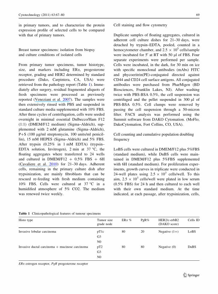
Table 1 Clinicopathological features of tumour specimens
Histo type Tumor size
grade node
ERa % PgR% HER2/c-erbB2
(DAKO score)
Cells ID
Invasive lobular carcinoma pT1c
G3
N0
80 20 Negative (1?) LoBS
Invasive ductal carcinoma ? mucinose carcinoma pT2
G3
N0
80 80 Negative (0) DuBS
ERa estrogen receptor, PgR progesterone receptor
Cytotechnology (2011) 63:67–80 69
123
maintained in sterile environment, were counted in a
hemocytometer chamber, and re-plated 1:2 to new
wells. Growth experiments were performed two times
per sample. To determine the proliferation rate of the
cells, we measured the cumulative population dou-
bling frequency (cpdf) in continuous subculture from
a known number of cells. The cpdf at each subcul-
tivation was calculated from the cell count by the
formula: Ln(No/Nn)/Ln2, where Ln is the natural log
and No and Nn are, respectively, initial and final cell
numbers. The sum of cpdf of subcultivation periods
result in the final total counts.
Sphere formation assay and growth in soft agar
To test the ability of LoBS and DuBS cells to form
mammospheres, cells were dissociated and seeded,
by serial dilution, in ultra low attachment surface
24-well plates in DMEM/F12 plus 0.5% FCS medium
(low serum). The final cell dilutions ranged from 10
to 1,000 cells/well. Sphere formation experiments
were carried out at least four times per sample.
For colony growth in soft agar spheres were
trypsinized, counted, and 104 cells/dish were plated in
60 mm triplicate dishes with 0.3% agar on a 0.5%
agar (Type I, Sigma) underlayer DMEM/F12 con-
taining 0.5% FBS. Colonies, cultured for 60 days,
were counted in ten fields per dish. The fields to be
counted were ID numbered fields on a 7 9 7
horizontal-vertical transparency grid of 60 mm of
diameter. The same ID fields were counted for all the
dishes. Results reported are mean ± SEM of three
different experiments performed in triplicate.
Protein extraction and western blot analysis
Western blots were performed, at least three times, on
LoBS and DuBS extracts as indicated in the figures.
MCF7 cells and MDA-MB231 served as controls.
Spheres, transferred for 21–30 days to cell culture
dishes, were adherent and proliferating; floating
aggregates were detached by trypsin–EDTA, pooled
and proteins were extracted. Protein preparations
were obtained by lysing samples in 50 mM TRIS pH
7.5, 100 mM NaCl, 1% Nonidet P40, 0.1% Triton,
2 mM EDTA, 10 lg/ml aprotinin, 100 lg/ml phe-
nylmethylsulfonyl-fluoride. Protein concentration
was measured by the Bio-Rad protein assay (Milan,
Italy) and polyacrylamide gels (from 8 to 15%) were
prepared as described (Veneziani et al. 2007).
Prestained molecular weight standards were from
Biorad (Milan, Italy). Proteins separated on the
polyacrylamide gels were blotted on a nitrocellulose
membrane (Hybond-C pure, Amersham Italia, Milan,
Italy). The membrane was stained with Ponceau S
(Sigma) to locate the molecular weight markers. The
membranes were stained with secondary antisera,
conjugated with horse radish peroxidase (Sigma–
Aldrich Corp.) diluted 1:2,000. The luminescent
signal was visualized with the ECL Western blotting
detection reagent kit (Amersham, Italy) and quanti-
fied by scanning with a Discover Pharmacia scanner
equipped with a Sun Spark Classic Workstation.
Data analysis
As indicated, flow cytometry, cell counting, sphere
formation assay, growth in soft agar, and western
blots experiments were carried out 2–4 times and
found to be reproducible. Human tissue samples were
not pooled, therefore each sample served as its own
control. Error bars are presented as standard error of
the mean (SEM). We used analysis of variance to
identify statistically significant differences among
means.
Results
Isolation and propagation of spheres from breast
cancer specimens
The cultures of LoBS cells derived from a specimen
of breast carcinoma were histologically classified as
lobular, pT1c (1–2 cm2), grade 3, node negative
(N0); the cultures of DuBS cells derived from a
specimen of breast carcinoma were histologically
classified as mixed ductal mucinous, pT2 (2–5 cm2),
grade 3, node negative (N0); both specimens were
estrogen receptor positive and cerbB2/Neu/HER2-
negative (Table 1) (Hammond et al. 2010).
From primary cultures we isolated and cultured
human breast cancer sphere-like aggregates of cells
in DMEM/F12 plus 0.5%FBS and 6H to disadvantage
fibroblasts adhesion and proliferation. Within
3–4 weeks the cultures formed aggregates of growing
cells, whose morphology is shown in Fig. 1. We
detached these cells with a gentle trypsinization
70 Cytotechnology (2011) 63:67–80
123

(2 min at 37 �C), and collected only the floating
aggregates (300 cells/aggregate) to propagate cells.
To this aim we conducted experiments to define
culture conditions (data not shown) and found that the
cells derived from lobular carcinoma (LoBS cells)
adapted to grow in DMEM/F12 plus 5%FBS, and
cells derived from ductal carcinoma (DuBS cells)
were adapted to grow in DMEM/F12?5%FBS sup-
plemented with 6H. When plated in their own
medium, the cells attached and, after 3–4 weeks,
formed new aggregates of growing cells. This cell
propagating procedure reduced doubling time and
generated long-term cultures (30 months, more than
60 passages).
We next evaluated whether the cell aggregates
possessed the ability to form spheres. To this aim, we
passed, by serial dilution, enzymatically digested
aggregates in low-attachment plates with low serum
(0.5% FBS); we found that, within few days (2–3),
when wells contained 100 cells and more, cells
formed floating aggregates that had the characteristic
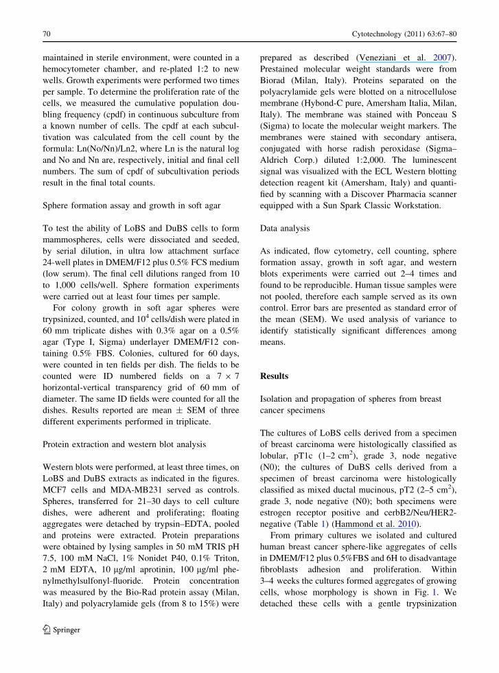
morphology of floating mammospheres (Fig. 2). In
these floating conditions, cell counting showed that,
during the initial 4–6 weeks of culture, cells prolif-
erate at low rate, with a doubling time of 15 days,
while during subsequent long term culture
(10–12 weeks) the cells arrested growth. This process
is reversible because, as expected, when mammo-
spheres were plated in adherent culture dishes, the
cells adhered to the dish and reached confluence
(Fig. 3).
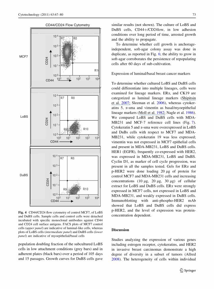
Identification of CD44?/CD24low cells
In breast cancer CD44?/CD24low cells are suggested
as a population of cells that contain potential breast
cancer stem cells (Al-Hajj et al. 2003; Dontu et al.
2003; Ponti et al. 2005). We evaluated the expression
of CD44 and CD24 by flow cytometry. To this aim
we collected and analyzed cell floating aggregates
which represent a small fraction of all the cells in the
dissected tissue (less than 10–15% of the total cell
number). As shown (Fig. 4), MCF7 control cells
consisted mainly of CD24? expressing cells (lumi-
nal-like), whereas LoBS cells and DuBS cells, as
indicated, consisted of CD44?/CD24low cell popu-
lations (myoepithelial and basal cells). This result
suggest that the floating aggregates are enriched for
CD44?/CD24low cells, and that the CD44?/
CD24low phenotype is not related to the tumor
histotype since cells displaying features of stem-like/
progenitor can be isolated from either lobular and
ductal breast cancer specimens.
Growth properties of CD44?/CD24low cells
To evaluate whether the CD44?/CD24low cells were
able to proliferate in different culture conditions, we
measured the growth rate of the cells dissociated
from mammospheres in adherent and non-adherent
conditions. To this aim cells were enzymatically
detached to single-cell suspension and 2 9 104 cells
were seeded in DMEM/F12 plus 0.5% FBS medium
50 µm 50 µm
Primary co-cultureDuctal Lobular
Fig. 1 Sphere-like aggregates in primary cultures. Primary
co-cultures of ductal (left) and lobular (right) dissociated
specimens, maintained in low serum and 6H, to form
aggregates. Aggregates transferred, one per well, to 24-well
dishes, within few days, adhere to the well and reach
confluence. (Phase-contrast microscopy)
Cytotechnology (2011) 63:67–80 71
123
(low serum) either in ultra low attachment or in
adherent 24-well plates for 24 h. The medium was
then substituted with standard medium, i.e. 5%FBS
for LoBS cells and 5%FBS?6H for DuBS cells. The
effect of adhesion on the long-term growth rates is
depicted in Fig. 5. The graph shows the cumulative
DuBS LoBS
Non-adherent culture
50 µm 50 µm
C
100 µm
D
100 µm
B
A
Fig. 2 Spheres under non-
adherent culture conditions.
Cells from confluent wells
are transferred, 100 cells/
well, to low-attachment
multiwells. Within few days
DuBS cells (left panels,
a higher and b lower
magnification) and LoBS
cells (right panels, c higher
and d lower magnification)
form floating spheroids. In
these conditions, cells
initially proliferate at low
rate (doubling time of
15 days), then arrest
growth. (Phase-contrast
microscopy)
DuBS cells LoBS cells
Adherent culture
100 µm
B
100 µm
D
50 µm
A
50 µm
C
Fig. 3 Spheres in adherent
culture conditions. Spheres
of DuBS (left panels,
a higher and b lower
magnification) and LoBS
cells (right panels, c higher
and d lower magnification)
transferred to cell culture
dishes, adhere to the dish
and restart proliferation.
Cells, detached from
adherent wells are pooled
and analyzed for flow
cytometry and protein
expression. (Phase-contrast
microscopy)
72 Cytotechnology (2011) 63:67–80
123
population doubling fraction of the subcultured LoBS
cells in low attachment conditions (grey bars) and in
adherent plates (black bars) over a period of 105 days
and 15 passages. Growth curves for DuBS cells gave
similar results (not shown). The culture of LoBS and
DuBS cells, CD44?/CD24low, in low adhesion
conditions over long period of time, arrested growth
and the ability to propagate.
To determine whether cell growth is anchorage-
independent, soft-agar colony assay was done in
duplicate, as reported in Fig. 6; the ability to grow in
soft-agar corroborates the persistence of repopulating
cells after 60 days of sub-cultivation.
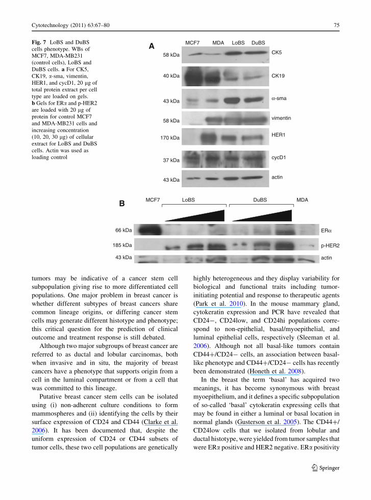
Expression of luminal/basal breast cancer markers
To determine whether cultured LoBS and DuBS cells
could differentiate into multiple lineages, cells were
examined for lineage markers. ERa, and CK19 are
categorized as luminal lineage markers (Shipitsin
et al. 2007; Sleeman et al. 2006), whereas cytoker-
atins 5, a-sma and vimentin as basal/myoepithelial
lineage markers (Moll et al. 1982; Nagle et al. 1986).
We compared LoBS and DuBS cells with MDA-
MB231 and MCF-7 reference cell lines (Fig. 7).
Cytokeratin 5 and a-sma were overexpressed in LoBS
and DuBs cells with respect to MCF7 and MDA-
MB231, while cytokeratin 19 was less expressed;
vimentin was not expressed in MCF7 epithelial cells
and present in MDA-MB231, LoBS and DuBS cells.
HER1 (EGFR), frequently co-expressed with HER2,
was expressed in MDA-MB231, LoBS and DuBS.
Cyclin D1, as marker of cell cycle progression, was
present in all the samples tested. Gels for ERa and
p-HER2 were done loading 20 lg of protein for
control MCF7 and MDA-MB231 cells and increasing
concentrations (10 lg, 20 lg, 30 lg) of cellular
extract for LoBS and DuBS cells. ERa were strongly
expressed in MCF7 cells, not expressed in LoBS and
MDA-MB231, and weakly expressed in DuBS cells.
Immunoblotting with anti-phospho-HER2 mAb
showed that LoBS and DuBS cells did express
p-HER2, and the level of expression was protein-
concentration dependent.
Discussion
Studies analyzing the expression of various genes
including estrogen receptor, cytokeratins, and HER2
in invasive breast carcinomas demonstrate a high
degree of diversity in a subset of tumors (Allred
2008). The heterogeneity of cells within individual
CD44/CD24 Flow Cytometry
MCF7
LoBS
CD44
CD
24
CD44
CD
24
DuBS
CD44
CD
24
Fig. 4 CD44/CD24 flow cytometry of control MCF7, of LoBS
and DuBS cells. Sample cells and control cells were detached
incubated with specific monoclonal antibodies against CD44
and CD24 cell surface antigens. FACS plots of MCF7 control
cells (upper panel) are indicative of luminal-like cells, whereas
plots of LoBS cells (intermediate panel) and DuBS cells (lowerpanel) are indicative of myoepithelial/basal cells
Cytotechnology (2011) 63:67–80 73
123

0
50
100
150
200
250
300
350
1 2 3 4 5 6 7 8 9 10 11 12 13 14 15
7 14 21 28 35 42 49 56 63 70 77 84 91 98 105
passage
days
AdherentNon Adherent
Long-term culture
Cum
ulat
ive
popu
latio
n do
ublin
g fr
eque
ncy
Fig. 5 Long-term growth curve of LoBS cells under non-
adherent and adherent conditions. 2.5 9 103 cells/well, were
cultured with standard medium for 105 days. At each passage,
cells were enzymatically dissociated to single cells from each
triplicate well in a sterile environment, counted, and passaged
1:2 to new wells
Growth in soft agar
60 days0
10
20
30
40
50
60
70
80
90
100
nu
mb
er o
f co
lon
ies
/ dis
h
LoBS
DuBS
LoBS
DuBS
100 µm
100 µm
Fig. 6 Colony growth in
soft agar of LoBS and
DuBS cells. Left,microphotographs of LoBS
and DuBS cells in soft-agar.
Right, bar-graph of number
of colonies; colonies were
counted in ten fields per
dish on an a horizontal-
vertical grid
74 Cytotechnology (2011) 63:67–80
123
tumors may be indicative of a cancer stem cell
subpopulation giving rise to more differentiated cell
populations. One major problem in breast cancer is
whether different subtypes of breast cancers share
common lineage origins, or differing cancer stem
cells may generate different histotype and phenotype;
this critical question for the prediction of clinical
outcome and treatment response is still debated.
Although two major subgroups of breast cancer are
referred to as ductal and lobular carcinomas, both
when invasive and in situ, the majority of breast
cancers have a phenotype that supports origin from a
cell in the luminal compartment or from a cell that
was committed to this lineage.
Putative breast cancer stem cells can be isolated
using (i) non-adherent culture conditions to form
mammospheres and (ii) identifying the cells by their
surface expression of CD24 and CD44 (Clarke et al.
2006). It has been documented that, despite the
uniform expression of CD24 or CD44 subsets of
tumor cells, these two cell populations are genetically
highly heterogeneous and they display variability for
biological and functional traits including tumor-
initiating potential and response to therapeutic agents
(Park et al. 2010). In the mouse mammary gland,
cytokeratin expression and PCR have revealed that
CD24-, CD24low, and CD24hi populations corre-
spond to non-epithelial, basal/myoepithelial, and
luminal epithelial cells, respectively (Sleeman et al.
2006). Although not all basal-like tumors contain
CD44?/CD24- cells, an association between basal-
like phenotype and CD44?/CD24- cells has recently
been demonstrated (Honeth et al. 2008).
In the breast the term ‘basal’ has acquired two
meanings, it has become synonymous with breast
myoepithelium, and it defines a specific subpopulation
of so-called ‘basal’ cytokeratin expressing cells that
may be found in either a luminal or basal location in
normal glands (Gusterson et al. 2005). The CD44?/
CD24low cells that we isolated from lobular and
ductal histotype, were yielded from tumor samples that
were ERa positive and HER2 negative. ERa positivity
CK5
α-sma
actin
CK19
cycD1
HER1
vimentin
MCF7 DuBSLoBS
p-HER2
58 kDa
43 kDa
43 kDa
40 kDa
37 kDa
170 kDa
58 kDa
185 kDa
MCF7 LoBS DuBS MDA
43 kDa actin
ERα66 kDa
A
B
MDAFig. 7 LoBS and DuBS
cells phenotype. WBs of
MCF7, MDA-MB231
(control cells), LoBS and
DuBS cells. a For CK5,
CK19, a-sma, vimentin,
HER1, and cycD1, 20 lg of
total protein extract per cell
type are loaded on gels.
b Gels for ERa and p-HER2
are loaded with 20 lg of
protein for control MCF7
and MDA-MB231 cells and
increasing concentration
(10, 20, 30 lg) of cellular
extract for LoBS and DuBS
cells. Actin was used as
loading control
Cytotechnology (2011) 63:67–80 75
123

and HER2 negativity are features of tumors defined as
‘‘luminal A’’ according to molecular profiling (Perou
et al. 2000), while carcinomas belonging to the basal-
like subtype of breast cancers are assumed to be stem
cell-derived or to have acquired properties of stem
cells during transformation (Sorlie et al. 2001). In
general, long-term culture leads to selection for basal/
myoepithelial cells. All established human breast
epithelial cell lines display a partial loss of the
myoepithelial differentiation program along with a
partial gain of a luminal differentiation program.
Although in vivo equivalent of these cells has not been
found, this particular culture profile is thought to
reflect the existence of human breast epithelial stem
cells in the basal compartment (Petersen and Polyak
2010). The cancer stem cell hypothesis postulates that
tumors are driven by cells that display the properties of
self-renewal and differentiation found in stem cells.
Self-renewal is demonstrated by the ability of the
enriched stem cell populations to form mammospheres
which can grow in an anchorage-independent manner.
The long-term culture of LoBS and DuBS cells under
non-adherent culture conditions generates mammo-
spheres that initially proliferate, albeit at low rate, then
arrest growth; these resting, non proliferating cells,
when transferred to adherent dishes, may still be
propagated. In addition, their growth is anchorage
independent because single cells, dissociated from
mammospheres, grow in soft agar. The low growth
rate in non-adherent conditions could be due to
nutrient and oxygen limitation for the cells within
the sphere; indeed these cells, potentially proliferating,
restart growth when allowed to form an adherent
monolayer.
Myoepithelial cells are cells from the basal layer,
defined as cells that express both epithelial charac-
teristics and contractile proteins (Anbazhagan et al.
1998) These cells are distinguished from basal cells
in stratified squamous epithelium because they
exhibit features of mesenchymal cells, including
expression of vimentin (Ronnov-Jessen et al. 1995),
smooth muscle actin (a-sma) (Gusterson et al. 1982),
the high-molecular-weight cytokeratin CK5 (cur-
rently known as basal cytokeratin) (Moll et al.
1982; Nagle et al. 1986). However, studies aimed to
define the cell of origin in experimental model
systems and in breast tumors show that intermediate
filament expression can be modulated in tissue
culture and expression of CK5 is not restricted to
myoepithelial cells (Gusterson et al. 2005). Recent
studies showed that these cells may represent early
progenitors and are candidates for precursor cells of
basal-like breast cancer (Ginestier et al. 2007;
Villadsen et al. 2007; Lim et al. 2009; Pece et al.
2010). As expected, our long-term cultured LoBS and
DuBS cells express vimentin, CK5, and smooth
muscle actin (a-sma), all markers of basal/myoepi-
thelial cells.
The majority of human breast cancer cell lines, as
well as MCF7 and MDA-MB231, are cytokeratin 19
(CK19) positive. All established breast epithelial cell
lines from long-term, primary-cell-derived cultures of
reductive mammoplasties are completely cytokeratin
CK19-negative (Bartek et al. 1985), and CK19
staining of luminal cells in the fetal breast is
homogeneously positive (Anbazhagan et al. 1998).
However, recent developments in cell culture tech-
nology have allowed long-term expansion of CK19-
positive cells from primary tissues in the absence of
immortalization (Garbe et al. 2009). With respect to
MCF7 and MDA-MB231, LoBS and DuBS cells are
weakly CK19-positive thus reinforcing the idea that
these cells are likely to be originated from the basal
layer.
The lack of the CK19 lineage differentiation is
consistent with the lack of estrogen receptor (ERa) in
the nonmalignant precursor cells (Petersen and
Polyak 2010). In the adult virgin mouse mammary
epithelium the majority of stem/progenitor cell activ-
ity is located in the basal compartment (Shackleton
et al. 2006; Sleeman et al. 2006; Stingl et al. 2006)
and the ERa-positive cells are distinct from the
mammary stem cell population (Asselin-Labat et al.
2008; Sleeman et al. 2007). In the adult mammary
gland only a subset of cells express ERa; these cells
can either form a stem cell compartment that is
directly stimulated by circulating hormones (Clarke
et al. 2005) or ERa-positive cells may stimulate
proliferation of a separate stem cell compartment in a
paracrine manner (Brisken and Duss 2007). Pulse-
chase experiments suggest that ERa-positive cells
form a slowly cycling cell compartment (Booth and
Smith 2006) and the stimulation with estrogens
down-regulates ERa before the proliferative response
(Chen et al. 2008), indeed ERa-positive cells do not
express markers of proliferation (Clarke et al. 2005).
ERa-expressing luminal epithelial subpopulation
contains little in vivo stem cell activity. The hormone
76 Cytotechnology (2011) 63:67–80
123
susceptibility of mammary epithelial stem cells has
been extensively investigated. Notwithstanding, there
are conflicting reports regarding the hormone recep-
tor status of these cells in both mouse and human
experiments. In mouse, pulse-chase experiments
(Booth and Smith 2006) predicted that the putative
mouse mammary epithelial stem cells were ERa-
positive, while other studies (Asselin-Labat et al.
2008) reported that stem cells identified by specific
cell surface markers were negative for both ERa and
HER2. Similar divergent results have been described
for human mammary epithelial stem cells (Oliveira
et al. 2010). To ascertain that the lack of ERa was due
to the lack of expression we performed WB exper-
iments loading increasing concentration of cellular
extract. We show that, with respect to MCF7, LoBS
cells do not express ERa, while DuBS cells do
express the receptor in a concentration-dependent
fashion. Breast cancer is not a single disease, but
rather a group of diseases displaying heterogeneous
features at both the molecular and clinical level, and
each subtype has its own stable phenotype maintained
during tumor evolution (Dontu et al. 2003; Shipitsin
et al. 2007; Sorlie et al. 2001). Our data confirm the
divergence, that may be presumed to reflect the well-
established heterogeneity of breast cancer.
Epidermal growth factor receptor (HER1) and
erbB2 (HER2) are two RTKs with oncogenic prop-
erties, and two of the most targeted oncoproteins in
cancer. HER1 and HER2 overexpression is generally
associated with more aggressive tumors and poor
prognosis (Sorlie et al. 2003; Sotiriou et al. 2003;
Suzuki et al. 2008). Overexpression of HER2 occurs
in about 20% of breast carcinomas, and is more
frequent in estrogen receptor-negative than in estro-
gen receptor-positive cases (Sotiriou et al. 2003). In
patients with ERa-positive tumors and active growth
factor receptor signaling there is growing evidence
that crosstalk between ERa and growth factor recep-
tor signaling pathways, especially the HER family, is
one of the mechanisms for resistance to endocrine
therapy in breast cancer (Knowlden et al. 2003;
Osborne et al. 2005). In ERa-positive and HER2-
positive metastatic breast cancer two trials demon-
strated that the strategy of combining hormone
deprivation with antiHER therapy, i.e., anastrozole
and herceptin (Kaufman et al. 2009) or letrozole and
lapatinib (Johnston et al. 2009), significantly pro-
longed PFS (Progression Free Survival, defined as
time from random assignment until the earliest date
of disease progression or death as a result of any
cause in the HER2-positive population). In experi-
mental models of ERa-positive breast cancer cells,
initially HER2 negative and hormone-responsive,
EGFR and HER2 pathways may become upregulated
on development of endocrine resistance over time
and a combined hormonal-and growth factor recep-
tor-targeted treatment may delay acquired resistance
(Schiff and Osborne 2005; Massarweh et al. 2008). It
has been reported that activated phospo-HER2 is
expressed also in HER2-negative tumors (Frogne
et al. 2009; Singer et al. 2009), and expression of
activated HER2 is associated with poor prognosis in
the series of hormone receptor-positive breast cancer
patients (Frogne et al. 2009). We report data demon-
strating the expression of HER1 and activated HER2
in the LoBS and DuBS cells; this supports the idea
that subpopulations of cells expressing activated
RTK, do exist within the context of a tumor mass
that exhibits features of luminal A breast cancer, i.e.,
ERa-positivity and HER2-negativity. This observa-
tion confirms other reports that HER2 expression
increases the stem cell population of normal and
malignant mammary cells and that the effects of
HER2 overexpression on mammary tumorigenesis
and invasion are mainly due to its effects on the stem
cells population (Korkaya et al. 2008). Our results are
also in agreement with studies reporting that HER2-
negative tumors express an amount of activated
HER2 sufficient to elicit signal transduction, and may
explain why some patients with HER2-negative
tumors are responsive to herceptin (Paik et al. 2008).
The expression profiling classification of breast
cancer into five main molecular subtypes (basal,
luminal A and B, HER2-positive/ERa-negative, and
normal breast-like) (Sorlie et al. 2001) correlates with
clinical outcome, is predictive of response to treat-
ment and prognostic (Sorlie et al. 2001; van de Vijver
et al. 2002; Shipitsin et al. 2007). Our approach
suggests that the molecular classification can be
enriched of intermediate or new subtypes as new
methods for the quantitative assessment of intratumor
diversity are introduced in clinical practice.
Acknowledgments We declare no conflict of interest. This
work was supported by: Associazione Italiana per la Ricerca
sul Cancro, CRPO Centro Regionale di Prevenzione
Oncologica, Regione Campania, Ministero dell’Universita‘ e
Ricerca, and Ministero della Salute, Italia.
Cytotechnology (2011) 63:67–80 77
123
References
Al-Hajj M, Wicha MS, Benito-Hernandez A, Morrison SJ,
Clarke MF (2003) Prospective identification of tumori-
genic breast cancer cells. Proc Natl Acad Sci USA
100:3983–3988
Allred DC (2008) The utility of conventional and molecular
pathology in managing breast cancer. Breast Cancer Res
10 (suppl 4):S4
Anbazhagan R, Osin PP, Bartkova J, Nathan B, Lane EB,
Gusterson BA (1998) The development of epithelial
phenotypes in the human fetal and infant breast. J Pathol
184:197–206
Asselin-Labat ML, Vaillant F, Shackleton M, Bouras T,
Lindeman GJ, Visvader JE (2008) Delineating the epi-
thelial hierarchy in the mouse mammary gland. Cold
Spring Harb Symp Quant Biol 73:469–478
Bartek J, Taylor-Papadimitriou J, Miller N, Millis R (1985)
Patterns of expression of keratin 19 as detected with
monoclonal antibodies in human breast tissues and
tumours. Int J Cancer 36:299–306
Booth BW, Smith GH (2006) Estrogen receptor-alpha and
progesterone receptor are expressed in label-retaining
mammary epithelial cells that divide asymmetrically and
retain their template DNA strands. Breast Cancer Res
8:R49
Brisken C, Duss S (2007) Stem cells and the stem cell niche in
the breast: an integrated hormonal and developmental
perspective. Stem Cell Rev 3:147–156
Campbell LL, Polyak K (2007) Breast tumor heterogeneity:
cancer stem cells or clonal evolution? Cell Cycle
37:2332–2338
Cavaliere C, Corvigno S, Galgani M, Limite G, Nardone A,
Veneziani BM (2010) Combined inhibitory effect of for-
mestane and herceptin on a subpopulation of CD44?/
CD24low breast cancer cells. Cancer Sci 107:1661–1669
Chen ST, Dou J, Temple R, Agarwal R, Wu KM, Walker S
(2008) New therapies from old medicines. Nat Biotechnol
26:1077–1083
Clarke RB, Spence K, Anderson E, Howell A, Okano H, Potten
CS (2005) A putative human breast stem cell population is
enriched for steroid receptor-positive cells. Dev Biol
277:443–456
Clarke MF, Dick JE, Dirks PB, Eaves CJ, Jamieson CHM,
Jones DL, Visvader J, Weissman IL, Wahl GM (2006)
Cancer stem cells-perspectives on current status and
future directions: AACR workshop on cancer stem cells.
Cancer Res 66:9339–9344
Collins AT, Berry PA, Hyde C, Stower MJ, Maitland NJ
(2005) Prospective identification of tumorigenic prostate
cancer stem cells. Cancer Res 65:10946–10951
Dontu G, Abdallah WM, Foley JM, Jackson KW, Clarke MF,
Kawamura MJ, Wicha MS (2003) In vitro propagation
and transcriptional profiling of human mammary stem/
progenitor cells. Genes Dev 17:1253–1270
Dowsett M, Houghton J, Iden C, Salter J, Farndon J, A’Hern R,
Sainsbury R, Baum M (2006) Benefit from adjuvant
tamoxifen therapy in primary breast cancer patients
according oestrogen receptor, progesterone receptor, EGF
receptor and HER2 status. Ann Oncol 17:818–826
Frogne T, Laenkholm AV, Lyng MB, Henriksen KL,
Lykkesfeldt AE (2009) Determination of HER2 phos-
phorylation at tyrosine 1221/1222 improves prediction of
poor survival for breast cancer patients with hormone
receptor-positive tumors. Breast Cancer Res 11:R11
Garbe JC, Bhattacharya S, Merchant B, Bassett E, Swisshelm
K, Feiler HS, Wyrobek AJ, Stampfer MR (2009) Molec-
ular distinctions between stasis and telomere attrition
senescence barriers shown by long-term culture of normal
human mammary epithelial cells. Cancer Res 69:
7557–7568
Ginestier C, Hur MH, Charafe-Jauffret E, Monville F, Dutcher
J, Brown M, Jacquemier J, Viens P, Kleer CG, Liu S,
Schott A, Hayes D, Birnbaum D, Wicha MS, Dontu G
(2007) ALDH1 is a marker of normal and malignant
human mammary stem cells and a predictor of poor
clinical outcome. Cell Stem Cell 1:555–567
Gusterson BA, Warburton MJ, Mitchell D, Ellison M, Neville
AM, Rudland PS (1982) Distribution of myoepithelial
cells and basement membrane proteins in the normal
breast and in benign and malignant breast disease. Cancer
Res 42:4763–4770
Gusterson BA, Ross DT, Heath VJ, Stein T (2005) Basal
cytokeratins and their relationship to the cellular origin
and functional classification of breast cancer. Breast
Cancer Res 7:143–148
Hammond ME, Hayes DF, Dowsett M, Allred DC, Hagerty
KL, Badve S, Fitzgibbons PL, Francis G, Goldstein NS,
Hayes M, Hicks DG, Lester S, Love R, Mangu PB,
McShane L, Miller K, Osborne CK, Paik S, Perlmutter J,
Rhodes A, Sasano H, Schwartz JN, Sweep FC, Taube S,
Torlakovic EE, Valenstein P, Viale G, Visscher D,
Wheeler T, Williams RB, Wittliff JL, Wolff AC (2010)
American society of clinical oncology/College of Amer-
ican pathologists guideline recommendations for immu-
nohistochemical testing of estrogen and progesterone
receptors in breast cancer. Arch Pathol Lab Med
134:907–922
Honeth G, Bendahl PO, Ringner M, Saal LH, Gruvberger-Saal
SK, Lovgren K, Grabau D, Ferno M, Borg A, Hegardt C
(2008) The CD44?/CD24- phenotype is enriched in
basal-like breast tumors. Breast Cancer Res 10:R53
Johnston S, Pippen J Jr, Pivot X, Lichinitser M, Sadeghi S,
Dieras V, Gomez HL, Romieu G, Manikhas A,
Kennedy MJ, Press MF, Maltzman J, Florance A,
O’Rourke L, Oliva C, Stein S, Pegram M (2009)
Lapatinib combined with letrozole versus letrozole and
placebo as first-line therapy for postmenopausal hor-
mone receptor-positive metastatic breast cancer. J Clin
Oncol 27:5538–5546
Kaufman B, Mackey JR, Clemens MR, Bapsy PP, Vaid A,
Wardley A, Tjulandin S, Jahn M, Lehle M, Feyereislova
A, Revil C, Jones A (2009) Trastuzumab plus anastrozole
versus anastrozole alone for the treatment of postmeno-
pausal women with human epidermal growth factor
receptor 2-positive, hormone receptor-positive metastatic
breast cancer: results from the randomized phase III
TAnDEM study. J Clin Oncol 27:5529–5537
Knowlden JM, Hutcheson IR, Jones HE, Madden T, Gee JM,
Harper ME, Barrow D, Wakeling AE, Nicholson RI
78 Cytotechnology (2011) 63:67–80
123

(2003) Elevated levels of epidermal growth factor recep-
tor/c-erbB2 heterodimers mediate an autocrine growth
regulatory pathway in tamoxifen-resistant MCF-7 cells.
Endocrinology 144:1032–1044
Korkaya H, Paulson A, Iovino F, Wicha MS (2008) HER2
regulates the mammary stem/progenitor cell population
driving tumorigenesis and invasion. Oncogene
27:6120–6130
Lim E, Vaillant F, Wu D, Forrest NC, Pal B, Hart AH, Asselin-
Labat ML, Gyorki DE, Ward T, Partanen A, Feleppa F,
Huschtscha LI, Thorne HJ, ConFab FoxSB, Yan M,
French JD, Brown MA, Smyth GK, Visvader JE, Lind-
eman GJ (2009) Aberrant luminal progenitors as the
candidate target population for basal tumor development
in BRCA1 mutation carriers. Nat Med 15:907–913
Massarweh S, Osborne CK, Creighton CJ, Qin L, Tsimelzon A,
Huang S, Weiss H, Rimawi M, Schiff R (2008) Tamox-
ifen resistance in breast tumors is driven by growth factor
receptor signaling with repression of classic estrogen
receptor genomic function. Cancer Res 68:826–833
Moll R, Franke WW, Schiller DL (1982) The catalog of human
cytokeratins: patterns of expression in normal epithelia,
tumors and cultured cells. Cell 31:11–24
Nagle RB, Bocker W, Davis JR, Heid HW, Kaufmann M,
Lucas DO, Jarasch ED (1986) Characterization of breast
carcinomas by two monoclonal antibodies distinguishing
myoepithelial from luminal epithelial cells. J Histochem
Cytochem 34:869–881
Nardone A, Cavaliere C, Corvigno S, Limite G, De Placido S,
Veneziani BM (2009) A banking strategy toward cus-
tomized therapy in breast cancer. Cell Tissue Bank
10:301–308
Oliveira LR, Jeffrey SS, Ribeiro-Silva A (2010) Stem cells in
human breast cancer. Histol Histopathol 25:371–385
Osborne CK, Shou J, Massarweh S, Schiff R (2005) Crosstalk
between estrogen receptor and growth factor receptor
pathways as a cause for endocrine therapy resistance in
breast cancer. Clin Cancer Res 11:865s–870s
Paik S, Kim C, Wolmark N (2008) HER2 status and benefit
from adjuvant trastuzumab in breast cancer. N Engl J Med
358:1409–1411
Park SY, Gonen M, Kim HJ, Michor F, Polyak K (2010)
Cellular genetic diversity in the progression of in situ
human breast carcinomas to an invasive phenotype. J Clin
Invest 120:636–644
Pece S, Tosoni D, Confalonieri S, Mazzarol G, Vecchi M,
Ronzoni S, Bernard L, Viale G, Pelicci PG, Di Fiore PP
(2010) Biological and molecular heterogeneity of breast
cancers correlates with their cancer stem cell content. Cell
140:62–73
Perou CM, Sorlie T, Eisen MB, van de Rijn M, Jeffrey SS,
Rees CA, Pollack JR, Ross DT, Johnsen H, Akslen LA,
Fluge O, Pergamenschikov A, Williams C, Zhu SX,
Lonning PE, Borresen-Dale AL, Brown PO, Botstein D
(2000) Molecular portraits of human breast tumours.
Nature 406:747–752
Petersen OW, Polyak K (2010) Stem cells in the human breast.
Cold Spring Harb Perspect Biol 2. doi: 10.1101/
cshperspect.a003160
Ponti D, Costa A, Zaffaroni N, Pratesi G, Petrangolini G,
Coradini D, Pilotti S, Pierotti MA, Daidone MG (2005)
Isolation and in vitro propagation of tumorigenic breast
cancer cells with stem/progenitor cell properties. Cancer
Res 65:5506–5511
Ronnov-Jessen L, Petersen OW, Koteliansky VE, Bissell MJ
(1995) The origin of the myofibroblasts in breast cancer.
Recapitulation of tumor environment in culture unravels
diversity and implicates converted fibroblasts and recrui-
ted smooth muscle cells. J Clin Invest 95:859–873
Schiff R, Osborne CK (2005) Endocrinology and hormone
therapy in breast cancer: new insight into estrogen
receptor-alpha function and its implication for endocrine
therapy resistance in breast cancer. Breast Cancer Res
7:205–211
Shackleton M, Vaillant F, Simpson KJ, Stingl J, Smyth GK,
Asselin-Labat ML, Wu L, Lindeman GJ, Visvader JE
(2006) Generation of a functional mammary gland from a
single stem cell. Nature 439:84–88
Shipitsin M, Campbell LL, Argani P, Weremowicz S,
Bloushtain-Qimron N, Yao J, Nikolskaya T, Serebryis-
kaya T, Beroukhim R, Hu M, Halushka MK, Sukumar S,
Parker LM, Anderson KS, Harris LN, Garber JE,
Richardson AL, Schnitt SJ, Nikolsky Y, Gelman RS,
Polyak K (2007) Molecular definition of breast tumor
heterogeneity. Cancer Cell 11:259–273
Singer CF, Gschwantler-Kaulich D, Fink-Retter A, Pfeiler G,
Walter I, Hudelist G, Helmy S, Spiess AC, Lamm W,
Kubista E (2009) HER2 overexpression and activation,
and tamoxifen efficacy in receptor-positive early breast
cancer. J Cancer Res Clin Oncol 135:807–813
Sleeman KE, Kendrick H, Ashworth A, Isacke CM, Smalley
MJ (2006) CD24 staining of mouse mammary gland cells
defines luminal epithelial, myoepithelial/basal and non-
epithelial cells. Breast Cancer Res 8:R7
Sleeman KE, Kendrick H, Robertson D, Isacke CM, Ashworth
A, Smalley MJ (2007) Dissociation of estrogen receptor
expression and in vivo stem cell activity in the mammary
gland. J Cell Biol 176:19–26
Sorlie T, Perou CM, Tibshirani R, Aas T, Geisler S, Johnsen H,
Hastie T, Eisen MB, van de Rijn M, Jeffrey SS, Thorsen
T, Quist H, Matese JC, Brown PO, Botstein D, Eystein
Lonning P, Borresen-Dale AL (2001) Gene expression
patterns of breast carcinomas distinguish tumor subclasses
with clinical implications. Proc Natl Acad Sci USA
98:10869–10874
Sorlie T, Tibshirani R, Parker J, Hastie T, Marron JS, Nobel A,
Deng S, Johnsen H, Pesich R, Geisler S, Demeter J, Perou
CM, Lønning PE, Brown PO, Børresen-Dale AL, Botstein
D (2003) Repeated observation of breast tumor subtypes
in independent gene expression data sets. Proc Natl Acad
Sci USA 100:8418–8423
Sotiriou C, Neo SY, McShane LM, Korn EL, Long PM, Jazaeri
A, Martiat P, Fox SB, Harris AL, Liu ET (2003) Breast
cancer classification and prognosis based on gene
expression profiles from a population-based study. Proc
Natl Acad Sci USA 100:10393–10398
Stingl J, Eirew P, Ricketson I, Shackleton M, Vaillant F, Choi D,
Li HI, Eaves CJ (2006) Purification and unique properties of
mammary epithelial stem cells. Nature 439:993–997
Suzuki T, Miki Y, Ohuchi N, Sasano H (2008) Intratumoral
estrogen production in breast carcinoma: significance of
aromatase. Breast Cancer 15:270–277
Cytotechnology (2011) 63:67–80 79
123

van de Vijver MJ, He YD, van’t Veer LJ, Dai H, Hart AA,
Voskuil DW, Schreiber GJ, Peterse JL, Roberts C, Marton
MJ, Parrish M, Atsma D, Witteveen A, Glas A, Delahaye
L, van der Velde T, Bartelink H, Rodenhuis S, Rutgers
ET, Friend SH, Bernards R (2002) A gene-expression
signature as a predictor of survival in breast cancer.
N Engl J Med 347:1999–2009
Veneziani BM, Criniti V, Cavaliere C, Corvigno S, Nardone A,
Picarelli S, Tortora G, Ciardiello F, Limite G, De Placido
S (2007) In vitro expansion of human breast cancer epi-
thelial and mesenchymal stromal cells: optimization of a
coculture model for personalized therapy approaches. Mol
Cancer Ther 6:3091–3100
Villadsen R, Fridriksdottir AJ, Rønnov-Jessen L, Gudjonsson
T, Rank F, Labarge MA, Bissell MJ, Petersen OW (2007)
Evidence for a stem cell hierachy in the adult human
breast. J Cell Biol 177:87–101
80 Cytotechnology (2011) 63:67–80
123
Copyright © 2022 FDOKUMEN