Notch2 is required for progression of pancreatic intraepithelial neoplasia and development of...
6
Notch2 is required for progression of pancreatic intraepithelial neoplasia and development of pancreatic ductal adenocarcinoma Pawel K. Mazur a , Henrik Einwächter a , Marcel Lee a , Bence Sipos b , Hassan Nakhai c , Roland Rad d , Ursula Zimber-Strobl e , Lothar J. Strobl e , Freddy Radtke f , Günter Klöppel g , Roland M. Schmid a , and Jens T. Siveke a,1 a Second Department of Internal Medicine and g Institute of Pathology, Technical University of Munich, 81675 Munich, Germany; b Department of Pathology, University Hospital Tübingen, 72076 Tubingen, Germany; c Department of Biological Sciences, University of Warwick, Coventry CV4 7AL, United Kingdom; d The Wellcome Trust Sanger Institute, Cambridge CB10 1SA, United Kingdom; e Department of Gene Vectors, Helmholtz Center, 81377 Munich, Germany; and f Ecole Polytechnique Fédérale de Lausanne (EPFL SV ISREC), SV/Swiss Institute for Experimental Cancer Research, 1015 Lausanne, Switzerland Edited by Elliott Kieff, Harvard Medical School and Brigham and Women’s Hospital, Boston, MA, and approved June 8, 2010 (received for review February 26, 2010) Pancreatic cancer is one of the most fatal malignancies lacking effective therapies. Notch signaling is a key regulator of cell fate specification and pancreatic cancer development; however, the role of individual Notch receptors and downstream signaling is largely unknown. Here, we show that Notch2 is predominantly expressed in ductal cells and pancreatic intraepithelial neoplasia (PanIN) lesions. Using genetically engineered mice, we demonstrate the effect of conditional Notch receptor ablation in Kras G12D -driven pancreatic carcinogenesis. Deficiency of Notch2 but not Notch1 stops PanIN progression, prolongs survival, and leads to a phenotypical switch toward anaplastic pancreatic cancer with epithelial–mesenchymal transition. By expression profiling, we identified increased Myc sig- naling regulated by Notch2 during tumor development, placing Notch2 as a central regulator of PanIN progression and malignant transformation. Our study supports the concept of distinctive roles of individual Notch receptors in cancer development. genetically engineered mice | K-Ras | Myc | Notch | pancreatic cancer P ancreatic ductal adenocarcinoma (PDAC) remains a devas- tating disease despite tremendous therapeutical efforts. PDAC derives from several preneoplastic lesions, including pancreatic intraepithelial neoplasia (PanIN), intraductal papillary mucinous neoplasm, and mucinous cystic neoplasm (MCN), of which PanINs are the most common precursors (1). PanINs typically progress through defined histological and molecular stages, with the most advanced PanIN3 lesion being defined as carcinoma in situ (2). Because of early metastatic spread, PanIN3 represents the latest curable precursor lesion. Thus, defining the regulators of PanIN initiation and progression is of utmost importance. Recapitulation of human pancreatic carcinogenesis was greatly advanced by generating mice with pancreas-specific activation of endogenous oncogenic Kras G12D (3). The ongoing characteriza- tion of relevant signaling pathways in pancreatic carcinogenesis using genetically engineered mouse models has helped to depict the enormous plasticity in precursors to PDAC. Despite activa- tion of cell fate regulating signaling pathways such as Hedgehog, Wnt, and Notch signaling (3–9), the precise role of these pathways remains largely unclear. The Notch signaling pathway plays a pivotal role in cell fate and differentiation decisions, and its activation early in the carcino- genic process suggests a role in initiation of transformation. Al- though the cell of origin in PDAC has not been decisively identified, activation of Notch signaling during PanIN initiation probably presents a pivotal step for transformation. In several murine models of PDAC, expression of the Notch target gene Hes1 was increased in PanIN lesions (3, 5, 8, 9). In a recent study, chemical inhibition of Notch activation completely blocked tumor progression in vivo (10). Conversely, Murtaugh and co-workers (11) described a PanIN-promoting effect of Notch activation in Kras G12D -driven PanIN development. However, the specific role of individual Notch receptors and the downstream events have so far not been determined. Here, we describe the effect of pancreas-specific ablation of Notch1 and Notch2 in Kras G12D -driven pancreatic carcinogenesis, taking advantage of the nonessential role of Notch1 and Notch2 during pancreatogenesis (12). We show that Notch1 and Notch2 are expressed in pancreatic acinar and ductal cells, respectively. Conditional ablation of Notch2 but not Notch1 leads to an abro- gation of PanIN progression, development of MCN-like lesions, and increased survival. Identification of Notch2-regulated Myc signaling during carcinogenesis points to a central role of Notch2 in controlling PanIN progression and tumor differentiation. Results Notch1 and Notch2 Are Expressed in Different Compartments in Adult Pancreata and Are Activated in Kras Mice During PanIN Development. To determine the expression of members of the Notch signaling family during pancreatic carcinogenesis, Kras +/LSL-G12D mice were crossed to Ptf1a +/Cre(ex1) mice (referred to as Kras; Fig. S1C), as previously described (9). Notch1 and Notch2 were pre- dominantly expressed in whole-tissue mRNA from WT and Kras G12D -induced pancreata compared with low expression of Notch3 and Notch4 (Fig. 1A). In Kras pancreata at 9 wk of age, when only a few PanIN1 lesions are notable, increased expression of Notch2 and the Notch target gene Hes1 but not Notch1 was observed, similar to previous reports (5). During progression, we noted a significant increase in Notch2 and Hes1 expression, whereas Notch1 was further reduced. Notch3 was also increased, albeit at lower total expression levels (Fig. 1B). This expression pattern correlated well with an increase in CK19 and a decrease in amylase expression, suggesting that Notch2 is expressed in CK19 + PanINs, whereas Notch1 may be predominantly expressed in acinar cells. To test this hypothesis, we used transgenic Notch1- GFP and Notch2 lacZ knockin reporter mice (13, 14) to localize Notch1 and Notch2 expression in WT and Kras mice. In WT pancreata, we found X-Gal as a surrogate for Notch2 expression in ductal but not acinar or islet cells (Fig. 1C). Moreover, X-Gal + cells were notable in the typical centroacinar position thought to be a presumed progenitor cell compartment (15) (Fig. 1C). In Author contributions: R.M.S. and J.T.S. designed research; P.K.M., H.E., M.L., B.S., H.N., R.R., and J.T.S. performed research; U.Z.-S., L.J.S., and F.R. contributed new reagents/ analytic tools; P.K.M., H.E., M.L., B.S., H.N., R.R., G.K., and J.T.S. analyzed data; and P.K.M. and J.T.S. wrote the paper. The authors declare no conflict of interest. This article is a PNAS Direct Submission. 1 To whom correspondence should be addressed. E-mail: [email protected]. This article contains supporting information online at www.pnas.org/lookup/suppl/doi:10. 1073/pnas.1002423107/-/DCSupplemental. 13438–13443 | PNAS | July 27, 2010 | vol. 107 | no. 30 www.pnas.org/cgi/doi/10.1073/pnas.1002423107
Transcript of Notch2 is required for progression of pancreatic intraepithelial neoplasia and development of...
Notch2 is required for progression of pancreaticintraepithelial neoplasia and development ofpancreatic ductal adenocarcinomaPawel K. Mazura, Henrik Einwächtera, Marcel Leea, Bence Siposb, Hassan Nakhaic, Roland Radd,Ursula Zimber-Stroble, Lothar J. Stroble, Freddy Radtkef, Günter Klöppelg, Roland M. Schmida, and Jens T. Sivekea,1
aSecond Department of Internal Medicine and gInstitute of Pathology, Technical University of Munich, 81675 Munich, Germany; bDepartment of Pathology,University Hospital Tübingen, 72076 Tubingen, Germany; cDepartment of Biological Sciences, University of Warwick, Coventry CV4 7AL, United Kingdom;dThe Wellcome Trust Sanger Institute, Cambridge CB10 1SA, United Kingdom; eDepartment of Gene Vectors, Helmholtz Center, 81377 Munich, Germany; andfEcole Polytechnique Fédérale de Lausanne (EPFL SV ISREC), SV/Swiss Institute for Experimental Cancer Research, 1015 Lausanne, Switzerland
Edited by Elliott Kieff, Harvard Medical School and Brigham and Women’s Hospital, Boston, MA, and approved June 8, 2010 (received for review February26, 2010)
Pancreatic cancer is one of the most fatal malignancies lackingeffective therapies. Notch signaling is a key regulator of cell fatespecification and pancreatic cancer development; however, the roleof individual Notch receptors and downstream signaling is largelyunknown.Here,we show thatNotch2 is predominantly expressed inductal cells and pancreatic intraepithelial neoplasia (PanIN) lesions.Using genetically engineered mice, we demonstrate the effect ofconditional Notch receptor ablation in KrasG12D-driven pancreaticcarcinogenesis. Deficiency of Notch2 but not Notch1 stops PanINprogression, prolongs survival, and leads to a phenotypical switchtoward anaplastic pancreatic cancer with epithelial–mesenchymaltransition. By expression profiling, we identified increased Myc sig-naling regulated by Notch2 during tumor development, placingNotch2 as a central regulator of PanIN progression and malignanttransformation. Our study supports the concept of distinctive rolesof individual Notch receptors in cancer development.
genetically engineered mice | K-Ras | Myc | Notch | pancreatic cancer
Pancreatic ductal adenocarcinoma (PDAC) remains a devas-tating disease despite tremendous therapeutical efforts. PDAC
derives from several preneoplastic lesions, including pancreaticintraepithelial neoplasia (PanIN), intraductal papillary mucinousneoplasm, andmucinous cystic neoplasm (MCN), of which PanINsare the most common precursors (1). PanINs typically progressthrough defined histological and molecular stages, with the mostadvanced PanIN3 lesion being defined as carcinoma in situ (2).Because of early metastatic spread, PanIN3 represents the latestcurable precursor lesion. Thus, defining the regulators of PanINinitiation and progression is of utmost importance.Recapitulation of human pancreatic carcinogenesis was greatly
advanced by generating mice with pancreas-specific activation ofendogenous oncogenic KrasG12D (3). The ongoing characteriza-tion of relevant signaling pathways in pancreatic carcinogenesisusing genetically engineered mouse models has helped to depictthe enormous plasticity in precursors to PDAC. Despite activa-tion of cell fate regulating signaling pathways such as Hedgehog,Wnt, andNotch signaling (3–9), the precise role of these pathwaysremains largely unclear.The Notch signaling pathway plays a pivotal role in cell fate and
differentiation decisions, and its activation early in the carcino-genic process suggests a role in initiation of transformation. Al-though the cell of origin in PDAC has not been decisivelyidentified, activation of Notch signaling during PanIN initiationprobably presents a pivotal step for transformation. In severalmurine models of PDAC, expression of the Notch target geneHes1 was increased in PanIN lesions (3, 5, 8, 9). In a recent study,chemical inhibition of Notch activation completely blocked tumorprogression in vivo (10). Conversely, Murtaugh and co-workers(11) described a PanIN-promoting effect of Notch activation in
KrasG12D-driven PanIN development. However, the specific roleof individual Notch receptors and the downstream events have sofar not been determined.Here, we describe the effect of pancreas-specific ablation of
Notch1 and Notch2 in KrasG12D-driven pancreatic carcinogenesis,taking advantage of the nonessential role of Notch1 and Notch2during pancreatogenesis (12). We show that Notch1 and Notch2are expressed in pancreatic acinar and ductal cells, respectively.Conditional ablation of Notch2 but not Notch1 leads to an abro-gation of PanIN progression, development of MCN-like lesions,and increased survival. Identification of Notch2-regulated Mycsignaling during carcinogenesis points to a central role of Notch2in controlling PanIN progression and tumor differentiation.
ResultsNotch1 and Notch2 Are Expressed in Different Compartments in AdultPancreata and Are Activated in KrasMice During PanIN Development.To determine the expression of members of the Notch signalingfamily during pancreatic carcinogenesis, Kras+/LSL-G12D micewere crossed to Ptf1a+/Cre(ex1)mice (referred to as Kras; Fig. S1C),as previously described (9). Notch1 and Notch2 were pre-dominantly expressed in whole-tissue mRNA from WT andKrasG12D-induced pancreata compared with low expression ofNotch3 and Notch4 (Fig. 1A). In Kras pancreata at 9 wk of age,when only a few PanIN1 lesions are notable, increased expressionof Notch2 and the Notch target gene Hes1 but not Notch1 wasobserved, similar to previous reports (5). During progression, wenoted a significant increase in Notch2 and Hes1 expression,whereas Notch1 was further reduced. Notch3 was also increased,albeit at lower total expression levels (Fig. 1B). This expressionpattern correlated well with an increase in CK19 and a decreasein amylase expression, suggesting that Notch2 is expressed inCK19+ PanINs, whereas Notch1may be predominantly expressedin acinar cells. To test this hypothesis, we used transgenic Notch1-GFP and Notch2lacZ knockin reporter mice (13, 14) to localizeNotch1 and Notch2 expression in WT and Kras mice. In WTpancreata, we found X-Gal as a surrogate for Notch2 expressionin ductal but not acinar or islet cells (Fig. 1C). Moreover, X-Gal+
cells were notable in the typical centroacinar position thought tobe a presumed progenitor cell compartment (15) (Fig. 1C). In
Author contributions: R.M.S. and J.T.S. designed research; P.K.M., H.E., M.L., B.S., H.N.,R.R., and J.T.S. performed research; U.Z.-S., L.J.S., and F.R. contributed new reagents/analytic tools; P.K.M., H.E., M.L., B.S., H.N., R.R., G.K., and J.T.S. analyzed data; andP.K.M. and J.T.S. wrote the paper.
The authors declare no conflict of interest.
This article is a PNAS Direct Submission.1To whom correspondence should be addressed. E-mail: [email protected].
This article contains supporting information online at www.pnas.org/lookup/suppl/doi:10.1073/pnas.1002423107/-/DCSupplemental.
13438–13443 | PNAS | July 27, 2010 | vol. 107 | no. 30 www.pnas.org/cgi/doi/10.1073/pnas.1002423107
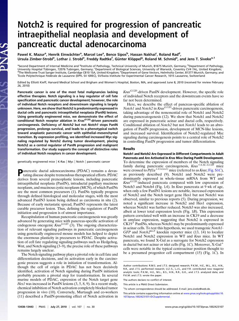
Kras:Notch2lacZ mice, X-Gal expression was detectable in PanINlesions and the surrounding stroma (Fig. 1C). GFP expression asa surrogate for Notch1 was found in normal acinar cells, as pre-viously described (16), but was hardly ever detectable in PanINlesions (Fig. 1C). In summary, these expression data are consis-tent with Notch2 as the predominant Notch receptor in ductal,centroacinar, and PanIN cells as suggested previously (5).
PanIN Development and Progression in Notch-Ablated Pancreata. Toanalyze the effect of Notch1 and Notch2 deficiency in pancreaticcarcinogenesis, we crossed previously described floxed Notch1fl/fl
and Notch2fl/fl mice (17) with Ptf1a+/Cre(ex1) mice (18) for gener-ation of Ptf1a+/Cre(ex1);Notch1fl/fl and Ptf1a+/Cre(ex1);Notch2fl/fl
mice, respectively (called N1ko and N2ko mice hereafter). Thesemice were born at the expected Mendelian ratio, and successfulrecombination of the floxed loci was confirmed by PCR (Fig. S1 Aand B). N1ko mice have been previously described to show nomajor pancreatic abnormalities (16). Similarly, N2ko adult pan-creata displayed no obvious morphological or functional abnor-malities (Fig. S2). However, in mice older than 12 mo of age, weoften noted a slight to moderate degree of focal exocrine atrophywith adipose tissue accumulation.To study the role of Notch1 and Notch2 during pancreatic
carcinogenesis, we crossed N1ko and N2ko mice with Kras micefor generation of Kras;N1ko and Kras;N2ko mice, respectively.Notably,Kras;N2komice showed no PanIN progression over time,whereas Kras and Kras;N1komice developed higher grade PanINlesions, suggesting that Notch2 is involved in PanIN progression(Fig. 1 D and E). PanIN lesions from all genotypes expressedtypical markers such as CK19 and MUC5AC and, somewhatsurprisingly, HES1 (Tables S1–S3).
Development of MCN-Like Lesions in Kras;N2ko Mice. Frequently,albeit not in all mice, Kras;N2komice developed moderate to verylarge multilocular cysts. These cysts most often developed in thesplenic part of the pancreas and showed a mucinous columnarepithelium resembling human MCN (Fig. S3 A and B). Rarely,goblet cells, high-grade dysplasia, and invasion into the adjacentstroma were noted. To characterize these lesions further, variousmarkers, including those found in human MCNs, were analyzed.The cystic epithelial cells expressed PDX1,MUC5AC, andHES1,thus showing similar characteristics as PanIN lesions (Table S3).Consistent with the observation of an MCN-like preneoplasticlesion, we found an ovarian-like stroma surrounding the cysticlesions with estrogen receptor (ER)-positive and progesteronereceptor-positive nuclei characteristic for humanMCNs (19) (Fig.S3B and Table S7). To see whether the MCN-like lesions werederived from Notch2-deficient cells, cell lineage analysis wasperformed by crossing the Rosa26R+/LSL-lacZ reporter strain toKras;N2komice. Indeed, we found all PanIN and MCN lesions tobe X-Gal+ (Fig. 2C).
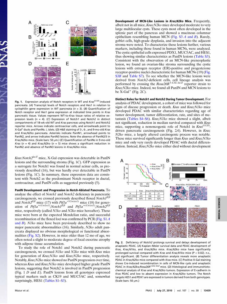
Distinct Roles for Notch1 and Notch2 During Tumor Development. Foranalysis of PDAC development, a cohort of mice was followed forsigns of disease progression or death. Kras and Kras;N1ko micedeveloped PDAC with similar characteristics regarding age oftumor development, tumor differentiation, rate, and sites of me-tastasis (Tables S4–S6). Kras;N1ko mice showed a slight, albeitnot significant, reduction in median survival compared with Krasmice, supporting a nononcogenic role of Notch1 in KrasG12D-driven pancreatic carcinogenesis (Fig. 2A). However, in Kras;N2ko mice, a largely altered carcinogenic process was notable.These mice survived significantly longer than Kras and Kras;N1komice and only very rarely developed PDAC with ductal differen-tiation. Instead, Kras;N2ko mice either died without development
A
B
CWT Kras
N2-L
acZ
N1-G
FP
Am
ylase
N1-G
FP
CK
19
i
9 weeks 12 weeks 18 weeks
Kras Kras;N2koD E
3 m
onth
s9 m
onth
s6 m
onth
s
**
*
*
Re
lativ
e m
RN
A le
vels
ratio
Kra
s /
WT
mR
NA c
opy
num
ber
per 1
000
copi
es o
f Cyp
Fig. 1. Expression analysis of Notch receptors in WT and KrasG12D-inducedpancreata. (A) Transcript levels of Notch receptors and Hes1 in relation tocyclophilin gene expression in WT pancreata (n = 3). (B) Quantification ofNotch receptor and Hes1 gene expression at indicated time points in Kraspancreatic tissue. Values represent WT-to-Kras tissue ratios of relative ex-pression levels (n = 4). (C) Expression of Notch1 and Notch2 in distinctcompartments of 18-wk-old WT and Kras pancreas using Notch1 and Notch2reporter mice. Arrows indicate centroacinar cells, and arrowheads point toX-Gal+ ducts and PanINs. i, islets. (D) H&E staining of 3-, 6-, and 9-mo-old Krasand Kras;N2ko pancreata. Asterisks indicate PanIN1, arrowhead points toPanIN2, and arrow indicates PanIN3 lesions. Note the absence of PanIN2/3 inKras;N2ko mice. (Scale bars: 50 μm.) (E) Quantification of PanINs in 9-mo-oldKras (n = 4) and Kras;N2ko (n = 5) mice shows a significant reduction inPanIN2 and absence of PanIN3 lesions in Kras;N2ko mice.
Kras;N2ko; ROSA26R-LacZ
Kras Kras;N1ko Kras;N2ko
Med. surv. [day] 425 336 521PDAC 54% 39% 52%
Kras
; N2k
oKr
as
HE E-cadherin HES1 PDX1
N 25 23 21
A B
C
D
Fig. 2. Deficiency of Notch2 prolongs survival and delays development ofanaplastic PDAC. (A) Kaplan–Meier survival data and PDAC development ofKras, Kras;N1ko, and Kras;N2ko mice. Kras;N2ko mice have significantlyprolonged survival compared with Kras and Kras;N1ko mice (P < 0.02). n.s.,not significant. (B) Tumor differentiation analysis reveals more anaplasticPDAC in Kras;N2komice compared with Krasmice. (C) Positive X-Gal stainingshows Cre-induced recombination in cells of MCN-like cysts and anaplasticPDAC in Kras;N2ko;Rosa26R+/LSL-lacZ mice. (D) Histological and immunohisto-chemical analysis of Kras and Kras;N2ko tumors. Expression of E-cadherin inKras PDAC and low to absent expression in Kras;N2ko tumors. The Notchtargets HES1 and PDX1 are expressed in tumors derived from both genotypes.(Scale bars: 50 μm.)
Mazur et al. PNAS | July 27, 2010 | vol. 107 | no. 30 | 13439
MED
ICALSC
IENCE
S
of PDACor developedhighly aggressive anaplastic PDACat a veryadvanced age (Fig. 2 A and B and Tables S4–S6). Histologically,most of these tumors were very large, showing a sarcomatoid cellpattern with a high proliferative index. Although we observed tu-mor areas that displayed features of poorly differentiated PDAC,we practically never observed G1/2 grades. Anaplastic PDACshowed an absence or low expression of E-cadherin and expressedPDX1, indicating its pancreatic origin (Fig. 2D). Lineage tracingshowed PanIN and anaplastic PDAC development from Notch2-ablated pancreatic cells (Fig. 2C). Surprisingly, as was seen inMCN-like lesions, many cells expressedHES1, suggesting Notch2-independent regulation (Fig. 2D). Kras;N1ko and Kras;N2koPDACshowedanabsence of the respectiveNotch receptor,where-as expression was notable in Kras cancer cells (Figs. S1D and S4).To determine whether deficiency of Notch2 led to up-regulationof other Notch receptors, we tested Kras and Kras;N2ko PDACcells for expression of Notch1–4. Here, we did not detect a consis-tent compensatory expression pattern of other Notch receptors inKras;N2ko mice (Fig. S4).
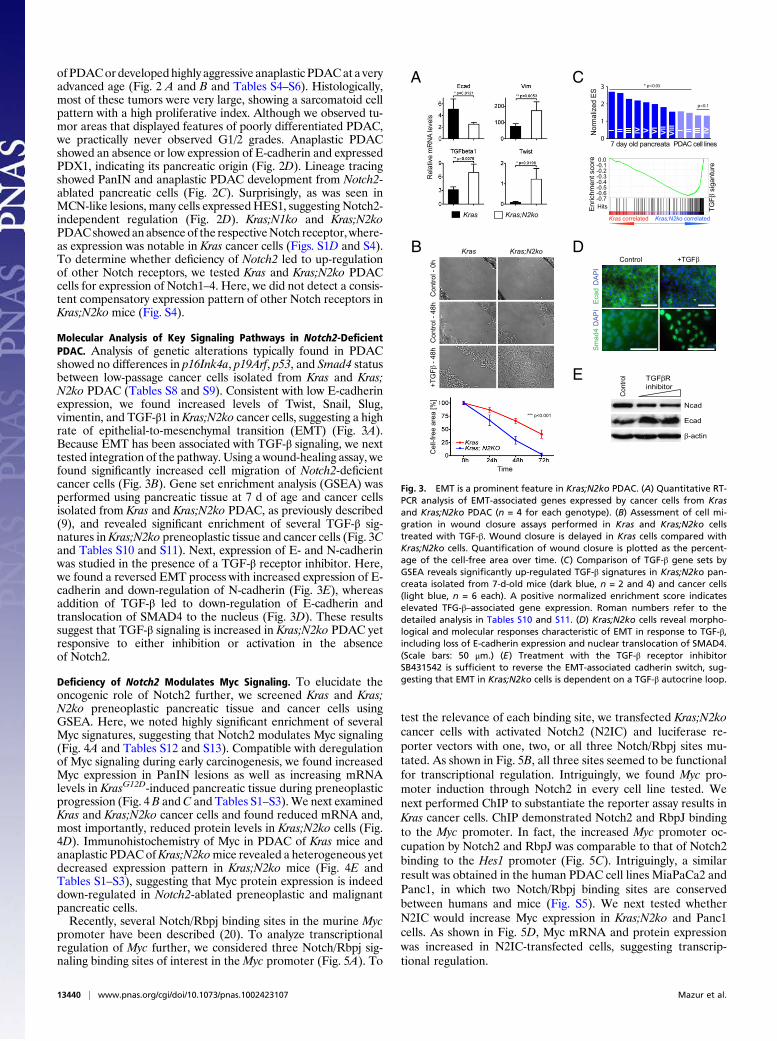
Molecular Analysis of Key Signaling Pathways in Notch2-DeficientPDAC. Analysis of genetic alterations typically found in PDACshowed no differences in p16Ink4a, p19Arf, p53, and Smad4 statusbetween low-passage cancer cells isolated from Kras and Kras;N2ko PDAC (Tables S8 and S9). Consistent with low E-cadherinexpression, we found increased levels of Twist, Snail, Slug,vimentin, and TGF-β1 in Kras;N2ko cancer cells, suggesting a highrate of epithelial-to-mesenchymal transition (EMT) (Fig. 3A).Because EMT has been associated with TGF-β signaling, we nexttested integration of the pathway. Using a wound-healing assay, wefound significantly increased cell migration of Notch2-deficientcancer cells (Fig. 3B). Gene set enrichment analysis (GSEA) wasperformed using pancreatic tissue at 7 d of age and cancer cellsisolated from Kras and Kras;N2ko PDAC, as previously described(9), and revealed significant enrichment of several TGF-β sig-natures in Kras;N2ko preneoplastic tissue and cancer cells (Fig. 3Cand Tables S10 and S11). Next, expression of E- and N-cadherinwas studied in the presence of a TGF-β receptor inhibitor. Here,we found a reversed EMT process with increased expression of E-cadherin and down-regulation of N-cadherin (Fig. 3E), whereasaddition of TGF-β led to down-regulation of E-cadherin andtranslocation of SMAD4 to the nucleus (Fig. 3D). These resultssuggest that TGF-β signaling is increased in Kras;N2ko PDAC yetresponsive to either inhibition or activation in the absenceof Notch2.
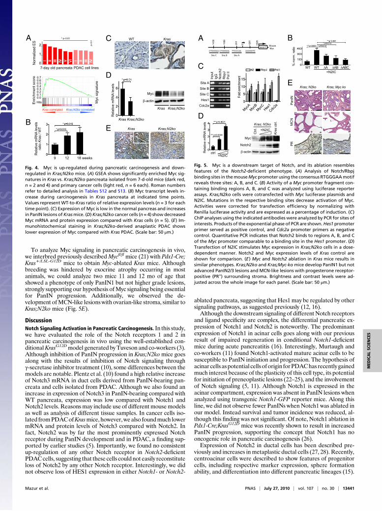
Deficiency of Notch2 Modulates Myc Signaling. To elucidate theoncogenic role of Notch2 further, we screened Kras and Kras;N2ko preneoplastic pancreatic tissue and cancer cells usingGSEA. Here, we noted highly significant enrichment of severalMyc signatures, suggesting that Notch2 modulates Myc signaling(Fig. 4A and Tables S12 and S13). Compatible with deregulationof Myc signaling during early carcinogenesis, we found increasedMyc expression in PanIN lesions as well as increasing mRNAlevels in KrasG12D-induced pancreatic tissue during preneoplasticprogression (Fig. 4B andC and Tables S1–S3).We next examinedKras and Kras;N2ko cancer cells and found reduced mRNA and,most importantly, reduced protein levels in Kras;N2ko cells (Fig.4D). Immunohistochemistry of Myc in PDAC of Kras mice andanaplastic PDACofKras;N2komice revealed a heterogeneous yetdecreased expression pattern in Kras;N2ko mice (Fig. 4E andTables S1–S3), suggesting that Myc protein expression is indeeddown-regulated in Notch2-ablated preneoplastic and malignantpancreatic cells.Recently, several Notch/Rbpj binding sites in the murine Myc
promoter have been described (20). To analyze transcriptionalregulation of Myc further, we considered three Notch/Rbpj sig-naling binding sites of interest in the Myc promoter (Fig. 5A). To
test the relevance of each binding site, we transfected Kras;N2kocancer cells with activated Notch2 (N2IC) and luciferase re-porter vectors with one, two, or all three Notch/Rbpj sites mu-tated. As shown in Fig. 5B, all three sites seemed to be functionalfor transcriptional regulation. Intriguingly, we found Myc pro-moter induction through Notch2 in every cell line tested. Wenext performed ChIP to substantiate the reporter assay results inKras cancer cells. ChIP demonstrated Notch2 and RbpJ bindingto the Myc promoter. In fact, the increased Myc promoter oc-cupation by Notch2 and RbpJ was comparable to that of Notch2binding to the Hes1 promoter (Fig. 5C). Intriguingly, a similarresult was obtained in the human PDAC cell lines MiaPaCa2 andPanc1, in which two Notch/Rbpj binding sites are conservedbetween humans and mice (Fig. S5). We next tested whetherN2IC would increase Myc expression in Kras;N2ko and Panc1cells. As shown in Fig. 5D, Myc mRNA and protein expressionwas increased in N2IC-transfected cells, suggesting transcrip-tional regulation.
Re
lativ
e m
RN
A le
vels
D
Eca
d
DA
PI
Control +TGFβ
Sm
ad
4
DA
PI
A
Kras Kras;N2ko
0.0-0.1-0.2-0.3-0.4-0.5-0.6-0.7
En
rich
me
nt
sco
re
Kras correlated Kras;N2ko correlated
Hits TG
Fβ
sig
an
ture
C
E
Ncad
Ecad
β-actin
TGFβR
inhibitor
Contr
ol
I II III IV V VI
VII
VIII I II III IVNo
rma
lize
d E
S
7 day old pancreata PDAC cell lines
3
2
1
0
* p<0.05
p<0.1
B Kras
Co
ntr
ol -
0h
Kras;N2ko
Co
ntr
ol -
48
h+
TG
Fβ
- 4
8h
Time
Ce
ll-fr
ee
are
a [
%]
*** p<0.001
Fig. 3. EMT is a prominent feature in Kras;N2ko PDAC. (A) Quantitative RT-PCR analysis of EMT-associated genes expressed by cancer cells from Krasand Kras;N2ko PDAC (n = 4 for each genotype). (B) Assessment of cell mi-gration in wound closure assays performed in Kras and Kras;N2ko cellstreated with TGF-β. Wound closure is delayed in Kras cells compared withKras;N2ko cells. Quantification of wound closure is plotted as the percent-age of the cell-free area over time. (C) Comparison of TGF-β gene sets byGSEA reveals significantly up-regulated TGF-β signatures in Kras;N2ko pan-creata isolated from 7-d-old mice (dark blue, n = 2 and 4) and cancer cells(light blue, n = 6 each). A positive normalized enrichment score indicateselevated TFG-β–associated gene expression. Roman numbers refer to thedetailed analysis in Tables S10 and S11. (D) Kras;N2ko cells reveal morpho-logical and molecular responses characteristic of EMT in response to TGF-β,including loss of E-cadherin expression and nuclear translocation of SMAD4.(Scale bars: 50 μm.) (E) Treatment with the TGF-β receptor inhibitorSB431542 is sufficient to reverse the EMT-associated cadherin switch, sug-gesting that EMT in Kras;N2ko cells is dependent on a TGF-β autocrine loop.
13440 | www.pnas.org/cgi/doi/10.1073/pnas.1002423107 Mazur et al.
To analyze Myc signaling in pancreatic carcinogenesis in vivo,we interbred previously describedMycfl/fl mice (21) with Pdx1-Cre;Kras+/LSL-G12D mice to obtain Myc-ablated Kras mice. Althoughbreeding was hindered by exocrine atrophy occurring in mostanimals, we could analyze two mice 11 and 12 mo of age thatshowed a phenotype of only PanIN1 but not higher grade lesions,strongly supporting our hypothesis ofMyc signaling being essentialfor PanIN progression. Additionally, we observed the de-velopment ofMCN-like lesions with ovarian-like stroma, similar toKras;N2ko mice (Fig. 5E).
DiscussionNotch Signaling Activation in Pancreatic Carcinogenesis. In this study,we have evaluated the role of the Notch receptors 1 and 2 inpancreatic carcinogenesis in vivo using the well-established con-ditionalKrasG12Dmodel generated by Tuveson and co-workers (3).Although inhibition of PanIN progression in Kras;N2komice goesalong with the results of inhibition of Notch signaling throughγ-secretase inhibitor treatment (10), some differences between themodels are notable. Plentz et al. (10) found a high relative increaseof Notch3 mRNA in duct cells derived from PanIN-bearing pan-creata and cells isolated from PDAC. Although we also found anincrease in expression of Notch3 in PanIN-bearing compared withWT pancreata, expression was low compared with Notch1 andNotch2 levels. Reasons may include use of different mousemodelsas well as analysis of different tissue samples. In cancer cells iso-lated fromPDACofKrasmice, however, we also foundmuch lowermRNA and protein levels of Notch3 compared with Notch2. Infact, Notch2 was by far the most prominently expressed Notchreceptor during PanIN development and in PDAC, a finding sup-ported by earlier studies (5). Importantly, we found no consistentup-regulation of any other Notch receptor in Notch2-deficientPDACcells, suggesting that these cells could not easily reconstituteloss of Notch2 by any other Notch receptor. Interestingly, we didnot observe loss of HES1 expression in either Notch1- or Notch2-
ablated pancreata, suggesting that Hes1 may be regulated by othersignaling pathways, as suggested previously (12, 16).Although the downstream signaling of different Notch receptors
and ligand specificity are complex, the differential pancreatic ex-pression of Notch1 and Notch2 is noteworthy. The predominantexpression of Notch1 in acinar cells goes along with our previousresult of impaired regeneration in conditional Notch1-deficientmice during acute pancreatitis (16). Interestingly, Murtaugh andco-workers (11) found Notch1-activated mature acinar cells to besusceptible to PanIN initiation and progression. The hypothesis ofacinar cells as potential cells of origin forPDAChas recently gainedmuch interest because of the plasticity of this cell type, its potentialfor initiation of preneoplastic lesions (22–25), and the involvementof Notch signaling (5, 11). Although Notch1 is expressed in theacinar compartment, expression was absent in PanIN lesions whenanalyzed using transgenic Notch1-GFP reporter mice. Along thisline, we did not observe fewer PanINs when Notch1 was ablated inour model. Instead survival and tumor incidence was reduced, al-though this finding was not significant. Of note, Notch1 ablation inPdx1-Cre;KrasG12D mice was recently shown to result in increasedPanIN progression, supporting the concept that Notch1 has nooncogenic role in pancreatic carcinogenesis (26).Expression of Notch2 in ductal cells has been described pre-
viously and increases in metaplastic ductal cells (27, 28). Recently,centroacinar cells were described to show features of progenitorcells, including respective marker expression, sphere formationability, and differentiation into different pancreatic lineages (15).
En
rich
me
nt
sco
re 0.70.60.50.40.30.20.10.0
Hits Myc s
ign
atu
re
Kras correlated Kras;N2ko correlated
A C
E Kras Kras;N2ko
KrasWT
I II III IV I II III IV
B
Re
lativ
e m
RN
A le
vels
ratio
Kra
s /
WT
9 12 18 weeks
No
rma
lize
d E
S
I II III IV V VI I II III IV
7-day old pancreata PDAC cell lines
2
1
0
* p<0.05
p<
0.1
Re
lativ
e m
RN
A le
vels
Myc
β-actin
Kras Kras;N2ko
Kras Kras;N2ko
D
Fig. 4. Myc is up-regulated during pancreatic carcinogenesis and down-regulated in Kras;N2ko mice. (A) GSEA shows significantly enriched Myc sig-natures in Kras vs. Kras;N2ko pancreata isolated from 7-d-old mice (dark red,n = 2 and 4) and primary cancer cells (light red, n = 6 each). Roman numbersrefer to detailed analysis in Tables S12 and S13. (B) Myc transcript levels in-crease during carcinogenesis in Kras pancreata at indicated time points.Values represent WT-to-Kras ratio of relative expression levels (n = 3 for eachtime point). (C) Expression of Myc is low in the normal pancreas and increasesin PanIN lesions ofKrasmice. (D)Kras;N2ko cancer cells (n = 4) show decreasedMyc mRNA and protein expression compared with Kras cells (n = 5). (E) Im-munohistochemical staining in Kras;N2ko-derived anaplastic PDAC showslower expression of Myc compared with Kras PDAC. (Scale bar: 50 μm.)
Rela
tive m
RN
A le
vels
ctrl+N2IC
Myc
Notch2
β-actin
+N2IC
Ctr
l.
Kra
s ctr
l.
Kras; N2ko
A
Kras; N2ko Kras; Myc ko
PanIN
MC
N
E
PR
exon1
+5
26
+2
81
-20
1
-11
89
-14
36
0
Site ASite C
Mouse
Myc
Site B
CTGGGAA CTGGGAA TTCCCAA AAGGGTC
D
C
Site A
Site B
Site C
Hes1
Cdc2a
Input
IgG
Notc
h2
RbpJ
Pol II
WT WT ΔA ΔAB ΔABC
% n
orm
. ra
tio
+N2IC
B
Fold
enric
hm
ent
Fig. 5. Myc is a downstream target of Notch, and its ablation resemblesfeatures of the Notch2-deficient phenotype. (A) Analysis of Notch/Rbpjbinding sites in themouseMyc promoter using the consensus RTGGGAAmotifreveals three sites: A, B, and C. (B) Activity of a Myc promoter fragment con-taining binding regions A, B, and C was analyzed using luciferase reporterassays. Kras;N2ko cells were cotransfected with Myc luciferase plasmids andN2IC. Mutations in the respective binding sites decrease activation of Myc.Activities were corrected for transfection efficiency by normalizing withRenilla luciferase activity and are expressed as a percentage of induction. (C)ChIP analyses using the indicated antibodies were analyzed by PCR for sites ofinterests. Products of the exponential phase of PCR are shown.Hes1 promoterprimer served as positive control, and Cdc2a promoter primers as negativecontrol. Quantitative PCR indicates that Notch2 binds to regions A, B, and Cof the Myc promoter comparable to a binding site in the Hes1 promoter. (D)Transfection of N2IC stimulates Myc expression in Kras;N2ko cells in a dose-dependent manner. Notch2 and Myc expression levels of Kras control areshown for comparison. (E) Myc and Notch2 ablation in Kras mice results insimilar phenotypes. Kras;N2ko and Kras;Myc-komice develop PanIN1 but notadvanced PanIN2/3 lesions and MCN-like lesions with progesterone receptor-positive (PR+) surrounding stroma. Brightness and contrast levels were ad-justed across the whole image for each panel. (Scale bar: 50 μm.)
Mazur et al. PNAS | July 27, 2010 | vol. 107 | no. 30 | 13441
MED
ICALSC
IENCE
S

These and our results suggest that a potential progenitor com-partment in small ducts such as centroacinar cells expressesNotch2, a hypothesis supported by our expression studies usingNotch2+/lacZ reporter mice. Because we observed PanIN1 initia-tion but no higher grade PanINs in Kras;N2ko mice, activation ofNotch2 may be required for progression of PanIN lesions. How-ever, other explanations remain possible. Because PanIN1 lesionsare often encountered in pancreata of elderly people, it is possiblethat PanIN1 lesionsmay not actually precede PanIN2 and PanIN3lesions but are mainly default lesions that may form from differentpancreatic cells, including the acinar compartment. Consistentwith this hypothesis is the induction of PanIN lesions but usuallyno development of invasive PDAC from acinar cells in Ela-Cre-ER;KrasG12D mice. Although our study did not directly addressthis intriguing question, it remains possible that PanIN1 lesionsmay originate from acinar cells, whereas initiation or progressionof PanIN2/3 lesions may require a Notch-regulated potentialprogenitor compartment or an additional stimulus such as ongo-ing inflammation (25, 29).
Development of MCN-Like Lesions and Anaplastic PDAC in Kras;N2koMice. The blockade of PanIN progression and PDAC de-velopment in Notch2-deficient KrasG12D mice goes along with thelonger survival of thesemice. Eventually, thesemice develop largecysts resembling MCNs and succumb from either pancreatic in-sufficiency or from the development of anaplastic PDAC. De-velopment of MCN-like lesions may thus be a bypass route forpancreatic cells undergoing oncogenic stress. However, two sce-narios are possible with either (i) a common cell of origin forPanIN andMCN development, in which the route to higher gradePanINs is blocked by Notch2 deficiency, or (ii) different cells oforigin for each lesion type that respond differentially to KrasG12D
in the presence or absence of Notch2.Interestingly, an association of anaplastic PDAC and MCN has
been repeatedly described in patients (30). However, we do nothave enough evidence to conclude that MCNs are the direct pre-cursors for PDAC in Kras;N2ko mice. Further analysis is requiredto understand the cellular and molecular cues in Notch2-deficientmalignant transformation. However, the clinical and experimentalobservations of the combined occurrence of MCN and anaplasticPDAC highlight the potential predictive capability of genotype-phenotype correlations in complex cancer mouse models.
TGF-β Signaling and EMT in Notch2-Deficient PDAC. Molecularcharacterization of the anaplastic PDAC in Kras;N2ko miceshowed evidence of EMT. Several reports have described an ac-tivating role of increased Notch signaling in EMT by regulation ofE-cadherin repressors such as Snail or interaction with TGF-βsignaling (31–34). TGF-β is known to play an ambivalent role incancer biology. In the pancreas, conditional inactivation of TGF-βreceptor 2 led to accelerated development and progression ofwell-differentiated PDAC (35). The development of late-occur-ring anaplastic PDAC with increased EMT is compatible with thedual role of TGF-β signaling in epithelial tumorigenesis. The ef-fect of TGF-β receptor inhibition on E- and N-cadherin expres-sion and exogenous TGF-β–induced nuclear translocation ofSMAD4 suggest an intact TGF-β signaling axis. Indirect regula-tion of TGF-β may occur through deregulated Myc signaling,which is known to suppress the activation of TGF-β–inducedgenes such as p21CIP1, which has been shown to interact withNotch in various organs (36, 37). However, we could not detectconsistent differences in p21CIP1 expression or related signaturesbetween Kras and Kras;N2ko tumors.
Myc Signaling Is Regulated by Notch2 in PDAC. Decreased Myc sig-naling in Kras;N2ko mice supports the hypothesis of Notch2-de-pendent Myc signaling as a key regulator of the carcinogenicprocess in the pancreas. Deregulation of Myc in PDAC has been
described in many studies, and amplification occurs in about 30%of human PDAC as well as in murine PDAC (38–40). In recentstudies, Myc signaling has been identified to play a key role in cellcycle regulation of PDAC cells (41, 42). Although these studiesdemonstrate the importance of deregulated Myc signaling inPDAC, our results suggest an early role during PanIN progressionsupported by early Myc amplification in precursor lesions (38). Ina recent quantitative proteomic screen of preneoplastic PanINlesions, Myc expression was identified in PanIN3 lesions (43).We and others have previously characterized the important role
of Myc in progenitor and acinar cell proliferation during de-velopment and adult homeostasis (21, 44, 45). Consistently, wefound increasedMyc expression throughout PanIN development inKras mice. It is tempting to speculate that Myc and Ras signalingcooperatively promote tumor progression in a setting of activeNotch. Notch signaling has been reported to cooperate with Ras,and several studies have reporteddirect transcriptional regulationofMyc by Notch1 (20, 46–48). Our finding that active Notch2 inducesMyc expression in PDAC cells supports these reports. Althoughpreliminary, the phenotypical similarities of Notch2 and Myc-ab-latedKrasG12D-inducedpancreatawithdevelopmentof cystic lesionsand a PanIN progression stop strongly support this hypothesis. Ofconsideration is the use of different Cre mice, Ptf1a+/Cre(ex1) andPdx1-Cre mice, in Kras;N2ko and Kras;Myc-ko mice, respectively,because of extensive exocrine hypoplasia and early postnatal deathof Ptf1a+/Cre(ex1);Mycfl/fl mice (21). Although we cannot rule outdifferent target compartments in both Cre lines, this seems unlikely,given the similar phenotype in KrasG12D-activated mice (3).The results from luciferase reporter and ChIP assays suggest
that all three reported Notch/Rbpj binding sites in the Myc pro-moter are relevant for transcriptional regulation of Myc. On thebasis of our findings, we report that Myc is regulated by Notch2.Why Notch1 ablation did not lead to similar alterations in earlytumor progression in our model is not clear. A possible explana-tion would be a context- and cell-specific role of Myc and its reg-ulation through Notch. A possible scenario may thus be thata progenitor cell (e.g., within the centroacinar compartment) is thetarget cell for cooperative Myc-Ras–induced tumor developmentpropagated by Notch2 activation. The success of Notch inhibitionas a chemopreventive approach to inhibit PanIN progression hasbeen shown (10). This outcome is supported by our results. Ofnote, the same group has reportedMyc amplification in KrasG12D-driven PDACmouse models, adding evidence for a key role of thissignaling pathway during the carcinogenic process (40). It will be ofgreat interest to study the integration of the transcriptional pro-grams regulated by Myc and Notch signaling in further detail,which may eventually help to explain the permissive signals regu-lating pancreatic plasticity and malignant transformation.In summary, our results provide evidence for an essential role of
Notch2 and Myc in the initiation of a neoplastic transformationprogram in pancreatic cells, whereas Notch1 has no oncogenicrole, supporting the concept of distinctive roles of individual Notchreceptors in cancer development. In addition, the data demon-strate the integrative interaction of regulators of cell fate and cellcycle signaling, thereby enhancing our biological understanding forunique approaches in this still untreatable disease.
Materials and MethodsMouse Strains. Kras+/LSL-G12D, Notch1fl/fl, Notch2fl/fl, Mycfl/fl, Ptf1a+/Cre(ex1),Pdx1-Cre, and Rosa26+/LSL-lacZ mice have been described before (3, 9, 17, 21).All experiments were performed according to the guidelines of the localanimal use and care committees.
Detailed descriptions of additional procedures, including protein andmRNA analysis, immunohistochemistry, microarray/GSEA, luciferase-basedreporter assays, and ChIP, are provided in SI Text.
ACKNOWLEDGMENTS. We thank W. Gao (Genentech, Inc., CA), Y. Hamada(National Institute for Basic Biology, Okazaki, Japan), and C. A. Klug (Universityof Alabama, Birmingham, AB) for the generous gift of Notch reporter mice
13442 | www.pnas.org/cgi/doi/10.1073/pnas.1002423107 Mazur et al.
and A. Klinakis (Biochemical Research Foundation, Athens, Greece) for Mycplasmids. We are grateful to T. Sudo (Toray Industries Inc., Kamakura, Japan) forHES1 and to C. V.Wright (Vanderbilt UniversityMedical Center, Nashville, TN) forPDX1 antibodies. We thank M. Neuhofer, S. Ruberg, and C. Köhler for excellent
technical assistance. This work was supported by grants from the German CancerAid (Grant 107195), German Federal Ministry of Education and Research (Grant01GS08115), Lustgarten Foundation (RFP05-14 and 06-12), and German ResearchFoundation (Grant SI 1549/1-1).
1. Hezel AF, Kimmelman AC, Stanger BZ, Bardeesy N, Depinho RA (2006) Genetics andbiology of pancreatic ductal adenocarcinoma. Genes Dev 20:1218–1249.
2. Hruban RH, Iacobuzio-Donahue C, Wilentz RE, Goggins M, Kern SE (2001) Molecularpathology of pancreatic cancer. Cancer J 7:251–258.
3. Hingorani SR, et al. (2003) Preinvasive and invasive ductal pancreatic cancer and itsearly detection in the mouse. Cancer Cell 4:437–450.
4. Pasca di Magliano M, et al. (2006) Hedgehog/Ras interactions regulate early stages ofpancreatic cancer. Genes Dev 20:3161–3173.
5. Miyamoto Y, et al. (2003) Notch mediates TGF alpha-induced changes in epithelialdifferentiation during pancreatic tumorigenesis. Cancer Cell 3:565–576.
6. Pasca di Magliano M, et al. (2007) Common activation of canonical Wnt signaling inpancreatic adenocarcinoma. PLoS ONE 2:e1155.
7. Thayer SP, et al. (2003) Hedgehog is an early and late mediator of pancreatic cancertumorigenesis. Nature 425:851–856.
8. Stanger BZ, et al. (2005) Pten constrains centroacinar cell expansion and malignanttransformation in the pancreas. Cancer Cell 8:185–195.
9. Siveke JT, et al. (2007) Concomitant pancreatic activation of Kras(G12D) and Tgfaresults in cystic papillary neoplasms reminiscent of human IPMN. Cancer Cell 12:266–279.
10. Plentz R, et al. (2009) Inhibition of gamma-secretase activity inhibits tumor progression ina mouse model of pancreatic ductal adenocarcinoma. Gastroenterology 136:1741–1749.e6.
11. De La O JP, et al. (2008) Notch and Kras reprogram pancreatic acinar cells to ductalintraepithelial neoplasia. Proc Natl Acad Sci USA 105:18907–18912.
12. Nakhai H, et al. (2008) Conditional ablation of Notch signaling in pancreaticdevelopment. Development 135:2757–2765.
13. Lewis AK, Frantz GD, Carpenter DA, de Sauvage FJ, Gao WQ (1998) Distinct expressionpatterns of notch family receptors and ligands during development of themammalian inner ear. Mech Dev 78:159–163.
14. Hamada Y, et al. (1999) Mutation in ankyrin repeats of the mouse Notch2 geneinduces early embryonic lethality. Development 126:3415–3424.
15. Rovira M, et al. (2010) Isolation and characterization of centroacinar/terminal ductalprogenitor cells in adult mouse pancreas. Proc Natl Acad Sci USA 107:75–80.
16. Siveke JT, et al. (2008) Notch signaling is required for exocrine regeneration afteracute pancreatitis. Gastroenterology 134:544–555.
17. Besseyrias V, et al. (2007) Hierarchy of Notch-Delta interactions promoting T celllineage commitment and maturation. J Exp Med 204:331–343.
18. Nakhai H, et al. (2007) Ptf1a is essential for the differentiation of GABAergic andglycinergic amacrine cells and horizontal cells in the mouse retina. Development 134:1151–1160.
19. Zamboni G, et al. (1999) Mucinous cystic tumors of the pancreas: clinicopathologicalfeatures, prognosis, and relationship to other mucinous cystic tumors. Am J SurgPathol 23:410–422.
20. Klinakis A, et al. (2006) Myc is a Notch1 transcriptional target and a requisite forNotch1-induced mammary tumorigenesis in mice. Proc Natl Acad Sci USA 103:9262–9267.
21. Nakhai H, Siveke JT, Mendoza-Torres L, Schmid RM (2008) Conditional inactivation ofMyc impairs development of the exocrine pancreas. Development 135:3191–3196.
22. Habbe N, et al. (2008) Spontaneous induction of murine pancreatic intraepithelialneoplasia (mPanIN) by acinar cell targeting of oncogenic Kras in adult mice. Proc NatlAcad Sci USA 105:18913–18918.
23. Ji B, et al. (2009) Ras activity levels control the development of pancreatic diseases.Gastroenterology, 137:1072–1082 1082.e1–6..
24. Zhu L, Shi G, Schmidt CM, Hruban RH, Konieczny SF (2007) Acinar cells contribute tothe molecular heterogeneity of pancreatic intraepithelial neoplasia. Am J Pathol 171:263–273.
25. Guerra C, et al. (2007) Chronic pancreatitis is essential for induction of pancreaticductal adenocarcinoma by K-Ras oncogenes in adult mice. Cancer Cell 11:291–302.
26. Hanlon L, et al. (2010) Notch1 functions as a tumor suppressor in a model of K-ras-induced pancreatic ductal adenocarcinoma. Cancer Res 70:4280–4286.
27. Rooman I, et al. (2006) Expression of the Notch signaling pathway and effect onexocrine cell proliferation in adult rat pancreas. Am J Pathol 169:1206–1214.
28. Lee KM, Yasuda H, Hollingsworth MA, Ouellette MM (2005) Notch 2-positiveprogenitors with the intrinsic ability to give rise to pancreatic ductal cells. Lab Invest85:1003–1012.
29. Morris JP, IV, Cano DA, Sekine S, Wang SC, Hebrok M (2010) Beta-catenin blocks Kras-dependent reprogramming of acini into pancreatic cancer precursor lesions in mice.J Clin Invest 120:508–520.
30. Pan ZG, Wang B (2007) Anaplastic carcinoma of the pancreas associated witha mucinous cystic adenocarcinoma. A case report and review of the literature. JOP 8:775–782.
31. Sahlgren C, Gustafsson MV, Jin S, Poellinger L, Lendahl U (2008) Notch signalingmediates hypoxia-induced tumor cell migration and invasion. Proc Natl Acad Sci USA105:6392–6397.
32. Wang Z, et al. (2009) Acquisition of epithelial-mesenchymal transition phenotype ofgemcitabine-resistant pancreatic cancer cells is linked with activation of the notchsignaling pathway. Cancer Res 69:2400–2407.
33. Timmerman LA, et al. (2004) Notch promotes epithelial-mesenchymal transitionduring cardiac development and oncogenic transformation. Genes Dev 18:99–115.
34. Zavadil J, Cermak L, Soto-Nieves N, Böttinger EP (2004) Integration of TGF-beta/Smadand Jagged1/Notch signalling in epithelial-to-mesenchymal transition. EMBO J 23:1155–1165.
35. Ijichi H, et al. (2006) Aggressive pancreatic ductal adenocarcinoma in mice caused bypancreas-specific blockade of transforming growth factor-beta signaling in cooperationwith active Kras expression. Genes Dev 20:3147–3160.
36. Liu M, et al. (2009) p21CIP1 attenuates Ras- and c-Myc-dependent breast tumorepithelial mesenchymal transition and cancer stem cell-like gene expression in vivo.Proc Natl Acad Sci USA 106:19035–19039.
37. Rangarajan A, et al. (2001) Notch signaling is a direct determinant of keratinocytegrowth arrest and entry into differentiation. EMBO J 20:3427–3436.
38. Schleger C, Verbeke C, Hildenbrand R, Zentgraf H, Bleyl U (2002) c-MYC activation inprimary and metastatic ductal adenocarcinoma of the pancreas: Incidence, mechanisms,and clinical significance. Mod Pathol 15:462–469.
39. Schreiner B, et al. (2003) Murine pancreatic tumor cell line TD2 bears the characteristicpattern of genetic changes with two independently amplified gene loci. Oncogene22:6802–6809.
40. Bardeesy N, et al. (2006) Both p16(Ink4a) and the p19(Arf)-p53 pathway constrainprogression of pancreatic adenocarcinoma in the mouse. Proc Natl Acad Sci USA 103:5947–5952.
41. Konig A, et al. (2010) NFAT-induced histone acetylation relay switch promotes c-myc-dependent growth in pancreatic cancer cells. Gastroenterology 138:1189–1199.e1-2.
42. Schild C, et al. (2009) PI3K signaling maintains c-myc expression to regulatetranscription of E2F1 in pancreatic cancer cells. Mol Carcinog 48:1149–1158.
43. Pan S, et al. (2009) Quantitative proteomics investigation of pancreatic intraepithelialneoplasia. Electrophoresis 30:1132–1144.
44. Bonal C, et al. (2009) Pancreatic inactivation of c-Myc decreases acinar mass andtransdifferentiates acinar cells into adipocytes in mice. Gastroenterology 136:309–319.e9.
45. Strom A, et al. (2007) Unique mechanisms of growth regulation and tumorsuppression upon Apc inactivation in the pancreas. Development 134:2719–2725.
46. Palomero T, et al. (2006) NOTCH1 directly regulates c-MYC and activates a feed-forward-loop transcriptional network promoting leukemic cell growth. Proc NatlAcad Sci USA 103:18261–18266.
47. Sharma VM, et al. (2006) Notch1 contributes to mouse T-cell leukemia by directlyinducing the expression of c-myc. Mol Cell Biol 26:8022–8031.
48. Weng AP, et al. (2006) c-Myc is an important direct target of Notch1 in T-cell acutelymphoblastic leukemia/lymphoma. Genes Dev 20:2096–2109.
Mazur et al. PNAS | July 27, 2010 | vol. 107 | no. 30 | 13443
MED
ICALSC
IENCE
S