Observations on Solitary Versus Multiple Isolated Pancreatic ...
24
cancers Review Observations on Solitary Versus Multiple Isolated Pancreatic Metastases of Renal Cell Carcinoma: Another Indication of a Seed and Soil Mechanism? Franz Sellner Surgical Department, Kaiser-Franz-Josef-Hospital, 1100 Wien, Austria; [email protected] Received: 5 August 2019; Accepted: 4 September 2019; Published: 17 September 2019 Abstract: Isolated pancreas metastases are a rare type of metastasis of renal cell carcinoma, characterized by the presence of pancreatic metastases, while all other organs remain unaffected. In a previous study, we determined arguments from the literature which (a) indicate a systemic–haematogenic metastasis route (uniform distribution of the metastases across the pancreas and independence of the metastatic localization in the pancreas of the side of the renal carcinoma); and (b) postulate a high impact of a seed and soil mechanism (SSM) on isolated pancreatic metastasis of renal cell carcinoma (isPM) as an explanation for exclusive pancreatic metastases, despite a systemic haematogenous tumor cell embolization. The objective of the study presented was to search for further arguments in favor of an SSM with isPM. For that purpose, the factor’s histology, grading, and singular/multiple pancreas metastases were analyzed on the basis of 814 observations published up to 2018. While histology and grading allowed for no conclusions regarding the importance of an SSM, the comparison of singular/multiple pancreas metastases produced arguments in favor of an SSM: 1. The multiple pancreas metastases observed in 38.1% prove that multiple tumor cell embolisms occur with isPM, the exclusive “maturation” of which in the pancreas requires an SSM; 2. The survival rates (SVR), which are consistent with singular and multiple pancreas metastases (despite the higher total tumor load with the latter), prove that the metastasized tumor cells are not able to survive in all other organs because of an SSM, which results in identical SVR when the pancreatic foci are treated adequately. Keywords: renal cell carcinoma; pancreatic metastasis; seed and soil mechanism 1. Introduction Two fundamental mechanisms determine and regulate the metastatic potential of solid carcinomas. On the one hand, it is the imperatively necessary invasion of blood and lymph vessels by tumor cells: there can be no metastasis without circulating tumor cells or tumor cell clusters. The fate of the embolized tumor cells is just as significant, however, since clinical experience shows that not every tumor cell embolism inevitably results in clinically manifest metastasis. That is rather the result of a multi-stage interaction of tumor cells and host organ (seed and soil), since neither each tumor cell nor each host organ can provide all necessary properties required for successful interaction. Large-scale studies determined the complexity of this mechanism, which takes place in five main steps [1–3]: 1. The invasion of tumor cells in the vascular system “intravasion”; 2. The transport of the tumor cells in the vascular system; 3. The docking of the tumor cells, mechanically or by way of organ–tumor cell-specific adhesion to the vascular wall [1–6]; 4. The extravasion of tumor cells through the capillary wall in the extracellular matrix (ECM)—after distraction or death of endothelial cells [2,3,6–12], and 5. After previous dormancy [3,13–16] or without it, the maturation of the cells into metastasis (colonization); a process which requires the formation of a vessel network [17] and the Cancers 2019, 11, 1379; doi:10.3390/cancers11091379 www.mdpi.com/journal/cancers
-
Upload
khangminh22 -
Category
Documents
-
view
0 -
download
0
Transcript of Observations on Solitary Versus Multiple Isolated Pancreatic ...
cancers
Review
Observations on Solitary Versus Multiple IsolatedPancreatic Metastases of Renal Cell Carcinoma:Another Indication of a Seed and Soil Mechanism?
Franz Sellner
Surgical Department, Kaiser-Franz-Josef-Hospital, 1100 Wien, Austria; [email protected]
Received: 5 August 2019; Accepted: 4 September 2019; Published: 17 September 2019�����������������
Abstract: Isolated pancreas metastases are a rare type of metastasis of renal cell carcinoma,characterized by the presence of pancreatic metastases, while all other organs remain unaffected.In a previous study, we determined arguments from the literature which (a) indicate asystemic–haematogenic metastasis route (uniform distribution of the metastases across the pancreasand independence of the metastatic localization in the pancreas of the side of the renal carcinoma); and(b) postulate a high impact of a seed and soil mechanism (SSM) on isolated pancreatic metastasis ofrenal cell carcinoma (isPM) as an explanation for exclusive pancreatic metastases, despite a systemichaematogenous tumor cell embolization. The objective of the study presented was to search forfurther arguments in favor of an SSM with isPM. For that purpose, the factor’s histology, grading,and singular/multiple pancreas metastases were analyzed on the basis of 814 observations publishedup to 2018. While histology and grading allowed for no conclusions regarding the importance ofan SSM, the comparison of singular/multiple pancreas metastases produced arguments in favorof an SSM: 1. The multiple pancreas metastases observed in 38.1% prove that multiple tumor cellembolisms occur with isPM, the exclusive “maturation” of which in the pancreas requires an SSM;2. The survival rates (SVR), which are consistent with singular and multiple pancreas metastases(despite the higher total tumor load with the latter), prove that the metastasized tumor cells arenot able to survive in all other organs because of an SSM, which results in identical SVR when thepancreatic foci are treated adequately.
Keywords: renal cell carcinoma; pancreatic metastasis; seed and soil mechanism
1. Introduction
Two fundamental mechanisms determine and regulate the metastatic potential of solid carcinomas.On the one hand, it is the imperatively necessary invasion of blood and lymph vessels by tumorcells: there can be no metastasis without circulating tumor cells or tumor cell clusters. The fate ofthe embolized tumor cells is just as significant, however, since clinical experience shows that notevery tumor cell embolism inevitably results in clinically manifest metastasis. That is rather theresult of a multi-stage interaction of tumor cells and host organ (seed and soil), since neither eachtumor cell nor each host organ can provide all necessary properties required for successful interaction.Large-scale studies determined the complexity of this mechanism, which takes place in five mainsteps [1–3]: 1. The invasion of tumor cells in the vascular system “intravasion”; 2. The transport ofthe tumor cells in the vascular system; 3. The docking of the tumor cells, mechanically or by wayof organ–tumor cell-specific adhesion to the vascular wall [1–6]; 4. The extravasion of tumor cellsthrough the capillary wall in the extracellular matrix (ECM)—after distraction or death of endothelialcells [2,3,6–12], and 5. After previous dormancy [3,13–16] or without it, the maturation of the cellsinto metastasis (colonization); a process which requires the formation of a vessel network [17] and the
Cancers 2019, 11, 1379; doi:10.3390/cancers11091379 www.mdpi.com/journal/cancers
Cancers 2019, 11, 1379 2 of 24
overwhelming of the immune system of the host (interaction with ECM components [18] and stromacells [2,19,20]).
In the light of recent studies, a pre-stage “0” still has to be added: the influence of the primarytumor on potential target organs, which takes place a long time before tumor cell embolization inorder to prepare them for the subsequent metastatic settlement: the development of a pre-metastaticniche [3].
Paget recognized the essential significance of an interaction between circulating tumor cellsand host organ in 1889 and concisely termed it the seed and soil mechanism (SSM) [21]. The mainargument for his thoughts was the clinical observation that the individual malignant tumors do notspread the metastases diffusely in all host organs following an even distribution of the tumor cellswith the bloodstream, but apparently have points of predilection (e.g., breast carcinoma and bonemetastases). The second argument for a seed and soil mechanism is the relative resistance of individualorgans and organ systems, respectively, to metastases, such as muscles or spleen. This behavior givesrise to the suspicion that local factors which can prevent the attack of metastases take effect in theseorgans [5]. Another clinical phenomenon could only be added to these two main arguments in favor ofthe effectiveness of an SSM in human medicine decades later; the absence of diffuse lung metastasesafter the placement of a peritoneo-venous shunt in the treatment of malignant ascites [22,23].
For that reason, it was all the more surprising that two studies [24,25] delivered substantialevidence indicating that, with isolated pancreas metastases in renal cell carcinoma (isPM), anotherclinical entity exists, the development and progression of which can be explained by an SSM.
The assumed great significance of an SSM with isPM [25] was based on the sole chain ofproof presuming that the epidemiological studies favored a systemic-haematogenic metastasis route.That again calls for an SSM with isPM, since the exclusive growth of metastases in the pancreas despitea diffuse haematogenous tumor cell spreading requires a tumor cell selection. The objective of thestudy presented here was therefore to pursue the question as to whether no other unusual behaviorpatterns could be discovered among the numerous parameters presented for the characterization ofisPM, which deliver arguments in favor of an SSM.
2. Results
2.1. History and Literature Compilation
The first description of a singular isPM is from Jenssen [26] from the year 1952; multiple pancreaticmetastases in isPM patients were reported for the first time in 1984 [27,28]. In the time following, isPMwere observed in very rare cases. Up until 1996, only 66 cases could be found in the literature [29].That only changed with the progress that was made in pancreas diagnostics, which allowed the morefrequent diagnosing of these tumors: In 2006, our working group was able to find as many as 239reports [24], and 666 by 2016 [25]. By the end of 2018, 148 additional reports were added, whichincreased the total to 814 reports [24,26–195] (440 case records and 374 cases from summaries), whichwere used for the search for arguments in favor of an SSM.
2.2. Epidemiology and Pathology of isPM
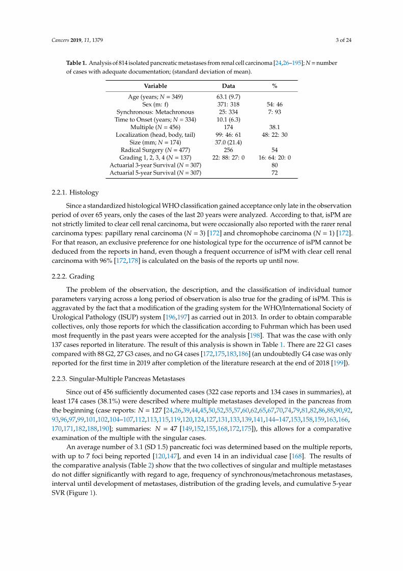
The results of the literature analysis of the casuistic reports are presented in Table 1 [24,26–195].Taken together, it produced the following results: 1. mean age 63.1 years; 2. 46% female, 54%
male; 3. metachronous metastases in 93%; 4. interval from tumornephrectomy to manifest pancreaticmetastasis 10.1 years (maximum 33 years [104]; 5. multiple metastases 38.1%; 6. Localization: head ofpancreas 48%, body 22%, and tail 30%; 7. Grading: G1 16%, 2 64%, G3 20%, G4 0%; 8. cumulative fiveyear survival rate (SVR) 72%.
Cancers 2019, 11, 1379 3 of 24
Table 1. Analysis of 814 isolated pancreatic metastases from renal cell carcinoma [24,26–195]; N = numberof cases with adequate documentation; (standard deviation of mean).
Variable Data %
Age (years; N = 349) 63.1 (9.7)Sex (m: f) 371: 318 54: 46
Synchronous: Metachronous 25: 334 7: 93Time to Onset (years; N = 334) 10.1 (6.3)
Multiple (N = 456) 174 38.1Localization (head, body, tail) 99: 46: 61 48: 22: 30
Size (mm; N = 174) 37.0 (21.4)Radical Surgery (N = 477) 256 54Grading 1, 2, 3, 4 (N = 137) 22: 88: 27: 0 16: 64: 20: 0
Actuarial 3-year Survival (N = 307) 80Actuarial 5-year Survival (N = 307) 72
2.2.1. Histology
Since a standardized histological WHO classification gained acceptance only late in the observationperiod of over 65 years, only the cases of the last 20 years were analyzed. According to that, isPM arenot strictly limited to clear cell renal carcinoma, but were occasionally also reported with the rarer renalcarcinoma types: papillary renal carcinoma (N = 3) [172] and chromophobe carcinoma (N = 1) [172].For that reason, an exclusive preference for one histological type for the occurrence of isPM cannot bededuced from the reports in hand, even though a frequent occurrence of isPM with clear cell renalcarcinoma with 96% [172,178] is calculated on the basis of the reports up until now.
2.2.2. Grading
The problem of the observation, the description, and the classification of individual tumorparameters varying across a long period of observation is also true for the grading of isPM. This isaggravated by the fact that a modification of the grading system for the WHO/International Society ofUrological Pathology (ISUP) system [196,197] as carried out in 2013. In order to obtain comparablecollectives, only those reports for which the classification according to Fuhrman which has been usedmost frequently in the past years were accepted for the analysis [198]. That was the case with only137 cases reported in literature. The result of this analysis is shown in Table 1. There are 22 G1 casescompared with 88 G2, 27 G3 cases, and no G4 cases [172,175,183,186] (an undoubtedly G4 case was onlyreported for the first time in 2019 after completion of the literature research at the end of 2018 [199]).
2.2.3. Singular-Multiple Pancreas Metastases
Since out of 456 sufficiently documented cases (322 case reports and 134 cases in summaries), atleast 174 cases (38.1%) were described where multiple metastases developed in the pancreas fromthe beginning (case reports: N = 127 [24,26,39,44,45,50,52,55,57,60,62,65,67,70,74,79,81,82,86,88,90,92,93,96,97,99,101,102,104–107,112,113,115,119,120,124,127,131,133,139,141,144–147,153,158,159,163,166,170,171,182,188,190]; summaries: N = 47 [149,152,155,168,172,175]), this allows for a comparativeexamination of the multiple with the singular cases.
An average number of 3.1 (SD 1.5) pancreatic foci was determined based on the multiple reports,with up to 7 foci being reported [120,147], and even 14 in an individual case [168]. The results ofthe comparative analysis (Table 2) show that the two collectives of singular and multiple metastasesdo not differ significantly with regard to age, frequency of synchronous/metachronous metastases,interval until development of metastases, distribution of the grading levels, and cumulative 5-yearSVR (Figure 1).
Cancers 2019, 11, 1379 4 of 24
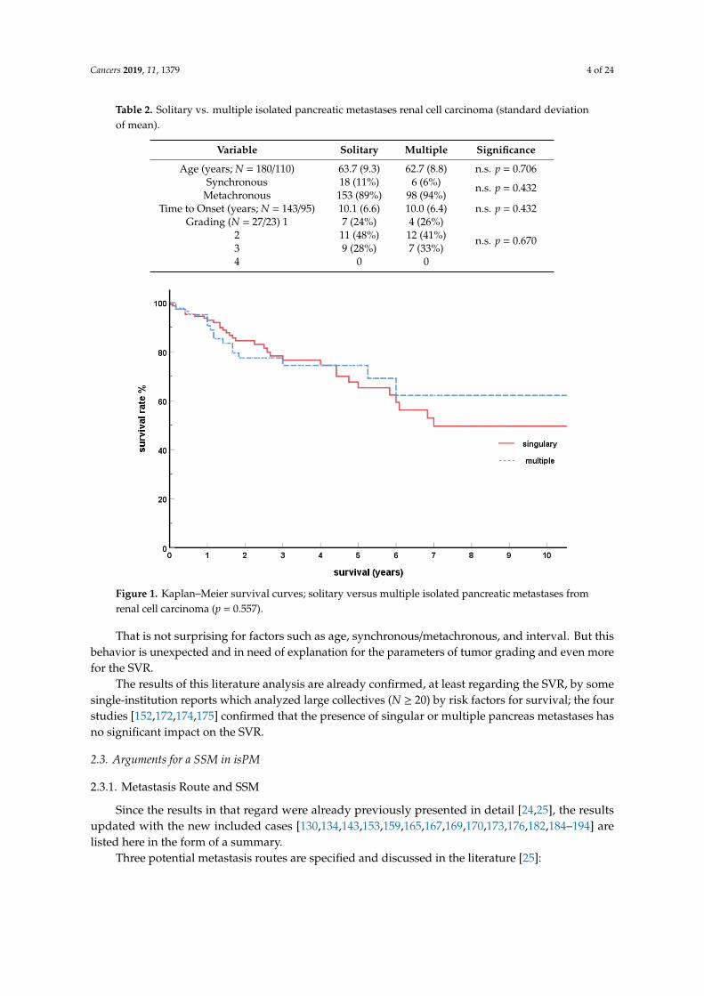
Table 2. Solitary vs. multiple isolated pancreatic metastases renal cell carcinoma (standard deviationof mean).
Variable Solitary Multiple Significance
Age (years; N = 180/110) 63.7 (9.3) 62.7 (8.8) n.s. p = 0.706Synchronous 18 (11%) 6 (6%) n.s. p = 0.432
Metachronous 153 (89%) 98 (94%)Time to Onset (years; N = 143/95) 10.1 (6.6) 10.0 (6.4) n.s. p = 0.432
Grading (N = 27/23) 1 7 (24%) 4 (26%)
n.s. p = 0.6702 11 (48%) 12 (41%)3 9 (28%) 7 (33%)4 0 0
Cancers 2019, 11, x FOR PEER REVIEW 4 of 23
Table 2. Solitary vs multiple isolated pancreatic metastases renal cell carcinoma (standard deviation of mean).
Variable Solitary Multiple Significance Age (years; N = 180/110) 63.7 (9.3) 62.7 (8.8) n.s. p = 0.706
Synchronous 18 (11%) 6 (6%) n.s. p = 0.432
Metachronous 153 (89%) 98 (94%) Time to Onset (years; N = 143/95) 10.1 (6.6) 10.0 (6.4) n.s. p = 0.432
Grading (N = 27/23) 1 2 3 4
7 (24%) 11 (48%) 9 (28%)
0
4 (26%) 12 (41%) 7 (33%)
0
n.s. p = 0.670
Figure 1. Kaplan–Meier survival curves; solitary versus multiple isolated pancreatic metastases from renal cell carcinoma (p = 0.557).
That is not surprising for factors such as age, synchronous/metachronous, and interval. But this behavior is unexpected and in need of explanation for the parameters of tumor grading and even more for the SVR.
The results of this literature analysis are already confirmed, at least regarding the SVR, by some single-institution reports which analyzed large collectives (N ≥ 20) by risk factors for survival; the four studies [152,172,174,175] confirmed that the presence of singular or multiple pancreas metastases has no significant impact on the SVR.
2.3. Arguments for a SSM in isPM
2.3.1. Metastasis Route and SSM
Since the results in that regard were already previously presented in detail [24,25], the results updated with the new included cases [130,134,143,153,159,165,167,169,170,173,176,182,184–194] are listed here in the form of a summary.
Three potential metastasis routes are specified and discussed in the literature [25]: 1. A local lymphogenic metastasis route [48,88,101,106,150,161,168], where pancreatic metastases
are supposed to develop by way of pre-existent or tumor-induced local lymph routes between kidney and pancreas after blockage of the regional lymph nodes by a retrograde lymphatic flow.
Figure 1. Kaplan–Meier survival curves; solitary versus multiple isolated pancreatic metastases fromrenal cell carcinoma (p = 0.557).
That is not surprising for factors such as age, synchronous/metachronous, and interval. But thisbehavior is unexpected and in need of explanation for the parameters of tumor grading and even morefor the SVR.
The results of this literature analysis are already confirmed, at least regarding the SVR, by somesingle-institution reports which analyzed large collectives (N ≥ 20) by risk factors for survival; the fourstudies [152,172,174,175] confirmed that the presence of singular or multiple pancreas metastases hasno significant impact on the SVR.
2.3. Arguments for a SSM in isPM
2.3.1. Metastasis Route and SSM
Since the results in that regard were already previously presented in detail [24,25], the resultsupdated with the new included cases [130,134,143,153,159,165,167,169,170,173,176,182,184–194] arelisted here in the form of a summary.
Three potential metastasis routes are specified and discussed in the literature [25]:
Cancers 2019, 11, 1379 5 of 24
1. A local lymphogenic metastasis route [48,88,101,106,150,161,168], where pancreatic metastasesare supposed to develop by way of pre-existent or tumor-induced local lymph routes betweenkidney and pancreas after blockage of the regional lymph nodes by a retrograde lymphatic flow.
2. A local venous spread route, where pre-existent, porto-renal anastomoses [106,157,200,201], ordraining, collateral veins of hyper-vascularized tumors [27,48,88,101,106,168]—enabling a tumorcell embolism in the pancreas—and that independently of whether there is a renal vein thrombosisor not [48].
3. The systemic haematogenic metastasis route
An analysis of the literature data produces the following results relevant for the issue of themetastasis route:
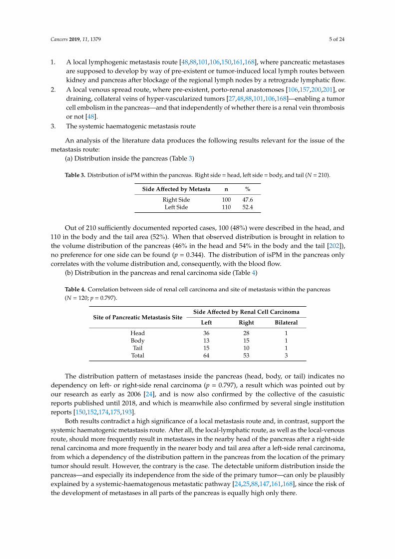
(a) Distribution inside the pancreas (Table 3)
Table 3. Distribution of isPM within the pancreas. Right side = head, left side = body, and tail (N = 210).
Side Affected by Metasta n %
Right Side 100 47.6Left Side 110 52.4
Out of 210 sufficiently documented reported cases, 100 (48%) were described in the head, and110 in the body and the tail area (52%). When that observed distribution is brought in relation tothe volume distribution of the pancreas (46% in the head and 54% in the body and the tail [202]),no preference for one side can be found (p = 0.344). The distribution of isPM in the pancreas onlycorrelates with the volume distribution and, consequently, with the blood flow.
(b) Distribution in the pancreas and renal carcinoma side (Table 4)
Table 4. Correlation between side of renal cell carcinoma and site of metastasis within the pancreas(N = 120; p = 0.797).
Site of Pancreatic Metastasis SiteSide Affected by Renal Cell Carcinoma
Left Right Bilateral
Head 36 28 1Body 13 15 1Tail 15 10 1
Total 64 53 3
The distribution pattern of metastases inside the pancreas (head, body, or tail) indicates nodependency on left- or right-side renal carcinoma (p = 0.797), a result which was pointed out byour research as early as 2006 [24], and is now also confirmed by the collective of the casuisticreports published until 2018, and which is meanwhile also confirmed by several single institutionreports [150,152,174,175,193].
Both results contradict a high significance of a local metastasis route and, in contrast, support thesystemic haematogenic metastasis route. After all, the local-lymphatic route, as well as the local-venousroute, should more frequently result in metastases in the nearby head of the pancreas after a right-siderenal carcinoma and more frequently in the nearer body and tail area after a left-side renal carcinoma,from which a dependency of the distribution pattern in the pancreas from the location of the primarytumor should result. However, the contrary is the case. The detectable uniform distribution inside thepancreas—and especially its independence from the side of the primary tumor—can only be plausiblyexplained by a systemic-haematogenous metastatic pathway [24,25,88,147,161,168], since the risk ofthe development of metastases in all parts of the pancreas is equally high only there.

Cancers 2019, 11, 1379 6 of 24
In addition to this main argument, we found more indications in the literature which argueagainst a high significance of the local-lymphatic and local-venous metastasis route, but in favor of thesystemic route:
a. In general, the lymphatic system is rarely involved with isPM. Regional lymph nodemetastases were only present at the time of renal carcinoma surgery in 7.1% of the reportscommunicated [74,150,160,175,176], and at the time of pancreas metastases surgery, paraaortal lymphnodes were reported only once [142], and peripancreatic lymph nodes were found in only 18 of 309reports (5.8%) [124,152,163,172,174,177]. That makes a great significance of a lymphatic tumor celltransport with isPM seem unlikely. b. A tumorous infiltration of the renal vein stems (category IIIb),condition for a flow reversal in the direction of the pancreas, was only reported in 9.6% [129,145,148,150],which argues against a great significance of tumor infiltration or tumor occlusion of the renal veins,respectively, in the development of isPM. c. Porto-renal anastomoses [200,201] which drain blood fromthe kidney region in the portal vascular system of the pancreas would, following the hepatopedalbloodstream in the portal vein system, also have to drain this blood with the tumor cells contained in itin the liver—with subsequent liver metastases. That is not the case, since no clustering of metachronousliver metastases is detectable epidemiologically. d. A high significance of a systemic-haematogenicmetastatic route, however, emphasises that out of 27 extra-pancreatic, resectable metastases whichdeveloped time-wise between renal carcinoma surgery and diagnosis of pancreas metastases, 20(72%) were undoubtedly of a haematogenic, systemic origin [25,153], and out of the 94 metastaseswhich developed after the resection of the pancreas metastases, 70 (74.1%) were also undoubtedlyof a haematogenic-systemic origin [25,130,167,169,170]. Consequently, the predominant number ofmetastases which were occasionally observed before the development or after the removal of pancreaticmetastases are of a systemic haematogenic origin.
In sum, these data make a particular significance of the lymphogenic and local-venous routeappear unlikely. By contrast, the systemic-haematogenic route seems to be more significant, since itcorrelates well with the epidemiological data. This comes, however, at the price that the question as towhy—despite systemic-haematogenic spread—clinical manifest metastases develop exclusively in thepancreas remains unanswered [148,154,161,175].
2.3.2. Histology, Grading, and SSM
The study found two relevant peculiarities. On the one hand, the highly specific metastaticpathway is not strictly linked to clear cell renal carcinoma, but was also occasionally observed with therarer, histological forms [172]. On the other hand, the study shows that this highly specific metastaticbehavior of the tumor cells gets lost with the degree of tumor cell de-differentiation; until the end of2018, there was no G4 observation [172,175,183,186].
Particularities which indicate a special importance of an SSM cannot be deduced from the literaturedata for histology and grading.
2.3.3. Multiple Pancreas Metastases and SSM
The comparative study of singular and multiple pancreas metastases presented here producedthree results which are remarkable for the influence of an SSM:
1. The risk of multiple metastases in the pancreas, which is only 120–180 g, is high at 38.1%—witha simultaneous absence of metastases in other organs.
It shows that multiple tumor cell embolisms undoubtedly occur in the case of isPM. If manifestedmetastases remain limited to the pancreas despite multiple cell embolisms in the vascular system,however, that can only be explained by an SSM.
2. No different SVR for singular and multiple isPM.That the SVR with metastasising renal cell carcinoma is influenced by the total tumor mass and
therefore also by the number of metastases is to be expected, but it was only examined later in studies.When an effective drug treatment for metastasising renal cell carcinoma became available for the first
Cancers 2019, 11, 1379 7 of 24
time with the establishment of targeted therapies [169,203–206], the question as to prognostic factorsgained clinical importance and resulted in corresponding studies. These studies [207–210], whichdiffer little with regard to patient selection, determination of the tumor burden, and drug treatmentshow concordantly that the baseline tumor burden correlates significantly negatively with the overallsurvival time. Regarding the question asked here, this means that shorter SVR are to be expected forpatients with multiple pancreas metastases and, consequently, a greater tumor load. However, theliterature analysis presented shows—just like five major institution reports [150,152,172,174,175]—adiametrically opposite behavior; singularity or multiplicity of pancreatic metastases has no influenceon the SVR.
3. A grading level distribution identical for singular and multiple metastases.It is a known fact that the risk of tumor cell embolization and the subsequent number of metastases
is co-determined by the degree of cell degeneration, i.e., the grading, in case of solid tumors, andthat is the methodical reason for the grading of solid tumors. Regarding our question, however, thismeans that a shift to the higher grading levels should be detectable in the case of multiple metastases.However, the analysis revealed no such shift.
For that reason, the absence of the expected results for grading, and even more for SVR whencomparing singular/multiple metastases, requires an explanation. However, the unusual clinicalbehavior can easily be reconciled with the effect of an exquisite SSM which is maintained over alonger period of time, often over several years. On the one hand, the embolised renal carcinoma cellswith isPM have properties which make metastasis of them in the pancreas particularly easy, resultingin multiple metastases, while they cannot settle in the other organs on the other hand. Then, thehigh 38.1% rate of multiple pancreas metastases are not the consequence of more aggressive primarytumor cells with a larger number of embolized tumor cells, which cannot be reconciled with thenon-different grading. The high rate is rather the consequence of a share of tumor cells able to settle inthe pancreas which is exquisitely high with isPM. It is not the cell aggressiveness which determinesthe clinical course, but the exquisite cell adaptation to the pancreas, which is often maintained foryears—consequently, a SSM. This SSM makes the non-different SVR plausible as well. In the case ofsingular as well as multiple metastases, the effect of the high organ specificity is that these cells arenot able to metastasise outside of the pancreas and die. Since this mechanism affects tumor cells ofsingular metastases just like cells of multiple pancreatic metastases, a pattern of metastasis which isstrictly limited to the pancreas persists for a prolonged period of time with both types of progression,which explains the consistent SVR after adequate treatment of the pancreatic foci.
3. Discussion
With over 330,000 cases world-wide, renal cell carcinoma is the ninth most commonmalignant tumor [211], which is already in a stage of generalisation in 20–30% of the patientsat the time of diagnosis [171,172,175,211], and even after supposedly radical surgery, 15–25%of the patients develop a stage of generalisation [171,172,183,203] with metastases in lungs,bones, liver, and brain later on [157,178,183,212]. A special characteristic of renal carcinomasis that the disease is characterised by a protracted course in about 20%, with periods of slowtumor growth or stability for many years [144,171,181,212,213]. The exquisitely rare entityof isPM, of which approximatively 800 cases were reported world-wide, is also part of thislast-mentioned group. The clinical presentation is typically characterised by a late onset (10years after primary tumor), often multiple occurrence (38%), and a good prognosis. The onlytherapeutic option for a long period of time was surgical resection—depending on the metastaticsite in the pancreas in the form of duodenopancreatectomy (DP) [26,116,148,171,175,186,193],pylorus preserving DP [120,121,127,131,132,135,137,140,150,174,179,183], distalpancreatectomy [29,116,145,157,181], total pancreatectomy [32,33,139,144,151,186,188], central(midsegment) pancreatectomy [68,121,131,146,147,152,171,175,186,193], or local tumor resections, therole of which is still controversial [25,121,125,146,150,152,158,160,163,172]. With cumulative 5-year
Cancers 2019, 11, 1379 8 of 24
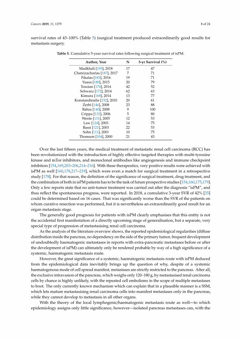
survival rates of 43–100% (Table 5) (surgical treatment produced extraordinarily good results formetastasis surgery.
Table 5. Cumulative 5-year survival rates following surgical treatment of isPM.
Author, Year N 5-yr Survival (%)
Madkhali [193], 2018 17 47Chatzizacharias [187], 2017 7 71
Fikatas [183], 2016 19 71Yuasa [180], 2015 20 79
Tosoian [174], 2014 42 52Schwarz [172], 2014 62 63Kimura [168], 2014 13 77
Konstandinidis [152], 2010 20 61Zerbi [146], 2008 23 88Bahra [140], 2008 9 100
Crippa [132], 2006 5 80Wente [131], 2005 12 53Law [124], 2003 14 75Bassi [121], 2003 22 53Sohn [111], 2001 10 75
Thomson [104], 2000 21 43
Over the last fifteen years, the medical treatment of metastatic renal cell carcinoma (RCC) hasbeen revolutionized with the introduction of highly effective targeted therapies with multi-tyrosinekinase and mTor inhibitors, and monoclonal antibodies like angiogenesis and immune checkpointinhibitors [154,169,203–206,214–216]. With these therapeutics, very positive results were achieved withisPM as well [160,178,217–219], which were even a match for surgical treatment in a retrospectivestudy [178]. For that reason, the definition of the significance of surgical treatment, drug treatment, andthe combination of both in isPM patients has to be the task of future prospective studies [154,160,175,179].Only a few reports state that no anti-tumor treatment was carried out after the diagnosis “isPM”, andthus reflect the spontaneous progress, were reported. In 2018, a cumulative 3-year SVR of 42% [25]could be determined based on 16 cases. That was significantly worse than the SVR of the patients onwhom curative resection was performed, but it is nevertheless an extraordinarily good result for anorgan metastasis stage.
The generally good prognosis for patients with isPM clearly emphasises that this entity is notthe accidental first manifestation of a directly upcoming stage of generalisation, but a separate, veryspecial type of progression of metastasising renal cell carcinoma.
As the analysis of the literature overview shows, the reported epidemiological regularities (diffusedistribution inside the pancreas, no dependency on the side of the primary tumor, frequent developmentof undoubtedly haematogenic metastases in reports with extra-pancreatic metastases before or afterthe development of isPM) can ultimately only be rendered probable by way of a high significance of asystemic, haematogenic metastasis route.
However, the great significance of a systemic, haematogenic metastasis route with isPM deducedfrom the epidemiological data inevitably brings up the question of why, despite of a systemichaematogenous mode of cell spread manifest, metastases are strictly restricted to the pancreas. After all,the exclusive intravasion of the pancreas, which weighs only 120–180 g, by metastasised renal carcinomacells by chance is highly unlikely, with the repeated cell embolisms in the scope of multiple metastasesto boot. The only currently known mechanism which can explain that in a plausible manner is a SSM,which lets mature metastasising renal carcinoma cells into manifest metastases only in the pancreas,while they cannot develop to metastasis in all other organs.
With the theory of the local lymphogenic/haematogenic metastasis route as well—to whichepidemiology assigns only little significance, however—isolated pancreas metastases can, with the
Cancers 2019, 11, 1379 9 of 24
simultaneously very rare presence of metastases in the complete tissue crossed by the tumor cellsbetween renal tumor and pancreas (one paraaortal lymph node involvement [142]; only in 7% infiltratedperipancreatic lymph nodes [124,152,163,172,174,177]), also only be explained by a highly specificinteraction between tumor cells and pancreas, which allows a maturation into metastases exclusivelyin the pancreas.
To date, the argumentation for the existence of an SSM for isPM was limited exclusively toconclusions drawn from the systemic-haematogenic metastasis route, which was rendered probable.The presented comparative studies of solitary and multiple metastases deliver a second chain ofargumentation independent of the metastasis route for an SSM with isPM: the multiple pancreasmetastases observed in 38%, the consistent grading level distribution for isPM with solitary andmultiple metastases, and above all, the SVR, which are also consistent. That behavior can also onlybe plausibly explained by a highly specific SSM, which makes it much easier for these tumor cells tosettle in the pancreas and makes it almost impossible in all other organs. Consequently, the diseaseremains strictly limited to the pancreas with solitary as well as multiple metastases, which resultsin equally good SVR with multiple as well as singular pancreatic metastases following an adequate“radical” treatment of the pancreas metastases (In context with the unusual clinical behavior of isPMit is noteworthy that a recent clinical investigation [220] revealed a second unexpected behaviorof pancreatic metastases from renal cell carcinoma. In patients with multi-organ metastasis fromRCC those seem to have an unexpected better outcome, in which the pancreas was affected frommetastasis too, when compared with patients with multiorgan metastases but without pancreaticmetastases—although the pancreatic metastasis group had a higher median number of affected organsites than the non-pancreatic metastasis group. This indicates that RCC capable of metastasizing intothe pancreas represent a special subgroup of RCC and that detailed molecular studies may providevaluable information on the molecular drivers of tumor progression [220]).
In summary, the study delivers another argument for considering isPM a model entity forthe taking effect of an SSM, since the reports about singular and multiple metastases deliver anadditional, second chain of argumentation. According to the current state of knowledge, severalarguments consequently suggest that this type of metastasis should not exclusively be considered amechanical tumor cell transport phenomenon, but to regard it as a biochemical tumor cell settlementphenomenon [3].
Limits of the studyIt is a retrospective study of casuistic reports, and a hidden bias in the published casuistic reports
cannot be excluded on principle. The methodical limitation of an analysis of casuistic reports is alreadycompensated, however—at least as far as the SVR with singular/multiple metastases is concerned—byvarious high volume single institution reports [152,172,174,175] which confirm the results of thecasuistic reports.
3.1. Pathomechanism
The precise biochemical processes involved in the development of isPM are not yet investigatedand therefore unknown. Even in extensive overviews, detailed investigations concerning pancreaticmetastasis of RCC could not be found [221]. There are, however, studies of more frequent tumorentities that identified some biochemical mechanisms that may also be at work in isPM.
3.1.1. Genetic/Epigenetic Alterations and isPM
Due to the scarcity of isPM, detailed systematic studies on the genetic/epigenetic profile of isPMhave, to our best knowledge, not yet been presented. A MEDLINE (PubMed) based literature researchthus revealed only one single presentation [222], reporting on two cases of pancreatic metastasis in RCCin which mutation analysis was performed, in order to evaluate the use of moleculary targeted therapies.There are, however, a few publications comparing non-metastatic with metastatic RCC, however thesereport on metastatic RCC as one group without taking into consideration different distant metastasis
Cancers 2019, 11, 1379 10 of 24
sites or the inclusion of pancreatic metastasis. On the one hand, a recent investigation [223] did notaddress variability between matched primary tumors and metastasis or changes in the genomic of RCC.On the other hand, in a few publications [224–228], reporting on metastatic RCC as one group, it could beproved that the microRNA profile in metastatic RCC differs from non metastatic RCC [225,229], as wellas the epithelial-mesenchymal transition associated microRNA/mRNA signature [230] (The numberof differently expressed miRNA in metastatic RCC was determined to be 12 [229], 14 [224], 15 [230],20 [227], and 21 [225], respectively; 11 of these miRNAs were mentioned just in one report: miRNA10a–p5, 21, 30c–5p, 30e, 31, 130b, 149, 199–5p, 200b, 429, 455; and 10 in two or more reports: miRNA10b–5p, 30a–3p, 30a–5p, 139–5p, 144, 200a, 200c, 204, 223–3p, 451). In addition, the microRNAexpression in distant RCC metastasis differed according to the site of metastasis in lung, bone, andbrain [224]. These results reveal a relation between microRNA signature and metastatic potential anddistant metastatic site. Whether analogous a special microRNA signature is involved in the isPMphenomenon remains, of course, uncertain, and will have to be established by further investigations.
Furthermore, several investigations have shown the great number of altered miRNA involvedin renal cell carcinoma also [228,231–248]. These miRNA regulate cancer metastasis also,because of their capacity to inhibit numerous target genes involved in different steps of themetastatic cascade, e.g., EMT [233,241,244,245], migration [234,236,238,240,241,246], and metastasissettlement [231,235,239,242,243]. The variable interactions of all these miRNS in various tumor cellsbrings about manifold different capabilities for metastasis, which increases the odds that one of theembolized tumor cell exactly “fits” the properties of the target organ (soil)—a necessary prerequisitefor the metastatic process.
3.1.2. Organotropism and SSM
The genetic mechanism responsible for this extreme organotropism of the metastatic patterncan naturally not be determined at present. In general, there will be an organ preference duringmetastasis if steps which require a precisely fitting interaction of tumor cell-specific properties withorgan-specific properties occur in the scope of the early multi-stage process of metastasis. At leastthree such mechanisms are already known:
1. The pre-metastatic niche (PMN) [3,249–252]. PMN conceptualized as a fertile “soil” conduciveto the survival and outgrowth of metastatic seed results from the interaction of three components:(a) Primary tumor derived components (such as tumor-derived secreted factors or tumor-derivedexosomes/microvesicles [253,254]), (b) Tumor mobilized bone marrow derived cells (such as myeloidderived suppressor cells [255]), and (c) Organ components of the future host organ, (such ascellular and molecular factors, fibroblasts, endothelium cells of vascular structures, and extracellularmatrix) [256,257]. Since properties of the primary tumor, as well as of the host organ, are involvedin PMN formation, this results in an organotropism during niche formation, which facilitates latermetastasis in the target organ and prevents it in non-target organs, respectively. The ability to provide aPMN is documented for RCC also, by the proof of a PMN in the lung [256]. This study also pointed outthe complexity of the process, as only CD105+ tumor stem cells were capable of doing so. The abilityto form a PMN in the pancreas, however, is so far not documented for RCC.
2. A successful interaction of a chemokine receptor on the tumor cell surface and a suitable ligandis the prerequisite for the activation of numerous signal transducing pathways, which are critical incell proliferation, migration, angiogenesis, invasion, and proliferation [1,258]. Since the equipment ofthe individual tumor cells with chemokine receptors is tumor cell-specific and the type and the levelof the ligand are organ-specific, successful interaction is only possible in tissues where cell receptorsand ligand match precisely [1–3]. For example, breast cancer was found to express the chemokinereceptors CXCR4 and CCR7 at high levels. The corresponding ligands on the other hand, CXCL12 andCCL21, are present at elevated levels in lymph nodes, lung, liver, and bone marrow—preferred distantmetastatic sites of breast cancer [1,3].
Cancers 2019, 11, 1379 11 of 24
3. Due to the lack of qualified investigations on pancreatic metastasis considerations on the impactof different immunoediting [259] in different distant metastasis sites, creating an organotropism leadingto isPM must remain speculative, although conceivable, if one interprets the above-mentioned differentmiRNA profile at different distant metastatic organ sites as a consequence of cell selectioning process. Itis therefore tempting to assume that in isPM in all host organs except the pancreas immune-surveillancedetects and correctly eliminates the metastasized tumor cells by natural tumor specific T-cell mediatedimmune response, or keeps them in a dormant “equilibrium” state [259]. Only in the pancreas (soil)is an immunosuppression present which enables the carcinomas cells to evade immune control andto mature to manifest metastasis. IsPM would thus represent a “single organ deficiency of immuneresponse”. The reasons for this immunosuppression in the pancreas are of course not yet known.In this context, it is worth recalling that the pancreas consists of two different components, an exocrineand an endocrine, and that at the moment it remains unknown whether the metastasis settlementcommences in the vascular structures of the exocrine or endocrine component.
Currently, the question as to whether and how these biochemical processes, but also otherphenomena still unknown to date, are involved in isPM remains unanswered, and has to remain thetask of future research.
4. Materials and Methods
A literature compilation concerning isPM dating up to the end of 2018 was evaluated as to whether1. The lines of argumentation presented so far are still tenable, and 2. Whether further epidemiologicalparticularities and phenomena which can be explained by an SSM can be proven. Histology, grading,and a comparison of singular versus multiple metastases in isPM were examined and analyzed forcorrelations with the SSM for that purpose.
4.1. Data Sources
The literature research was based on MEDLINE (PubMed) registry and data on the epidemiology,pathology, and clinical variables i.e., age, sex, site of primary RCC, time to onset of metastatic disease,number, size, and site of metastases and survival time, were collected and analyzed.
4.2. Inclusion and Exclusion Criteria
For the analysis we considered as isPM the very rare observations of metastasising renal carcinomawhere solitary or multiple metastases definitely, or at least across a period of years, synchronously,as well as metachronously to RCC, developed exclusively in the pancreas. For the selection of theindividual reports, the criterions defined in 2006 (24) were used; e.g., for rating metastases as solitaryor multiple, only those reports were considered that specified the number of lesions or that usedwording clearly indicative of singularity or multiplicity. For defining the site of the metastatic lesions(head, body, or tail), only solitary isPMs that were unequivocally assignable to a specific part of thepancreas by preoperative imaging, the surgeons report, or the resected specimen were considered. Itgoes without saying that in a retrospective review, not every report contained data on all variablesinvestigated, thus reducing the number of observations for subset analysis. The actual number ofobservations that provided information on a given variable was specified [24].
4.3. Statistics
Continuous data are presented as means (standard deviation). Differences were evaluatedwith the X2test, Fisher’s exact test, and Student’s t-test. Survival was calculated according to theKaplan–Meier method and differences among subgroups were compared by log-rank test. P < 0.05was considered significant.
Cancers 2019, 11, 1379 12 of 24
5. Conclusions
IsPM constitute a very rare, well-defined tumor entity, for the development of which a highlydeveloped SSM is responsible with a high probability. Furthermore, the uniform clinical course givesrise to the suspicion that the phenomenon is based on a uniform pathomechanism which remainsconstant for years. That indicates that genetic investigations would be meaningful to examine themechanism which causes the exclusive presence of metastases in the pancreas and their absence in otherorgans, respectively. A clarification of these factors can contribute to a more profound understandingof the complex metastatic process, which is the fundamental requirement for the development oftherapeutics which can block the metastatic process.
Funding: This research received no external funding.
Conflicts of Interest: The author declares that there is no conflict of interest. There was no funding source forthis study.
References
1. Chambers, A.F.; Groom, A.C.; McDonald, I.C. Metastasis: Dissemination and growth of cancer cells inmetastatic sites. Nat. Rev. Cancer 2002, 2, 563–572. [CrossRef] [PubMed]
2. Shibue, T.; Weinberg, R.A. Metastatic colonization: Settlement, adaptation and propagation of tumor cells ina foreign tissue environment. Semin. Cancer Biol. 2011, 21, 99–106. [CrossRef] [PubMed]
3. Talmadge, J.E.; Fidler, I.J. AACR centennial series: The biology of cancer metastasis: Historical perspective.Cancer Res. 2010, 70, 5649–5669. [CrossRef]
4. Schluter, K.; Gassmann, P.; Enns, A.; Korb, T.; Hemping-Bovenkerk, A.; Hölzen, J.; Haier, J. Organ specifictumor cell adhesion and extravasion of colon carcinoma cells with different metastatic potential. Am. J. Pathol.2006, 169, 1064–1073. [CrossRef] [PubMed]
5. Nicolson, G.L. Organ specificity of tumor metastasis: Role of preferential adhesion, invasion and growth ofmalignant cells at specific secondary sites. Cancer Metastasis Rev. 1988, 7, 143–188. [CrossRef] [PubMed]
6. Strell, C.; Entschladen, F. Extravasation of leukocytes in comparison to tumor cells. Cell. Commun. Signal.2008, 6, 10. [CrossRef] [PubMed]
7. Miles, F.L.; Pruitt, F.L.; van Golen, K.L.; Cooper, C.R. Stepping out the flow: Capillary extravasion in cancermetastasis. Clin. Exp. Metastasis 2008, 25, 305–324. [CrossRef] [PubMed]
8. Gandalovicová, A.; Rosel, D.; Fernandes, M.; Veselý, P.; Heneberg, P.; Cermák, V.; Petruželka, L.; Kumar, S.;Sanz-Moreno, V.; Brábek, J. Migrastatics—Anti-Metastatic and anti-invasion drugs: Promises and challenges.Trends Cancer 2017, 3, 391–406. [CrossRef]
9. Zhang, Q.; Yang, M.; Shen, J.; Gerhold, L.M.; Hoffman, R.M.; Xing, H.R. The role of the intravascularmicroenvironment in spontaneous metastasis development. Int. J. Cancer 2010, 126, 2534–2541. [CrossRef]
10. Clark, A.; Vignjevic, D. Modes of cancer cell invasion and the role of the microenvironment. Curr. Opin.Cell Biol. 2015, 36, 13–22. [CrossRef]
11. Friedl, P.; Wolf, K. Tumour-cell invasion and migration: Diversity and escape mechanisms. Nat. Rev. Cancer2003, 3, 362–374. [CrossRef] [PubMed]
12. Wong, M.S.; Sidik, S.M.; Mahmud, R.; Stanlas, J. Molecular targets in the discovery and development of novelantimetastatic agents: Current progress and future prospects. Clin. Exp. Pharmacol. Physiol. 2013, 40, 307–319.[CrossRef] [PubMed]
13. Hedley, B.D.; Chambers, A.F. Tumor dormancy and metastasis. Cancer Res. 2009, 102, 67–101.14. Luzzi, K.J.; MacDonald, I.C.; Schmidt, E.E.; Kerkvliet, N.; Morris, V.L.; Chambers, A.F.; Groom, A.C. Multistep
nature of metastatic efficiency: Dormancy of solitary cells after successful extravasation and limited survivalof early micrometastasis. Am. J. Pathol. 1998, 153, 865–873. [CrossRef]
15. Naumov, G.N.; MacDonald, I.C.; Weinmeister, P.M.; Kerkvliet, N.; Nadkarni, K.V.; Wilson, S.M.; Morris, V.L.;Groom, A.C.; Chambers, A.F. Persistence of solitary mammary carcinoma cells in a secondary site: A possiblecontributor to dormancy. Cancer Res. 2002, 62, 2162–2168. [PubMed]
Cancers 2019, 11, 1379 13 of 24
16. Goss, P.; Allan, A.L.; Rodenhiser, D.I.; Foster, P.J.; Chambers, A.F. New clinical and experimental approachesfor studying tumor dormancy: Does tumor dormancy offer a therapeutic target? APMIS 2008, 116, 552–568.[CrossRef] [PubMed]
17. Folkman, J. Angiogenesis. Annu. Rev. Med. 2006, 57, 1–18. [CrossRef]18. Hynes, R.O. The extracellular matrix: Not just pretty fibrils. Science 2009, 326, 1216–1219. [CrossRef]19. Killion, J.J.; Fidler, I.J. Therapy of cancer metastasis by tumoricidal activation of tissue macrophages using
liposome-encapsulated immunomodulators. Pharmacol. Ther. 1998, 78, 141–154. [CrossRef]20. Fidler, I.J.; Gersten, D.M.; Hart, I.R. The biology of cancer invasion and metastasis. Adv. Cancer Res.
1978, 28, 149–250.21. Paget, S. The distribution of secondary growths in cancer of the breast. Lancet 1889, 1, 99–101. [CrossRef]22. Gassmann, P.; Haier, J. The tumor cell-host interface in the early onset of metastatic organ colonisation.
Clin. Exp. Metastasis 2008, 25, 171–181. [CrossRef] [PubMed]23. Tarin, D.; Price, J.E.; Kettlewell, M.G.; Souter, R.G.; Vass, A.C.; Crossley, B. Mechanisms of human tumor
metastasis studied in patients with peritoneovenous shunt. Cancer Res. 1984, 44, 3584–3592. [PubMed]24. Sellner, F.; Tykalsky, N.; De Santis, M.; Pont, J.; Klimpfinger, M. Solitary and multiple isolated metastases
of clear cell renal carcinoma to the pancreas: An indication for pancreatic surgery. Ann. Surg. Oncol.2006, 13, 75–85. [CrossRef] [PubMed]
25. Sellner, F. Isolated pancreatic metastases from renal cell carcinoma: An outcome of a special metastaticpathway or of a specific tumor cell selection? Clin. Exp. Metastasis 2018, 35, 91–102. [CrossRef] [PubMed]
26. Jenssen, E.A. Metastatic hypernephroma to the pancreas. Acta Chir. Scand. 1952, 104, 177–180. [PubMed]27. Py, J.M.; Arnaud, J.P.; Cinqualbre, J.; Adloff, M.; Bollack, C. Pancreatic metastases of nephro-epitheliomas.
Apropos of 2 cases. Acta Chir. Belg. 1984, 84, 117–121. [PubMed]28. Skaarup, P.; Jorgensen, T.; Larsen, S. Asynchronous metastasizing renal cell carcinoma associated with
progressive immune complex glomerulonephritis and proteinuria. Scand. J. Urol. Nephrol. 1984, 18, 351–356.[CrossRef] [PubMed]
29. Hirota, T.; Tomida, T.; Iwasa, M.; Takahashi, K.; Kaneda, M.; Tamaki, H. Solitary pancreatic metastasis,occuring 8 years after nephrectomy for renal cell carcinoma. Inter. J. Pancreatol. 1996, 19, 145–153.
30. Lawson, L.I.; Holt, L.P.; Rooke, H.W. Recurrent duodenal haemorrhage from renal carcinoma. Br. J. Urol.1966, 38, 133–137. [CrossRef]
31. Franciosi, R.A.; Russo, J. Renal cell carcinoma metastatic to the pancreas thirteen years following nephrectomy.Mil. Med. 1969, 134, 200–203. [CrossRef]
32. Marquand, J.; Giraud, B.; Maliakas, S. Pancreatic metastasis revealing a kidney neoplasm. J. Urol. Nephrol.1971, 77, 595–601.
33. Guttman, F.M.; Ross, M.; Lachance, C. Pancreatic metastasis of renal cell carcinoma treated by totalpancreatectomy. Arch. Surg. 1972, 105, 782–784. [CrossRef] [PubMed]
34. Gillet, M.; Camelit, G.; Runser, G.; Clement, D. Duodenopancreatic metastasis from a carcinoma of the kidneyrevealed by digestive haemorrhage treated by cephalic duodeno-pancreatectomy. Chirurgie 1974, 100, 226–230.[PubMed]
35. Hermanutz, K.D.; Sonnenberg, G.E. Late metastasis of a hypernephroid kidney carcinoma to the pancreaswith tumor invasion to the duodenum. Fortschr. Röntgenstr. 1977, 127, 595–597. [CrossRef] [PubMed]
36. Saxon, A.; Gottesman, J.; Doolas, A. Bilateral hypernephroma with solitary pancreatic metastasis. J. Surg. Oncol.1980, 13, 317–322. [CrossRef]
37. Yazaki, T.; Ishikawa, S.; Ogawa, Y.; Takahashi, S.; Nemoto, S.; Rinsho, K.; Kanoh, S.; Kitagawa, R. Silentpancreatic metastasis from renal cell carcinoma. Acta Urol. Jpn. 1981, 27, 1517–1522.
38. Audisio, R.A.; La Monica, G. Solitary pancreatic metastasis occuring 20 years after nephrectomy for carcinomaof the kidney. Tumori J. 1985, 71, 197–200. [CrossRef]
39. Kishimoto, H.; Niumra, Y.; Okamoto, K.; Tsuchie, K.; Yamase, H.; Maeda, S.; Kamija, J.; Hasegawa, H.;Hayakawa, N.; Yamamoto, M. A case of resected renal cell carcinoma with massive pancreatic metastasis.Jpn. J. Cancer Clin. 1985, 31, 91–96.
40. Amamiya, H.; Iizumi, T.; Yazaki, T.; Waku, M.; Yasuda, H.; Takada, T.; Shikata, J.; Nagai, J. A solitarypancreatic metastasis from renal cell carcinoma. Hinyouki Geka 1988, 2, 167–170.
41. Carini, M.; Selli, C.; Barbanti, G.; Bianchi, S.; Muraro, G. Pancreatic late recurrence of bilateral renal cellcarcinoma after conservative surgery. Eur. Urol. 1988, 14, 258–260. [CrossRef] [PubMed]

Cancers 2019, 11, 1379 14 of 24
42. Hirano, M.; Douden, K.; Bantou, H.; Sakatoku, M.; Saitoh, H.; Tachikawa, H.; Takizawa, T.; Horigami, K.;Kameya, T.; Nagai, T.; et al. Solitary pancreatic metastasis occuring 10 years after nephrectomy for carcinomaof the kidney. Tan Sui 1988, 9, 233–237.
43. Sharma, S.K.; Kumar, A.; Madhusoodnan, P.; Banerjee, C.; Suri, S.; Dhar, M. Solitary pancreatic metastasisfrom renal cell carcinoma. A rare metastatic site. Indian J. Cancer 1988, 25, 29–32. [PubMed]
44. Guyenne, C.; Rat, P.; Haas, O.; Baudet, J.G.; Favre, J.P. Triple metastase pancreatique d´un cancer du reintraitee par duodenopancreatectomie subtotale. Presse Med. 1989, 18, 231. [PubMed]
45. Iwanami, M.; Nakayoshi, A.; Yagi, H.; Shimizu, K.; Kimura, K.; Suzuki, K.; Matsumoto, K.; Kai, Y.; Ueno, M.;Sagawa, F. A resected case of the asymptomatic pancreatic metastasis in the body and tail of the pancreasfrom renal cell carcinoma. J. Jpn. Panc. Soc. 1989, 4, 100–106.
46. Roland, C.F.; Van Heerden, J.A. Nonpancreatic primary tumors with metastasis to the pancreas.Surg. Gynec. Obstet. 1989, 168, 345–347.
47. Simpson, N.S.; Mulholland, C.; Lioe, T.; Spence, R. Late solitary metastatic renal carcinoma in the pancreas.Ulst. Med. J. 1989, 58, 198–199.
48. Strijk, S.P. Pancreatic metastases of renal cell carcinoma: Report of two cases. Gastrointest. Radiol.1989, 14, 123–126. [CrossRef]
49. Temellini, F.; Bavosi, M.; Lamarra, M.; Quagliarini, P.; Giuliani, F. Pancreatic metastasis 25 years afternephrectomy for renal cancer. Tumori J. 1989, 75, 503–504. [CrossRef]
50. Gohji, K.; Matsumoto, O.; Kamidono, S. Solitary pancreatic metastasis from renal cell carcinoma.Hinyokika Kiyo 1990, 36, 677–681.
51. Terashima, M.; Abe, H.; Suga, K.; Matsuya, F.; Kobayashi, K.; Itoh, S.; Sasaki, R.; Kanno, S.; Tomichi, N. Twocases of renal cell carcinoma metastasized to the pancreas and to the gallbladder. Jpn. J. Gastroenterol. Surg.1990, 23, 1952–1956. [CrossRef]
52. Furukawa, T.; Hattori, R.; Ohtake, H.; Souma, T.; Kinukawa, T.; Hirai, A.; Kimura, J.; Sakata, T.; Ishii, M.;Hayashi, N.; et al. A resectable case of pancreatic head metastasis from renal cell carcinoma. Hinyouki Geka1991, 4, 111–114.
53. Kubo, K.; Morita, J.; Mizoe, J.; Ogawa, H.; Irie, G. Renal cell carcinoma metastatic to the pancreas 8 yearsfollowing nephrectomy. Jpn. J. Clin. Radiol. 1991, 36, 509–512.
54. Nishida, O.; Matsunaga, Y.; Dekigai, H.; Um, S.H.; Hsieh, C.C.; Kimura, F.; Yoshioka, H.; Murakami, M.;Inoue, R.; Murai, A. Three elderly cases of renal cell carcinoma with pancreatic metastasis. Jpn. J. Geriatr.1991, 28, 392–396. [CrossRef] [PubMed]
55. Oka, H.; Hatayama, T.; Taki, Y.; Ueyama, H.; Hida, S.; Noguchi, M. A resected case of renal cell carcinomawith metastasis to pancreas. Hinyokika Kiyo 1991, 37, 1531–1534. [PubMed]
56. Tabata, T.; Kuroda, Y.; Nishimatsu, S.; Satoh, Y. A resected case of pancreatic tumor metastasized from renalcell carcinoma. J. Jpn. Panc. Soc. 1991, 6, 245–250.
57. Yamamoto, S.; Tobinaga, K.; Taketomi, K.; Kimino, K.; Ashizuka, S.; Kishikawa, M. Pancreatic metastasis ofrenal cell carcinoma occuring 17 years after nephrectomy. J. Jpn. Soc. Clin. Surg. 1991, 52, 3006–3011.
58. Fujii, M.; Kogawa, T.; Matsuyama, K.; Yamamoto, H.; Kawahito, Y.; Iinuma, S.; Kokura, S.; Takemura, S.;Yoshikawa, T.; Kondo, M.; et al. A case of metastatic renal cell carcinoma to pancreas ten years afternephrectomy. J. Kyoto Pref. Univ. Med. 1992, 101, 589–596.
59. Melo, C.R.; Melo, I.S.; Monteiro, A.Z.; de Mello, E.S. Pancreatic metastasis from renal cell carcinoma.Arq. Gastroenterol. 1992, 29, 110–112.
60. Nakagawa, K.; Tsuchiya, T.M.; Momono, S.; Sasaki, Y.; Sato, T. A case of pancreatic metastasis of renal cellcarcinoma. Jpn. J. Gastroenterol. Surg. 1992, 25, 2200–2204. [CrossRef]
61. Rypens, F.; Gansbeke, V.; Lambiliotte, J.; Regemorter, V.; Verhesi, A.; Struyven, J. Pancreatic metastasis fromrenal cell carcinoma. Br. J. Radiol. 1992, 65, 547–548. [CrossRef] [PubMed]
62. Stankard, C.E.; Karl, R.C. The treatment of isolated pancreatic metastases from renal cell carcinoma: A surgicalreview. Am. J. Gastroenterol. 1992, 87, 1658–1660. [PubMed]
63. Aikou, S.; Tokura, Y.; Yamafuji, K.; Takahashi, T.; Yoshihide, O.; Kishii, K.; Fujii, S.; Katsumata, K.; Tamiya, M.A resected case of pancreatic metastasis from renal cell carcinoma presenting with acute duodenal bleeding.J. Jpn. Soc. Clin. Surg. 1993, 54, 2666–2672. [CrossRef]
64. Calmes, J.M.; Meyer, A. Pancreatic hypernephroma manifested by a duodenal hemorrhage. Rev. Med.Suisse Romande 1993, 113, 629–631. [PubMed]
Cancers 2019, 11, 1379 15 of 24
65. Ishikawa, T.; Horimi, T.; Majima, K. A resected case of pancreatic tumor metastasized from renal cellcarcinoma. A review of 11 cases in the Japanese and 13 cases in the foreign literature. J. Jpn. Soc. Clin. Surg.1993, 51, 1642–1647.
66. Marcote-Valdivieso, E.; Arlandis, F.; Baltasar, A.; Martinez-Castro, R.; Vierna-Garcia, J. Synchronous pancreaticmetastasis of renal carcinoma. Rev. Esp. Enferm. Dig. 1993, 83, 471–473. [PubMed]
67. Nan, Y.; Kuno, N.; Kurimoto, K.; Nakamura, T.; Kobayashi, S. A resected case of pancreatic tumormetastasized from renal cell carcinoma diagnosed by endoscopic biopsy through the main pancreatic duct.Gastroenterol. Endosc. 1993, 35, 1380–1385.
68. Sauvanet, A.; Barthes, T.; Levy, P.; Flejou, J.F.; Delcenserie, R.; Bernades, P.; Belghiti, J. Late pancreaticmetastasis from renal cell carcinoma. Pancreas 1993, 8, 742–744. [CrossRef] [PubMed]
69. Takeuchi, H.; Konaga, E.; Harano, M.; Watanabe, K.; Takeuchi, Y.; Hara, M.; Mano, S. Solitary pancreaticmetastasis from renal cell carcinoma. Acta Med. Okayama 1993, 47, 63–66. [PubMed]
70. Vergara, V.; Marucci, M.; Marcarino, C.; Brunello, F.; Capussotti, L. Metastatic involvement of the pancreasfrom renal cell carcinoma treated by surgery. Ital. J. Gastroenterol. 1993, 25, 388–390.
71. Yanagisawa, T.; Nakayama, K.; Kashiwagi, M.; Tanaka, J.; Kashiwagi, T.; Mizusaki, K.; Itoh, A.; Akimoto, H.;Takahashi, T.; Aoki, T.; et al. Three cases of resectable pancreatic metastases from renal cell carcinoma.Geka Shinryo 1993, 35, 651–655.
72. Zugel, N.; Leipprand, F.; Weckermann, D.; Witte, J. Solitary metastasis to the head of the pancreas inhypernephroid carcinoma. Fortschr. Med. 1994, 112, 388–390. [PubMed]
73. Dousset, B.; Andant, C.; Guimbaud, R.; Roseau, G.; Tulliez, M.; Gaudric, M.; Palazzo, L.; Chaussade, S.;Chapuis, Y. Late pancreatic metastasis from renal cell carcinoma diagnosed by endoscopic ultrasonography.Surgery 1995, 117, 591–594. [CrossRef]
74. Fabre, J.M.; Rouanet, P.; Dagues, F.; Blanc, F.; Baumel, H.; Domergue, J. Various features and surgical approachof solitary pancreatic metastasis from renal cell carcinoma. Eur. J. Surg. Oncol. 1995, 21, 683–686. [CrossRef]
75. Onishi, T.; Ohishi, Y.; Iizuka, N.; Suzuki, Y.; Shirakawa, H.; Hatano, T.; Tom, M. Clinical characteristics of7 renal cell carcinoma patients developing a solitary pancreatic metastasis after nephrectomy. Jpn. J. Urol.1995, 86, 1538–1542. [CrossRef] [PubMed]
76. Orita, M.; Morita, N.; Hiraoka, H.; Noshima, S.; Takaimashi, T.; Esato, K. A case of resected pancreaticmetastasis from renal cell carcinoma 14 years after radical nephrectomy. J. Jpn. Pancreas Soc. 1995, 10, 63–68.
77. Takashi, M.; Takagi, Y.; Sakata, T.; Shimoji, T.; Miyake, K. Surgical treatment of renal cell carcinoma metastases:Prognostic significance. Int. Urol. Nephrol. 1995, 27, 1–8. [CrossRef] [PubMed]
78. Barras, J.P.; Baer, H.; Stenzl, A.; Czerniak, A. Isolated late metastasis of a renal cell cancer treated by radicaldistal pancreatectomy. HPB Surg. 1996, 10, 51–53. [CrossRef]
79. Palazzo, L.; Borotto, E.; Cellier, C.; Roseau, G.; Chaussade, S.; Couturier, D.; Paolaggi, J. Endosonographicfeatures of pancreatic metastases. Gastrointest. Endosc. 1996, 44, 433–436. [CrossRef]
80. Paz, A.; Koren, R.; Gal, R.; Wolloch, Y. Late solitary pancreatic metastasis from renal cell carcinoma. Isr. J.Med. Sci. 1996, 32, 1319–1321.
81. Chambers, T.P.; Fishman, E.K.; Hruban, R.H. Pancreatic metastases from renal cell carcinoma in vonHippel-Lindau disease. Clin. Imaging 1997, 21, 40–42. [CrossRef]
82. Robbins, E.G.; Franceschi, D.; Barkin, J.S. Solitary metastatic tumors to the pancreas: A case report andreview of the literature. Am. J. Gastroenterol. 1996, 91, 2414–2417.
83. Harrison, L.; Merchant, N.; Cohen, A.; Brennan, M. Pancreaticoduodenectomy for nonperiampullary primarytumors. Am. J. Surg. 1997, 174, 393–395. [CrossRef]
84. Adem, C.; Chetritt, J.; Guymar, S.; Bellil, K.; Ladouch-Badre, A.; Benlagha, N.; Bedossa, P. Pancreaticmetastasis of a renal adenocarcinoma. Apropos on 2 cases. Ann. Pathol. 1998, 18, 481–483.
85. Altschuler, E.L.; Ray, A. Spontaneous regression of a pancreatic metastasis of a renal cell carcinoma.Arch. Fam. Med. 1998, 7, 516–517. [CrossRef]
86. Butturini, G.; Bassi, C.; Falconi, M.; Salvia, R.; Caldiron, E.; Iannucci, A.; Zamboni, G.; Graziani, R.;Procacci, C.; Pederzoli, P. Surgical treatment of pancreatic metastases from renal cell carcinomas. Dig. Surg.1998, 15, 241–246. [CrossRef]
87. Gupta, R.K.; Lallu, S.; Delahunt, B. Fine-needle aspiration cytology of metastatic clear-cell renal carcinomapresenting as a solitary mass in the head of the pancreas. Diagn. Cytopathol. 1998, 19, 194–197. [CrossRef]
Cancers 2019, 11, 1379 16 of 24
88. Hashimoto, M.; Watanabe, G.; Matsuda, M.; Dohi, T.; Tsurumaru, M. Management of the pancreatic metastasesfrom renal cell carcinoma: Report of four resected cases. Hepatogastroenterology 1998, 45, 1150–1154.
89. Jingu, K.; Watanabe, K.; Yamamoto, H.; Fujita, Y.; Honda, I.; Watanabe, S.; Nagata, M.; Sugimoto, K.;Watanabe, Y. Surgical treatment of a solitary pancreatic metastasis from renal cell carcinoma: Report of acase. Surg. Today 1998, 28, 91–94. [CrossRef]
90. Merkle, E.M.; Boaz, T.; Kolokythas, O.; Haaga, J.R.; Lewin, J.S.; Brambs, H.J. Metastases to the pancreas.Br. J. Radiol. 1998, 71, 1208–1214. [CrossRef]
91. Sahin, M.; Foulis, A.A.; Poon, F.W.; Imrie, C.W. Late focal pancreatic metastasis of renal cell carcinoma.Dig. Surg. 1998, 15, 72–74. [CrossRef]
92. Z’graggen, K.; Fernandez-del Castillo, C.; Rattner, D.W.; Sigala, H.; Warshaw, A.L. Metastases to the pancreasand their surgical extirpation. Arch. Surg. 1998, 133, 418–419.
93. Augustin, H.; Bacher, H.; Uggowitzer, M.; Ott, A.; Hubmer, G.; Mischinger, H.J. Pancreatic metastases fromrenal cell carcinoma mimicking insulinomas. BJU Int. 1999, 83, 140–141. [CrossRef]
94. Carucci, L.R.; Siegelman, E.S.; Feldman, M.D. Pancreatic metastasis from clear cell renal carcinoma: Diagnosiswith chemical shift MRI. J. Comput. Assist. Tomogr. 1999, 23, 934–936. [CrossRef]
95. Erigushi, N.; Aoyagi, S.; Hara, M.; Miyazaki, T.; Hashino, K.; Imamura, I.; Jimi, A.; Naito, H. A resected caseof pancreatic metastasis from primary renal cell carcinoma. Kurume Med. J. 1999, 46, 119–122. [CrossRef]
96. Ng, C.S.; Loyer, E.M.; Iyer, R.B.; David, C.L.; DuBrow, R.A.; Charnsangavej, C. Metastases to the pancreas fromrenal cell carcinoma: Findings on three-phase contrast-enhanced CT. Am. J. Roentgenol. 1999, 172, 1555–1559.[CrossRef]
97. Sugiyama, M.; Katsura, M.; Yamamoto, K.; Nouchi, W.; Abe, N.; Hatano, N.; Atomi, Y. Pancreaticmetastasis from renal cell carcinoma causing massive gastrointestinal bleeding in von Hippel-Lindaudisease. Hepatogastroenterology 1999, 46, 1199–1201.
98. Yavascaoglu, I.; Korun, N.; Oktay, B.; Simsek, U.; Ozyurt, M. Renal cell carcinoma with solitarysynchronous pancreaticoduodenal and metachronous periprostatic metastases: Report of a case. Surg. Today1999, 29, 364–366. [CrossRef]
99. Fricke, P.; Schulz, H.U.; Buhtz, P.; Lippert, H. The pancreas as a site of multiple metastases from renal cellcarcinoma. Report of one case and review of the literature. Chirurg 2000, 71, 575–579. [CrossRef]
100. Ghavamian, R.; Klein, K.A.; Stephens, D.H.; Welch, T.J.; LeRoy, A.J.; Richards, R.L.; Burch, P.A.; Zincke, H.Renal cell carcinoma metastatic to the pancreas: Clinical and radiological features. Mayo Clin. Proc.2000, 75, 581–585. [CrossRef]
101. Kassabian, A.; Stein, J.; Jabbour, N.; Parsa, K.; Skinner, B.; Parekh, D.; Cosenza, C.; Selby, R. Renal cellcarcinoma metastatic to the pancreas: A single institution series and review of the literature. Urology2000, 56, 211–215. [CrossRef]
102. Le Borgne, J.; Partensky, C.; Glemain, P.; Dupas, B.; de Kerviller, B. Pancreaticoduodenectomy for metastaticampullary and pancreatic tumors. Hepatogastroenterology 2000, 47, 540–544.
103. Mehta, N.; Volpe, C.; Haley, T.; Balos, L.; Bradley, E.L.; Doerr, R.J. Pancreaticoduodenectomy for metastaticrenal cell carcinoma: Report of a case. Surg. Today 2000, 30, 94–97. [CrossRef]
104. Thompson, L.D.; Heffess, C.S. Renal cell carcinoma to the pancreas in surgical pathology material. Cancer2000, 89, 1076–1089. [CrossRef]
105. Espinoza, R.; Rossi, R.; Rossi, R.; Rosenberg, H. Metachronous pancreatic metastasis of a renal cell carcinoma:3 new cases. Rev. Med. Chile 2001, 129, 86–90.
106. Faure, J.P.; Tuech, J.J.; Richer, J.P.; Pessaux, P.; Arnaud, J.P.; Carretier, M. Pancreatic metastasis of renal cellcarcinoma: Presentation, treatment and survival. J. Urol. 2001, 165, 20–22. [CrossRef]
107. Hashimoto, M.; Miura, Y.; Matsuda, M.; Watanabe, G. Concomitant duodenal and pancreatic metastasesfrom renal cell carcinoma: Report of a case. Surg. Today 2001, 31, 180–183. [CrossRef]
108. Marusch, F.; Koch, A.; Dietrich, F.; Hoschke, B.; Gastinger, I. A singular late metastasis of renal cell carcinomain the pancreas. An uncommon pancreatic tumor. Zentralbl. Chir. 2001, 126, 391–395. [CrossRef]
109. Ruibal Moldes, M.; Quintana de la Rosa, J.L.; Farina Perez, L.A.; Tardaguila, F.; Ortiz Rey, J.A.; Zungri Telo, E.Late pancreatic metastasis from renal carcinoma. Acta Urol. Esp. 2001, 25, 122–124. [CrossRef]
110. Scatarige, J.C.; Horton, K.M.; Sheth, S.; Fishman, E.K. Pancreatic parenchymal metastases. Am. J. Roentgenol.2001, 176, 695–699. [CrossRef]
Cancers 2019, 11, 1379 17 of 24
111. Sohn, T.A.; Yeo, C.J.; Cameron, Y.L.; Nakeeb, A.; Lillemoe, K.D. Renal cell carcinoma metastatic to thepancreas: Results of surgical management. J. Gastrointest. Surg. 2001, 5, 346–351. [CrossRef]
112. Tada, T.; Kobayashi, G.; Noda, Y.; Kimura, K.; Ito, K.; Fujita, N. A resected case with multiple pancreaticmetastasis of renal cell carcinoma. Jpn. J. Gastro Enterol. 2001, 98, 1368–1373.
113. Bechade, D.; Palazzo, I.; Desrame, J.; Duvic, C.; Herody, M.; Didelot, F.; Coutant, G.; Algayres, J.P. Pancreaticmetastasis of renal carcinoma: Report of three cases. Rev. Med. Intern. 2002, 23, 862–866.
114. Chou, Y.H.; Chiou, H.J.; Hong, T.M.; Tiu, C.M.; Chiou, S.Y.; Su, C.H.; Tsay, S.H. Solitary metastasis from renalcell carcinoma presenting as diffuse pancreatic enlargement. J. Clin. Ultrasound 2002, 30, 499–502. [CrossRef][PubMed]
115. Eloubeidi, M.A.; Jhala, D.; Chhieng, D.C.; Jhala, N.; Eltoum, I.; Wilcox, C.M. Multiple late asymptomaticpancreatic metastases from renal cell carcinoma: Diagnosis by endoscopic ultrasound-guided fine needleaspiration biopsy with immunocytochemical correlation. Dig. Dis. Sci. 2002, 47, 1839–1842. [CrossRef][PubMed]
116. Hiotis, S.P.; Klimstra, D.S.; Conlon, K.C.; Brennan, M.F. Results after pancreatic resection for metastaticlesions. Ann. Surg. Oncol. 2002, 9, 675–679. [CrossRef] [PubMed]
117. Lisii, D.; Gaimant, A.; Sautereau, D.; Paraf, F.; Maubon, A. Duodenal bleeding revealing a renal cell carcinoma.Gastroenterol. Clin. Biol. 2002, 26, 1044–1046. [PubMed]
118. Peschaud, F.; Cheynel, N.; Hagry, O.; Tremeaux, J.C.; Rat, P.; Favre, J.P. Surgical treatment of pancreaticmetastases from renal carcinoma. Ann. Chir. 2002, 127, 527–531. [CrossRef]
119. Roviello, F.; Nastri, G.; Hako, L.; Marrelli, D.; De Stefano, A.; Cioppa, T.; Pinto, E. Pancreatic metastasis fromclear cell carcinoma. Chir. Ital. 2002, 54, 873–877.
120. Yachida, S.; Fukushima, N.; Kanai, Y.; Nimura, S.; Shimada, K.; Yamamoto, J.; Sakamoto, M. Pancreaticmetastasis from renal cell carcinoma extending into the main pancreatic duct: A case report. Jpn. J. Clin. Oncol.2002, 32, 315–317. [CrossRef]
121. Bassi, C.; Butturini, G.; Falconi, M.; Sargenti, W.; Mantovavi, W.; Pederzoli, P. High recurrence rate afteratypical resection for pancreatic metastases from renal cell carcinoma. Br. J. Surg. 2003, 90, 555–559.[CrossRef] [PubMed]
122. Giulini, S.; Portolani, N.; Bonardelli, S.; Baiocchi, G.; Zampatti, M.; Coniglio, A.; Baronchelli, C. Distalpancreatic resection with splenic preservation for metastasis of renal carcinoma diagnosed 24 years laterfrom nephrectomy. Ann. Ital. Chir. 2003, 74, 93–96. [PubMed]
123. Hermandez, D.J.; Kavoussi, L.R.; Ellison, L.M. Laparoscopic pancreatectomy for metastatic renal cellcarcinoma. Urology 2003, 62, 551. [CrossRef]
124. Law, C.H.; Wei, A.C.; Hanna, S.S.; Al-Zahrani, M.; Taylor, B.R.; Greig, P.D.; Langer, B.; Gallinger, S.Pancreatic resection for metastatic renal carcinoma: Presentation, treatment and outcome. Ann. Surg. Oncol.2003, 10, 922–926. [CrossRef] [PubMed]
125. Nakagohri, T.; Konishi, M.; Inoue, K.; Nakamura, T.; Kinoshita, T. Partial pancreatic head resection forpancreatic metastasis from renal cell carcinoma. Hepatogastroenterology 2003, 50, 2236–2238. [PubMed]
126. Pecchi, A.; Cesinaro, A.; Torricelli, P. Solitary pancreatic metastasis from renal cell carcinoma. Radiol. Med.2003, 105, 386–390.
127. Zacharoulis, D.; Asopa, V.; Karvounis, E.; Williamson, R. Resection of renal metastases to the pancreas:A surgical challenge. HPB 2003, 5, 137–141. [CrossRef]
128. Moussa, A.; Mitry, E.; Hammel, P.; Sauvanet, A.; Nassif, T.; Palazzo, L. Pancreatic metastasis: A multicentricstudy of 22 patients. Gastroenterol. Clin. Biol. 2004, 28, 872–876. [CrossRef]
129. Ninan, S.; Jain, P.; Paul, A.; Menon, K. Synchronous pancreatic metastases from asymptomatic renal cellcarcinoma. JOP 2005, 6, 26–28.
130. Sotiropoulos, G.C.; Lang, H.; Liu, C.; Brokalaki, E.I.; Molmenti, E.I.; Broelsch, C.E. Surgical treatment ofpancreatic metastases of renal cell carcinoma. JOP 2005, 6, 339–343.
131. Wente, M.N.; Kleeff, J.; Esposito, I.; Hartel, M.; Müller, M.W.; Fröhlich, B.E.; Büchler, M.W.; Friess, H. Renalcancer cell metastasis into the pancreas: A single center experience and overview of the literature. Pancreas2005, 30, 218–222. [CrossRef] [PubMed]
132. Crippa, S.; Angelini, C.; Mussi, C.; Bonardi, C.; Romano, F.; Sartori, P.; Uggeri, F.; Bovo, G. Surgical treatmentof metastatic tumors to the pancreas: A single center experience and review of the literature. World J. Surg.2006, 30, 1536–1542. [CrossRef] [PubMed]
Cancers 2019, 11, 1379 18 of 24
133. Köhler, K.; Haroske, G.; Ludwig, K. Management of pancreatic metastases from renal cell carcinoma. Reportof five cases. Zentralbl. Chir. 2006, 131, 425–428. [CrossRef] [PubMed]
134. Shrikhande, S.V.; Büchler, P.; Esposito, I.; Loos, M.; Büchler, M.W.; Friess, H. Splenic and portal veinthrombosis in pancreatic metastasis from renal cell carcinoma. World J. Surg. Oncol. 2006, 4, 25. [CrossRef][PubMed]
135. Eidt, S.; Jergas, M.; Schmidt, R.; Siedek, M. Metastasis to the pancreas—An indication for pancreatic resection?Langenbecks Arch. Surg. 2007, 392, 539–542. [CrossRef] [PubMed]
136. Karimi, K.M.; McFadden, D.W. Pancreatic resection for metastatic renal cell cancer to the pancreas. Am. Surg.2007, 73, 58–60.
137. Maeda, H.; Okabayashi, T.; Nishimori, I.; Kobayashi, M.; Sugimoto, T.; Kohsaki, T.; Onishi, S.; Hanazaki, K.Duodenum-preserving pancreatic head resection for pancreatic metastasis from renal cell carcinoma: A casereport. Langenbecks Arch. Surg. 2007, 392, 649–652. [CrossRef]
138. Varker, K.A.; Muscarella, P.; Wall, K.; Ellison, C.; Bloomston, M. Pancreatectomy for non-pancreaticmalignancies results in improved survival after R0 resection. World J. Surg. Oncol. 2007, 5, 145. [CrossRef]
139. Aimoto, T.; Uchida, E.; Yamahatsu, K.; Yoshida, H.; Hiroi, M.; Tajiri, T. Surgical treatment for isolated multiplepancreatic metastases from renal cell carcinoma: Report of a case. J. Nippon Med. Sch. 2008, 75, 221–224.[CrossRef]
140. Bahra, M.; Jacob, D.; Langrehr, J.M.; Glanemann, M.; Schumacher, G.; Lopez-Hänninen, E.; Neuhaus, P.Metastatic lesions to the pancreas. When is resection reasonable? Chirurg 2008, 79, 241–248. [CrossRef]
141. Kawakami, H.; Kuwatani, M.; Yamato, H.; Shinada, K.; Hirano, S.; Kondo, S.; Yonemori, A.; Matsuno, Y.;Asaka, M. Pancreatic metastasis from renal cell carcinoma with intraportal tumor thrombus. Inter. Med.2008, 47, 1967–1970. [CrossRef]
142. Koide, N.; Yokoyama, Y.; Oda, K.; Nishio, H.; Ebata, T.; Abe, T.; Igami, T.; Nimura, Y.; Nagino, M. Pancreaticmetastasis from renal cell carcinoma. Results of the surgical management and pathologic findings. Pancreas2008, 37, 104–107. [CrossRef]
143. Matsutani, T.; Sasajima, K.; Miyamoto, M.; Yokoyama, T.; Maruyama, H.; Yanagi, K.; Matsuda, A.;Kashiwabara, M.; Suzuki, S.; Tajiri, T. Resection of pancreatic metastasis from renal cell carcinoma and anearly gastric cancer. J. Nippon Med. Sch. 2008, 75, 41–45. [CrossRef]
144. Schauer, M.; Vogelsang, H.; Siewert, J.R. Pancreatic resection for metastatic renal cell carcinoma: A singlecenter experience and review of the literature. Anticancer Res. 2008, 28, 361–366.
145. Tuech, J.J.; Lefebure, R.; Bridoux, V.; Albouy, B.; Lermite, E.; Le Pessot, F.; Le Blanc-Louvry, I.; Michot, F.Combined resection of the pancreas and inferior vena cava for pancreatic metastasis from renal cell carcinoma.J. Gastrointest. Surg. 2008, 12, 612–615. [CrossRef]
146. Zerbi, A.; Ortolano, E.; Balzano, G.; Borri, A.; Beneduce, A.; Di Carlo, V. Pancreatic metastasis from renal cellcarcinoma: Which patients benefit from surgical resection? Ann. Surg. Oncol. 2008, 15, 1161–1168. [CrossRef]
147. Deguchi, Y.; Shimada, K.; Nara, S.; Esaki, M.; Sakamoto, Y.; Kosuge, T.; Hiraoka, N. Pancreaticojejunostomywith invagination of the punched pancreatic remnant after medial pancreatectomy and enucleation formultiple metastases of renal cell carcinoma: Report of a case. Surg. Today 2009, 39, 1086–1090. [CrossRef]
148. Machado, N.O.; Chopra, P. Pancreatic metastasis from renal carcinoma managed by Whipple resection.A case report and literature review of metastatic pattern, surgical management and outcome. J. Pancreas2009, 10, 13–18.
149. Tanis, P.J.; van der Gaag, N.A.; Busch, O.R.; van Gulik, T.M.; Gouma, D.J. Systematic review of pancreaticsurgery for metastatic renal cell carcinoma. Br. J. Surg. 2009, 96, 579–592. [CrossRef]
150. Volk, A.; Kersting, S.; Konopke, R.; Dobowolski, F.; Franzen, S.; Ockert, D.; Grützmann, R.; Saeger, H.D.;Bergert, H. Surgical therapy of intrapancreatic metastasis from renal cell carcinoma. Pancreatology2009, 9, 392–397. [CrossRef]
151. Hijioka, S.; Hifumi, M.; Mekky, M.; Takekuma, Y.; Kawaguchi, T.; Yokomizo, H.; Sato, T. Total pancreatectomyfor metastatic renal carcinoma with marked extension into the main pancreatic duct. Inter. Med.2010, 49, 557–562. [CrossRef]
152. Konstantinidis, I.; Dursun, A.; Zheng, H.; Wargo, J.; Thayer, S.P.; Fernandez-del Castillo, C.; Warshaw, A.L.;Ferrone, C.R. Metastatic tumors in the pancreas in the modern era. J. Am. Coll. Surg. 2010, 211, 749–753.[CrossRef]
Cancers 2019, 11, 1379 19 of 24
153. Mourra, N.; Arrive, L.; Balladur, P.; Flejou, J.F.; Tiret, E.; Paye, F. Isolated metastatic tumors to the pancreas:Hôpital St-Antoine experience. Pancreas 2010, 39, 577–580. [CrossRef]
154. Ballarin, R.; Spaggiari, M.; Cautero, N.; De Ruvo, N.; Montalti, R.; Longo, C.; Pecchi, A.; Giacobazzi, P.;De Marco, G.; D’Amico, G.; et al. Pancreatic metastases from renal cell carcinoma: The state of the art.World J. Gastroenterol. 2011, 17, 4747–4756. [CrossRef]
155. D’Ambra, M.; Ricci, C.; Casadei, R.; Minni, F. Pancreatic metastasis from renal cell carcinoma. Urologia2011, 78, 5–8. [CrossRef]
156. Miyao, M.; Naito, S.; Ozono, S.; Shinohara, N.; Masumori, N.; Igarashi, T.; Nakao, M.; Tsushima, T.; Senga, Y.;Horie, S.; et al. Late recurrence of renal cell carcinoma: Retrospective and collaborative study of the japanesesociety of renal cancer. Urology 2011, 77, 379–384. [CrossRef]
157. Thadani, A.; Pais, S.; Savino, J. Metastasis of renal cell carcinoma to the pancreas 13 years postnephrectomy.Gastroenterol. Hepatol. 2011, 7, 697–699.
158. You, D.; Choi, D.; Choi, S.; Heo, J.; Kim, W.; Ho, C.; Lee, H. Surgical resection of metastasis to the pancreas.J. Korean Surg. 2011, 80, 278–282. [CrossRef]
159. Alzahrani, M.A.; Schmulewitz, N.; Grewal, S.; Lucas, F.V.; Turner, K.O.; McKenzie, J.T.; Sussman, J.J.;Ahmad, S.A. Metastases to the pancreas: The experience of a high volume center and a review of theliterature. J. Surg. Oncol. 2012, 105, 156–161. [CrossRef]
160. Firek, P.; Richter, S.; Jaekel, J.; Brehmer, B.; Heidenreich, A. Metastasectomy in renal cell cancer afterneoadjuvant therapy with multi-tyrosine kinase inhibitors. Urologe 2012, 51, 398–402. [CrossRef]
161. Hung, J.; Wang, S.; Shyr, Y.; Su, C.; Chen, T.; Wu, C. Resection for secondary malignancy of the pancreas.Pancreas 2012, 41, 121–129. [CrossRef]
162. Katsourakis, A.; Noussios, G.; Hadjis, I.; Alatsakis, M.; Chatzitheoklitos, E. Late solitary pancreatic metastasisfrom renal cell carcinoma: A case report. Case Rep. Med. 2012, 2012, 464808. [CrossRef]
163. Yazbek, T.; Gayet, B. The place of enucleation and enucleo-resection in the treatment of pancreatic metastasisof renal cell carcinoma. JOP 2012, 13, 433–438.
164. Hata, T.; Sakata, N.; Aoki, T.; Yoshida, H.; Kanno, A.; Fujishima, F.; Motoi, F.; Masamune, A.; Shimosegawa, T.;Unno, M. Repeated pancreatectomy for metachronous duodenal and pancreatic metastases of renal cellcarcinoma. Case Rep. Gastroenterol. 2013, 7, 442–448. [CrossRef]
165. Mqirage, M.; Zabala Egurrola, J.; Rodríguez, J.G.; Peña, C.P. Metachronous pancreatic metastasis of renal cellcarcinoma: A case report. Can. Urol. Assoc. J. 2013, 7, e460–e461. [CrossRef]
166. Niess, H.; Conrad, C.; Kleespies, A.; Haas, F.; Bao, Q.; Jauch, K.W.; Graeb, C.; Bruns, C. Surgery for metastasisto the pancreas: Is it safe and effective? J. Surg. Oncol. 2013, 107, 859–864. [CrossRef]
167. Espinoza, E.; Hassani, A.; Vaishampayan, U.; Shi, D.; Pontes, J.E.; Weaver, D.W. Surgical excision ofduodenal/pancreatic metastatic renal cell carcinoma. Front. Oncol. 2014, 4, 218. [CrossRef]
168. Kimura, Y.; Keira, Y.; Imamura, M.; Ito, T.; Nobuoka, T.; Mizuguchi, T.; Masumori, N.; Hasegawa, T.; Hirata, K.Histopathological aspects of pancreatic metastases in renal cell carcinoma: Does the mode of invasion permitlimited resections? Pancreat. Disord. Ther. 2014, 4, 2.
169. Lauro, S.; Onesti, E.C.; Righini, R.; Carbonetti, F.; Cremona, A.; Marchetti, P. A synchronous pancreaticmetastasis from renal clear cell carcinoma, with unusual CT characteristics, completely regressed aftertherapy with sunitinib. Case Rep. Med. 2014, 2014, 473431. [CrossRef]
170. Minni, F.; Casadei, R.; Perence, B.; Greco, V.; Marrano, N.; Margiotta, A.; Marrano, D. Pancreatic metastasis:Observations of three cases and review of the literature. Pancreatology 2014, 4, 509–520. [CrossRef]
171. Moletta, L.; Milanetto, A.C.; Vincenzi, V.; Alaggio, R.; Pedrazzoli, S.; Pasquali, C. Pancreatic secondary lesionsfrom renal cell carcinoma. World J. Surg. 2014, 38, 3002–3006. [CrossRef]
172. Schwarz, L.; Sauvanet, A.; Regenet, N.; Mabrut, J.Y.; Gigot, J.F.; Housseau, E.; Millat, B.; Ouaissi, M.;Gayet, B.; Fuks, D.; et al. Long-term survival after pancreatic resection for renal cell carcinoma metastasis.Ann. Surg. Oncol. 2014, 21, 4007–4013. [CrossRef]
173. Takeshi, A.; Mitsuhiro, I.; Hiromitsu, A.; Naoyuki, Y.; Taiichiro, S.; Hiroki, S.; Takeaki, K.; Tatsuya, S.;Futoshi, O.; Hiroharu, S.; et al. Middle segment-preserving pancreatectomy for recurrent metastasis of renalcell carcinoma after pancreatoduodenectomy: A case report. Case Rep. Surg. 2014, 2014, 3. [CrossRef]
174. Tosoian, J.J.; Cameron, J.L.; Allaf, M.E.; Hruban, R.H.; Nahime, C.B.; Pawlik, T.M.; Pierorazio, P.M.; Reddy, S.;Wolfgang, C.L. Resection of isolated renal cell carcinoma metastases of the pancreas: Outcomes from theJohns Hopkins Hospital. J. Gastrointest. Surg. 2014, 18, 542–548. [CrossRef]
Cancers 2019, 11, 1379 20 of 24
175. Benhaim, R.; Oussoultzoglou, E.; Saeedi, Y.; Mouracade, P.; Bachellier, P.; Lang, H. Pancreatic metastasis fromclear cell renal cell carcinoma: Outcome of an aggressive approach. Urology 2015, 85, 135–140. [CrossRef]
176. Chang, Y.H.; Liaw, C.; Chuang, C.K. The role of surgery in renal cell carcinoma with pancreatic metastasis.Biomed. J. 2015, 38, 173–176.
177. Gajendra, S.; Sachdew, R.; Mohapatra, I.; Goel, R.; Goel, S. Metastatic renal cell carcinoma: An unusual causeof bleeding pancreatic mass. J. Clin. Diagn. Res. 2015, 9, 15–17. [CrossRef]
178. Santoni, M.; Conti, A.; Partelli, S.; Porta, C.; Sternberg, C.N.; Procopio, G.; Bracarda, S.; Basso, U.; De Giorgi, U.;Derosa, L.; et al. Surgical resection does not improve survival in patients with renal metastases to thepancreas in the era of tyrosine kinase inhibitors. Ann. Surg. Oncol. 2015, 22, 2094–2100. [CrossRef]
179. Wiltberger, G.; Bucher, J.N.; Krenzien, F.; Benzing, C.; Atanasov, G.; Schmelzle, M.; Hau, H.M.; Bartels, M.Extended resection in pancreatic metastases: Feasibility, frequency, and long-term outcome: A retrospectiveanalysis. BMC Surg. 2015, 15, 126. [CrossRef]
180. Yuasa, T.; Inoshita, N.; Saiura, A.; Yamamoto, S.; Urakami, S.; Masuda, H.; Fujii, Y.; Fukui, I.; Ishikawa, Y.;Yonese, J. Clinical outcome of patients with pancreatic metastases from renal cell cancer. BMC Cancer2015, 15, 46. [CrossRef]
181. Boussios, S.; Zerdes, J.; Batsi, O.; Papakostas, V.P.; Seraj, E.; Pentheroudakis, G.; Glantzounis, G. Pancreaticresection for renal cell carcinoma metastasis: An exceptionally rare coexistence. Int. J. Surg. Case Rep.2016, 27, 198–201. [CrossRef]
182. Dong, J.; Cong, L.; Zhang, T.P.; Zhao, Y.P. Pancreatic metastasis of renal cell carcinoma. Hepatobiliary Pancreat.Dis. Int. 2016, 15, 30–38. [CrossRef]
183. Fikatas, P.; Klein, F.; Andreou, A.; Schmuck, R.B.; Pratschke, J.; Bahra, M. Long-term survival after surgicaltreatment of renal cell carcinoma metastasis within the pancreas. Anticancer Res. 2016, 36, 4273–4278.
184. Miura, T.; Nakamura, N.; Ogawa, K.; Watanabe, Y.; Yonekura, K.; Sanada, T.; Kuwabara, H.; Goseki, N.Resection of pancreatic metastasis from renal cell carcinoma 21 years after nephrectomy. Cancer Chemother.2016, 43, 2187–2189.
185. Nihei, K.; Sakamoto, K.; Suzuki, S.; Mishina, T.; Otaki, M. A case of pancreatic metastasis of renal cellcarcinoma. Cancer Chemother. 2016, 43, 2274–2276.
186. Rückert, F.; Distler, M.; Ollmann, D.; Lietzmann, A.; Birgin, E.; Teoule, P.; Grützmann, R.; Wilhelm, T.J.Retrospective analysis of survival after resection of pancreatic renal cell carcinoma metastases. Int. J. Surg.2016, 26, 64–68. [CrossRef]
187. Chatzizacharias, N.A.; Rosich-Medina, A.; Dajani, K.; Harper, S.; Huguet, E.; Liau, S.S.; Praseedom, R.K.; Jah, A.Surgical management of hepato-pancreatic metastasis from renal cell carcinoma. World J. Gastrontest. Oncol.2017, 15, 70–77. [CrossRef]
188. Choi, Y.J.; Lee, J.H.; Lee, C.R.; Han, W.K.; Kang, C.M.; Lee, W.J. Laparoscopic total pancreatectomy for multiplemetastasis of renal cell carcinoma of the pancreas: A case report and literature review. Ann. HepatobiliaryPancreat. Surg. 2017, 21, 96–100. [CrossRef]
189. Ko, S.; Yun, S.; Kim, S.; Kim, T.N.; Seo, H. Pancreatic resection for renal cell carcinoma metastasis: A casereview. Ann. Hepatobiliary Pancreat. Surg. 2017, 21, 176–179. [CrossRef]
190. Zianne, M.; Takahashi, N.; Tsujibata, A.; Miwa, K.; Goto, Y.; Matano, Y. Asymptomatic pancreaticmetastasis from renal cell carcinoma diagnosed 21 years after nephrectomy. Case Rep. Gastrointest. Med.2017, 2017, 8765264. [CrossRef]
191. Boni, A.; Cochetti, G.; Ascani, S.; Del Zingaro, M.; Quadrini, F.; Paladini, A.; Diego Cocca, D.; Mearini, E.Robotic treatment of oligometastatic kidney tumor with synchronous pancreatic metastasis: Case report andreview of the literature. BMC Surg. 2018, 18, 40. [CrossRef]
192. Limaiem, F.; Bouraoui, S. Metastasis of renal cell carcinoma to the pancreas 11 years post nephrectomy.Pan Afr. Med. J. 2018, 30, 53. [CrossRef]
193. Madkhali, A.A.; Shin, S.; Song, K.B.; Lee, J.H.; Hwang, D.W.; Park, K.M.; Lee, Y.J.; Kim, S.C. Pancreatectomyfor secondary metastasis of the pancreas. Medicine 2018, 97, e12653. [CrossRef]
194. Nogueira, M.; Dias, S.C.; Silva, A.C.; Pinto, J.; Machado, J. Solitary pancreatic renal cell carcinoma metastasis.Autopsy Case Rep. 2018, 8, e2018023. [CrossRef]
195. Yamashita, H.; Toyama, H.; Terai, S.; Mukubou, H.; Shirakawa, S.; Ishida, J.; Asakura, Y.; Shimizu, T.; Lee, D.;Tanaka, M.; et al. A patient with multiple pancreatic metastases undergoing total pancreatectomy 18 yearsafter renal cell carcinoma resection. Cancer Chemother. 2018, 45, 2214–2216.
Cancers 2019, 11, 1379 21 of 24
196. Delahunt, B.; Cheville, J.C.; Martignoni, G.; Humphrey, P.A.; Magi-Galluzzi, C.; McKenney, J.; Egevad, L.;Algaba, F.; Moch, H.; Grignon, D.J.; et al. The international society of urological pathology (ISUP) gradingsystem for renal cell carcinoma and other prognostic parameters. Am. J. Surg. Pathol. 2013, 37, 1490–1504.[CrossRef]
197. Moch, H. WHO classification 2016 and first S3 guidelines on renal cell cancer: What is important in practice?Pathologe 2016, 37, 127–133. [CrossRef]
198. Fuhrman, S.; Lasky, L.; Limas, C. Prognostic significance of morphologic parameters in renal cell carcinoma.Am. J. Surg. Pathol. 1982, 6, 655–663. [CrossRef]
199. Brozzetti, S.; Sterpetti, A.V. Unexpected prolonged survival after extended and emergent resection ofpancreatic metastases from renal cell carcinoma. J. Gastrointest. Cancer 2019. [CrossRef]
200. Loré, J.M.; Madden, J.L.; Gerold, F.P. Pre-existing portocaval shunts. A hypothesis for the bizarre metastasesof some carcinomas. Cancer 1958, 11, 24–27. [CrossRef]
201. Saitoh, H.; Yoshida, K.; Uchijima, Y.; Kobayashi, N.; Suwata, J.; Nakame, Y. Possible metastatic routes viaportocaval shunts in renal adenocarcinoma with liver metastasis. Urology 1991, 37, 598–601. [CrossRef]
202. Allen-Mersh, T. Significance of the site of origin of pancreatic exocrine adenocarcinoma. J. Clin. Pathol.1982, 35, 544–546. [CrossRef]
203. Motzer, R.J.; Hutson, T.E.; Tomczak, P.; Michaelson, M.D.; Bukowski, R.M.; Oudard, S.; Negrier, S.; Szczylik, C.;Pili, R.; Bjarnason, G.A.; et al. Overall survival and updated results for sunitinib compared with interferonalfa in patients with metastatic renal cancer. J. Clin. Oncol. 2009, 27, 3584–3590. [CrossRef]
204. Escudier, B.; Bellmunt, J.; Négrier, S.; Bajetta, E.; Melichar, B.; Bracarda, S.; Ravaud, A.; Golding, S.; Jethwa, S.;Sneller, V. Phase III trial of bevacizumab plus interferon alfa-2a in patients with metastatic renal cell carcinoma(AVOREN): Final analysis of overall survival. J. Clin. Oncol. 2010, 28, 2144–2150. [CrossRef]
205. Beck, J.; Bellmunt, J.; Escudier, B. Long-term stable disease in metastatic renal cell carcinoma: Sorafenibsequenced to sunitinib and everolimus: A case study. Med. Oncol. 2011, 28, 1379–1383. [CrossRef]
206. Rini, B.; Escudier, B.; Tomczak, P.; Kaprin, A.; Szczyloik, C.; Hutson, T.E.; Michaelson, M.D.; Gorbunova, V.;Gore, M.E.; Rusakov, I.G.; et al. Comparative effectiveness of axitinib versus sorafenib in advanced renal cellcaarcinoma (AXIS): A randomized phase III trial. Lancet 2012, 378, 1931–1939. [CrossRef]
207. Ishikara, H.; Kondo, T.; Yoshida, K.; Omae, K.; Takagi, T.; Iizuka, H.; Kobayashi, H.; Tanabe, K. Evaluation oftumor burden after sequential molecular-targeted therapy in patients with metastatic renal cancer. Jpn. J.Clin. Oncol. 2017, 47, 226–232. [CrossRef]
208. Iacovelli, R.; Lanoy, E.; Albiges, L.; Escudier, B. Tumour burden is an independent prognosic factor inmetastatic renal cell carcinoma. BJU Int. 2012, 110, 1747–1754. [CrossRef]
209. Basappa, N.S.; Elson, P.; Golshayan, A.R.; Wood, L.; Garcia, J.A.; Dreicer, R.; Rini, B.I. The impact oftumor burden characteristics in patients with metastatic renal cell carcinoma treated with sunitinib. Cancer2011, 117, 1183–1189. [CrossRef]
210. Stein, W.D.; Huang, H.; Menefee, M.; Edgerly, M.; Kotz, H.; Dwyer, A.; Yang, J.; Bates, S.E. Other paradigms:Growth rate constants and tumor burden determinated using computed tomography data correlate stronglywith the overall survival of patients with renal cell carcinoma. Cancer J. 2009, 15, 441–447. [CrossRef]
211. Jonasch, E.; Gao, J.; Rathmell, K.W. Renal cell carcoinoma. BMJ 2014, 349, g4797. [CrossRef]212. McNichols, D.W.; Segura, J.W.; DeWeerd, J.H. Renal cell carcinoma: Long-term survival and late recurrence.
J. Urol. 1981, 126, 17–23. [CrossRef]213. Dekernion, J.B.; Ramming, K.P.; Smith, R.B. The natural history of metastatic renal cell carcinoma—A
computer analysis. J. Urol. 1978, 120, 148–152. [CrossRef]214. Chara, L.; Rodriguez, B.; Holgado, E.; Ramirez, N.; Fernández-Rañada, I.; Mohedano, N.; Arcediano, A.;
García, I.; Cassinello, J. An unusual metastatic renal cell carcinoma with maintained complete response tosunitinib treatment. Case Rep. Oncol. 2011, 4, 583–586. [CrossRef]
215. Mazza, C.; Escudier, B.; Albiges, L. Nivolumab in renal cell carcinoma: Latest evidence and clinical potential.Ther. Adv. Med. Oncol. 2017, 9, 171–181. [CrossRef]
216. Flippot, R.; Escuduer, B.; Albiges, L. Immune checkpoint inhibitors: Toward new paradigms in renal cellcarcinoma. Drugs 2018, 78, 1443–1457. [CrossRef]
217. Shatveryan, G.A.; Chardarov, N.K.; Bagmet, N.N.; Ratnikova, N.P.; Bedzhanyan, A.L.; Petrenko, K.N.;Polishchuk, L.O.; Karagyozyan, G.A. Isolated pancreatic metastases of renal cell carcinoma. Khirurgiia2017, 12, 36–40. [CrossRef]
Cancers 2019, 11, 1379 22 of 24
218. Sbitti, Y.; Debbagh, A.; Slimani, K.; Mahi, M.; Errihani, H.; Ichou, M. When tyrosine kinase inhibitor sunitinibcan be discontinued in metastatic renal cell carcinoma to pancreas: A case report. J. Med. Case Rep. 2018, 20, 80.[CrossRef]
219. Medioni, J.; Choueiri, T.K.; Zinzindohoue, F.; Cho, D.; Fournier, L.; Oudard, S. Response of renal cellcarcinoma pancreatic metastasis to Sunitinib treatment: A retrospective analysis. J. Urol. 2009, 181, 2470–2475.[CrossRef]
220. Kalra, S.; Atkinson, B.J.; Matrana, M.R.; Matin, S.F.; Wood, C.G.; Karam, J.A.; Tamboli, P.; Sircar, K.; Rao, P.;Corn, P.G.; et al. Prognosis of patients with metastatic cell carcinoma and pancreatic metastases. BJU Int.2016, 117, 761–765. [CrossRef]
221. Liu, Q.; Zhang, H.; Jiang, C.; Qian, C.; Liu, Z.; Luo, D. Factors involved in cancer metastasis: A betterunderstanding to “seed and soil” hypothesis. Mol. Cancer 2017, 16, 176. [CrossRef]
222. Li, Y.; Zhang, Z.; Zhang, Y.; Yang, J.; Zhu, D.; Li, D.; Zhou, J. Clinical diagnosis and detection of geneticmutations of pancreatic metastases: A report of four cases and review of the literature. Oncol. Lett.2017, 14, 1017–1024. [CrossRef]
223. De Velasco, G.; Wankowicz, S.A.; Madison, R.; Ali, S.M.; Norton, C.; Duquette, A.; Ross, J.S.; Bosse, D.;Lalani, A.A.; Miller, V.A.; et al. Targeted genomic landscape of metastases compasred to primary tumours inclear cell metastatic renal carcinoma. Br. J. Cancer 2018, 118, 1238–1242. [CrossRef]
224. Heinzelmann, J.; Unrein, A.; Wickmann, U.; Baumgart, S.; Stapf, M.; Szendroi, A.; Grimm, M.O.; Gajda, M.;Wunderlich, H.; Junker, K. MicroRNAs with prognostic potential for metastasis in clear cell renal cellcarcinoma: A comparison of primary tumors and distant metastasis. Ann. Surg. Oncol. 2014, 21, 1046–1054.[CrossRef]
225. Wu, X.; Weng, L.; Li, X.; Guo, C.; Pal, S.K.; Jin, J.M.; Li, Y.; Nelson, R.A.; Mu, B.; Onami, S.H.; et al.Identification of a 4-microRNA signature for clear cell renal carcinoma metastasis and prognosis. PLoS ONE2012, 7, e35661. [CrossRef]
226. Qi, Y.; Wang, L.; Wang, K.; Peng, Z.; Ma, Y.; Zheng, Z.; Shang, D.; Xu, W.; Zheng, J. New mechanisticinsights of clear cell renal carcinoma from integrated miRNA and mRNA expression profiling studies.Biomed. Pharmacother. 2019, 111, 821–834. [CrossRef]
227. Kowalik, C.G.; Palmer, D.A.; Sullivan, T.B.; Teebagy, P.A.; Dugan, J.M.; Libertino, J.A.; Burks, E.J.; Canes, D.;Rieger-Christ, K.M. Profiling microRNA from nephrectomy and biopsy specimens: Predictors of progressionand survival in clear cell renal cell carcinoma. BJU Int. 2017, 128, 428–440. [CrossRef]
228. Heinzelmann, J.; Arndt, M.; Pleyers, R.; Fehlmann, T.; Hoelters, S.; Zeuschner, P.; Vogt, A.; Pryalukhin, A.;Schaeffeler, E.; Bohle, R.M.; et al. 4-miRNA score predicts the individual metastatic risk of renal cell carcinomapatients. Ann. Surg. Oncol. 2019. [CrossRef]
229. Yu, L.; Xiang, L.; Feng, J.; Li, B.; Zhou, Z.; Li, J.; Lin, Y.; Lv, Y.; Zou, D.; Lei, Z.; et al. miRNA-21 andmiRNA-223 expression singnature as a predictor for lymph node metastasis, distant metastasis and survivalin kidney renal clear cell carcinoma. J. Cancer 2018, 9, 3651–3659. [CrossRef]
230. Mlcochova, H.; Machakova, T.; Rabien, A.; Radova, L.; Fabian, P.; Iliev, R.; Slaba, K.; Poprach, A.; Kilic, E.;Stanik, M.; et al. Epithelial-mesenchymal transition associated microRNA/mRNA signature is linked tometastasis and prognosis in clear-cell renal cell carcinoma. Sci. Rep. 2016, 6, 31852. [CrossRef]
231. Shiomi, E.; Sugai, T.; Ishida, K.; Osakabe, M.; Tsuyukubo, T.; Kato, Y.; Takata, R.; Obara, W. Analysis ofexpression patterns of microRNAs that are closely associated with renal carcinogenesis. Front. Oncol.2019, 9, 431. [CrossRef]
232. Yuan, J.; Dong, R.; Liu, F.; Zhan, L.; Liu, Y.; Wei, J.; Wang, N. The miR-183/182/96 cluster functions as apotential carcinogenic factor and prognostic factor in kidney renal clear cell carcinoma. Exp. Ther. Med.2019, 17, 2457–2464. [CrossRef]
233. Dasgupta, P.; Kulkarni, P.; Majid, S.; Varahram, S.; Hashimoto, Y.; Bhat, N.S.; Shiina, M.; Deng, G.;Saini, S.; Tabatabai, Z.L.; et al. MicroRNA-203 inhibits long noncoding RNA HOTAIR and regulatestumorigenesis through epithelial-to-mesenchymal transition pathway in renal cell carcinoma. Mol. Cancer Ther.2018, 17, 1061–1069. [CrossRef]
234. Li, Y.; Chen, D.; Su, Z.; Li, Y.; Liu, J.; Jin, L.; Shi, M.; Jiang, Z.; Qi, Z.; Gui, Y.; et al. MicroRNA-106b functionsas an oncogene in renal cell carcinoma by affecting cell proliferation, migration and apoptosis. Mol. Med. Rep.2016, 13, 1420–1426. [CrossRef]
Cancers 2019, 11, 1379 23 of 24
235. Xiao, W.; Wang, X.; Wang, T.; Xing, J. Mir-223-3p promotes cell proliferation and metastasis by downregulatingSLC4A4 in clear cell renal cell carcinoma. Aging 2019, 11, 615–631. [CrossRef]
236. Su, Z.; Chen, D.; Zhang, E.; Li, Y.; Yu, Z.; Shi, M.; Jiang, Z.; Ni, L.; Yang, S.; Gui, Y.; et al. MicroRNA-509-3pinhibits cancer cell proliferation and migration by targeting the mitogen- activated protein kinase kinasekinase 8 oncogene in renal cell carcinoma. Mol. Med. Rep. 2015, 12, 1535–1543. [CrossRef]
237. Tong, Z.; Meng, X.; Wang, J.; Wang, L. MicroRNA-212 inhibits the proliferation and invasion of human renalcell carcinoma by targeting FOXA1. Mol. Med. Rep. 2018, 17, 1361–1367. [CrossRef]
238. Gu, C.; Wang, Z.; Jin, Z.; Li, G.; Kou, Y.; Jia, Z.; Yang, J.; Tian, F. MicroRNA-212 inhibits the proliferation,migration ad invasion of renal cell carcinoma by targeting X-linked inhibitor of apoptosis protein (XIAP).Oncotarget 2017, 8, 92119–92133. [CrossRef]
239. He, C.; Chen, Z.Y.; Li, Y.; Yang, Z.Q.; Zeng, F.; Cui, Y.; He, Y.; Chen, J.B.; Chen, H.Q. miR-10b suppresses cellinvasion and metastasis through targeting HOXA3 regulated by FAK/YAP signalling pathway in clear-cellrenal cell carcinoma. BMC Nephrol. 2019, 20, 127. [CrossRef]
240. Yamasaki, T.; Seki, N.; Yoshino, H.; Itesako, T.; Hidaka, H.; Yamada, Y.; Tatarano, S.; Yonezawa, T.; Kinoshita, T.;Nakagawa, M.; et al. MicroRNA-218 inhibits cell migration and invasion in renal cell carcinoma throughtargeting caveolin-2 involved in focal adhesion pathway. J. Urol. 2013, 190, 1059–1068. [CrossRef]
241. Ji, S.; Su, X.; Zhang, X.; Han, Z.; Zhao, Y.; Liu, Q. MicroRNA-372 functions as a tumor suppressor in cellinvasion, migration and epithelial-mesenchymal transition by targeting ATAD2 in renal cell carcinoma.Oncol. Lett. 2019, 17, 2400–2408. [CrossRef]
242. Li, Y.; Guan, B.; Liu, J.; Zhang, Z.; He, S.; Zhan, Y.; Su, B.; Han, H.; Zhang, X.; Wang, B.; et al. MicroRNA-200bis downregulated and suppresses metastasis by targeting LAMA4 in renal cell carcinoma. EBioMedicine2019, 44, 439–451. [CrossRef]
243. Zhou, H.; Tang, K.; Liu, H.; Zeng, J.; Li, H.; Yan, L.; Hu, J.; Guan, W.; Chen, K.; Xu, H.; et al. Regulatorynetwork of two tumor-suppressive noncoding RNAs interferes with the growth and metastasis of renal cellcarcinoma. Mol. Ther. Nucleic Acids 2019, 16, 554–565. [CrossRef]
244. Wang, M.; Sun, Y.; Xu, J.; Lu, J.; Wang, K.; Yang, D.R.; Yang, G.; Li, G.; Chang, C. Preclinical studiesusing miR-32-5p to suppress clear cell renal carcinoma metastasis via altering the miR-32-5p/TR4/HGF/Metsignaling. Int. J. Cancer 2018, 143, 100–112. [CrossRef]
245. Ding, X. MicroRNAs: Regulators of cancer metastasis and epithelial-mesenchymal transition (EMT).Chin. J. Cancer 2014, 33, 140–146. [CrossRef]
246. Wei, R.; Ye, X.; Zhao, Y.; Jia, N.; Liu, T.; Lian, W.; Wei, H.; Zhang, G.; Song, L. MicroRNA-218 inhibits the cellproliferation and migration in clear cell renal cell carcinoma through targeting cancerous inhibitor of proteinphosphatase 2A. Oncol. Lett. 2019, 17, 3211–3218. [CrossRef]
247. Chen, X. Expression of microRNA-3133 correlates with the prognosis in patients with clear cell renal cellcarcinoma. Medicine 2019, 98, e16008. [CrossRef]
248. The Cancer Genome Atlas Research Network. Comprehensive molecular characterization of clear renal cellcarcinoma. Nature 2013, 499, 43–49. [CrossRef]
249. Kaplan, R.N.; Riba, R.D.; Zacharoulis, S.; Bramley, A.H.; Vincent, L.; Costa, C.; MacDonald, D.D.; Jin, D.K.;Shido, K.; Kerns, S.A.; et al. VEGFR1-positive haematopoietic bone marrow progenitors initiate thepre-metastatic niche. Nature 2005, 438, 820–827. [CrossRef]
250. Sceneay, J.; Smyth, M.J.; Möller, A. The pre-metastatic niche: Finding common ground. Cancer Metastasis Rev.2013, 32, 449–464. [CrossRef]
251. Liu, Y.; Cao, X. Characteristics and significance of the pre-metastatic niche. Cancer Cell 2016, 30, 668–681.[CrossRef]
252. Peinado, H.; Zhang, H.; Matei, I.R.; Costa-Silva, B.; Hoshino, G.; Rodrigues, G.; Psaila, B.; Kaplan, R.N.;Bromberg, J.F.; Kang, Y.; et al. Pre-metastatic niches: Organ-specific homes for metastases. Nat. Rev. Cancer2017, 17, 302–317. [CrossRef]
253. Grange, C.; Brossa, A.; Bussolati, B. Extracellular vesicles and carried miRNAs in the progression of renalcell carcinoma. Int. J. Mol. Sci. 2019, 20, 1832. [CrossRef]
254. Wang, L.; Yang, G.; Zhao, D.; Wang, J.; Bai, Y.; Peng, Q.; Wang, H.; Fang, R.; Chen, G.; Wang, Z.; et al.CD103-positive CSC exosomes promotes EMT of clear cell renal carcinoma: Role of remote MiR-19b-3p.Mol. Cancer 2019, 18, 86. [CrossRef]
Cancers 2019, 11, 1379 24 of 24
255. Wang, Y.; Ding, Y.; Guo, N.; Wang, S. MDSCs: Key criminals of tumor pre-metastatic niche formation.Front. Immunol. 2019, 10, 172. [CrossRef]
256. Grange, C.; Tapparo, M.; Collino, F.; Vitillo, L.; Damasco, C.; Deregibus, M.C.; Tetta, C.; Bussolati, B.;Camussi, G. Microvesicles released from human renal cancer stem cells stimulate angiogenesis and formationof lung premetastatic niche. Cancer Res. 2011, 71, 5346–5356. [CrossRef]
257. Gai, C.; Pomatto, M.A.; Grange, C.; Deregibus, M.C.; Camussi, G. Extracellular vesicles in onco-nephrology.Exp. Mol. Med. 2019, 51, 29. [CrossRef]
258. Walenkamp, A.M.; Lapa, C.; Herrmann, K.; Wester, H.J. CXCR4 ligands: The next big hit? J. Nucl. Med.2017, 58, 77S–82S. [CrossRef]
259. Münst, S.; Läubli, H.; Soysal, S.D.; Zippelius, A.; Tzankov, A.; Hoeller, S. The immune system and cancerevasion strategies: Therapeutic concepts. J. Intern. Med. 2016, 279, 541–562. [CrossRef]
© 2019 by the author. Licensee MDPI, Basel, Switzerland. This article is an open accessarticle distributed under the terms and conditions of the Creative Commons Attribution(CC BY) license (http://creativecommons.org/licenses/by/4.0/).