Chemopreventive efficacy of Andrographis paniculata on azoxymethane-induced aberrant colon crypt...
12
Chemopreventive Efficacy of Andrographis paniculata on Azoxymethane-Induced Aberrant Colon Crypt Foci In Vivo Nawal Al-Henhena 1 , Rozaida Poh Yuen Ying 1 , Salmah Ismail 4 , Wala Najm 1 , Shaden A. M. Khalifa 2 , Hesham El-Seedi 3 , Mahmood Ameen Abdulla1* 1 Department of Biomedical Science, Faculty of Medicine, University of Malaya, 50603 Kuala Lumpur, Malaysia, 2 Department of Experimental Hematology, Karolinska University Hospital, SE-141 86 Stockholm, Sweden, 3 Division of Pharmacognosy, Department of Medicinal Chemistry, Uppsala University, Box 574, SE-75 123, Uppsala, Sweden, 4 Institute of Biological Science, Faculty of Science, University of Malaya, 50603 Kuala Lumpur, Malaysia Abstract Andrographis paniculata is a grass-shaped medicinal herb, traditionally used in Southeast Asia. The aim of this study was to evaluate the chemoprotective effects of A. paniculata on colorectal cancer. A. paniculata ethanol extract was tested on azoxymethane (AOM)-induced aberrant crypt foci (ACF) in vivo and in vitro. A. paniculata treated groups showed a significant reduction in the number of ACF of the treated rats. Microscopically, ACF showed remarkably elongated and stratified cells, and depletion of the submucosal glands of AOM group compared to the treated groups. Histologically, staining showed slightly elevated masses above the surrounding mucosa with oval or slit-like orifices. Immunohistochem- ically, expression of proliferating cell nuclear antigen (PCNA) and b-catenin protein were down-regulated in the A. paniculata treated groups compared to the AOM group. When colon tissue was homogenized, malondialdehyde (MDA) and nitric oxide (NO) levels were significantly decreased, whereas superoxide dismutase (SOD) activity was increased in the treated groups compared to the AOM group. A. paniculata ethanol extract showed antioxidant and free radical scavenging activity, as elucidated by the measure of oxidative stress markers. Further, the active fractions were assessed against cell lines of CCD841 and HT29 colon cancer cells. Citation: Al-Henhena N, Ying RPY, Ismail S, Najm W, Khalifa SAM, et al. (2014) Chemopreventive Efficacy of Andrographis paniculata on Azoxymethane-Induced Aberrant Colon Crypt Foci In Vivo. PLoS ONE 9(11): e111118. doi:10.1371/journal.pone.0111118 Editor: Renping Zhou, Rutgers University, United States of America Received June 13, 2014; Accepted September 29, 2014; Published November 12, 2014 Copyright: ß 2014 Al-Henhena et al. This is an open-access article distributed under the terms of the Creative Commons Attribution License, which permits unrestricted use, distribution, and reproduction in any medium, provided the original author and source are credited. Data Availability: The authors confirm that all data underlying the findings are fully available without restriction. All relevant data are within the paper and its Supporting Information files. Funding: Funding provided by University of Malaya, Grant No. (PS246-2010B) and High Impact Research Grant UM-MOHE UM.C/625/1/HIR/MOHE/SC/09 from the Ministry of Higher Education Malaysia. The funders had no role in study design, data collection and analysis, decision to publish, or preparation of the manuscript. Competing Interests: The authors have declared that no competing interests exist. * Email: [email protected] Introduction Colorectal cancer continues to afflict several thousands of males and females per year, accounting for about 10% of cancer-related deaths and weighed as the third commonest malignancy in Malaysia as reported in 2007 [1]. Therapeutic modalities such as chemotherapy and radical colostomy are considered curative for localized manageable cases, in fact they are acknowledged for the noticed increase in the five-year survival [2]. The fluoropyrimidine 5-fluorouracil (5-FU) is an anti-tumour agent widely used in the treatment of solid tumours, including colorectal cancers [3]. It is the most potent agent that can be used solely for colorectal cancer treatment in an advanced stage [4]. Fluorouracil is one of the chemotherapeutic drugs of choice for the treatment of colon, rectum, stomach, and pancreatic tumours and is also regularly used to alleviate breast benign and malignant lumps. Unfortu- nately, fluorouracil among other chemotherapies has side effects, which include diarrhoea, heartburn and sores in mouth and on lips. Approximately 20% of the patients develop symptomatic organ failures, manifested as stomach cramps, painful urination, and difficulty in breathing accompanied by fever or chills. The complications of the standard chemotherapies were attributed to their nature as antimetabolites that impair the production of the essential proteins followed by secondary cellular degradation. The strong impact of the fluorouracil on the body tissues increases the risk for delayed unwanted consequences. However, cessation of fluorouracil therapy is not always a choice, because the symptoms of the polyp formation and carcinogenic early/late stage affect the patient’s quality of life even more. Currently, a great number of natural products have been found possessing anti-carcinogenic property by counteracting different etiological factors [5]. Habit- ual consumption of medicinal plants are known to improve mitochondrial bioenergetics and inhibit various secondary sources of reactive oxygen species (ROS), thus reducing the risk for carcinogenesis [6]. Studying specific natural product molecules selected on the basis of the inhibition of multiple processes responsible for the production of proinflammatory mediators and stimulating the transcriptional machinery necessary for mitochon- drial biosynthesis may be a feasible approach for the prevention and treatment of numerous types of cancer. Andrographis paniculata is a potential cure for several malignancies [7] and treatment of several non-malignant disorders PLOS ONE | www.plosone.org 1 November 2014 | Volume 9 | Issue 11 | e111118
Transcript of Chemopreventive efficacy of Andrographis paniculata on azoxymethane-induced aberrant colon crypt...

Chemopreventive Efficacy of Andrographis paniculata onAzoxymethane-Induced Aberrant Colon Crypt Foci InVivoNawal Al-Henhena1, Rozaida Poh Yuen Ying1, Salmah Ismail4, Wala Najm1, Shaden A. M. Khalifa2,
Hesham El-Seedi3, Mahmood Ameen Abdulla1*
1 Department of Biomedical Science, Faculty of Medicine, University of Malaya, 50603 Kuala Lumpur, Malaysia, 2 Department of Experimental Hematology, Karolinska
University Hospital, SE-141 86 Stockholm, Sweden, 3 Division of Pharmacognosy, Department of Medicinal Chemistry, Uppsala University, Box 574, SE-75 123, Uppsala,
Sweden, 4 Institute of Biological Science, Faculty of Science, University of Malaya, 50603 Kuala Lumpur, Malaysia
Abstract
Andrographis paniculata is a grass-shaped medicinal herb, traditionally used in Southeast Asia. The aim of this study was toevaluate the chemoprotective effects of A. paniculata on colorectal cancer. A. paniculata ethanol extract was tested onazoxymethane (AOM)-induced aberrant crypt foci (ACF) in vivo and in vitro. A. paniculata treated groups showed asignificant reduction in the number of ACF of the treated rats. Microscopically, ACF showed remarkably elongated andstratified cells, and depletion of the submucosal glands of AOM group compared to the treated groups. Histologically,staining showed slightly elevated masses above the surrounding mucosa with oval or slit-like orifices. Immunohistochem-ically, expression of proliferating cell nuclear antigen (PCNA) and b-catenin protein were down-regulated in the A.paniculata treated groups compared to the AOM group. When colon tissue was homogenized, malondialdehyde (MDA) andnitric oxide (NO) levels were significantly decreased, whereas superoxide dismutase (SOD) activity was increased in thetreated groups compared to the AOM group. A. paniculata ethanol extract showed antioxidant and free radical scavengingactivity, as elucidated by the measure of oxidative stress markers. Further, the active fractions were assessed against celllines of CCD841 and HT29 colon cancer cells.
Citation: Al-Henhena N, Ying RPY, Ismail S, Najm W, Khalifa SAM, et al. (2014) Chemopreventive Efficacy of Andrographis paniculata on Azoxymethane-InducedAberrant Colon Crypt Foci In Vivo. PLoS ONE 9(11): e111118. doi:10.1371/journal.pone.0111118
Editor: Renping Zhou, Rutgers University, United States of America
Received June 13, 2014; Accepted September 29, 2014; Published November 12, 2014
Copyright: � 2014 Al-Henhena et al. This is an open-access article distributed under the terms of the Creative Commons Attribution License, which permitsunrestricted use, distribution, and reproduction in any medium, provided the original author and source are credited.
Data Availability: The authors confirm that all data underlying the findings are fully available without restriction. All relevant data are within the paper and itsSupporting Information files.
Funding: Funding provided by University of Malaya, Grant No. (PS246-2010B) and High Impact Research Grant UM-MOHE UM.C/625/1/HIR/MOHE/SC/09 from theMinistry of Higher Education Malaysia. The funders had no role in study design, data collection and analysis, decision to publish, or preparation of the manuscript.
Competing Interests: The authors have declared that no competing interests exist.
* Email: [email protected]
Introduction
Colorectal cancer continues to afflict several thousands of males
and females per year, accounting for about 10% of cancer-related
deaths and weighed as the third commonest malignancy in
Malaysia as reported in 2007 [1]. Therapeutic modalities such as
chemotherapy and radical colostomy are considered curative for
localized manageable cases, in fact they are acknowledged for the
noticed increase in the five-year survival [2]. The fluoropyrimidine
5-fluorouracil (5-FU) is an anti-tumour agent widely used in the
treatment of solid tumours, including colorectal cancers [3]. It is
the most potent agent that can be used solely for colorectal cancer
treatment in an advanced stage [4]. Fluorouracil is one of the
chemotherapeutic drugs of choice for the treatment of colon,
rectum, stomach, and pancreatic tumours and is also regularly
used to alleviate breast benign and malignant lumps. Unfortu-
nately, fluorouracil among other chemotherapies has side effects,
which include diarrhoea, heartburn and sores in mouth and on
lips. Approximately 20% of the patients develop symptomatic
organ failures, manifested as stomach cramps, painful urination,
and difficulty in breathing accompanied by fever or chills. The
complications of the standard chemotherapies were attributed to
their nature as antimetabolites that impair the production of the
essential proteins followed by secondary cellular degradation. The
strong impact of the fluorouracil on the body tissues increases the
risk for delayed unwanted consequences. However, cessation of
fluorouracil therapy is not always a choice, because the symptoms
of the polyp formation and carcinogenic early/late stage affect the
patient’s quality of life even more. Currently, a great number of
natural products have been found possessing anti-carcinogenic
property by counteracting different etiological factors [5]. Habit-
ual consumption of medicinal plants are known to improve
mitochondrial bioenergetics and inhibit various secondary sources
of reactive oxygen species (ROS), thus reducing the risk for
carcinogenesis [6]. Studying specific natural product molecules
selected on the basis of the inhibition of multiple processes
responsible for the production of proinflammatory mediators and
stimulating the transcriptional machinery necessary for mitochon-
drial biosynthesis may be a feasible approach for the prevention
and treatment of numerous types of cancer.
Andrographis paniculata is a potential cure for several
malignancies [7] and treatment of several non-malignant disorders
PLOS ONE | www.plosone.org 1 November 2014 | Volume 9 | Issue 11 | e111118

functioning as anti-diabetic [8], anti-ulcerative [9], anti-bacterial
and anti-fungal [10], antioxidant [11], anti- HIV [12], antioedema
and analgesic [13], anti-inflammatory [14], wound healing [15]
renal-protective [8], cardio-protective and hepato-protective [16]
agent. An increase in plasma concentration of andrographolide
(AP) and 14-deoxy-11, 12-didehydroandrographolide (DIAP) were
observed from 30 min to 3 hours after oral administration of the
A. paniculata extract [17].
Today, a combination therapy of A. paniculata and Eluther-ococcus senticosus is commonly used in traditional Chinese
medicine [18]. A. paniculata (Burm.f.) Nees, known as Kalmegh
or ‘‘King of Bitters’’ is a common grass-like species, genus
Andrographis (family Acanthaceae). It is indigenous to the
Southeast Asian countries (e.g., China and India). It has been
documented before 2500 BC in the ancient Ayurvedic system of
healthcare and longevity [19]. It is also popular in Chinese
medicine, and was recently approved as an anti-influenza remedy
in Scandinavia [20]. Both fresh and dried A. paniculata leaves, as
well as the juice extract, are widely endorsed in folk medicines as a
curative lead for liver disorders, bowel and colic pain, general
debility, and convalescence after fevers [21]. A. paniculata is
currently being used as a substitute for chemotherapeutic agents
such as fluorouracil to avoid serious side effects for long-term
survivors. Although clinical results of A. paniculata are constantly
improving, the mechanisms of the early events in the prevention of
colorectal cancer formation have not yet been satisfactorily
elucidated. In this study we wanted to examine the effect of A.paniculata against azoxymethane (AOM)-induced aberrant crypt
foci (ACF) in colon.
Experimentally, AOM is a standard carcinogen that is usually
used to induce colon cancer in vivo. AOM has proven to be useful
in the chemopreventive screening of dietary supplements such as
indigestible sugars, red meat, and green tea [22]. To examine the
mechanisms involved in the colorectal carcinogen associated with
AOM induction, Sprague Dawley rats were used for testing the
anti-oxidative and anti-proliferative influence of A. paniculataethanol extract and its corresponding fractions. In this report,
AOM was administered twice over a time span of two weeks to
develop cryptal foci that mimic the features of early colorectal
carcinogenic lesion. Application of AOM exhibited various
histopathological, biochemical and immunohistochemical alter-
ations of colorectal adenoma precursors. Our observations
throughout the two weeks of injection and eight weeks following
up of experimental course reflect the effects of A. paniculatatreatment.
Further, the rat colorectal model of ACF was developed in our
group as part of our on-going investigations to examine the
biological implication of natural products from medicinal plants
versus synthetic compounds in the in vivo situation. Accumulating
body of data recognised ACF as benign adenomatous polyps that
progress into an advanced adenoma with high-grade dysplasia that
later can lead to an invasive cancer [23]. The development of
cryptal foci in the colon is generally accepted as precancerous
lesions. The dosage of A. paniculata extracts and fractions were
adjusted to simulate the effects of fluorouracil regimen used in
practice. In total, this study and our future investigation goal will
be to shed light on the mechanism underlying the prevention of
colon cancer and the development of safer alternatives in order to
enhance prophylactic treatment strategies and/or optimize the
current regimen by adding complementary natural products.
Material and Methods
Plant materialA. paniculata was obtained from Ethno Resources Sdn. Bhd.,
Selangor, Malaysia and identified at the Herbarium of Rimba
Ilmu, Institute of Biological Science, University of Malaya, Kuala
Lumpur. The plant was washed, dried, and grounded. The
ethanol extract was obtained by soaking 100 g of the powdered
plant in 1000 ml of 95% ethanol at room temperature for 3–4
days. This was followed by evaporation under low pressure at
45uC to obtain the crude ethanolic extract. The final yield was
8.7 mg crude extract for every 100 mg plant material. The crude
extract was stored at 220uC until further use.
Experimental animalsHealthy adult female SD rats (6–8 weeks old, between 150–
180 g) were placed in individual cages at the Animal House,
Faculty of Medicine, University of Malaya. This study was
conducted on rats because of the ease in handling, injecting, and
feeding these rats. In addition, the processing of blood samples and
organs of rats was easier compared to those of mice. The study was
approved by the Ethics Committee for Animal Experimentation,
Faculty of Medicine, University of Malaya, Malaysia [Ethic No
PM/07/05/2011/MMA (a) (R)]. Thirty rats were equally divided
into five groups within this study. Group 1 (normal group) was
injected subcutaneously with normal saline once a week for two
weeks and fed orally with 10% Tween 20 for two months. Group 2
(AOM control group), Group 3 (FU treated group), Groups 4 (A.paniculata treated groups) and group 5 (A. paniculata sub-lethal
treated groups) were injected subcutaneously, each with 15 mg/kg
AOM once a week for two weeks, and continued to be fed, orally
with 10% Tween 20 (daily), intra-peritoneal injection with
fluorouracil (5-FU) 35 mg/kg twice a week for a month, and the
oral administration of A. paniculata plant extract was scheduled at
two doses of 250 mg/kg and 500 mg/kg (daily), respectively.
For the acute toxicity study, one dosage level was chosen in such
a way that the dose was ten times the low dose of chemopreventive
study (2500 mg/kg).
ChemicalsAzoxymethane (AOM; Sigma Chemical Co., St. Louis, MO,
USA) was dissolved in 0.9% normal saline and administered
subcutaneously at a dosage of 15 mg/kg body weight once a week
for 2 weeks, to induce ACF in colon [24]. The reference drug,
fluorouracil (5-FU; Sigma Chemical Co., St. Louis, MO, USA),
was dissolved in 0.9% normal saline and administered intraper-
itoneally at a dosage of 35 mg/kg body weight twice a week in
accordance with a previously described protocol [25].
Column chromatographyColumn chromatography was performed to fractionate the
plant extract [26] by a Kontes glass cylinder column (2.0650 cm
glass column) containing packed silica gel 60 (0.063–0.200 mm,
70–230 mesh (Merck, Germany)). Starting from the lowest to the
highest polarity, different concentrations of eluting solvents
(hexane, ethyl acetate, methanol, acetone, acetonitrile, and water)
were passed through the column (1 g of the plant extract per 5 ml
solvent was used). The eluted fractions were collected.
Thin layer chromatography (TLC)The eluted fractions were analyzed via thin layer chromatog-
raphy using aluminium foils precoated with silica 60 F254 plate
(20 cm 620 cm width and 0.2 thickness) (Merck, Darmstadt,
Germany). The TLC analysis was performed using a mixture of
Andrographis paniculata and Aberrant Crypt Foci
PLOS ONE | www.plosone.org 2 November 2014 | Volume 9 | Issue 11 | e111118

ethyl acetate and ethanol. After UV light visualization at 240 nm
and 360 nm, the similar collected fractions were combined to
obtain the main fractions of the extract depending on the
similarities of retention factor and spot colours. Six major fractions
(ANF1, ANF2, ANF3, ANF4, ANF5, and ANF6) were obtained.
The chemopreventive effects of the fractions were assessed in vitro
against human colorectal adenocarcinoma cell line HT29 and
cytotoxicity for human colon epithelial cell line CCD 841.
Liquid chromatography/mass spectrometry (LC/MS)The fractions that showed positive results were further identified
by HPLC and LC-MS. The Agilent 1200 Series HPLC system
with capillary pump and degasser, micro well plate sampler with
thermostat, and agilent 6520 accurate-mass Q-TOF mass
spectrometer with dual ESI source was used. The Agilent Zorbax
SB-C18 (0.56150 mm, 5 mm) with the following conditions: Part
no - 5064–8256; flow rate 218 mL/min from agilent 1200 series
capillary pump (micro flow); injection volume: 1 uL; positive
polarity 20.1% formic acid in water (solvent A); and 90%
acetonitrile in water with 0.1% formic acid (solvent B) was
employed. Analysis was performed by negative polarity 5 mM
ammonium formate in water (solvent A) and 90% acetonitrile in
water with 5 mM ammonium format (solvent B). Sample analysis
was performed with the following elution gradients: 0 min 23%
(B) in (A); 30 min 2100% (B) in (A); 38 min 2100% (B) in (A);
38.1 min 23% (B) in (A); and 46 min 20% (B) in (A).
The 2, 2-diphenyl-1-picryl-hydrazyl (DPPH) assayDetermination of the free radical scavenging activity of the plant
extract was established spectrophotometrically using DPPH.
DPPH is originally violet, which changes to yellow upon
interaction with the antioxidants [27]. The DPPH working
solution was prepared using 95% ethanol in a concentration of
3.94 mg/100 ml. The sample and the DPPH reagent were mixed
well and incubated for 20 min at room temperature in a dark
place. The absorbance was measured at 517 nm in triplicates.
Vitamin C was used as a standard indicator following the protocol
of Ikram [29]. The activity of the extract was calculated based on
the percentage of scavenged DPPH.
Total phenolic and total flavonoid contentsTotal phenolic content (TPC) was determined based on the
Folin-Ciocalteu spectrophotometric method with slight modifica-
tions. The measurement was compared to a standard curve of
gallic acid (GA) solution and the value was expressed as milligrams
of GA equivalents (GAE) per gram of the plant extract (mg GAE/g
db) [29]. Total flavonoid (TF) content was measured using the
aluminum chloride colorimetric method. The measurement was
expressed as quercetin equivalents in mg (QE)/g of the extract
[30]. The assays were carried out in triplicates.
Ferric-reducing antioxidant potential (FRAP) assayFerric-reducing antioxidant potential (FRAP) assay was per-
formed as described by Benzie and Strain [31]. The FRAP reagent
was prepared by mixing 16.7 mMFeCl4.6H2O and 8.3 mM 2,4,6-
tripyridyl- s-triazine (TPTZ) with 250 mM acetate buffer, pH 3.6.
The ethanol extract was added to the freshly prepared FRAP
reagent in a 96-well plate. The antioxidant potential of the plant
extract was calculated using FeSO4.7H2O as a standard at 400–
1000 mM and the absorbance was read at 0 and 4 min at 593 nm.
Vitamin C, gallic acid (GA), butylated hydroxytoluene, and
quercetin were used as positive controls [28].
Nitric oxide (NO) concentrationFor the determination of nitric oxide concentration, the stable
conversion product of nitric oxide to nitrite (NO2) was measured
using the Griess reagent as described earlier [32]. A series of
dilutions was prepared from a 1 mg/ml stock of the plant extract.
At every concentration, 50 ml of the extract was transferred into
96-well microplates and an equal volume of sodium nitroprusside
was added. The preparation was incubated for 1 h under visible
polychromatic light (light and heat). Griess reagent (100 ml) was
then added to the mixture at room temperature. The percentage
inhibition was obtained by calculating the ability of the plant
extracts to inhibit nitric oxide formation compared to the control
(vitamin C), which was set at zero percent inhibition [33]. The
absorbance was read at 550 nm using sodium nitrite as a standard
[34]. Percentage inhibition was defined as [(Ao – As)/Ao)]*100,
where Ao is the absorbance of control and As is the absorbance of
test samples.
Toxicity testThe acute toxicity study was performed preliminarily to
determine the safe dosage of A. paniculata. For this purpose,
sixteen healthy rats were divided randomly into two groups. The
two groups were vehicled either with10% Tween 20 or 2500 mg/
kg plant extract, respectively. The rats were deprived of food and
water one night before the administration. After vehicling, water
and food was further withheld for 3–4 h. The rats were initially
observed for 30 min; then the observation continued for 2, 4, 24
and 48 h. The appearance of clinical or toxicological symptoms of
mortality over a period of two weeks was recorded and the body
weights of the two groups were measured weekly. All rats were
sacrificed with overdose of anaesthesia consisting of ketamine and
xylazine at 50 and 5 mg/kg body weight, respectively. Biochem-
ical and histological (liver and kidney) parameters were evaluated
according to the standard methods of Organization for Economic
Co-operation and Development (OECD) [35]. Additionally, the
colon, liver and spleen weights of all groups were measured after
the rats were sacrificed.
ACF countingAll rats were sacrificed by cervical dislocation at the end of the
experimental ten weeks under a combined overdose of ketamine
and xylazine at 50 and 5 mg/kg body weight, respectively. Colons
of the sacrificed rats were removed and washed with cold
phosphate buffered saline (PBS). Colons were dissected longitudi-
nally and each colon was cut evenly into three segments (proximal,
middle, and distal). The colon segments were fixed in 10%
formalin for 5–10 min, and stained with methylene blue (0.2% in
PBS solution) for 15–20 min in order to visualize the crypts [36].
The colon segments were placed on inverted microscope with the
intestinal mucosa facing up and the number of the crypts foci was
counted on random observational fields at a magnification of 2X
and 4X.
Histopathological examinationColon tissue was sectioned using a programmed tissue-
processing machine, after which the 5 mm thick sections were
embedded in paraffin wax. The liver and kidney of normal and
treated groups were also histologically tested to determine the
histolopathological changes that occurred in response to the long-
time of plant oral administration. The sections were stained with
hematoxylin and eosin for histological and immunohistochemical
evaluation.
Andrographis paniculata and Aberrant Crypt Foci
PLOS ONE | www.plosone.org 3 November 2014 | Volume 9 | Issue 11 | e111118

Immunohistochemical stainingFive mm thick colon sections were mounted on slides coated
with poly-lysine then dried overnight in an oven at 50uC. Sections
were deparaffinised and rehydrated in graded alcohols. For the
retrieval of antigen target, a 10 mM sodium citrate buffer solution
was used. The tissue was then rinsed in PBS prior to
immunohistochemical staining. Immunohistochemical procedures
were done according to the manufacturer’s protocol (Dako ARK
USA) where the proliferating cell nuclear antigen (PCNA) and b-
catenin proteins are an indicator of proliferative alterations and
the avidin biotin and peroxidase reactions served as the fluorescent
markers. Negative control sections were processed similarly, but
with the omission of the primary antibodies. All sections were
viewed under a light microscope (100X magnification).
Biochemical parametersBiochemical parameters were performed using the standard
automated analyser at the Central Diagnostic Laboratory,
University of Malaya Medical Centre. Blood samples were
collected in gel activating tubes and centrifuged at 3000 rpm for
10 min. The serum was separated and calibrated for the presence
of creatinine, total protein, albumin and glucose levels, as well as
alkaline phosphatase (ALP), alanine aminotransferase (ALT),
aspartate aminotransferase (AST) and lactate dehydrogenase
(LDH).
Antioxidant activity in colon homogenateThe colon tissue was rinsed with ice-cold PBS solution, pH 7.4
to remove extra cells or debris. Protease (a mixture of protease
enzymes used for the inhibition of serine, cysteine, aspartic
proteases and amino peptidases) was employed to break the
cellular interstitial layer. One gram of colon tissue was homog-
enized in 5–10 ml of cold buffer, and centrifuged at 4500 rpm for
15 min at 4uC. The tissue homogenate supernatant was used for
the observation of superoxide dismutase (SOD) activity. Thiobar-
bituric acid reactive substances (TBARS) assay were used to
measure malondialdehyde (MDA) for lipid peroxidation levels and
Figure 1. The histological changes of major organs (Kidney and Liver) in A. paniculata-treated AOM-induced cancer. They showednormal and no related toxicity in liver and kidney tissues as they are the main organs for detoxification process in the body.doi:10.1371/journal.pone.0111118.g001
Table 1. Effect of A.paniculata on weights of body, liver, colon, spleen and kidney.
Group/Weight Body (gm) Colon (gm) Liver (gm) Spleen (gm) Kidney (gm)
Normal group 307.33612.65 2.6260.05 8.1660.23 0.8860.05 2.5160.15
AOM control group 314.50619.56 3.1460.24 9.8760.67 0.8660.05 2.8260.36
FU group (AOM +5-FU35 mg/kg) 383.50617.38 2.1860.16 10.0960.05 0.6660.03 2.9360.07
AOM +A. paniculata 250 mg 426.67628.48* 4.0860.32* 12.0460.93 0.8660.04 2.7960.31
AOM +A. paniculata 500 mg 419.33613.48* 4.1160.35* 12.5160.66 1.060.07 2.7060.27
Values expressed as mean 6 S.E.M. *Significant at the 0.05 level.5-FU: 5-Fluorouracil; AOM: azoxymethane.doi:10.1371/journal.pone.0111118.t001
Andrographis paniculata and Aberrant Crypt Foci
PLOS ONE | www.plosone.org 4 November 2014 | Volume 9 | Issue 11 | e111118

nitric oxide (NO) levels. All measurements were performed using
commercial kits (Cayman Chemical Company, U.S.A).
In vitro chemopreventive assessmentThe human epithelial colon cell line CCD 841 (ATCC CRL-
1790) and colorectal adenocarcinoma cell line HT29 (ATCC
HTB-38) was a gift from Department of Molecular Medicine,
Faculty of Medicine University of Malaya. They were grown in
RPMI medium supplemented with 10% (v/v) foetal bovine serum
(FBS) and 1% antibiotic (penicillin and streptomycin), and were
incubated at 37uC in a humidified incubator with 5% CO2. The
cells were sub-cultured periodically to keep them in an exponential
growth phase. The CCD 841 and HT29 cells were seeded in 96-
well plates at the density of 56105 viable cells/well. Both cell types
were exposed to different concentrations of plant extract and its
fractions (6 fractions), and were incubated for 24–48 h to allow cell
adherence. Then, 10 ml MTT (5 gm/ml PBS) was added to each
well and re-incubated for 4 h. The medium containing MTT was
removed and 100 ml DMSO was added. The absorbance was read
at 595 nm using the microplate reader and cell viability was
calculated.
Statistical analysisThe counts from different treatment groups were calculated
from numerous observational fields and analysed for each
individual experiment (n). Each experiment was repeated at least
3 times and triplicate samples were counted for each condition.
Recorded data were fed into a computer program (Window XP,
Excel). One-way ANOVA for independent samples was per-
formed followed by Tukey’s post-hoc test using SPSS version 19
(SPSS Inc. Chicago, IL, USA). A value of P,0.05 was considered
significant and all values were reported as mean 6 SD.
Revised Results
Acute toxicityNo mortalities were observed in the treated animal groups. In
addition, there were no visible manifestations of hepatotoxic and
nephrotoxic effects, at sub-lethal doses. Furthermore, the blood
biochemistry and histopathology demonstrated no major destruc-
tion of the affected tissues neither in the treated groups nor the
control. Thus, drug related toxicity was not detected even at the
highest dose investigated (2500 mg/kg in this case) and throughout
the observational course of 14 days. The sub-lethal dose was
previously recommended by OECD [35].
Body, colon, liver, spleen and kidney weights andhistological changes of liver and kidney
In terms of body weight, rats treated with the ethanolic extract
were heavier compared to the AOM control group, whereas rats
treated with 5-FU were lighter (Table 1). The local effect of the
treatments on the organs was tested and it was found that the
colon weights was higher in respect to the AOM control group
with no major differences in liver, spleen and kidney weights
between the groups. The long term oral administration of A.paniculata revealed no histopathological changes of liver and
kidney in plant treated groups with respect to the normal (vehicle)
group (Figure 1).
Aberrant crypt foci number in rats colonsRats were sacrificed and the colons were observed and scored
for ACF count (Table 2). No ACF formation was observed in the
control group not exposed to AOM. The treated rats had a lower
ACF count, with respect to the AOM control rats (p,0.01).
Additionally, the distribution of ACF in colon was different in
various colon segments. ACF were mainly found in the middle and
distal parts of colon in extract-treated groups, whereas ACF were
Table 2. Effect of A. paniculata on AOM-induced ACF in rat colon.
Group ACF ACF distribution at the colon segments
Total Proximal Middle Distal
AOM control group (AOM 15 mg/kg) 150621.01 36.864.60 83614.5 3069
FU group (AOM+5-FU35 mg/kg) 4169.68** 561.2** 3269.74** 3.661.7**
AOM +A. paniculata (250 mg/kg) 4965.78** 760.88** 15.661.0** 2667.06**
AOM +A. paniculata (500 mg/kg) 3864.17** 8.661.6** 16.662.1** 1362.1**
All values are expressed as mean 6 S.E.M. **Significant difference at p,0.01. 5-FU: 5-fluorouracil; ACF: aberrant crypt foci; AOM: azoxymethane.doi:10.1371/journal.pone.0111118.t002
Table 3. Effects of A. paniculata on the numbers of crypts per focus in AOM-induced ACF in rat colon.
Group Number of foci containing:
1 crypt 2 crypts 3 crypts 4 crypts $5 crypts
AOM control group (AOM) 2667.15 3668.37 3164.4 2561.4 30.867.3
FU group (AOM +5-FU) 1062.1** 962.3** 962.6** 561.5** 862.6**
AOM +A. paniculata 250 mg/kg 662.8** 1161.7** 1563.1** 962.06** 761.5**
AOM +A. paniculata 500 mg/kg 461.0** 1160** 1162.0** 9.661.4** 661.3**
All values are in mean 6 S.E.M. **Significant difference at p,0.01 (ANOVA, Tukey’s post hoc).5-FU: 5-Fluorouracil; AOM: azoxymethane.doi:10.1371/journal.pone.0111118.t003
Andrographis paniculata and Aberrant Crypt Foci
PLOS ONE | www.plosone.org 5 November 2014 | Volume 9 | Issue 11 | e111118

mainly found in the middle and proximal parts of the colon in the
AOM control group and FU-treated group. The treated rats
showed a significant reduction in the number of ACF in each
colon segment. In total, the extract treatment significantly reduced
the number of crypts per focus (Table 3).
Blood biochemical parametersLactate dehydrogenase (LHD) was significantly lower in treated
groups compared to AOM group (p,0.05) (Table 4). However
there was no difference in the level of urea, creatinine, total
protein, albumin, glucose, and enzymes (ALT, AST, ALP)
(p.0.05).
Malondialdehyde, nitric oxide and superoxide dismutasebioassays in colon homogenate
Malondialdehyde (MDA), nitric oxide (NO) levels and super-
oxide dismutase (SOD) activity of the colon homogenate differed
among groups. Malondialdehyde levels were significantly lower in
A. paniculata (4.760.33, 4.160.46 at 250 mg/kg, 500 mg/kg,
respectively) and FU treated rats (3.660.12 mm) (Figure 2).
Likewise, nitric oxide levels were significantly lower in A.paniculata treated rats with a value of 10.960.32 mm at
250 mg/kg and 8.261.7 mm at 500 mg/kg (Figure 2). Moreover,
SOD activity showed values of 11.560.9 and 11.160.2 U/ml at
dosages of 250 and 500 mg/kg (Figure 2).
DPPH, FRAP, and NO bioassayAntioxidant activity was evaluated based on the free radical
scavenging activity of the samples using the 2, 2-diphenyl-1-
picrylhydrazyl (DPPH) free radical scavenging assay. A. paniculataethanol extract showed a remarkable activity with BHT, with an
IC50 value of 57.08 mmol (Table 5). Furthermore, the ferric
reducing antioxidant potential (FRAP) assay to confirm the above
results by measuring the reduction of iron from the ferric form
(Fe3+) to the ferrous form (Fe2+). The ability of the extract to
reduce Fe3+ to Fe2+, was 4676.260.07, mimicking those of BHT,
at 5228.660.01 (Table 5). Nitric oxide (NO) radical scavenging
activity (172.9%) was comparable to vitamin C at 183.7%. The
nitric oxide radical scavenging activity was utilized to evaluate the
power of the plant extract to scavenge free radicals in vitro.
Total flavonoids and phenolic contentsTotal phenolic and flavonoid contents (TPC and TFC,
respectively) were assessed for the plant extract. TPCs of the
extract were 242.6760.007 mg, expressed as gallic acid equivalent
in mg per g of the plant extract. TFC levels were 142.8660.005,
expressed as quercetin equivalents in mg per g of the plant extract
(Table 5).
Histopathological study using haematoxylin and eosinstain
The histological features of colon cells were observed by
haematoxylin and eosin (H&E) staining. Histological examination
of AOM colon tissue showed remarkably elongated and slightly
stratified cells. Additionally, the AOM colon tissue showed
proliferating mucosal glands and shrinkage of submucosal layer
with marks of mucin depletion. The morphological and histolog-
ical alterations represent early stages of cancer transformation
(Figure 3).
Methylene blue staining of ACFACF were visualized by staining with 0.2% methylene blue, and
was observed predominantly in the middle and distal colon and to
Ta
ble
4.
Effe
cto
fA
.p
an
icu
lata
on
bio
che
mic
alp
aram
ete
rs.
Do
seT
ota
lp
rote
in(g
/L)
Alb
um
in(g
/L)
Glu
cose
(mm
ol/
L)
Cre
ati
nin
e(u
mo
l/L
)A
LP
(IU
/L)
AL
T(I
U/L
)A
ST
(IU
/L)
LD
H*
(U/L
)
No
rmal
gro
up
63
.836
0.4
99
.836
0.3
17
.206
0.8
22
9.3
36
3.8
41
10
.676
9.9
24
7.1
76
4.1
19
9.0
61
2.1
61
37
3.6
76
13
7.2
9
AO
Mco
ntr
ol
gro
up
63
.676
0.6
51
2.8
60
.41
8.7
36
0.2
54
3.6
76
3.7
51
10
.676
8.8
51
.06
2.1
51
78
.56
9.4
18
23
.506
97
.96
FUtr
eat
ed
gro
up
65
.676
1.5
12
.166
0.1
77
.206
0.4
54
6.0
61
.32
13
3.1
76
7.2
52
.36
2.7
81
64
.836
10
.88
77
6.3
36
47
.32
*
(25
0m
g/k
g).
A.
Pa
nic
ula
ta6
8.0
+1.5
41
3.6
76
0.4
29
.526
0.9
94
1.1
676
3.2
81
17
.676
11
.02
87
.836
14
.56
18
6.4
26
23
.46
46
.676
68
.52
*
(50
0m
g/k
g).
A.
Pa
nic
ula
ta6
8.1
676
1.4
91
3.5
60
.62
9.6
46
0.5
33
3.3
61
.31
18
.176
15
.66
57
.06
2.4
13
3.0
61
2.6
86
82
.336
10
2.4
2*
All
valu
es
are
inm
ean
6S.
E.M
.**
Sig
nif
ican
td
iffe
ren
ceat
p,
0.0
1(A
NO
VA
,T
uke
y’s
po
sth
oc)
.5
-FU
:5
-Flu
oro
ura
cil;
AO
M:
azo
xym
eth
ane
.A
lkal
ine
ph
osp
hat
ase
(ALP
),al
anin
eam
ino
tran
sfe
rase
(ALT
),as
par
tate
amin
otr
ansf
era
se(A
ST)
lact
ate
de
hyd
rog
en
ase
(LD
H).
do
i:10
.13
71
/jo
urn
al.p
on
e.0
11
11
18
.t0
04
Andrographis paniculata and Aberrant Crypt Foci
PLOS ONE | www.plosone.org 6 November 2014 | Volume 9 | Issue 11 | e111118

a lesser extent in the proximal colon. ACF were slightly elevated
above the surrounding mucosa with oval or slit-like orifices
(Figure 4). The aberrant crypts had irregular luminal elongated
crypts and thicker epithelial lining with decreased goblet cells. The
lateral view demonstrated that the ACF protruded towards the
lumen.
Immunohistochemical stainingAOM group had high expressions of PCNA and b-catenin
proteins (Figures 5, 6) compared to the other three groups based
on the immunohistochemical appearance. The sections of the
AOM group showed the brownish positive colour revealing the
up-regulation of PCNA protein, in contrast to the treated groups
where the bluish appearance indicated PCNA protein down-
regulation. Overexpression of beta catenin protein in AOM
control group was observed with respect to treated groups.
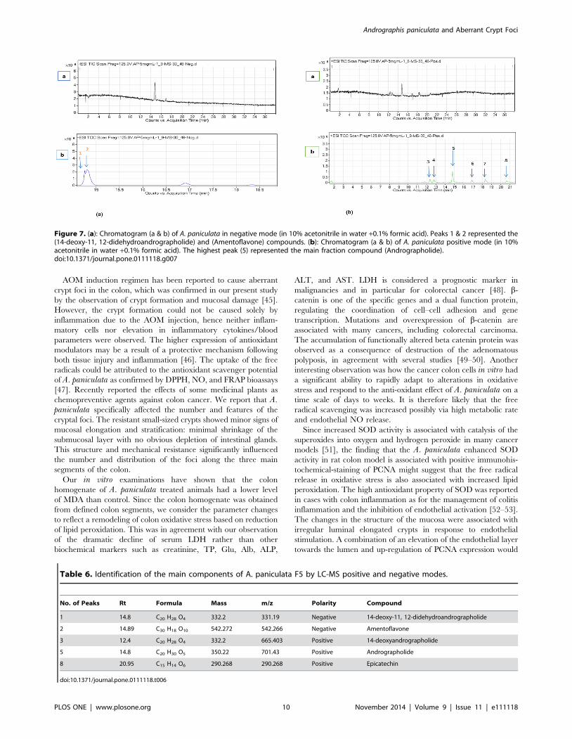
Chromatographic profiling of A. paniculataThe crude extract was separated by column chromatography
into six fractions, according to the differences in molecular size
and polarities. Their anti-proliferative effects were investigated
against HT29 and CCD-841. High inhibition was obtained with
fractions 4 and 5 (ANF4 and ANF5) (Figure S1) and moderate
inhibition with fractions 1 and 2 (ANF1 and ANF2). The cell
viability decreased to 3.53% and 6.10%, at concentrations of
50 mg of ANF4 and ANF5, respectively. In contrast, low inhibition
was observed in the CCD-841 colon cells with ANF4 and ANF5.
At the same concentration, the cell viability was detected at
58.74% and 55.8%, respectively. ANF5 was studied further by
LC-MS to identify the active compounds and eight peaks were
distinguished (Figure 7). The isolated compounds (Table 6) were
characterized and mainly diterpenoids were evident. Diterpenoids
in the fractions included andrographolide, 14-deoxyandrographo-
lide, and 14-deoxy-11, 12-didehydroandrographolide in addition
to amentoflavone and epicatechin.
Discussion
Colorectal epithelial homeostasis is attributed to the balance
between cell proliferation and cell death. Colorectal cancer arises
from abnormal growth of the colon cells with excessive prolifer-
ation [37–38]. Oxidative stress plays an important role in the
molecular mechanism of colon cancer development and progres-
sion [39]. Many studies have shown that a diet rich in
phytochemicals is associated with reduction in the risk of cancer
[40]. The wide range of chemical compounds that are present in
natural dietary products has been approved as antioxidant and
chemopreventive entities [41]. A. paniculata is known to be a rich
Figure 2. Effect of A. paniculata on superoxide dismutase (SOD) activity, malonaldehyde (MDA) level and nitric oxide (NO) level inAOM induced tissue. All values are represented as mean 6 SEM. Significant difference at *P,0.01, ** P,0.001(ANOVA, Tukey’s). FU: Fluorouracil;AOM: azoxymethane.doi:10.1371/journal.pone.0111118.g002
Table 5. Antioxidant activity of A. paniculata in vitro.
DPPH(IC50 mmol/L) FRAP (mmol/g) TPC (mg/g) TF (mg/g) Nitric oxide%
BHT 8.0660.28 5228.660.01 ------ ------ ------
Vitamin C ------ ------ ------ ------ 183.760.89
Gallic acid ------ ------ 1015.360.004 ------ ------
Quercetin ------ ------ ------ 775.7160.009 ------
A. paniculata 57.0861.04 4676.260.07 242.6760.007 142.8660.005 172.960.85
Values represent mean 6 SEM for triplicates; BHT-butylated hydroxytoluene.doi:10.1371/journal.pone.0111118.t005
Andrographis paniculata and Aberrant Crypt Foci
PLOS ONE | www.plosone.org 7 November 2014 | Volume 9 | Issue 11 | e111118

Figure 3. Cross-section of the rat colon stained with hematoxylin and eosin: (A) Normal group with normal crypts, (B) AOM controlgroup (C) FU treated group, D) 250 mg/kg A. paniculata treated group, (E) 500 mg/kg A. paniculata treated group (100Xmagnification). For (B), arrow indicated the elongated and slightly stratified nuclei found in AOM control group, showing depletion of mucin. For(A), (C), (D) and (E) which represented the normal cells, the arrows indicated crypts with round nuclei which are normal.doi:10.1371/journal.pone.0111118.g003
Figure 4. Effect of A. paniculata on ACF count: (A) Normal group with normal crypts, (B) AOM control group (ACF with multiplecrypts. five), (C) FU treated group, (D) 250 mg/kg A. paniculata treated group, (E) 500 mg/kg A. paniculata treated group (methyleneblue staining. (4X magnification). The arrows indicated the crypts that were more than five foci in AOM control group and less than five foci in alltreated groups.doi:10.1371/journal.pone.0111118.g004
Andrographis paniculata and Aberrant Crypt Foci
PLOS ONE | www.plosone.org 8 November 2014 | Volume 9 | Issue 11 | e111118

source of many bioactive compounds with a broad range of
pharmaceutical effects [42]. Extracts of A. paniculata were found
to inhibit the formation of oxygen derived free radicals [43]. Some
of the plant bioactive compounds of these extracts belong to the
diterpenoid and flavonoid classes [42]. Dietary phytochemicals
and herbs similar to A. paniculata, especially its polyphenolic
components have the potential in the prevention of colon cancer
[44].
Figure 5. (a): Regulation of PCNA in the colon tissue of rats (A) Normal group (B) AOM control group (C) FU treated group, (D) 250 mg/kg A.paniculata treated group, and (E) 500 mg/kg A. paniculata treated group (100X magnification). (b): Regulation of PCNA protein expression in thecolon tissue of normal and AOM induced rats. All values are in mean 6 SEM. FU: Fluorouracil; AOM: azoxymethane.doi:10.1371/journal.pone.0111118.g005
Figure 6. Regulation of b-catenin in the colon tissue of rats. (A) Normal group (B) AOM control group (C) FU treated group, (D) 250 mg/kg A.paniculata treated group, and (E) 500 mg/kg A. paniculata treated group (100X magnification). In the AOM and treated groups the arrows indicatedto the cells expressed the b-catenin protein. The b-catenin showed to be more expressed in AOM control group than that of treated groups.doi:10.1371/journal.pone.0111118.g006
Andrographis paniculata and Aberrant Crypt Foci
PLOS ONE | www.plosone.org 9 November 2014 | Volume 9 | Issue 11 | e111118
AOM induction regimen has been reported to cause aberrant
crypt foci in the colon, which was confirmed in our present study
by the observation of crypt formation and mucosal damage [45].
However, the crypt formation could not be caused solely by
inflammation due to the AOM injection, hence neither inflam-
matory cells nor elevation in inflammatory cytokines/blood
parameters were observed. The higher expression of antioxidant
modulators may be a result of a protective mechanism following
both tissue injury and inflammation [46]. The uptake of the free
radicals could be attributed to the antioxidant scavenger potential
of A. paniculata as confirmed by DPPH, NO, and FRAP bioassays
[47]. Recently reported the effects of some medicinal plants as
chemopreventive agents against colon cancer. We report that A.paniculata specifically affected the number and features of the
cryptal foci. The resistant small-sized crypts showed minor signs of
mucosal elongation and stratification: minimal shrinkage of the
submucosal layer with no obvious depletion of intestinal glands.
This structure and mechanical resistance significantly influenced
the number and distribution of the foci along the three main
segments of the colon.
Our in vitro examinations have shown that the colon
homogenate of A. paniculata treated animals had a lower level
of MDA than control. Since the colon homogenate was obtained
from defined colon segments, we consider the parameter changes
to reflect a remodeling of colon oxidative stress based on reduction
of lipid peroxidation. This was in agreement with our observation
of the dramatic decline of serum LDH rather than other
biochemical markers such as creatinine, TP, Glu, Alb, ALP,
ALT, and AST. LDH is considered a prognostic marker in
malignancies and in particular for colorectal cancer [48]. b-
catenin is one of the specific genes and a dual function protein,
regulating the coordination of cell–cell adhesion and gene
transcription. Mutations and overexpression of b-catenin are
associated with many cancers, including colorectal carcinoma.
The accumulation of functionally altered beta catenin protein was
observed as a consequence of destruction of the adenomatous
polyposis, in agreement with several studies [49–50]. Another
interesting observation was how the cancer colon cells in vitro had
a significant ability to rapidly adapt to alterations in oxidative
stress and respond to the anti-oxidant effect of A. paniculata on a
time scale of days to weeks. It is therefore likely that the free
radical scavenging was increased possibly via high metabolic rate
and endothelial NO release.
Since increased SOD activity is associated with catalysis of the
superoxides into oxygen and hydrogen peroxide in many cancer
models [51], the finding that the A. paniculata enhanced SOD
activity in rat colon model is associated with positive immunohis-
tochemical-staining of PCNA might suggest that the free radical
release in oxidative stress is also associated with increased lipid
peroxidation. The high antioxidant property of SOD was reported
in cases with colon inflammation as for the management of colitis
inflammation and the inhibition of endothelial activation [52–53].
The changes in the structure of the mucosa were associated with
irregular luminal elongated crypts in response to endothelial
stimulation. A combination of an elevation of the endothelial layer
towards the lumen and up-regulation of PCNA expression would
Figure 7. (a): Chromatogram (a & b) of A. paniculata in negative mode (in 10% acetonitrile in water +0.1% formic acid). Peaks 1 & 2 represented the(14-deoxy-11, 12-didehydroandrographolide) and (Amentoflavone) compounds. (b): Chromatogram (a & b) of A. paniculata positive mode (in 10%acetonitrile in water +0.1% formic acid). The highest peak (5) represented the main fraction compound (Andrographolide).doi:10.1371/journal.pone.0111118.g007
Table 6. Identification of the main components of A. paniculata F5 by LC-MS positive and negative modes.
No. of Peaks Rt Formula Mass m/z Polarity Compound
1 14.8 C20 H28 O4 332.2 331.19 Negative 14-deoxy-11, 12-didehydroandrographolide
2 14.89 C30 H18 O10 542.272 542.266 Negative Amentoflavone
3 12.4 C20 H28 O4 332.2 665.403 Positive 14-deoxyandrographolide
5 14.8 C20 H30 O5 350.22 701.43 Positive Andrographolide
8 20.95 C15 H14 O6 290.268 290.268 Positive Epicatechin
doi:10.1371/journal.pone.0111118.t006
Andrographis paniculata and Aberrant Crypt Foci
PLOS ONE | www.plosone.org 10 November 2014 | Volume 9 | Issue 11 | e111118

lead to major alterations of the colon contractility signalling. These
data and the suppression of crypt formation are also consistent
with a mechanism where the apoptotic cascade is compensating
for proliferation alterations of colorectal mucosa.
The in vivo measurements also clearly showed an increase of
the animal weight, which suggests not only healthy animals but the
satisfactory nutritional supply under our proposed regimen and
specifically the remarkable digestion and absorption pharmaco-
dynamics. Liver and kidney tissues revealed no histopathological
changes on long term A. paniculata oral administration. Possibly
the increased colon weight of the treated animals is related to
water retention and dilution of the free radical. It should be noted
that the improvement of nutritional values following A. paniculatawould further indicate promotion of the structural and functional
integrity of the colon segments. It is noteworthy that the acute
toxicity test did not reveal any histopathological changes in the
liver and kidney tissues treated with the plant extracts compared to
control.
The findings from this study suggested that A. paniculatapossesses antioxidant, anti-proliferative and antimetastatic activi-
ties and hence, has the potential to offer new anti-cancer
therapeutic agent. The bioactivity and bioavailability of A.paniculata against cancer was suggested to be due to its
diterpenoid and flavonoid contents. Previous studies have
discussed the relationship between diterpenoids and cancer
prevention [54]. Furthermore, diterpenoids have been tested on
HT29, the colon cancer cell line, and have potent effect on the
inhibition of HT-29 proliferation [55–56]. Flavonoid and diter-
penes isolated from A. paniculata have been reported for their
antioxidant properties and their ability to protect cells from lipid
peroxidation [43]. Taken together, our results highlight the
chemoprotective attribution of A. paniculata on the colon mucosal
and submucosal layers and propose some possible components of
the early/mid stages of carcinogenic cells proliferation and
filtration that could be interrupted efficiently after two weeks of
administration, guaranteeing the preservation of the colon
integrity and functionality.
Conclusion
When orally administered to rats, the ethanol extract of A.paniculata played a role in the intervention of cancer formation.
A. paniculata changed the morphological identity of the crypts in
the mucosa and showed histological alterations that varied from
hyperplasia to dysplasia. The formation of less ACF excludes one
of the earlier abnormalities that occur during the colorectal cancer
induction. Furthermore, A. paniculata interfered with the
intermediate biomarker for colon cancer development as evi-
denced by the various levels of certain oxidative stress markers.
This may be due to the antioxidative properties of the extracts as
free radical scavengers. In addition, A. paniculata regulated the
genetic and epigenetic features displaying a chemoprotective effect
on the colon cells against a potent carcinogen i.e., AOM as
documented on the apoptotic and proliferative levels. The effects
were generally comparable to those of rats treated with 5-FU.
Supporting Information
Figure S1 Effect of A. paniculata fractions ANF4 and ANF5 on
the viability of (a & c) HT29 colon cancer cells and (b & d) CCD
841 normal colon cells. Data were expressed as the mean 6 SEM
for triplicates.
(TIF)
Author Contributions
Conceived and designed the experiments: NA-H RPYY MAA SI.
Performed the experiments: NA-H WN MAA. Analyzed the data: NA-H
RPYY MAA HE-S. Contributed reagents/materials/analysis tools: NA-H
RPYY MAA. Wrote the paper: NA-H RPYY MAA SAMK HE-S.
References
1. Cheah PL, Looi LM, Teoh KH, Rahman NA, Wong LX, et al. (2014)
Colorectal carcinoma in Malaysians: DNA mismatch repair pattern in a
multiethnic population. Asian Pacific Journal Cancer Prevention 15(7): 3287–
3291.
2. Norwati D, Harmy MY, Norhayati MN, Amry AR (2014) Colorectal cancer
screening practices of primary care providers: results of a national survey in
Malaysia. Asian Pacific Journal Cancer Prevention 15(6): 2901–4.
3. Pinedo, Herbert M, Peters GF (1988) Fluorouracil: biochemistry and
pharmacology. Journal of Clinical Oncology 6(10): 1653–1664.
4. Raderer M, Scheithauer W (1995) Treatment of advanced colorectal cancer
with 5-fluorouracil and interferon-a: an overview of clinical trials. European
Journal of Cancer 31(6): 1002–1008.
5. Khalifa S, Medina P, Erlandsson A, El-Seedi HR, Silvente-Poirot S, et al. (2014)
The novel steroidal alkaloids dendrogenin A and B promote proliferation of
adult neural stem cells. Biochem Biophys Res Commun 446: 681–6.
6. El-Seedi HR, Burman R, Mansour A, Turki Z, Boulos L, et al. (2013) The
traditional medical uses and cytotoxic activities of sixty-one Egyptian plants:
Discovery of active cardiac glycosides from Urginea maritima. Journal of
Ethnopharmacology 145: 746–757.
7. Sheeja K, Kuttan G (2007) Activation of cytotoxic T lymphocyte responses and
attenuation of tumor growth in vivo by Andrographis paniculata extract and
andrographolide. Immunopharmacology and Immunotoxicology 29: 81–93.
8. Rao NK (2006) Anti-hyperglycemic and renal protective activities of
Andrographis paniculata roots chloroform extract. Iranian Journal of Pharma-
cology and Therapeutics 5: 47–50.
9. Wasman S, Mahmood A, Chua LS, Alshawsh MA, Hamdan S (2011)
Antioxidant and gastroprotective activities of Andrographis paniculata (hem-
pedu bumi) in sprague dawley rats. Indian Journal of Experimental Biology 49:
767–772.
10. Singha PK, Roy S, Dey S (2003) Antimicrobial activity of Andrographispaniculata. Fitoterapia 74: 692–694.
11. Qader SW, Abdulla MA, Chua LS, Najim N, Zain MM, et al. (2011)
Antioxidant, total phenolic content and cytotoxicity evaluation of selected
Malaysian plants. Molecules 16: 3433–3443.
12. Calabrese C, Berman SH, Babish JG, Ma X, Shinto L, et al. (2000) A phase I
trial of andrographolide in HIV positive patients and normal volunteers.
Phytotherapy Research 14: 333–338.
13. Lin F, Wu S, Lee S, Ng L (2009) Antioxidant, antioedema and analgesic
activities of Andrographis paniculata extracts and their active constituent
andrographolide. Phytotherapy Research 23: 958–964.
14. Sheeja K, Shihab P, Kuttan G (2006) Antioxidant and anti-inflammatory
activities of the plant Andrographis paniculata nees. Immunopharmacology and
Immunotoxicology 28: 129–140.
15. Al-Bayaty FH, Abdulla MA, Hassan MIA, Ali HM (2012) Effect of
Andrographis paniculata leaf extract on wound healing in rats. Natural Product
Research 26: 423–429.
16. Yoopan N, Thisoda P, Rangkadilok N, Sahasitiwat S, Pholphana N, et al. (2007)
Cardiovascular effects of 14-deoxy-11, 12-didehydroandrographolide and
Andrographis paniculata extracts. Planta Medica 73: 503–511.
17. Akowuah GA, Zhari I, Mariam A, Yam MF (2009) Absorption of
andrographolides from Andrographis paniculata and its effect on CCl sub 4
sub-induced oxidative stress in rats. Food and Chemical Toxicology 47(9):
2321–2326.
18. Amaryan G, Astvatsatryan V, Gabrielyan E, Panossian A, Panosyan V, et al.
(2003) Double-blind, placebo-controlled, randomized, pilot clinical trial of
Immuno Guard sup a standardized fixed combination of Andrographispaniculata Nees, with Eleutherococcus senticosus Maxim Schizandra chinensis
Bail. And Glycyrrhiza glabra L. extracts in patients with Familial Mediterranean
Fever. Phytomedicine 10(4): 271–285.
19. Samy RP, Thwin MM, Gopalakrishnakone P, Ignacimuthu S (2008)
Ethnobotanical survey of folk plants for the treatment of snakebites in southern
part of tamilnadu, india. Journal of Ethnopharmacology 115: 302–312.
20. Ko HC, Wei BL, Chiou WF (2006) The effect of medicinal plants used in
chinese folk medicine on rantes secretion by virus-infected human epithelial cells.
Journal of Ethnopharmacology 107: 205–210.
21. Roy S, Rao K, Bhuvaneswari C, Giri A, Mangamoor LN (2010) Phytochemical
analysis of Andrographis paniculata extract and its antimicrobial activity. World
Journal of Microbiology and Biotechnology 26: 85–91.
Andrographis paniculata and Aberrant Crypt Foci
PLOS ONE | www.plosone.org 11 November 2014 | Volume 9 | Issue 11 | e111118

22. Marotta F, Naito Y, Minelli E, Tajiri H, Bertuccelli J, et al. (2003)
Chemopreventive effect of a probiotic preparation on the development of
preneoplastic and neoplastic colonic lesions: An experimental study. Hepato-
gastroenterology 50: 1914.
23. Traverso G, Shuber A, Olsson L, Levin B, Johnson C, et al. (2002) Detection of
proximal colorectal cancers through analysis of faecal DNA. Lancet 359: 403–4.
24. Ohishi T, Kishimoto Y, Miura N, Shiota G, Kohri T, et al. (2002) Synergistic
effects of (-)-epigallocatechin gallate with sulindac against colon carcinogenesis of
rats treated with azoxymethane. Cancer Letters 177: 49–56.
25. Tomimatsu T, Horie T (2005) Enhanced glucose absorption in the rat small
intestine following repeated doses of 5-fluorouracil. Chemico-Biological
Interactions 155: 129–139.
26. Fraga CG, Martino VS, Ferraro GE, Coussio JD, Boveris A (1987) Flavonoids as
antioxidants evaluated by in vitro and in situ liver chemiluminescence.
Biochemical Pharmacology 36(5): 717–720.
27. Sharma OP, Bhat TK (2009) DPPH antioxidant assay revisited. Food Chemistry
113: 1202–1205.
28. Ikram, Khairul H, Eng KH, Jalil AMM, Ismail A, et al. (2009) Antioxidant
capacity and total phenolic content of Malaysian underutilized fruits. Journal of
Food Composition and Analysis 22: 388–393.
29. Gan RY, Xu XR, Song FL, Kuang L, Li HB (2010) Antioxidant activity and
total phenolic content of medicinal plants associated with prevention and
treatment of cardiovascular and cerebrovascular diseases. Journal of Medicinal
Plants Research 4: 2438–2444.
30. Sathuvan M, Vignesh A, Thangam R, Palani P, Rengasamy R, et al. (2012) Invitro antioxidant and anticancer potential of bark of costus pictus D. Don. Asian
Pacific Journal of Tropical Biomedicine 2: S741–S749.
31. Benzie IF, Strain J (1996) The ferric reducing ability of plasma (FRAP) as a
measure of ‘‘antioxidant power’’: the FRAP assay. Analytical biochemistry
239(1): 70–76.
32. Chi YC, Tung HK, Huei KC (2005) Nitric oxide reduces Cu toxicity and Cu-
induced NH accumulation in rice leaves. Journal of plant physiology 162(12):
1319-1330.
33. Yen GC, Lai HH, Chou HY (2001) Nitric oxide-scavenging and antioxidant
effects of Uraria crinita root. Food Chemistry 74: 471–478.
34. Saha K, Lajis NH, Israf DA, Hamzah AS, Khozirah S, et al. (2004) Evaluation
of antioxidant and nitric oxide inhibitory activities of selected Malaysian
medicinal plants. Journal of Ethnopharmacology 92: 263–267.
35. OECD. 2005. DOFO. OECD guideline for testing of chemicals [Online].
Available: http://www.env.go.jp/water/dojo/noyaku/hisan_risk/hyoka_tih/
com07/ref05.pdf.
36. Bird RP (1987) Observation and quantification of aberrant crypts in the murine
colon treated with a colon carcinogen: Preliminary findings. Cancer Letters 37:
147–151.
37. Violette S, Poulain L, Dussaulx E, Pepin D, Faussat AM, et al. (2002) Resistance
of colon cancer cells to long-term 5-fluorouracil exposure is correlated to the
relative level of Bcl-2 and Bcl-XL in addition to Bax and p53 status.
International Journal of Cancer 98: 498–504.
38. Yamaguchi H, Bhalla K, Wang HG (2003) Bax plays a pivotal role in
thapsigargin-induced apoptosis of human colon cancer HCT116 cells by
controlling Smac/Diablo and Omi/Htra2 release from mitochondria. Cancer
Research 63: 1483–1489.
39. Seril DN, Liao J, Yang GY, Yang CS (2003) Oxidative stress and ulcerative
colitis-associated carcinogenesis: Studies in humans and animal models.Carcinogenesis 24: 353–362.
40. Murthy N, Mukherjee S, Ray G, Ray A (2009) Dietary factors and cancer
chemoprevention: An overview of obesity-related malignancies. Journal ofPostgraduate Medicine 55: 45.
41. Bagchi D, Bagchi M, Stohs SJ, Das DK, Ray SD, et al. (2000) Free radicals andgrape seed proanthocyanidin extract: Importance in human health and disease
prevention. Toxicology. 148: 187–197.
42. Jarukamjorn K, Nemoto N (2008) Pharmacological aspects of Andrographispaniculata on health and its major diterpenoid constituent andrographolide.
Journal of Health Science 54: 370–381.43. Niranjan A, Tewari S, Lehri A (2010) Biological activities of kalmegh
(Andrographis paniculata nees) and its active principles-a review. Indian JournalNatural Products Resourses 1: 125–135.
44. Chan AT, Giovannucci EL (2010) Primary prevention of colorectal cancer.
Gastroenterology 138(10): 2029–2043.45. Hosono K, Endo H, Takahashi H, Sugiyama M, Uchiyama T, et al. (2010)
Metformin suppresses azoxymethane-induced colorectal aberrant crypt foci byactivating amp-activated protein kinase. Molecular Carcinogenesis 49: 662–671.
46. Skrovankova S, Misurcova L, Machu L (2012) Chapter three - antioxidant
activity and protecting health effects of common medicinal plants. In:Jeyakumar, H. Advances in Food and Nutrition Research. Academic Press.
47. Shafie NH, Mohd EN, Ithnin H, Md Akim A, Saad N, et al. (2013) Preventiveinositol hexaphosphate extracted from rice bran inhibits colorectal cancer
through involvement of Wnt/b-Catenin and COX-2 pathways. BioMedResearch International 2013.
48. Koukourakis M, Giatromanolaki A, Sivridis E (2009) Colorectal cancer: Lactate
dehydrogenase (LDH) activity as a prognostic marker. In: M.A. HayatColorectal Cancer. Springer Netherlands.
49. Macdonald BT, Tamai K, He X (2009) Wnt/b-catenin signaling: components,mechanisms, and diseases. Developmental Cell 17: 9–26.
50. Tetsu O, Mccormick F (1999) b-Catenin regulates expression of cyclin D1 in
colon carcinoma cells. Nature 398: 422–426.51. Neelwarne B (2012) Red Beet Biotechnology: Food and Pharmaceutical
Applications: Springer.52. Seguı J, Gil F, Gironella M, Alvarez M, Gimeno M, et al. (2005) Down-
regulation of endothelial adhesion molecules and leukocyte adhesion bytreatment with superoxide dismutase is beneficial in chronic immune
experimental colitis. Inflammatory Bowel Diseases 11: 872–882.
53. Jubeh TT, Nadler-Milbauer M, Barenholz Y, Rubinstein A (2006) Localtreatment of experimental colitis in the rat by negatively charged liposomes of
catalase, TMN and SOD. Journal of Drug Targeting 14: 155–163.54. Al Batran R, Al-Bayaty F, Al-Obaidi MMJ, Abdulla MA (2013) Acute toxicity
and the effect of andrographolide on porphyromonas gingivalis-induced
hyperlipidemia in rats. BioMed Research International 2013.55. Rajagopal S, Kumar RA, Deevi DS, Satyanarayana C, Rajagopalan R (2003)
Andrographolide, a potential cancer therapeutic agent isolated from Andro-graphis paniculata. Journal of Experimental Therapeutics and Oncology 3: 147–
158.56. Zhao F, He EQ, Wang L, Liu K (2008) Anti-tumor activities of andrographo-
lide, a diterpene from Andrographis paniculata by inducing apoptosis and
inhibiting VEGF level. Journal of Asian Natural Products Research 10: 467–473.
Andrographis paniculata and Aberrant Crypt Foci
PLOS ONE | www.plosone.org 12 November 2014 | Volume 9 | Issue 11 | e111118