Photodynamic inactivation of Candida albicans by BAM-SiPc
19
RESEARCH ARTICLE Photodynamic Inactivation of Candida albicans with Imidazoacridinones: Influence of Irradiance, Photosensitizer Uptake and Reactive Oxygen Species Generation Aleksandra Taraszkiewicz 1 , Grzegorz Szewczyk 2 , Tadeusz Sarna 2 , Krzysztof P. Bielawski 1 , Joanna Nakonieczna 1 * 1 Laboratory of Molecular Diagnostics, Intercollegiate Faculty of Biotechnology University of Gdansk and Medical University of Gdansk, Gdansk, Poland, 2 Department of Biophysics, Faculty of Biochemistry, Biophysics and Biotechnology, Jagiellonian University, Krakow, Poland * [email protected] Abstract The increasing applicability of antifungal treatments, the limited range of available drug clas- ses and the emergence of drug resistance in Candida spp. suggest the need for new treat- ment options. To explore the applicability of C. albicans photoinactivation, we examined nine structurally different imidazoacridinone derivatives as photosensitizing agents. The most effective derivatives showed a >10 4 -fold reduction of viable cell numbers. The fungi- cidal action of the three most active compounds was compared at different radiant powers (3.5 to 63 mW/cm 2 ), and this analysis indicated that 7 mW/cm 2 was the most efficient. The intracellular accumulation of these compounds in fungal cells correlated with the fungicidal activity of all 9 derivatives. The lack of effect of verapamil, an inhibitor targeting Candida ABC efflux pumps, suggests that these imidazoacridinones are not substrates for ABC transporters. Thus, unlike azoles, a major class of antifungals used against Candida, ABC transporter-mediated resistance is unlikely. Electron paramagnetic resonance (EPR)-spin trapping data suggested that the fungicidal light-induced action of these derivatives might depend on the production of superoxide anion. The highest generation rate of superoxide anion was observed for 1330H, 1610H, and 1611. Singlet oxygen production was also de- tected upon the irradiation of imidazoacridinone derivatives with UV laser light, with a low to moderate yield, depending on the type of compound. Thus, imidazoacridinone derivatives examined in the present study might act via mixed type I/type II photodynamic mechanism. The presented data indicate lack of direct correlation between the structures of studied imi- dazoacridinones, cell killing ability, and ROS production. However, we showed for the first time that for imidazoacridinones not only intracellular accumulation is necessary prerequi- site of lethal photosensitization of C. albicans, but also localization within particular cellular structures. Our findings present IA derivatives as efficient antifungal photosensitizers with a potential to be used in local treatment of Candida infection. PLOS ONE | DOI:10.1371/journal.pone.0129301 June 8, 2015 1 / 19 OPEN ACCESS Citation: Taraszkiewicz A, Szewczyk G, Sarna T, Bielawski KP, Nakonieczna J (2015) Photodynamic Inactivation of Candida albicans with Imidazoacridinones: Influence of Irradiance, Photosensitizer Uptake and Reactive Oxygen Species Generation. PLoS ONE 10(6): e0129301. doi:10.1371/journal.pone.0129301 Academic Editor: Michael Hamblin, Massachusetts General Hospital, UNITED STATES Received: December 21, 2014 Accepted: May 8, 2015 Published: June 8, 2015 Copyright: © 2015 Taraszkiewicz et al. This is an open access article distributed under the terms of the Creative Commons Attribution License, which permits unrestricted use, distribution, and reproduction in any medium, provided the original author and source are credited. Data Availability Statement: All relevant data are within the paper and its Supporting Information files. Funding: Financial support for this work was obtained from the National Science Center grant no: NCN 2013/09/B/NZ7.00410 (JN), 2011/03/B/NZ1/ 00007 (TS), and NCN 2013/09/N/NZ1/00188 (AT). AT is supported by LiSMIDoS – Life Sciences and Mathematics Interdisciplinary Doctoral Studies. The funders had no role in study design, data collection and analysis, decision to publish, or preparation of the manuscript.
Transcript of Photodynamic inactivation of Candida albicans by BAM-SiPc
RESEARCH ARTICLE
Photodynamic Inactivation of Candidaalbicans with Imidazoacridinones: Influenceof Irradiance, Photosensitizer Uptake andReactive Oxygen Species GenerationAleksandra Taraszkiewicz1, Grzegorz Szewczyk2, Tadeusz Sarna2, KrzysztofP. Bielawski1, Joanna Nakonieczna1*
1 Laboratory of Molecular Diagnostics, Intercollegiate Faculty of Biotechnology University of Gdansk andMedical University of Gdansk, Gdansk, Poland, 2 Department of Biophysics, Faculty of Biochemistry,Biophysics and Biotechnology, Jagiellonian University, Krakow, Poland
AbstractThe increasing applicability of antifungal treatments, the limited range of available drug clas-
ses and the emergence of drug resistance in Candida spp. suggest the need for new treat-
ment options. To explore the applicability of C. albicans photoinactivation, we examined
nine structurally different imidazoacridinone derivatives as photosensitizing agents. The
most effective derivatives showed a >104-fold reduction of viable cell numbers. The fungi-
cidal action of the three most active compounds was compared at different radiant powers
(3.5 to 63 mW/cm2), and this analysis indicated that 7 mW/cm2 was the most efficient. The
intracellular accumulation of these compounds in fungal cells correlated with the fungicidal
activity of all 9 derivatives. The lack of effect of verapamil, an inhibitor targeting CandidaABC efflux pumps, suggests that these imidazoacridinones are not substrates for ABC
transporters. Thus, unlike azoles, a major class of antifungals used against Candida, ABCtransporter-mediated resistance is unlikely. Electron paramagnetic resonance (EPR)-spin
trapping data suggested that the fungicidal light-induced action of these derivatives might
depend on the production of superoxide anion. The highest generation rate of superoxide
anion was observed for 1330H, 1610H, and 1611. Singlet oxygen production was also de-
tected upon the irradiation of imidazoacridinone derivatives with UV laser light, with a low to
moderate yield, depending on the type of compound. Thus, imidazoacridinone derivatives
examined in the present study might act via mixed type I/type II photodynamic mechanism.
The presented data indicate lack of direct correlation between the structures of studied imi-
dazoacridinones, cell killing ability, and ROS production. However, we showed for the first
time that for imidazoacridinones not only intracellular accumulation is necessary prerequi-
site of lethal photosensitization of C. albicans, but also localization within particular cellular
structures. Our findings present IA derivatives as efficient antifungal photosensitizers with a
potential to be used in local treatment of Candida infection.
PLOS ONE | DOI:10.1371/journal.pone.0129301 June 8, 2015 1 / 19
OPEN ACCESS
Citation: Taraszkiewicz A, Szewczyk G, Sarna T,Bielawski KP, Nakonieczna J (2015) PhotodynamicInactivation of Candida albicans withImidazoacridinones: Influence of Irradiance,Photosensitizer Uptake and Reactive OxygenSpecies Generation. PLoS ONE 10(6): e0129301.doi:10.1371/journal.pone.0129301
Academic Editor: Michael Hamblin, MassachusettsGeneral Hospital, UNITED STATES
Received: December 21, 2014
Accepted: May 8, 2015
Published: June 8, 2015
Copyright: © 2015 Taraszkiewicz et al. This is anopen access article distributed under the terms of theCreative Commons Attribution License, which permitsunrestricted use, distribution, and reproduction in anymedium, provided the original author and source arecredited.
Data Availability Statement: All relevant data arewithin the paper and its Supporting Information files.
Funding: Financial support for this work wasobtained from the National Science Center grant no:NCN 2013/09/B/NZ7.00410 (JN), 2011/03/B/NZ1/00007 (TS), and NCN 2013/09/N/NZ1/00188 (AT). ATis supported by LiSMIDoS – Life Sciences andMathematics Interdisciplinary Doctoral Studies. Thefunders had no role in study design, data collectionand analysis, decision to publish, or preparation ofthe manuscript.

IntroductionDue to longer life span and increasing numbers of people with compromised immune systems,the number of fungal infections caused by yeasts has significantly increased in recent years[1].This phenomenon is also related to a dramatic increase in antifungal drug resistance. The new-est classes of antifungals (e.g., triazoles and echinocandins) are widely used and effective, how-ever, optimal therapy may be complicated through emergence of at least four resistancemechanisms, the most prominent being the induction of efflux pumps leading to low drug con-centration [2]. Antimicrobial agents that can be activated with light are commonly known asphotoantimicrobials [3]. In the presence of oxygen, these agents use the energy of visible lightto produce highly reactive oxygen species, e.g., singlet oxygen or hydroxyl radicals, which aretoxic to microbial cells. The process is known as antimicrobial photodynamic inactivation(PDI). The main advantage of photoantimicrobial agents, also known as photosensitizers, overclassical antimicrobials is the multiple-target actions of these compounds. Reactive oxygen spe-cies, as long as they are localized in the close vicinity of a specific molecular target, act on pro-teins, nucleic acids or unsaturated lipids. As a consequence, photoantimicrobials are relativelyimmune to individual resistance mechanisms, and can act effectively against wild-type and an-tifungal-resistant isolates. Indeed, there has been no reports indicating the in vitro induction ofresistance to the action of light-dependent drugs [4–6]. Due to their short life times, reactiveoxygen species act locally. The selectivity issue remains a concern, particularly in the case ofyeast cells (i.e., eukaryotic pathogens), but the potential ‘therapeutic window’ although morenarrow compared with bacterial cells, is still possible to obtain [7–8]. Photoantimicrobials areonly active after light treatment, thus localized skin and soft tissue infections caused by fungiare good targets for PDI. Thus, this treatment assures the selectivity and minimization of theside effects.
The fluence of light has been, for years, considered the most important parameter in photo-dynamic action against microbial cells, based on the reciprocity principle [9]. However, somewell documented reports exist pointing to the importance of the time parameter over whichthe light is being delivered to cells [10]. Influence of two fluence rates of light (artificial orsolar) on the efficiency of photoinactivation was studied in the past with the use of cationicporphyrins, where it was demonstrated that under 620 W/m2 of solar light, photoinactivationof bioluminescent E. coli was faster over artificial 40 W/m2 due to higher light fluence rate [11].The effect of light source, light dose, fluence rate was also shown to be of critical meaning forphage photoinactivation in a extensive studies using light sources within range of 40 W/m2–
1690 W/m2[12].The finding that fluence rate and exposure time play important role in effec-tiveness of antimicrobial PDI is of profound meaning and opens the possibility to optimizeirradiation protocols for in vivo studies.
Candida spp. plays a major role in localized and systemic infections in humans. The fungalinfections are particularly risky for a growing number of immune-compromised, and post-trauma patients, individuals with tumours and those who underwent surgical interventions.Candida albicans is most frequently isolated fungi species from infections [13].
Several studies on planktonic and biofilm forms of C. albicans culture have shown effectivekilling using various photosensitizing compounds, including phenothiazinium photosensitizers[7,14–16], phthalocyanines[17] and porphyrins[18–24]. Antifungal PDI research is currentlyat the stage of ex vivo experiments, i.e., using yeast cell cultures, rather than infected animalmodels. However, some in vivo studies on animal models of human infections demonstratedthe potential of PDI in eradicating pathogenic fungi. Indeed, a significant decrease in the num-ber of viable C. albicans cells was observed following PDI in rat model of oral candidiasis usinga hematoporphyrin derivative[25] or methylene blue[26].However, the complete elimination
Photoinactivation of Candida albicans
PLOSONE | DOI:10.1371/journal.pone.0129301 June 8, 2015 2 / 19
Competing Interests: The authors have declaredthat no competing interests exist.

of viable cells was not achieved. In other studies, antifungal PDI was shown to reduce micro-scopic lesions of experimentally induced candidiasis in rats and inhibit the proteinase activityof C. albicans. Albeit, a reduction in the number of viable cells was not achieved[27,28]. The ad-equate efficacy of a new methylene blue-based PDI was also established for a cutaneous skin in-fection[29] in rat model of skin burn wound infections[30], and in C. albicans-induced murinevaginitis[31]. Thus, despite considerable progress, antifungal PDI remains at the stage of devel-opment, and in particular, the effective killing of pathogen cells remains a challenge. Therefore,there is a need for the development of new and improved photoantimicrobials, and the exami-nation of the mechanism underlying actions of these compounds.
The objective of the present study were therefore:(i) to investigate the irradiance-dependentefficacy of C. albicans photoinactivation with the use of imidazoacridinone derivatives, (ii) tostudy cellular accumulation of these derivatives, and the type of reactive oxygen species pro-duced during the photodynamic process.
Materials and Methods
StrainsCandida albicans Quality Control strains were used: ATTC 10231, ATCC 90028, and ATCC14053. For photoinactivation studies and imidazoacridinone accumulation experiments, theyeast cells were grown overnight in BHI medium (Brain Heart Infusion) with shaking (37°C,150 rpm). ATCC strain 10231 was used in photoinactivation experiments (impact of variableirradiant power under a fixed light dose), and examining imidazoacridinone accumulation,and apoptosis induction. The latter two strains, ATCC 90028 and ATCC 14053were used in ac-cumulation studies, and we also assessed the reduction of survival for these two strains underparticular illumination conditions: 20J/cm2, 7mW/cm2).
ChemicalsThe basic forms of three imidazoacridinone derivatives (IA), namely 1330, 1415, and 1558were obtained from A. Skladanowski of Gdansk University of Technology, Poland. These com-pounds were prepared according to the method described previously [32–36]. Compounds1610 and 1611 were synthesized according the same method as IA described above [32–36].The hydrochloride form of the following imidazoacridinone derivatives, namely 1330H,1415H, 1558H, and 1610H were prepared using the same protocol, and subsequently convertedinto hydrochloride salts using the following procedure: a slight molar excess of hydrogen chlo-ride in absolute ethyl ether was added dropwise to the compound solution in the mixture ofchloroform:methanol, at temperature 5°C. The yellow-orange solid was precipitated using an-hydrous ethyl ether, followed by separation and crystalization with the ethyl ether. The generalstructure of the compounds used in the study is presented in Fig 1. The compounds were firstdissolved in pure (100%) DMSO (Sigma-Aldrich, Germany) to obtain 10 mM stock solutions,and stored at -20°C until further used.All other chemicals were of analytical grade, includingmeso-Tetra(N-methyl-4-pyridyl)porphinetetratiosylate (TMPyP) (Sigma-Aldrich, Germany);Verapamil (Sigma-Aldrich, Germany); 5,5-Dimethyl-1-pyroline N-oxide (DMPO)(Sigma-Al-drich, Germany).
Antimicrobial photodynamic studies on C. albicansCandida albicans was grown overnight in standard BHI (Brain Heart Infusion) medium(Biomerieux, France) at 37°C, with shaking (150 rpm). C. albicans standardized suspension inBHI (5x10⁶ CFU/ml) was prepared and added to the wells on sterile 96 well flat-bottom
Photoinactivation of Candida albicans
PLOSONE | DOI:10.1371/journal.pone.0129301 June 8, 2015 3 / 19

microtiterplate. The studied concentrations (50 or 100 μM) of each photosensitizer imidazoa-cridinone derivative were added. The plate was incubated in the dark at 37°C for 30 min with-out shaking and the samples were irradiated (LED illuminator, output power of 11W/12mm2,blue light of 405 nm, custom designed for laboratory use(Secure Media, Poland). After irradia-tion serial dilutions were obtained from every sample in sterile PBS and 10 μl aliquots werespotted on BHI agar plates. The plates were incubated overnight at 37°C and the colony form-ing units (CFU) were counted and the results were statistically analysed. Three types of con-trols were included in the photoinactivation experiment: (i) cells treated with light only, (ii)cells incubated in the dark in the presence of imidazoacridinone derivatives, and (iii) cells incu-bated in the dark in the absence of imidazoacridinones. Each experiment was performed as aseparate biological replicate from cultures grown independently.
Accumulation of photosensitizers in C. albicansStandardized suspensionsof C. albicans were prepared in PBS (106 CFU/ml) from overnightgrown independent cultures in BHI (Brain Heart Infusion) medium (Biomerieux, France).Each assay was performed as a separate biological replicate of the experiment. Photosensitizers(IAs) were added at concentrations of 50 and 100 μMto 1 ml of C. albicans suspension. Thesamples were incubated in the dark at 37°C for the 30 min without shaking. Samples were cen-trifuged at room temperature for 5 mins, at 5000 rpm (2.3xg), and supernatant was
Fig 1. Chemical structures of imidazoacridinone derivatives.
doi:10.1371/journal.pone.0129301.g001
Photoinactivation of Candida albicans
PLOSONE | DOI:10.1371/journal.pone.0129301 June 8, 2015 4 / 19

spectrophotometrically analysed to determine the amount of photosensitizer remaining out-side the cells. The imidazoacridinone concentrations were calculated based on the Lambert-Beer law and the extinction coefficient values were experimentally measured for each com-pound. The concentrations of accumulated imidazoacridinone in these cells were obtainedafter subtracting the calculated concentration of imidazoacridinone present in the supernatantfrom the concentration used for cell incubation (50 or 100 μM). These values are presented inTable 1 (IAs accumulated in cells). Subsequently, the cells were centrifuged at room temp for5 mins, at 5000 rpm (2.3xg), re-suspended in PBS, and further incubated in the dark at 37°Cfor 30 min without shaking. The cells were subsequently pelleted (5 min, 5000 rpm [2.3xg],room temp.),and absorbance of the supernatants was analysed to determine the amount of thephotosensitizers transported out of the cells (Table 1, IAs remained after washing). The absorp-tion measurements were performed on the Beckman DU-640 Spectrophotometer at 420 nm.To measure imidazoacridinones accumulation in the presence of verapamil, the cells (5 McFar-land, 106CFU/ml) were centrifuged at room temp for 5 mins, at 5000 rpm (2.3xg), washed withPBS, incubated with 200 μM verapamil, and subsequently, without further washing, incubatedwith each imidazoacridinone derivative. the cells were then pelleted under the above conditionsand the absorbance of the supernatants was measured.
Imidazoaridinone derivatives fluorescence was observed using fluorescent microscopy. Thecells (106 CFU/mL) were harvested at room temp for 5 mins, at 5000 rpm (2.3xg), washed withPBS, and incubated with 50 μM of imidazoacridinone suspension. Subsequently, the cells wereplaced ontoa cohesive microscopic slide, covered with a glass coverslip and observed underfluorescent microscopy at an excitation of 360–370 nm, and emission of>420 nm (OlympusBX51 fluorescence microscope with F-View-II CCD camera). The measurements and imageanalysis were conducted using AnalySIS software.
Table 1. Accumulation and phototoxic effect of imidazoacridinones inCandida albicansATCC 10231 strain.
IAs Initial IAs conc. 50 μM = 0.05 μmol/106 cells Initial IAs conc. 100 μM = 0.1 μmol/106 cells PDI effect (20 J/cm2,7 mW/ cm2)
IAs accumulated incellsa
IAs remained afterwashinga
IAs accumulated incellsa
IAs remained afterwashinga
50 μMIA
100 μMIA
μmol/106 cells ± SD μmol/106 cells ± SD Reduction insurvival [log10 units
CFU/ml]b,c,d
1330 0.033 ± 0.002 0.023 ± 0.002 0.076 ± 0.004 0.064 ± 0.0067 2 ± 0.03 >5 ± 0.00
1415 0.023 ± 0.002 0.013 ± 0.002 0.054 ± 0.006 0.036 ± 0.0066 1 ± 0.1 1 ± 0.1
1558 0.031 ± 0.001 0.020 ± 0.005 0.065 ± 0.010 0.048 ± 0.0119 0 ± 0.5 2 ± 0.1
1330h 0.042 ± 0.001 0.032 ± 0.007 0.086 ± 0.001 0.077 ± 0.0048 1 ± 0.22 2 ± 0.19
1415h 0.019 ± 0.002 0.006 ± 0.002 0.042 ± 0.002 0.024 ± 0.0036 1 ± 0.13 1.5 ± 0.22
1558h 0.024 ± 0.002 0.010 ± 0.004 0.049 ±0.004 0.026 ± 0.0065 0 ± 0.65 1 ± 0.12
1610 0.036 ± 0.002 0.023 ± 0.003 0.065 ± 0.004 0.046 ± 0.0055 1 ± 0.00 2.5 ± 0.00
1610h 0.035 ± 0.004 0.026 ± 0.011 0.063 ± 0.003 0.044 ± 0.0062 1 ± 0.55 2 ± 0.11
1611 0.011 ± 0.002 0.013 ± 0.010 0.028 ± 0.004 0.022 ± 0.0047 0 ± 0.80 0 ± 0.07
IAs—imidazoacridinone derivatives; PDI—antifungal photodynamic effect, The values were calculated by subtracting log10 CFU/ml of tested samples from
those of untreated controls (0 J/cm2; 0 μM IA). At least three biological replicates were used for calculation of the mean reduction values.amean values of three replicates ± standard deviation of the meanbin samples incubated in the dark without imidazoacridinone derivatives, the number of C. albicans cells was c.a. 5x10⁶ CFU/mlcin the samples incubated in the dark with 50 μM or 100 μM of each imidazoacridinone derivative, the number of cells was c.a. 5x10⁶ CFU/mldin the samples exposed to light only (20J/cm2, 7mW/cm2), the number of C. albicans cells was c.a. 5x10⁶ CFU/ml
doi:10.1371/journal.pone.0129301.t001
Photoinactivation of Candida albicans
PLOSONE | DOI:10.1371/journal.pone.0129301 June 8, 2015 5 / 19

Time-resolved spectroscopic detection of singlet oxygen -1O2
Time-resolved luminescence of 1O2 was measured at 1270 nm. Solutions of imidazoacridi-nones (100 μM) in 1-cm fluorescence cuvettes (QA-1000, HelmaOptik) were excited with 355nm microjoule pulses (750 ps duration) generated using a microchip Nd:YAG laser (Pulselas-P-1064-FC, Alphalas GmbH, Germany) operating with a 2–10 kHz repetition rate. To filterout the first and third harmonics of laser radiation, 50-cm water filter and dichroic mirrors(BK7series, Eksma Optics, Lithuania) were used. Near-infrared luminescence was measuredperpendicularly to the excitation beam in a photon counting mode using a thermoelectriccooled NIR PMT module (Model H10330-45, Hamamatsu, Japan) equipped with 1100-nmcutoff filter and additional selected narrow-band filters (NB series, NDC Infrared EngineeringLTD, UK). A computer-mounted PCI-board multichannel scaler was used (NanoHarp 250,PicoQuant GmbH, Germany), and the data collection was synchronized with laser pulses usingan ultrafast photodiode (UGP-300-SP, Alphalas GmbH, Germany) as a trigger. First-order lu-minescence decay fitting was achieved using the Levenberg-Marquardt algorithm in custom-written software. Each measurement of singlet oxygen phosphorescence was repeated threetimes on independent samples.
Detection of oxygen radicals through Electron Paramagnetic Resonansespin trappingEPR spin-trapping was employed using DMPO as a spin trap at concentrations of 10 mM.Samples of imidazoacridinone derivatives (100 μM each) were irradiated in EPR quartz flatcells in the resonant cavity with blue light (70 mW/cm2) derived from a 300 W high pressurecompact arc xenon lamp (Cermax, PE300CE–13FM/Module300W, Perkin-Elmer) equippedwith a water filter, heat reflecting hot mirror, cutoff filter blocking light below 390 nm, and di-chroic filter transmitting light at a range of 400–500 nm. The EPR samples were run using amicrowavepower of 10.55 mW, modulation amplitude of 0.05 mT, scan width 8 mT, and ascan time of 80 s, while the kinetics measurements were obtained at an overmodulated ampli-tude (0.2 mT).The EPR measurements were performed using a BrukerEMX–AA EPR spec-trometer (BrukerBioSpin, Germany). Simulations of EPR spectra were performedusingWinSIM (Version 0.98) software. TheEPR spin trapping measurements were carried outusing three independent samples yielding with very similar results.
Statistical analysisEach experiment was performed at least in triplicate. The primary data are presented as themeans with standard deviations of the mean. The statistical analysis was performed using one-way analysis of variance (ANOVA) with Tukey’s post-hoc test. The hypotheses were tested at asignificance level of 0.05. All analyses were performed using the STATISTICA version 8.0 soft-ware (StatSoft Inc. 2008, data analysis software system, Tulsa, USA).
Results
Irradiance influences the antifungal PDI outcomeWe first examined the impact of variable irradiant power under a fixed light dose. Hence, thetime of irradiation was accordingly varied, testing 3 different most active imidazoacridinonederivatives, 1330, 1558 and 1610H and measuring the survival of yeasts cells. The total dose oflight applied was 20 J/cm2, and the dose rate varied at 3.5, 7, 15, 30, and 63 mW/cm2. The re-sults indicated a non-linear correlation between the applied power density and cell killing. Nei-ther the highest applied power density (of 63 mW/cm2, the shortest exposure time), nor the
Photoinactivation of Candida albicans
PLOSONE | DOI:10.1371/journal.pone.0129301 June 8, 2015 6 / 19

lowest power density(3.5mW/cm2, the longest irradiation time)were effective. The best resultswere obtained at medium irradiance (7 mW/cm2) and hence, at a medium irradiation time(Fig 2). A similar pattern of medium irradiance being most active in photokilling, was observedat a higher light dose (30 J/cm2; Fig 2). However, a dose rate of7 mW/cm2 was the most effec-tive in 1330 and 1610H activation, whereas 15 mW/cm2 was the most efficient in case of 1558activation. The compound 1330 was previously photoactivated at a lower light dose of 20J/cm2, so that further increase to 30 J/cm2 had no significant effect on photokilling. However,for the remaining 2 compounds, 1558 and 1610H, the higher light dose applied exerted im-proved killing efficacy, reaching 5 log10 units of killing at 30 J/cm
2 versus only 2 log10 units at20 J/cm2 as observed for 1558 (15 mW/cm2). A similar tendency was observed for 1610H, al-though the differences at 7 mW/cm2 between optimal photokilling with a total light dose of 30J/cm2 versus 20 J/cm2 were not so explicitly pronounced: 3.5 log10 units of killing and 3 log10units of killing, respectively (Fig 2). Overall, we observed that, beyond the light dose itself, thetime of exposure, and hence the irradiation power is a critical determinant of the effectivenessof antimicrobial photoinactivation.
Imidazoacridinones accumulation in Candida albicansWe examined 3 ATCC reference strains of C. albicans (10231, 90028, and 14053) to quantifythe amounts of photosensitizers that accumulated within these cells. We applied the spectro-photometric measurements, as described in Materials and Methods. We observed no signifi-cant differences in the accumulation profiles among the studied strains with respect to theexamined imidazoacridinone derivative (Table 1, S1–S2 Tables). Therefore the next analysis fo-cused on C. albicans strain 10231.
The quantity of imidazoacridinones accumulated in C. albicans strain 10231 after incuba-tion with 50 μM (0.050 nmol/106 cells) of the tested compounds ranged from 0.015 to 0.042nmol/106 cells (Table 1). Compound 1611 was an exception, exhibiting the lowest value of ac-cumulation (0.01 nmol/106 cells). This low accumulation level correlated with a lack of thephototoxic activity at both 50 and 100 μM concentrations tested (Table 1). The compounds, re-sulting in the highest photokilling, namely 1330, 1330H, 1558, 1610 and 1610H, accumulatedmost effectively (more than 60% of initial concentration used). Moreover, for the most photo-active compounds, the amount remaining within the cells after washing with PBS, was> 70%of the initially accumulated amount. Notably, 1611, which did not exhibit any phototoxic activ-ity, accumulated in C. albicans cells only at 20%, and this level remained unchanged afterPBS washing.
We next examined the accumulation of the applied imidazoacridinone derivatives in C. albi-cans strain 10231 using fluorescent microscopy. The optimal imidazoacridinones concentra-tion and time of incubation in the dark were selected based on previous studies [37]. Thefluorescent microscope images (Fig 3), revealed quantitative differences in accumulation andthe distribution pattern inside the cells. For example, after 30 min of incubation, 1330H wasobserved inside the cell, with the highest concentration detected within the subcellular mem-brane fraction. This trend was similar with 1558H, 1610, and 1610H. The remaining two deriv-atives, namely 1611 and 1415H, accumulated poorly compared with the four above mentionedcompounds, and showed a more peripheral distribution. A completely different pattern of in-tracellular distribution was observed for 1330. In this case, the signal corresponded with small,explicit fluorescing points distributed inside the cell (Fig 3).
Photoinactivation of Candida albicans
PLOSONE | DOI:10.1371/journal.pone.0129301 June 8, 2015 7 / 19
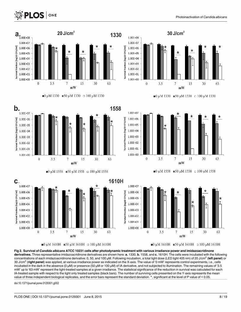
Fig 2. Survival ofCandida albicansATCC 10231 cells after photodynamic treatment with various irradiance power and imidazoacridinonederivatives. Three representative imidazoacridinone derivatives are shown here: a. 1330, b. 1558, and c. 1610H. The cells were incubated with the followingconcentrations of each imidazoacridinone derivative: 0, 50, and 100 μM. Following incubation, a total light dose (LED light 405 nm) of 20 J/cm2 (left panel) or30 J/cm2 (right panel) was applied, at various irradiance power as indicated on the X-axis. The value of ‘0 mW’ represents control experiments, i.e., cellsincubated in the dark in the absence (0 μM) or presence (50 μM or 100 μM) of IA derivative, and not subjected to illumination. The remaining values of ‘3.5mW’ up to ‘63 mW’ represent the light-treated samples at a given irradiance. The statistical significance of the reduction in survival was calculated for eachIA-treated sample with respect to the light only treated samples (black bars). The number of surviving cells presented on the Y-axis represents the meanvalue of three independent biological replicates, and the error bars represent the standard deviation. *, significant at the level of P value of < 0.05.
doi:10.1371/journal.pone.0129301.g002
Photoinactivation of Candida albicans
PLOSONE | DOI:10.1371/journal.pone.0129301 June 8, 2015 8 / 19

Verapamil inhibits imidazoacridinone accumulationWe determined whether the ABC transporters in C. albicans recognized the newly applied IAs,and whether resistance to PDI based on these photosensitizers might occur. To this end, wepre-treated C. albicans with verapamil (200μM), a potent ABC pump inhibitor, and subse-quently measured the levels of IA accumulation. The results, presented in Fig 4, indicated a de-crease in IAs accumulation after verapamil pre-treatment, consistent with free diffusion ratherthan active transport. We also observed differences between the overall accumulation levelsand accumulation in the presence of verapamil with respect to particular compounds. Verapa-mil had negligible effect on the most effectively accumulated compounds (1330 and 1330H; theestimated decrease in accumulation in the presence of verapamil accounted for 15% and 8%,respectively compared with the amount accumulated in the absence of verapamil). In case of1415, 1558, 1610, and 1610H verapamil pre-treatment had a more noticeable effect (30% de-crease in accumulation with respect to non-verapamil treated samples). The highest decreasein accumulation after verapamil treatment was observed for 1415H and 1558H (> 60%). Theeffect was apparent for 1611, as yeast cells accumulated a low amount of this compound andverapamil had no visible effect on this process (Fig 4).
Fig 3. Accumulation of imidazoacridinone derivatives inCandida albicansATCC 10231 cells. The cells were incubated in the dark with the denotedimidazoacridinone derivatives (50 μM, 30 min, 37°C). Imidazoacridinone fluorescence (right panel) was observed under a fluorescence microscope(excitation wavelength 360–370 nm, emission >420 nm). The same cells visualized under white light are presented in the left panel.
doi:10.1371/journal.pone.0129301.g003
Photoinactivation of Candida albicans
PLOSONE | DOI:10.1371/journal.pone.0129301 June 8, 2015 9 / 19

Photochemistry of imidazoacridinones in model systemsWe used two experimental approaches to determine the type of photochemical processes medi-ated through imidazoacridinones in simple model systems. The approaches included: near-in-frared time-resolved luminescence, a direct method to monitor the formation and decay ofsinglet oxygen (1O2), and EPR-spin trapping, an indirect method for detecting short-lived freeradicals. It is important to emphasize that EPR-spin trapping is a canonical method for study-ing oxygen radicals formed during type I photosensitized oxidation reactions.
Quantum yields for singlet oxygen photogeneration through IAs were determined by mea-suring time-resolved luminescence in acetonitrile solutions at 1270 nm, after excitation of thestudied compounds with 355 nm laser light. Rose Bengal was used as a reference, following ex-citation at 355 nm. The photoexcitation of IAs resulted in singlet oxygen generation (Fig 5), ev-ident by the observed dependence of the intensities of the time-resolved luminescence signalson oxygen presence in the samples studied and wavelength at which the signals were detected.Importantly, the corresponding quantum yields varied from low (2%) for 1610 to moderate(16%) for 1330H compared with the well known efficient singlet oxygen generators Rose Ben-gal (70%) and TMPyP (75%). Electron paramagnetic resonance (EPR)-spin trapping using5,5-dimethyl-1-pyrroline N-oxide (DMPO) as a spin trap, revealed the photogeneration of su-peroxide anions (O2 _̄) through all imidazoacridinones (using as solvent DMSO:H2O, at 9:1ratio). Fig 6 shows the exemplary spectra of the DMPO spin adduct after the photoexcitation
Fig 4. Accumulation of imidazoacridinone derivatives inCandida albicansATCC 10231 cells in the presence of verapamil. The cells (106 CFU/ml)were incubated with 50 μM of a particular imidazoacridinone derivative (indicated on the X-axis) in the dark, and the accumulation was measured throughspectrophotometry as described in Materials and Methods (black bars). The cells treated as described, but pretreated with 200 μM of verapamil beforeincubation with the imidazoacridinone derivative, are represented with grey bars. The mean values from three independent biological experiments areshown and the error bars represent the standard deviation.*, significant at the level of P value of < 0.05.
doi:10.1371/journal.pone.0129301.g004
Photoinactivation of Candida albicans
PLOSONE | DOI:10.1371/journal.pone.0129301 June 8, 2015 10 / 19

of 1330 compound in the presence and absence of reduced nicotinamide adenine dinucleotide(NADH). The simulated spectrum of the DMPO spin adduct using superoxide anion is shownin the inset of Fig 6. Comparison of the simulated and experimental spectra indicates that 1330produces superoxide anion upon excitation with UV light (355 nm) and the photoformation ofthe O2 _̄signal is enhanced by about seventy-fold in the presence of an electron donor such asNADH. Similar spectra were recorded for all the remaining imidazoacridinone derivatives.The accumulation of the DMPO spin adduct as a function of irradiation time for derivative1330, in the presence and in the absence of NADH, is shown in Fig 7A. These data indicatethat the relative rates of spin adduct formation can be calculated with respect to riboflavin (areference superoxide anion generator). Such measurements performed for different imidazoa-cridinones indicate that the rate of superoxide anion photoformation is 30–60 times lowerthan that of riboflavin (Fig 7B). Notably, one of the highest rates of superoxide radical genera-tion was observed with 1611.
Notably, the rate of O2 _̄generation was dramatically increased for all tested imidazoacridi-nones when NADH was included in the reaction mixture. The observed acceleration of O2 _̄
generation in the presence of NADH, varied among the studied compounds. The highest en-hancement of the rate of superoxide anion photoformation (~140-fold) was observed for1330H, 1610H, and 1611, while the lowest rate was detected for 1610(~30-fold).
DiscussionThe apparent increase in a number of studies on C. albicans reflects two phenomena: the grow-ing number of isolates resistant to the currently applied antifungals, and, the increasing num-ber of patients with local candidiasis symptoms alongside the growing number of neutropenic,immune-compromised patients [38]. Thus, antifungal photodynamic therapy can be consid-ered as an alternative promising strategy to control localized C. albicans infections [18,39]. No-tably, although encouraging data on photoinactivation of Candida spp. have been published,
Fig 5. Singlet oxygen generation through imidazoacridinone derivatives. Time-resolved luminescenceof singlet oxygen (1O2) was measured at 1270 nm. The excitation wavelength used was 355 nm. RoseBengal, anthracene, and TMPyP were used as standards with known singlet oxygen quantum yields. The Y-axis represents the quantum yield of singlet oxygen generation, where a maximal value of 100% indicatesthat the entire energy of triplet state is transformed into a singlet excited state of molecular oxygen. Thequantum yields are determined within +/- 5% accuracy.
doi:10.1371/journal.pone.0129301.g005
Photoinactivation of Candida albicans
PLOSONE | DOI:10.1371/journal.pone.0129301 June 8, 2015 11 / 19

there are only few reports concerning the characterization and/or mechanism of action of newphotosensitizers. Here, we addressed photosensitizer cellular accumulation, the nature of gen-erated reactive oxygen species, and light-induced photosensitizers activation. These aspects ofphotodynamic therapy are important, particularly regarding the potential clinical applicationsof antifungal PDI.
Porphyrins and phenothaziniums were the first compounds shown to be effective in thephotosensitization of C. albicans cells in suspension [7,20]. These results were later confirmedin mouse models of local Candida infections [25,26] observing antifungal effects, includingstrains that were azole-resistant[40]. However, due to the variability in experimental parame-ters, the comparison of outcomes from different studies is difficult. When analysing photo-chemical and photobiological phenomena, fluence (the energy delivered per unit area) is a keyradiation parameter to quantitatively characterize the observable effects. According to theBunsen-Roscoe law (the reciprocity rule), the observable effect is directly proportional to thetotal radiation energy dose, irrespective of the administered regime[9]. However, it has beendemonstrated that certain photobiological reactions critically depend on the fluence rate(power of radiation per unit area incident on a surface)[9]. Accordingly, Henderson et al.showed that low fluence rate significantly decreases the number of clonogenic cells, while aninsignificant reduction was observed following the high fluence rate treatment of tumour cellswith Photofrin [41]. A similar trend was also observed in the present study (Fig 2). The efficacy
Fig 6. A representative single Electron Paramagnetic Resonance (EPR) spectrum obtained upon photoexcitation of imidazoacridione derivative1330. The predicted (inlet) and experimental spectra of photoexcited imidazoacridinone 1330 in a DMSO/DMPOmixture, upon exposure to light at awavelength range of 540–740 nm are shown.
doi:10.1371/journal.pone.0129301.g006
Photoinactivation of Candida albicans
PLOSONE | DOI:10.1371/journal.pone.0129301 June 8, 2015 12 / 19

of photodynamic therapy could therefore be significantly increased through modifications inthe irradiation protocol. The fact that fluence rates affect imidazoacridinone-mediated photo-dynamic treatment, may be considered as a valuable means of increasing the selectivity of pho-totherapy. Recent data, and the results published by another group using methylene blue [10]indicate that output power and/or time of exposure should be considered as important deter-minants of the photokilling of yeast cells. Similar conclusions can be drawn based on sewagebacteriophage inactivation with cationic porphyrins [12]. Although different molecular andcellular mechanisms could be responsible for the observed effects, oxygen depletion inducedthrough photosensitized reactions, might play an important role. It is conceivable that oxygenphotouptake at higher fluence rates exceeds oxygen replenishment due to its diffusion from
Fig 7. Electron Paramagnetic Resonance (EPR)-spin trapping experiments. a. An example of an EPR spectrum of superoxide anion created uponphotoexcitation of imidazoacridinone 1330 alone and in the presence of NADH. b. Comparison of the coefficient a indicating the efficiency in superoxideanion creation derived from the EPR spectra of different irradiated imidazoacridinone derivatives (indicated in X-axis) in the presence and absence of NADH.Riboflavin was used as a positive control photosensitizer that efficiently generates superoxide anion in the absence of NADH.
doi:10.1371/journal.pone.0129301.g007
Photoinactivation of Candida albicans
PLOSONE | DOI:10.1371/journal.pone.0129301 June 8, 2015 13 / 19

oxygen rich surroundings. As a result, the efficiency of photodynamic cell killing will decreaseat higher fluence rates.
In our previous work on 1330, we did initial experiments concerning photo- and cytotoxici-ty of this compound towards human HaCaT cells. We observed that 200J/cm2 of 380–480 nmlight only treatment causes about 25% decrease in the survival of human HaCaT cell line withrespect to non-light treated cells [37]. This indicates certain degree of light only toxicity, how-ever, still at an acceptable level. With the following PDI treatment conditions: 200J/cm2, 50 μMof 1330 compound, we obtained satisfactory therapeutic window (3.1 log10 decrease in C. albi-cans cells survival, and 43% decrease in HaCaT cell line survival), which points out that C1330may potentially be used as a candidate photosensitizer in the light-dependent killing of C.albicans cells.
Another key factor is the photosensitizer used. Employing different imidazoacridinone de-rivatives, we have shown that fast intake and slow clearance from the treated yeast is a crucialparameter. Indeed, the best derivatives, in terms of PDI efficacy, could be detected within thetreated cells, even after prolonged incubation (up to 30 min). This observation is in contrastwith the previously studied cationic porphyrin TriP[4], which was slowly taken up and ineffi-ciently accumulated by yeast cells. It has been suggested that membrane damage is responsiblefor the killing of C. albicans(21). These authors also showed that vacuoles remained intactduring PDI treatment. Notably, imidazoacridinone accumulation can be easily reversed asdemonstrated after washing the cells with PBS, which decreased the observed imidazoacridi-none-dependent phototoxicity. The data obtained here and particularly the lack of inhibitionby verapamil, suggested that imidazoacridinones are largely imported into cells via spontane-ous diffusion through the membrane. In contrast to hypericin [42], where the photosensitizerlocalized within a large intracellular patch, most likely within a vacuole, imidazoacridinones lo-calized in other cytosolic organelles with the vacuole remaining unstained. PhthalocyaninePc4, another frequently used photosensitizer, was shown to localize in the mitochondria[43].Our most active derivative, imidazoacridinone 1330, showed an intracellular accumulation pat-tern, most likely in the cytoplasm. This unique pattern of intracellular distribution might be as-sociated with a dramatic increase in lethality of photosensitized cells. The remaining IAsexhibited a rather dispersed pattern of cytoplasmic distributions. A recent study on humanovarian carcinoma cells treated with 3,6-bis(imidazolidine)acridine, which is structurally simi-lar to imidazoacridinones, indicated that this compound primary localizes to lysosomes andmitochondria, but not in the nuclei [44]. Similarly, the lysosomal localization of imidazoacridi-nones was observed in human A549 cell line [45]. Therefore, it is likely that 1330 localizes inCandida lysosomes, and this localization pattern results in highly effective lethal photosensiti-zation (as further discussed below).
The key mechanism of drug resistance in C. albicans is the low intracellular accumulation ofantifungal agents due to elevated levels of drug efflux pumps such as ABC (ATP binding cas-sette) transporters, primarily of CaCdr1p and CaCdr2p [46]. Following a genetic modification,Candida albicans strains overexpressing ABC efflux pumps (CaCDR1/CaCDR2) resisted meth-ylene blue-mediated photodynamic action [47]. Photosensitizers comprising substrates forABC-type transporters therefore have limited efficacy for antifungal photodynamic treatment[47]. As shown here, imidazoacridinones are not substrates for ABC transporters, as indicatedby the lack of verapamil inhibition, which blocks the cellular efflux pump CaCDR1. The ob-served decrease in imidazoacridinones accumulation in C. albicansmight reflect either the dis-turbed uptake of these compounds in the presence of verapamil or CaCDR1-independentleakage. The observed differences in accumulation profiles of particular imidazoacridinones inthe presence and in the absence of verapamil are relatively small, and might therefore reflectthe multi-effect action of verapamil on yeast cells. CaCDR1 transporters have been implicated
Photoinactivation of Candida albicans
PLOSONE | DOI:10.1371/journal.pone.0129301 June 8, 2015 14 / 19
in multiple functions, including the transport of inorganic ions, efflux of steroids, and phos-pholipid translocation. These mechanisms might indirectly contribute to the small differencesobserved in the absence and presence of verapamil [48]. Taken together, CaCDR1 is unlikely toexpel the studied imidazoacridinones.
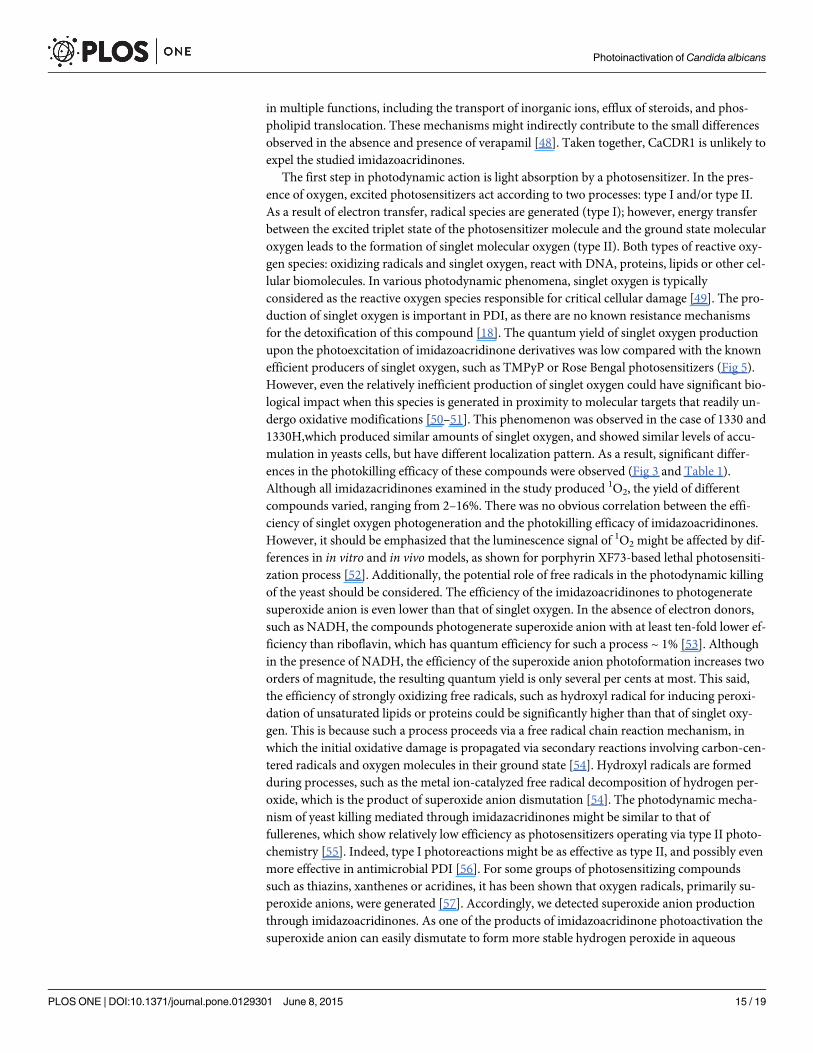
The first step in photodynamic action is light absorption by a photosensitizer. In the pres-ence of oxygen, excited photosensitizers act according to two processes: type I and/or type II.As a result of electron transfer, radical species are generated (type I); however, energy transferbetween the excited triplet state of the photosensitizer molecule and the ground state molecularoxygen leads to the formation of singlet molecular oxygen (type II). Both types of reactive oxy-gen species: oxidizing radicals and singlet oxygen, react with DNA, proteins, lipids or other cel-lular biomolecules. In various photodynamic phenomena, singlet oxygen is typicallyconsidered as the reactive oxygen species responsible for critical cellular damage [49]. The pro-duction of singlet oxygen is important in PDI, as there are no known resistance mechanismsfor the detoxification of this compound [18]. The quantum yield of singlet oxygen productionupon the photoexcitation of imidazoacridinone derivatives was low compared with the knownefficient producers of singlet oxygen, such as TMPyP or Rose Bengal photosensitizers (Fig 5).However, even the relatively inefficient production of singlet oxygen could have significant bio-logical impact when this species is generated in proximity to molecular targets that readily un-dergo oxidative modifications [50–51]. This phenomenon was observed in the case of 1330 and1330H,which produced similar amounts of singlet oxygen, and showed similar levels of accu-mulation in yeasts cells, but have different localization pattern. As a result, significant differ-ences in the photokilling efficacy of these compounds were observed (Fig 3 and Table 1).Although all imidazacridinones examined in the study produced 1O2, the yield of differentcompounds varied, ranging from 2–16%. There was no obvious correlation between the effi-ciency of singlet oxygen photogeneration and the photokilling efficacy of imidazoacridinones.However, it should be emphasized that the luminescence signal of 1O2 might be affected by dif-ferences in in vitro and in vivomodels, as shown for porphyrin XF73-based lethal photosensiti-zation process [52]. Additionally, the potential role of free radicals in the photodynamic killingof the yeast should be considered. The efficiency of the imidazoacridinones to photogeneratesuperoxide anion is even lower than that of singlet oxygen. In the absence of electron donors,such as NADH, the compounds photogenerate superoxide anion with at least ten-fold lower ef-ficiency than riboflavin, which has quantum efficiency for such a process ~ 1% [53]. Althoughin the presence of NADH, the efficiency of the superoxide anion photoformation increases twoorders of magnitude, the resulting quantum yield is only several per cents at most. This said,the efficiency of strongly oxidizing free radicals, such as hydroxyl radical for inducing peroxi-dation of unsaturated lipids or proteins could be significantly higher than that of singlet oxy-gen. This is because such a process proceeds via a free radical chain reaction mechanism, inwhich the initial oxidative damage is propagated via secondary reactions involving carbon-cen-tered radicals and oxygen molecules in their ground state [54]. Hydroxyl radicals are formedduring processes, such as the metal ion-catalyzed free radical decomposition of hydrogen per-oxide, which is the product of superoxide anion dismutation [54]. The photodynamic mecha-nism of yeast killing mediated through imidazacridinones might be similar to that offullerenes, which show relatively low efficiency as photosensitizers operating via type II photo-chemistry [55]. Indeed, type I photoreactions might be as effective as type II, and possibly evenmore effective in antimicrobial PDI [56]. For some groups of photosensitizing compoundssuch as thiazins, xanthenes or acridines, it has been shown that oxygen radicals, primarily su-peroxide anions, were generated [57]. Accordingly, we detected superoxide anion productionthrough imidazoacridinones. As one of the products of imidazoacridinone photoactivation thesuperoxide anion can easily dismutate to form more stable hydrogen peroxide in aqueous
Photoinactivation of Candida albicans
PLOSONE | DOI:10.1371/journal.pone.0129301 June 8, 2015 15 / 19

solutions. Hydrogen peroxide can decompose in a free radical process to create the most reac-tive oxygen species (ROS), hydroxyl radical. The presence of these ROS might be responsiblefor the efficacy of imidazaocridinones photokilling.
Here, we have characterized for the first time the mechanism underlying IA action againstC. albicans. We have shown that structurally different imidazoacridinone derivatives are ableto inactivate C. albicans cells in light dependent manner. Moreover, we provide indicationsthat fluence rate should be considered as a crucial parameter to improve efficiency of IA-medi-ated PDI. A correlation between ROS production by particular IA derivative and cell killingwas not observed. Interestingly, we have shown, that IA accumulation and localization eventsare critical factors for cell killing, which is in accordance with results obtained for other photo-sensitizing agents. The results presented here show that IA not only efficiently accumulate incells but importantly they are not actively expelled from C. albicans cells via CaCDR1 ABCtransporter. Therefore, IA are promising candidates for the long-term treatment of localizedinfections. Overall, this study contributes to a better understanding of the mode of action ofimidazoacridinones, and suggests that these compounds are potential reagents for the photo-killing of C. albicans.
Supporting InformationS1 Table. Accumulation and phototoxic effect of imidazoacridinone derivatives in Candidaalbicans ATCC 14053 cells.(DOCX)
S2 Table. Accumulation and phototoxic effect of imidazoacridinone derivatives in Candidaalbicans ATCC 90028 cells.(DOCX)
Author ContributionsConceived and designed the experiments: JN TS. Performed the experiments: AT GS. Analyzedthe data: JN AT GS TS. Contributed reagents/materials/analysis tools: JN AT TS KPB. Wrotethe paper: JN AT TS.
References1. Rodrigues ME, Silva S, Azeredo J, Henriques M. Novel strategies to fight Candida species infection.
Crit Rev Microbiol 2014;1–13.
2. Pfaller MA. Antifungal drug resistance: mechanisms, epidemiology, and consequences for treatment.Am J Med 2012; 125:S3–13. doi: 10.1016/j.amjmed.2011.11.001 PMID: 22196207
3. Wainwright M. 'Safe' photoantimicrobials for skin and soft-tissue infections. Int J Antimicrob Agents2010; 36:14–8. doi: 10.1016/j.ijantimicag.2010.03.002 PMID: 20382003
4. Tavares A, Carvalho CM, Faustino MA, Neves MG, Tome JP, Tome AC et al. Antimicrobial photody-namic therapy: study of bacterial recovery viability and potential development of resistance after treat-ment. Mar Drugs 2010; 8:91–105. doi: 10.3390/md8010091 PMID: 20161973
5. Giuliani F, Martinelli M, Cocchi A, Arbia D, Fantetti L, Roncucci G. In vitro resistance selection studiesof RLP068/Cl, a new Zn(II) phthalocyanine suitable for antimicrobial photodynamic therapy. AntimicrobAgents Chemother 2010; 54:637–42. doi: 10.1128/AAC.00603-09 PMID: 20008782
6. Costa L, Tome JP, Neves MG, Tome AC, Cavaleiro JA, Faustino MA et al. Evaluation of resistance de-velopment and viability recovery by a non-enveloped virus after repeated cycles of aPDT. Antiviral Res2011; 91:278–82. doi: 10.1016/j.antiviral.2011.06.007 PMID: 21722673
7. Zeina B, Greenman J, Purcell WM, Das B. Killing of cutaneous microbial species by photodynamic ther-apy. Br J Dermatol 2001; 144:274–8. PMID: 11251558
8. Zeina B, Greenman J, Corry D, Purcell WM. Antimicrobial photodynamic therapy: assessment of geno-toxic effects on keratinocytes in vitro. Br J Dermatol 2003; 148:229–32. PMID: 12588372
Photoinactivation of Candida albicans
PLOSONE | DOI:10.1371/journal.pone.0129301 June 8, 2015 16 / 19

9. Schindl A, Rosado-Schlosser B, Trautinger F. [Reciprocity regulation in photobiology. An overview].Hautarzt 2001; 52:779–85. PMID: 11572068
10. Prates RA, da Silva EG, Yamada AM Jr, Suzuki LC, Paula CL, Ribeiro MS. Light parameters influencecell viability in antifungal photodynamic therapy in a fluence and ratefluence-dependent manner. LaserPhysics 2009; 19:1038–44.
11. Alves E, Carvalho CM, Tome JP, Faustino MA, Neves MG, Tome AC et al. Photodynamic inactivationof recombinant bioluminescent Escherichia coli by cationic porphyrins under artificial and solar irradia-tion. J Ind Microbiol Biotechnol 2008; 35:1447–54. doi: 10.1007/s10295-008-0446-2 PMID: 18712538
12. Costa L, Carvalho CM, Faustino MA, Neves MG, Tome JP, Tome AC et al. Sewage bacteriophage in-activation by cationic porphyrins: influence of light parameters. Photochem Photobiol Sci 2010;9:1126–33. doi: 10.1039/c0pp00051e PMID: 20563346
13. Arendrup MC. Epidemiology of invasive candidiasis. Curr Opin Crit Care 2010; 16:445–52. doi: 10.1097/MCC.0b013e32833e84d2 PMID: 20711075
14. Munin E, Giroldo LM, Alves LP, Costa MS. Study of germ tube formation by Candida albicans after pho-todynamic antimicrobial chemotherapy (PACT). J Photochem Photobiol B 2007; 88:16–20. PMID:17566757
15. Souza RC, Junqueira JC, Rossoni RD, Pereira CA, Munin E, Jorge AO. Comparison of the photody-namic fungicidal efficacy of methylene blue, toluidine blue, malachite green and low-power laser irradia-tion alone against Candida albicans. Lasers Med Sci 2010; 25:385–9. doi: 10.1007/s10103-009-0706-zPMID: 19579004
16. Pereira CA, Romeiro RL, Costa AC, Machado AK, Junqueira JC, Jorge AO. Susceptibility of Candidaalbicans, Staphylococcus aureus, and Streptococcus mutans biofilms to photodynamic inactivation: anin vitro study. Lasers Med Sci 2011; 26:341–8. doi: 10.1007/s10103-010-0852-3 PMID: 21069408
17. Mantareva V, Angelov I, Kussovski V, Dimitrov R, Lapok L, Wohrle D. Photodynamic efficacy of water-soluble Si(IV) and Ge(IV) phthalocyanines towards Candida albicans planktonic and biofilm cultures.Eur J Med Chem 2011; 46:4430–40. doi: 10.1016/j.ejmech.2011.07.015 PMID: 21816518
18. Pereira GF, Maisch T. Photodynamic inactivation for controlling Candida albicans infections. FungalBiol 2012; 116:1–10. doi: 10.1016/j.funbio.2011.10.001 PMID: 22208597
19. Cormick MP, Quiroga ED, Bertolotti SG, Alvarez MG, Durantini EN. Mechanistic insight of the photody-namic effect induced by tri- and tetra-cationic porphyrins on Candida albicans cells. Photochem Photo-biol Sci 2011; 10:1556–61. doi: 10.1039/c1pp05074e PMID: 21748182
20. Bliss JM, Bigelow CE, Foster TH, Haidaris CG. Susceptibility of Candida species to photodynamic ef-fects of photofrin. Antimicrob Agents Chemother 2004; 48:2000–6. PMID: 15155191
21. Lambrechts SA, Aalders MC, Van MJ. Mechanistic study of the photodynamic inactivation of Candidaalbicans by a cationic porphyrin. Antimicrob Agents Chemother 2005; 49:2026–34. PMID: 15855528
22. Eichner A, Gonzales FP, Felgentrager A, Regensburger J, Holzmann T, Schneider-Brachert W et al.Dirty hands: photodynamic killing of human pathogens like EHEC, MRSA and Candida within seconds.Photochem Photobiol Sci 2013; 12:135–47. doi: 10.1039/c2pp25164g PMID: 22855122
23. Gonzales FP, Felgentrager A, Baumler W, Maisch T. Fungicidal photodynamic effect of a twofold posi-tively charged porphyrin against Candida albicans planktonic cells and biofilms. Future Microbiol 2013;8:785–97. doi: 10.2217/fmb.13.44 PMID: 23701333
24. Beirao S, Fernandes S, Coelho J, Faustino MA, Tome JP, Neves MG et al. Photodynamic inactivationof bacterial and yeast biofilms with a cationic porphyrin. Photochem Photobiol 2014; 90:1387–96. doi:10.1111/php.12331 PMID: 25112506
25. Mima EG, Pavarina AC, Dovigo LN, Vergani CE, Costa CA, Curachi C et al. Susceptibility of Candidaalbicans to photodynamic therapy in a murine model of oral candidosis. Oral Surg Oral Med Oral PatholOral Radiol Endod 2010; 109:392–401. doi: 10.1016/j.tripleo.2009.10.006 PMID: 20060338
26. Teichert MC, Jones JW, UsachevaMN, Biel MA. Treatment of oral candidiasis with methylene blue-me-diated photodynamic therapy in an immunodeficient murine model. Oral Surg Oral Med Oral PatholOral Radiol Endod 2002; 93:155–60. PMID: 11862203
27. Junqueira JC, Martins JS, Faria RL, Colombo CE, Jorge AO. Photodynamic therapy for the treatmentof buccal candidiasis in rats. Lasers Med Sci 2009; 24:877–84. doi: 10.1007/s10103-009-0673-4PMID: 19408038
28. Martins JS, Junqueira JC, Faria RL, Santiago NF, Rossoni RD, Colombo CE et al. Antimicrobial photo-dynamic therapy in rat experimental candidiasis: evaluation of pathogenicity factors of Candida albi-cans. Oral Surg Oral Med Oral Pathol Oral Radiol Endod 2011; 111:71–7. doi: 10.1016/j.tripleo.2010.08.012 PMID: 21176823
Photoinactivation of Candida albicans
PLOSONE | DOI:10.1371/journal.pone.0129301 June 8, 2015 17 / 19

29. Dai T, Bil dA, V, Tegos GP, Hamblin MR. Blue dye and red light, a dynamic combination for prophylaxisand treatment of cutaneous Candida albicans infections in mice. Antimicrob Agents Chemother 2011;55:5710–7. doi: 10.1128/AAC.05404-11 PMID: 21930868
30. Yang YT, Chien HF, Chang PH, Chen YC, Jay M, Tsai T et al. Photodynamic inactivation of chlorin e6-loaded CTAB-liposomes against Candida albicans. Lasers Surg Med 2013; 45:175–85. doi: 10.1002/lsm.22124 PMID: 23508377
31. Machado-de-Sena RM, Correa L, Kato IT, Prates RA, Senna AM, Santos CC et al. Photodynamic ther-apy has antifungal effect and reduces inflammatory signals in Candida albicans-induced murine vagini-tis. Photodiagnosis Photodyn Ther 2014; 11:275–82. doi: 10.1016/j.pdpdt.2014.03.013 PMID:24792453
32. CholodyWM, Martelli S, Paradziej-Lukowicz J, Konopa J. 5-[(Aminoalkyl)amino]imidazo[4,5,1-de]acri-din-6-ones as a novel class of antineoplastic agents. Synthesis and biological activity. J Med Chem1990; 33:49–52. PMID: 2296035
33. CholodyWM, Horowska B, Paradziej-Lukowicz J, Martelli S, Konopa J. Structure-activity relationshipfor antineoplastic imidazoacridinones: synthesis and antileukemic activity in vivo. J Med Chem 1996;39:1028–32. PMID: 8676337
34. CholodyWM, Martelli S, Konopa J. 8-Substituted 5-[(aminoalkyl)amino]-6H-v-triazolo[4,5,1-de]acridin-6-ones as potential antineoplastic agents. Synthesis and biological activity. J Med Chem 1990;33:2852–6. PMID: 2213837
35. Cholody, W. M. and Konopa, J. K. Antineoplastic modified imidazoacridines. British Technology GroupLimited. 760,694(United Staes Patent 5,231,100). 7-27-1993. England. Ref Type: Patent
36. Assaraf, Y. G., Misgav, D-N., Bram, E. E., HaSharon, D-N. L., Bney-Moshe, Y., and Skladanowski, A.M. Imidazoacridinone derivative compounds and methods for their use. Technion Research and Devel-opment Foundation Ltd. 12/585,252(US 2010/0137351 A1). 6-3-2010. Haifa Israel. Ref Type: Patent
37. Taraszkiewicz A, Grinholc M, Bielawski KP, Kawiak A, Nakonieczna J. Imidazoacridinone derivativesas efficient sensitizers in photoantimicrobial chemotherapy. Appl Environ Microbiol 2013; 79:3692–702. doi: 10.1128/AEM.00748-13 PMID: 23563951
38. Maubon D, Garnaud C, Calandra T, Sanglard D, Cornet M. Resistance of Candida spp. to antifungaldrugs in the ICU: where are we now? Intensive Care Med 2014; 40:1241–55. doi: 10.1007/s00134-014-3404-7 PMID: 25091787
39. Dai T, Fuchs BB, Coleman JJ, Prates RA, Astrakas C, St Denis TG et al. Concepts and principles ofphotodynamic therapy as an alternative antifungal discovery platform. Front Microbiol 2012; 3:120. doi:10.3389/fmicb.2012.00120 PMID: 22514547
40. Chibebe JJ, Sabino CP, Tan X, Junqueira JC, Wang Y, Fuchs BB et al. Selective photoinactivation ofCandida albicans in the non-vertebrate host infection model Galleria mellonella. BMCMicrobiol 2013;13:217. doi: 10.1186/1471-2180-13-217 PMID: 24083556
41. Henderson BW, Busch TM, Snyder JW. Fluence rate as a modulator of PDT mechanisms. Lasers SurgMed 2006; 38:489–93. PMID: 16615136
42. Lopez-Chicon P, Paz-Cristobal MP, Rezusta A, Aspiroz C, Royo-Canas M, Andres-Ciriano E et al. Onthe mechanism of Candida spp. photoinactivation by hypericin. Photochem Photobiol Sci 2012;11:1099–107. doi: 10.1039/c2pp25105a PMID: 22566080
43. LamM, Jou PC, Lattif AA, Lee Y, Malbasa CL, Mukherjee PK et al. Photodynamic therapy with Pc 4 in-duces apoptosis of Candida albicans. Photochem Photobiol 2011; 87:904–9. doi: 10.1111/j.1751-1097.2011.00938.x PMID: 21521233
44. Cizekova L, Grolmusova A, Ipothova Z, Barbierikova Z, Brezova V, Hunakova L et al. Novel 3,6-bis(imi-dazolidine)acridines as effective photosensitizers for photodynamic therapy. Bioorg Med Chem 2014;22:4684–93. doi: 10.1016/j.bmc.2014.07.013 PMID: 25096820
45. Adar Y, Stark M, Bram EE, Nowak-Sliwinska P, van den Bergh H, Szewczyk G et al. Imidazoacridi-none-dependent lysosomal photodestruction: a pharmacological Trojan horse approach to eradicatemultidrug-resistant cancers. Cell Death Dis 2012; 3:e293. doi: 10.1038/cddis.2012.30 PMID: 22476101
46. Prasad R, DeWP, Goffeau A, Balzi E. Molecular cloning and characterization of a novel gene of Candi-da albicans, CDR1, conferring multiple resistance to drugs and antifungals. Curr Genet 1995; 27:320–9. PMID: 7614555
47. Prates RA, Kato IT, Ribeiro MS, Tegos GP, Hamblin MR. Influence of multidrug efflux systems on meth-ylene blue-mediated photodynamic inactivation of Candida albicans. J Antimicrob Chemother 2011;66:1525–32. doi: 10.1093/jac/dkr160 PMID: 21525022
48. Prasad R, Goffeau A. Yeast ATP-binding cassette transporters conferring multidrug resistance. AnnuRev Microbiol 2012; 66:39–63. doi: 10.1146/annurev-micro-092611-150111 PMID: 22703054
Photoinactivation of Candida albicans
PLOSONE | DOI:10.1371/journal.pone.0129301 June 8, 2015 18 / 19

49. Alves E, Faustino MA, Neves MG, Cunha A, Tome J, Almeida A. An insight on bacterial cellular targetsof photodynamic inactivation. Future Med Chem 2014; 6:141–64. doi: 10.4155/fmc.13.211 PMID:24467241
50. Clennan EP, Pace A. Advances in singlet oxygen chemistry. Tetrahedron 2005; 61:6665–91.
51. Kozinska A, Oles T, Sarna T. Photoactivation and detection of photoexcited molecules and photochem-ical products. Israel Journal of Chemistry 2012; 52:745–56.
52. Felgentrager A, Gonzales FP, Maisch T, BaumlerW. Ion-induced stacking of photosensitizer moleculescan remarkably affect the luminescence detection of singlet oxygen in Candida albicans cells. J BiomedOpt 2013; 18:045002. doi: 10.1117/1.JBO.18.4.045002 PMID: 23552633
53. Krishna CM, Uppuluri S, Riesz P, Zigler JS Jr., Balasubramanian D. A study of the photodynamic effi-ciencies of some eye lens constituents. Photochem Photobiol 1991; 54:51–8. PMID: 1658825
54. Halliwell B, Gutteridge JMC. Free Radicals in Biology and Medicine. Oxford: Oxford University Press,2000.
55. Milanesio ME, Spesia MB, Cormick MP, Durantini EN. Mechanistic studies on the photodynamic effectinduced by a dicationic fullerene C60 derivative on Escherichia coli and Candida albicans cells. Photo-diagnosis Photodyn Ther 2013; 10:320–7. doi: 10.1016/j.pdpdt.2013.01.007 PMID: 23993859
56. Mroz P, Tegos GP, Gali H, Wharton T, Sarna T, Hamblin MR. Photodynamic therapy with fullerenes.Photochem Photobiol Sci 2007; 6:1139–49. PMID: 17973044
57. Martin JP, Logsdon N. Oxygen radicals are generated by dye-mediated intracellular photooxidations: arole for superoxide in photodynamic effects. Arch Biochem Biophys 1987; 256:39–49. PMID: 3038028
Photoinactivation of Candida albicans
PLOSONE | DOI:10.1371/journal.pone.0129301 June 8, 2015 19 / 19