Microevolution of Candida albicans in macrophages restores filamentation in a nonfilamentous mutant
Upload
independentCategory
view
3download
0
A Genetic Code Alteration Is a Phenotype DiversityGenerator in the Human Pathogen Candida albicansIsabel Miranda, Rita Rocha, Maria C. Santos, Denisa D. Mateus, Gabriela R. Moura, Laura Carreto, Manuel A. S. Santos*
Department of Biology, Centro de Estudos do Ambiente e do Mar (CESAM), University of Aveiro, Aveiro, Portugal
Background. The discovery of genetic code alterations and expansions in both prokaryotes and eukaryotes abolished thehypothesis of a frozen and universal genetic code and exposed unanticipated flexibility in codon and amino acid assignments.It is now clear that codon identity alterations involve sense and non-sense codons and can occur in organisms with complexgenomes and proteomes. However, the biological functions, the molecular mechanisms of evolution and the diversity ofgenetic code alterations remain largely unknown. In various species of the genus Candida, the leucine CUG codon is decodedas serine by a unique serine tRNA that contains a leucine 59-CAG-39anticodon (tRNACAG
Ser). We are using this codon identityredefinition as a model system to elucidate the evolution of genetic code alterations. Methodology/Principal Findings. Wehave reconstructed the early stages of the Candida genetic code alteration by engineering tRNAs that partially reverted theidentity of serine CUG codons back to their standard leucine meaning. Such genetic code manipulation had profound cellularconsequences as it exposed important morphological variation, altered gene expression, re-arranged the karyotype, increasedcell-cell adhesion and secretion of hydrolytic enzymes. Conclusion/Significance. Our study provides the first experimentalevidence for an important role of genetic code alterations as generators of phenotypic diversity of high selective potential andsupports the hypothesis that they speed up evolution of new phenotypes.
Citation: Miranda I, Rocha R, Santos MC, Mateus DD, Moura GR, et al (2007) A Genetic Code Alteration Is a Phenotype Diversity Generator in theHuman Pathogen Candida albicans. PLoS ONE 2(10): e996. doi:10.1371/journal.pone.0000996
INTRODUCTIONA number of exceptions to the standard genetic code have been
discovered in prokaryotic and eukaryotic organisms, involving
nonsense-to-sense and sense-to-sense codon identity changes [1,2].
Twenty five of such alterations have been recorded in mitochon-
drial genetic codes of metazoan, fungi, red algae, green plants,
alveolates, stramenopiles, haptophtes and euglenozoans [3]. The
most remarkable alterations involve metazoan arginine AGA and
AGG (AGR) codons, which changed their identity to serine at the
base of the phylogenetic tree, and later on to translation-stop in
vertebrates and to glycine in urochordates [4,5].
In bacteria and eukaryotic cytoplasmic systems, 18 genetic code
alterations have also been recorded, but unlike in mitochondria,
they involve, with one exception, nonsense-to-sense codon identity
changes or codon unassignments (codons that vanished from
genomes). For example, in Micrococcus spp., Mycoplasma spp. and
Pseudomicrothorax dubius the AGA/AUA, CGG and UGA codons
are unassigned, respectively. In Bacillus subtilis UGA codons are
used to terminate mRNA translation (stop codons) and to insert
tryptophan, creating readthrough proteins [6]. In various species
of ciliates, 1 or 2 stop codons changed their identity to either
glutamine (UAA and UAG), glutamate (UAA) or cysteine (UGA).
Interestingly, these genetic code alterations apparently minimize
nonsense errors arising from re-assembly of the ciliates fragmented
genome [7–10].
Those genetic code alterations show that certain codons, namely
codons that start with uridine (UNN) or adenosine (ANN), in
particular stop (UAA, UAG or UGA), arginine (AGR), serine
(AGY) and isoleucine (AUA) codons are more prone to identity
changes than others. The only exception to this first codon base
rule occurs in yeast mitochondria where cytosine starting codons
(CNN), namely leucine CUN codons, changed their identity to
threonine. Also, the leucine CUG codon altered its identity to
serine in the cytoplasm of various Candida and Debaryomyces species
[11–13]. These findings suggest that the strength of the interaction
of the first codon-anticodon base pair plays an important role in
the evolution of genetic code alterations. Indeed, the change of
identity of UAA and UAG stop codons to glutamine in various
ciliates involved first base misreading by glutamine tRNAs, which
decode glutamine CAA and CAG codons [8,14]. But, it also
indicates that other forces beyond codon-anticodon interaction
play important roles in codon identity redefinition.
The unexpected flexibility of the genetic code described above is
further highlighted by insertion of selenocystein (21st amino acid)
in the active sites of prokaryotic and eukaryotic selenoproteins and
insertion of pyrrolysin (22sd amino acid) in the active site of
monomethylamine methyltransferase of Methanosarcina barkeri
[15,16]. These expansions involving reprogramming of UGA
and UAG stop codons, respectively, highlight the potential of
genetic code alterations/expansions to generate functional in-
novation. This hypothesis is supported by recent artificial
expansion of the genetic code through synthetic biology
methodologies. Indeed, 49 non-natural amino acids have already
been incorporated into E. coli, yeast and mammalian cells [17–20],
to produce novel proteins of biotechnological and biomedical
interest. This dramatic demonstration of genetic code flexibility
also unveiled an extraordinary capacity of complex organisms to
tolerate partial codon identity redefinition [21].
Academic Editor: Rosemary Redfield, University of British Columbia, Canada
Received July 9, 2007; Accepted September 18, 2007; Published October 3, 2007
Copyright: � 2007 Miranda et al. This is an open-access article distributed underthe terms of the Creative Commons Attribution License, which permitsunrestricted use, distribution, and reproduction in any medium, provided theoriginal author and source are credited.
Funding: This study was supported by FCT/FEDER projects POCI/BIA-MIC/55466/04, POCI/BIA-PRO/55472/2004 and POCI/SAU-MMO/55476/2004. IM, RR aresupported by FCT/FEDER, BD/19807/99 and BD/8296/2002 PhD grants, re-spectively. MASS was supported by an EMBO YIP and a Human Frontier ScienceProgramme Grant (REF: RGP45/2005).
Competing Interests: The authors have declared that no competing interestsexist.
* To whom correspondence should be addressed. E-mail: [email protected]
PLoS ONE | www.plosone.org 1 October 2007 | Issue 10 | e996
We are using C. albicans as a model system to elucidate the
evolution of genetic code alterations. In this case, a unique serine
tRNA (tRNACAGSer) decodes leucine CUG codons as serine [22–
24]. However, the tRNACAGSer is recognized by both leucyl- and
seryl-tRNA synthetases (LeuRS and SerRS) and it is aminoacy-
lated in vitro with both serine (97%) and leucine (3%) [25]. This
unusual dual aminoacylation of the tRNACAGSer has been
preserved to the present day [26,27], raising the intriguing
possibility that it may play a role in C. albicans biology. It also
provides strong support for the hypothesis that codon identity
redefinition is driven through codon decoding ambiguity [28,29].
In here, we have reconstructed the early stages of the C. albicans
genetic code alteration to shed new light into those questions. This
partial reversion of CUG identity from serine back to leucine
triggered morphogenesis, phenotypic switching, and up-regulated
expression of genes involved in cell adhesion and hyphal
development and increased secretion of proteinases and phospho-
lipases. Important karyotype alterations were also observed. The
overall data suggest that C. albicans CUG ambiguity is an
important phenotypic diversity generator and highlight important
and yet overlooked functional roles for genetic code alterations.
RESULTS
Reverting CUG identity from serine back to leucineThe CUG identity alteration from leucine to serine was initiated
272625My ago through a mutant serine tRNA (tRNACAGSer),
containing a 59-CAG-39 anticodon, which could decode CUG
codons (see introduction) [26,22]. Initially, this unique tRNA-
CAGSer competed with endogenous tRNALeu, which decoded CUG
codons as leucine [30], creating a new situation where both
leucines and serines were inserted at CUG positions on a genome
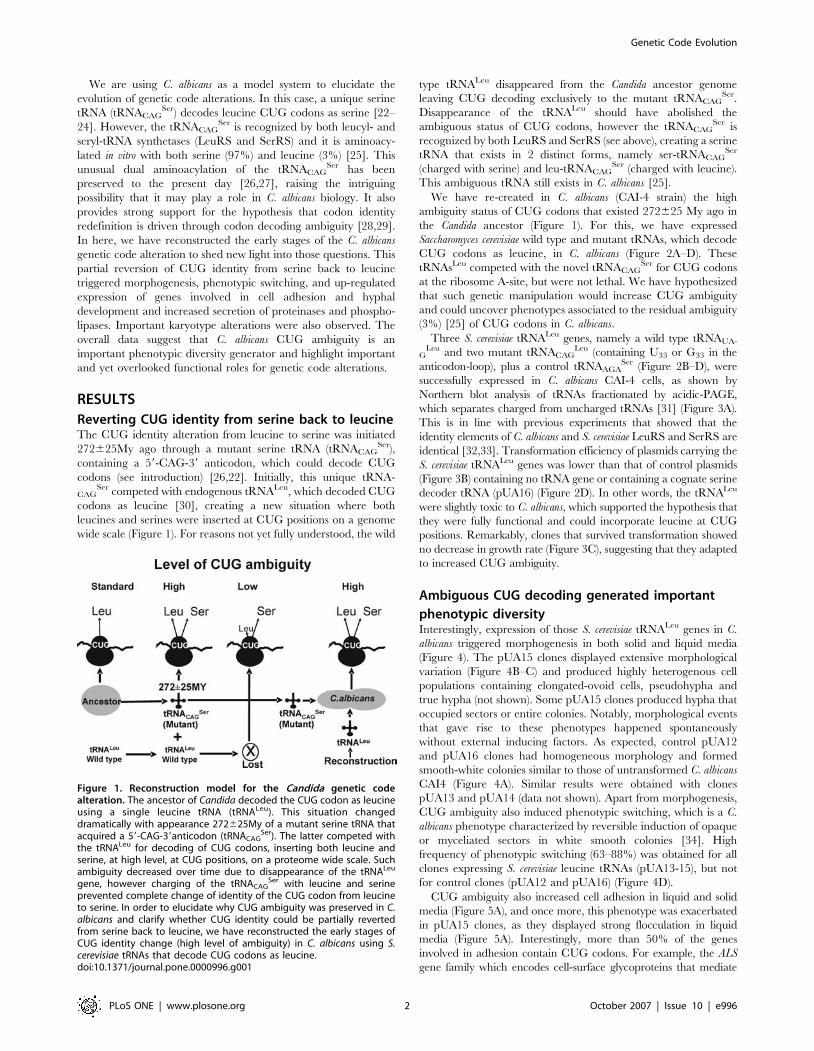
wide scale (Figure 1). For reasons not yet fully understood, the wild
type tRNALeu disappeared from the Candida ancestor genome
leaving CUG decoding exclusively to the mutant tRNACAGSer.
Disappearance of the tRNALeu should have abolished the
ambiguous status of CUG codons, however the tRNACAGSer is
recognized by both LeuRS and SerRS (see above), creating a serine
tRNA that exists in 2 distinct forms, namely ser-tRNACAGSer
(charged with serine) and leu-tRNACAGSer (charged with leucine).
This ambiguous tRNA still exists in C. albicans [25].
We have re-created in C. albicans (CAI-4 strain) the high
ambiguity status of CUG codons that existed 272625 My ago in
the Candida ancestor (Figure 1). For this, we have expressed
Saccharomyces cerevisiae wild type and mutant tRNAs, which decode
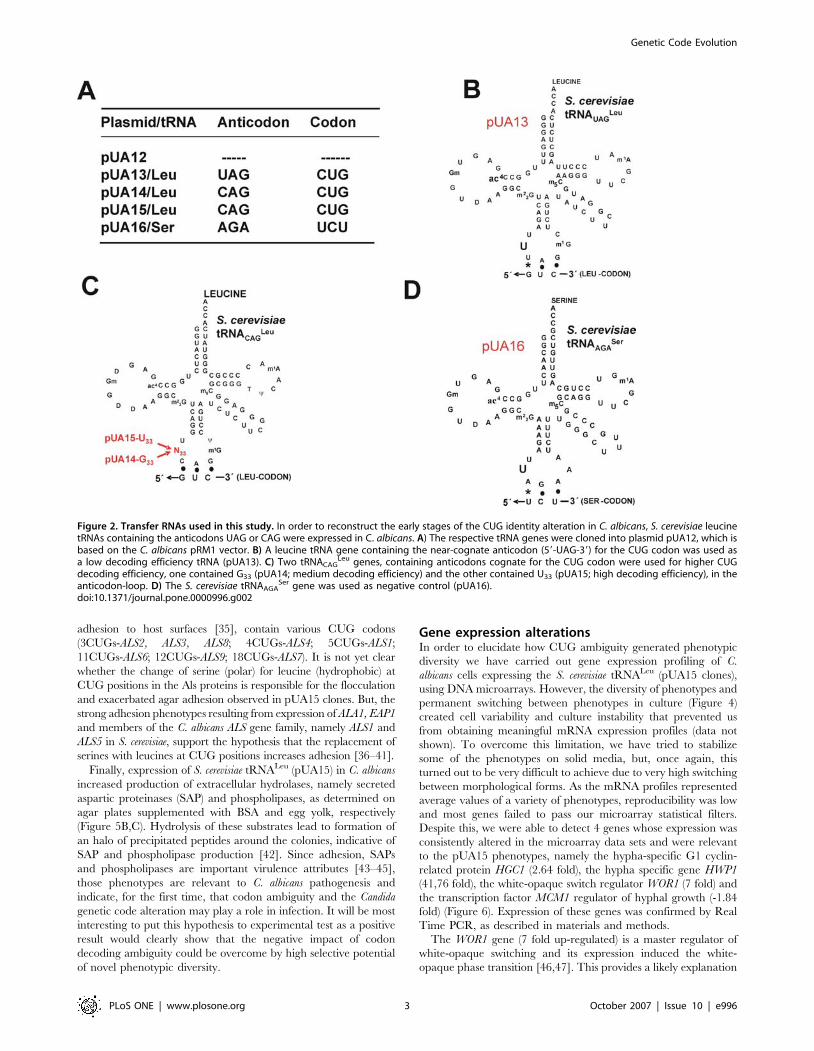
CUG codons as leucine, in C. albicans (Figure 2A–D). These
tRNAsLeu competed with the novel tRNACAGSer for CUG codons
at the ribosome A-site, but were not lethal. We have hypothesized
that such genetic manipulation would increase CUG ambiguity
and could uncover phenotypes associated to the residual ambiguity
(3%) [25] of CUG codons in C. albicans.
Three S. cerevisiae tRNALeu genes, namely a wild type tRNAUA-
GLeu and two mutant tRNACAG
Leu (containing U33 or G33 in the
anticodon-loop), plus a control tRNAAGASer (Figure 2B–D), were
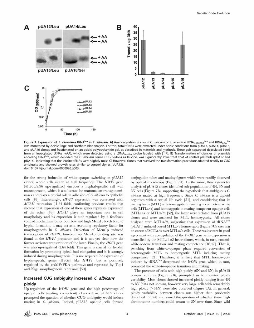
successfully expressed in C. albicans CAI-4 cells, as shown by
Northern blot analysis of tRNAs fractionated by acidic-PAGE,
which separates charged from uncharged tRNAs [31] (Figure 3A).
This is in line with previous experiments that showed that the
identity elements of C. albicans and S. cerevisiae LeuRS and SerRS are
identical [32,33]. Transformation efficiency of plasmids carrying the
S. cerevisiae tRNALeu genes was lower than that of control plasmids
(Figure 3B) containing no tRNA gene or containing a cognate serine
decoder tRNA (pUA16) (Figure 2D). In other words, the tRNALeu
were slightly toxic to C. albicans, which supported the hypothesis that
they were fully functional and could incorporate leucine at CUG
positions. Remarkably, clones that survived transformation showed
no decrease in growth rate (Figure 3C), suggesting that they adapted
to increased CUG ambiguity.
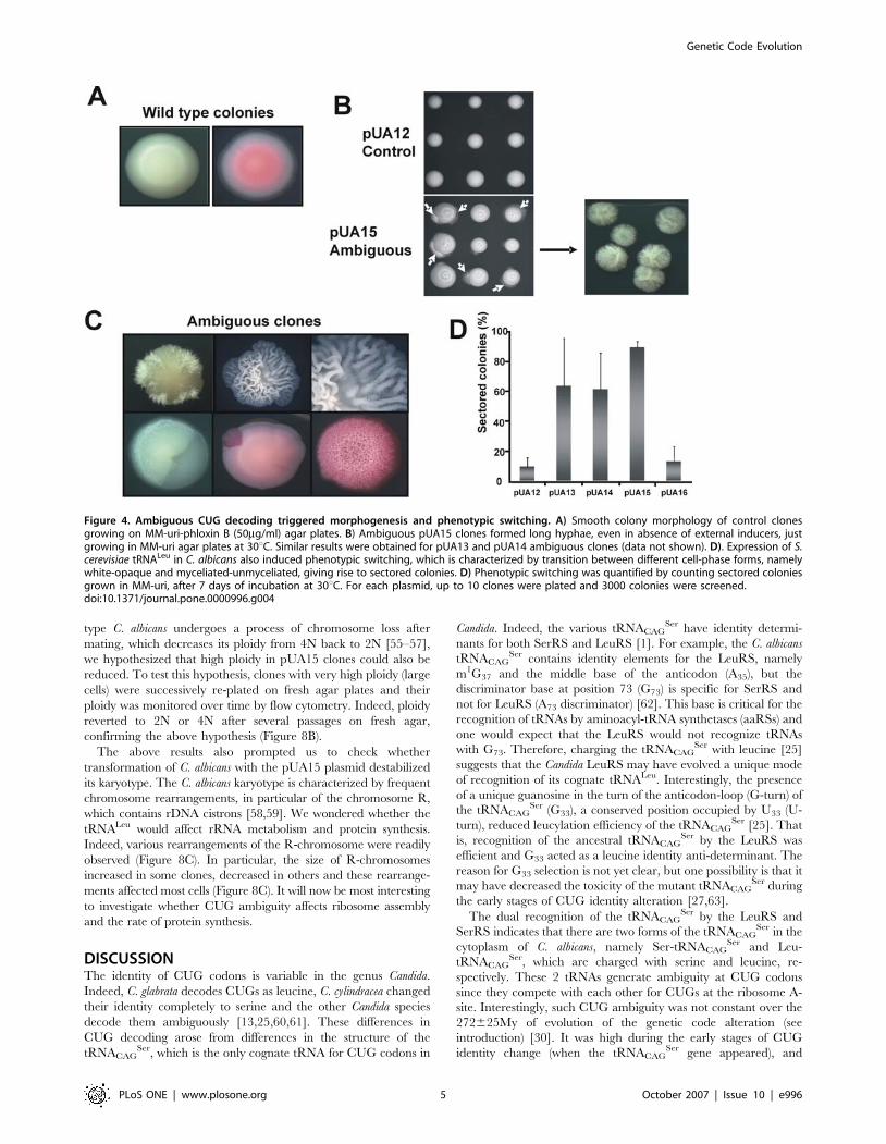
Ambiguous CUG decoding generated important
phenotypic diversityInterestingly, expression of those S. cerevisiae tRNALeu genes in C.
albicans triggered morphogenesis in both solid and liquid media
(Figure 4). The pUA15 clones displayed extensive morphological
variation (Figure 4B–C) and produced highly heterogenous cell
populations containing elongated-ovoid cells, pseudohypha and
true hypha (not shown). Some pUA15 clones produced hypha that
occupied sectors or entire colonies. Notably, morphological events
that gave rise to these phenotypes happened spontaneously
without external inducing factors. As expected, control pUA12
and pUA16 clones had homogeneous morphology and formed
smooth-white colonies similar to those of untransformed C. albicans
CAI4 (Figure 4A). Similar results were obtained with clones
pUA13 and pUA14 (data not shown). Apart from morphogenesis,
CUG ambiguity also induced phenotypic switching, which is a C.
albicans phenotype characterized by reversible induction of opaque
or myceliated sectors in white smooth colonies [34]. High
frequency of phenotypic switching (63–88%) was obtained for all
clones expressing S. cerevisiae leucine tRNAs (pUA13-15), but not
for control clones (pUA12 and pUA16) (Figure 4D).
CUG ambiguity also increased cell adhesion in liquid and solid
media (Figure 5A), and once more, this phenotype was exacerbated
in pUA15 clones, as they displayed strong flocculation in liquid
media (Figure 5A). Interestingly, more than 50% of the genes
involved in adhesion contain CUG codons. For example, the ALS
gene family which encodes cell-surface glycoproteins that mediate
Figure 1. Reconstruction model for the Candida genetic codealteration. The ancestor of Candida decoded the CUG codon as leucineusing a single leucine tRNA (tRNALeu). This situation changeddramatically with appearance 272625My of a mutant serine tRNA thatacquired a 59-CAG-39anticodon (tRNACAG
Ser). The latter competed withthe tRNALeu for decoding of CUG codons, inserting both leucine andserine, at high level, at CUG positions, on a proteome wide scale. Suchambiguity decreased over time due to disappearance of the tRNALeu
gene, however charging of the tRNACAGSer with leucine and serine
prevented complete change of identity of the CUG codon from leucineto serine. In order to elucidate why CUG ambiguity was preserved in C.albicans and clarify whether CUG identity could be partially revertedfrom serine back to leucine, we have reconstructed the early stages ofCUG identity change (high level of ambiguity) in C. albicans using S.cerevisiae tRNAs that decode CUG codons as leucine.doi:10.1371/journal.pone.0000996.g001
Genetic Code Evolution
PLoS ONE | www.plosone.org 2 October 2007 | Issue 10 | e996
adhesion to host surfaces [35], contain various CUG codons
(3CUGs-ALS2, ALS3, ALS8; 4CUGs-ALS4; 5CUGs-ALS1;
11CUGs-ALS6; 12CUGs-ALS9; 18CUGs-ALS7). It is not yet clear
whether the change of serine (polar) for leucine (hydrophobic) at
CUG positions in the Als proteins is responsible for the flocculation
and exacerbated agar adhesion observed in pUA15 clones. But, the
strong adhesion phenotypes resulting from expression of ALA1, EAP1
and members of the C. albicans ALS gene family, namely ALS1 and
ALS5 in S. cerevisiae, support the hypothesis that the replacement of
serines with leucines at CUG positions increases adhesion [36–41].
Finally, expression of S. cerevisiae tRNALeu (pUA15) in C. albicans
increased production of extracellular hydrolases, namely secreted
aspartic proteinases (SAP) and phospholipases, as determined on
agar plates supplemented with BSA and egg yolk, respectively
(Figure 5B,C). Hydrolysis of these substrates lead to formation of
an halo of precipitated peptides around the colonies, indicative of
SAP and phospholipase production [42]. Since adhesion, SAPs
and phospholipases are important virulence attributes [43–45],
those phenotypes are relevant to C. albicans pathogenesis and
indicate, for the first time, that codon ambiguity and the Candida
genetic code alteration may play a role in infection. It will be most
interesting to put this hypothesis to experimental test as a positive
result would clearly show that the negative impact of codon
decoding ambiguity could be overcome by high selective potential
of novel phenotypic diversity.
Gene expression alterationsIn order to elucidate how CUG ambiguity generated phenotypic
diversity we have carried out gene expression profiling of C.
albicans cells expressing the S. cerevisiae tRNALeu (pUA15 clones),
using DNA microarrays. However, the diversity of phenotypes and
permanent switching between phenotypes in culture (Figure 4)
created cell variability and culture instability that prevented us
from obtaining meaningful mRNA expression profiles (data not
shown). To overcome this limitation, we have tried to stabilize
some of the phenotypes on solid media, but, once again, this
turned out to be very difficult to achieve due to very high switching
between morphological forms. As the mRNA profiles represented
average values of a variety of phenotypes, reproducibility was low
and most genes failed to pass our microarray statistical filters.
Despite this, we were able to detect 4 genes whose expression was
consistently altered in the microarray data sets and were relevant
to the pUA15 phenotypes, namely the hypha-specific G1 cyclin-
related protein HGC1 (2.64 fold), the hypha specific gene HWP1
(41,76 fold), the white-opaque switch regulator WOR1 (7 fold) and
the transcription factor MCM1 regulator of hyphal growth (-1.84
fold) (Figure 6). Expression of these genes was confirmed by Real
Time PCR, as described in materials and methods.
The WOR1 gene (7 fold up-regulated) is a master regulator of
white-opaque switching and its expression induced the white-
opaque phase transition [46,47]. This provides a likely explanation
Figure 2. Transfer RNAs used in this study. In order to reconstruct the early stages of the CUG identity alteration in C. albicans, S. cerevisiae leucinetRNAs containing the anticodons UAG or CAG were expressed in C. albicans. A) The respective tRNA genes were cloned into plasmid pUA12, which isbased on the C. albicans pRM1 vector. B) A leucine tRNA gene containing the near-cognate anticodon (59-UAG-39) for the CUG codon was used asa low decoding efficiency tRNA (pUA13). C) Two tRNACAG
Leu genes, containing anticodons cognate for the CUG codon were used for higher CUGdecoding efficiency, one contained G33 (pUA14; medium decoding efficiency) and the other contained U33 (pUA15; high decoding efficiency), in theanticodon-loop. D) The S. cerevisiae tRNAAGA
Ser gene was used as negative control (pUA16).doi:10.1371/journal.pone.0000996.g002
Genetic Code Evolution
PLoS ONE | www.plosone.org 3 October 2007 | Issue 10 | e996
for the strong induction of white-opaque switching in pUA15
clones, whose cells switch at high frequency. The HWP1 gene
(41,7669,96 up-regulated) encodes a hyphal-specific cell wall
mannoprotein, which is a substrate for mammalian transglutami-
nases and plays a crucial role in adhesion of C. albicans to epithelial
cells [48]. Interestingly, HWP1 expression was correlated with
MCM1 repression (-1.84 fold), confirming previous results that
showed that expression of one of these genes represses expression
of the other [49]. MCM1 plays an important role in cell
morphology and its expression is auto-regulated by a feedback
control mechanism. Since both low and high Mcm1p levels lead to
hyphal formation, it may act as a recruiting regulatory factor for
morphogenesis in C. albicans. Depletion of Mcm1p induced
transcription of HWP1, however no Mcm1p binding site was
found in the HWP1 promotor and it is not yet clear how the
former activates transcription of the later. Finally, the HGC1 gene
was also up-regulated (2.64 fold). This gene is crucial for hyphal
formation by promoting apical bud elongation and it is strongly
induced during morphogenesis. It is not required for expression of
hypha-specific genes (HSGs), like HWP1, but is positively
regulated by the cAMP/PKA pathways and repressed by Tup1
and Nrg1 morphogenesis repressors [50].
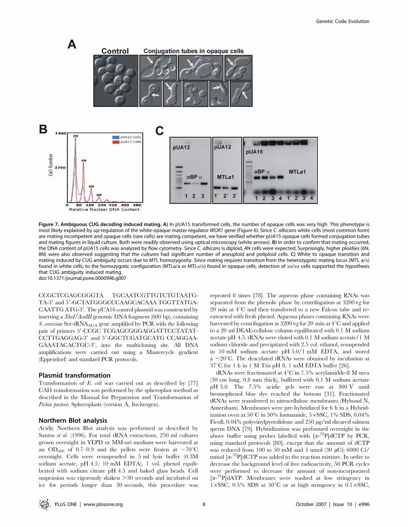
Increased CUG ambiguity increased C. albicans
ploidyUp-regulation of the WOR1 gene and the high percentage of
opaque cells (mating competent) observed in pUA15 clones
prompted the question of whether CUG ambiguity would induce
mating in C. albicans. Indeed, pUA15 opaque cells formed
conjugation tubes and mating figures which were readily observed
by optical microscopy (Figure 7A). Furthermore, flow cytometry
analysis of pUA15 clones identified sub-populations of 4N, 6N and
8N cells (Figure 7B), supporting the hypothesis that ambiguous C.
albicans mated at high frequency. Since C. albicans is a diploid
organism with a sexual life cycle [51], and considering that its
mating locus (MTL) is heterozygotic in mating incompetent white
cells (MTLa/a) and homozygotic in mating competent opaque cells
(MTLa/a or MTLa/a) [52], the latter were isolated from pUA15
clones and were analysed for MTL homozygozity. All clones
analysed were MTLa/a, suggesting that expression of tRNALeu
(pUA15) induced biased MTLa/a homozygoty (Figure 7C), creating
an excess of MTLa/a over MTLa/a cells. These results were in good
agreement with up-regulation of the WOR1 gene as its expression is
controlled by the MTLa1-a2 heterodimer, which, in turn, controls
white-opaque transition and mating competence [46,47]. That is,
switching from white-to-opaque phase required conversion of
heterozygotic MTL to homozygotic MTL inducing mating
competence [52]. Therefore, it is likely that MTL homozygoty
induced by tRNALeu derepressed the WOR1 gene, which, in turn,
promoted the white-to-opaque transition and mating.
The presence of cells with high ploidy (6N and 8N) in pUA15
opaque cultures (Figure 7B), prompted us to monitor ploidy
variability. Most clones showed increased ploidy ranging from 4N
to 8N (data not shown), however very large cells with remarkably
high ploidy (.64N) were also observed (Figure 8A). In general,
ploidy variability between clones was higher than previously
described [53,54] and raised the question of whether those high
chromosome numbers could return to 2N over time. Since wild
Figure 3. Expression of S. cerevisiae tRNAleu in C. albicans. A) Aminoacylation in vivo in C. albicans of S. cerevisiae tRNAUAG/CAGLeu and tRNAAGA
Ser
was monitored by Acidic Page and Northern Blot analysis. For this, total tRNAs were extracted under acidic conditions from pUA13, pUA14, pUA15,and pUA16 clones and fractionated on an acidic polyacrylamide gel, as described in materials and methods. These gels separated deacylated (-AA)from aminoacylated tRNAs (+AA), which were detected using a tDNALeu/Ser probe labeled with [32P]. B) Transformation efficiencies of plasmidsencoding tRNALeu, which decoded the C. albicans serine CUG codons as leucine, was significantly lower that that of control plasmids (pUA12 andpUA16), indicating that the leucine tRNAs were slightly toxic. C) However, clones that survived the transformation procedure adapted readily to CUGambiguity and showed growth rates similar to control clones (pUA12).doi:10.1371/journal.pone.0000996.g003
Genetic Code Evolution
PLoS ONE | www.plosone.org 4 October 2007 | Issue 10 | e996
type C. albicans undergoes a process of chromosome loss after
mating, which decreases its ploidy from 4N back to 2N [55–57],
we hypothesized that high ploidy in pUA15 clones could also be
reduced. To test this hypothesis, clones with very high ploidy (large
cells) were successively re-plated on fresh agar plates and their
ploidy was monitored over time by flow cytometry. Indeed, ploidy
reverted to 2N or 4N after several passages on fresh agar,
confirming the above hypothesis (Figure 8B).
The above results also prompted us to check whether
transformation of C. albicans with the pUA15 plasmid destabilized
its karyotype. The C. albicans karyotype is characterized by frequent
chromosome rearrangements, in particular of the chromosome R,
which contains rDNA cistrons [58,59]. We wondered whether the
tRNALeu would affect rRNA metabolism and protein synthesis.
Indeed, various rearrangements of the R-chromosome were readily
observed (Figure 8C). In particular, the size of R-chromosomes
increased in some clones, decreased in others and these rearrange-
ments affected most cells (Figure 8C). It will now be most interesting
to investigate whether CUG ambiguity affects ribosome assembly
and the rate of protein synthesis.
DISCUSSIONThe identity of CUG codons is variable in the genus Candida.
Indeed, C. glabrata decodes CUGs as leucine, C. cylindracea changed
their identity completely to serine and the other Candida species
decode them ambiguously [13,25,60,61]. These differences in
CUG decoding arose from differences in the structure of the
tRNACAGSer, which is the only cognate tRNA for CUG codons in
Candida. Indeed, the various tRNACAGSer have identity determi-
nants for both SerRS and LeuRS [1]. For example, the C. albicans
tRNACAGSer contains identity elements for the LeuRS, namely
m1G37 and the middle base of the anticodon (A35), but the
discriminator base at position 73 (G73) is specific for SerRS and
not for LeuRS (A73 discriminator) [62]. This base is critical for the
recognition of tRNAs by aminoacyl-tRNA synthetases (aaRSs) and
one would expect that the LeuRS would not recognize tRNAs
with G73. Therefore, charging the tRNACAGSer with leucine [25]
suggests that the Candida LeuRS may have evolved a unique mode
of recognition of its cognate tRNALeu. Interestingly, the presence
of a unique guanosine in the turn of the anticodon-loop (G-turn) of
the tRNACAGSer (G33), a conserved position occupied by U33 (U-
turn), reduced leucylation efficiency of the tRNACAGSer [25]. That
is, recognition of the ancestral tRNACAGSer by the LeuRS was
efficient and G33 acted as a leucine identity anti-determinant. The
reason for G33 selection is not yet clear, but one possibility is that it
may have decreased the toxicity of the mutant tRNACAGSer during
the early stages of CUG identity alteration [27,63].
The dual recognition of the tRNACAGSer by the LeuRS and
SerRS indicates that there are two forms of the tRNACAGSer in the
cytoplasm of C. albicans, namely Ser-tRNACAGSer and Leu-
tRNACAGSer, which are charged with serine and leucine, re-
spectively. These 2 tRNAs generate ambiguity at CUG codons
since they compete with each other for CUGs at the ribosome A-
site. Interestingly, such CUG ambiguity was not constant over the
272625My of evolution of the genetic code alteration (see
introduction) [30]. It was high during the early stages of CUG
identity change (when the tRNACAGSer gene appeared), and
Figure 4. Ambiguous CUG decoding triggered morphogenesis and phenotypic switching. A) Smooth colony morphology of control clonesgrowing on MM-uri-phloxin B (50mg/ml) agar plates. B) Ambiguous pUA15 clones formed long hyphae, even in absence of external inducers, justgrowing in MM-uri agar plates at 30uC. Similar results were obtained for pUA13 and pUA14 ambiguous clones (data not shown). D). Expression of S.cerevisiae tRNALeu in C. albicans also induced phenotypic switching, which is characterized by transition between different cell-phase forms, namelywhite-opaque and myceliated-unmyceliated, giving rise to sectored colonies. D) Phenotypic switching was quantified by counting sectored coloniesgrown in MM-uri, after 7 days of incubation at 30uC. For each plasmid, up to 10 clones were plated and 3000 colonies were screened.doi:10.1371/journal.pone.0000996.g004
Genetic Code Evolution
PLoS ONE | www.plosone.org 5 October 2007 | Issue 10 | e996

decreased gradually due to elimination of the tRNALeu gene from
the genome of the Candida ancestor [30]. Reconstruction of the
high level of CUG ambiguity, which existed during the early stages
of CUG identity alteration, provided the first insight on how the
genetic chaos created at the onset of CUG identity change may
have generated phenotypic diversity of evolutionary and adaptive
relevance. In extant C. albicans, morphological variation alters cell
surface antigens and it is likely that this pathogen uses its
sophisticated capacity to generate morphological variation as
a strategy to escape the immune system. Furthermore, secreted
proteinases and phospolipases are important C. albicans virulence
attributes and their increased activity in pUA15 clones may also be
relevant to virulence. It will now be interesting to engineer stable
high level mistranslation in C. albicans and test the virulence of the
recombinant strains in mice models.
The high phenotypic diversity of pUA13, pUA14 and pUA15
clones and constant transition between phenotypes prevented us
from carrying out a detailed study of the impact of CUG
ambiguity on gene expression. However, the strong up-regulation
of the hyphal specific gene (HWP1) and of the master regulator of
the white-opaque transition (WOR1), may explain, at least in part,
some of the phenotypes observed. As a hyphal specific gene
(HSG), high HWP1 expression, induced by CUG ambiguity, is in
agreement with spontaneous morphogenesis events that generate
filamentous cell populations. Furthermore, HWP1 up-regulation
may also increase adhesion since Hwp1p is a glycosylphosphatidy-
linositol cell wall adhesin (GPI-CWP) and mediates attachment of
C. albicans cells to human endothelial and epithelial cells [44]. The
observed adhesion phenotype (Figure 5A) may have also resulted
from the combined up-regulation of HWP1 and WOR1 genes,
since the later, previously known as EAP2 (enhanced adhesion to
polystyrene), mediates C. albicans and S. cerevisiae adhesion to
polystyrene and epithelial cells [41].
Apart from its putative role in adhesion, up-regulation of WOR1
may also explain the white-opaque phenotype since Wor1p is
a transcriptional regulator of white-opaque switching [46,47].
Indeed, Wor1p is present in very low amounts in white cells and
accumulates in opaque cells. WOR1 is repressed by the hetero-
dimer MTL a1/a2 and is activated by Wor1p itself when cells
become homozygous MTL aa/aa or MTL aa/aa. Increased
accumulation of Wor1p triggered white-opaque switching and
repressed its own transcription by a feedback regulatory
mechanism [46,47]. The up-regulation of the HGC1 gene
(Figure 6), which is a hypha-specific gene encoding a G1 cyclin-
related protein, that plays a role in hyphal morphogenesis, was also
significant. Since it is transcriptionally regulated by hypha-
inducing rather than cell cycle signals [50], its up-regulation in
ambiguous cells supports the hypothesis that it functions in-
dependently of other cell cycle cyclins.
CUG ambiguity also generated karyotype alterations and
formation of polyploids and aneuploids. Ploidy-shift has been
associated with chromosomal rearrangements [53,54,64] which
also generates morphological variation [65–67], antifungal re-
sistance [68], adaptation to alternative carbon sources [69,70], or
even with homozigosity of chromosome-V [71]. This suggests that
part of the phenotypic diversity observed in pUA13, pUA14 and
pUA15 clones may have resulted from karyotype alterations, or
from a combination of up-regulation of the genes described above
Figure 5. Increased CUG ambiguity resulted in higher hydrolytic activity and increased adhesion. A) Highly ambiguous cells (pUA15) exhibitedstrong adhesion phenotypes both in solid and liquid media. Adhesion to the solid agar surface resulted from cell-cell and cell-agar adhesion. In liquidmedia, cells showed a strong flocculation phenotype and sedimented even when grown with agitation (30uC for 2 days). B–C) Cells transformed withpUA13-14 (data not shown) and with pUA15 plasmids, had higher SAP and phospholipase activity than control cells, as determined by hydrolysis ofBSA and egg yolk, respectively. Hydrolytic activity was quantified by measuring precipitation zones formed around the colonies, corrected by thecolony diameter, in order to obtain Pz values.doi:10.1371/journal.pone.0000996.g005
Genetic Code Evolution
PLoS ONE | www.plosone.org 6 October 2007 | Issue 10 | e996

and karyotype destabilization. Since some clones did not have
karyotype alterations (Figure 8C), but still displayed phenotypic
variability, it is likely that the former does not play a main role in
expression of phenotypic diversity. However, one should not
exclude the hypothesis that genome destabilization contributes to
exposure of hidden phenotypes through ambiguous CUG
decoding. Finally, in E. coli, genetic code ambiguity induced by
misreading tRNAs triggered translational stress-induced mutagen-
esis (TSM), due to synthesis of error-prone DNA polymerases [72].
This general mistranslation resulted in increased global error rates
during DNA replication leading to heritable genetic changes.
Since these hypermutagenic phenotypes result in rapid adaptation,
an unexpected consequence of genetic code ambiguity (and
genetic code alterations) is acceleration of genome variability
and fast evolution of new phenotypes. This may explain evolution
of the high plasticity of Candida morphology and the very high
heterozigosity of its genome [73].
ConclusionsGenetic code alterations pose important new biological questions
whose answers remain elusive. It is now clear that a number of
them evolved through codon decoding ambiguity, required
significant structural change of protein synthesis machineries and
reprogrammed codon usage [1,5,30]. However, codon decoding
ambiguity is toxic, decreases fitness and may ultimately lead to cell
death, as is the case in multicellular organisms [19,74]. For these
reasons, evolution of genetic code alterations through codon
ambiguity is most puzzling. This study unveiled possible ways of
overcoming the negative impact of codon ambiguity by high
selective potential generated through generation of phenotypic
diversity. The molecular mechanism used to generate such
phenotypic diversity remains to be elucidated. However, the
adaptive potential of the unveiled phenotypes strongly suggests
that CUG ambiguity may have been preserved in Candida spp. as
a novel generator phenotypic diversity. We have previously shown
that codon ambiguity in S. cerevisiae creates a competitive edge
under stress by inducing the general stress response and pre-
adapting cells to sudden environmental changes [75]. Therefore,
the toxicity of codon ambiguity is not an impediment to codon
identity redefinition, supporting the hypothesis that codon mis-
reading plays a critical role in the evolution of genetic code
alterations and genetic code expansion.
MATERIALS AND METHODS
Strains and growth conditionsEscherichia coli strain JM109 (recA1 SupE44 endA1 hsdR17
gyrA96 relA1 thi D(Lac-proAB) F’[traD36 proAB-lacI lacZ
DM15] was used has a host for all DNA manipulations. Candida
albicans CAI4 (ura3D::imm434/ura3::imm434) was grown at
30uC in YEPD (2% glucose; 1% yeast extract, 1% peptone). After
transformation with pUA12, pUA13, pUA14, pUA15, and
pUA16 plasmids, cells were grown in minimal medium lacking
uridine (MM-uri) (0.67% yeast nitrogen base without amino acids,
2% glucose, 2% agar and 100mg/ml of the required amino acids).
Solid MM-uri was supplemented with phloxin B (50mg/ml) in
order to detect macroscopic growth of opaque cells.
PlasmidsA multi-cloning site was inserted (NruI/EcoRV) in the low-copy C.
albicans vector pRM1 [76]. The resulting vector was named pUA12.
For heterologous expression of the S. cerevisiae tRNAs genes in CAI4,
a genomic DNA fragment containing S. cerevisiae tRNA gene
(700 bp), previously amplified by PCR, was cloned into the multi
cloning site of pUA12 plasmid. S. cerevisiae Leu-tRNAUAG and
a 300 bp flanking region, upstream and downstream of the gene, was
amplified with the following set of primers: 59-CCGCTCGAGC-
GGCGACTGTCCAGACTTAGTAAAG CT-39 and 59-GCTC-
TAGAGCCCGCTGTCGCCAGCGTTAGC-39. Genomic DNA
containing S. cerevisiae tRNAGAGLeu gene was used as a template for
PCR amplification using the forward primers: 59-GCTATGGGC-
CCGCCTCCGGGTAGTTGCAACGGTACTCTGG CCGAG-
TGGTCTAAGGCG-39 and 59-GCTATGGGCCCTAGTTGCA-
ACGG TATCTGGCCGAGTGGTCTAAGGCGTCAGGTTC-
AGGTCC-39; and reverse primer 59-ATGCATAAAAACAAAA-
TTTGTTGAAA-39. These primers introduced a mutation in the
first position of the anticodon changing it from 59-GAG-39 to 59-
CAG-39 and allowed insertion of G or T at position 33 of the tRNA
anticodon-loop. Upstream of this gene, a 250 bp fragment with the
same sequence of the 59 flanking C. albicans Ser-tRNACAG gene, was
also inserted. This fragment was amplified by PCR from a XhoI/ApaI
genomic DNA fragment, with the following primers: 59-
Figure 6. Increased CUG ambiguity up-regulated morphogenesisgenes. Cells of pUA15 clones showed significant up-regulation of theWOR1 (7.062.5) gene and hyphal-specific genes CaHWP1 (41.7669.96)and HGC1 (2.6461.12). Since WOR1 increases the frequency of thewhite-opaque transition the very high percentage of opaque cellsfound in transformed clones may be a consequence of WOR1 up-regulation. On the other hand, expression of the hypha-specific genes,CaHWP1 and CaHGC1, supported the hypothesis that morphogenesisand hyphal growth triggered by CUG ambiguity resulted fromexpression of morphogenesis regulators. Induction of the CaHWP1gene was accompanied by repression of the CaMCM1 (21.8460.44)gene, which controls cell morphology through the recruitment of othermorphogenesis regulatory factors.doi:10.1371/journal.pone.0000996.g006
Genetic Code Evolution
PLoS ONE | www.plosone.org 7 October 2007 | Issue 10 | e996
CCGCTCGAGCGGGTA TGCAATCGTTGTCTGTAATG-
TA-39 and 59-GCTATGGGCCCAAGCACAAA TGGTTATGA-
CAATTG ATG-39. The pUA16 control plasmid was constructed by
inserting a XhoI/AvaIII genomic DNA fragment (600 bp), containing
S. cerevisiae Ser-tRNAAGA gene amplified by PCR with the following
pair of primers 59-CCGC TCGAGCGGGAGGATTCCTATAT-
CCTTGAGGAG-39 and 59-GGCTCGATGCATG CCAGGAA-
GAAATACACTGC-39, into the multicloning site. All DNA
amplifications were carried out using a Mastercycle gradient
(Eppendorf) and standard PCR protocols.
Plasmid transformationTransformation of E. coli was carried out as described by [77]
CAI4 transformation was performed by the spheroplast method as
described in the Manual for Preparation and Transformation of
Pichia pastoris Spheroplasts (version A, Invitrogen).
Northern Blot analysisAcidic Northern Blot analysis was performed as described by
Santos et al. (1996). For total tRNA extractions, 250 ml cultures
grown overnight in YEPD or MM-uri medium were harvested at
an OD600 of 0.7–0.9 and the pellets were frozen at 270uCovernight. Cells were resuspended in 5 ml lysis buffer (0.3M
sodium acetate, pH 4.5, 10 mM EDTA), 1 vol. phenol equili-
brated with sodium citrate pH 4.5 and baked glass beads. Cell
suspension was vigorously shaken .30 seconds and incubated on
ice for periods longer than 30 seconds, this procedure was
repeated 8 times [78]. The aqueous phase containing RNAs was
separated from the phenolic phase by centrifugation at 32006g for
20 min at 4uC and then transferred to a new Falcon tube and re-
extracted with fresh phenol. Aqueous phases containing RNAs were
harvested by centrifugation at 32006g for 20 min at 4uC and applied
to a 20 ml DEAE-cellulose column equilibrated with 0.1 M sodium
acetate pH 4.5. tRNAs were eluted with 0.1 M sodium acetate/1 M
sodium chloride and precipitated with 2.5 vol. ethanol, resuspended
in 10 mM sodium acetate pH 5.0/1 mM EDTA, and stored
a 220uC. The deacylated tRNAs were obtained by incubation at
37uC for 1 h in 1 M Tris pH 8, 1 mM EDTA buffer [26].
tRNAs were fractionated at 4uC in 7.5% acrylamidde-8 M urea
(30 cm long, 0.8 mm thick), buffered with 0.1 M sodium acetate
pH 5.0. The 7.5% acidic gels were run at 300 V until
bromophenol blue dye reached the bottom [31]. Fractionated
tRNAs were transferred to nitrocellulose membranes (Hybond N,
Amersham). Membranes were pre-hybridized for 6 h in a Hybrid-
ization oven at 50uC in 50% formamide, 56SSC, 1% SDS, 0.04%
Ficoll, 0.04% polyvinylpyrrolidone and 250 mg/ml sheared salmon
sperm DNA [79]. Hybridization was performed overnight in the
above buffer using probes labelled with [a-32P]dCTP by PCR,
using standard protocols [80], except that the amount of dCTP
was reduced from 100 to 50 mM and 5 nmol (30 mCi) 6000 Ci/
mmol [a-32P]dCTP was added to the reaction mixture. In order to
decrease the background level of free radioactivity, 50 PCR cycles
were performed to decrease the amount of non-incorporated
[a-32P]dATP. Membranes were washed at low stringency in
16SSC, 0.5% SDS at 50uC or at high stringency in 0.16SSC,
Figure 7. Ambiguous CUG decoding induced mating. A) In pUA15 transformed cells, the number of opaque cells was very high. This phenotype ismost likely explained by up-regulation of the white-opaque master regulator WOR1 gene (Figure 6). Since C. albicans white cells (most common form)are mating incompetent and opaque cells (rare cells) are mating competent, we have verified whether pUA15 opaque cells formed conjugation tubesand mating figures in liquid culture. Both were readily observed using optical microscopy (white arrows). B) In order to confirm that mating occurred,the DNA content of pUA15 cells was analyzed by flow cytometry. Since C. albicans is diploid, 4N cells were expected. Surprisingly, higher ploidies (6N,8N) were also observed suggesting that the cultures had significant number of aneuploid and poliploid cells. C) White to opaque transition andmating induced by CUG ambiguity occurs due to MTL homozygosity. Since mating requires transition from the heterozygotic mating locus (MTL a/a)found in white cells, to the homozygotic configuration (MTLa/a or MTLa/a) found in opaque cells, detection of aa/aa cells supported the hypothesisthat CUG ambiguity induced mating.doi:10.1371/journal.pone.0000996.g007
Genetic Code Evolution
PLoS ONE | www.plosone.org 8 October 2007 | Issue 10 | e996

0.5% SDS at 65uC for 1 h. The membranes were exposed
overnight with intensifying screens and developed using a Molec-
ular Imager FX (Biorad).
Switching frequencies and phenotypic
characterizationC. albicans grown overnight at 30uC in MM-uri were serially
diluted to 1000 cells per ml. Approximately 50 cells were plated
onto fresh agar plates and then allowed to grow at 30uC for 7 days
in a humidified incubator to prevent drying of the agar surface.
Sectored colonies displaying atypical colonies were scored and the
data was analyzed for statistical significance using ANOVA.
Colonies were photographed using a Stemi 2000-C dissecting
microscope equipped with AxioCam HRc camera and Axio
Vision Software from Zeiss. Cells were photographed using a Zeiss
MC80 Axioplan 2 light microscope.
Real Time RT-PCRTotal RNA was prepared from C. albicans using hot acid phenol
[81]. First-strand cDNA synthesis was carried out using the
Superscript II RT kit from Invitrogen and its quantification was
carried out in an Applied Biosystems 7500 Real Time PCR system
using the SYBR Green I dye quantification assay (Power SYBR
Green PCR master Mix). Primer concentrations were tested (0,2
to 0,4 mM) to ensure the lowest threshold cycle (CT) and the
highest signal magnitude against the target template and to ensure
non-specific product formation, resulting from primer dimmeriza-
tion. After amplification, reactions were checked for presence of
non-specific products through dissociation curve analysis. Each
gene was quantified using 9 replicas of both control (pUA12) and
ambiguous (pUA15) clones and a mean value was calculated.
Outliers were rejected using critical values of Dixon’s ‘‘Q’’
parameter at 95% confidence level [82].
Determination of extra cellular hydrolytic activityC. albicans strains were screened for the production of extra cellular
phospholipase and secreted aspartic proteinase activity by growing
cells on MM-uri agar supplemented with 10% egg yolk (Merck)
and 10% bovine albumin (Sigma), respectively. A 362 ml
suspension of 107cells/ml in PBS was plated on the surface of
the agar medium and left to dry at room temperature. The culture
Figure 8. Ambiguous CUG decoding induced karyotype rearrangements and ploidy-shift. A) In ambiguous cell lines (pUA15) polyploidy waspredominant and very high ploidy was often detected (.32N). Aneuploidy was also observed (6N). B) However, after plating cells several consecutivetimes on fresh agar chromosome numbers were reduced indicating that most cells returned to low ploidy (2N or 4N). Ploidy reduction after matingnormally occurs by chromosome loss in C. albicans and it is likely that such mechanism also played a role in ploidy reduction in pUA15 transformedcells. C) CUG ambiguity also promoted extensive rearrangements of the R-chromosome (highlighted in white circles). Chromosomes were separatedby PFGE on 0.6% agarose gels under the following conditions: 120–300 s for 24h at 80 V, then 420–900 s for 48 h at 80 V. The numbers 1–7 and Ridentify C. albicans chromosomes.doi:10.1371/journal.pone.0000996.g008
Genetic Code Evolution
PLoS ONE | www.plosone.org 9 October 2007 | Issue 10 | e996
was then incubated at 30uC for 3 days, after which the diameter of
the colony and the precipitation zone around the colony was
determined. Three different clones of pUA12, pUA13, pUA14
and pUA15 transformed cells were tested twice. The experiment
was carried out on two different occasions. The extra cellular
hydrolase activity was calculated using the formula [(1/Pz)-1],
where Pz value represents the hydrolyse zone, i. e., the
cloudy-zone-around-plus-colony diameter divided by the colony
diameter [83]. Data obtained was submitted to an ANOVA
statistical test.
Genome analysisDNA content of C. albicans cells was determined using FACS
analysis [84]. Karyotype analysis was performed using Pulsed-
Field Gel Electrophoresis (PFGE) [85].
MTL analysisPCR analysis of the MTL configuration was carried out in pUA12
and pUA15 cells, through amplification of oBPa and MTLa1
genes, respectively, using the following pairs of primers: 59
GTGGTCAATGGAGCTGATAC 39and 59 ACATGTGGTC-
GCCCAACTCC 39; 59 TTGAAGCGTGAGAGGCAGGAG 39
and 59 ATCAATTCCCTTTCTCTTCGATTAGG 39.
ACKNOWLEDGMENTSWe are most grateful to Jorge Rino for helping with the light microscopy
studies, Alexander Johnson for providing oBP primers and Concha Gil for
the pRM1 plasmid.
Author Contributions
Conceived and designed the experiments: MS. Performed the experiments:
GM RR IM MS DM. Analyzed the data: MS GM IM LC. Contributed
reagents/materials/analysis tools: MS. Wrote the paper: MS IM.
REFERENCES1. Santos MA, Moura G, Massey SE, Tuite MF (2004) Driving change: the
evolution of alternative genetic codes. Trends Genet 20: 95–102.
2. Miranda I, Silva R, Santos MA (2006) Evolution of the genetic code in yeasts.
Yeast 23: 203–213.
3. Knight RD, Landweber LF, Yarus M (2001) How mitochondria redefine the
code. J Mol Evol 53: 299–313.
4. Castresana J, Feldmaier-Fuchs G, Paabo S (1998) Codon reassignment and
amino acid composition in hemichordate mitochondria. Proc Natl Acad Sci U S A
95: 3703–3707.
5. Knight RD, Freeland SJ, Landweber LF (2001) Rewiring the keyboard:
evolvability of the genetic code. Nat Rev Genet 2: 49–58.
6. Lovett PS, Ambulos NP Jr, Mulbry W, Noguchi N, Rogers EJ (1991) UGA can
be decoded as tryptophan at low efficiency in Bacillus subtilis. J Bacteriol 173:
1810–1812.
7. Caron F, Meyer E (1985) Does Paramecium primaurelia use a different genetic
code in its macronucleus? Nature 314: 185–188.
8. Harper DS, Jahn CL (1989) Differential use of termination codons in ciliated
protozoa. Proc Natl Acad Sci U S A 86: 3252–3256.
9. Sanchez-Silva R, Villalobo E, Morin L, Torres A (2003) A new noncanonical
nuclear genetic code: translation of UAA into glutamate. Curr Biol 13: 442–447.
10. Le Mouel A, Butler A, Caron F, Meyer E (2003) Developmentally regulated
chromosome fragmentation linked to imprecise elimination of repeated
sequences in paramecia. Eukaryot Cell 2: 1076–1090.
11. Tuite MF, Santos MA (1996) Codon reassignment in Candida species: an
evolutionary conundrum. Biochimie 78: 993–9.
12. Jukes TH, Osawa S (1996) CUG codons in Candida spp. J Mol Evol 42: 321–322.
13. Sugita T, Nakase T (1999) Non-universal usage of the leucine CUG codon and
the molecular phylogeny of the genus Candida. Syst Appl Microbiol 22: 79–86.
14. Hanyu N, Kuchino Y, Nishimura S, Beier H (1986) Dramatic events in ciliate
evolution: alteration of UAA and UAG termination codons to glutamine codons
due to anticodon mutations in two Tetrahymena tRNAs. EMBO J 5:
1307–1311.
15. Hao B, Gong W, Ferguson TK, James CM, Krzycki JA, et al. (2002) A new
UAG-encoded residue in the structure of a methanogen methyltransferase.
Science 296: 1462–1466.
16. Lee BJ, Worland PJ, Davis JN, Stadtman TC, Hatfield DL (1989) Identification
of a selenocysteyl-tRNA(Ser) in mammalian cells that recognizes the nonsense
codon, UGA. J Biol Chem 264: 9724–9727.
17. Bacher JM, Ellington AD (2001) Selection and characterization of Escherichia
coli variants capable of growth on an otherwise toxic tryptophan analogue.
J Bacteriol 183: 5414–5425.
18. Bacher JM, Crecy-Lagard V, Schimmel PR (2005) Inhibited cell growth and
protein functional changes from an editing-defective tRNA synthetase. Proc Natl
Acad Sci U S A 102: 1697–1701.
19. Nangle LA, Motta CM, Schimmel P (2006) Global effects of mistranslation from
an editing defect in mammalian cells. Chem Biol 13: 1091–1100.
20. Chin JW, Cropp TA, Anderson JC, Mukherji M, Zhang Z, et al. (2003) An
expanded eukaryotic genetic code. Science 301: 964–967.
21. Hendrickson TL, Crecy-Lagard V, Schimmel P (2004) Incorporation of
nonnatural amino acids into proteins. Annu Rev Biochem 73: 147–176.
22. Santos MA, Perreau VM, Tuite MF (1996) Transfer RNA structural change is
a key element in the reassignment of the CUG codon in Candida albicans.
Embo J 15: 5060–8.
23. Santos MA, Tuite MF (1995) The CUG codon is decoded in vivo as serine and
not leucine in Candida albicans. Nucleic Acids Res 23: 1481–1486.
24. Ohama T, Suzuki T, Mori M, Osawa S, Ueda T, et al. (1993) Non-universal
decoding of the leucine codon CUG in several Candida species. Nucleic Acids
Res 21: 4039–45.
25. Suzuki T, Ueda T, Watanabe K (1997) The ‘polysemous’ codon--a codon with
multiple amino acid assignment caused by dual specificity of tRNA identity.
Embo J 16: 1122–34.
26. Santos MA, Keith G, Tuite MF (1993) Non-standard translational events inCandida albicans mediated by an unusual seryl-tRNA with a 59-CAG-39
(leucine) anticodon. Embo J 12: 607–16.
27. Perreau VM, Keith G, Holmes WM, Przykorska A, Santos MA, et al. (1999)The Candida albicans CUG-decoding ser-tRNA has an atypical anticodon stem-
loop structure. J Mol Biol 293: 1039–1053.
28. Schultz DW, Yarus M (1996) On malleability in the genetic code. J Mol Evol 42:597–601.
29. Schultz DW, Yarus M (1994) Transfer RNA mutation and the malleability of the
genetic code. J Mol Biol 235: 1377–1380.
30. Massey SE, Moura G, Beltrao P, Almeida R, Garey JR, et al. (2003)Comparative evolutionary genomics unveils the molecular mechanism of
reassignment of the CTG codon in Candida spp. Genome Res 13: 544–557.
31. Varshney U, Lee CP, RajBhandary UL (1991) Direct analysis of aminoacylationlevels of tRNAs in vivo. Application to studying recognition of Escherichia coli
initiator tRNA mutants by glutaminyl-tRNA synthetase. J Biol Chem 266:24712–24718.
32. O’Sullivan JM, Davenport JB, Tuite MF (2001) Codon reassignment and the
evolving genetic code: problems and pitfalls in post-genome analysis. Trends
Genet 17: 20–22.
33. O’Sullivan JM, Mihr MJ, Santos MA, Tuite MF (2001) Seryl-tRNA synthetase is
not responsible for the evolution of CUG codon reassignment in Candida
albicans. Yeast 18: 313–322.
34. Soll DR (2002) Phenotypic Switching. Candida and Candidiasis. pp 123–142.
35. Hoyer LL (2001) The ALS gene family of Candida albicans. Trends Microbiol 9:
176–80.
36. Fu Y, Rieg G, Fonzi WA, Belanger PH, Edwards JE, et al. (1998) Expression ofthe Candida albicans gene ALS1 in Saccharomyces cerevisiae induces
adherence to endothelial and epithelial cells. Infect Immun 66: 1783–1786.
37. Rauceo JM, Gaur NK, Lee KG, Edwards JE, Klotz SA, et al. (2004) Global cell
surface conformational shift mediated by a Candida albicans adhesin. InfectImmun 72: 4948–4955.
38. Gaur NK, Smith RL, Klotz SA (2002) Candida albicans and Saccharomyces
cerevisiae expressing ALA1/ALS5 adhere to accessible threonine, serine, oralanine patches. Cell Commun Adhes 9: 45–57.
39. Gaur NK, Klotz SA, Henderson RL (1999) Overexpression of the Candida
albicans ALA1 gene in Saccharomyces cerevisiae results in aggregation followingattachment of yeast cells to extracellular matrix proteins, adherence properties
similar to those of Candida albicans. Infect Immun 67: 6040–6047.
40. Gaur NK, Klotz SA (1997) Expression, cloning, and characterization of a Candidaalbicans gene, ALA1, that confers adherence properties upon Saccharomyces
cerevisiae for extracellular matrix proteins. Infect Immun 65: 5289–5294.
41. Li F, Palecek SP (2003) EAP1, a Candida albicans gene involved in bindinghuman epithelial cells. Eukaryot Cell 2: 1266–1273.
42. Ibrahim AS, Mirbod F, Filler SG, Banno Y, Cole GT, et al. (1995) Evidence
implicating phospholipase as a virulence factor of Candida albicans. InfectImmun 63: 1993–8.
43. Naglik JR, Challacombe SJ, Hube B (2003) Candida albicans Secreted Aspartyl
Proteinases in Virulence and Pathogenesis. Microbiol Mol Biol Rev 67:400–428.
Genetic Code Evolution
PLoS ONE | www.plosone.org 10 October 2007 | Issue 10 | e996
44. Sundstrom P (2002) Adhesion in Candida spp. Cell Microbiol 4: 461–469.
45. Ghannoum MA (2000) Potential role of phospholipases in virulence and fungal
pathogenesis. Clin Microbiol Rev 13: 122–43.
46. Zordan RE, Galgoczy DJ, Johnson AD (2006) Epigenetic properties of white-
opaque switching in Candida albicans are based on a self-sustaining
transcriptional feedback loop. Proc Natl Acad Sci U S A 103: 12807–12812.
47. Huang G, Wang H, Chou S, Nie X, Chen J, et al. (2006) Bistable expression of
WOR1, a master regulator of white-opaque switching in Candida albicans. Proc
Natl Acad Sci U S A 103: 12813–12818.
48. Staab JF, Bradway SD, Fidel PL, Sundstrom P (1999) Adhesive and mammalian
transglutaminase substrate properties of Candida albicans Hwp1. Science 283:
1535–8.
49. Rottmann M, Dieter S, Brunner H, Rupp S (2003) A screen in Saccharomyces
cerevisiae identified CaMCM1, an essential gene in Candida albicans crucial for
morphogenesis. Mol Microbiol 47: 943–959.
50. Zheng X, Wang Y, Wang Y (2004) Hgc1, a novel hypha-specific G1 cyclin-
related protein regulates Candida albicans hyphal morphogenesis. EMBO J 23:
1845–1856.
51. Hull CM, Johnson AD (1999) Identification of a mating type-like locus in the
asexual pathogenic yeast Candida albicans. Science 285: 1271–5.
52. Miller MG, Johnson AD (2002) White-opaque switching in Candida albicans is
controlled by mating-type locus homeodomain proteins and allows efficient
mating. Cell 110: 293–302.
53. Iwaguchi SI, Kanbe T, Tohne T, Magee PT, Suzuki T (2000) High-frequency
occurrence of chromosome translocation in a mutant strain of Candida albicans
by a suppressor mutation of ploidy shift. Yeast 16: 411–422.
54. Suzuki T, Hitomi A, Magee PT, Sakaguchi S (1994) Correlation between
polyploidy and auxotrophic segregation in the imperfect yeast Candida albicans.
J Bacteriol 176: 3345–3353.
55. Bennett RJ, Johnson AD (2003) Completion of a parasexual cycle in Candida
albicans by induced chromosome loss in tetraploid strains. EMBO J 22:
2505–2515.
56. Magee BB, Magee PT (2000) Induction of mating in Candida albicans by
construction of MTLa and MTLalpha strains. Science 289: 310–3.
57. Hull CM, Raisner RM, Johnson AD (2000) Evidence for mating of the
‘‘asexual’’ yeast Candida albicans in a mammalian host. Science 289: 307–10.
58. Lasker BA, Carle GF, Kobayashi GS, Medoff G (1989) Comparison of the
separation of Candida albicans chromosome-sized DNA by pulsed-field gel
electrophoresis techniques. Nucleic Acids Res 17: 3783–3793.
59. Magee BB, Magee PT (1987) Electrophoretic karyotypes and chromosome
numbers in Candida species. J Gen Microbiol 133: 425–430.
60. Santos MA, Ueda T, Watanabe K, Tuite MF (1997) The non-standard genetic
code of Candida spp.: an evolving genetic code or a novel mechanism for
adaptation? Mol Microbiol 26: 423–431.
61. O’Sullivan JM, Santos MA, Tuite MF (2002) Standard and nonstandard mRNA
decoding in Candida. Candida and Candidiasis. pp 279–292.
62. Breitschopf K, Gross HJ (1994) The exchange of the discriminator base A73 for
G is alone sufficient to convert human tRNA(Leu) into a serine-acceptor in vitro.
EMBO J 13: 3166–3167.
63. Santos MA, Perreau VM, Tuite MF (1996) Transfer RNA structural change is
a key element in the reassignment of the CUG codon in Candida albicans.
EMBO J 15: 5060–5068.
64. Selmecki A, Bergmann S, Berman J (2005) Comparative genome hybridization
reveals widespread aneuploidy in Candida albicans laboratory strains. Mol
Microbiol 55: 1553–1565.
65. Rustchenko-Bulgac EP, Howard DH (1993) Multiple chromosomal and
phenotypic changes in spontaneous mutants of Candida albicans. J GenMicrobiol 139 Pt 6: 1195–1207.
66. Rustchenko-Bulgac EP, Sherman F, Hicks JB (1990) Chromosomal rearrange-
ments associated with morphological mutants provide a means for geneticvariation of Candida albicans. J Bacteriol 172: 1276–83.
67. Suzuki T, Kobayashi I, Kanbe T, Tanaka K (1989) High frequency variation ofcolony morphology and chromosome reorganization in the pathogenic yeast
Candida albicans. J Gen Microbiol 135: 425–434.
68. Perepnikhatka V, Fischer FJ, Niimi M, Baker RA, Cannon RD, et al. (1999)Specific chromosome alterations in fluconazole-resistant mutants of Candida
albicans. J Bacteriol 181: 4041–4049.69. Rustchenko EP, Howard DH, Sherman F (1997) Variation in assimilating
functions occurs in spontaneous Candida albicans mutants having chromosomalalterations. Microbiology 143 ( Pt 5): 1765–1778.
70. Janbon G, Sherman F, Rustchenko E (1998) Monosomy of a specific
chromosome determines L-sorbose utilization: a novel regulatory mechanismin Candida albicans. Proc Natl Acad Sci U S A 95: 5150–5155.
71. Legrand M, Lephart P, Forche A, Mueller FM, Walsh T, et al. (2004)Homozygosity at the MTL locus in clinical strains of Candida albicans:
karyotypic rearrangements and tetraploid formation. Mol Microbiol 52:
1451–1462.72. Dorazi R, Lingutla JJ, Humayun MZ (2002) Expression of mutant alanine
tRNAs increases spontaneous mutagenesis in Escherichia coli. Mol Microbiol44: 131–141.
73. Jones T, Federspiel NA, Chibana H, Dungan J, Kalman S, et al. (2004) Thediploid genome sequence of Candida albicans. Proc Natl Acad Sci U S A 101:
7329–7334.
74. Lee JW, Beebe K, Nangle LA, Jang J, Longo-Guess CM, et al. (2006) Editing-defective tRNA synthetase causes protein misfolding and neurodegeneration.
Nature 443: 50–55.75. Santos MA, Cheesman C, Costa V, Moradas-Ferreira P, Tuite MF (1999)
Selective advantages created by codon ambiguity allowed for the evolution of an
alternative genetic code in Candida spp. Mol Microbiol 31: 937–947.76. Pla J, Gil C, Monteoliva L, Navarro-Garcia F, Sanchez M, et al. (1996)
Understanding Candida albicans at the molecular level. Yeast 12: 1677–702.77. Sambrook J, Fritsch EF, Maniatis T (1989) Molecular cloning: a laboratory
manual. Cold Spring Harbor, N.Y.: Cold Spring Harbor Laboratory Press.78. Weygand-Durasevic I, Nalaskowska M, Soll D (1994) Coexpression of
eukaryotic tRNASer and yeast seryl-tRNA synthetase leads to functional amber
suppression in Escherichia coli. J Bacteriol 176: 232–239.79. Heitzler J, Marechal-Drouard L, Dirheimer G, Keith G (1992) Use of a dot blot
hybridization method for identification of pure tRNA species on differentmembranes. Biochim Biophys Acta 1129: 273–277.
80. Innis MA, Gelfand DH, Sninsky JJ, White TJ (1990) PCR protocols: A Guide to
Methods and Applications.81. Kohrer K, Domdey H (1991) Preparation of high molecular weight RNA.
Methods Enzymol 194: 398–405.82. Rorabacher DB (1991) Statistical Treatment for Rejection of Deviant Values -
Critical-Values of Dixon Q Parameter and Related Subrange Ratios at the 95-Percent Confidence Level. Analytical Chemistry 63: 139–146.
83. Samaranayake LP, Raeside JM, MacFarlane TW (1984) Factors affecting the
phospholipase activity of Candida species in vitro. Sabouraudia 22: 201–207.84. Fortuna M, Sousa MJ, Corte-Real M, Leao C, Salvador A, et al. (2000) Cell
Cycle Analysis of Yeasts. Current Protocols in Cytometry. pp 11.13.1–11.13.9.85. Chu WS, Magee BB, Magee PT (1993) Construction of an SfiI macrorestriction
map of the Candida albicans genome. J Bacteriol 175: 6637–6651.
Genetic Code Evolution
PLoS ONE | www.plosone.org 11 October 2007 | Issue 10 | e996
Copyright © 2022 FDOKUMEN