Five DNA markers predict Legionella pneumophila pathogenicity
Upload
independentCategory
view
0download
0
Chapter 2
Virulence and Pathogenicity of Fungal
Pathogens with Special Reference
to Candida albicans
Mohd Sajjad Ahmad Khan, Iqbal Ahmad, Farrukh Aqil, Mohd Owais,
Mohd Shahid, and Javed Musarrat
Abstract The frequency of severe systemic fungal diseases has increased in the last
few decades. The clinical use of broad spectrum antibacterial drugs and immuno-
suppressive agents after organ transplantation, cancer chemotherapy, and advance-
ments in surgery are associated with increasing risk of fungal infection. Despite the
effectiveness of available antifungals in combating such infections, the emergence
of drug resistance to antifungals, and problems of toxicity and poor delivery of drugs
at the target site in systemic infections, have necessitated a systematic approach to
the study of fungal pathogens, host–fungi interactions, and identification of viru-
lence factors. Characterization of virulence factors is expected to improve under-
standing of fungal pathogenesis and to help explore new drug targets. In this article
we discuss the process of fungal infections, virulence factors and pathogenicity of
fungal pathogens, with special reference to Candida albicans. Adherence, dimor-
phism, phenotypic switching, secretion of hydrolytic enzymes, biofilm formation,
M.S.A. Khan (*) and I. Ahmad
Department of Agricultural Microbiology, Faculty of Agricultural Sciences, Aligarh Muslim
University, Aligarh 202002, India
e-mail: [email protected]
F. Aqil
Brown Cancer Center, University of Louisville, Louisville, KY 40202, USA
M. Owais
Interdisciplinary Biotechnology Unit, Aligarh Muslim University, Aligarh 202002, India
M. Shahid
Department of Microbiology, JN Medical College, Aligarh Muslim University, Aligarh 202002,
India
J. Musarrat
DNA Research Chair, Department of Zoology, King Saud University, Riyadh, Saudi Arabia
I. Ahmad et al. (eds.), Combating Fungal Infections,DOI 10.1007/978-3-642-12173-9_2, # Springer-Verlag Berlin Heidelberg 2010
21

and ability to adapt at host body temperature are some of the well-known virulence
factors among pathogenic fungi and are discussed in relation to C. albicans.
2.1 Introduction
Fungi are eukaryotic microorganisms that are more closely related to humans than
bacteria at cellular level. They belong to the group Eumycota, and are chemoheter-
otrophs with a chitinous cell wall. More than 100,000 species have been described.
Most species grow as multicellular filaments called hyphae-forming mycelium such
as molds; some species also grow as single cells like yeasts. Some groups of fungi
are pathogenic to humans and require control measures. Human fungal pathogens
belong to four main groups, namely zygomcetes, ascomycetes, deuteromycetes,
and basidiomycetes. Fungi can cause significant number of human diseases repre-
sented by pathogens such as Trichophyton sp, Epidermophyton sp, Histoplasma sp,
Blastomyces sp, Sporothrix sp, Coccidioides sp, and Paracoccidioides sp, capable ofinfecting healthy people, or opportunistic invaders such as Aspergillus sp, Candidasp, Cryptococcus sp, Fusarium sp, and Rhizopus sp, which are normally avirulent
in healthy people but could be disseminated to deep tissue and cause fatal disease in
unhealthy people (Chakrabarti 2005; Reedy et al. 2007). The morbidity and mortality
rates caused by fungal species such as Candida, Aspergillus, Fusarium, and Trichos-porum are relatively higher (Fluckiger et al. 2006). In Europe, fungal infections
account for 17% cases associated with intensive care units (Rupp 2007), while in
the USA it has become the seventh most common cause of deaths among hospitalized
patients (Martin et al. 2003). About 15% of allogenic haemopoietic stem cell trans-
plant recipients and 20% of lung transplant recipients suffered fungal infections
(Ribaud et al. 1999). Approximately 60% and 20% of AIDS patients present with
pneumonia and esophageal candidiasis respectively (Moore and Chaisson 1996).
Data from the late 1950s and early 1960s indicate that invasive fungal infections
were extremely rare, even in immunocompromised cancer patients (Chakrabarti
2005). Now, fungal infections have dramatically increased in the past two decades
as a result of improved diagnostics, high frequency of catheterization, instrumenta-
tion and an increasing number of immunosuppressed patients. Particularly, invasive
fungal infections are showing extremely high mortality rate. The use of antineo-
plastic and immunosuppressive agents, broad-spectrum antibiotics, prosthetic
devices and grafts, and more aggressive surgery have led to the development of
complicated infections, including invasive fungal infections. Furthermore, patients
with burns, neutropenia, and HIV infections are now seriously exposed to fungal
infections (Kuleta et al. 2009).
Fungal infections have now also become more common in the healthy population.
The National Nosocomial Infections Surveillance System has reported Candida spp.as the fourth most common bloodstream isolates in nosocomial infections in USA.
Over 95% of all fungal infections have been associated with Candida albicans,Aspergillus fumigates, and Cryptococcus neoformans (Richardson 2005).
22 M.S.A. Khan et al.

2.2 Diseases Caused by Human Pathogenic Fungi
Fungal diseases can be broadly classified on the basis of causative agents as:
(a) dermatophytosis, (b) histoplasmosis, (c) blastomycosis, (d) coccidiomycosis,
(e) candidiasis, (f) cryptococcosis, (g) aspergillosis, (h) hyalohyphomycosis, and
(i) zygomycosis, as described by many authors (Sullivan et al. 2005). These
diseases differ in their nature, causative agents, and distribution. Description of
such fungal diseases, their causative agents and major organs involved etc are given
in Table 2.1. However, candidiasis is described here briefly. Candidiasis encom-
passes secondary or opportunistic infections ranging from acute, sub-acute, and
chronic to life-threatening mycoses. Infections are localized to mouth, throat, skin,
vagina, fingers, bronchi, lungs, and gastrointestinal tract, or sometimes become
Table 2.1 Examples of commonly caused fungal diseases
Fungal diseases Causative agent Site of infection Transmission
Dermatophytosis T. rubrum,T. mentagrophytes,E. floccosusm,M. gypseum,M. canis
Skin, hair, nails, feet Soil, contact with
arthrospores or
conidia from
contaminated
animals and
humans
Histoplasmosis H. capsulatum Lungs Soil, inhalation of
microconidia
Blastomycosis B. dermatididis Lungs, skin, genitourinary
tract, brain
Soil, inhalation of
conidia
Coccidioidomycosis C. immitis,C. posadasii
Lungs, bones, joints,
meninges
Soil, inhalation of
arthroconidia
Candidiasis C. albicans,C. tropicalis,C. glabrata,C. dubliniensis,C. krusei
Intestinal tract, vaginal
tract, skin, fingers,
oral cavity
Endogenous flora,
contact with
secretions from
infected person
Cryptococcosis C. neoformans Lungs, meninges, kidney,
liver, prostate, bones
Soil, contamination
with bird feces
Aspergillosis A. fumigatus, A. flavus,A. niger
Lungs Soil, inhalation of
spores
Hyalohyphomycosis Fusarium sp.,
Phecilomyces sp,Scedosporium sp,Scopulariopsis sp,and Acremonium sp
Keratin, nails, lungs Soil, plant debris,
ingestion of toxin
contaminated
plant parts
Zygomycosis Rhizopus sp, Mucor spand Absidia sp
Skin, cerebral, blood,
lungs, genitourinary
and gastrointestinal
system
Soil, decaying plant
material,
inhalation or
percutaneous
contact of spores
Adapted from Weitzman and Summerbell (1995), Hogan et al. (1996), Pommerville (2004),
Rappleye and Goldman (2006), Willey et al. (2008)
2 Virulence and Pathogenicity of Fungal Pathogens 23

systemic as candidemia, endocarditis, and meningitis. A number of Candida spp areencountered in candidiasis such as C. albicans, C. glabrata, C. tropicalis, C. krusei,C. dubliniensis, C. parapsilosis (Hayens and Westerneng 1996). C. albicans is amember of the commensal microflora of the intestine. It is pleomorphic and under-
goes reversible morphogenic transitions between budding yeast, pseuodohyphal,
and hyphal growth forms. Healthy persons generally encounter superficial infec-
tions but in immunocompromised patients invasive infections could also occur.
Approximately 70% of woman experience vaginal candidiasis once in a life,
and 20% suffer from recurrence (Fidel et al. 1999). Among other Candida spp,
C. glabrata has emerged as a frequent pathogen due to increased use of immune
suppressive agents. C. krusei is a pathogen of importance in patients with hemato-
logical malignancies and transplants. C. parapsilosis is frequently isolated from
blood cultures due to insertive medical devices. C. tropicalis is one of the causativeagents of candidemia and isolated from patients with leukemia and those who have
undergone bone-marrow transplantation. C. dubliniensis is found associated with
systemic infections in AIDS patients.
2.3 Host–Fungi Interaction: The Process of Infection
Like any other microbial pathogen, fungal infection also involves some basic steps
such as (1) entry and adherence to the host tissue, (2) invasion of the host tissue,
(3) multiplication, colonization and dissemination in the tissues, and (4) evasion of
the host immune system and damage to the tissues.
2.3.1 Entry or Adherence to the Host Tissue
Humans are first exposed to fungus C. albicans when passing through the vaginal
canal during birth. In this course the fungus colonizes the buccal cavity, and upper
and lower parts of the gastrointestinal tract of the newborn, where it becomes
commensal (Khan and Gyanchandani 1998; Claderone and Fonzi 2001). Other
fungi of human diseases come from exogenous sources of soil and decaying vege-
tation as saprophytes. Generally, they enter through respiratory portals. Fungi rarely
cause disease in immunocompetent hosts, though often exposed to infectious
spores. Disease results when fungi accidentally penetrate host barriers or when
immunologic defects or other debilitating conditions exist that favor fungal entry
and growth (Hogan et al. 1996). Infection of a host starts with the adherence of
fungi at epithelial surface layers and further dissemination to different host sites.
Invasion of various tissues and resistance to attack by the host immune system is
necessary for a pathogen to establish infection.
24 M.S.A. Khan et al.

2.3.2 Adaptation and Propagation
For a fungus to survive in its niche it has to adapt to constantly changing para-
meters. Therefore, fungi respond to change in a specific environmental component
by inducing transcriptional and translational changes that promote survival under
the newest environmental conditions. When fungi enter the mammalian host their
lifestyle changes from saprophytic to parasitic. As saprophytes, fungi survive in an
environment with a moderate ambient temperature and pH, essential nutrients such
as carbon and metal ions, and atmospheric concentrations of carbon dioxide and
oxygen. Once having invaded a human host, these environmental factors are
suddenly replaced by drastic changes. In the different niches of a host, completely
different nutrient compositions may exist and specialized features of fungal patho-
gens may be involved in the establishment, dissemination, and manifestation of an
infection (Brock 2009). For example, ambient temperature is replaced by the high
temperature of the human body. Fungal survival at the elevated temperature of a
human host is essential for virulence. The fungal pathogens C. neoformans and
A. fumigatus are simply better able to survive at 37�C than their nonpathogenic
counterparts (Hogan et al. 1996). Fungi often develop morphogenetic virulence
mechanisms, e.g., formation of yeasts, hyphae, and spherules that facilitate their
multiplication within the host at higher temperature. Yeast cells of many Candidaspecies form filamentous pseudohyphae and hyphae in tissues, whereas C. neofor-mans yeasts become coated with a capsule, and Coccidioides immitis develops
swollen, septated spherules in the host. Other fungi such as Histoplasma capsula-tum, Blastomyces dermatitidis, and Penicillium marneffei form filamentous mycelia
in the environment, but convert to yeast morphology upon contact with the human
host (Rappleye and Goldman 2006). Hyphae that grow in the skin or nail as
dermatophytes can fragment into arthroconidia or other conidial types. On the
other hand, ambient pH is replaced with acidic conditions of mucosal surfaces or
neutral to slightly alkaline pH of blood and tissues. One pathway used by fungi in
response to changing pH involves activation of the transcription factors such as
PacC in A. nidulans and Rim101 in C. albicans (De Bernardis et al. 1998). Carbonand metal ions are lacking in host tissues; iron is sequestered from microbes by iron
carrier proteins in tissues, creating an iron-limited environment. In order to survive,
fungi encode certain mechanisms by employing siderophores, high affinity iron
chelators, to efficiently bind host iron into fungal cytoplasm (Haas et al. 2008).
Also, fungi have to face hypoxia and high levels of carbon dioxide in tissues. In
C. albicans, the response to hypoxia is dependent on coordination of specific
transcriptional regulators; for example, transcription factor Ace2 represses oxida-
tive metabolic processes and promotes filamentation (Mulhern et al. 2006).
All these specialized adaptations help fungi in sustaining infection at the host
site. Most of the free-living pathogenic fungi possess an extremely versatile metab-
olism which allows them to adapt immediately to changes in the environmental
conditions during life in the soil. Therefore, success of infection depends on rapid
adaptation to changing micro-environments.
2 Virulence and Pathogenicity of Fungal Pathogens 25

2.3.3 Dissemination
Dissemination of fungi in the host body is facilitated by severe endocrinopathies
and immune disorders. A fungus utilizes various mechanisms to deceive or destroy
the immune cells and spread to various organs. Dissemination depends on interac-
tions of factors from host and fungi, as described by several authors (Casadevall and
Pirofski 2001; Latge and Calderone 2002).
2.3.3.1 Host Factors
Considering the interaction between host and pathogen, immune cells are the major
antagonists to the survival of fungal pathogens inside the host. However, primary
resistance to fungal invasion and colonization is contributed by cutaneous and
mucosal physical barriers. The non-specific host defenses include (1) the antifungal
activity of saliva and sweat, (2) the competition for space and nutrients by the
normal microbiota of the skin and mucous membranes, which limits the growth of
potential pathogens, and (3) the mechanical barrier of the skin and mucous mem-
branes which prevent entry of fungi. Inflammatory systems to combat fungal
proliferation involving the action of neutrophils, mononuclear phagocytes, and
other granulocytes are also considered to be nonspecific. The specific host defenses
or acquired immunity consist basically of the cell-mediated immunity regulated by
T-lymphocytes. In humans, mycoses acquired by exposure to fungal spores through
the respiratory tract are checked primarily by the first line of defense, i.e., muco-
ciliary clearance. Remaining spores are ingested and killed by monocytes or
macrophages through phagocytosis as adaptive innate immunity (Wanner et al.
1996). In addition, healthy individuals employ a second line of defense formed by
neutrophilic granulocytes. They mainly attack hyphae, which are too large for
ingestion. These in turn are killed by oxidative and non-oxidative mechanisms,
including different defensins. Each of these two defense systems alone is able to
protect the host against large spores over long time periods. Fungal pathogens can
cause invasive disease only if both protective lines are surpassed (Murphy 1991).
Overall, severity of disease depends on factors such as inoculum size of the
attacking pathogen, magnitude of tissue damages, ability of fungi to multiply in
the tissue, and the immune status of the host cells.
2.3.3.2 Fungal Factors
Production of extracellular enzymes such as keratinases, collagenases, gelatinases,
phospholipases, lipases, and acid proteinases by dermatophytes, Aspergillus sp,
Candida sp, and Cryptococcus sp is considered to be the fungal-associated factor
that helps fungi in nutrient uptake, tissue invasion, adherence, and dissemination
inside the host. In some fungi such as C. neoformans, the presence of capsule may be
26 M.S.A. Khan et al.

an important factor. Similarly, the ability to grow at 37�C, dimorphism, and other
factors contribute to fungal pathogenesis, which involves a complex interplay of
many fungal and host factors.
2.4 Virulence and Pathogenicity
Pathogenesis is the ability of a microorganism to infect the host and produce disease
resulting from interaction of pathogen with host via expression of certain factors on
both sides. Pathogenicity of a fungus depends on its ability to adapt to the tissue
environment and to withstand the lytic activity of the host’s defenses. Several
determinants including genes or gene products such as enzyme molecules known
as virulence factors are involved in this relationship, producing superficial to
invasive infections in humans. Virulence refers specifically to a property of the
pathogen and, according to modern definitions, virulence is the ability of a pathogen
to multiply and cause harm to its host (Casadevall 2007). For a fungus to produce
disease in a patient, it must be actively invading tissues. Diseases caused by fungi
without invasion of live tissues include mould allergies and cutaneous dermato-
phyte infections (ringworm), in which fungi invade and damage only the nonviable
epidermis. Further, potentially lethal mycoses involving deep tissues result from
fungal dissemination and invasion throughout the body (Fluckiger et al. 2006).
Many human fungal pathogens are dimorphic (capable of reversible transitions
between yeast and hyphal forms), and the morphogenetic transition between these
forms is often stimulated by growth in the host and correlated with host invasion.
However, the nature of association between fungal morphogenesis and host inva-
sion is a highly debated aspect of fungal virulence (Molero et al. 1998; Klein and
Tebbets 2007).
Determinants of pathogenicity are called virulence factors. Pathogenic microbes
often possess a number of virulence factors and mechanisms. These factors deter-
mine whether the organism (the host) lives or dies during host–microbe interac-
tions. The factors can be inducible or constitutive, the direct product of genetic
elements (proteins), or the products of complex biosynthetic pathways such as
polysaccharides or lipid mediators. The virulence factor can be assessed by com-
paring biological response in fungi with and without the factor. The most convinc-
ing evidence for a factor to be considered as a virulence determinant is the
simultaneous loss of the factor and loss of virulence, and the regaining of virulence
when the factor is restored. Virulence factors must help the pathogen to grow at
elevated temperatures, facilitate adherence, penetration and dissemination, or assist
in resistance against innate immune defenses, e.g., phagocytosis and complement,
evasion from adaptive immune defenses, or nutritional and metabolic factors,
necrotic factors, or morphology variation. The ability of a fungus to grow at 37�Cand physiological pH is a virulence factor for fungi that invade deep tissue, and the
transition to parasitic form is essential for the pathogenicity of dimorphic fungi.
A size compatible with alveolar deposition is a virulence factor for fungi producing
2 Virulence and Pathogenicity of Fungal Pathogens 27

infections by inhalation of airborne spores (Tomee and Kauffman 2000). Some
kinds of virulence factors are commonly required for all pathogens, such as the
ability to recognize and adhere to host tissues, to respond rapidly to changes in the
external environment and to secrete hydrolases; all are thought to be important
in virulence. But the complex nature of the host–fungus interaction has resulted in
some factors that are absolutely required for fungal virulence. Some properties are
frequently associated with pathogenesis across all fungal pathogens and others have
been found to be important for specific pathogens. Because pathogenesis is com-
plex phenomenon, possession of a single putative virulence factor is not sufficient
for a fungus to become pathogenic; rather, a complex combination of properties is
usually required. Several kinds of processes are thought to be involved in virulence
in a wide range of fungal pathogens. Virulence factors associated with certain well-
characterized fungi have been described in the literature, and we have listed some
of them associated with medically important fungi in Table 2.2.
2.5 Candida albicans: An Opportunistic Fungus
Candida spp are asexual yeasts of the genus ascomycetes and genetically diploid
with the presence of eight chromosomes. Some species have shown phenotypic
switching, variant colony morphology and dimorphism, and transition from yeast
to filamentous form. Out of more than 200 species, the most commonly encountered
in medical practices are C. albicans, C. dubliniensis, C. glabrata, C. krusei,C. parapsilosis, and C. tropicalis. About 8%–15% of nosocomial blood stream
infections are reported to be caused by Candida spp (Pfaller and Diekema 2002).
Candidal infections are a serious problem in individuals with weakened immune
defense. Interestingly, C. albicans differs from other medically important fungi
such as H. capsulatum, A. fumigatus, and C. neoformans in rarely being isolated
from soil. Therefore, infections caused are categorized as endogenous and not
exogenous as with others. C. albicans and related spp have been isolated from
several body locations as a carrier in the oral cavities, gastrointestinal tract, anus,
groin, vaginal canal, and vulva of healthy people, and may attain sufficiently high
density without symptoms of disease. Among these, C. albicans was predominant
at all body locations (70%), C. glabrata and C. tropicalis (7%) (Odds 1988). In
normal conditions, it exists with other normal microbial flora of host organs; about
50% of a healthy population is supposed to be a benign carrier of Candida spp, but
in immunocompromised patients who have undergone chemotherapy, bone-mar-
row transplantations, or diabetic treatment, it behaves like an opportunistic patho-
gen and produces superficial to systemic infection. Broad-spectrum antibiotic
therapy may also alter the population of normal bacterial flora, resulting in Candidasp taking over the niche and assisting in flourishing and establishing secondary
infections. Oral and vaginal thrushes are very common even in individuals with
slightly weakened immunity (Soll 2002a; Fluckiger et al. 2006; Odds et al. 2006).
The ability of C. albicans and other Candida spp to colonize and survive at different
28 M.S.A. Khan et al.

Table 2.2 Role of virulence factors in pathogenic fungi
Fungal pathogen Type of virulence factors Role in pathogenicity
Dermatophytes Keratinase Damage of keratinous layer in epidermis
Elastase Destruction of elastin in tissues
Acid proteinase Cleavage of peptide bonds in host cells to obtain
nutrients and invasion
Phospholipase Cleavage of phosphodiester bond in membrane
lipids for invasion
Aspergillus spp Cell wall component,
b-1,3 glucan
Cell adhesion
Conidial size (2–3 m) Escape from mucocilliary extrusion
Adhesin, tronchin Binding of conidia to lung tissue
cAMP, rasA, rasB Nutritional uptake and growth of pathogen,
germination of conidia, branching of hyphae
Elastase-alkaline serine
proteinase
Degradation of elastin in lung tissue
Phospholipase C (plb1,2,3) Tissue damage and penetration
Catalases (catA, catB and
cat2), Superoxide
dismutase
Prevention from oxidative damage in
macrophages
Gliotoxin, helvolic acid Immunosuppressive properties, prevention from
oxidative burst of macrophages
Ribotoxin Cleavage of phosphodiester bond in eukaryotic
28s rRNA
Siderophore (sidA gene) Uptake of iron from blood heme
Growth at 37�C, hsp1, cgrA Ability to invade host tissue and survival at
elevated temperature of host
Histoplasmacapsulatum
Dimorphism Altered cell surface adhesion, tissue invasion by
hyphal phase, dissemination by yeast phase
a-1,3-glucan in cell wall Required for adhesion
Growth inside macrophage Evasion from immune cells, dissemination to
other organ tissues
Catalase Protection from oxidative killing
Coccidiodesimmitis
Dimorphism Sheer size of spherule is required for
dissemination, hyphal phase tolerate pH 2–12
Elastase Destruction of lung insertium and blood vessels
Estrogen binding protein Acceleration of spherule maturation and
endospore release
Blastomycesdermatitidis
Dimorphism Tissue invasion and dissemination
Adhesin (BAD1) Suppression of immune response
a-1,3-glucan in cell wall Adhesion and masking of cell surface receptors
being recognized by immune cells
Cryptococcusneoformans
Capsule Inhibition to phagocytosis
Melanin Prevention from oxidative damages
Mannitol Scavenging of hydroxyl radical during
respiratory burst
Phospholipases A,B,C,D
(plb1,2,3)
Tissue invasion and adherence
Acid proteinases Tissue invasion and dissemination
Candida albicans Adhesin (Als family,
HWP1)
Adherence to epithelial cells, fibronectin, biofilm
establishment
(continued)
2 Virulence and Pathogenicity of Fungal Pathogens 29
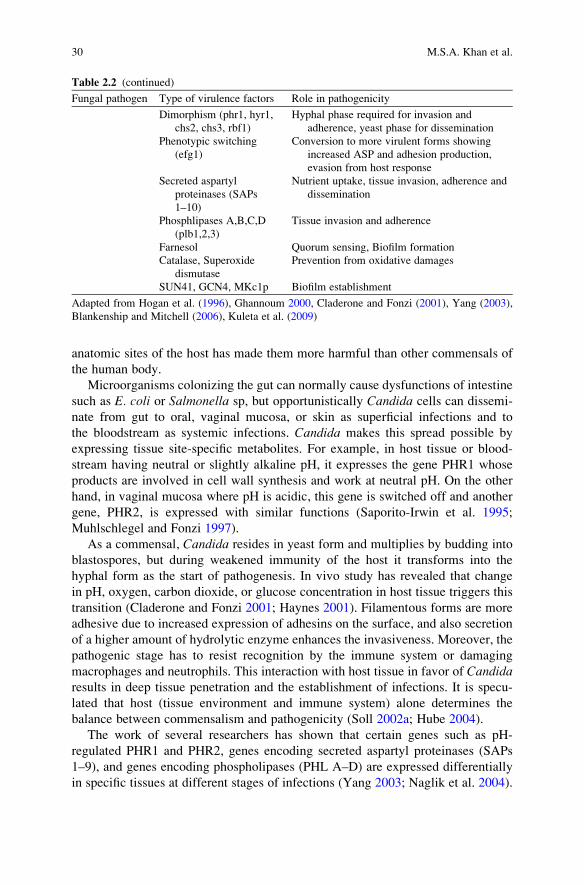
anatomic sites of the host has made them more harmful than other commensals of
the human body.
Microorganisms colonizing the gut can normally cause dysfunctions of intestine
such as E. coli or Salmonella sp, but opportunistically Candida cells can dissemi-
nate from gut to oral, vaginal mucosa, or skin as superficial infections and to
the bloodstream as systemic infections. Candida makes this spread possible by
expressing tissue site-specific metabolites. For example, in host tissue or blood-
stream having neutral or slightly alkaline pH, it expresses the gene PHR1 whose
products are involved in cell wall synthesis and work at neutral pH. On the other
hand, in vaginal mucosa where pH is acidic, this gene is switched off and another
gene, PHR2, is expressed with similar functions (Saporito-Irwin et al. 1995;
Muhlschlegel and Fonzi 1997).
As a commensal, Candida resides in yeast form and multiplies by budding into
blastospores, but during weakened immunity of the host it transforms into the
hyphal form as the start of pathogenesis. In vivo study has revealed that change
in pH, oxygen, carbon dioxide, or glucose concentration in host tissue triggers this
transition (Claderone and Fonzi 2001; Haynes 2001). Filamentous forms are more
adhesive due to increased expression of adhesins on the surface, and also secretion
of a higher amount of hydrolytic enzyme enhances the invasiveness. Moreover, the
pathogenic stage has to resist recognition by the immune system or damaging
macrophages and neutrophils. This interaction with host tissue in favor of Candidaresults in deep tissue penetration and the establishment of infections. It is specu-
lated that host (tissue environment and immune system) alone determines the
balance between commensalism and pathogenicity (Soll 2002a; Hube 2004).
The work of several researchers has shown that certain genes such as pH-
regulated PHR1 and PHR2, genes encoding secreted aspartyl proteinases (SAPs
1–9), and genes encoding phospholipases (PHL A–D) are expressed differentially
in specific tissues at different stages of infections (Yang 2003; Naglik et al. 2004).
Table 2.2 (continued)
Fungal pathogen Type of virulence factors Role in pathogenicity
Dimorphism (phr1, hyr1,
chs2, chs3, rbf1)
Hyphal phase required for invasion and
adherence, yeast phase for dissemination
Phenotypic switching
(efg1)
Conversion to more virulent forms showing
increased ASP and adhesion production,
evasion from host response
Secreted aspartyl
proteinases (SAPs
1–10)
Nutrient uptake, tissue invasion, adherence and
dissemination
Phosphlipases A,B,C,D
(plb1,2,3)
Tissue invasion and adherence
Farnesol Quorum sensing, Biofilm formation
Catalase, Superoxide
dismutase
Prevention from oxidative damages
SUN41, GCN4, MKc1p Biofilm establishment
Adapted from Hogan et al. (1996), Ghannoum 2000, Claderone and Fonzi (2001), Yang (2003),
Blankenship and Mitchell (2006), Kuleta et al. (2009)
30 M.S.A. Khan et al.

Expression or modulation of these genes on the same mucosal surfaces only during
transition from the commensal to the parasitic stage reflects a weakness in the
immune system responsible for this shift (Casadevall and Pirofski 2001). Candidasurvives and proliferates as commensal in competition with other microbial flora
and is affected by epithelial cell proliferation and the immune system. Proliferation
of epithelial cells constitutively forces Candida to attempt deeper invasion into
tissues. Prolonged antibiotic therapy provides more available nutrients and space
for Candida to multiply as other commensal microbial flora are diminished (Senet
1998). Immune suppression in HIV patients and inhibition of epithelial cell prolif-
eration such as in cancer therapy changes the tissue environment in terms of pH,
osmolarity, and oxidative stress. This changed condition is perceived by the
candidal cell and subsequently down- or up-regulation of certain genes provokes
Candida to switch over from commensal to opportunistic pathogens (Claderone and
Fonzi 2001; Hube 2004).
Advanced medical equipment and surgery has also led to the increased spread
of commensal Candida to tissues as pathogens. Medical devices such as catheter,
dental implants, artificial joints, pacemakers, central nervous system shunts, and
others have provided the opportunity to form biofilms, a stage more resistant to
drugs and capable of greater invasion to tissues. These devices are easily colo-
nized by candidal cells from mucosal surfaces and blood stream, and frequently
get spread from one tissue site to another. Further, candidal cells can also migrate
via blood flow to all inner organs, leading to septicemia and life-threatening
diseases (Douglas 2003; Hall-Stoodley et al. 2004). Biofilm-forming cells have
been reported to be more virulent than planktonic cells (Ramage et al. 2005;
Seneviratne et al. 2007). Recently, several workers have reported increased
production of proteinases, phospholipases, and adhesins in biofilm compared to
planktonic cells (Chandra et al. 2001; Al-Fattani and Douglas 2006; Seneviratne
et al. 2007). All these collectively aid in establishing infections by heightening
the adherence and invasion of tissues, leading to increased virulence.
Further, genetic changes in biofilms result in elevated drug resistance, pro-
nounced quorum sensing and regulated carbohydrate synthesis, thereby influen-
cing the pathogenicity of Candida. Therefore, biofilms-forming capacity has
greatly increased the potency of Candida to convert from the commensal stage
into a virulent pathogen.
2.6 Virulence Factors Involved in Pathogenicity
of Candida albicans
Like other fungal pathogens, C. albicans also regulates expression of certain genes
and their products as virulence factors to produce disease. This is the most common
opportunistic pathogen, utilizing several kinds of virulence factors. Some of the
commonly studied virulence factors in C. albicans are briefly described here.
2 Virulence and Pathogenicity of Fungal Pathogens 31

2.6.1 Adhesion
Adherence of candidal cells to host tissues is a complex multifactorial phenomenon
utilizing several types of adhesins expressed on morphogenetically changing cell
surfaces. But the striking feature ofCandida cells is the formation of biofilms in host
tissue, resulting in enhanced adherence. Ramage et al. (2006) have reported that in
the last few decades, Candida-related infections have been found associated with
biofilm-forming capacity. Well-known adhesins are agglutinin-like sequences
(ALS) that are members of a family of seven glycosylated proteins. Als1p, Als3p
and Als5p (Ala1p) on the cell surface of hyphae adhere to human buccal epithelial
cells (HBEC) and fibronectin, collagen, laminin, and endothelial cells (Hawser and
Douglas 1994; Hoyer 2001). Als6p and Als9p bind to collagen and laminin respec-
tively. Als4p binds to endothelial cells and Als5p is additionally needed for cell
aggregation. However, the role of Als7p is unclear (Filler et al. 2006; Kuleta et al.
2009). Another 34 kDa adhesin molecule, Hwp1 (hyphal wall protein), encodes an
outer surface mannoprotein on the hyphal wall; the amino terminal sequences of this
adhesin are recognized as mammalian transglutaminase substrate (TGase) and form
covalent binding with HBEC. Studies with hwp1� knockout mutant and HWP1�
deficient mutant of C. albicans have shown reduced adherences and mortality in
murine models (Chaffin et al. 1998; Staab et al. 1999). An integrin-like protein
(Int1p) which is a plasma membrane receptor and antigenic functionally similar to
human complement receptors 3 and 4, has been isolated from C. albicans and foundto bind with extracellular matrix (ECM) ligands such as fibronectin, laminin, and
collagen I and IV, and induce morphological changes in response to extracellular
signals (Claderone and Fonzi 2001; Ruiz-Herrera et al. 2006).
2.6.2 Morphogenesis
Morphogenesis in C. albicans is defined as transition from unicellular yeast form to
filamentous form (pseudohyphae or hyphae). Of all the species only C. albicans andC. dubliniensis are able to undergo morphogenesis. Transition from yeast form to
hyphal form is facilitated by nutrients, near-neutral pH, temperature of 37�C–40�C,CO2 concentration about 5.5%, and presence of N-acetyl-D-glucosamine, serum,
some amino acids, and biotin. Reverse transition from hyphal to yeast form is
provoked by lower temperatures, acidic pH, absence of serum, and higher concen-
tration of glucose (Corner and Magee 1997; Eckert et al. 2007). This transition is
strongly required for pathogenesis. Yeast forms are more suited for dissemination in
tissues and to other hosts, whereas hyphal forms are required for tissue damage and
invasion. For example, the yeast cell, when phagocytosed by macrophages, pro-
duces hyphae and secretes hyphae-associated proteinases that kill macrophages;
these factors also prevent hyphal cells from being killed by neutrophils. In addition,
hyphal cells have been shown to induce phagocytosis by endothelial cells, helping
32 M.S.A. Khan et al.

Candida cells to escape from the bloodstream (Molero et al. 1998; Gow et al.
2002; Hube 2004). Further, hyphal cells have stronger adherence capacity due to
expression of ALS adhesins and also exhibit greater invasiveness to tissues.
Increased expression of superoxide dismutase (SOD) antagonizes oxidative burst
of phagocytic cells. Several genes have been identified which regulate phase
transition, namely PHR1, ECE1, HYR1, RBF1, CHS2, CHS3 which are differen-
tially expressed during morphogenesis (Haynes 2001; Claderone and Fonzi 2001).
Of these, ECE1 correlates with hyphal elongation although ECE1 null mutants
displayed no morphological alterations. Similarly, null mutants for expression of
CHS2, CHS3 and HYR1 did not show any obvious morphological type. But
disruption of RBF1 demonstrated alteration in cell morphology and strongly
involved in yeast–hypha transition (Yang 2003). Studies with homozygous null
mutants for Hst7p, Cph1p and Cst20p have shown defective hyphal formation
(Leberer et al. 1996); in addition, three genes TUP1, EFG1, CLA4 were found to
be regulating candidal morphogenesis (Liu 2001). Transcription factors such as
Tup1 and Rbp1 are negative regulators of filamentation (Braun and Johnson 2000).
A tup1 mutant strain resulted in constitutive filamentous growth under all condi-
tions, indicating a role in filament formation. Deletion of homozygous allele of
Ste20 encoded by CLA4 showed impaired hyphal formation in a wide range of
medium, and decrease in virulence in a murine model (Braun and Johnson 1997;
Celera and Claderone 2001). A protein of bHLH class encoded by EFG1 acts as
transcriptional activator as well as repressor, and is required for pseudohyphal and
hyphal morphogenesis (Liu 2001; Noffiz et al. 2008). A study with homozygous
mutants cfg1 and cph1 showed failure of germ tube and hyphae production in a
murine model (Noffiz et al. 2008).
2.6.3 Phenotypic Switching
Unlike other pathogens, phenotypic switching in Candida is pleitropic by affecting
several phenotypic and metabolic parameters, with subsequently a number of
virulence traits such as SAP gene regulation. This allows Candida to adapt to a
different host environment during infection (Soll 1992; Soll 2002b). Colonies of
C. albicans show morphological variation, including smooth, rough, star, stippled,
hat, wrinkle, and fuzzy at high frequency. This switching is reversible, occurs
spontaneously in stress, and results in changes in cell surface behavior, colony
appearance, and metabolic, biochemical and molecular attributes to become more
virulent and effective during infection (Soll 2002b; Odds et al. 2006). Strains
isolated from vaginitis or systemically infected patients showed higher frequencies
of switching, indicating a strong role for the switching phenomenon in establishing
diseases (Kvaal et al. 1999). In the case of yeast–hypha transition, all cells of a
population express the same phenotype under the same environmental conditions,
whereas in the case of switching, some cells of a population express different
phenotypes under the same set of environmental conditions. Earlier research had
2 Virulence and Pathogenicity of Fungal Pathogens 33

reported that laboratory isolate 3153A, grown on amino acid rich agar which was
limiting for zinc and incubated at 25�C, showed a smooth phenotype as dominant,
while variant colonies of star, ring, irregular, and wrinkle occurred spontaneously.
Such types of variation were also observed with cells of strain 3153A treated with
low doses of UV irradiation (Soll 1992). At present, of all the phenotypes described,
the white-opaque system in strain WO-1 is the most studied. This system is
characterized by transition from smooth, white colonies to flat, gray opaque colo-
nies. White cells are round ovoid while opaque cells are elongated or bean shaped
(Soll 2002b). Study of gene expression with the WO-1 system revealed an associa-
tion of OPA1 (SAP1) and SAP3 in opaque cells, in contrast to SAP2, WH11and
EFG1 in white cells (Soll 1997; Miller and Johnson 2002). Study with efg1 null
mutants exhibited no involvement of EFG1 in switching, but rather control of
phenotypic characteristics. It has been reported that white cells in WO-1 hardly
form hyphal stages, but this was achieved by opaque cells (Staib et al. 2002). There
is good evidence that opaque cells are more virulent than white cells in several
murine models (Yang 2003).
2.6.4 Phospholipases
Phospholipases are enzymes that hydrolyze ester linkages of glycophospholipids
and hence impart tissue invasiveness to Candida cells. In C. albicans, four typesof phospholipases are classified by researchers on the basis of the ester bond they
cleaved, viz., phospholipase A, B, C, and D. All types possess hydrolase activity,
but PLB in addition also possesses lysophospholipase transacylase activities; there-
fore, it is able to release fatty acids from phospholipids and the remaining fatty acid
from lysophospholipids, and then transfer a free fatty acid to lysophospholipids,
producing phospholipids. Of these, only PLB1, a 84 kDa glycoprotein isolated from
hyphal tip in the course of tissue invasion, has been shown to be required for
virulence in a murine model of candidiasis (Ghannoum 2000; Yang 2003; Theiss
et al. 2006). A study conducted by Ibrahim et al. (1995) revealed an increased level
of phospholipase production in blood isolates compared to commensal isolates.
2.6.5 Proteinases
Secretion of proteinases by pathogen is mandatory in order to degrade the tissue
barriers and obtain nutrition at the infection site. Secreted aspartyl proteinases
(SAPs) from Candida have been reported that hydrolyze many proteins such as
albumin, hemoglobin, keratin, collagen, laminin, fibronectin, mucin, salivary lacto-
ferin, interleukin1b, cystatin A, and Immunoglobulin A (Hube et al. 1998). To date,
ten proteins have been recognized as SAP family (SAPs 1–10) and found to be
responsible for tissue invasion. Several researchers have reported that production of
SAPs is also correlated with hyphal formation, adherence, and phenotypic switching
34 M.S.A. Khan et al.

(Monod and Zepelin 2002; Naglik et al. 2003). Such researches have highlighted the
complex role played by SAPs in the pathogenicity of C. albicans. Several models
using SAP inhibitors such as pepstatin A and SAP-disrupted or over-expressing
mutants demonstrated the need for these factors in candidal pathogenesis. In vitro
studies have reported that SAPs 1, 2 and 3 are expressed by the yeast phase, only
while SAPs 4, 5 and 6 are expressed in the hyphal phase (Hube et al. 1998; Schaller
et al. 1999; Naglik et al. 2004).Whereas, SAPs 9 and 10 are expressed by both forms
(Albrecht et al. 2006). Structural analysis revealed that SAPs 1–8 are secreted
extracellularly, but that SAPs 9 and 10 are anchored to the cell wall by glycopho-
sphotidyinositol (GPI) protein (Naglik et al. 2003; Albrecht et al. 2006). Models of
epidermal and vaginitis candidiasis revealed involvement of SAPs 4–6 in invasive
systemic disease whereas SAP 7 was never detected in vitro. The role of SAPs 1–3 isassociated with early adherence, invasion, and cutaneous infections as studied in the
WO1 strain, whereas SAP8 is associated with extensive penetration. SAPs 6 and 9
were found expressed in later stages of hyphal growth (Hube et al. 1998; Schaller
et al. 1999; Kvaal et al. 1999). Different properties of SAPs are exploited in the
pathogenicity of Candida. For example, SAPs are active across a broad range of pH
2.0–7.0, as SAPs 1–3 are active at pH 3.5, SAPs 4–6 at pH 5.0–7.0, and therefore
make Candida capable of colonizing and invading different tissue sites of varying
pH. In addition, SAPs show varied levels of protein specificity, as SAPs 1, 2, 3 and 6
cleave peptide bonds in larger hydrophobic amino acids; SAPs 1, 2 and 6 act on
phenylalinine, whereas SAP 3 attacks leucine and SAPs 9 and 10 hydrolyze yapsin
and kexins (Naglik et al. 2004). This attribute enables Candida to obtain nitrogen atdifferent tissue makeups, and aids pathogenicity by revealing potential binding sites
from tissue for adhesion of candidal cells, and also dissemination via circulatory
systems. In vivo studies have confirmed the role of SAPs in colonization, increased
adhesion and tissue penetration (Naglik et al. 2004; Hube and Naglik 2001).
Disruption of SAPs 1, 2 and 3 have resulted in decreased virulence in mouse models
(Hube et al. 1997). Several reports have supported functional role of SAP2 in
invasion and dissemination of systemic infections (Kvaal et al. 1999; Naglik et al.
2004; De Bernardis et al. 1999). Further research data have indicated increased
expression of SAP genes, especially SAPs 5, 6 and 9 mRNA transcripts, in biofilm
rather than planktonic cells (Green et al. 2004; Naglik et al. 2008). Recently, in
addition to SAPs, a 60 kDa metallopeptidase and 50 kDa serine peptidases have also
been isolated, and reported to hydrolyze extracellular matrix proteins and serums
(dos Santos et al. 2006). Expression of SAPs has been found to be correlated with
other virulence determinants to enhance the pathogenicity of C. albicans. Correla-tion of SAPs with other virulence factors in C. albicans is illustrated in Fig. 2.1.
2.6.6 Biofilm Formation
Biofilms are the organized structures involving microbial communities that are
attached to some inanimate surfaces or tissues and circumvented in a matrix of
2 Virulence and Pathogenicity of Fungal Pathogens 35

exopolymeric materials. Biofilm formation is initiated by irreversible adherence of
microbial cells to tissues or devices and followed by growth and maturation to form
a mesh of cells with altered phenotype, growth rate, and gene expression compared
to planktonic cells. Studies with scanning electron microscopy of biofilms revealed
the presence of both adherent yeast cells and invasive hyphal forms constructing
basal and upper layers respectively, enclosed in an extracellular polymer matrix
consisting of polysaccharides and proteins and forming a three-dimensional struc-
tures with water channels (Dominic et al. 2007). These forms differ in ultrastruc-
ture, physiological behavior and composition of cell walls, and are required for
candidal pathogenicity, as mutants lacking genes for any one became less virulent
both in vitro and in vivo (Chandra et al. 2001). Heterogeneity of these biofilms
depends on the substrate composition, environmental conditions, and type of strains
involved. Although yeast–hypha transition is necessary for full maturation of the
biofilm, strains that are unable to grow as yeasts or to form hyphae can still form
biofilms but are easily detachable (Baille and Douglas 1999). In addition, some
authors have reported a change in biofilm-forming ability of candidal cells, with
Dissemination
Secreted aspartylproteinase production
(SAPs 1-10)
Adherence
Invasion and tissue damage
Nut
rien
t upt
ake
Hydrolysis of several types
of membrane proteins
to release N-source
Cle
avag
e o
f p
epti
de
bo
nd
s in
dif
fere
nt
kin
ds
of
pro
tein
Exposure of receptor s
ite
after ti
ssue damage for
strong binding
SAPs 1-3 for adherence
to buccal epithelial cells
Co
nve
rsio
n f
rom
hyp
hal
to y
east
ph
ase:
re
vers
ible
dim
orp
his
m
SA
P 2
fo
r H
ydro
lysi
s o
f im
mu
ne
cells
; S
AP
s 4-
6 fo
r h
yph
al t
ran
siti
on
an
d
esca
pe
fro
m m
acro
ph
ages
Biofilm form
ationIn
crea
sed
viru
lenc
e
Increased expression of
SAPs 5,6,9 in biofilm
stage
Survival and establishment
of infection
Incr
ease
d e
xpre
ssio
n
of
AL
S a
dh
esin
s
Spread to various tissues
SAP 1,3 are expressed
in more virulent opaque
cells (WO1)
More threatening
systemic mycoses
SAPs 1-3 for epithelial
infections; SAPs4-6 for
systemic infections
Co
nve
rsio
n f
rom
ye
ast
to m
ore
in
vasi
ve h
yph
alfo
rm
SA
Ps4
-6
Fig. 2.1 Correlation of secreted aspartyl proteinases with other virulence attributes in Candidaalbicans. Partially adapted from Naglik et al. (2004)
36 M.S.A. Khan et al.

alteration in variants produced during phenotypic switching (Brown and Gow 1999;
Berman and Sudbery 2002).
The ability of Candida to form biofilms on catheters, endotracheal tubes, pace-
makers and other prosthetic devices has contributed to its predominant prevalence
in nosocomial infections (Douglas 2003; Ramage et al. 2005). Such devices, in
addition to providing a platform for candidal cells to form biofilm, grow and
develop, provide a route through host barrier defenses for dissemination. During
weakened immunity, hematogenous dissemination of candidal cells from biofilms
to deep-seated organs could occur, resulting in candidemia and septicemia. Recent
studies have confirmed biofilm growth in the majority of diseases caused by
Candida spp (Chandra et al. 2001; Douglas 2003; Chandra et al. 2005). Dental
plaque is a well-known example of biofilm formation from Candida cells, and is
responsible for oral candidiasis. Biofilm formation on such tissues is favored by a
high concentration of glucose, serum, and other proteins. Biofilm formation was
found to be linked to dimorphism and phenotypic switching, well-known virulence
traits for candidal cells (Baille and Douglas 1999; Chandra et al. 2001). Also,
alerted phenotypes exhibited reduced susceptibility to the host immune system
and to antifungal drug therapy (Chandra et al. 2001). These biofilm-specific cell
properties are an indicator for virulence, and have prompted much recent interest in
C. albicans biofilm structure, physiology, and regulation. Therefore, knowing the
ability of C. albicans to populate a surface and produce a biofilm as a virulence trait,
exhaustive research is being focused on the prevention of biofilm infection by
Candida cells.
Adherence is the critical property for biofilm-forming cells and is mediated by
hydrophobic interactions, electrostatic forces, and adhesion–ligand interactions;
multiple adhesion molecules function in the successful establishment of biofilm.
A variety of genes are involved in adhesion, and penetrations are associated deeply
with the biofilm-forming capability of C. albicans. Here, we would discuss some of
them for their role in pathogenicity. A number of adhesins, termed glycosylpho-
sphatidylinositol-dependent cell wall proteins (GPI-CWPs), encoded by ALS1,
ALS2, ALS4, ALS5 (ALA1), HWP1, and EAP1, mediate adhesion to organic
and inorganic surfaces, extracellular matrix proteins, human endothelial cells, and
epithelial cells (Blankenship andMitchell 2006; Filler et al. 2006; Zhao et al. 2006).
Experimental results showed upregulated ALS family gene expression in biofilm-
forming cells compared with planktonic cells (Hoyer et al. 1998; Chandra et al.
2001; Nobile and Mitchell 2005; Green et al. 2004) and ALS 3 was also found
necessary for biofilm formation on silicone substrates in vitro (Nobile et al. 2006a).ALS1, ALS3, and HWP1 are regulated by transcription factor BCR1 (biofilm and
cell wall regulator), a zinc finger protein, which is under the control of transcription
factor Tec1. An als3/als3 mutant strain was found defective in biofilm formation
in vitro, and overexpression of ALS3 permitted biofilm formation by a bcr1/bcr1
mutant in vitro and in vivo. Studies with the bcr gene revealed involvement of
BCR1 in governing the mechanism of biofilm formation only and not the filamen-
tation (Nobile and Mitchell 2005; Lopez-Ribot 2005; Nobile et al. 2006a; Nobile
et al. 2006b). Hwp1 is a cell surface protein covalently linked to the cell wall glucan
2 Virulence and Pathogenicity of Fungal Pathogens 37

through a remnant of its GPI anchor. Functional analysis showed its requirement
for tight adherence to oral epithelial cells (Chaffin et al. 1998; Staab et al. 1999;
Mendes-Giannini et al. 2008). A role for Hwp1 in C. albicans cell–cell adherence isexhibited from the finding that it is induced by mating factor and is deposited on the
surface of the bridge between mating partners (Staab et al. 1999; Hoyer et al. 1998).
Recent studies showed that Hwp1is required as first cell surface protein in vivo for
biofilm formation (Nobile et al. 2006a; Nobile et al. 2006b). Recently, work from
Granger et al. (2005) described the role of Ywp1 (Yeast cell wall specific protein)
as anti-adhesin. The mutant for Ywp1 led to enhanced adherence of yeast cells,
therefore highlighting its negative role in biofilm establishment. In addition, studies
with mutants for transcription factor Ace2 (activation of CUP1 expression 2)
resulted in inhibition of biofilm formation (Kelly et al. 2004). A study conducted
by Li et al. (2007) showed eap1 mutants exhibiting reduced adhesion to plastic
surfaces and epithelial cells, and that Eap1p was able to mediate adhesion to yeast
cells. The same study also showed of the need for eap1 gene expression in biofilm
formation under shear flow in vitro and in central venous catheter biofilm model
in vivo. In a study, another cell wall protein Ecm33 was found to be necessary for
cell wall integrity and yeast-to-hypha transition (Martinez-Lopez et al. 2004), and
Mp65 (Norice et al. 2007) is also required for full virulence in a disseminated
infection model, illustrating that cell wall proteins may have diverse functions that
are relevant to infection. Norice et al. (2007) also showed that protein SUN41
plays major roles in biofilm formation, cell wall integrity, and virulence in both
oropharyngeal and disseminated candidiasis.
Richard et al. (2005) showed involvement of genes sun3, nup85, mds3, kem1 in
hyphal development and biofilm formation. Some studies have also demonstrated
that the hyphal regulatory gene efg1 is required for normal biofilm growth, and
efg1/efg1 and efg1/efg1 cph1/cph1 mutants have exhibited defective biofilms and
also adhered poorly to the substrate (Lewis et al. 2002; Watamoto et al. 2009). Gcn4,
a general amino acid control regulatory gene, was shown to be required for full
biofilm biomass production (Blankenship and Mitchell 2006). A contact-activated
protein kinase, Mkc1p, is also required for biofilm development, suggesting that
C. albicans may respond uniquely to surface contact during biofilm formation.
Further, an experiment by Ramage et al. (2002) showed that biofilms are
organized communities under tight regulation of gene expression controlled
through quorum sensing which in turn is regulated by farnesol and tyrosol mole-
cules. Several workers have observed these organized communities under the
control of a signaling molecule (Hogan 2006). This cell-to-cell communication
prevents and controls unnecessary overpopulation and nutritional competition, and
has implications in dissemination and establishment of infection at the distal site
from old biofilm (Alem et al. 2006). Farnesol, a quorum-sensing molecule that
inhibits C. albicans biofilm formation by inhibiting yeast-to-hypha transition,
decreases HWP1 expression in biofilms (Ramage et al. 2002). Farnesol acts on
yeast cells to prevent filamentation but elongated hyphae continue to form biofilms.
A report highlighted differential expression of genes associated with hyphae forma-
tion in farnesol-treated biofilms, such as genes involved in cell wall maintenance,
38 M.S.A. Khan et al.

iron transport, stress response and upregulation of TUP1 and downregulation of
CSH1 protein associated with cell surface hydrophobicity. Farnesol also prevents
induction of Tup1-regulated filament specific genes hwp1, rbt1, cph1 and hst7 (Cao
et al. 2005; Dominic et al. 2007). In contrast to farnesol, another quorum-sensing
molecule, tyrosol, induces filamentation under conditions conducive to germ tube
formation, but its role in biofilms has not been much investigated (Alem et al.
2006). However, a recent study highlighted a two-component signal transduction
protein Chk1p regulating both quorum sensing and biofilm formation by negatively
regulating hyphal development in C. albicans. However, it is not clear whether
chk1p is directly involved in response to farnesol or not (Kruppa et al. 2004;
Blankenship and Mitchell 2006). Involvement of different virulence factors in
forming biofilms and associated pathogenicity is depicted in Fig. 2.2.
2.7 Conclusion
Based on the review of the literature on fungal infection, virulence, and
pathogenicity, it is clear that at present the frequency of fungal infection rate is
increasing, up to 90% for patients with disseminated candidiasis, aspergillosis or
Increased adherence and invasionIncreased secretion of SAPsIncreased secretion of phospholipasesReduced susceptibility to host immune systemIncreased level of drug resistance
Planktonic yeast cells
Cell surfaces or PVC
Adherence of yeast cellsforming basal adherent layer
Filamentation to form upper invasive hyphal layer(efg1, sun3, nup85)
Secretion of exopolysaccharides(adh1)
b
a
c
d
Matu
ration
of b
iofilm
Increased expression of virulence factors inbiofilm; increased virulence and pathogenicity
Biofilm dispersal and dissemination to deepseated organs; systemic infections, septicemia
Dimorphism andphenotypic switching
Upregulation of adhesins
Ywp1, chk1p
ALS, hwp1, eap1ace2, bcr1, tec1
ALS, hwp1, eap1, ace2, bcr1,tec1, efg1. adh1, ecm33, sun3
ALS, hwp1, eap1, ace2, bcr1, tec1, efg1,adh1, nup85, sun3, mkc1p, gcn4
Fig. 2.2 Illustration of virulence factors involved in establishing biofilm and associated pathoge-
nicity in Candida albicans: (a) planktonic yeast cells, (b) initiation of biofilm formation on living
tissues or inert object like polyvinyl chloride (PVC) by adherence of yeast cells forming basal
layer, (c) development of biofilm by initiation of upper invasive hyphal layer and production of
exoploysaccharide, and (d) maturation of biofilm by more filamentation and exopolysaccharide
production. Partially adapted from Blankenship and Mitchell (2006)
2 Virulence and Pathogenicity of Fungal Pathogens 39

cryptococcosis. Fungal pathogenesis is a multifactorial phenomenon; therefore, the
nature of fungal pathogens, their virulence factors, and their interaction with host
defense mechanisms need to be explored for the development of more effective
antifungal therapy. Although phenomenal progress has been made on molecular
characterization of various virulence factors and host–fungi interactions; this issue
needs further investigation in order to know the exact contribution of each virulence
factor under different disease conditions.
References
Albrecht A, Felk A, Pichova I, Naglik JR, Schaller M, De Groot P, MacCallum D, Odds FC,
Schafer W, Klis F, Monod M, Hube B (2006) Glycosylphosphatidylinositol anchored proteases
of Candida albicans target proteins necessary for both cellular process and host pathogen
interactions. J Biol Chem 281:668–694
Alem MAS, Oteef MDY, Flowers TH, Douglas LJ (2006) Production of tyrosol by Candidaalbicans biofilms and its role in quorum sensing and biofilm development. Eukaryotic Cell
5:1770–1779
Al-Fattani MA, Douglas LJ (2006) Biofilm matrix of Candida albicans and Candida tropicalis:chemical composition and role in drug resistance. J Med Microbiol 55:999–1008
Baille GS, Douglas LJ (1999) Role of dimorphism in the development of Candida albicansbiofilm. J Med Microbiol 48:671–679
Berman J, Sudbery PE (2002) Candida albicans: a molecular revolution built on lessons from
budding yeast. Nat Rev Genet 3:918–930
Blankenship JR, Mitchell AP (2006) How to build a biofilm: a fungal perspective. Curr Opin
Microbiol 9:588–594
Braun BR, Johnson AD (1997) Control of filament formation in Candida albicans by the
transcriptional repressor TUP1. Science 277:105–109
Braun BR, Johnson AD (2000) TUP1, CPH1 and EFG1 make independent contributions to
filamentation in Candida albicans. Genetics 157:57–67Brock M (2009) Fungal metabolism in host niches. Curr Opin Microbiol 12:371–376
Brown AJ, Gow NA (1999) Regulatory networks controlling Candida albicans morphogenesis.
Trends Microbiol 7:333–338
Cao YY, Cao YB, Xu Z, Ying K, Li Y, Xie Y, Zhu ZY, Chen WS, Jiang YY (2005) cDNA
microarray analysis of differential gene expression in Candida albicans biofilm exposed to
farnesol. Antimicrob Agents Chemother 49:584–589
Casadevall A (2007) Determinants of virulence in the pathogenic fungi. Fungal Biol Rev
21:130–132
Casadevall A, Pirofski LA (2001) Host-pathogen interactions: the attributes of virulence. J Infect
Dis 184:337–344
Celera JA, Claderone R (2001) Signalling and the biology of human fungal pathogens. In:
Claderone R, Cihlar R (eds) Fungal pathogenesis: principles and clinical applications. Marcel
Dekker, New York, pp 115–137
Chaffin WL, Lopez-Ribot Jl, Casanova M, Gozalbo D, Martinez JP (1998) Cell wall and secreted
proteins of Candida albicans: identification, function and expression. Microbiol Mol Biol Rev
62:130–180
Chakrabarti A (2005) Microbiology of systemic fungal infection. J Postgrad Med 51(Suppl1):
S16–S20
Chandra J, Kuhn DM, Mukherjee PK, Hoyer LL, McCormik T, Ghannoum MA (2001) Biofilm
formation by the fungal pathogen Candida albicans: development, architecture and drug
resistance. J Bacteriol 18:5385–5394
40 M.S.A. Khan et al.

Chandra J, Patel JD, Li J, Zhou G, Mukherjee PK, McCormick TS et al (2005) Modification of
surface properties of biomaterials influences the ability of Candida albicans to form biofilms.
Appl Environ Microbiol 71:8795–8801
Claderone RA, Fonzi WA (2001) Virulence factors of Candida albicans. Trends Microbiol
9:327–336
Corner BE, Magee PT (1997) Candida pathogenesis: unraveling the threads of infection. Curr Biol
2:R691–R694
de Bernardis F, Muhlschlegel FA, Cassone A, Fonzi WA (1998) The pH of the host niche controls
the gene expression in and virulence of Candida albicans. Infect Immun 66:3317–3325
de Bernardis F, Arancia S, Morelli L, Hube B, Sanglard D, Schafer W, Cassone A (1999) Evidence
that members of aspartyl proteinase gene family, in particular SAP 2, are virulence factors for
Candida vaginitis. J Infect Dis 179:201–208
Dominic RM, Shenoy S, Baliga S (2007) Candida biofilms in medical devices: evolving trends.
Kathmandu Univ Med J 5:431–436
Dos Santos AL, de Carvalno IM, daSilva BA, Portela MB, Alviano CS, de Aroujo Soares RM
(2006) Secretion of serine peptidase by a clinical strain of Candida albicans: influence of
growth condition and cleavage of human serum proteins and extracellular matrix components.
FEMS Immunol Med Microbiol 46:209–220
Douglas LJ (2003) Candida biofilms and their role in infection. Trends Microbiol 11:30–36
Eckert SE, Sheth CC, Muhlschlegel FA (2007) Regulation of morphogenesis in Candia species.
In: d’Enfert CH, Hube B (eds) Candida. Comparative and functional genomics. Caister
Academic, Norfolk, pp 263–291
Fidel PL Jr, Vanquez JA, Sobel JD (1999) Candida glabrata: a review of epidemiology, patho-
genesis and clinical disease with comparison to Candida albicans. Clin Microbiol Rev
12:80–96
Filler SG, Sheppard DC, Edwards JE Jr (2006) Molecular basis of fungal adherence to endothelial
and epithelial cells. In: Heitman J, Filler SG, Edwards JE Jr, Mitchell AP (eds) Molecular
principles of fungal pathogenesis. ASM, Washington, DC, pp 187–196
Fluckiger U, Marchetti O, Bille J, Eggiman P, Zimmerli S, Imhof A, Garbino J, Ruef C, Pittet D,
Tauber M, Glauser M, Calandra T (2006) Treatment options of invasive fungal infections in
adults. Swiss Med Wkly 136:447–463
GhannoumMA (2000) Potential role of phospholipases in virulence and fungal pathogenesis. Clin
Microbiol Rev 13:122–143
Gow NAR, Brown AJP, Odds FC (2002) Fungal morphogenesis and host invasion. Curr Opin
Microbiol 5:366–371
Granger BL, Flenniken ML, Davis DA, Mitchell AP, Cutler JE (2005) Yeast wall protein 1 of
Candida albicans. Microbiol 151:1631–1644
Green CB, Cheng G, Chandra J, Mukherjee P, Ghannoum MA, Hoyer LL (2004) RT-PCR
detection of Candida albicans ALS gene expression in the reconstituted human epithelium
(RHE) model of oral candidiasis and in model biofilms. Microbiol 150:267–275
Haas H, Eisendle M, Turgeon BG (2008) Siderophores in fungal physiology and virulence. Ann
Rev Phytopathol 46:149–187
Hall-Stoodley L, Costerton JW, Stoodley P (2004) Bacterial biofilms: from the natural environ-
ment to infectious diseases. Nat Rev Microbiol 2:95–108
Hawser SP, Douglas LJ (1994) Biofilm formation by Candida species on the surface of catheter
materials in vitro. Infect Immun 62:915–921
Hayens KA, Westerneng TJ (1996) Rapid identification of Candida albicans, C. glabrata,C. parapsilosis and C. krusei by species specific PCR of large subunit ribosomal DNA.
J Med Microbiol 44:390–396
Haynes K (2001) Virulence in Candida species. Trends Microbiol 9:591–596
Hogan DA (2006) Talking to themselves: autoregulation and quorum sensing in fungi. Eukaryotic
Cell 5:613–619
2 Virulence and Pathogenicity of Fungal Pathogens 41

Hogan LH, Klein BS, Levitz SM (1996) Virulence factors of medically important fungi. Clin
Microbiol Rev 9:469–488
Hoyer LL (2001) The ALS gene family of Candida albicans. Trends Microbiol 9:176–180
Hoyer LL, Payne TL, Bell M, Myers AM, Scherer S (1998) Candida albicans AL S3 and insights
into the nature of the ALS gene family. Curr Genet 33:451–459
Hube B (2004) From commensal to pathogen: stage and tissue specific gene expression of Candidaalbicans. Curr Opin Microbiol 7:336–341
Hube B, Naglik J (2001) Candida albicans proteinases resolving the mystery of a gene family.
Microbiol 147:1997–2005
Hube B, Sanglard D, Odds FC, Hess D, Monod M, Schafer W, Brown AJ, Gow NA (1997)
Disruption of each of the secreted aspartyl proteinase genes SAP1, SAP2 and SAP3 of Candidaalbicans attenuates virulence. Infect Immun 65:3529–3538
Hube B, Ruchel R, MonodM, Sanglard D, Odds FC (1998) Functional aspects of secreted Candidaproteinases. Adv Exp Med Biol 436:339–344
Ibrahim AS, Mirbod F, Filler SG, Banno Y, Cole GT, Kitajima Y, Edwards JE Jr, Nozawa Y,
Ghannoum MA (1995) Evidence implicating phospholipase as a virulence factor of Candidaalbicans. Infect Immun 63:1993–1998
Kelly MT, MacCallum DM, Clancy SD, Odds FC, Brown AJ, Butler G (2004) The Candidaalbicans CaACE 2 gene affects morphogenesis, adherence and virulence. Mol Microbiol
53:969–983
Khan ZK, Gyanchandani A (1998) Candidiasis: a review. PINSA 64:1–34
Klein BS, Tebbets B (2007) Dimorphism and virulence in fungi. Curr Opin Microbiol 10:314–319
Kruppa M, Krom BP, Chauhan N, Bambach AV, Cihlar RL, Calderone RA (2004) The two-
component signal transduction protein Chk1p regulates quorum sensing in Candida albicans.Eukaryotic Cell 3:1062–1065
Kuleta JK, Kozik MR, Kozik A (2009) Fungi pathogenic to humans: molecular basis of virulence
of Candida albicans, Cryptococcus neoformans and Aspergillus fumigatus. Acta Biochim Pol
56:211–224
Kvaal C, Lachke SA, Srikantha T, Daniels K, McCoy J, Soll DR (1999) Misexpression of the
opaque phase specific gene PEP1 (SAP1) in the white phase of Candida albicans confers
increased virulence in a mouse model of cutaneous infection. Infect Immun 67:6652–6662
Latge JP, Calderone R (2002) Host–microbe interactions: fungi Invasive human fungal opportu-
nistic infections. Curr Opin Microbiol 5:355–358
Leberer E, Harcus D, Broadbent ID, Clark KL, Dignard D, Ziegelbauer K, Schmidt A, Gow NAR,
Brown AJP, Thomas DY (1996) Signal transduction through homologs of the Ste20p and Ste7p
protein kinases can trigger hyphal formation in the pathogenic fungus Candida albicans. ProcNatl Acad Sci USA 93:13217–13222
Lewis RE, Lo HJ, Raad II, Kontoyiannis DP (2002) Lack of catheter infection by the efg1/efg1cph1/cph1 double-null mutant, a Candida albicans strain that is defective in filamentous
growth. Antimicrob Agents Chemother 46:1153–1155
Li F, Svarovsky MJ, Karlsson AJ, Wagner JP, Marchillo K, Oshel P, Andes D, Palecek SP (2007)
Eap1p, a adhesin that mediates Candida albicans biofilm formation in vitro and in vivo.Eukaryotic Cell 6:931–939
Liu H (2001) Transcriptional control of dimorphism in Candida albicans. Curr Opin Microbiol
4:728–735
Lopez-Ribot Jl (2005) Candida albicans biofilms: more than filamentation. Curr Biol 15:
R453–R455
Martin GS, Mnnino DM, Eaton S, Moss M (2003) The epidemiology of sepsis in the United States
from 1979 through 2000. N Engl J Med 348:546–1554
Martinez-Lopez R, Monteoliva L, Diez-Orejas R, Nombela C, Gil C (2004) The GPI-anchored
protein CaEcm33p is required for cell wall integrity, morphogenesis and virulence in Candidaalbicans. Microbiol 150:3341–3354
42 M.S.A. Khan et al.

Mendes-Giannini MJS, Da Silva JLM, Da Silva JF, Donofrio FC, Miranda ET, Andreotti PF,
Soares CP (2008) Interactions of Paracoccidioides brasiliensis with host cells: recent
advances. Mycopathologia 165:237–248
Miller MG, Johnson AD (2002) White opaque switching in Candida albicans is controlled by
mating type locus homeodomain proteins and allows efficient mating. Cell 110:293–302
Molero G, Dies-Oreja R, Navarro-Garcia F, Monteoliva L, Pla J, Gill C, Sanchez-Perez M,
Nambela C (1998) Candida albicans: genetics, dimorphism and pathogenicity. Int J Microbiol
1:95–106
Monod M, Zepelin MB (2002) Secreted proteinases and other virulence mechanisms of Candidaalbicans. Chem Immunol 81:114–128
Moore RD, Chaisson RE (1996) Natural history of opportunistic disease in an HIV infected urban
clinical cohort. Ann Intern Med 124:633–642
Muhlschlegel FA, Fonzi WA (1997) PHR2 of Candida albicans encodes a functional homolog of
the pH regulated gene PHR1 with an inverted pattern of pH dependent expression. Mol Cell
Biol 17:5960–5967
Mulhern SM, Logue ME, Butler G (2006) Candida albicans transcription factor Ace2 regulates
metabolism and is required for filamentation in hypoxic conditions. Eukaryotic Cell
5:2001–2013
Murphy JW (1991) Mechanisms of natural resistance to human pathogenic fungi. Annu Rev
Microbiol 45:509–538
Naglik JR, Challacombe SJ, Hube B (2003) Candia albicans secreted aspartyl proteinases in
virulence and pathogenesis. Microbiol Mol Biol Rev 67:400–428
Naglik JR, Albercht A, Bader O, Hube B (2004) Candida albicans proteinases and host pathogen
interactions. Cellular Microbiol 6:915–926
Naglik JR, Moyes D, Makwana J, Kanzaria P, Tsichlaki E, Weindl G, Tappuni AR, Rodgers CA,
Woodman AJ, Challacombe SJ, Schaller M, Hube B (2008) Quantitative expression of the
Candida albicans secreted aspartyl proteinase gene family in human oral and vaginal candidi-
asis. Microbiol 154:3266–3280
Nobile CJ, Mitchell AP (2005) Regulation of cell surface genes and biofilm formation by the
Candida albicans transcription factor Bcr1. Curr Biol 15:1150–1155
Nobile CJ, Andes DR, Nett JE, Smith FJ, Yue F, Phan QT, Edwards JE, Filler SG, Mitchell AP
(2006a) Critical role of Bcr1- dependent adhesins in Candida albicans biofilms formation
in vitro and in vivo. PLoS Pathog 2:636–649
Nobile CJ, Nett JE, Andes DR, Mitchell AP (2006b) Function of Candida albicans adhesin Hwp1in biofilm formation. Eukaryotic Cell 5:1604–1610
Noffiz CS, Liedschulte V, Lengeler K, Ernst JF (2008) Functional mapping of the Candidaalbicans Efg1 regulator. Eukaryotic Cell 7:881–893
Norice CT, Smith FJ Jr, Solis N, Filler SG, Mitchell AP (2007) Requirement for Candida albicansSun41 in biofilm formation and virulence. Eukaryot Cell 6:2046–2055
Odds FC (1988) Candida and Candidiasis: a review and bibliography. Bailliere Tindall, London,
UK, p 67
Odds FC, Gow NAR, Brown AJP (2006) Toward a molecular understanding of Candida albicansvirulence. In: Heitman J, Filler SG, Edwards JE Jr, Mitchell AP (eds) Molecular principles of
fungal pathogenesis. ASM, Washington, DC, pp 305–319
Pfaller MA, Diekema DJ (2002) Role of sentinel surveillance of candidemia: trends in species
distribution and antifungal susceptibility. J Clin Microbiol 40:3551–3557
Pommerville JC (2004) Alcamo’s fundamentals of microbiology, 7th edn. Jones and Bartlett,
Sudbury, MA
Ramage G, Saville SP, Wickes BL, Lopez-Ribot Jl (2002) Inhibition of Candida albicans biofilmformation by farnesol, a quorum sensing molecule. Appl Environ Microbiol 68:5459–5463
Ramage G, Saville SP, Thomas DP, Lopez-Ribot JL (2005) Candida biofilms: an update. Eukary-
otic Cell 4:633–638
2 Virulence and Pathogenicity of Fungal Pathogens 43

Ramage G, Ghannoum MA, Lopez-Ribot JL (2006) Fungal biofilms: agents of disease and drug
resistance. In: Hetman J, Filler SG, Edwards JE Jr, Mitchell AP (eds) Molecular principles of
fungal pathogenesis. ASM, Washington, DC, pp 177–185
Rappleye CA, Goldman WE (2006) Defining virulence genes in the dimorphic fungi. Annu Rev
Microbiol 60:281–303
Reedy JL, Bastidas RJ, Heitman J (2007) The virulence of human pathogenic fungi: notes from the
South of France. Cell Host Microbe 2:77–83
Ribaud P, Chastang C, Latge JP, BAffroy-Lafitte C, Parquet N, Devergie A, Esperou H, Selini F,
Rocha V, Derouin F, Socie G, Gluckman E (1999) Outcome and prognostic factors of invasive
aspergillosis after allogenic bone marrow transplantation. Clin Infect Dis 28:322–330
Richard ML, Nobile CJ, Bruno VM, Mitchell AP (2005) Candida albicans biofilm-defective
mutants. Eukaryote Cell 4:1493–1502
Richardson MD (2005) Changing pattern and trends in systemic fungal infections. J Antimicrob
Chemother 56:5–11
Ruiz-Herrera J, Elorza MV, Valentin E, Sentandreu R (2006) Molecular organization of the cell
wall of Candida albicans and its relation to pathogenicity. FEMS Yeast Res 6:14–29
Rupp S (2007) Interactions of the fungal pathogen Candida albicans with the host. Future
Microbiol 2:141–151
Saporito-Irwin SM, Birse CE, Sypherd PS, Fonzi WA (1995) PHR1, a pH regulated gene of
Candida albicans, is required for morphogenesis. Mol Cell Biol 15:601–613
Schaller M, Hube B, Ollert MW, Schafer W, Borg-Von ZM, Thoma-Greber E, Korting HC (1999)
In vivo expression and localization of Candida albicans secreted aspartyl proteinases during
oral candidiasis in HIV infected patients. J Invest Dermatol 112:383–386
Senet JM (1998) Candida adherence pheneomenon, from commensalisms to pathogenicity. Int
Microbiol 1:117–122
Seneviratne CJ, Jin L, Samaranayke LP (2007) Biofilm lifestyle of Candida: a mini review. Oral
Dis 14:582–590
Soll DR (1992) High frequency switching in Candida albicans. Clin Microbiol Rev 5:183–203
Soll DR (1997) Gene regulation during high frequency switching in Candida albicans. Microbiol
143:279–288
Soll DR (2002a) Phenotypic switching. In: Claderone R (ed) Candida and candidiasis. ASM,
Washington, DC, pp 123–142
Soll DR (2002b) Candida commensalism and virulence: the evolution of phenotypic plasticity.
Acta Trop 81:101–110
Staab JF, Bradway SD, Fidel PL, Sundstrom P (1999) Adhesive and mammalian transglutaminase
substrate properties of Candida albicans Hwp1. Science 283:1535–1538Staib P, Kretschmar M, Nichterlein T, Hof H, Morschhauser J (2002) Transcriptional regulators
cph1p and Efg1p mediate activation of the Candida albicans virulence gene SAP5 during
infection. Infect Immun 70:921–927
Sullivan D, Moran G, Coleman D (2005) Fungal diseases of humans. In: Kavanagh K (ed) Fungi:
biology and applications. Wiley, Chichester, UK, pp 171–190
Theiss S, Ishdorj G, Brenot A, Kretschmar M, Lan CY, Nichterlein T, Hacker J, Nigam S, Agabian
N, Kohler GA (2006) Inactivation of the phospholipase B gene PLB5 in wild type Candidaalbicans reduces cell associated phospholiupase A2 activity and attenuates virulence. Int J Med
Microbiol 296:405–420
Tomee JFCH, Kauffman HF (2000) Putative virulence factors of Aspergillus fumigatus. Clin Exp
Allergy 30:476–484
Wanner A, Salathe M, O’Riordan TG (1996) Mucociliary clearance in the airways. Am J Respir
Crit Care Med 154:1868–1902
Watamoto T, Samaranayake LP, Jayatilake JAMS, Egusa H, Yatani H, Seneviratne CJ (2009)
Effect of filamentation and mode of growth on antifungal susceptibility of Candida albicans.Int J Antimicrob Agents 34:333–339
Weitzman I, Summerbell RC (1995) The dermatophytes. Clin Microbiol Rev 8:240–259
44 M.S.A. Khan et al.

Willey JM, Sherwood LM, Woolverton CJ (2008) Prescott, Harley and Klein’s microbiology, 7th
edn. McGraw Hill, Singapore
Yang YL (2003) Virulence factors of Candida species. J Microbiol Immunol Infect 36:223–228
Zhao X, Daniels KJ, Oh SH, Green CB, Veater KM, Soll DR, Hoyer LL (2006) Candida albicansAls3p is required for wild type biofilm formation on silicone elastomer surface. Microbiology
152:2287–2299
2 Virulence and Pathogenicity of Fungal Pathogens 45
Copyright © 2022 FDOKUMEN