Appraisal of Microbial evolution to commensalism and pathogenicity in Humans
12
Clinical Medicine Insights: Gastroenterology 2013:6 1–12 doi: 10.4137/CGast.S11858 This article is available from http://www.la-press.com. © the author(s), publisher and licensee Libertas Academica Ltd. This is an open access article published under the Creative Commons CC-BY-NC 3.0 license. OPEN ACCESS Full open access to this and thousands of other papers at http://www.la-press.com. Clinical Medicine Insights: Gastroenterology REVIEW Clinical Medicine Insights: Gastroenterology 2013:6 1 Appraisal of Microbial Evolution to Commensalism and Pathogenicity in Humans Asit Ranjan Ghosh Centre for Infectious Diseases and Control, Division of Medical Biotechnology, School of Biosciences and Technology, VIT University, India. Corresponding author email: [email protected]; [email protected] Abstract: The human body is host to a number of microbes occurring in various forms of host-microbe associations, such as commensals, mutualists, pathogens and opportunistic symbionts. While this association with microbes in certain cases is beneficial to the host, in many other cases it seems to offer no evident benefit or motive. The emergence and re-emergence of newer varieties of infectious diseases with causative agents being strains that were once living in the human system makes it necessary to study the environment and the dynamics under which this host microbe relationship thrives. The present discussion examines this interaction while tracing the origins of this association, and attempts to hypothesize a possible framework of selective pressures that could have lead microbes to inhabit mammalian host systems. Keywords: commensalism, pathogenicity, evolution, host-microbe interaction, humans, microflora
Transcript of Appraisal of Microbial evolution to commensalism and pathogenicity in Humans

Clinical Medicine Insights: Gastroenterology 2013:6 1–12
doi: 10.4137/CGast.S11858
This article is available from http://www.la-press.com.
© the author(s), publisher and licensee Libertas Academica Ltd.
This is an open access article published under the Creative Commons CC-BY-NC 3.0 license.
Open AccessFull open access to this and thousands of other papers at
http://www.la-press.com.
Clinical Medicine Insights: Gastroenterology
R e v I e w
Clinical Medicine Insights: Gastroenterology 2013:6 1
Appraisal of Microbial evolution to commensalism and pathogenicity in Humans
Asit Ranjan GhoshCentre for Infectious Diseases and Control, Division of Medical Biotechnology, School of Biosciences and Technology, vIT University, India. Corresponding author email: [email protected]; [email protected]
Abstract: The human body is host to a number of microbes occurring in various forms of host-microbe associations, such as commensals, mutualists, pathogens and opportunistic symbionts. While this association with microbes in certain cases is beneficial to the host, in many other cases it seems to offer no evident benefit or motive. The emergence and re-emergence of newer varieties of infectious diseases with causative agents being strains that were once living in the human system makes it necessary to study the environment and the dynamics under which this host microbe relationship thrives. The present discussion examines this interaction while tracing the origins of this association, and attempts to hypothesize a possible framework of selective pressures that could have lead microbes to inhabit mammalian host systems.
Keywords: commensalism, pathogenicity, evolution, host-microbe interaction, humans, microflora

Ghosh
2 Clinical Medicine Insights: Gastroenterology 2013:6
IntroductionThe human body, along with bodies of most warm-blooded mammals, plays host to a large number of microbial organisms. While the adult human body is estimated to be composed of approximately 1013 eukaryotic cells, it also serves as a natural habitat for 10 times this number of microbial cells, whose collective genome, microbiome, is at least 100 times greater than human genome.1 We, therefore, are not alone. These microbes occupy different anatomical niches in the human body and are variedly associated with the human system and physiology. There are different types of associations between the host and the microbe. The host-microbe interaction has been defined and re-defined in light of a knowledge domain developed gradually since the introduction of Koch’s postulates. Most microbes inhabiting the human body, called commensals, are harmless, and association with them does not affect the host. However, the term “commensalism” is debatable, demonstrating indiscernible relationship between the host and microbes.2 In general, the host-microbe relationship is an ecological one, where one species receives benefits from such association with other species without harming the other. Commensalism appears to be an innocuous state of association of microbes where the host either receives benefits without being harmed, or supports their shelter without any visible effect. A large number of microbiota fall under such a definition and very often they are called “normal flora”, residential microbiota, indigenous microbiota, or autochthonous.3 The human body harbors a large microbial population on skin and mucosal membranes of some organs where such microbiota come in constant contact and make an almost permanent home. However, in this review, we consider the term “commensalism” to mean a symbiotic relationship between 2 organisms of different species in which one derives some benefit while the other is unaffected, according to the American Heritage Dictionary.4 Henceforth, microbes showing commensalism will be termed as commensals. There are other forms of association such as mutualist and symbiotic. In these cases both the host and the microbe derive benefit from each other. Boucher et al5 differentiated the 2 terms; “the term mutualism is defined as an interaction between species that is beneficial to both of them” and “symbiosis is the living together of two organisms in close association”.
Other associations are pathogenic, where the host is harmed by the microbe. In yet another association, certain microbes carry pathogenicity genes, but do not express them. Such a condition is termed asymptomatic (carrier stage). Certain commensal microbes can behave as opportunistic pathogens that they may cause infection, given the right conditions (ie, under immune-suppressed conditions). While some strains of microbes are not pathogenic in a host, the same strain may show moderate-to-severe virulence in another host. Again, within the same host species, different individuals show varied susceptibility to the strain causing infection. Several new strains are encountered regularly in humans. Until now, these either were not known to be pathogenic, or they are entirely new inhabitants of the human system. Such a scenario, coupled with a sudden explosion in the gamut of infectious diseases, necessitates the study of how the human body came to be colonized by microbes.
Our planet is very beautiful with diverse forms of life. It is also crowded, and larger forms of life like humanity cannot help coming into contact of other forms of life regularly throughout evolution. Microbes also find means of survival, developing certain or no relationships through interactions with other life forms. Microbes in a host system work in complex environments with multiple relations, though there is little understanding of the relative importance of cooperation and conflict in these associations. Studies are flooded with the information on sole, isolated microbes rather than real-time natural conditions with mixed populations. Little is also known about the prokaryotic-prokaryotic, prokaryotic-eukaryotic cell-cell interactions that form these multispecies consortia.6 However, it is critical to understand the potential for cooperation among microbes. It is also important to discern how and when natural selection can shape cooperation among microbes. It appears that the evolutionary process gives possible means to survive and sustain both the partners of association. However, many more critical observations are necessary to address the evolution of commensalism and pathogenicity, which might assist in general understanding of how newer virulent strands emerge in the human body.7,8
There are several questions surrounding this issue that have no proper answer. Some of these questions are: (i) whether both commensals and human

Microbial evolution to commensalism and pathogenicity
Clinical Medicine Insights: Gastroenterology 2013:6 3
development evolved together to sustain the survival ability of both life forms, (ii) whether the occurrence of commensals become essential for the needs of human development, (iii) whether the parasitism shown by both commensals and pathogens and/or asymptomatic pathogens illustrate similar behavioral response as womb to a mother, (iv) whether shape, size and physico-chemical architecture of microbes possess several characteristics to be habituated to a certain anatomical niches, and (v) whether the same microbe can be a commensal in one system but play the role of pathogen in another. This would indicate that the immune response is governed by a local system rather than a global system.
The Beginnings of commensalism in Humans: entry into Warm- Blooded HostsThe association between hosts and microbes finds its origin in the evolution of eukaryotes. The idea that the eukaryotic cell evolved by symbiogenesis, as a symbiotic association between microbes was first suggested by Russian botanist Konstantin Mereschkowski in 1905,9–11 while in the 1920’s the American biologist Ivan Wallin extended it12 into a theory called the Endosymbiotic Theory of Evolution. In 1981, Margulis published the first edition of her book entitled “Symbiosis in Cell Evolution” in which she proposed that eukaryotic cells originated as communities of interacting entities that joined together in a specific order.13 With time, the members of this union became the organelles of a single host. The organelle progenitors could have gained entry into a host cell as undigested prey or as an internal parasite, after which the combination became mutually beneficial to both organisms. As the organisms became more interdependent, an obligatory symbiosis evolved.
Mammalian young are sterile in the uterus and become colonized as they are born.14 The fetus acquires microorganisms first passing through the birth canal, and then through the newborn’s immediate surroundings in the post-natal period. The question arises of how did the microbes evolve into adapting the human body as a habitat? This brings one to the next question of whether and how the human body assisted the microbes in this colonization. Can we assume a co-evolution of the host and the microbe?
As an answer to this question, one could lead to the understanding of the mechanisms that play vital roles in selecting host microbe specificities, selecting a few and keeping the rest away from the host system.15,16
The changing environment, with a shift from anaerobic to aerobic atmospheres during the early times of earth’s history, could have possibly provided the selection pressure that made the primarily anaerobic microbes adopt to either an aerobic life (resulting from which we find free living facultative aerobes today) or enter newer habitats providing anaerobic growth conditions. The human body, with anaerobic anatomic niches, could have thus provided the microbes with an ideal growth environment for these microbes to settle in and counter the selection pressure (Fig. 1).17 Better food availability, feeding mobility, increased mobility, and protection from predators and disturbances are possibly the driving forces behind the establishment of commensal and symbiotic relationships in nature16,18 and the microbes could have followed a similar path into the human body.
In an environment where nutritional requirements are high and nutrients are short in supply, natural selection favored a host–microbe interaction where the microbes gained an ideal growth environment and the host gained nutrition, explaining why such
Candidate hosts
Candidate microbes
Human, bird, snake, frog, fish, etc.
Selection pressure
Human — E. coli associationin Gut
E. coli, and other bacteria
E. coli
Figure 1. Sketch showing plausible selective pressure for making an association (commensalism) of E. coli selectively to humans’ gut in com-petition/comparison to other microbes such as Leptospira spp., Shigella spp., and Staphylococcus spp. as examples.
Ghosh
4 Clinical Medicine Insights: Gastroenterology 2013:6
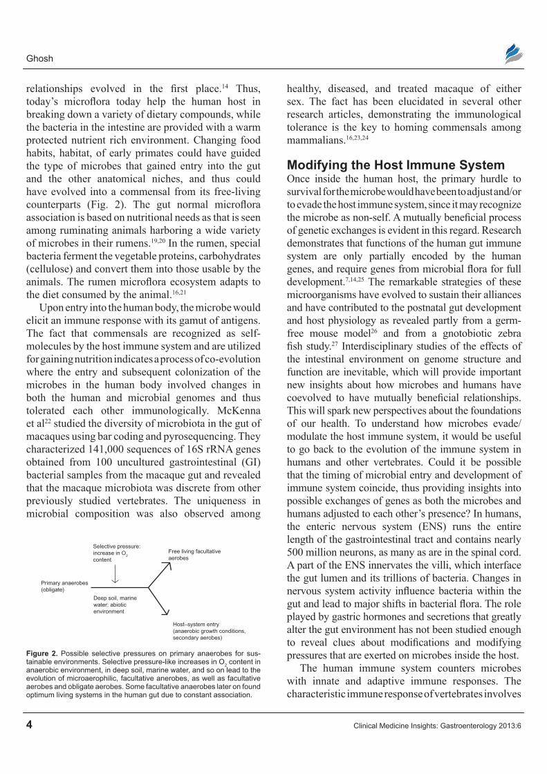
relationships evolved in the first place.14 Thus, today’s microflora today help the human host in breaking down a variety of dietary compounds, while the bacteria in the intestine are provided with a warm protected nutrient rich environment. Changing food habits, habitat, of early primates could have guided the type of microbes that gained entry into the gut and the other anatomical niches, and thus could have evolved into a commensal from its free-living counterparts (Fig. 2). The gut normal microflora association is based on nutritional needs as that is seen among ruminating animals harboring a wide variety of microbes in their rumens.19,20 In the rumen, special bacteria ferment the vegetable proteins, carbohydrates (cellulose) and convert them into those usable by the animals. The rumen microflora ecosystem adapts to the diet consumed by the animal.16,21
Upon entry into the human body, the microbe would elicit an immune response with its gamut of antigens. The fact that commensals are recognized as self-molecules by the host immune system and are utilized for gaining nutrition indicates a process of co-evolution where the entry and subsequent colonization of the microbes in the human body involved changes in both the human and microbial genomes and thus tolerated each other immunologically. McKenna et al22 studied the diversity of microbiota in the gut of macaques using bar coding and pyrosequencing. They characterized 141,000 sequences of 16S rRNA genes obtained from 100 uncultured gastrointestinal (GI) bacterial samples from the macaque gut and revealed that the macaque microbiota was discrete from other previously studied vertebrates. The uniqueness in microbial composition was also observed among
healthy, diseased, and treated macaque of either sex. The fact has been elucidated in several other research articles, demonstrating the immunological tolerance is the key to homing commensals among mammalians.16,23,24
Modifying the Host Immune systemOnce inside the human host, the primary hurdle to survival for the microbe would have been to adjust and/or to evade the host immune system, since it may recognize the microbe as non-self. A mutually beneficial process of genetic exchanges is evident in this regard. Research demonstrates that functions of the human gut immune system are only partially encoded by the human genes, and require genes from microbial flora for full development.7,14,25 The remarkable strategies of these microorganisms have evolved to sustain their alliances and have contributed to the postnatal gut development and host physiology as revealed partly from a germ-free mouse model26 and from a gnotobiotic zebra fish study.27 Interdisciplinary studies of the effects of the intestinal environment on genome structure and function are inevitable, which will provide important new insights about how microbes and humans have coevolved to have mutually beneficial relationships. This will spark new perspectives about the foundations of our health. To understand how microbes evade/modulate the host immune system, it would be useful to go back to the evolution of the immune system in humans and other vertebrates. Could it be possible that the timing of microbial entry and development of immune system coincide, thus providing insights into possible exchanges of genes as both the microbes and humans adjusted to each other’s presence? In humans, the enteric nervous system (ENS) runs the entire length of the gastrointestinal tract and contains nearly 500 million neurons, as many as are in the spinal cord. A part of the ENS innervates the villi, which interface the gut lumen and its trillions of bacteria. Changes in nervous system activity influence bacteria within the gut and lead to major shifts in bacterial flora. The role played by gastric hormones and secretions that greatly alter the gut environment has not been studied enough to reveal clues about modifications and modifying pressures that are exerted on microbes inside the host.
The human immune system counters microbes with innate and adaptive immune responses. The characteristic immune response of vertebrates involves
Selective pressure:increase in O2
content
Free living facultativeaerobes
Host–system entry(anaerobic growth conditions,secondary aerobes)
Deep soil, marinewater: abioticenvironment
Primary anaerobes(obligate)
Figure 2. Possible selective pressures on primary anaerobes for sus-tainable environments. Selective pressure-like increases in O2 content in anaerobic environment, in deep soil, marine water, and so on lead to the evolution of microaerophilic, facultative anerobes, as well as facultative aerobes and obligate aerobes. Some facultative anaerobes later on found optimum living systems in the human gut due to constant association.

Microbial evolution to commensalism and pathogenicity
Clinical Medicine Insights: Gastroenterology 2013:6 5
the production of immunoglobulin in light and heavy chains, the capacity to generate diverse variable region sequences, and mechanisms to rearrange gene segments during development.28,29 The central feature of the adaptive immune system is the evolution of diverse unique antigen receptors that lymphocytes bear in our vertebrate ancestors, leading to a greater survival advantage. Immunologists and evolutionary biologists long believed that this adaptive immune system was exclusive to the higher vertebrates. However, T-cell-like lymphocytes or thymus-like tissue in the lamprey—a fish-like living fossil which evolved around 500 million years ago—had the capacity to form antibodies, has been discovered recently.30 All jawed vertebrates assemble their antigen-receptor genes through recombinatorial rearrangement of different immunoglobulin or T cell receptor gene segments while the surviving jawless vertebrates, lampreys and hagfish, which evolved through recombinatorial assembly of leucine-rich-repeat genetic modules to encode variable lymphocyte receptors.29,31,32 The immunoglobulin (Ig) is said to be a vertebrate invention.33,34 The immunoglobulin heavy chain (IgH) class switch recombination requires activation-induced cytidine deaminase.33 The occurrence of light weight immunoglobulin chains, with heavy chains differing from Immunoglobulin M, have been shown to have started at the phylogenetic stage when vertebrates emerged out of the water to land, concurrent with the evolution of high-pressure blood vascular systems in these organisms.31,32,34,35 Changes in the rate of circulation, osmotic pressure, and hydrostatic pressure of blood could have possibly affected both migration of cells from the vessels and their movement into the extravascular compartments, necessitating a need for low molecular weight immunoglobulins in vertebrates.24,34 Thus, immunoglobulin-like Immunoglobulin G is less prominent in amphibians today. Other Ig like IgA (immunoglobulin A) class are found in birds and mammals but are not seen in other poikilothermal animals. While IgE (immunoglobulin E) seem confined to higher advanced vertebrates like mammals.36,37
Again, given the fact that the mainstay of vertebrate immune system, particularly in birds and mammals, has the ability to produce variable antigen-specific immunoglobulins, it is difficult to envision
the evolution and production of an effective and highly specific immune response without the ability to rearrange a limited number of immunoglobulin genes present in these organisms.38 The evolution of adaptive immunity seems to have been caused by the insertion of a putative immunoglobulin-like gene by a transposable element, which many researchers believe to be a retrotransposon.24,36,39,40 This gave the genes for immunoglobulins the ability to rearrange and thus generate genetic diversity. What could have been the carrier source of this mobile gene? Could it have been initially carried by a microbe, and transferred to the human/vertebrate host in the process of gaining entry into this system? Certain evidence suggests that this could be a possible mechanism.28,41
The Recombination Activating Genes (RAG 1 and 2) are both critical in rearranging the immunoglobulin gene segments in T and B cells.28,32 Again, a homology detected in these DNA processing enzyme genes and the site-specific recombinases in prokaryotes suggests a relation between the vertebrate immune system and the rearrangement and recombination mechanisms of prokaryotes. Furthermore, the RAG 1 homology domain resembles the integrase (INT) family of microbial site-specific recombinases, in particular the E. coli site specific fim recombinases appeared to be the most related to the RAG 1 protein,28 suggesting that the recombination and rearrangement capacity of the human immunoglobulin system is possibly derived from an association with microbes during the evolutionary stages (Fig. 3). Similarly RAG 2
MicrobeMicrobiome
Putativeretrotransposons
Human genome
Variability in lgVariable antigenon T and B cells
Human
Figure 3. Possible changes in the human genome due to residential microflora. Human genome underwent possible rearrangement and reassortment with the intervention of retrotransposon from commensals leading to the variability in immunoglobulin (Ig) and hence in variable receptors on T and B cells.

Ghosh
6 Clinical Medicine Insights: Gastroenterology 2013:6
resembles the bacterial integration host factor (IHF) proteins, a fact that further bolsters the argument that an exchange of genes took place to allow colonization of microbes in the hosts.28,42–44
The adaptive immune response arsenal of the human involves the T and B lymphocytes, including their activation and maturation. Lymphocytes undergo key parts of their maturation in the gut, in specific lymphoid structures called Peyer’s patches. In Peyer’s patches, immature T and B cells are exposed to antigenic epitopes, enabling them to differentiate between self and non-self antigens through a process termed Lymphocyte Education.45 These epitopes are provided by the commensal microbial populations of the gut.46 This can thus be explained as a co- evolutionary adaptation of host and the microbe.
The fact that commensal microbes express self-antigens of humans is an adaptation in order to evade the host immune system, as it enables the commensal to gain recognition by the host as “self”. A possible horizontal exchange of genes encoding these antigens could have resulted in the commensal mimicking the host self-antigens.46 Simultaneously, as the microbes started producing the antigens, the human genome lost the use of these genes and hence ceased to carry them on it. Thus the function of providing antigens for lymphocyte education could have been passed on to the commensal microbes (Fig. 3). In mice and rabbits, the commensal flora also drives the formation of Peyer’s patches in the gut. In addition, the timing for development of memory B cells in the host coincides with the advent of the gram-negative bacteria in the gut, indicating a process of co-evolution.25,45,47,48
The innate response is the primary immune reaction mounted upon first entry of the microbe. But the need for maintaining large microbial populations to serve host nutritional needs forced the intestinal immune system to tolerate intestinal microbial antigens, in a way that assists their entry and colonization. Indeed, a commensal bacterium modulates the expression of genes that regulate several important intestinal functions like nutrient adsorption, xenobiotic metabolism, and post natal intestinal maturation.46,49 A simple example for this is the paneth cells on the gut lining that release granules containing anti microbial components upon sensing invading microbes.50 The cluster of differentiation (cd) 1d (cd1d), a major histo-compatibility complex (MHC) class I-like molecule
play a vital role in regulating intestinal colonization through mechanisms that include the control of paneth cell function.51 These cells synthesize and secrete Angiogenin 4, a bacterial killer. Angiogenin levels in paneth cells are regulated as per changes in gut microflora using bacterial muramyl dipeptide (MDP) through nucleotide-binding oligomerization domain 2 (NOD2) activation.52 Again, Angiogenin 4 specifically targets gram-positive bacteria and leaves the gram-negative bacteria (that constitute the bulk of adult human intestinal flora) untouched, pointing to an adaptation by the gram negative bacteria like E. coli to both evade and regulate the amounts of antibacterial component expressed by the genes. At the same time, this selective expression of Angiogenin 4 allows the human intestine to remove the invading bacteria and prevent commensal bacteria from invading intestinal barriers and containing their populations in the gut. In fact, the intestinal tract has evolved to regulate commensals through various strategies that allow a symbiotic relationship with the host and restrict the invasion of micro-organisms through the epithelial barrier. Innate immune sensors belonging to the cytoplasmic NLR [NOD-like receptor; or nucleotide-binding domain (NBD)- and leucine-rich repeat (LRR)-containing receptor] family of proteins perform a vital role in formative intestinal microbiota.53
Furthermore, the post-natal gut flora consists of predominantly gram-negative bacteria that start shifting towards gram-positive bacteria after weaning and the onset of sold diet. To allow for this change, the production of Angiogenin 4 is increased, exemplifying evolutionary interaction between the host and the microbes. Thus bacterial and host genomes collaborate in shaping the composition of the gut micro flora.54 Again, toll-like receptors (TLRs) are transmembrane proteins expressed by sentinel cells of the innate immune system, which recognize invading microbes (pathogens) and activate signaling pathways that initiate immune and inflammatory responses to regulate the homeostasis. 13 different proteins (TLR1-TLR13) of the mammalian TLR family consist of an extracellular portion containing leucine-rich repeats, a transmembrane region and a cytoplasmic tail, called the TIR (Toll-IL-1R (Interleukin-1-Receptor)) homology domain and serve as receptors for diverse ligands like bacterial cell wall components, viral

Microbial evolution to commensalism and pathogenicity
Clinical Medicine Insights: Gastroenterology 2013:6 7
double-stranded RNA and small-molecule anti-viral or immunomodulatory compounds. Thus, TLRs are pattern recognition receptors (PRR) that have evolved to differentiate commensals (the self) from pathogens (the non-self) by pathogen-associated molecular patterns (PAMP). TLRs are thus an earliest conserved entity in immune system to restore sustainable homeostasis in host.55 Some questions remain, surrounding the nature of this collaboration, whether it is at the genetic level, and where exchange of genes has occurred between the host and microbial genome.
Mimicking the Host physiology Aids colonizationNot only does a microbe show immunological adaptation towards the host, there are certain other changes that the microbe singularly undergoes to be able to colonize in the host tissue.56,57 These changes include the production of receptors and proteins for adhesion to tissues, formation of pilli to avoid desquamation, and use of nutrients like iron for growth and colonization. Commensal microbes are known to produce hemolysins, to break host hemoglobin and use the iron from it, or to produce human transferring-like receptors for iron-capturing in the gut, that compete with the human transferrin receptors. This could have evolved as a result of the exchange of genes encoding the transferring receptors from host to microbe, or through polymorphisms in the microbial genes.7,14 Commensal and pathogens also mimic signaling pathways of the human host to survive and evade an antagonistic host response. Secretory IgA, in the host gut, promotes bacterial biofilm formation within normal gut flora but also limits the expansion and translocation of undesirable pathogenic populations. Host intestinal epithelium secretes bacteria-specific fucosylated glycoconjugates; expression of the glycoconjugates provides lectin-like receptors for the attachment of intestinal flora.
The microorganism trying to settle in either as a commensal or a pathogen has been helpful for studying the intestinal physiology of complex animals.58 For example the cyclic guanosine monophosphate (GMP) signaling pathway has been mimicked by the microbes, and they produce an agonist (EAST1- enteroaggregative E. coli-heat stable enterotoxin 1) for guanylin, the natural ligand of the GC (guanyl
cyclase)/cyclic GMP signaling. Some microbes also synthesize a long-lived super agonist of guanylin. For example the LT (heat labile toxin)/CT (cholera toxin) secreted by E. coli like ETEC (Enterotoxicogenic E. coli)30 and V. cholerae O1, O139,59,60 that interact with the monosialotetrahexosyl ganglioside 1 (GM1) gangliosides and increase adenosine monophosphate (AMP) levels, decrease sodium–potassium and chlorine absorption and aid in the etiology of diarrhoeae.8,61,62 What is interesting here is the question of whether changes like these, that help a microbe survive as a pathogen in the short term, help it become a commensal eventually in the long run. Certain polymorphisms are the key actors of immune system like human leukocyte antigens (HLA) and interleukin 1 in the host show evolutionary processes that assisted the colonization of commensal.47,63
Do pathogens have commensal Tendencies or Vice Versa?Why do certain microbes become pathogens in the host, while certain microbes remain commensals? In order to be a successful enteric pathogen, a microbe has to be a good colonizer, and to be able to interact with the target cells effectively, competing for nutrients as well as inducing secretion of water and electrolytes.62,64 Mechanisms for colonization are common for both commensals and pathogens inside the host. What differentiates them are certain virulent genes that are carried exclusively by the pathogens called genome islands (such as Pathogenicity Islands (PAIs)), and that are either absent or silent in the commensal strain. Did a pathogen loose virulent gene sequences and eventually turn into a commensal in the present day, with genes for colonization intact? Or are today’s commensals slowly gaining virulent genes while competing for survival with the present-day pathogens occupying the same anatomical niche in the host system? In addition, the triggers (selective pressure?) of this loss or gain of genes are not yet understood.
Once inside the host, the commensal genome is under constant pressure and it exists in a dynamic mutable state,65,66 exchanges mobile genetic elements, and/or single base polymorphisms have helped the commensal to face newer challenges the host offered to its survival. The transition from a commensal state to a pathogenic state was studied in Staphylococcus

Ghosh
8 Clinical Medicine Insights: Gastroenterology 2013:6
epidermidis.67 This study revealed the involvement of certain single nucleotide polymorphisms (SNPs) for such a transition. Host agents, like reactive oxygen species produced by phagocytes, that induce an SOS repair response in the microbe genome, have promoted the transfer of mobile genetic elements from different microbial strains coexisting in the host.65 SOS response in particular has enhanced the phage and plasmid transfer of DNA and horizontal transfer of conjugative elements to the microbial genome. Could this possibly be the route in which commensal microbes acquired certain virulence genes, either in the form of SNPs or genomic islands through horizontal gene transfer? Expression of virulence by an otherwise commensal strain would have elicited an immune response from the host, and restarted the competition for survival. Thus, it is possible that this selective pressure paved the way for existence of carrier stage microbes that have the virulent gene but do not express it.
Carrier stage is explained as a situation where host and microbes adapt to one another, and underscores a mutability that defines host–microbial interactions.7 A possible polymorphism in the regulatory regions of the virulence genes (like promoters and silencers) in a carrier stage might explain the non-virulence of the carrier stage. Again, carrier stages have also been viewed as a phase of the commensal development of a pathogenic strain to establish a niche in the host tissue.7,68
Virulence is a Result of Loss or Gain of Genes via Horizontal TransferWhat makes a commensal acquire virulence genes? It is partly the immune system itself that provides the selective pressure, where acquisition of virulence factors facilitates colonization in the face of immune response, and over a period of time the commensal becomes a pathogen.25,47 However, could it also be argued that the same immune response to a pathogen can lead it to evolve into a commensal through progressively decreased virulence? The emergence of a pathogen from a previously commensal strain might involve exchange of gene segments between a host and a microbe, further implying that the host assists the microbe in settling down in the system, as well as in the uptake of gene segments from phages and other sources in the host. An example in this regard is
the Vibrio cholerae O1, in which the genes upstream cholera toxin (CTX) belong to filamentous phage (CTXǾ) that replicates as a plasmid and is responsible for the horizontal transfer of a pathogen element (CTX) to the non-toxigenic V. cholerae.62,69 Further, is gaining genes of virulence alone responsible for the conversion of a commensal into a pathogen, or vice versa? It has been conclusively proven that virulence traits can not only be acquired with the gaining of virulence genes but also by the shedding of genes that are detrimental to new pathogenic lifestyles.70 Shigella spp. is another case, which is known to have evolved from E. coli to become a pathogen not only by acquiring virulence genes on a plasmid, but also by loosing genes that exerted inhibitory effects on toxin functions.70–72 Another example is E. coli O157:H7. The commensal E. coli gained the property of verotoxigenicity by the intervention of phage 933. In true sense, commensal E. coli thus has 4 different genetic groups now, including A, B1, B2, D and E with 181 O antigens, 80 K antigens and 65 H antigens respectively, and also in all forms of relationship with the host, may it be a commensal, an asymptomatic carrier and/or a symptomatic pathogen.73 This provides us an evolutionary mechanism for commensals to become virulent. Thus, while such genetic “black holes” triggered production of pathogens from related commensal strains, could the same process not commensalize an existing pathogen? It can thus be argued that pathogens of today do show commensal tendencies and might be normal harmless colonizers of the human body tomorrow.
Now if a genetic exchange of transposable virulent genes and other factors are necessary for colonization and if it is so common in the host system, why do we have so few pathogens compared to the large numbers of commensal in the body? Why is pathogenicity so rare? The answer to this could again lie in the arrangement of genes required for pathogenicity.73 Bacterial populations are clonal in nature;68,74,75 thus, distinct bacterial clones are often the cause of diseases, as well as an increase in outbreaks and infection frequencies. The inheritance of PAI and other mobile elements responsible for virulence does not necessarily create a new pathogenic species. The analysis of pathogenic bacterial species indicates that a certain unique combination of the pathogenic genes may arise only once during the evolutionary

Microbial evolution to commensalism and pathogenicity
Clinical Medicine Insights: Gastroenterology 2013:6 9
process of the same species,21,68,75 but due to an inappropriate arrangement of virulent genes, they remain non pathogenic, and do not express their pathogenicity. In other words, they become non-virulent (or asymptomatic), yet they remain virulent gene-containing carriers. Moreover, very rarely, a pathogen horizontally transfers its entire set of virulent genes to another related or unrelated strain in the same dangerous arrangement, explaining the limited rise of newer pathogenic strains (Fig. 4). Could this be an evolutionarily ingrained control mechanism evolved by the host system to check the growing number of pathogens? A deeper understanding of this mechanism might hold the answer to the changing virulence patterns faced by people in recurring bouts of infectious diseases, where each new outbreak pops up a new causative strain of the same species.
Co-evolution of host and the microbe favors an outcome in which the cost of eliminating the microbe is high and hence the microbe settles as a commensal or a carrier. The remaining question concerns whether the host immune response can promote transfer of genetic element among strains, helping them either lose or gain virulence factors. The fact that DNA mutation rates are higher in the strains within a host compared to free-living strains supports this argument.62 For example, the increase in frequency of lysogeny in E. coli in the intestine in vivo suggests that host promotes the spread of the phage containing a fitness factor.65,66,76 Thus the journey from commensal to carrier to pathogen and vice versa can be viewed
as a dynamic ongoing process of evolution. Studying the various changes assisting the colonization of these microbes in hosts such as humans, at the gene level, can help one to understand the mechanisms involved in the process of development of newer virulent strains (Fig. 4).
Final RemarksRecent progress in the human microbiome project, genome research and molecular epidemiology, as well as functional investigations at the transcriptome level, have revealed intriguing new insights into the genetics and physiology of several commensals. Many of the commensals live on the edge of commensalism and pathogenicity. This becomes evident among immuno-compromised hosts. Gene transfer makes the innocuous commensal pathogenic, which leads it to emerge as a new pathogen. A potential virulence factor may enable microflora to find a new habitat or a new host. Traditional cultivation, microscopy and determination of fermentation/degradation products are still important. However, future studies of the microbe-microbe and the microbe-host cross-talk will strengthen our knowledge about the composition and function of the microflora under the light of the germ-free associated characteristic-microflora associated characteristic (GAC-MAC) concept.77 Gnotobiotic study may help to get some useful answers to questions raised in this review. Recently, Al-Asmakh et al78 found that two genes implicated in anxiety, nerve growth factor-inducible clone A (NGF1-A) and brain-derived
Commensal
Loss of virulence genes,black holes
Pathogen
Gain of virulence from phages,horizontal transfer
Rearrangement of silentvirulent genes or acquiringregulators for their expression
Invasion of silentvirulent genes
Eventual loss ofthe virulent genes as a response toselective pressurefo competetion tosurvive
Asymptomatic carrier
Inheritance of virulentgenes in randomarrangement renderingthe genes silent
A possible mechanism of genetic exchange between the microflora of the humanintestine
Figure 4. Possible mechanism of genetic exchange between the microflora of the human intestine. Gut is the best anatomical niche in humans where commensals, asymptomatic carriers and symptomatic pathogens sustain and survive well. It is assumed that either certain commensals gain virulence by horizontal gene transfer and turn into pathogens, or pathogenic counterparts lose the traits required to become commensals. Likely, an asymptomatic carrier evolved either from commensal or pathogens by different mechanisms to survive better in the host system, maybe by gene silencing or gene aug-menting. It is assumed that the order of evolution occurred from commensals to asymptomatic pathogens to pathogens, or vice versa.

Ghosh
10 Clinical Medicine Insights: Gastroenterology 2013:6
neurotrophic factor (BDNF) were down-regulated in multiple brain regions in the germ-free animals, and these behavioural changes were found to be interrelated with the commensal gut bacteria, which influenced the expression of nearly 40 genes of the human brain. The commensal either in the gut or on the skin and elsewhere are involved in the regulation of multiple host metabolic pathways. Gut microflora in particular give rise to interactive host-microbiota metabolic, signaling, and immune-inflammatory alliances that physiologically connect the gut, liver, muscle, and brain79 It is possible to genetically manipulate the host epithelia, associated immune systems, and potential residential microflora. The biofilm formation, space distribution of neighbouring microbe populations and the competition to sustain in an ecological niche, is very intriguing.
Nineteenth-century French microbiologist Louis Pasteur believed that animals cannot exist without a population of commensal and mutual organisms, and early experiments to raise germ-free animals were met with failure. All germ-free animals have weak, poorly developed immune systems. This suggests that the roles of normal microscopic organisms are very important. The immune system in the gut mainly recognizes commensals from pathogens, and tolerates the resident flora, while at the same time mounting inhibitory immune responses against the pathogens. This is possible only when there is a large amount of cross talk between the microbial cell and epithelial cell at the mucosal layer. The exact nature of this cross talk is as yet unclear, but can be studied by observing the differential response that the epithelium gives in response to a pathogen and a commensal. Again, it is possible that given the plasticity and dynamic nature of microbial genome, co culture of a commensal and a pathogen at the intestinal epithelium might cause the exchange of survival advantages between 2 types of microbial strains, or one might outnumber the other, synergistically with the epithelium cells. Investigations to this effect can help to understand the basis of the molecular cross talk that helps the intestine tolerate certain microbes and eliminate others. It would also help us appreciate the manner in which a microbe slips from commensalism to pathogenicity in its quest to survival in the intestinal or likely micro-environment. Similarly
to “which came first, the chicken or the egg”, the question here remains: “which evolved first, pathogen or commensal?” The more we learn about these relations, the more we will be benefitted.
Author contributionsConceived and designed the concept: ARG. Analyzed the data: ARG. Wrote the first draft of the manuscript: ARG. Developed the structure and arguments for the paper: ARG. Made critical revisions and approved final version: ARG. The author reviewed and approved of the final manuscript.
FundingIntramural fund of VIT University for the promotion of research and development.
competing InterestsAuthor(s) disclose no potential conflicts of interest.
Disclosures and ethicsAs a requirement of publication the author has provided signed confirmation of compliance with ethical and legal obligations including but not limited to compliance with ICMJE authorship and competing interests guidelines, that the article is neither under consideration for publication nor published elsewhere, of their compliance with legal and ethical guidelines concerning human and animal research participants (if applicable), and that permission has been obtained for reproduction of any copyrighted material. This article was subject to blind, independent, expert peer review. The reviewers reported no competing interests.
References1. Alekshun MN, Levy SB. Commensals upon us. Biochemica Pharmacol.
2006;71:893–900.2. Zapalski MK. Is absence of proof a proof of absence? Comments on com-
mensalism. Palaeogeogr Palaeoclimatol Palaeoecol. 2011;302:484–8.3. Todar K. Todar’s online textbook of Bacteriology. University of Wisconsin
Department of Bacteriology. The normal bacterial flora of humans. 2008. Avail-able at http://textbookofbacteriology.net/normalflora_4.html (May18, 2013).
4. The Heritage Dictionary. Commensalism. Available at: http://www.answers.com/topic/commensalism. Accessed May 10, 2013.
5. Boucher DH, James S, Keeler KH. The ecology of mutualism. Ann Rev Ecol Syst. 1982;13:315–34.
6. Sachs JL, Hollowell AC. The Origins of cooperative bacterial communities. mBio. 2012;3:e00099–12.
7. Casadevall A, Pirofski LA. Host–pathogen interactions—basic concepts of microbial commensalism, colonization, infection, and disease. Infect Immun. 2000;68:6511–8.

Microbial evolution to commensalism and pathogenicity
Clinical Medicine Insights: Gastroenterology 2013:6 11
8. Alteri CJ, Mobley HL. Escherichia coli physiology and metabolism dictates adaptation to diverse host microenvironment. Curr Opinion Microbiol. 2012;15:1–7.
9. Mereschkowski C. Uber Natur und ursprung der chromatophoren im Pflanzenreiche. Biol Centralblatt. 1905;25:593–604.
10. Sapp J, Carrapiço F, Zolotonosov M. Symbiogenesis: the hidden face of Constantin Merezhkowsky. Hist Philos Life Sci. 2002;24:413–40.
11. Kutschera U. Symbiogenesis, natural selection, and the dynamic Earth. Theory Biosci. 2009;128:191–203.
12. Fausto-Sterling A. Is nature really red in tooth and claw? Discover. 1993;14: 24–7.
13. Margulis L. Symbiosis in Cell Evolution. 1st ed. New York: Freeman; 1981.
14. Cash HL, Hooper LV. Commensal bacteria shape intestinal immune system development. ASM News. 2005;71:77–83.
15. Frese SA, Benson AK, Tannock GW, et al. The evolution of host specialization in the vertebrate gut symbiont Lactobacillus reuteri. PLoS Genet. 2011;7:e1001314.
16. Taschuk R, Griebel PJ. Commensal microbiome effects on mucosal immune system development in the ruminant gastrointestinal tract. Anim Health Res Rev. 2012;13:129–41.
17. Bauer E, Williums BA, Smidt H, Verstegen MWA, Mosenthin R. Influence of the gastrointestinal microbiota on the development of the immune system in young animals. Curr Issues Intest Microbiol. 2006;7: 35–52.
18. Tokeshi M. On the evolution of commensalism in the Chironomidae. Fresh Water Biol. 1993;29:481–9.
19. Ley RE, Lozupone CA, Hamady M, Knight R, Gordon JI. Worlds within worlds: evolution of the vertebrate gut microbiota. Nat Rev Microbiol. 2008;6:776–88.
20. Stevens CE, Hume ID. Contributions of microbes in vertebrate gastrointestinal tract to production and conservation of nutrients. Physiol Rev. 1998;78:393–427.
21. Guiney D. Regulation of bacterial virulence gene expression by the host environment. J Clin Invest. 1997;99:565–9.
22. McKenna P, Hoffmann C, Minkah N, Aye PP, Lackner A, et al. The Macaque gut microbiome in health, lentiviral Infection, and chronic enterocolitis. PLoS Pathog. 2008;4:e20.
23. Guarino A, Wudy A, Basile F, Ruberto E, Buccigrossi V. Composition and roles of intestinal microbiota in children. J Matern Fetal Neonatal Med. 2012;25(Suppl 1):63–6.
24. McFall-Ngai M. Adaptive immunity: care for the community. Nature. 2007;445:153.
25. Hooper LV. Bacterial contributions to mammalian gut development. Trends Microbiol. 2004;12:129–34.
26. Xu J, Gordon JI. Honor thy symbionts. Proc Natl Acad Sci U S A. 2003;100: 10452–9.
27. Rawls JF, Samuel BS, Gordon JI. Gnotobiotic zebrafish reveal evolution-arily conserved responses to the gut microbiota. Proc Natl Acad Sci U S A. 2004;101(13):4596–601.
28. Bernstein RM, Schulutzeer SF, Bernstein H, Marchalonis JJ. Primordial emergence of the recombination activating gene 1(RAG 1): sequence of the complete shark gene indicates homology to microbial integrases. Proc Natl Acad Sci U S A. 1996;93:9454–9.
29. Cooper MD, Alder MN. The evolution of adaptive immune systems. Cell. 2006;124:815–22.
30. Ghosh AR, Koley H, De D, Paul M, Nair GB, Sen D. Enterotoxigenic Escherichia coli associated diarrhoea among infants aged less than six months in Calcutta, India. Eur J Epid. 1996;12:81–4.
31. Das S, Hirano M, Tako R, MacCallister C, Nicolaidis N. Evolutionary genomics of immunoglobulin-encoding loci in vertibrates. Curr Genomics. 2012;12:1389–2029.
32. Saha NR, Smith J, Amemiya CT. Evolution of adaptive immune recognition in jawless vertebrates. Semin Immunol. 2010;22:25–33.
33. Chaudhuri J, Basu U, Zarrin A, et al. Evolution of the immunoglobulin heavy chain class switch recombination mechanism. Adv Immunol. 2007;94: 157–214.
34. Manning MJ. Evolution of the vertebrate immune system: symposium, anti microbial defense systems. J Roy Soc Med. 1979;72:683–9.
35. Manning MJ, Turner RJ. Comparitive Immunobiology. Glasgow: Blackie and Sons Ltd; 1976.
36. Jurd RD. In: Solomon JB, Horton JD, editors. Developmental Immunobiology. 1979; Amsterdam: North Holland Biomedical Press; 1979.
37. Ley RE, Peterson DA, Gordon JI. Ecological and evolutionary forces shaping microbial diversity in the human intestine. Cell. 2006;124:837–48.
38. Bartl S, Baltimore D, Weissman IL. Molecular evolution of the vertebrate immune system. Proc Natl Acad Sci U S A. 1994;91:10769–70.
39. Berg JM, Tymockzo JL, Stryer L. Biochemistry. 5th ed. New York: WH Freeman; 2002.
40. Schatz DG, Oettinger MA, Baltimore D. The V(D)J Recombination Activating Gene, RAG 1. Cell. 1989;59:1035–48.
41. Lee YK, Mazmanian SK. Has the microbiota played a critical role in the evolution of the adaptive immune system? Science. 2010;330:1768–73.
42. Cuomo CC, Oettinger MA. Analysis of regions of RAG-2 important for V(D)J recombination. Nucleic Acids Res. 1994;22:1810–4.
43. Sadofsky MJ, Hesse JE, Gellert M. Definition of a core region of RAG-2 that is functional in V(D)J recombination. Nucleic Acids Res. 1994;22: 1805–9.
44. Schatz DG, Chun JJ. V(D)J recombination and the transgenic brain blues. New Biol. 1992;4:188–96.
45. Lotz M, Menard S, Hornef M. Innate immune recognition on the intestinal mucosa. Int J Med Microbiol. 2007;297:379–92.
46. Chung H, Kasper DL. Microbiota-stimulated immune mechanisms to maintain gut homeostasis. Curr Opinion Immunol. 2010;22:455–60.
47. Albini B, van Oss CJ. Microbial pathogenicity and host-parasite relationships. In: Milgrom F, Flanagan TD, editors. Medical Microbiology. Ann Arbor; 1982:81–106.
48. Kurtz J. Memory in the innate and adaptive immune systems. Microbes Infect. 2004;6:1410–7.
49. Hooper LV, Wong MH, Thelin A, Hanson L, Falk PG, Gordon JI. Molecular analysis of commensal host–microbial relationship in the intestine. Science. 2001;291:881–4.
50. Persing DH, Tenover FC, Versalovic J, et al. In: Molecular Microbiology: Diagnostic Principles and Applications. Washington: ASM Press; 2004.
51. Nieuwenhuis EES, Matsumoto T, Lindenbergh D, et al. Cd1d-dependent regulation of bacterial colonization in the intestine of mice. J Clin Invest. 2009;119:1241–50.
52. Kobayashi K, Chamaillard M, Ogura Y, et al. Nod2-dependent regulation of innate and adaptive immunity in the intestinal tract. Science. 2005;307: 731–4.
53. Biswas A, Kobayashi KS. Regulation of intestinal microbiota by the NLR protein family. Int Immunol. 2013;25:207–14.
54. Pamer EG. Immune responses to commensal and environmental microbes. Nat Immunol. 2007;8:1173–8.
55. Lee J, Sayed N, Hunter A, et al. Activation of innate immunity is required for efficient nuclear reprogramming. Cell. 2012;151:547–8.
56. Ghosh AR, Nair GB, Naik TN, Paul M, Pal SC, Sen D. Enteroadherent Escherichia coli: an important diarrhoeagenic agent in infants aged below six months in Calcutta, India. J Med Microbiol. 1992;33:264–8.
57. Ghosh AR, Sen D, Sack DA, Hoque AT. Evaluation of conventional media for detection of colonization factor antigens of enterotoxigenic Escherichia coli. J Clin Microbiol. 1993;31:2163–6.
58. Koley H, Ghosh AN, Paul M, Ghosh AR, Ganguly PK, Nair GB. Colonization ability and intestinal pathology of rabbits orally fed with Vibrio cholerae O139 Bengal. Indian J Med Res. 1995;101:57–60.
59. Ghosh AR, Koley H, De D, et al. Incidence and toxigenicity of Vibrio cholerae in a freshwater lake during the epidemic of cholera caused by serogroup O139 Bengal in Calcutta, India. FEMS Microbiol. Ecol. 1994;14: 281–5.
60. Roy S, Dutta B, Ghosh AR, et al. Molecular tracking of the lineage of strains of Vibrio cholerae O1 biotype El Tor associated with a cholera outbreak in Andaman and Nicobar Islands, India. Trop Med Int Health. 2005;10: 604–11.

Ghosh
12 Clinical Medicine Insights: Gastroenterology 2013:6
61. Fasano A. Cellualr microbiology: can we learn cell physiology from microorganisms? Am J Physiol. 1999;276:C765–6.
62. Fasano A. Toxins and the gut: role in human disease. Gut. 2002;50:III9–14. 63. Macpherson AJ, Harris NL. Interactions between commensal intestinal
bacteria and the immune system. Nat Rev Immunol. 2004;4:478–85. 64. Ghosh AR. Bacterial toxin and diarrhea. In: Pandey VD, Singh SK, editors.
Microbial Toxins and Toxigenic Microbes. New Delhi: Studium Press; 2012:59–83.
65. Brown NF, Mark EW, Coomber B, Finlay BB. Crossing the line: selection and evolution of virulence traits. ASM News. 2006;63:159–65.
66. Laohachai KN, Bahadi R, Hardo MB, Hardo PG, Kourie JI. The role of bacterial and non bacterial toxins in the induction of changes in membrane transport: implications for diarrhea. Toxicon. 2003;42:687–707.
67. Wei W, Cao ZW, Zhu YL, et al. Conserved genes in a path from commensalism to pathogenicity: comparative phylogenetic profiles of Staphylococcus epidermidis RP62A and ATCC12228. BMC Genomics. 2006;7:112.
68. Ochman H, Moran NA. Genes lost and genes found: evolution of bacterial pathogenesis and symbiosis. Science. 2001;292:5519.
69. Waldor MK, Mekalanos JJ. Lysogenic conversion by a filamentous phage encoding cholera toxin. Science. 1996;272:1910–4.
70. Maurelli AT, Fernandez RE, Bloch CA, Rode CK, Fasano A. Black holes and bacterial pathogenixity: a large genomic deletion that enhances the virulence of Shigella spp and enteroinvasive Eschericia coli. Proc Natl Acad Sci U S A. 1998;95:3943–8.
71. Bertin Y, Girardeau JP, Chaucheyras-Durand F, et al. Enterohaemorrhagic Escherichia coli gains a competitive advantage by using ethanolamine as a nitrogen source in the bovine intestinal content. Environ Microbiol. 2011;13: 365–77.
72. Garsin DA. Ethanolamine utilization in bacterial pathogens: roles and regu-lation. Nature Rev Microbiol. 2010;8:290–5.
73. Touchon M, Hoede C, Tenaillon O, et al. Organised genome dynamics in the Escherichia coli species results in highly diverse adaptive paths. PLoS Genet. 2009;5:e1000344.
74. Falkow S. The evolution of pathogenicity in Escherichia, Shigella and Salmonella. In: Neidhart N (Ed). Escheichia Coli and Salmonella: Cellular and Molecular Biology. Washington: ASM Press; 1996:2723–9.
75. Falkow S. Perspectices series: host/pathogen interactions. Invasion and intracellular sorting of bacteria: searching for bacterial genes expressed during host/pathogen interactions. J Clin Invest. 1997;100:239–43.
76. Salyers AA, Moon K, Schlesinger D. The human intestinal tract—a hotbed of resistance gene transfer? Clin Microbiol Newslet. 2007;29:40.
77. Midtvedt E. Interactions of bacteria with the host alteration of microflora-associated characteristics of the host; non-immune functions. Microbial Ecol Health Dis. 2000;12(2):186–93.
78. Al-Asmakh M, Anuar F, Zadjali F, Rafter J, Pettersson S. Gut microbial communities modulating brain development and function. Gut Microbes. 2012;3:366–73.
79. Nicholson JK, Holmes E, Kinross J, et al. Host-gut microbiota metabolic interactions. Science. 2012;8(336):1262–7.