Virulence and Pathogenicity of Fungal Pathogens with Special Reference to Candida albicans
Upload
khangminh22Category
view
1download
0
Journal of Tropical Forest Science 21(4): 289–298 (2009) Mohd Farid et al.
289
PATHOGENICITY OF RIGIDOPORUS MICROPORUS AND PHELLINUS NOXIUS AGAINST FOUR MAJOR PLANTATION TREE SPECIES IN PENINSULAR MALAYSIA
A Mohd Farid1, *, SS Lee1, Z Maziah2 & M Patahayah1
1Forest Research Institute Malaysia, 52109 Kepong, Selangor Darul Ehsan, Malaysia2School of Biological Sciences, Universiti Sains Malaysia, 18000 Minden, Penang, Malaysia
Received October 2008
E-mail: [email protected]
MOHD FARID A, LEE SS, MAZIAH Z & PATAHAYAH M. 2009. Pathogenicity of Rigidoporus microporus and Phellinus noxius against four major plantation tree species in Peninsular Malaysia. Rigidoporus microporus and Phellinus noxius are destructive fungi in tropical forest plantations. Early stages of infection by these pathogens are often difficult to detect while trees bearing visible disease symptoms are mostly beyond treatment. Little research has been conducted to determine the aggressiveness of these two pathogens on different tree species. This study was conducted on 24-month-old saplings of Acacia mangium, Azadirachta excelsa, Tectona grandis and Hevea brasiliensis. Inoculum blocks of R. microporus and P. noxius were placed next to roots of saplings and left for 30 weeks for infection to take place. The study revealed that R. microporus and P. noxius were pathogenic to all hosts. However, the degree of pathogenicity varied. Phellinus noxius was more pathogenic to H. brasiliensis than to A. excelsa, T. grandis and A. mangium, while R. microporus was more pathogenic to H. brasiliensis and A. excelsa than to A. mangium and T. grandis. In terms of host susceptibility, H. brasiliensis was the most susceptible to R. microporus followed by A. excelsa, T. grandis and A. mangium. Against P. noxius, T. grandis was the most susceptible, followed by H. brasiliensis, A. excelsa and A. mangium. Saplings infected by both pathogens manifested similar aboveground symptoms, but symptoms belowground varied depending on the fungal species. The presence of adhering soil particles and dark brown mycelial crust on the root was common on saplings inoculated with P. noxius, while white rhizomorphs were usually present on the root surface of saplings inoculated with R. microporus.
Keywords: Host susceptibility, root disease, symptom, forest plantation
MOHD FARID A, LEE SS, MAZIAH Z & PATAHAYAH M. 2009. Kepatogenan kulat Rigidoporus microporus dan Phellinus noxius terhadap empat spesies pokok ladang yang utama di Semenanjung Malaysia. Rigidoporus microporus dan Phellinus noxius merupakan kulat pemusnah di ladang hutan di kawasan tropika. Peringkat awal jangkitan oleh kulat-kulat ini selalunya sukar dikesan manakala kebanyakan pokok yang telah pun menunjukkan gejala penyakit biasanya tidak dapat dirawat lagi. Tidak banyak kajian yang telah dijalankan untuk menentukan tahap keagresifan kedua-dua patogen tersebut terhadap pokok yang berlainan spesies. Kajian ini telah dijalankan terhadap anak-anak pokok Acacia mangium, Azadirachta excelsa, Tectona grandis dan Hevea brasiliensis yang berusia 24 bulan. Blok-blok inokulum kulat R. microporus dan P. noxius diletakkan pada bahagian akar anak-anak pokok tersebut dan dibiarkan selama 30 minggu supaya jangkitan boleh berlaku. Hasil daripada kajian ini mendapati kulat R. microporus dan P. noxius adalah patogenik terhadap kesemua perumah tersebut, tetapi tahap kepatogenan adalah berbeza. Kulat P. noxius didapati lebih patogenik terhadap H. brasiliensis berbanding A. excelsa, T. grandis dan A. mangium manakala kulat R. microporus lebih patogenik terhadap H. brasiliensis dan juga A. excelsa berbanding A. mangium dan T. grandis. Dari segi kerentanan perumah, H. brasiliensis merupakan perumah yang paling rentan terhadap jangkitan kulat R. microporus diikuti oleh A. excelsa, T. grandis dan A. mangium. Sebaliknya, T. grandis merupakan perumah yang paling rentan terhadap jangkitan P. noxius, diikuti oleh H. brasiliensis, A. excelsa dan A .mangium. Anak-anak pokok yang dijangkiti kedua-dua patogen ini menunjukkan gejala serupa pada bahagian silara tetapi gejala berbeza pada bahagian akar bergantung kepada spesies kulat. Kehadiran partikel-partikel tanah dan kerak miselium berwarna coklat kehitaman pada permukaan akar sering kelihatan pada anak pokok yang dijangkiti oleh kulat P. noxius manakala rizomorf putih biasanya hadir pada permukaan akar anak-anak pokok yang telah diinokulasi dengan kulat R. microporus.
INTRODUCTION
Although agroplantations have existed in Peninsular Malaysia since the 1880s, plantations of forest tree species are still relatively recent.
The majority of our older forest plantations were established by the British as a reforestation effort, mainly of forest destroyed during World War II
Journal of Tropical Forest Science 21(4): 289–298 (2009) Mohd Farid et al.
290
(Krishnapillay & Appanah 2002). In the 1980s, the acreage of forest plantations grew rapidly due to the forecasted shortage of timber from the natural forest (Chong 1979). Thus large-scale forest plantations of fast-growing exotic species were established for general utility wood. With the increase in acreage of forest plantations, pest and disease problems have become a greater concern, especially since very little information or data are available about them. In many cases, failures during the early phase of plantation establishment have been linked to serious pest and disease problems (Wingfield 1999). The susceptibility of a particular species to pests and diseases will usually determine its success in the future (Gibson 1979, Ivory 1987, Harrington & Wingfield 1998). Disease surveys conducted in several forest plantations in Peninsular Malaysia have shown that root disease is the main cause of mortality in these plantations (Lee 1997, Mohd Farid et al. 2005). Root disease fungi such as Rigidoporus microporus and Phellinus noxius are known to be pathogenic and destructive to trees in the tropics. These pathogens cause significant economic losses in rubber, cocoa, tea and fruit tree plantations (Fox 1977, Johnston 1989). Trees are rapidly killed by these pathogens and detection during early stages of infection is difficult. In the field, the aboveground symptoms associated with both pathogens are almost similar but belowground they differ. Normally, the presence of aboveground symptoms indicate that the trees are mostly beyond treatment and recovery, as rapid progress of infection makes death imminent (Ismail & Azaldin 1985). However, information on the susceptibility and aggressiveness of these pathogens on forest plantation species in Malaysia remains scanty. Therefore, the aims of this study were, firstly, to assess the susceptibility of four hosts, namely, Acacia mangium, Azadirachta excelsa, Hevea brasiliensis and Tectona grandis against white root (R. microporus) and brown root (P. noxius) diseases using foliar symptoms. Secondly, we aimed to improve skills in detecting incidence of white root and brown root diseases on the selected hosts based on observation of root symptoms in the field.
MATERIALS AND METHODS
A pathogenicity study was conducted on 24- month-old healthy plants of A. mangium, A.
excelsa, H. brasiliensis and T. grandis. The two root disease fungi used in this study were R. microporus (isolate 590) obtained from the Lembaga Getah Malaysia (LGM—Malaysian Rubber Board), Kuala Lumpur and P. noxius (isolate FRIM154) obtained from the Forest Research Institute Malaysia (FRIM) collection. Prior to inoculation, young rubber tree branches obtained from within the FRIM campus were cut into smaller blocks measuring approximately 8 × 1.5−2.0 cm diameter. Six to eight debarked blocks were placed into individual autoclavable plastic bags and 50 ml of 2% malt extract (ME) were added. The mouth of each bag was fitted with a PVC ring and stoppered with a cotton wool plug, covered with aluminium foil and autoclaved at 121 °C for 15 min. The autoclaved bags were transferred into a culture room to cool. Four to five mycelial discs taken from the edge of one-week-old actively growing cultures with a sterilized 5-mm diameter cork borer were used to inoculate the sterilized wood blocks in each plastic bag. The inoculated bags were then incubated in the dark at 28 ± 2 °C for about a month to allow good colonization of the wood blocks by the fungi. In the pathogenicity test, all plants used were obtained from known sources. Acacia mangium and A. excelsa seeds collected from healthy mother trees at FRIM’s campus were germinated and raised at the FRIM nursery. For H. brasiliensis and T. grandis clone T16, seedlings were obtained from the LGM nursery, Sg. Buloh, Selangor and FRIM substation, Mata Ayer, Perlis respectively. The plants were maintained at the FRIM nursery until they were 24 months old and transferred into plastic pots measuring 60 cm diameter × 45 cm height. They were left for a month in the open in the nursery for hardening.
Experimental layout and data analysis
Arrangement of the saplings was according to randomized complete block design (RCBD) with three replications. Two treatment combinations were assigned in each replicate with four types of plant species, two types of fungi and one control. Each treatment consisted of five healthy and uniform saplings. Each sapling was inoculated with six to seven inoculum blocks placed in contact with the taproot. Uninoculated plants were used as controls. Plants were observed at two-weekly intervals for the appearance of aboveground disease symptoms for up to

Journal of Tropical Forest Science 21(4): 289–298 (2009) Mohd Farid et al.
291
30 weeks. Evaluation of disease infection was conducted according to Nandris et al. (1983) with the following modification: the scale used for rating the severity of attack aboveground was 1 (healthy), 2 (yellowing of foliage), 3 (wilting), 4 (defoliation) and 5 (death of plant). The data were then subjected to Fisher’s exact test (2-tail) to determine the interaction between pathogens and symptom development on the hosts using SAS (Statistical Analysis System) package, Version 6.03 (1989). Prior to the test, frequency of symptom appearance was grouped into three categories, namely, healthy (scales 1 and 2), moderate (scales 3 and 4) and severe (scale 5). Plants bearing disease symptoms were brought to the laboratory and fungal isolation was made from the roots to confirm the presence of causal pathogens. Diseased tissues were directly transferred onto Ganoderma selective media (GSM) (Ariffin & Idris 1992) and incubated at room temperature. The diseased tissues were then transferred onto malt extract agar (MEA) and left to grow. Identification of the causal organisms was done based on cultural and microscopic characteristics as described by Stalpers (1987).
RESULTS
Comparison of the pathogenicity of R. microporus and P. noxius for each host
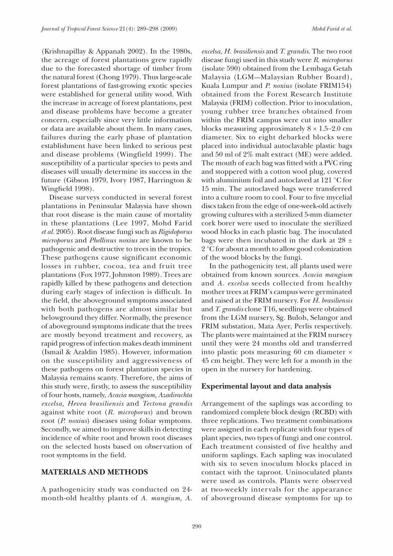
Results of this study revealed that all uninoculated control saplings with the exception of A. excelsa were healthy without any symptoms of root disease infection until the experiment was terminated (Figures 1a, d and g). Belowground, all uninoculated saplings possessed vigorous and clean root systems compared with inoculated saplings. Roots of symptomatic saplings could be separated into two categories depending on the inoculated pathogens. For saplings inoculated with R. microporus, rough white rhizomorphs were present on the surface of roots of all symptomatic
saplings. In contrast a dark brown mycelial crust with soil particles covering the roots was found on diseased saplings inoculated with P. noxius. Table 1 shows the percentage of saplings exhibiting root symptoms for all treatments. All H. brasiliensis saplings inoculated with R. microporus exhibited characteristic white root disease symptoms (Table 1). In contrast all A. excelsa and T. grandis saplings and 93% of H. brasiliensis saplings inoculated with P. noxius exhibited characteristic brown root disease symptoms. Acacia mangium had the fewest symptomatic saplings against both pathogens. Further detailed results, particularly of the aboveground symptoms for each species, are described below.
Acacia mangium
Results indicated that P. noxius was more pathogenic than R. microporus on A. mangium saplings (Figures 1b and c). At week 8, although the percentage of saplings with yellowing symptoms was similar (13.3%) for saplings inoculated with P. noxius and R. microporus, progression of disease symptoms was generally more rapid on saplings inoculated with P. noxius than with R. microporus. In addition, severe symptoms such as defoliation and death of saplings appeared thereafter on saplings inoculated with P. noxius. Sapling death caused by P. noxius and R. microporus was first observed at week 20 and week 22 respectively. At the end of 30 weeks, P. noxius inoculated plants suffered 20% mortality compared with 13% for plants inoculated with R. microporus (Figure 1c).
Hevea brasiliensis
Yellowing symptoms on H. brasiliensis saplings inoculated with P. noxius were observed on 13.3% of saplings at week 4. Symptom severity increased rapidly and by the 12th week, all
Table 1 Percentage of uninoculated control saplings and saplings inoculated with Rigidoporus microporus or Phellinus noxius showing belowground symptoms
Pathogen Acacia mangium Azadirachta excelsa Tectona grandis Hevea brasiliensis
R. microporus 13.33 86.67 53.34 100
P. noxius 46.67 100 100 93.33
Uninoculated control
0 0 0 0
Journal of Tropical Forest Science 21(4): 289–298 (2009) Mohd Farid et al.
292
A. m
angi
um ×
con
trol
A. m
angi
um ×
P. n
oxiu
sA
. man
gium
× R
. mic
ropo
rus
% symptom development
% symptom development
% symptom development
Wee
kW
eek
Wee
k
(a)
(b)
(c)
% symptom development
% symptom development
Wee
kW
eek
(d)
(f)
H. b
rasi
liens
is ×
con
trol
Hea
lthy
Yello
win
gW
iltin
gD
efol
iatio
nD
ead
% symptom development
Wee
k
(e)
H. b
rasi
liens
is ×
P. n
oxiu
sH
. bra
silie
nsis
× R
. mic
ropo
rus
Figu
re 1
D
evel
opm
ent o
f sym
ptom
s on
sapl
ings
of A
caci
a m
angi
um, H
evea
bra
silie
nsis
, Tec
tona
gra
ndis
and
Aza
dira
chta
exce
lsa
inoc
ulat
ed w
ith R
igid
opor
us m
icro
poru
s an
d Ph
ellin
us n
oxiu
s
(con
tinue
d)
Journal of Tropical Forest Science 21(4): 289–298 (2009) Mohd Farid et al.
293
(g)
Wee
k
% symptom development % symptom development
% symptom development % symptom development
% symptom development % symptom development
T. g
rand
is ×
con
trol
T. g
rand
is ×
P. n
oxiu
sT.
gra
ndis
× R
. mic
ropo
rus
Wee
kW
eek
(h)
(i)
A. e
xcel
sa ×
con
trol
A. e
xcel
sa ×
P. n
oxiu
sA
. exc
elsa
× R
. mic
ropo
rus
Wee
kW
eek
Wee
k
(j)(k
)(l)
Hea
lthy
Yello
win
gW
iltin
gD
efol
iatio
nD
ead
Figu
re 1
(co
ntin
ued)
Journal of Tropical Forest Science 21(4): 289–298 (2009) Mohd Farid et al.
294
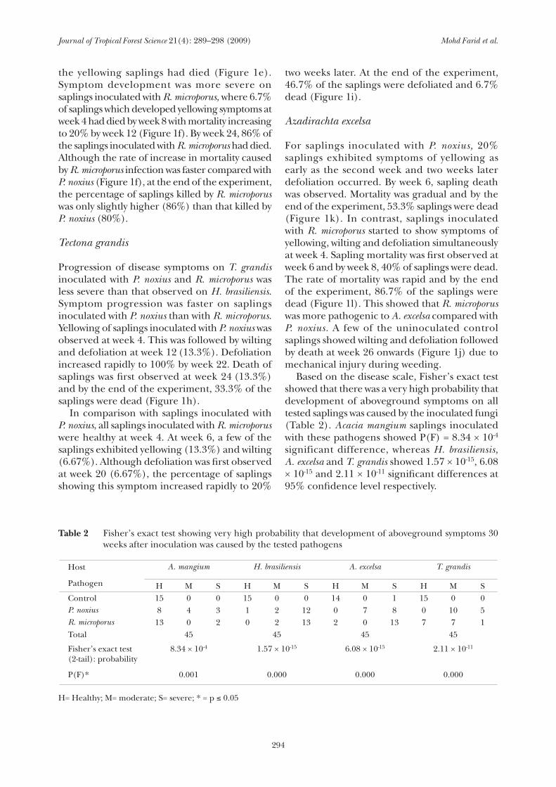
Table 2 Fisher’s exact test showing very high probability that development of aboveground symptoms 30 weeks after inoculation was caused by the tested pathogens
Host A. mangium H. brasiliensis A. excelsa T. grandis
Pathogen H M S H M S H M S H M SControl 15 0 0 15 0 0 14 0 1 15 0 0P. noxius 8 4 3 1 2 12 0 7 8 0 10 5R. microporus 13 0 2 0 2 13 2 0 13 7 7 1Total 45 45 45 45
Fisher’s exact test (2-tail): probability
8.34 × 10⁻⁴ 1.57 × 10⁻¹⁵ 6.08 × 10⁻¹⁵ 2.11 × 10⁻¹¹
P(F)* 0.001 0.000 0.000 0.000
H= Healthy; M= moderate; S= severe; * = p ≤ 0.05
the yellowing saplings had died (Figure 1e). Symptom development was more severe on saplings inoculated with R. microporus, where 6.7% of saplings which developed yellowing symptoms at week 4 had died by week 8 with mortality increasing to 20% by week 12 (Figure 1f). By week 24, 86% of the saplings inoculated with R. microporus had died. Although the rate of increase in mortality caused by R. microporus infection was faster compared with P. noxius (Figure 1f), at the end of the experiment, the percentage of saplings killed by R. microporus was only slightly higher (86%) than that killed by P. noxius (80%).
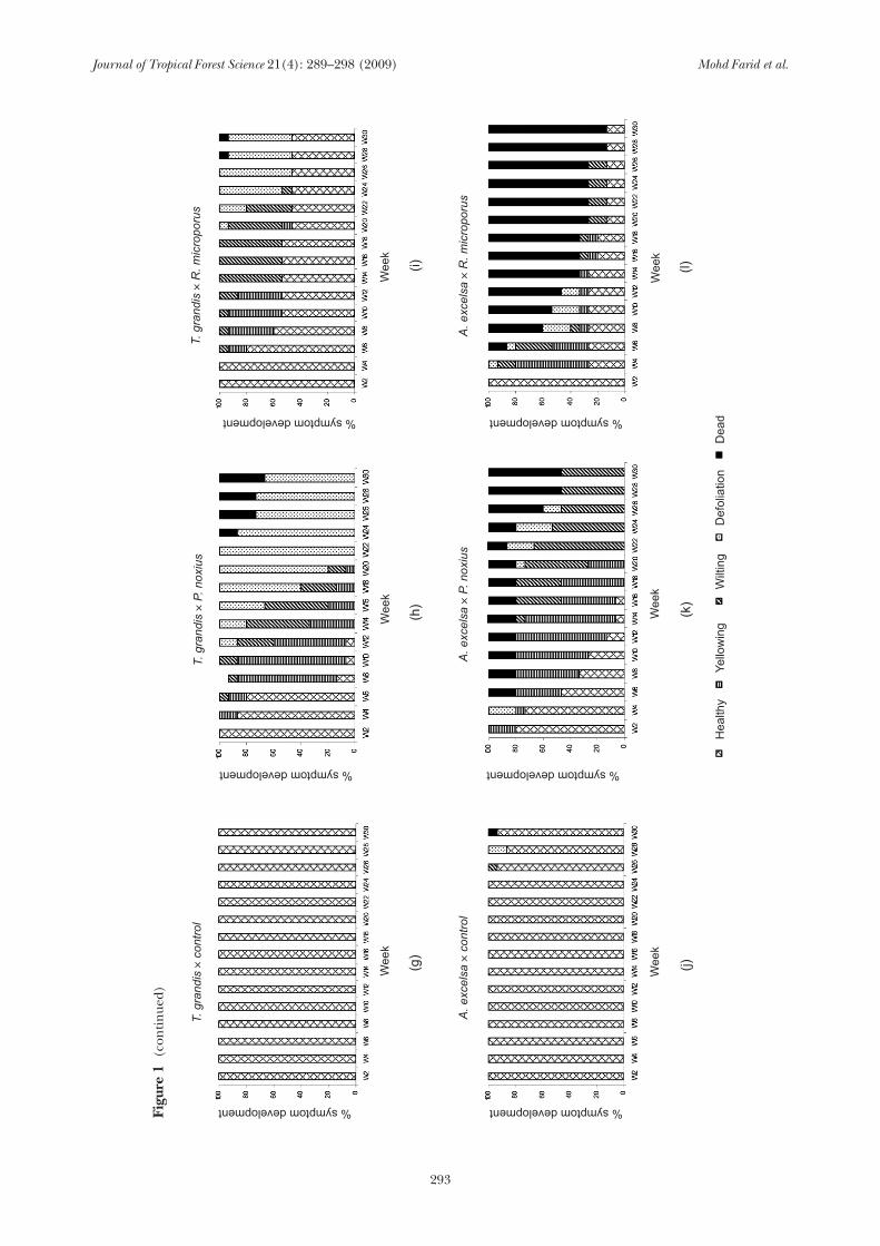
Tectona grandis
Progression of disease symptoms on T. grandis inoculated with P. noxius and R. microporus was less severe than that observed on H. brasiliensis. Symptom progression was faster on saplings inoculated with P. noxius than with R. microporus. Yellowing of saplings inoculated with P. noxius was observed at week 4. This was followed by wilting and defoliation at week 12 (13.3%). Defoliation increased rapidly to 100% by week 22. Death of saplings was first observed at week 24 (13.3%) and by the end of the experiment, 33.3% of the saplings were dead (Figure 1h). In comparison with saplings inoculated with P. noxius, all saplings inoculated with R. microporus were healthy at week 4. At week 6, a few of the saplings exhibited yellowing (13.3%) and wilting (6.67%). Although defoliation was first observed at week 20 (6.67%), the percentage of saplings showing this symptom increased rapidly to 20%
two weeks later. At the end of the experiment, 46.7% of the saplings were defoliated and 6.7% dead (Figure 1i).
Azadirachta excelsa
For saplings inoculated with P. noxius, 20% saplings exhibited symptoms of yellowing as early as the second week and two weeks later defoliation occurred. By week 6, sapling death was observed. Mortality was gradual and by the end of the experiment, 53.3% saplings were dead (Figure 1k). In contrast, saplings inoculated with R. microporus started to show symptoms of yellowing, wilting and defoliation simultaneously at week 4. Sapling mortality was first observed at week 6 and by week 8, 40% of saplings were dead. The rate of mortality was rapid and by the end of the experiment, 86.7% of the saplings were dead (Figure 1l). This showed that R. microporus was more pathogenic to A. excelsa compared with P. noxius. A few of the uninoculated control saplings showed wilting and defoliation followed by death at week 26 onwards (Figure 1j) due to mechanical injury during weeding. Based on the disease scale, Fisher’s exact test showed that there was a very high probability that development of aboveground symptoms on all tested saplings was caused by the inoculated fungi (Table 2). Acacia mangium saplings inoculated with these pathogens showed P(F) = 8.34 × 10⁻⁴ significant difference, whereas H. brasiliensis, A. excelsa and T. grandis showed 1.57 × 10⁻¹⁵, 6.08 × 10⁻¹⁵ and 2.11 × 10⁻¹¹ significant differences at 95% confidence level respectively.
Journal of Tropical Forest Science 21(4): 289–298 (2009) Mohd Farid et al.
295
Comparison of the susceptibility of the four hosts to each pathogen
In general, the pathogenicity test revealed that saplings of A. mangium, H. brasiliensis, T. grandis and A. excelsa were susceptible to both P. noxius and R. microporus. However, their degree of susceptibility differed. Against R. microporus, both H. brasiliensis and A. excelsa were the most susceptible followed by A. mangium and T. grandis. Against P. noxius, H. brasiliensis was the most susceptible followed by A. excelsa, T. grandis and A. mangium (Figure 1). Acacia mangium was equally susceptible to P. noxius and R. microporus but T. grandis was more susceptible to the former than to the latter (Figure 1). In contrast, A. excelsa was more susceptible to R. microporus compared with P. noxius. In general, H. brasiliensis was the most susceptible host to both pathogens, whereas A. mangium was the least susceptible host.
DISCUSSION
Results of this study showed a good association between the appearance of foliar symptoms and damage to roots belowground. Nevertheless, observation of foliar symptoms is generally more important for tree disease diagnosis as it is often difficult, costly and time consuming to excavate roots for observation of symptoms. Foliar inspection was often relied upon in identifying infected trees in the field (Nandris et al. 1987, Ann et al. 2002). In the present study, all saplings inoculated with R. microporus and P. noxius appeared to exhibit almost similar foliar symptoms. Progress of disease was generally observed first as yellowing followed by wilting, defoliation and finally death of the host. In addition, progress of these symptoms concurs with those observed by Mohd Farid et al. (2001, 2006a, b) who worked on root diseases of A. excelsa and T. grandis. It has been reported that foliar symptoms in Swietenia macrophylla, Cordia alliodora and Gmelina arborea infected by P. noxius progressed rapidly from yellowing to wilting and finally death of trees (Ivory 1990). Similar observations have also been recorded on H. brasiliensis infected by root diseases where general discoloration of the foliage was linked to the interruption in root function (Anonymous 1974, Nandris et al. 1987). In root disease, wilting and defoliation of leaves are considered symptoms
of advanced disease development. At this stage, chemical treatments are usually not effective as the presence of these symptoms indicate that the tree is beyond the point of recovery (Ismail & Azaldin 1985, Chee 1986). Although the infected hosts exhibited very similar foliar symptoms, root symptoms differed depending on pathogens. Roots of saplings inoculated with R. microporus had white rhizomorphs on their surface while those inoculated with P. noxius had encrustations of rusty brown mycelia with soil particles on the root surface. Similar observations were also reported by Holliday (1980), Nandris et al. (1987), Lee (2000), Ann et al. (2002) and Mohd Farid et al. (2005). Furthermore, these symptoms were consistent for the fungus irrespective of the host species. The high probability of Fisher’s exact test P(F) showed that symptoms developed on saplings inoculated with R. microporus and P. noxius were due to the effect of the inoculated fungi and most certainly not the effect of chance. Planters could use these symptoms as a guide to detect and control the spread of root disease infection in the field. However, emphasis should be given on trees showing yellowing symptoms as these are considered to be at an early stage of infection. Fungicide treatments to combat rubber root disease are recommended for newly infected trees or trees with mild infection levels (Ismail & Shamsuri 1998). Thus, it is vital to conduct regular disease monitoring in plantations so that fungicide treatments can be applied on yellowing trees as early as possible. The high mortality of A. excelsa (86.67%) and H. brasiliensis (86.67%) saplings inoculated with R. microporus indicated that the pathogen was equally aggressive in killing both hosts and that both hosts were very susceptible to the white root pathogen. Hevea brasiliensis is well known to be very susceptible to R. microporus in Malaysia, Sri Lanka, Indonesia and Gabon (Anonymous 1974, Fox 1977, Nandris et al. 1987, Liyanage 1997, Rajalakshmy & Jayarathnam 2000, Semangun 2000, Guyot & Flori 2002), while A. excelsa had previously been reported to be susceptible to white root disease in plantations established on areas previously planted with H. brasiliensis (Mohd Farid et al. 1999, 2005). The study also revealed that A. mangium and T. grandis were less susceptible to R. microporus as indicated by the lower mortality
Journal of Tropical Forest Science 21(4): 289–298 (2009) Mohd Farid et al.
296
rate. Furthermore, the white rhizomorphs often associated with infection by this pathogen were seldom found on the root surface of infected saplings compared with those of H. brasiliensis and A. excelsa. This suggests that the presence of rhizomorphs may be directly linked to susceptibility of the host to the pathogen. Similar observations have been made for root disease caused by Armillaria ostoyae (Omdal et al. 1995, Prospero et al. 2006) where dense rhizomorphs were often observed on the root surface of trees killed by the pathogen. This could indicate that production of rhizomorphs is strongly correlated with the ability to cause disease. Rhizomorph production was a prerequisite in assessing pathogenicity and virulence (Morrison & Pellow 2002). Susceptibility and damage by root disease fungi vary with tree species (Filip 1999). These results appear to indicate that both A. mangium and T. grandis may have some resistance to R. microporus. In this study, cumulative mortality was used to assess host susceptibility since it could show the aggressiveness of pathogens in killing their hosts, particularly in the plantations. The overall results suggested that P. noxius was a less aggressive pathogen to H. brasiliensis and A. excelsa than R. microporus as it took a longer time to kill the test plants. Similar results have been reported by Nicole et al. (1986) and Mohd Farid et al. (2001). The inoculated saplings that were not infected by the disease were probably vigorous enough to withstand the infection as evidenced by the presence of more vigorous, greener and denser canopy prior to infection. The difference in disease severity is often attributed to differences in tree vigour (Rosso & Hansen 1998). This could suggest that hosts need to be weakened before infection can occur. This is in agreement with a study that reported that pathogenicity of Armillaria sp. was often greater when trees were somewhat weakened and stressed (Wargo & Harrington 1991). This also indicates that trees under stress are more susceptible to infection by a pathogen (Agrios 2005). Another possible reason for the lack of disease development in some of the inoculated saplings could have been due to the lack of viable inoculum in some wood blocks. This could have occurred in wood blocks which had been buried too shallowly in the soil, thereby becoming exposed to sunlight and drying out.
CONCLUSIONS
In the field, brown root disease caused by P. noxius can be recognized by the presence of adhering soil particles and dark brown mycelial crust, while white root disease caused by R. microporus is usually identified based on the presence of rough white rhizomorphs on the tree root surface. Although results presented in this study are limited to cultivars of the four tree species, it is likely that similar symptoms will be observed in other tree species as well. The pathogenicity test confirmed that both R. microporus and P. noxius were pathogenic to H. brasiliensis, A. excelsa, T. grandis and A. mangium saplings. In terms of host susceptibility, H. brasiliensis and A. excelsa were more susceptible to R. microporus followed by A. mangium and T. grandis. Against P. noxius, H. brasiliensis was again the most susceptible followed by A. excelsa, T. grandis and A. mangium. Overall, H. brasiliensis was the most susceptible and A. mangium was the least susceptible to root rot disease caused by these two pathogens. Results from the present study suggest that A. excelsa is not suitable for planting in areas with a previous history of white root and brown root diseases, particularly in ex-rubber plantations, as it is extremely susceptible to both diseases. Hence, planting A. mangium and T. grandis trees in these areas could be an alternative since they show a lower degree of susceptibility to the diseases. Although A. mangium trees were least susceptible to white root and brown root disease, interplanting H. brasiliensis and A. mangium is not recommended. Hevea brasiliensis trees may not perform well in the field due to the high competitiveness of the fast-growing A. mangium for light and space. Consequently, latex production from H. brasiliensis may be reduced considerably. Since all infected saplings exhibited identical foliar symptoms which are generally detected too late for efficient treatments, cultural practices would be more practical to reduce and/or prevent incidence of root disease in the future. Good land clearing, regular disease monitoring and implementation of effective control measures are still required to prevent the spread of root disease in plantations. It may also be advisable to identify and treat the diseased areas with suitable chemicals before planting.
Journal of Tropical Forest Science 21(4): 289–298 (2009) Mohd Farid et al.
297
ACKNOWLEDGEMENTS
We would like to thank Ab. Rasip Ab. Ghani for the experimental design and Victor Netto from UPM for the statistical analysis. The help of Fakaruddin Baharuddin and Zakaria Yusoff in establishing and maintaining the experimental plot is highly appreciated. We would also like to thank the Forest Research Institute Malaysia, especially staff of the Mycology and Pathology Laboratory for their continued support. This study was funded by the Ministry of Science, Technology and Innovation (MOSTI) through IRPA grant No. 01-04-01-10013-EA001.
REFERENCES
Agrios gN. 2005. Plant Pathology. Fifth edition. Elsevier Academic Press, San Diego.
Ann PJ, ChAng TT & Ko WH. 2002. Phellinus noxius brown rot of fruit and ornamental trees in Taiwan. Plant Disease 86: 820−826.
Anonymous. 1974. Root diseases of Hevea. Planter’s Bulletin of the Rubber Research Institute Malaysia 133: 109−200.
Ariffin D & iDris AS. 1992. The Ganoderma Selective Media (GSM). PORIM Information Series. Palm Oil Research Institute Malaysia, Bangi.
Chee Kh. 1986. Present status of rubber diseases and their control. Review on Plant Pathology 69: 423–430.
Chong PW. 1979. The growing domestic demand for timber and its influence on forest management. Malaysian Forester 42: 378−389.
filiP gM. 1999. Ecology, identification and management of forest root diseases in Oregon. The Woodland Workbook: Forest Protection. Oregon State University, Oregon.
fox rA. 1977. The impact of ecological, cultural and biological factors on the strategy and costs of controlling root diseases in tropical plantation crops as exemplified by Hevea brasiliensis. Journal of Rubber Research Institute Sri Lanka 54: 329−362.
gibson iAS. 1979. Diseases of Forest Trees Widely Grown as Exotics in the Tropics and Southern Hemisphere. Part II. The Genus Pinus. Commonwealth Mycological Institute and Commonwealth Forestry Institute, Kew and Oxford.
hArringTon TC & WingfielD mJ. 1998. Diseases and the ecology of indigenous and exotic pines. Pp. 381−404 in Richardson DM (Ed.) Ecology and Biogeography of Pinus. Cambridge University Press, Cambridge.
guyoT J & flori A. 2002. Comparative study for detecting Rigidoporus lignosus on rubber trees. Crop Protection 21: 461−466.
holliDAy P. 1980. Fungus Diseases of Tropical Crops. Cambridge University Press, Cambridge.
ismAil h & AzAlDin mY. 1985. Interaction of sulfur with soil pH and root disease of rubber. Journal Rubber Research Institute of Malaysia 33: 59−69
ismAil h & shAmsuri mH. 1998. Current status of root diseases of rubber. Paper presented at CABI Workshop on Ganoderma Diseases. 5−8 October 1998, Serdang. Malaysian Agricultural Research and Development Institute Training Center, Serdang.
ivory mH. 1987. Diseases and Disorders of Pines in the Tropics. Overseas Research Publication Number 31. Oxford Forestry Institute, Oxford.
ivory mh. 1990. Brown root-rot of tropical forest trees in the south-west pacific region. Pp. 100–107 in Hutacharern C et al. (Eds). Proceedings of the IUFRO Workshop on Pests and Diseases of Forest Plantations. 5−11 June 1988. RAPA Publication 1990/9 FAO, Bangkok.
JohnsTon A. 1989. Diseases and pests. Pp. 415−458 in Webster CC & Baulkwill W J (Eds.) Rubber. Longman Scientific and Technical, New York.
KrishnAPillAy b & APPAnAh s. 2002. Forest plantation: historical background. Pp. 3–11 in Krishnapillay B (Ed.) A Manual for Forest Plantation Establishment in Malaysia. Malayan Forest Records No. 45. Forest Research Institute Malaysia, Kepong.
lee sS. 1997. Diseases of some tropical plantation Acacias in Peninsular Malaysia. Pp. 53−61 in Old KM, Lee SS & Sharma JK (Eds.) Diseases of Tropical Acacias. Proceedings of the International Workshop. Subanjeriji (South Sumatra). 28 April−3 May 1996. CIFOR Special Publication. Center for International Forestry Research, Jakarta.
lee sS. 2000. The current status of root diseases of Acacia mangium Willd. Pp. 71−79 in Flood J, Bridge PD & Holderness M (Eds.) Ganoderma Diseases of Perennial Crops. CAB International, Wallingford.
liyAnAge ADes. 1977. Rubber. Pp. 331−347 in Hillock RJ & Walter JM (Eds.) Soilborne Diseases of Tropical Crops. CAB International, Wallingford.
mohD fAriD A, lee sS, mAziAh z, rosli h & norWATi m. 2006a. Basal root rot, a new disease of teak (Tectona grandis) in Malaysia caused by Phellinus noxius. Malaysian Journal of Microbiology 1: 40−45.
mohD fAriD A, lee sS, mAziAh z, rosli h & norWATi m. 2006b. Root rot in tree species other than A. mangium. Pp. 60−66 in Potter K et al. (Eds.) Heart Rot and Root Rot in Tropical Acacia Plantations. ACIAR Proceedings No. 124. ACIAR, Canberra.
mohD fAriD A, lee sS, PATAhAyAh m & mAziAh z. 2005. Current root disease incidence status of forest plantations in Peninsular Malaysia. Pp. 264−271 in Lee SS & Lim HF (Eds.) Proceedings of the Conference on Forestry and Forest Product Research 2005. Investment for Sustainable Heritage and Wealth. 22−24 November 2005, Kuala Lumpur. Forest Research Institute Malaysia, Kepong.
mohD fAriD A, mAziAh z, Ab rAsiP AG & norAini siKin y. 2001. Preliminary study on pathogenicity of three root disease fungi on Azadirachta excelsa (sentang). Journal of Tropical Forest Science 13: 554−558.
mohD fAriD A, mAziAh z & norAini siKin y. 1999. Preliminary study on the pathogenicity of four root rot fungi on Azadirachta excelsa (Jack) Jacob (Sentang). Book of Abstracts. Malaysian Science and Technology Congress ’99, Kuala Lumpur.
Journal of Tropical Forest Science 21(4): 289–298 (2009) Mohd Farid et al.
298
morrison DJ & PelloW KW. 2002. Variation in virulence among isolates of Armillaria ostoyae. Forest Pathology 32: 99−107.
nAnDris D, niCole m, geiger JP & mAlleT b. 1983. Root rot disease in the forest and plantations of the Ivory Coast. Pp. 286−295 in Proceedings of the Sixth International Conference on Root and Butt Rot of Forest Trees. 25−31 August 1983, Melbourne, Victoria and Gympie, Queensland. CSIRO, Melbourne.
nAnDris D, niCole m & geiger JP. 1987. Root rot disease of rubber. Plant Disease 71: 298−306.
niCole m, geiger JP & nAnDris D. 1986. Penetration and degradation of suberized cells of Hevea brasiliensis infected with root rot fungi. Physiological and Molecular Plant Pathology 28: 181−185.
omDAl DW, shAW gC, JACobi Wr & WAger TC. 1995. Variation in pathogenicity and virulence of isolate Armillaria ostoyae on eight tree species. Plant Disease 79: 939−944.
rosso P & hAnsen e. 1998. Tree vigour and the susceptibility of Douglas fir to Armillaria root disease. European Journal of Forest Pathology 28: 43−52.
ProsPero s, holDerrieDer o & rigling D. 2006. Rhizomorph production and stump colonization by co-occurring Armillaria cepistipes and Armillaria ostoyae: an experimental study. Forest Pathology 36: 21−31.
rAJAlAKshmy vK & JAyArAThnAm K. 2000. Root diseases and non-microbial maladies. Pp. 309–336 in George PJ & Jacob CK (Eds.) Natural Rubber: Agromanagement and Crop Processing. Rubber Research Institute of India, Kottayam.
semAngun h. 2000. Diseases of Plantation Crops in Indonesia. Gadjah Mada University Press, Yogyakarta.
sTAlPers JA. 1987. Identification of Wood Inhibiting Aphyllophorales in Pure Culture. Studies in Mycology No. 16. Institute of the Royal Netherlands Academy of Arts and Sciences, Baarn.
WArgo rH & hArringTon TC. 1991. Host stress and susceptibility. Pp. 88−101 in Shaw CG & Kile GA (Eds.) Armillaria Root Disease. Agriculture Handbook No. 69. USDA Forest Service, Washington.
WingfielD mJ. 1999. Pathogens in exotic plantation forestry. International Forestry Review 1: 163−167.
Copyright © 2022 FDOKUMEN