Ellerman House defines luxury, with stunning views, personal ...
Upload
independentCategory
view
1download
0
The EMBO Journal Vol.16 No.15 pp.4519–4530, 1997
Mutagenesis of phospholipase D defines asuperfamily including a trans-Golgi viral proteinrequired for poxvirus pathogenicity
and choline (reviewed in Exton, 1994). Several cellularTsung-Chang Sung1, Rachel L.Roper2,functions have been proposed for PLD, including criticalYue Zhang3, Simon A.Rudge1, Ryan Temel1,roles in regulation of secretion, reorganization of the actinScott M.Hammond1, Andrew J.Morris1,4,cytoskeleton and control of cell proliferation, but all ofBernard Moss2, JoAnne Engebrecht1,4 andthe hypotheses remain unproven (reviewed in Kleinet al.,Michael A.Frohman1,4,5
1995). Until recently, a major factor hindering progressin this field was the absence of molecular tools.1Department of Pharmacological Sciences,3Molecular and Cellular
Biology Program and4The Institute for Cell and Developmental A breakthrough was made with the cloning of a plantBiology, SUNY at Stony Brook, Stony Brook, NY 11794-8651 and PLD cDNA (Wang et al., 1994). The properties of the2Laboratory of Viral Diseases, National Institute of Allergy and
plant PLD are distinct from mammalian PLD at a biochem-Infectious Diseases, Bethesda, MD 20892, USAical level (Brownet al., 1995). However, the plant protein5Corresponding author contained novel motifs that were sufficiently well con-e-mail: [email protected] across a broad phylogenetic range to identify ayeast gene required for meiosis as a PLD (Roseet al.,Phospholipase D (PLD) genes are members of a super-1995), and to provide an entry into the cloning offamily that is defined by several highly conservedmammalian PLD cDNAs (Hammondet al., 1995; Colleymotifs. PLD in mammals has been proposed to play aet al., 1997). Scrutiny of the mammalian PLD1 cDNArole in membrane vesicular trafficking and signalrevealed none of the motifs that might have been expectedtransduction. Using site-directed mutagenesis, 25 pointby analogy to phospholipase C, such as SH2, SH3 or PHmutants have been made in human PLD1 (hPLD1)domains (Hammondet al., 1995). In addition, PLD1 doesand characterized. We find that a motif (HxKxxxxD)not encode sequences previously shown to interact withand a serine/threonine conserved in all members ofRhoA (Reid et al., 1996). However, database searchesthe PLD superfamily are critical for PLD biochemicalusing mammalian PLD1 revealed the existence of PLDactivity, suggesting a possible catalytic mechanism.homologs in numerous species (Hammondet al., 1995)Functional analysis of catalytically inactive pointand defined four regions of highly conserved sequencemutants for yeast PLD demonstrates that the meiotic(Figure 1; reviewed in Morriset al., 1996). The mostphenotype ensuing from PLD deficiency in yeast derivesprominent sequence contains an invariant charged motif,from a loss of enzymatic activity. Finally, mutation ofHxKxxxxD, which was proposed to mediate catalysisan HxKxxxxD motif found in a vaccinia viral protein(Hammondet al., 1995). Two separated copies of thisexpressed in the Golgi complex results in loss of efficientmotif, embedded in surrounding sequences that are alsovaccinia virus cell-to-cell spreading, implicating theconserved but less rigorously duplicated, are found in allviral protein as a member of the superfamily andPLD homologs (Morriset al., 1996).suggesting that it encodes a lipid modifying or binding
This ‘HKD’ motif is also found in two phospholipidactivity. The results suggest that vaccinia virus andsynthesis enzymes, cardiolipin synthase (CLS) and phos-hPLD1 may act through analogous mechanisms tophatidyl serine synthase (PSS) (reviewed in Morriset al.,effect viral cellular egress and vesicular trafficking,1996) and in a pair of proteins encoded by the Poxviridaerespectively.family of viruses (reviewed in Koonin, 1996; Ponting andKeywords: phospholipase D (PLD)/SPO14/vacciniaKerr, 1996). One of these proteins, VP37, which containsvirus/VP37only a single, partially conserved form of the HKD motif,is required for efficient cell-to-cell spreading by vacciniavirus and has been studied extensively. Although its
Introduction mechanism of action is unknown, it is expressed in theGolgi complex and plays a role in the wrapping of viralThe initiation of signal transduction cascades throughparticles intrans-Golgi membranes and their subsequentG-protein-coupled receptors or tyrosine kinase receptorstransit from the cell (Hiller and Weber, 1985). Thisresults in the activation of membrane-associated lipid-prompts the hypothesis that VP37 and mammalian PLD1modifying enzymes, including the phospholipases A2, Care homologous in a functional sense, since PLD1 is alsoand D (reviewed in Divecha and Irvine, 1995). Moreexpressed in the Golgi complex (Colleyet al., 1997) anddirectly, phospholipase D1 (PLD1) can be activatedin vitrohas been proposed to regulate vesicular budding (reviewedby protein kinase C (PKC) and members of the ARF andin Bednareket al., 1996).Rho small G-protein families, suggesting that its activation
In this study, we have used site-directed mutagenesisduring agonist stimulation ensues from the controlledand a combination ofin vitro and in vivo systems toregulation of these effectors (Hammondet al., 1997).determine whether the HKD motif and other conservedPhospholipase D (PLD) catalyzes the hydrolysis of phos-
phatidylcholine (PC) to yield phosphatidic acid (PA) sequences are required by the PLD superfamily. Our
© Oxford University Press 4519
T.-C.Sung et al.
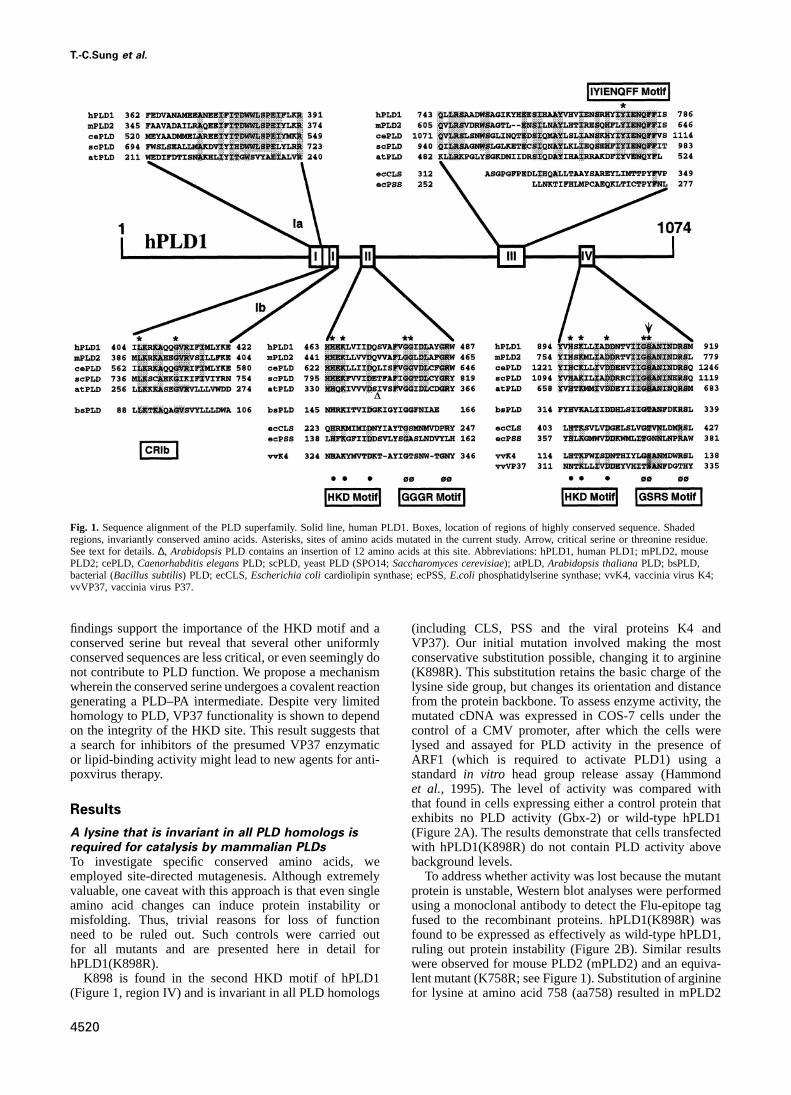
Fig. 1. Sequence alignment of the PLD superfamily. Solid line, human PLD1. Boxes, location of regions of highly conserved sequence. Shadedregions, invariantly conserved amino acids. Asterisks, sites of amino acids mutated in the current study. Arrow, critical serine or threonine residue.See text for details.∆, ArabidopsisPLD contains an insertion of 12 amino acids at this site. Abbreviations: hPLD1, human PLD1; mPLD2, mousePLD2; cePLD,Caenorhabditis elegansPLD; scPLD, yeast PLD (SPO14;Saccharomyces cerevisiae); atPLD,Arabidopsis thalianaPLD; bsPLD,bacterial (Bacillus subtilis) PLD; ecCLS,Escherichia colicardiolipin synthase; ecPSS,E.coli phosphatidylserine synthase; vvK4, vaccinia virus K4;vvVP37, vaccinia virus P37.
findings support the importance of the HKD motif and a (including CLS, PSS and the viral proteins K4 andVP37). Our initial mutation involved making the mostconserved serine but reveal that several other uniformly
conserved sequences are less critical, or even seemingly do conservative substitution possible, changing it to arginine(K898R). This substitution retains the basic charge of thenot contribute to PLD function. We propose a mechanism
wherein the conserved serine undergoes a covalent reaction lysine side group, but changes its orientation and distancefrom the protein backbone. To assess enzyme activity, thegenerating a PLD–PA intermediate. Despite very limited
homology to PLD, VP37 functionality is shown to depend mutated cDNA was expressed in COS-7 cells under thecontrol of a CMV promoter, after which the cells wereon the integrity of the HKD site. This result suggests that
a search for inhibitors of the presumed VP37 enzymatic lysed and assayed for PLD activity in the presence ofARF1 (which is required to activate PLD1) using aor lipid-binding activity might lead to new agents for anti-
poxvirus therapy. standardin vitro head group release assay (Hammondet al., 1995). The level of activity was compared withthat found in cells expressing either a control protein thatResultsexhibits no PLD activity (Gbx-2) or wild-type hPLD1(Figure 2A). The results demonstrate that cells transfectedA lysine that is invariant in all PLD homologs is
required for catalysis by mammalian PLDs with hPLD1(K898R) do not contain PLD activity abovebackground levels.To investigate specific conserved amino acids, we
employed site-directed mutagenesis. Although extremely To address whether activity was lost because the mutantprotein is unstable, Western blot analyses were performedvaluable, one caveat with this approach is that even single
amino acid changes can induce protein instability or using a monoclonal antibody to detect the Flu-epitope tagfused to the recombinant proteins. hPLD1(K898R) wasmisfolding. Thus, trivial reasons for loss of function
need to be ruled out. Such controls were carried out found to be expressed as effectively as wild-type hPLD1,ruling out protein instability (Figure 2B). Similar resultsfor all mutants and are presented here in detail for
hPLD1(K898R). were observed for mouse PLD2 (mPLD2) and an equiva-lent mutant (K758R; see Figure 1). Substitution of arginineK898 is found in the second HKD motif of hPLD1
(Figure 1, region IV) and is invariant in all PLD homologs for lysine at amino acid 758 (aa758) resulted in mPLD2
4520

PLD mutagenesis and function
Fig. 2. A PLD mutant in the HKD motif is catalytically inactive. (A) hPLD1 encoding a substitution of arginine for lysine at aa898 was transfectedinto COS-7 cells and examined for activity in comparison to wild-type PLD1 and a control vector. Wild-type PLD1 is largely inactive unlessstimulated by an effector such as ARF1. A small increase is observed for the control vector (Gbx-2, a homeobox gene that does not encode PLDactivity) in the presence of ARF1 due to activation of endogenous COS-7 cell PLD present in the cell extract. No additional activity over the controlis observed for hPLD1(K898R). (B) Western analysis of transfected cells. Aliquots of cells transfected with a control expression plasmid (Gbx-2; a50 kDa protein), the expression plasmid lacking any inserted cDNA (pCGN) or wild-type or mutant PLD1 or PLD2 cDNAs, were analyzed byWestern blotting using an anti-Flu tag monoclonal antibody. Mutant PLD1 and PLD2 are expressed at wild-type levels, demonstrating that theproteins were not destabilized by the K→R mutation. The slight difference seen here between wild-type and mutant PLD1 was not observedreproducibly. 59∆-PLD1 and 59∆-PLD2 constitute PLD structural mutants lacking ~300 aa at the amino-terminus. They are shown here to furtherillustrate the specificity of the bands identified for the full length wild-type and mutant PLD proteins. PLD1 exhibits heterogeneity with a major bandmigrating at 122 kDa and two minor slightly larger bands. PLD2 (108 kDa) breakdown products are frequently observed.
losing all measurable activityin vitro (data not shown),although the mutant protein is fully stable.
Another possibility was that the mutation inducedaltered PLD activity specifically in thein vitro assay.Although well accepted, thein vitro assay is not physio-logical in several respects. To address this question, wequantified PLD activityin vivo using the transphosphat-idylation assay (Wakelamet al., 1995). This methodmeasures production of phosphatidylalcohol, which is
Fig. 3. PLD mutants are catalytically inactivein vivo. Aformed by PLD when alcohol is used as a nucleophiletransphosphatidylation assay was performed to examine the behaviorinstead of water to cleave PC (see Figure 8 and Discussion).in vivo of mutated PLD proteins. Accumulation of phosphatidylbutanol
To determine mutant PLD activityin vivo, cells were (PtdBut) in COS-7 cells transfected with control and wild-type ortransfected with the expression constructs and assayed formutant PLD1 and PLD2 expression plasmids was determined. Some
culture dishes were additionally stimulated with 100 nM PMA duringphosphatidylbutanol (PBut) accumulation (Figure 3). Asthe 30 min assay period. Each lane represents an individual dish ofreported previously (Colleyet al., 1997), cells expressingcells. The experiment was repeated several times and a representativewild-type PLD1 or control plasmids exhibit little PLD result is shown.
activity in the basal state (Figure 3, first four lanes),whereas COS-7 cells expressing PLD2 manifest highlevels of PLD activity (Figure 3, lane 5). An ~200-fold recombinant PLD1 in the PLD1-transfected cells (Figure
3, lane 8). An ~4-fold increase in PBut accumulation isincrease in PBut accumulation is observed (data notshown). In contrast, COS-7 cells expressing mPLD2- observed over PMA-stimulated control cells (data not
shown). However, cells expressing hPLD1(K898R) do not(K758R) do not exhibit increased levels of PLD activityover background (Figure 3, lane 6), providing a striking exhibit increased PLD activity over background (Figure
3, lane 10), again confirming that the mutant PLDs aredemonstration that its lack of activityin vitro accuratelyreflects its behaviorin vivo. inactive in vivo.
In the absence of structural information, it is not possibleStimulation of cells with phorbol ester results in theactivation of several protein kinase C isoforms including to predict which mutations will cause problems in folding.
However, this can be addressed using approaches such asPKCα, which then activate endogenous PLD in controlcells (Figure 3, lanes 7 and 9) and both endogenous and the two-hybrid assay. Two-hybrid interactions are highly
4521

T.-C.Sung et al.
required for PLD1-mediated catalysis of PC and that theloss of enzymatic activity is not due to protein instability,misfolding or mislocalization.
Site-directed mutagenesis identifies critical
invariant amino acids
A series of amino acids were mutated. The mutant PLDswere then expressed in COS-7 cells and analyzed asdescribed above (Figure 5). Two mutant proteins wereunstable and could not be characterized (see legend toFigure 5). Several others failed to interact with RhoA inthe two-hybrid assay and might exhibit some degree ofstructural alteration (see legend to Figure 5). None ofthe stable mutant proteins displayed any alteration insubcellular localization (data not shown). All proteinactivities tested using transphosphatidylation assaysyielded results similar to those generated throughin vitroanalyses (data not shown). Finally, no differences inrelative activity were observed through activating PLDusing other types of effectors, such as RhoA, Rac1 orPKC (Table I).
HKD motifs. A series of conservative and non-conservativesubstitutions were made. Without exception, mutation ofany amino acid in either HKD motif resulted in completeor virtually complete loss of enzymatic activity. Thisfinding supports predictions that the motif is critical for
Fig. 4. RhoA and hPLD1 interactions in the two-hybrid assay. Yeast PLD catalytic function (Hammondet al., 1995; Morrisstrains were constructed harboring either a carboxy-terminal fragmentet al., 1996; Ponting and Kerr, 1996). These resultsof PLD1 fused to the LexA DNA binding domain or RhoA fused todistinguish between two existing models for catalysis:the VP16 activation domain. None of the PLD1 or RhoA fusion
proteins activated the reporter geneLacZ when expressed alone (lanes one model postulated that each motif would function1–4). When combined through mating of the respective yeast strains, independently, whereas the other proposed that each motifPLD1 was observed to interact with a dominant active form of RhoA
would constitute half of the actual active site (Ponting(RhoA-valine14), but not with wild-type RhoA (lanes 5 and 6).and Kerr, 1996). Since single point mutations in eitherPLD1(K898R), when combined with RhoA-V14, activated the reporter
plasmid almost as well as wild-type PLD1 (lane 7). Data points motif abolish activity completely, our results demonstraterepresent duplicate samples. Other controls demonstrating the that the duplicated motifs do not function independently.specificity of the RhoA–PLD1 interaction were performed but are notshown (Y.Zhang and M.A.Frohman, unpublished). GGGR/GSRS motif. Four of the seven substitutions resulted
in reduced but significant levels of catalysis. The resultssuggest that the small non-polar amino acids (glycines)sensitive to changes that result in the misfolding of target
proteins. Separate experiments have revealed that an are important but not as critical as the charged HKDmotifs and may effect correct positioning of other requisiteactivated allele of RhoA (RhoA-valine 14) interacts with
PLD1’s carboxy-terminus (Y.Zhang and M.A.Frohman, amino acids, since in some cases, double mutations‘rescued’ single mutations that inhibited catalysis [e.g.unpublished). To examine the consequence of the K898R
mutation on PLD1’s interaction with RhoA, the corres- Table I: G479A (1% residual activity in the presence ofARF) as compared with G478A-G479A (21% residualponding carboxy-terminus fragment of the mutant was
subcloned into the appropriate two-hybrid vector as a activity)].Contrasting results were observed with serine 911fusion protein and tested (Figure 4). The results indicate
that the mutant PLD1 peptide interacts with RhoA effici- (S911), which is highly conserved (as either a serine orthreonine) in the second HKD motif but is not present inently, making it unlikely that the mutation induced sig-
nificant conformational changes in PLD1. the first. Mutation of S911 to threonine resulted in retentionof significant enzymatic activity (39%), whereas mutationIn addition, hPLD1(K898R) was expressed in baculo-
virus. The recombinant protein is stable, electrophoreses to alanine completely abolished activity. In addition,mutation of S911 to alanine altered the migration ofwith the correct apparent mobility and is completely
inactive in the presence of ARF1 (as well as other known hPLD1 by Western analysis (data not shown). In brief,the heterogeneous band observed in Figure 2 can bePLD activators such as RhoA and PKC; data not shown).
Subcellular localization was also examined. PLD1 local- resolved into a major species and two minor bands oflarger apparent molecular weight. The relative ratio of theizes to a peri-nuclear region consistent with Golgi, endo-
plasmic reticulum and late endosomes (Colleyet al., major and minor bands is not altered by dephosphorylationusing alkaline phosphatase, indicating that the hetero-1997). hPLD1(K898R) localized similarly, suggesting that
the mutated PLD1 is membrane-associated and correctly geneity does not arise from phosphorylation (in contrast,yeast PLD is phosphorylated and undergoes a shift intargeted (data not shown).
Taken together, these results suggest that K898 is mobility after phosphatase treatment; A.Rudge and
4522

PLD mutagenesis and function
Fig. 5. Mutation of conserved amino acids in PLD1. 25 site-directed mutants of PLD1 were assayed for activity in the presence of ARF1. Activitiesrelative to wild-type PLD1 and the control plasmid (Gbx-2) are shown. Two mutants (Y778T and Y778F) were unstable as determined by Westernblot analysis and are not shown here. Mutants K898R and G910A-S911T interacted with RhoA-V14. Mutants at aa778 and D903E did not. Theexperiment was repeated several times. An average of two or more determinations is shown. CR; conserved region.
Table I. PLD activitiesa of individual hPLD1 mutants
Mutants Basal PLD activators
ARF PKCα Rac1 RhoA
CR IbL405D 0 6 5 0 6 1 0 6 2 0 6 1 0 6 2G412A 806 8 85 6 5 77 6 18 596 10 806 7G412E 646 23 886 4 95 6 8 70 6 12 836 31
CR IIH464D 36 3 0 6 1 0 6 2 0 6 1 0 6 1H464D-K466R 26 11 0 6 2 1 6 4 0 6 1 0 6 2H464G 216 14 0 6 1 0 6 2 3 6 6 1 6 4H464G-K466G 76 13 0 6 1 2 6 3 3 6 4 0 6 1H464G-K466R 326 12 1 6 1 1 6 2 3 6 4 1 6 1G478A-G479A 56 10 216 16 176 10 116 8 7 6 5G478V 156 2 17 6 2 11 6 9 8 6 9 9 6 10G479A 236 22 1 6 1 0 6 1 0 6 1 0 6 1G479V 06 7 0 6 1 0 6 1 0 6 2 0 6 2
CR IIIY778C 476 6 40 6 3 49 6 7 29 6 7 28 6 7Y778G 06 6 0 6 1 0 6 1 0 6 1 1 6 2Y778I 62 6 35 456 1 56 6 5 28 6 3 32 6 8Y778S 06 25 146 1 10 6 6 11 6 2 11 6 2
CR IVK898R 06 2 1 6 1 0 6 1 1 6 3 0 6 2D903E 06 7 5 6 3 3 6 1 2 6 1 4 6 3G910A-S911T 06 8 13 6 1 7 6 1 6 6 4 11 6 2
Data represent the mean SD of at least three independent experiments, each carried out in duplicate.aExpressed as a percentage of PLD activity with respect to PLD WT (100%, positive control) and Gbx2 (0%, negative control).
J.Engebrecht, submitted). The larger molecular weight PLDs, although computer analysis suggests that a regionwith significant similarity, highlighted by an invariantbands are also unaltered by mutations to the HKD motif,amino acid (F784), is present in CLS and PSS as well. Abut are reduced by mutation of S911 to threonine andpossible role for this site is suggested by studies ofeliminated by mutation of S911 to alanine (data notacetylcholinesterase, which have shown that choline inter-shown). One possibility raised by these results is thatacts with the enzyme via an aromatic region dominatedthese larger bands represent catalytic intermediates andby phenylalanine and tyrosines (Harelet al., 1993; Gilsonthat a critical step in catalysis involves modification ofet al., 1994). The interaction stabilizes the receptor–S911 by transphosphatidylation (see Discussion).substrate complex while catalysis takes place elsewhere.Thus, this aromatic-rich region in PLD represents aIYIENQFF motif.This motif is found only in eukaryotic
4523

T.-C.Sung et al.
possible binding site for the choline portion of PC. We spores. Tetrads (groups of four spores linked together) areobserved most frequently (Figure 6A). In yeast lackingtargeted one of the most conserved amino acids with a
series of substitutions (Y777→F, T, S, C, I and G) designed PLD, meiosis is blocked during chromosomal segregationand spores do not form (Figure 6B). The meiotic phenotypeto: (i) keep the aromaticity but remove the hydroxyl group
(F); (ii) remove the aromaticity but keep the hydroxyl can be rescued by transforming the mutant yeast with acentromere-containing plasmid bearing a fragment ofgroup (T and S); (iii) remove the aromaticity and the
hydroxyl group but keep the side group polar (C); genomic DNA containing theSPO14gene and flankingDNA (Figure 6C).(iv) remove the aromaticity and the hydroxyl group but
keep the size of the side group similar (I) or (v) minimize To examine the functionality of mutant PLDs in yeast,we generated three point mutants in the SPO14 rescuethe size of the small group (G). Since two of the six
mutant proteins were unstable (T and F) and since the plasmid and transformed them into yeast lacking SPO14.Levels of PLD activity were determined for the trans-others did not interact with RhoA, this would appear to
represent a sensitive site for PLD protein structure. The formed yeast, and their progression through meiosis wasexamined. Analogous to thein vitro analysis of the mutantloss of the aromatic part of the side chain (S, I and C)
correlated with a decrease in PLD activity regardless of mammalian PLDs, PLD activity was not observed abovebackground levels for mutants in which substitutions werewhether the side chain was polar or not. Substitution of
glycine for the tyrosine resulted in a complete loss of made in the HKD domain (Table II). Moreover, suchmutations eliminated the ability of the plasmid to rescueactivity, suggesting that additional structural constraints
were introduced. Taken together, the results suggest that the meiotic defect (Figure 6D). This result demonstratesthat the basis for the sporulation phenotype derives fromthis conserved site plays an important role in enzymatic
activity, although it is not as critical as the HKD motif. the loss of PLD catalysis. To address protein stability,Western analysis was performed using an anti-SPO14
CRIb. This region is conserved in all PLD homologs and antiserum on extracts from yeast expressing the K1098Rwe targeted two invariant amino acids. A non-conservative mutant. The mutant protein was present at levels compar-substitution at L405 resulted in complete loss of PLD able with that observed for yeast rescued by the wild-typeactivity. However, semi-conservative (G412A) or non- plasmid (data not shown).conserved (G412E) substitutions at the other site had no In contrast, the G744E mutation (analogous to theeffect. This result was surprising since the glycine residue G412E mutation made in hPLD1) exhibited full PLDat aa412 is completely invariant and presumably is import- activity (Table II) and rescued the meiotic phenotypeant for some aspect of PLD function common to all the equally as well as wild-type SPO14 (Figure 6C). Takenhomologs. This finding prompted us to investigate whether together, the results are paradoxical. On the one hand,the PLD mutants were functional in a biological context, G412 is invariantly conserved in all PLD enzymes. Onusing yeast. the other hand, it appears to be completely dispensable
for PLD function, at least when tested in both yeast andmammalian systems. Several reviews of PLD homologiesFunctional analysis of PLD mutants in yeast
Several hypothetical roles have been proposed for mamma- have discussed the conserved amino acids mutated herein detail (Koonin, 1996; Morriset al., 1996; Ponting andlian PLD in a cellular context (Frohman and Morris,
1996). PLD’s role in yeast is better understood, since it Kerr, 1996). Potential functions for genes that exhibit verylimited homology to the PLDs have also been discussed,was identified during a genetic screen as being required
for meiosis (Roseet al., 1995). Yeast PLD (SPO14) is a including an endonuclease and several viral proteins. Ourresults emphasize the importance of testing conservedlarge protein (1683 aa) compared with other PLD cognates.
For example, human PLDs range in size from 932 to 1074 relationships through experimental analyses.aa, castor bean PLD encodes 811 aa andStreptomycesPLD, 556 aa. It is not known what function(s) are mediated Requirement for the HKD motif in a vaccinia viral
protein with weak sequence homology to PLDby the non-conserved sequences in SPO14, althoughanalyses of yeast expressing only the (non-catalytic, non- Two genes encoding proteins with limited homology to
PLD are present in vaccinia virus and other poxvirusesconserved) amino-terminus suggested that this regioninteracts with unknown cellular components (Roseet al., (Figure 1). K4 contains two HKD motifs and adjacent
conserved sequences, while VP37 contains only one par-1995). When expressed in yeast lacking SPO14, the amino-terminal region induced a meiotic phenotype more severe tially conserved motif (NxKxxxxD), denoted here as
(H)KD. Functionally, however, VP37 appears morethan the one which was observed when the entire proteinwas absent. Moreover, the amino-terminal region exhibited important than K4. Vaccinia virus mutants lacking K4
replicate and spread as efficiently as the wild-type virus,dominant negative effects on meiosis when expressed inwild-type yeast, confirming that this region functions in suggesting that K4 is redundant, at least in the laboratory
setting (Blasco and Moss, 1991). In contrast, viral mutantssome manner other than to promote catalysis by the intactprotein. These results suggest that the meiotic block lacking the gene encoding VP37 (F13L) are seriously
compromised (Blasco and Moss, 1991). In cells infectedobserved in yeast lacking SPO14 might be caused byinsufficiencies other than a lack of hydrolysis of PC to by the wild-type virus, viral particles become enwrapped
sequentially in a double layer of membranes derived fromform PA. To test this hypothesis, site-directed mutagenesisof SPO14 and functional analyses were carried out. the intermediate compartment between the endoplasmic
reticulum and cis-Golgi, and then a double layer ofStarvation conditions trigger yeast to undergo meiosis.The most visible manifestation occurs during late meiosis, modifiedtrans-Golgi membranes, before leaving the cells
(Ichihashiet al., 1971; Sodeiket al., 1993; Schmelzet al.,when the meiotic products become encapsulated into
4524

PLD mutagenesis and function
Fig. 6. The HKD motif is critical for yeast PLD function. Completion of meiosis is marked by the formation of tetrads. (A) Wild-type yeast achievethis at high frequency when stimulated to undergo meiosis. (B) Yeast lacking SPO14 (PLD) do not form tetrads. (C) Yeast transformed with agenomic fragment containing SPO14 and its promoter, or the same fragment engineered to encode the G744E mutation, efficiently rescue the meioticphenotype. The results were indistinguishable for the wild-type and G744E mutated plasmid. Actually shown is the rescue by the G744E mutatedSPO14 gene. (D) Yeast transformed with a genomic fragment containing SPO14 engineered to encode the mutations in the HKD domain do notrescue the meiotic phenotype. The results were indistinguishable for both mutations. Actually shown is the rescue attempt by the SPO14 geneencoding the K1098R mutation.
1994; Strauss, 1995). In cells infected by viral mutantsTable II. Activity of SPO14– yeast rescued with wild-type and mutant
lacking VP37, thetrans-Golgi envelopment step does not forms of SPO14occur (Blasco and Moss, 1991). As a result, viral particle
Strain Rescue plasmid aPLD activity bSpores (%)egress is blocked and spread of the virus to adjacent cellsgreatly diminished. This is visualized as an inability of
SPO14– SPO14 1.00 62the mutant virus to form lytic plaques after infection of SPO14– vector alone 0.18 0monolayers of BS-C-1 cells (Blasco and Moss, 1991, SPO14– SPO14(K1098R) 0.20 0
SPO14– SPO14(G744E) 1.00 591995).VP37 localizes to thetrans-Golgi (Hiller and Weber,
aPLD activity was determined as previously described (Roseet al.,1985; Schmelzet al., 1994) and is retained in the viral1995). Densitometry scanning was performed and normalized for
membranes (Hirtet al., 1986; Schmutzet al., 1995). protein content. Activity from the wild-type extract was given ahPLD1 is also present in the Golgi apparatus (Colley relative value of 1.0.
bCultures were examined by phase microscopy after 48 h inet al., 1997). hPLD1 has been proposed to regulate buddingsporulation medium. The % sporulation was estimated by dividing theof vesicles (Stutchfield and Cockcroft, 1993; Chen andnumber of asci containing two, three or four spores by the totalShields, 1996; Frohman and Morris, 1996; Ktistakiset al., number of cells. At least 300 cells were examined for each strain.
1996; Seethaleret al., 1996; Simonet al., 1996) bygenerating PA which may act to recruit coatomer proteins(Ktistakis et al., 1996) or cause local alterations in in the subcellular localization and biological roles proposed
for the proteins raises two intriguing issues. First, VP37membrane structure (reviewed in Bednareket al., 1996;Seaman, 1996). Although the sequence homology between functionality might depend on the integrity of the (H)KD
motif. Second, VP37-mediated viral envelopment mightVP37 and hPLD1 is quite limited, the additional parallels
4525

T.-C.Sung et al.
Fig. 7. Efficient vaccinia virus cell-to-cell spread requires expression of VP37 encoding an intact (H)KD motif. BS-C-1 cells grown in six-wellplates were infected with wild-type virus (VP37 WT) or viruses mutated in the (H)KD motif at aa 314 (K→R) or 319 (D→E). After 48 h, cells werefixed and recombinant viruses stained blue by the addition of X-gal. Single wells of a six-well plate are observed in the top row, each wellcontaining at least 15 blue staining plaques. Boxes indicate regions containing infected plaques or cells equivalent to those shown magnified in thebottom row. All magnifications are equivalent. Arrows point to the blue-staining cells infected by mutant viruses.
constitute a novel model for the study of PLD-regulated mutants, VP37 was made in substantial quantity, approxim-ating wild-type levels (data not shown).vesicular budding.
Although VP37 is further diverged from PLD than CLSTo test the first hypothesis, recombinant vaccinia virusesor PSS, these results raised the possibility that VP37encoding normal or mutated VP37 proteins were con-encodes PLD activity. This possibility was addressedstructed (K314R and D319E; see Figure 1). The virusesusing several approaches. First, recombinant VP37 wasadditionally carried theLacZ gene as a marker. BS-C-1successfully expressed in COS-7 cells using the expressioncell monolayers were infected, and then fixed and stainedemployed for PLD1. Second, cell extracts prepared fromwith X-gal after 48 h of culture. Figure 7 shows the wild-monolayers of BS-C-1 cells infected with wild type ortype and mutant plaque sizes. The top panel showsmutant VV were assayed for PLD activity in the presenceindividual wells from a six-well plate, and the bottomor absence of ARF1. PLD activity was not observed abovepanel shows micrographs of wild-type and mutant plaquesbackground levels in any experiment, suggesting thatat equivalent magnifications. Both point mutations in theVP37 does not encode PLD activity (data not shown). It(H)KD motif drastically diminished the ability of theshould be noted, however, that thein vitro assay isviruses to form plaques. In fact, cells infected with theoptimized for human PLD1 and may fail to supportmutant viruses could be detected only microscopically, VP37 activity (for example, yeast PLD is inactive in the
because of the poor virus spread. mammalian assay system; see Roseet al., 1995). InMutant vaccinia virus lacking the F13L gene (VP37) addition, VP37 expressed alone as a recombinant protein
forms very small plaques (Blasco and Moss, 1995) which does not accumulate in thetrans-Golgi network, suggestingare indistinguishable from those observed for the (H)KD that the presence of other viral proteins is required for itsmutant viruses in Figure 7. To rule out the possibility correct localization and potentially for its activity (Payne,that the (H)KD point mutations reduced VP37 stability, 1992; Katzet al., 1997).Western blots were prepared from wild-type and mutant Finally, the (H)KD motif in VP37 is actually encodedvirus infected cells. Unlike the experiment shown in Figure as NKD. We used site-directed mutagenesis to change the7 for which limiting quantities of virus were used for VP37 sequence to HKD, but this did not result in VP37infection, in this experiment sufficient virus was added to exhibiting PLD activity (data not shown). In addition, weinfect all the cells at the beginning of the culture period. mutated the second HKD motif of PLD1 to NKD, whichConsequently, the amount of VP37 produced did not resulted in a complete loss of activity (Figure 5), suggesting
that His and Asn are not readily interchangeable.depend on subsequent viral spread. For both (H)KD
4526

PLD mutagenesis and function
Fig. 8. PLD, cardiolipin synthase and phosphatidylserine synthase catalytic reactions. Each reaction involves nucleophile attack on a phosphate bondas shown. Two biochemical activities for PLD have been observed: phosphodiester bond hydrolysis and phospholipid transphosphatidylation. UsingH2O as an electron donor, PLD functions as a phospholipid hydrolysis enzyme, cleaving phosphatidylcholine (PC) to produce phosphatidic acid (PA)and choline. Through the same mechanism but using alcohol instead of H2O as the electron donor, PLD can generate phosphatidylalcohols.Nucleophiles that can be used by hPLD1 include exogenously supplied butanol or endogenous diacylglycerol (DAG), yielding phosphatidylbutanol(PBut) and bisphosphatidic acid, respectively (Figure 3 and data not shown). CLS and PSS only carry out synthetic reactions in which alcohols areused as the nucleophile, as shown.
Discussion wide variety of agonists. Members of the ARF, RhoA andPKC families activate hPLD1 in the absence of other
The PLD superfamily protein cofactors, suggesting direct protein–protein con-The identification of non-PLD enzymes (PSS and CLS) tacts (Hammondet al., 1997). None of the mutants createdas part of the PLD superfamily is intriguing, given PLD’s in this study selectively affect the ability of these effectorscapacity for transphosphatidylation (Figures 3 and 8A). to activate PLD1, suggesting that the effector interactingPLD in fact greatly prefers alcohol over water as a sites are located separately from the motifs requirednucleophile, and it has never been understood why the for catalysis. This finding was not unexpected, sinceendogenous compound (water) is used less efficiently by mammalian PLD2, yeast PLD, plant PLD and prokaryotePLD. Comparisons of the reactions mediated by CLS and PLD are all active in the absence of effectors (MorrisPSS are illuminating in this respect (Figure 8B and C). et al., 1996; Colleyet al., 1997).Both enzymes carry out PLD-like reactions, but use Our finding that both HKD domains need to be intactsubstrates containing alcohols as the nucleophile. This for PLD to exhibit catalytic activity implies that therelationship suggests that PLD evolved from an ancestral structure mediating catalysis contains both motifs.enzyme that was an alcohol transphosphatase and raisesAlthough regions II and IV both encode HKD amino acidthe possibility that PLD catalyzes reactionsin vivo that residues, other highly conserved and requisite amino acidsrequire transphosphatidylation capacity. Several reports (i.e. S911) are unique to each region (see Figure 1). Thishave suggested that PLD may have a role in phospholipid suggests that the individual regions mediate analogous butsynthesis (Stanacev and Stuhne-Sekalec, 1970; Stanacevnot identical reactions, prompting the following modelet al., 1973; van Blitterswijk and Hilkmann, 1993), and we (Figure 9).have observed hPLD1-mediated synthesis of an intriguing PLD enzymes have two substrates: phosphatidylcholinephospholipidin vivo (below). and water. The PLD reaction could proceed via a ternary
complex (i.e. wherein PC and water are both bound toA model for catalysis the enzyme at the same site with direct hydrolysis ofhPLD has been studied intensively because it is involved the phosphodiester bond) or by a substituted enzyme
mechanism. The latter would involve the formation of ain the general signal transduction cascade initiated by a
4527

T.-C.Sung et al.
Fig. 9. A PLD catalytic model (see text for additional details). At the beginning of the reaction cycle, the amino-, but not the carboxy-terminal HKDhistidine is protonated. The first half-reaction involves the formation of a PA–PLD intermediate by covalent linkage of PA to S911. This is effectedby donation of S911’s proton to the carboxy-terminal HKD histidine, and liberation of a proton from the amino-terminal HKD histidine to formcholine. The second half-reaction then reverses this process, as a proton is transferred from a water molecule to the amino-terminal HKD histidine,freeing up the resulting hydroxyl group to hydrolyze the intermediate to form PA. Finally, the ionized serine recovers its proton from the carboxy-terminal HKD histidine.
covalent ‘phosphatidylated’ intermediate (i.e. PLD–PA) in Point mutants resulting in a defect in ability of
vaccinia virus to spreada half-reaction that would concurrently generate choline.Despite the fact that the vaccinia virus VP37 proteinThe intermediate would then be hydrolyzed by water,contains only one partially conserved HKD domain, itgenerating free phosphatidic acid. Although the PLDappears to require this site to be functional. This impliesreaction mechanism has been speculated to involve suchthat VP37 either binds to phospholipids or carries out aan intermediate (Stanacev and Stuhne-Sekalec, 1970), nomodifying activity. The former represents a more likelydirect evidence has been presented thus far to confirm it.possibility upon first inspection, since all other PLDThese types of intermediate generally involve the form-homologs exhibiting catalysis encode two HKD motifs.ation of a phosphodiester bond that couples the reactionHowever, VP37 can be immunoprecipitated as a dimerproduct to one of the enzyme’s serine or threonine residues.from infected cells (Schmutzet al., 1995), suggesting thatSuch an essential serine or threonine catalytic residueit may exist functionally in a form in which two inter-would be expected to be highly conserved among membersmolecular motifs are brought together. It does not appearof the PLD family, the most obvious choice for which islikely that VP37 encodes PLD activity. We have alsoS911. All members of the superfamily encode serine orsuccessfully expressed K4, but it does not exhibit PLDthreonine at aa911 except PSS, which encodes threonineactivity under ourin vitro or in vivo assay conditions (dataat the adjacent position (Figure 1).not shown).This two-step mechanism offers an attractive explan-
Since VP37 is considerably further diverged from PLDation for the apparent ‘duplication’ of essential catalyticthan CLS or PSS, it is not unlikely to generate or bindamino acid residues. Histidine residues are frequentlyphospholipid products other than PA. One possibilityinvolved in activation of nucleophiles in hydrolysis reac-for its function may involve an unusual but relatedtions, as has been demonstrated for RNaseA (reviewed inphospholipid, semilysobisphosphatidic acid (SLBPA).
Fersht, 1985). Intriguingly, a bacterial nuclease gene (nuc) Bisphosphatidic acid (BPA) is a phospholipid formedexists which is an extremely divergent member of the through transphosphatidylation when diacylglycerol isPLD superfamily (Ponting and Kerr, 1996). We propose used as a nucleophile donor instead of water (vanthat catalysis will proceed as follows: (i) activation of an Blitterswijk and Hilkmann, 1993). The resulting productenzyme-intrinsic nucleophile (S911) by transfer of a proton consequently contains a head group linked to four fattyto the carboxy-terminal HKD motif, which promotes acid side chains. SLBPA is potentially formed either bytransphosphatidylation of S911 with concomitant gener- PLA2-mediated hydrolysis of BPA, by the conjugation ofation of choline using a proton donated by the amino- lysoDAG to PC or by the conjugation of DAG to lysoPC,terminal HKD motif and (ii) activation of water as a all of which would result in the formation of a productnucleophile by transfer of a proton to the amino-terminal consisting of a head group linked to three fatty acid sideHKD motif, promoting the release of PA via hydrolysis chains. This unusual lipid should promote membraneof the intermediate through the donation of a proton from curvature (van Blitterswijk and Hilkmann, 1993) and thusthe carboxy-terminal HKD motif. Our findings that the might assist in the formation of small organelles such aspresence of a serine or threonine at position 911 is encapsulated viral particles. SLBPA is a normal componentcritical for enzyme activity and that a potential reaction of Golgi membranes at low levels (Cluett and Machamer,intermediate observed for wild-type PLD1 fails to be 1996), but is a major component of the outer envelope ofobserved for hPLD1(S911A) support this model. The Lys vaccinia virus (Sodeiket al., 1993). Thus, VP37 mightand Asp amino acids may play critical roles in stabilizing mediate concentration of SLBPA into viral membranes.
The alternative hypothesis, that VP37 might synthesizethe His and Ser residues, respectively.
4528

PLD mutagenesis and function
(Elledge and Davis, 1988). Plasmids were introduced into the diploidSLBPA, is less attractive because levels of SLBPA areyeast strain KR52-3C (Roseet al., 1995), which is homozygous for asimilar in infected and uninfected cells (Cluett andSPO14deletion, using the lithium acetate transformation procedure (Ito
Machamer, 1996). et al., 1983). To assess rescue of the meiotic phenotype, the transformedCuriously, PLD itself has been proposed to synthesize yeast were grown and sporulated as described previously (Roseet al.,
1990). Cultures were examined by phase microscopy after 48 h inBPA (van Blitterswijk and Hilkmann, 1993). We havesporulation medium. PLD assays were performed as previously describedexamined this by expressing hPLD1 and mPLD2in vivo.(Roseet al., 1990).Overexpression of hPLD1 in COS-7 cells leads to accumu-Construction and purification of recombinant viruslation of BPA and possibly SLBPA, whereas overexpres-Plasmid pRB20lacZ, containing theLacZ gene inserted between thesion of mPLD2 does not (data not shown). The resultsF13L gene and its downstream flanking sequence, was a gift of R.Blasco.suggest that the catalytic selectivities of PLD1 and PLD2 F13L encodes the 37 kDa Viral Protein (VP37). Recombinant viruses
may be subtly different, or may reflect the availability of expressing mutant F13L genes were derived from a recombinant vacciniathe appropriate substrates in the Golgi and in the plasmavirus, vRB12, whose F13 L gene was replaced by guanine phospho-
ribosyltransferase (gpt) (Blasco and Moss, 1995). BS-C-1 cells in six-membrane, where PLD1 and PLD2 localize, respectively.well plates were infected at a multiplicity of 2 p.f.u. per cell. At 3 hNonetheless, this leads to the hypothesis that PLD1 andafter infection, pRB20lacZ or derivatives containing mutated copies of
VP37 may work together to promote viral egress, and the F13L gene were transfected with DOTAP (Boehringer Mannheimsuggests that agents which target either activity may Biochemicals). The cells were harvested 36 h after transfection, frozen
and thawed three times, sonicated and then used to infect fresh BS-C-1represent novel approaches to inhibiting poxvirus patho-monolayers. After 2 h, the liquid medium was replaced with semi-solidgenicity.medium containing 1% low melting temperature agarose. After 2 daysof viral growth, semi-solid medium containing 1 mg/ml 5-bromo-4-chloro-3indolyl-β-D-galactoside (X-gal) (Promega, Madison, WI) wasMaterials and methodsadded.
Recombinant viruses staining blue were picked with a Pasteur pipetteGeneral reagentsand resuspended in 0.5 ml of medium, frozen and thawed three times,All phospholipids were purchased from Avanti polar lipids. PIP2 wassonicated and used for a subsequent round of infection. Virus isolatesisolated as described (Hammondet al., 1995).L-α-dipalmitoyl phosphat-were passaged and picked three times and then amplified on BS-C-1idylcholine [choline-methyl-3H] ([3H]PC) was obtained from Americancells. Viruses from infected cells were analyzed by PCR. All virusesRadiolabeled Chemicals and [32P]phosphoric acid from ICN pharma-isolated in this manner and analyzed by PCR retained thegptsequence ofceuticals. All other reagents were obtained from previously notedthe vRB12 virus, probably as the result of single crossover recombination.standard sources and were of analytical grade unless otherwise specifiedTherefore, the viruses were passed twice through STO cells in the(Hammondet al., 1995).presence of 6-thioguanine to select against virus containing thegpt gene(Isaacset al., 1990). Blue plaques were picked again under semi-solidSite-directed mutagenesismedium containing X-gal, and stocks were grown and analyzed by PCR.Site-directed mutagenesis of expression plasmids was carried out usingPure double crossover recombinant viruses were used in subsequentthe Quik-change kit (Stratagene). Plasmids were sequenced to confirmexperiments. For photography, infected monolayers were fixed at 48 hthe intended mutation and the integrity of the surrounding sequences forpost infection and stained with X-gal.at least 100 bp using Sequenase (US Biochemicals).
Western blotting and PLD assaysCell culture Cells in six-well plates were infected with 10 p.f.u. of virus per cell.COS-7 cells were maintained in DMEM with 10% FCS. For transfections, After 27 h, the cells were harvested and subsequently boiled in loadingthe cells were grown in 35 mm dishes and then switched into Opti- buffer containing 2% SDS, 50 mM Tris–HCl, 10% glycerol in theMEM I media (Gibco BRL). Forin vivo assays, the cells were washed presence of 2% 2-mercaptoethanol and electrophoresed on 4–20%into phosphoric acid-free DMEM media after transfection and labeled polyacrylamide gels (Integrated Separation Systems). Proteins werewith 5 µCi of [32P]phosphoric acid (Pi) per dish for 18 h (van Blitterswijk transferred to PVDF membrane (Millipore, Bedford, MA) overnight atand Hilkmann, 1993). 80 mA. Membranes were blocked in 0.1% NP-40, 1% powdered milk,
0.14 M NaCl, 25 mM Tris (pH 7.4) for 30 min followed by incubationPLD assays with anti-VP37 primary rat monoclonal antibody 15B6 (the kind gift ofRecombinant ARF, RhoA, Rac-1 and PKCα were purified and activated G.Hiller, see Hilleret al., 1981) supernatant for 2 h. Membranes wereusing GTPγS or PMA as previously described (Hammondet al., 1997). washed and incubated with a 1:1000 dilution of alkaline phosphataseMammalian PLD activity was measured using thein vitro head group conjugated anti-rat secondary (Promega, Madison, WI) for at leastrelease assay and thein vivo transphosphatidylation assay as previously 45 min. The membranes were washed three times in blocking bufferdescribed (Wakelamet al., 1991; Brown et al., 1993; Hammond and incubated with Western Blue (Promega) substrate solution. Theet al., 1995, 1997; Colleyet al., 1997). PLD cDNAs were transiently reaction was stopped by rinsing in water.overexpressed in COS-7 cells as previously described using the mamma- For recombinant PLD activity analyses, VP37 (F13L) and K4 DNAlian expression vector pCGN (Hammondet al., 1995, 1997; Colley coding regions were subcloned into pCGN, transfected into COS-7 cells,et al., 1997). The transfection efficiency was observed to be ~5–20%. and assayed as described for hPLD1. Intact virus was assayed by
infecting monolayers of BS-C-1 cells for 27 h as described above,Two-hybrid analysis of PLD1–RhoA interactions following which PLD activity was assayed in the presence or absenceDetails will be published elsewhere (Y.Zhang and M.A.Frohman, in of ARF as described previously.preparation). In brief, a carboxy-terminal fragment of wild-type ormutant PLD1 (674–1074 amino acids) was cloned into pBTM116 (Bartel Acknowledgementset al., 1993) as a fusion protein with the LexA DNA binding domain.In addition, the entire open reading frame of wild-type RhoA or a The authors thank S.Tsirka, T.Miller and S.Strickland for critical readingdominant active form of RhoA (RhoA-valine 14) was cloned into pAct of the manuscript and E.Dennis for discussion regarding the PLDas a fusion protein with the activation domain of herpes virus protein mechanism of action. This work was supported by grants from the NIH16 (VP16). The plasmids were used to transform yeast of opposite to A.J.M. (GM50388), J.E. (GM4863903) and M.A.F. (HD29758).mating types, and mated to each other or to yeast containing control S.M.H. was supported by an NIH Training Grant (GM07518) to theplasmids. The resulting hybrid strains were expanded in liquid culture Pharmacology graduate program. S.R. is a fellow of the American Heartand assessed forβ-gal activity. Association.
Yeast strains and plasmids ReferencesYeast media were prepared and genetic methods carried out usingstandard protocols (Roseet al., 1990). TheSPO14 complementing Bartel,P.L., Chien,C.-T., Sternglanz,R. and Fields,S. (1993) Using the
two-hybrid system to detect protein–protein interactions. Ingenomic fragment was subcloned into theLEU2 CENplasmid UN105
4529

T.-C.Sung et al.
Hartley,D.A. (ed.),Cellular Interactions in Development: A Practical glycoprotein to the outer envelope of nascent vaccinia virions.J. Virol.,71, 3178–3187.Approach. Oxford University Press, Oxford, UK, pp. 153–179.
Klein,J., Chalifa,V., Liscovitch,M. and Koffelholz, K. (1995) RoleBednarek,S.Y., Orci,L. and Schekman,R. (1996) Traffic COPS and theof phospholipase D activation in nervous system physiology andformation of vesicle coats.Trends Cell Biol., 6, 468–473.pathophysiology.J. Neurochem., 65, 1445–1455.Blasco,R. and Moss,B. (1991) Extracellular vaccinia virus formation
Koonin,E.V. (1996) A duplicated catalytic motif in a new superfamily ofand cell-to-cell virus transmission are prevented by deletion of thephosphohydrolases and phospholipid synthases that includes poxvirusgene encoding the 37000-Dalton outer envelope protein.J. Virol., 65,envelope proteins.Trends Biochem. Sci., 21, 242–243.5910–5920.
Ktistakis,N.T., Brown,H.A., Waters,M.G., Sternweis,P.C. and Roth,M.G.Blasco,R. and Moss,B. (1995) Selection of recombinant vaccinia viruses(1996) Evidence that phospholipase D mediates ARF-dependenton the basis of plaque formation.Gene, 158, 157–162.formation of Golgi coated vesicles.J. Cell Biol., 134, 295–306.Brown,H.A., Gutowski,S., Kahn, R.A. and Sternweis,P.C. (1995) Partial
Morris,A.J., Engebrecht,J. and Frohman,M.A. (1996) Structure andpurification and characterization of ARF-sensitive phospholipase Dregulation of phospholipase D.Trends Pharmacol. Sci., 5, 182–185.from porcine brain.J. Biol. Chem., 270, 14935–14943.
Payne,L.G. (1992) Characterization of vaccinia virus glycoproteins byBrown,H.A., Gutowski,S., Moomaw,C.R., Slaughter,C. and Sternweis,monoclonal antibody precipitation.Virology, 187, 251–260.P.C. (1993) ADP-ribosylation factor, a small GTP-dependent
Ponting,C.P. and Kerr,I.D. (1996) A novel family of phospholipase Dregulatory protein, stimulates phospholipase D activity.Cell, 75,homologues that includes phospholipid synthases and putative1137–1144.endonucleases.Protein Sci., 5, 914–922.Chen,Y.G. and Shields,D. (1996) ADP-ribosylation factor-1 stimulates
Reid,T., Furuyashiki,T., Ishizaki,T., Watanabe,G., Watanabe,N.,formation of nascent secretory vesicles from thetrans-Golgi networkFujisawa,K., Morii,N., Madaule,P. and Narumiya,S. (1996) Rhotekin,of endocrine cells.J. Biol. Chem., 271, 5297–5300.a new putative target for Rho bearing homology to a serine/threonineCluett,E.B. and Machamer,C.E. (1996) The envelope of vaccinia viruskinase, PKN, and rhophilin in the rho-binding domain.J. Biol. Chem.,reveals an unusual phospholipid in Golgi complex membranes.J. Cell271, 13556–13560.Sci., 109, 2121–2131.
Rose,K., Rudge,S.A., Frohman,M.A., Morris,A.J. and Engebrecht,J.Colley,W., Sung,T.C., Roll,R., Hammond,S.M., Altshuller,Y.M., Bar-(1995) Phospholipase D signaling is essential for meiosis.Proc. NatlSagi,D., Morris,A.J. and Frohman,M.A. (1997) Phospholipase D2, aAcad. Sci. USA, 92, 12151–12155.distinct phospholipase D isoform with novel regulatory properties that
Rose,M.D., Winston,F. and Hieter,P. (1990)Methods in Yeast Genetics.provokes cytoskeletal reorganization.Curr. Biol., 7, 191–201.Cold Spring Harbor Laboratory Press, Cold Spring Harbor, NY.Divecha,N. and Irvine,R.F. (1995) Phospholipid signaling.Cell, 80,
Schmelz,M., Sodeik,B., Ericsson,M., Wolffe,E.J., Shida,H., Hiller,G. and269–278.Griffiths,G. (1994) Assembly of vaccinia virus: the second wrappingElledge,S.J. and Davis,R.W. (1988) A family of versatile centromericcisterna is derived from thetrans-Golgi network.J. Virol., 68, 130–147.vectors designed for use in the sectoring-shuffle mutagenesis assay in
Schmutz,C., Rindisbacher,L., Galmiche,M.C. and Wittek,R. (1995)Saccharomyces cerevisiae. Gene, 70, 303–312.Biochemical analysis of the major vaccinia virus envelope antigen.Exton,J.H. (1994) Phosphatidylcholine breakdown and signalVirology, 213, 19–27.transduction.Biochem. Biophys. Acta, 1212, 26–42.
Seaman,M.N.J. (1996) Phospholipase D in vesicle budding.Trends CellFersht,A. (1985)Enzyme Structure and Mechanism. W.H.Freeman andBiol., 6, 473.Company, New York.
Seethaler,G., Tooze,S. and Shields,D. (1996) Fusion and confusion inFrohman,M.A. and Morris,A.J. (1996) Phospholipid signaling: Rho isthe secretory pathway.Trends Cell Biol., 6, 239–242.only ARF the story.Curr. Biol., 6, 945–947.
Simon,J.P., Ivanov,I.E., Adesnik,M. and Sabatini,D.D. (1996) TheGilson,M.K., Straatsma,T.P., McCammon,J.A., Ripoll,D.R., Faerman,production of post-Golgi vesicles requires a protein kinase-C likeC.H., Axelsen,P.H., Silman,I. and Sussman,J.L. (1994) Open ‘backmolecule, but not its phosphorylating activity.J. Cell Biol., 135,door’ in a molecular dynamics simulation of acetylcholinesterase.355–370.Science, 263, 1276–1278.
Sodeik,B., Doms,R.W., Ericsson,M., Hiller,G., Machamer,C.E., nan’tHammond,S.M., Altshuller,Y.M., Sung,T.-C., Rudge,S.A., Rose,K.,Hof,W., van Meer,G., Moss,B. and Griffiths,G. (1993) Assembly ofEngebrecht,J., Morris,A.J. and Frohman,M.A. (1995) Molecularvaccinia virus: role of the intermediate compartment between thecharacterization of a human ARF-activated phosphatidylcholine-endoplasmic reticulum and the Golgi stacks.J. Cell Biol., 121,
specific phospholipase D defines a new and highly conserved family521–541.
of genes.J. Biol. Chem., 270, 29640–29643. Stanacev,N.Z. and Stuhne-Sekalec,L. (1970) On the mechanism ofHammond,S.M., Jenco,J.M., Nakashima,S., Cadwallader,K., Cook,S., enzymatic phosphatidylation. Biosynthesis of cardiolipin catalyzed by
Nozawa,Y., Frohman,M.A. and Morris,A.J. (1997) Characterization phospholipase D.Biochim. Biophys. Acta, 210, 350–352.of phospholipase D1. Activation of the purified enzyme by Stanacev,N.Z., Stuhne-Sekalec,L. and Domazet,Z. (1973) Enzymaticphosphatidylinositol 4,5-bisphosphate, ARF and Rho family formation of cardiolipin from phosphatidylglycerol by theG-proteins and protein kinase C-α. J. Biol. Chem., 272, 3860–3868. transphosphatidylation mechanism catalyzed by phospholipase D.Can.
Harel,M., Schalk,I., Ehret-Sabatier,L., Bouet,F., Goeldner,M., Hirth,C., J. Biochem., 51, 747–753.Axelsen,P.H., Silman,I. and Sussman,J.L. (1993) Quaternary ligand Strauss,E.J. (1995) A virus joins the movement.Curr. Biol., 6, 504–507.binding to aromatic residues in the active-site gorge of Stutchfield,J. and Cockcroft,S. (1993) Correlation between secretion andacetylcholinesterase.Proc. Natl Acad. Sci. USA, 90, 9031–9035. phospholipase D activation in differentiated HL60 cells.Biochem. J.,
Hiller,G., Eibl,H. and Weber,K. (1981) Characterization of intracellular 293, 649–655.and extracellular vaccinia virus variants: N1-isonicotinoyl-N2–3- van Blitterswijk,W.J. and Hilkmann,H. (1993) Rapid attenuation ofmethyl-4-chlorobenzoylhydrazine interferes with cytoplasmic virus receptor-induced diacylglycerol and phosphatidic acid bydissemination and release.J. Virol., 39, 903–913. phospholipase D-mediated transphosphatidylation: formation of
Hiller,G. and Weber,K. (1985) Golgi-derived membranes that contain an bisphosphatidic acid.EMBO J., 12, 2655–2662.acylated viral polypeptide are used for vaccinia virus envelopment. Wakelam,M.J., Cooke,S.J., Currie,S., Palmer,S. and Plevin,R. (1991)J. Virol., 55, 651–659. Regulation of the hydrolysis of phosphatidylcholine in Swiss 3T3
Hirt, P., Hiller,G. and Wittek, R. (1986) Localization and fine structure cells.Biochem. Soc. Trans., 19, 321–324.of a vaccinia virus gene encoding an envelope antigen.J. Virol., 58, Wakelam,M.J.O., Hodgkin,M. and Martin,A. (1995) The measurement757–764. of phospholipase D-linked signaling in cells. In Kendall,D.A. and
Ichihashi,Y., Matsumoto,S. and Dales,S. (1971) Biogenesis of poxvirus: Hill,S.J. (eds),Signal Transduction Protocols. Humana Press Inc,role of A-type inclusions and host cell membranes in virus Totowa, NJ, Vol. 41, pp. 271–278.dissemination.Virology, 46, 507–532. Wang,X., Xu,L. and Zheng,L. (1994) Cloning and expression of
Isaacs,S.N., Kotwal,G.J. and Moss,B. (1990) Reverse guanine phosphatidylcholine-hydrolyzing phospholipase D fromRicinusphosphoribosyltransferase selection of recombinant vaccinia viruses. communis. J. Biol. Chem., 269, 20312–20317.Virology, 178, 626–630.
Ito,H., Fukuda,Y., Murata,K. and Kimura,A. (1983) Transformation of Received on February 12, 1997; revised on May 2, 1997intact yeast cells treated with alkali cations.J. Bacteriol., 153, 163–168.
Katz,E., Wolffe,E.J. and Moss,B. (1997) The cytoplasmic/transmembranedomains of the vaccinia virus B5R protein target a chimeric HIV-1
4530
Copyright © 2022 FDOKUMEN