
Host Response to the Attenuated Poxvirus Vector NYVAC: Upregulation of Apoptotic Genes and...
14
JOURNAL OF VIROLOGY, Jan. 2006, p. 985–998 Vol. 80, No. 2 0022-538X/06/$08.000 doi:10.1128/JVI.80.2.985–998.2006 Copyright © 2006, American Society for Microbiology. All Rights Reserved. Host Response to the Attenuated Poxvirus Vector NYVAC: Upregulation of Apoptotic Genes and NF-B-Responsive Genes in Infected HeLa Cells Susana Guerra, 1 Luis A. Lo ´pez-Ferna ´ndez, 2 Alberto Pascual-Montano, 3 Jose ´ Luis Na ´jera, 1 Angel Zaballos, 2 and Mariano Esteban 1 * Department of Molecular and Cellular Biology, 1 Department of Immunology and Oncology, 2 and Biocomputing Unit, 3 Centro Nacional de Biotecnologı ´a, CSIC, Ciudad Universitaria Cantoblanco, E-28049 Madrid, Spain Received 19 August 2005/Accepted 10 October 2005 NYVAC has been engineered as a safe, attenuated vaccinia virus (VV) vector for use in vaccination against a broad spectrum of pathogens and tumors. Due to the interest in NYVAC-based vectors as vaccines and current phase I/II clinical trials with this vector, there is a need to analyze the human host response to NYVAC infection. Using high-density cDNA microarrays, we found 368 differentially regulated genes after NYVAC infection of HeLa cells. Clustering of the regulated genes identified six discrete gene clusters with altered expression patterns. Clusters 1 to 3 represented 47.5% of the regulated genes, with three patterns of gene activation kinetics, whereas clusters 4 to 6 showed distinct repression kinetics. Quantitative real-time reverse transcription-PCR analysis of selected genes validated the array data. Upregulated transcripts correlated with genes implicated in immune responses, including those encoding interleukin-1 receptor 2 (IL-1R2), IL-6, ISG-15, CD-80, and TNFSF7. NYVAC upregulated several intermediates of apoptotic cascades, including caspase-9, correlating with its ability to induce apoptosis. NYVAC infection also stimulated the expression of NF-B1 and NF-B2 as well as that of NF-B target genes. Expression of the VV host range K1L gene during NYVAC infection prevented NF-B activation, but not the induction of apoptosis. This study is the first overall analysis of the transcriptional response of human cells to NYVAC infection and provides a framework for future functional studies to evaluate this vector and its derivatives as human vaccines. Attenuated strains of vaccinia virus (VV) were developed in response to the need for safer vaccines (37). NYVAC is a derivative of the VV Copenhagen strain in which 18 open reading frames were specifically deleted from the parental viral genome; genes involved in host range, virulence, and patho- genesis were thus lost (49). NYVAC-derived vectors are able to express antigens from a wide range of species (50). A num- ber of examples have been reported, using NYVAC as a re- combinant vaccine delivery system against pathogens and tu- mors (5, 10, 31, 46). Clinical trials using NYVAC-based vectors show a good safety profile, with induction of high levels of immunity against heterologous antigens (24, 34). Since phase I/II clinical trials using this vector are currently under way, particularly for human immunodeficiency virus type 1 (HIV-1) (www.eurovacc.org), it is essential to obtain a com- prehensive understanding of the effect of NYVAC infection on human host gene expression (34). DNA microarray technology allows monitoring of the expression of several thousand indi- vidual genes (22) and has been used to identify the differential expression of cellular genes in response to infection by several animal viruses, including VV strains WR (Western Reserve) and MVA (modified vaccinia Ankara) (15, 16, 29, 55). Host genes that govern vital cell processes such as replica- tion, transcription, and translation are downregulated during the course of WR infection. The expression of genes involved in apoptosis or the proteasome-ubiquitin degradation pathway is also repressed; few genes are upregulated after WR infection (15). A larger number of genes than that seen with WR is upregulated during strain MVA infection; most encode im- mune modulator proteins, some of which may be involved in host resistance and immune modulation during MVA infection (16). Wiskott-Aldrich syndrome protein (WASP) family mem- bers are upregulated after MVA or WR infection (15, 16). This family includes the gene that codes for the N-WASP protein, implicated in the actin-mediated motility of VV as a mechanism for intercellular viral spreading (8, 11). Additional studies show that WASP is required for VV pathogenesis (17). The results of microarray analysis have implicated cell signaling events and cell proteins as important regulators of VV infection. For this study, we used cDNA microarray technology to analyze host gene expression changes in HeLa cell cultures following NYVAC infection. This is the first large-scale anal- ysis of the transcriptional response of HeLa cells to NYVAC infection. It provides a better understanding of the mecha- nisms of vaccine protection against VV infection and will aid in the development of NYVAC recombinant-based vaccines against pathogens and tumors. MATERIALS AND METHODS Cells, viruses, and infection conditions. HeLa cells (ATCC) were cultured in Dulbecco’s medium supplemented with 10% newborn bovine serum and antibi- otics. The VV WR strain was cultured in monkey BSC-40 cells, purified by sucrose gradient banding, and titrated on BSC-40 cells by a plaque assay. The NYVAC and MVA strains were cultured in BHK-21 cells, purified by sucrose gradient banding, and titrated on BHK-21 cells by immunostaining of fixed infected cultures with a polyclonal anti-VV protein antibody (12). * Corresponding author. Mailing address: Centro Nacional de Biotec- nologı ´a/CSIC, Ciudad Universitaria Cantoblanco, 28049 Madrid, Spain. Phone: (34) 91/585-4553. Fax: (34) 91/585-4506. E-mail: mesteban@cnb .uam.es. 985
-
Upload
independent -
Category
Documents
-
view
5 -
download
0
Transcript of Host Response to the Attenuated Poxvirus Vector NYVAC: Upregulation of Apoptotic Genes and...

JOURNAL OF VIROLOGY, Jan. 2006, p. 985–998 Vol. 80, No. 20022-538X/06/$08.00�0 doi:10.1128/JVI.80.2.985–998.2006Copyright © 2006, American Society for Microbiology. All Rights Reserved.
Host Response to the Attenuated Poxvirus Vector NYVAC:Upregulation of Apoptotic Genes and NF-�B-Responsive
Genes in Infected HeLa CellsSusana Guerra,1 Luis A. Lopez-Fernandez,2 Alberto Pascual-Montano,3
Jose Luis Najera,1 Angel Zaballos,2 and Mariano Esteban1*Department of Molecular and Cellular Biology,1 Department of Immunology and Oncology,2 and Biocomputing Unit,3
Centro Nacional de Biotecnologıa, CSIC, Ciudad Universitaria Cantoblanco, E-28049 Madrid, Spain
Received 19 August 2005/Accepted 10 October 2005
NYVAC has been engineered as a safe, attenuated vaccinia virus (VV) vector for use in vaccination againsta broad spectrum of pathogens and tumors. Due to the interest in NYVAC-based vectors as vaccines andcurrent phase I/II clinical trials with this vector, there is a need to analyze the human host response to NYVACinfection. Using high-density cDNA microarrays, we found 368 differentially regulated genes after NYVACinfection of HeLa cells. Clustering of the regulated genes identified six discrete gene clusters with alteredexpression patterns. Clusters 1 to 3 represented 47.5% of the regulated genes, with three patterns of geneactivation kinetics, whereas clusters 4 to 6 showed distinct repression kinetics. Quantitative real-time reversetranscription-PCR analysis of selected genes validated the array data. Upregulated transcripts correlated withgenes implicated in immune responses, including those encoding interleukin-1 receptor 2 (IL-1R2), IL-6,ISG-15, CD-80, and TNFSF7. NYVAC upregulated several intermediates of apoptotic cascades, includingcaspase-9, correlating with its ability to induce apoptosis. NYVAC infection also stimulated the expression ofNF-�B1 and NF-�B2 as well as that of NF-�B target genes. Expression of the VV host range K1L gene duringNYVAC infection prevented NF-�B activation, but not the induction of apoptosis. This study is the first overallanalysis of the transcriptional response of human cells to NYVAC infection and provides a framework forfuture functional studies to evaluate this vector and its derivatives as human vaccines.
Attenuated strains of vaccinia virus (VV) were developed inresponse to the need for safer vaccines (37). NYVAC is aderivative of the VV Copenhagen strain in which 18 openreading frames were specifically deleted from the parental viralgenome; genes involved in host range, virulence, and patho-genesis were thus lost (49). NYVAC-derived vectors are ableto express antigens from a wide range of species (50). A num-ber of examples have been reported, using NYVAC as a re-combinant vaccine delivery system against pathogens and tu-mors (5, 10, 31, 46). Clinical trials using NYVAC-based vectorsshow a good safety profile, with induction of high levels ofimmunity against heterologous antigens (24, 34).
Since phase I/II clinical trials using this vector are currentlyunder way, particularly for human immunodeficiency virus type1 (HIV-1) (www.eurovacc.org), it is essential to obtain a com-prehensive understanding of the effect of NYVAC infection onhuman host gene expression (34). DNA microarray technologyallows monitoring of the expression of several thousand indi-vidual genes (22) and has been used to identify the differentialexpression of cellular genes in response to infection by severalanimal viruses, including VV strains WR (Western Reserve)and MVA (modified vaccinia Ankara) (15, 16, 29, 55).
Host genes that govern vital cell processes such as replica-tion, transcription, and translation are downregulated duringthe course of WR infection. The expression of genes involved
in apoptosis or the proteasome-ubiquitin degradation pathwayis also repressed; few genes are upregulated after WR infection(15). A larger number of genes than that seen with WR isupregulated during strain MVA infection; most encode im-mune modulator proteins, some of which may be involved inhost resistance and immune modulation during MVA infection(16). Wiskott-Aldrich syndrome protein (WASP) family mem-bers are upregulated after MVA or WR infection (15, 16). Thisfamily includes the gene that codes for the N-WASP protein,implicated in the actin-mediated motility of VV as a mechanismfor intercellular viral spreading (8, 11). Additional studies showthat WASP is required for VV pathogenesis (17). The results ofmicroarray analysis have implicated cell signaling events and cellproteins as important regulators of VV infection.
For this study, we used cDNA microarray technology toanalyze host gene expression changes in HeLa cell culturesfollowing NYVAC infection. This is the first large-scale anal-ysis of the transcriptional response of HeLa cells to NYVACinfection. It provides a better understanding of the mecha-nisms of vaccine protection against VV infection and will aid inthe development of NYVAC recombinant-based vaccines againstpathogens and tumors.
MATERIALS AND METHODS
Cells, viruses, and infection conditions. HeLa cells (ATCC) were cultured inDulbecco’s medium supplemented with 10% newborn bovine serum and antibi-otics. The VV WR strain was cultured in monkey BSC-40 cells, purified bysucrose gradient banding, and titrated on BSC-40 cells by a plaque assay. TheNYVAC and MVA strains were cultured in BHK-21 cells, purified by sucrosegradient banding, and titrated on BHK-21 cells by immunostaining of fixedinfected cultures with a polyclonal anti-VV protein antibody (12).
* Corresponding author. Mailing address: Centro Nacional de Biotec-nologıa/CSIC, Ciudad Universitaria Cantoblanco, 28049 Madrid, Spain.Phone: (34) 91/585-4553. Fax: (34) 91/585-4506. E-mail: [email protected].
985

Microarray production. The Research Genetics 40K sequence-verified humancDNA clone library (http://www.resgen.com/products/SVHcDNA.php3) was used togenerate cDNA arrays as described previously (15). Slides contained 15,360cDNAs, of which 13,295 correspond to known genes and 2,065 correspond tocontrol genes. The cDNAs were printed on CMT-GAPS II slides (Corning) witha Microgrid II apparatus (BioRobotics).
Microarray hybridization. Total RNA was isolated from purified NYVAC-infected (5 PFU/cell) or mock-infected HeLa cells cultured in 10-cm plates, usingUltraspect-II RNA (Biotecx). RNAs were then purified with Megaclear(Ambion), and their integrity was confirmed using an Agilent 2100 bioanalyzer(Agilent). Uninfected samples were isolated at each infection time point andprocessed in parallel with infected cells. Two independent replicates were pro-cessed for analysis. Total RNA (1.5 �g for each replicate) was amplified using anamino allyl MessageAmp aRNA kit (Ambion), and approximately 25 to 40 �g ofamplified RNA (aRNA) was obtained. The mean aRNA size was 1,500 nucleo-tides. For each sample, 5 �g aRNA was labeled with Cy3 or Cy5 (CyDyepostlabeling reactive dye pack; Amersham) and purified. We measured Cy3 andCy5 incorporation using 1 �l of probe in a Nanodrop spectrophotometer (Nano-drop Technologies Inc.). For each hybridization, Cy3 and Cy5 probes (150 pmoleach) were mixed, dried in a Speed-Vac machine, and resuspended in 9 �l ofRNase-free water. Two dye-swapped hybridizations were performed; in one, themock-infected sample was Cy3 labeled and the NYVAC-infected sample wasCy5 labeled; in the second, the labeling was reversed. Double labeling was usedto abolish dye-specific labeling and hybridization differences. Slides were prehy-bridized and hybridized as described previously (15, 16), dried by centrifugation,and scanned on an Axon 4000B instrument (Axon). Images and raw data wereobtained using GenePix 5.0 software (Axon) and were processed using SOLARsoftware (Bioalma, Spain). Briefly, the background was subtracted from the signal,log10 (signal) values were plotted versus log2 (ratio) values, and Lowess normaliza-tion was used to adjust most spots to a log ratio of 0. This was calculated for all fourreplicates, and a table was obtained with mean signals, degrees of change, log ratios,standard deviations of the log ratios, and z scores (40).
Gene expression analysis. The original data set, containing 13,295 clones perslide, was prepared for clustering. Genes with an interreplicate standard devia-tion of �1 were removed from the analysis. The resulting data set was reducedto 8,722 transcripts that showed consistent expression values among the fourreplicates. The z score (a measure of the proximity of one value [log ratio] toother values with similar signals) was used to eliminate genes that did not showsignificant expression under at least one experimental condition (40). Only geneswith a z score of �2 were thus selected for clustering. A new data set was createdwith the 368 transcripts that passed the filter. After data preprocessing, geneswere clustered using Kohonen’s self-organizing map (25). The resulting seven-by-five map was analyzed using the Engene software package (13) at http://www.engene.cnb.uam.es.
Quantitative real-time RT-PCR. RNAs (1 �g) were reverse transcribed usingthe Superscript first-strand synthesis system for reverse transcription-PCR (RT-PCR) (Invitrogen). A 1:40 dilution of the RT reaction mixture was used forquantitative PCR. The primer and probe sets used to amplify the genes forPCNT2, WASL, NF-�B2, interleukin-7 (IL-7), IL-6, gamma interferon (IFN-�),CASP-9, ATF-3, GADD34, and APEXL2 were purchased from Applied Biosys-tems. RT-PCRs were performed according to Assay-on-Demand, optimized forTaqMan Universal PCR master mix with no AmpErase UNG (15, 16). Allsamples were assayed in duplicate. Threshold cycle (CT) values were used to plota standard curve in which the CT decreased in linear proportion to the log of thetemplate copy number. Correlation values of standard curves were always �99%.
Gel retardation assay. Nuclear extracts (3 �g) from HeLa cells grown in6-cm-well plates, either uninfected or infected with WR, MVA, or NYVAC at 5PFU/cell, were analyzed using the synthetic [�-32P]dCTP-labeled double-stranded wild-type HIV enhancer oligonucleotide 5�-AGCTTACAAGGGACTTTCCGCTGGGGACTTTCCAGGGA-3�, containing two �B consensus motifs,as described previously (3). For supershift assays, anti-p50 antibody (1 �l; SantaCruz) was added 15 min before the labeled probe.
ELISA. IL-6 secreted into the medium of NYVAC-, MVA-, or WR-infectedHeLa cells (5 PFU/cell) in 12-well plates was measured with a quantitative humanIL-6 kit (BD Biosciences). Supernatants (100 �l) from uninfected or infected HeLacells at 2, 6, and 16 h postinfection (hpi) were used for enzyme-linked immunosor-bent assays (ELISAs). Captured IL-6 was quantified at 450 nm in a spectrophoto-meter. Duplicate samples were measured in two independent experiments.
Apoptosis assay. HeLa cells cultured in 12-well plates were infected (5 PFU/cell) with the indicated viruses and harvested at 24 hpi for determination of theabsorbance (405 nm). Cell death was detected with an ELISA kit (Roche) whichuses mouse anti-DNA and -histone monoclonal antibodies to estimate theamount of cytoplasmic histone-associated DNA.
Western blotting. HeLa cells grown in six-well plates were infected (5 PFU/cell) with WR, MVA, or NYVAC and collected, and cell extracts were preparedat 2, 6, and 16 hpi by lysis in buffer (50 mM Tris-HCl, pH 8.0, 0.5 M NaCl, 10%NP-40, 1% sodium dodecyl sulfate [SDS]) on ice for 5 min. Protein lysates (100�g) were separated by SDS-polyacrylamide gel electrophoresis (SDS-PAGE) on14% or 8% gels, transferred to nitrocellulose membranes, and incubated with thefollowing antibodies: anti-caspase-9 (Oncogene), -PARP (Cell Signaling), -actin(Santa Cruz), -E3L (a gift from B. L. Jacobs), -A14L (43), -NF-�B1, whichrecognizes p105 and p50 (Santa Cruz), -I�B� (Santa Cruz), -ATF-3 (SantaCruz), -initiation factor 2�-P (IF-2�-P; Biosource), and -eIF-2� (Santa Cruz),followed by secondary antibodies (mouse and rabbit peroxidase conjugates).Protein expression was detected using ECL reagents (Amersham).
Immunofluorescence. HeLa cells cultured on coverslips were infected withNYVAC or WR (5 PFU/cell). At 6 and 16 hpi, cells were washed with phosphate-buffered saline, fixed with 4% paraformaldehyde, and permeabilized with 0.1%Triton X-100 in phosphate-buffered saline (room temperature, 10 min). Cells wereincubated with the primary antibody anti-ATF-3, -caspase-9 (Calbiochem), -T7-tag(Novagen), or -E3L, followed by fluorescein- or Texas red-conjugated isotype-specific secondary antibodies and the DNA staining reagent ToPro (MolecularProbes). Images were obtained using a Bio-Rad Radiance 2100 confocal lasermicroscope.
Transfection assay. HeLa cells cultured on coverslips were infected (0.1 PFU/cell) with NYVAC, and after 1 h, were transfected with the expression plasmidpK1L (3 �g DNA) using JetPEI (Q-Biogene). At 40 h posttransfection, cellswere fixed and analyzed by immunofluorescence. The pK1L expression plasmidwas derived from pMJ601 by insertion of the VV (WR strain) K1L gene, con-taining the code for T7-Tag at the N terminus, under the transcriptional controlof the VV early-late promoter p7.5 (kindly provided by A. Alcamı). The trans-fection efficiency was approximately 80%, with �5% interexperiment variation.
RESULTS
Cellular gene expression pattern after NYVAC infection.cDNA microarray technology was used to analyze the overallchanges in human cellular gene expression following NYVACinfection. Due to the interest in NYVAC as a live recombinantvaccine vector, it is important to define the effects of NYVACinfection on human host gene expression. We thus used humanHeLa cells, as many fundamental studies in poxvirus biologyare based on this cell line.
We used chips carrying cDNAs from 13,295 human genes toprofile gene expression in HeLa cells which were mock in-fected or infected for 2, 6, and 16 h. We analyzed mRNAduplicates isolated at 2, 6, and 16 hpi in two independentexperiments and identified 368 genes that were differentiallyexpressed after NYVAC infection, which represent 2.8% ofthe genes in the array. Following detailed analysis of theirprofiles, we grouped these genes into six main clusters accord-ing to their behavior at three time points during NYVACinfection. The expression clusters of genes that were up- ordownregulated after infection and that passed the filteringconditions are depicted in Supplementary Fig. 1 on our web-site (http://www.cnb.uam.es/�sguerra/JVirol/supplementary.pdf). The clusters appear to represent specific regulation pat-terns; the average profile for each cluster is shown in Fig. 1.
Clusters 1 to 3 correspond to three patterns of gene activa-tion kinetics, whereas clusters 4 to 6 show distinct repressionkinetics. Cluster 1 contains 73 transcripts (20.16%) that areupregulated during infection; this is the only cluster that showsgeneralized induction from 2 to 16 hpi. Cluster 2 contains 58transcripts (16.02%), including genes with induction main-tained from 6 to 16 hpi that are unaffected at 2 hpi. Cluster 3contains 41 transcripts (11.32%) that are upregulated at 6 hpibut return to basal levels by 16 hpi. Cluster 4 contains 79transcripts (21.82%) that are downregulated at 6 hpi and are
986 GUERRA ET AL. J. VIROL.

unaltered at the other times studied. Clusters 5 and 6 contain111 transcripts (30.7%); both show downregulation throughoutinfection, with distinct profiles.
Representative human genes that are upregulated in NYVACinfection (clusters 1 to 3) are classified according to their biolog-ical functions in Table 1. Genes whose expression was repressedafter NYVAC infection represented 52.5% of the 368 genes;examples of human genes downregulated by NYVAC in clusters5 and 6 are detailed in supplementary Table 1 on our website(http://www.cnb.uam.es/�sguerra/JVirol/supplementary.pdf).
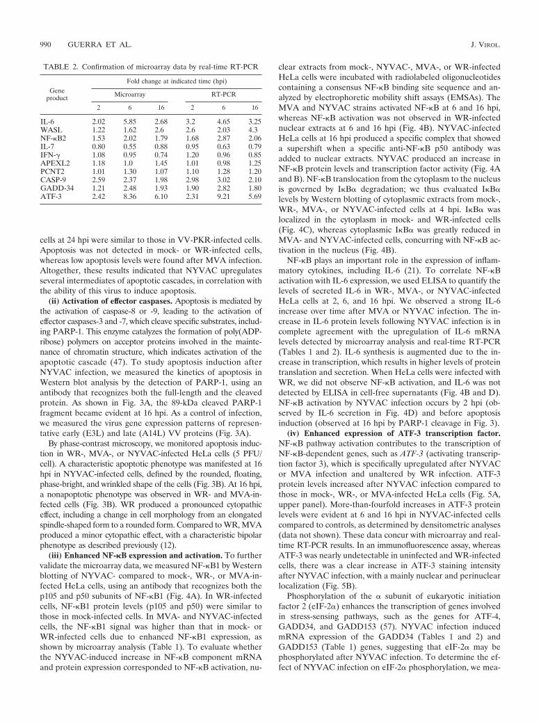
Confirmation of microarray data by quantitative real-timeRT-PCR. We used real-time RT-PCR to verify the transcrip-tional changes in selected genes, as detected by microarrayanalysis. We analyzed 10 genes, 6 of which were upregulated(WASL, NF-�B-2, CASP-9, ATF-3, GADD-34, and IL-6), 2 ofwhich were downregulated (IL-7 and IFN�), and 2 of whichwere unaltered (PCNT2 and APEXL); HPRT was used as aninternal control (Table 2). The assay was performed with thesame RNA samples from infected and uninfected HeLa cellsused in microarray experiments. The expression patterns andrelative mRNA abundances of the genes selected concurredwith the microarray data in all cases, with only slight variations,validating the microarray results.
Target verification and activation of representative cell pro-teins after NYVAC infection. Although changes in mRNA lev-els do not necessarily represent changes in protein expression,we used various approaches to analyze whether the gene ex-pression changes detected by microarray analysis correlatedwith protein levels and the activities of selected gene products.
(i) Caspase-9 activation. The caspase-9 gene was differen-tially expressed following NYVAC infection (Tables 1 and 2);thus, we analyzed the effect of the mRNA increase on theprotein level. Immunofluorescence experiments indicated thattranscriptional upregulation of caspase-9 after NYVAC infec-tion corresponded with an increase in active caspase-9 proteinlevels, confirming the microarray data. Whereas no activecaspase-9 signal was found in control or WR-infected HeLacells, a distinct punctate pattern was observed in NYVAC-infected cells at 6 and 16 hpi (Fig. 2A). In addition, we usedWestern blotting to measure the levels of procaspase cleavageto activated caspase-9 in HeLa cells infected with NYVACcompared to those in cells infected with other VV strains.Caspase-9 activation was detected, with a 10-kDa cleavageproduct from the 46-kDa procaspase. We observed a clearincrement in procaspase-9 and a notable increase in activecaspase-9 after NYVAC infection. In MVA-infected cells,
FIG. 1. Characteristic expression patterns represented in clusters 1 to 6. Mean values (left) and standard deviations (right) of the expressionprofiles of genes assigned to each cluster are shown. Experimental points on the x axis are indicated as follows: 1 for 2 h, 2 for 6 h, and 3 for 16hpi. The y axis shows normalized expression values. Each value in parentheses shows the percentage of genes in each cluster with reference to thetotal of 368 differentially expressed genes.
VOL. 80, 2006 MICROARRAY ANALYSIS OF NYVAC INFECTION IN HeLa CELLS 987

TABLE 1. Representative human genes in clusters 1 to 3 (upregulated by NYVAC)
Cluster, function, and gene name Accession no. Gene symbolaChange (fold) at indicated
time (hpi)
2.00 6.00 16.00
Cluster 1Cell cycle, apoptosis
V-Jun avian sarcoma virus 17 oncogene homolog W96155 JUN 2.14 13.75 8.01Activating transcription factor 3 H21042 ATF3 2.42 8.36 6.10Caspase-9, apoptosis-related cysteine protease AA281152 CASP9 2.59 2.37 1.98Dual-specificity phosphatase 2 AA759046 DUSP2 2.43 5.25 1.56JunB proto-oncogene T99236 JUNB 1.96 2.64 1.65Cytochrome P450, subfamily I (dioxin-inducible) N21019 CYP1B1 1.89 2.73 2.15
CytoskeletonConnective tissue growth factor AA598794 CTGF 4.24 11.45 5.14Pleckstrin homology-like domain, family A, member 1 AA258396 PHLDA1 4.19 8.24 5.28Pleckstrin homology-like domain, family A, member 1 AA258396 PHLDA1 3.85 5.22 4.11Calcineurin-binding protein calsarcin-1 AA065090 CS-1 1.70 2.30 2.55Kinesin family member 5A AA984728 KIF5A 1.89 5.45 4.64
Immune systemTumor necrosis factor (ligand) superfamily, member 7 AI347622 TNFSF7 1.79 3.08 3.41Coagulation factor III (thromboplastin, tissue factor) AI313387 F3 1.58 2.04 1.53Interleukin-6 (interferon beta 2) N98591 IL6 2.02 5.85 2.68
SignalingEarly growth response 1 AA488533 EGR1 4.74 10.38 6.83Nuclear factor kappa light polypeptide gene enhancer
in B cells 2 (p49/p100)AA952897 NFKB2 1.53 2.02 1.79
Serum-inducible kinase AA460152 SNK 1.57 6.49 2.44Tissue factor pathway inhibitor 2 AA399473 TFPI2 1.62 1.77 2.60Brain-derived neurotrophic factor AA262988 BDNF 1.78 4.35 2.12Small inducible cytokine subfamily A, member B AI268937 SCYA8 1.76 3.04 2.89
MiscellaneousNucleoporin, 88 kDa AA488609 NUP88 3.52 6.49 4.23DHHC1 protein W80739 LOC51304 1.96 8.09 4.32Ubiquitin-conjugating enzyme E2M AA449119 UBE2M 1.52 3.06 2.91Prostagtandin-endoperoxide synthase 2 AA644211 PTGS2 1.80 2.90 2.68Small nuclear ribonucleoprotein polypeptides B and B1 AA279662 SNRPB 1.66 2.85 2.87Hypothetical protein A-211C6.1 N39229 LOC57149 1.55 2.74 2.54
Cluster 2Adhesion and cytoskeleton
Collagen, type VII, alpha 1 AA598507 COL7A1 1.46 2.82 2.26Tight junction protein 1 (zona occludens 1) H50344 TJP1 1.18 1.93 2.96Protocadherin alpha 9 AA437139 PCDHA9 1.21 1.31 2.04
Cell cycle, apoptosisMyeloid cell leukemia sequence 1 (BCL2-related) AA488674 MCL1 1.31 3.61 2.18Growth arrest and DNA-damage-inducible 34 AA460168 GADD34 1.21 2.48 1.93SRC homology three (SH3) and cysteine-rich domain AA031284 STAC 1.11 2.41 2.84Growth arrest and DNA-damage-inducible 135 AA404666 GADD135 1.32 2.98 2.06Mitochondrial translational release factor 1 AI347695 MTRF1 1.19 2.06 2.21DnaJ (Hsp40) homolog, subfamily B, member 2 AA455298 DNAJB2 1.22 2.69 2.39Ras homolog gene family, member E W86282 ARHE 1.08 2.68 2.35GRO3 oncogene AA935273 GRO3 1.20 2.62 2.38v-Myc avian myelocytomatosis viral oncogene homolog AA464600 MYC 1.05 2.93 1.65v-Myc avian myelocytomatosis viral oncogene homolog W87741 MYC 1.08 2.93 1.59v-Akt murine thymoma viral oncogene homolog 2; AA457097 AKT2 1.22 2.15 1.57Murine leukemia viral (Bmi-1) oncogene homolog AA478036 BMI1 1.15 1.36 3.18
Immune systemCD80 antigen (CD28 antigen ligand 1, B7-1 antigen) AA983817 CD80 1.25 2.02 2.70Interleukin-1 receptor, type II H78386 IL1R2 1.29 1.58 4.02
Interferon stimulated proteinInterferon-stimulated protein, 15 kDa AA406020 ISG15 1.21 1.14 3.39
MetabolismFatty acid desaturase 3 AI123992 FADS3 1.45 2.94 2.41Carboxypeptidase A2 (pancreatic) AA844831 CPA2 1.48 2.83 3.45
MiscellaneousDiscs, large (Drosophila) homolog 1 AA521155 DLG1 1.24 2.16 2.80CGI-20 protein R02578 LOC51608 1.22 2.11 2.94Bromodomain, testis specific AA454222 BRDT 1.37 1.56 2.17RNA binding motif protein 5 AA456007 RBM5 1.18 3.20 2.42
Continued on facing page
988 GUERRA ET AL. J. VIROL.

weakly active caspase-9 was observed at 16 hpi, whereas therewas no evidence of active caspase-9 in WR-infected HeLa cellsat any time postinfection (Fig. 2B).
This apparent caspase activation in NYVAC-infected cellsprompted us to compare apoptosis in HeLa cells infected withdifferent VV strains. To measure apoptosis, we determined the
amount of cytoplasmic histone-associated DNA in mock-,WR-, MVA-, or NYVAC-infected cells (Fig. 2C). As a positivecontrol, we infected HeLa cells with an inducible VV recom-binant expressing protein kinase R (PKR) in an isopropyl--D-thiogalactopyranoside (IPTG)-dependent manner, whichproduces apoptosis (27). Apoptosis levels in NYVAC-infected
TABLE 1—Continued
Cluster, function, and gene name Accession no. Gene symbolaChange (fold) at indicated
time (hpi)
2.00 6.00 16.00
Spermidine/spermine N1-acetyltransferase AA598631 SAT 1.20 2.51 2.62Spermidine/spermine N1-acetyltransferase AA011215 SAT 1.20 2.47 2.25Splicing factor, arginine/serine-rich 10 AI583623 SFRS10 1.19 4.41 3.21CBP/p300-interacting transactivator 2 AA115078 CITED2 1.19 4.86 1.87Solute carrier family 2, member 3 AA406552 SLC2A3 1.43 2.12 1.77RAE1 (RNA export 1, from Schizosaccharomyces pombe) homolog AA504128 RAE1 1.39 2.07 1.82Apg12 (autophagy 12, S. cerevisiae)-like AA251737 APG12L 1.17 2.70 2.07Discs, large (Drosophila) homolog 1 H24708 DLG1 1.35 1.89 3.15
SignalingNuclear factor kappa light polypeptide gene enhancer AA451716 NFKB1 1.19 2.13 1.92A kinase (PRKA) anchor protein (gravin) 12 AA478543 AKAP12 1.43 4.55 2.86Phorbol-12-myristate-13-acetate-induced protein 1 AA458838 PMAIP1 1.10 2.46 2.93Glutamate receptor, ionotropic, N-methyl D-aspartate 2D AI565972 GRIN2D 1.30 2.36 2.24Receptor tyrosine kinase-like orphan receptor 2 N94921 ROR2 1.26 1.68 2.46Immediate-early response 3 AA457705 IER3 1.44 1.38 2.99Prostaglandin E receptor 3 (subtype EP3) AA406362 PTGER3 1.29 1.22 4.32Solute carrier family 17 (sodium phosphate) H60423 SLC17A2 1.29 1.60 2.14Adaptor-related protein complex 3, delta 1 subunit AA630776 AP3D1 1.18 2.05 2.34
Translation and transcriptionSplicing factor (CC1.3) H47069 CC1.3 1.24 2.36 2.40Splicing factor (CC1.3) AA171948 CC1.3 1.04 1.89 2.26Splicing factor, proline/glutamine-rich AA425853 SFPQ 1.09 1.69 2.55
Wiskott-Aldrich familyWiskott-Aldrich syndrome-like protein AI261600 WASL 1.22 1.62 2.60
Cluster 3Adhesion and cytoskeleton
Claudin 1 AA194833 CLDN1 1.48 2.29 1.30Catenin (cadherin-associated protein), alpha 1 (102 kDa) AA676957 CTNNA1 1.76 2.20 1.48Collagen, type III, alpha 1 COL3A1 COL3A1 1.19 2.03 1.09Cadherin 18, type 2 AA865745 CDH18 0.27 2.17 1.24
Cell cycle, apoptosisBCL2-associated athanogene AI017240 BAG1 1.06 2.05 1.32FOS-like antigen 1 T89996 FOSL1 1.19 2.08 1.10CDC10 (cell division cycle 10, S. cerevisiae), homolog AA633993 CDC10 1.07 3.76 1.45Transforming growth factor beta-stimulated protein TSC-22 AA664389 TSC22 1.20 3.16 1.41Transformer-2 alpha (hTra-2 alpha) AA701944 HSU53209 1.25 2.05 1.46
SignalingDelodinase, iodothyronine, type II AA864322 DIO2 1.17 3.86 1.40Mitogen-activated protein kinase kinase 3 H08749 MAP2K3 1.73 2.15 1.47Phosphatidylinositol-4-phosphate 5-kinase, type I, gamma AA482251 PIP5K1C 1.25 2.17 1.36Phosphatidylinositol-4-phosphate 5-kinase, type I, alpha AA255881 PIP5K1A 1.08 2.31 1.44
Immune systemInsulin-like growth factor binding protein 3 AA598601 IGFBP3 1.14 4.58 1.56Chemokine (C-X-C motif), receptor 4 (fusin) T62636 CXCR4 1.47 2.40 1.41Inhibitor of growth 1 family, member 1 N33574 ING1 1.49 2.24 1.76Coagulation factor III (thromboplastin, tissue factor) AI313387 F3 1.31 2.23 1.39
MiscellaneousSerum/glucocorticoid-regulated kinase AI375353 SGK 1.12 2.89 1.41Cofactor required for Sp1 transcriptional activation N92735 CRSP6 1.19 2.11 1.49Tryptophan 2,3-dioxygenase T72398 TDO2 1.26 2.12 1.44
Transiation and transcriptionSlug (chicken homolog), zinc finger protein H57310 SLUG 1.07 3.29 1.47Eukaryotic translation initiation factor 5A H99842 EIF5A 1.10 2.78 1.14Zinc finger protein 274 AI085519 ZNF274 1.07 2.08 1.33
a Representative human gene targets of NF-�B are underlined.
VOL. 80, 2006 MICROARRAY ANALYSIS OF NYVAC INFECTION IN HeLa CELLS 989
cells at 24 hpi were similar to those in VV-PKR-infected cells.Apoptosis was not detected in mock- or WR-infected cells,whereas low apoptosis levels were found after MVA infection.Altogether, these results indicated that NYVAC upregulatesseveral intermediates of apoptotic cascades, in correlation withthe ability of this virus to induce apoptosis.
(ii) Activation of effector caspases. Apoptosis is mediated bythe activation of caspase-8 or -9, leading to the activation ofeffector caspases-3 and -7, which cleave specific substrates, includ-ing PARP-1. This enzyme catalyzes the formation of poly(ADP-ribose) polymers on acceptor proteins involved in the mainte-nance of chromatin structure, which indicates activation of theapoptotic cascade (47). To study apoptosis induction afterNYVAC infection, we measured the kinetics of apoptosis inWestern blot analysis by the detection of PARP-1, using anantibody that recognizes both the full-length and the cleavedprotein. As shown in Fig. 3A, the 89-kDa cleaved PARP-1fragment became evident at 16 hpi. As a control of infection,we measured the virus gene expression patterns of represen-tative early (E3L) and late (A14L) VV proteins (Fig. 3A).
By phase-contrast microscopy, we monitored apoptosis induc-tion in WR-, MVA-, or NYVAC-infected HeLa cells (5 PFU/cell). A characteristic apoptotic phenotype was manifested at 16hpi in NYVAC-infected cells, defined by the rounded, floating,phase-bright, and wrinkled shape of the cells (Fig. 3B). At 16 hpi,a nonapoptotic phenotype was observed in WR- and MVA-in-fected cells (Fig. 3B). WR produced a pronounced cytopathiceffect, including a change in cell morphology from an elongatedspindle-shaped form to a rounded form. Compared to WR, MVAproduced a minor cytopathic effect, with a characteristic bipolarphenotype as described previously (12).
(iii) Enhanced NF-�B expression and activation. To furthervalidate the microarray data, we measured NF-�B1 by Westernblotting of NYVAC- compared to mock-, WR-, or MVA-in-fected HeLa cells, using an antibody that recognizes both thep105 and p50 subunits of NF-�B1 (Fig. 4A). In WR-infectedcells, NF-�B1 protein levels (p105 and p50) were similar tothose in mock-infected cells. In MVA- and NYVAC-infectedcells, the NF-�B1 signal was higher than that in mock- orWR-infected cells due to enhanced NF-�B1 expression, asshown by microarray analysis (Table 1). To evaluate whetherthe NYVAC-induced increase in NF-�B component mRNAand protein expression corresponded to NF-�B activation, nu-
clear extracts from mock-, NYVAC-, MVA-, or WR-infectedHeLa cells were incubated with radiolabeled oligonucleotidescontaining a consensus NF-�B binding site sequence and an-alyzed by electrophoretic mobility shift assays (EMSAs). TheMVA and NYVAC strains activated NF-�B at 6 and 16 hpi,whereas NF-�B activation was not observed in WR-infectednuclear extracts at 6 and 16 hpi (Fig. 4B). NYVAC-infectedHeLa cells at 16 hpi produced a specific complex that showeda supershift when a specific anti-NF-�B p50 antibody wasadded to nuclear extracts. NYVAC produced an increase inNF-�B protein levels and transcription factor activity (Fig. 4Aand B). NF-�B translocation from the cytoplasm to the nucleusis governed by I�B� degradation; we thus evaluated I�B�levels by Western blotting of cytoplasmic extracts from mock-,WR-, MVA-, or NYVAC-infected cells at 4 hpi. I�B� waslocalized in the cytoplasm in mock- and WR-infected cells(Fig. 4C), whereas cytoplasmic I�B� was greatly reduced inMVA- and NYVAC-infected cells, concurring with NF-�B ac-tivation in the nucleus (Fig. 4B).
NF-�B plays an important role in the expression of inflam-matory cytokines, including IL-6 (21). To correlate NF-�Bactivation with IL-6 expression, we used ELISA to quantify thelevels of secreted IL-6 in WR-, MVA-, or NYVAC-infectedHeLa cells at 2, 6, and 16 hpi. We observed a strong IL-6increase over time after MVA or NYVAC infection. The in-crease in IL-6 protein levels following NYVAC infection is incomplete agreement with the upregulation of IL-6 mRNAlevels detected by microarray analysis and real-time RT-PCR(Tables 1 and 2). IL-6 synthesis is augmented due to the in-crease in transcription, which results in higher levels of proteintranslation and secretion. When HeLa cells were infected withWR, we did not observe NF-�B activation, and IL-6 was notdetected by ELISA in cell-free supernatants (Fig. 4B and D).NF-�B activation by NYVAC infection occurs by 2 hpi (ob-served by IL-6 secretion in Fig. 4D) and before apoptosisinduction (observed at 16 hpi by PARP-1 cleavage in Fig. 3).
(iv) Enhanced expression of ATF-3 transcription factor.NF-�B pathway activation contributes to the transcription ofNF-�B-dependent genes, such as ATF-3 (activating transcrip-tion factor 3), which is specifically upregulated after NYVACor MVA infection and unaltered by WR infection. ATF-3protein levels increased after NYVAC infection compared tothose in mock-, WR-, or MVA-infected HeLa cells (Fig. 5A,upper panel). More-than-fourfold increases in ATF-3 proteinlevels were evident at 6 and 16 hpi in NYVAC-infected cellscompared to controls, as determined by densitometric analyses(data not shown). These data concur with microarray and real-time RT-PCR results. In an immunofluorescence assay, whereasATF-3 was nearly undetectable in uninfected and WR-infectedcells, there was a clear increase in ATF-3 staining intensityafter NYVAC infection, with a mainly nuclear and perinuclearlocalization (Fig. 5B).
Phosphorylation of the � subunit of eukaryotic initiationfactor 2 (eIF-2�) enhances the transcription of genes involvedin stress-sensing pathways, such as the genes for ATF-4,GADD34, and GADD153 (57). NYVAC infection inducedmRNA expression of the GADD34 (Tables 1 and 2) andGADD153 (Table 1) genes, suggesting that eIF-2� may bephosphorylated after NYVAC infection. To determine the ef-fect of NYVAC infection on eIF-2� phosphorylation, we mea-
TABLE 2. Confirmation of microarray data by real-time RT-PCR
Geneproduct
Fold change at indicated time (hpi)
Microarray RT-PCR
2 6 16 2 6 16
IL-6 2.02 5.85 2.68 3.2 4.65 3.25WASL 1.22 1.62 2.6 2.6 2.03 4.3NF-�B2 1.53 2.02 1.79 1.68 2.87 2.06IL-7 0.80 0.55 0.88 0.95 0.63 0.79IFN-� 1.08 0.95 0.74 1.20 0.96 0.85APEXL2 1.18 1.0 1.45 1.01 0.98 1.25PCNT2 1.01 1.30 1.07 1.10 1.28 1.20CASP-9 2.59 2.37 1.98 2.98 3.02 2.10GADD-34 1.21 2.48 1.93 1.90 2.82 1.80ATF-3 2.42 8.36 6.10 2.31 9.21 5.69
990 GUERRA ET AL. J. VIROL.

FIG. 2. NYVAC-induced apoptosis in HeLa cells. (A) Protein validation of microarray data by immunofluorescence analysis of the effect ofNYVAC infection on caspase-9 distribution in HeLa cells. Mock-, WR-, or NYVAC-infected cells were incubated at the indicated times with ananti-caspase-9 antibody (Calbiochem) that recognizes the active form of caspase-9, followed by the appropriate conjugated secondary antibody andthe ToPro reagent. The samples were analyzed by confocal immunofluorescence microscopy. (B) Activation of caspase-9 after WR, MVA, orNYVAC infection. Total proteins (100 �g) were separated by SDS-PAGE, transferred to nitrocellulose, and immunoblotted at various times (6and 16 hpi) with an anti-caspase-9 antibody (Oncogene) that recognizes the procaspase and the cleaved active form of caspase-9. Caspase-9presented a proform (46 kDa) that it is cleaved into a heterodimer of 35-kDa and 10-kDa chains. The molecular sizes (in kilodaltons) of theproteins are indicated on the right. Actin levels revealed that the same amount of protein was loaded into each lane. (C) Apoptosis assays afterWR, MVA, or NYVAC infection determined the amount of cytoplasmic histone-associated DNA by ELISA. HeLa cells grown in 12-well plateswere infected (5 PFU/cell) with the viruses indicated and harvested at 24 hpi for determinations with an ELISA kit of the absorbance at 405 nm.As a positive control for apoptosis, we used VV-PKR-infected HeLa cells. Duplicate samples were measured in two independent experiments.
VOL. 80, 2006 MICROARRAY ANALYSIS OF NYVAC INFECTION IN HeLa CELLS 991

FIG. 3. Apoptosis induction kinetics during NYVAC infection. (A) Time course of PARP-1 cleavage during NYVAC infection. HeLa cellswere infected with NYVAC, and at the indicated times, total proteins (100 �g) were separated by SDS-PAGE, transferred to nitrocellulose, andimmunoblotted with anti-PARP-1, which recognizes both the full-length and the cleaved protein. An 89-kDa cleavage product of PARP-1 wasobserved at 16 hpi. As a measure of infection, we used immunoblotting to visualize the viral proteins E3L and A14L. The molecular sizes (inkilodaltons) of the proteins are indicated on the right. Actin levels showed that the same amount of protein was loaded in each lane. (B) Apoptoticphenotype of NYVAC-infected HeLa cells. Monolayer cultures of HeLa cells were left uninfected or infected (5 PFU/cell) with the different VVstrains for 16 h, and the apoptotic phenotype was visualized by phase-contrast microscopy. NYVAC-infected cells showed characteristic apoptosis,defined by a rounded, floating, phase-bright, wrinkled shape.
992 GUERRA ET AL. J. VIROL.

FIG. 4. NYVAC-induced NF-�B signaling. (A) Validation of microarray data by NF-�B protein levels and comparison between WR, MVA,and NYVAC infections. Total proteins (100 �g) were separated by SDS-PAGE, transferred to nitrocellulose, and immunoblotted with anti-NF-�B1 antibody at various times (6 and 16 hpi). Actin levels indicated that the same amount of protein was loaded into each lane. The molecularsizes (in kilodaltons) of the proteins are indicated on the right. (B) Autoradiogram of NF-�B activity, determined by EMSA performed with nuclearextracts of mock-, MVA-, and NYVAC-infected HeLa cells at the indicated times. WR, nuclear extract from WR-infected HeLa cells at 16 hpi.The arrow indicates the 32P-labeled NF-�B/DNA complex and the nonspecific signal, and the asterisk indicates the supershift of NF-�B afterincubation of the nuclear extracts with the 32P-labeled probe and anti-p50 antibody. (C) I��� degradation kinetics of WR-, MVA-, andNYVAC-infected HeLa cells. Cytoplasmic extracts were prepared, as previously described (45), from uninfected and poxvirus-infected (5PFU/cell) HeLa cells. I��� levels were detected by immunoblotting at 4 hpi. The molecular sizes (in kilodaltons) of the proteins are indicated onthe right. Actin levels indicated that the same amount of protein was loaded into each lane of the gel. (D) Levels of IL-6 secreted from WR-, MVA-,and NYVAC-infected HeLa cells (5 PFU/cell), as determined by ELISA. Protein levels of IL-6 in supernatants of infected cells were measuredat 2, 6, and 16 hpi. Duplicate samples were measured in two independent experiments.
VOL. 80, 2006 MICROARRAY ANALYSIS OF NYVAC INFECTION IN HeLa CELLS 993

sured specific eIF-2� phosphorylation at Ser-51 by Westernblotting of mock-, WR-, MVA-, or NYVAC-infected HeLacells. Clear eIF-2� phosphorylation was observed at 6 and 16hpi in NYVAC-infected cells but not in WR- or MVA-infectedcells (Fig. 5A, lower panels). Thus, we found a correlation ofenhanced ATF-3 expression at the mRNA and protein levelsand enhanced GADD34 and GADD153 expression at theRNA level with eIF-2� phosphorylation, suggesting that thesestress pathways could be involved in apoptosis induction byNYVAC infection.
VV K1L gene prevents NF-�B activation but not apoptosisinduction after NYVAC infection. The VV K1L host rangegene acts as an NF-�B inhibitor (45). Since NYVAC lacks theK1L gene, we analyzed whether apoptosis induction andNF-�B activation by NYVAC can be inhibited by the K1Lgene. NYVAC-infected HeLa cells were transfected with aplasmid containing the K1L gene with a T7-Tag tag or with acontrol empty plasmid (pC), and apoptosis was measured byimmunofluorescence. The K1L gene is necessary for VV rep-lication in RK13 cells (38, 41), and after infection-transfection,we confirmed that the transfected K1L gene rescued the hostrange by producing plaques in RK13 cells (data not shown).Apoptotic cells were counted based on the characteristic in-crease in nucleus granularity and the appearance of apoptoticbodies. Cells were costained with E3L antibody to detect VV,ToPro for DNA detection, and anti-T7-Tag to detect trans-fected cells. The apoptotic phenotype of K1L-transfectedNYVAC-infected cells was comparable to that found in theabsence of K1L (not shown). We observed similar numbers ofapoptotic cells after K1L expression as for pC-transfected cells(Fig. 6A), indicating that the K1L gene was not responsible forthe apoptotic phenotype after NYVAC infection. To analyzethe ability of K1L to repress NF-�B activation after NYVACinfection, we used ELISA to quantify secreted IL-6, an indi-cator of NF-�B activity, in supernatants from NYVAC-in-fected HeLa cells that were untransfected or transfected withpC or pK1L. We observed a marked decrease in secreted IL-6in NYVAC-infected HeLa cells after pK1L transfection(Fig. 6B). This result indicated that K1L expression inhibitsNF-�B activity in NYVAC-infected cells.
DISCUSSION
Poxvirus vector technology has provided reagents for high-level expression of proteins for vaccine use. Due to their in-fection efficiencies, high gene product expression levels, andsafety, attenuated poxvirus vectors are useful for gene deliveryin immunotherapy (10, 35). NYVAC is a highly attenuated VVstrain, generated by the specific deletion of 18 open readingframes from the viral genome, which affects host range, viru-lence, and pathogenesis genes (32, 49, 50). This vector hasbeen applied as a recombinant vaccine delivery system in an-imal models and target species, including humans (34, 37).NYVAC-based vectors have been analyzed as vaccines forseveral tumors (23, 46) and are protective against pathogenssuch as retroviruses (5, 10, 31), Japanese encephalitis virus(24), and Plasmodium falciparum (34). The potential use ofvariola virus as a biological weapon requires the developmentof safer smallpox vaccines, using attenuated VV strains such asMVA and NYVAC (4, 9).
FIG. 5. ATF-3 protein expression is induced by NYVAC infection.(A) Levels of ATF-3, phosphorylated eIF2� at Ser-51, and total eIF2�were measured by immunoblot analysis of extracts of HeLa cells afterWR, MVA, and NYVAC infection. Total proteins (100 �g) wereseparated by SDS-PAGE, transferred to nitrocellulose, and immuno-blotted with anti-ATF-3, anti-phosphorylated-eIF2�, or anti-eIF2� an-tibody at various times (6 and 16 hpi). The molecular sizes (in kilo-daltons) of the proteins are indicated on the right. The antibodyagainst ATF-3 recognizes two or three bands, depending on the run-ning conditions, perhaps as a result of posttranslational modificationsof the transcription factor, which can be cell dependent. (B) Immuno-fluorescence analysis of the effect of NYVAC infection on ATF-3distribution in HeLa cells. Mock-, WR-, and NYVAC-infected cellswere labeled with anti-ATF-3 (Santacruz) at the indicated times, fol-lowed by the appropriate conjugated secondary antibody and ToProreagent. The samples were analyzed by confocal immunofluorescencemicroscopy.
994 GUERRA ET AL. J. VIROL.

The use of these vectors as vaccines requires a better un-derstanding of the impact of NYVAC infection in the humanhost, including the identification of differentially regulatedgenes and their functions. Using high-throughput cDNA mi-croarray screening of over 15,000 genes in NYVAC-infectedHeLa cells, we identified transcriptional alterations in a de-fined subset of human genes. When grouped into functionalcategories, a large proportion of altered transcripts were fromgenes involved in apoptosis, adhesion, cytoskeletal architec-ture, cell cycling, and immune modulation.
Apoptosis is regarded as an innate cellular response thatlimits virus propagation. The importance of apoptosis as anantiviral defense against poxvirus infection is suggested by thefinding that a number of poxvirus genes encode proteins thatinterfere with apoptosis in specific cell types (30, 39). DuringNYVAC infection, we found an upregulation of the caspase-9gene, an apoptosis-linked gene. Increased caspase-9 proteinlevels reflected an increase in caspase-9 activity and effectorcaspases, as determined by the appearance of a cleavage prod-uct from the target 89-kDa PARP-1 protein. Although wedetected caspase-9 activation at 6 hpi, as shown by cleavage ofthe 46-kDa procaspase, apoptosis and PARP-1 cleavage werenot evident until 16 hpi. These temporal differences in NYVAC-induced apoptosis may be due to distinct target specificities andthe sequence of biochemical events. The role of apoptosis inpoxvirus biology is not well understood; previous studies ofK1L gene-defective viruses found no correlation between ap-optosis suppression and virus growth (7), and similar resultswere obtained by inactivation of the cowpox virus crmA gene(42). CrmA/SPI-2 is the best-characterized apoptosis modula-tor encoded by poxvirus family members; it inhibits apoptosisby preventing caspase-8 activity (51, 56). An antiapoptotic
function was previously described for the VV E3L gene prod-uct that blocks PKR activity (27, 29) and was recently shownfor the viral protein F1L, which inhibits mitochondrial releaseof cytochrome c, which is essential for apoptosis induction (48,52). B13R and B14R, encoding the interleukin-1-convertingenzyme inhibitor, are also involved in antiapoptotic functions(39, 49). Since the antiapoptotic genes F1L and E3L arepresent in the NYVAC genome, other genes with antiapop-totic functions, such as B13R and B14R or others with un-known functions that are absent in NYVAC, may be the causefor apoptotic induction of this virus. Our results indicate thatthe K1L gene does not act as an inhibitor of apoptosis duringNYVAC infection, as we did not observe a blockade of apop-tosis by ectopic expression of this gene (Fig. 6).
With regard to immune modulators, our microarray exper-iments showed NF-�B upregulation, which induces the tran-scription of several genes that encode cytokines or are essentialfor immune response activation (14, 21). NF-�B forms a het-erodimer composed of 50- and 65-kDa subunits (p50 and p65)which is retained in the cytoplasm by its inhibitor, I���. Spe-cific stimuli cause proteolytic degradation of I��� in the cyto-plasm and subsequent NF-�B protein translocation to the nu-cleus, where it transactivates certain genes (14, 21). Ourmicroarray data indicated upregulated expression for both NF-�B1 and NF-�B2 messengers during NYVAC infection(Table 1). NF-�B2 upregulation was confirmed by real-timeRT-PCR. The increase in NF-�B1 mRNA corresponded toincreased levels of the p105 and p50 proteins (Fig. 4A). NYVACinfection also upregulated NF-�B transcriptional activity (Fig.4B). NF-�B was activated early in infection, although EMSAdid not detect activation at 2 hpi, when upregulation of IL-6,an indicator of NF-�B activation, was observed by real-time
FIG. 6. K1L is necessary for NF-�B inhibition but not for apoptotic induction in NYVAC infection. (A) Ectopic K1L expression in NYVAC-infected HeLa cells does not inhibit apoptosis during NYVAC infection. HeLa cells grown on coverslips were infected with NYVAC at 0.1PFU/cell, and at 1 hpi, cells were transfected (3 �g) with an expression plasmid containing the K1L gene with T7-Tag (pK1L) or with an emptycontrol plasmid (pC). At 40 h posttransfection, cells were fixed and processed to measure apoptosis by immunofluorescence, using antibodiesdirected against the E3L protein (red), T7-Tag (green), and ToPro for DNA staining (blue). The ratios of infected apoptotic cells to infectednonapoptotic cells were determined by counting (about 2,000 cells per experiment); each point represents the average of two independentexperiments. (B) Ectopic K1L expression inhibits NF-�B activation in NYVAC infection. Levels of IL-6 secreted from cells infected andtransfected as described above were quantified by ELISA. Duplicate samples were measured in two independent experiments.
VOL. 80, 2006 MICROARRAY ANALYSIS OF NYVAC INFECTION IN HeLa CELLS 995

RT-PCR and microarray analysis. The NF-�B activation path-way contributes to the transcription of �B-dependent genes.Many transcripts upregulated by NYVAC are known NF-�Btarget genes (Table 1), although other �B-dependent genessuch as TNF, A20, CCL2, CXCL1, and IL-8 were not upregu-lated during NYVAC infection. In view of the dual NF-�B-activating effect and apoptotic phenotype triggered by NYVACinfection, it is not surprising to find that not all of the NF-�B-dependent genes are upregulated by virus infection. One of theNF-�B-dependent genes that is upregulated is ATF-3, a stress-inducible gene that encodes an ATF/CREB transcription fac-tor (18, 19, 28). Recent reports describe the effect of ATF-3 inapoptosis induction (20, 53); ATF-3, considered a proapop-totic factor, might be involved in apoptosis activation inNYVAC-infected cells, although its physiological relevance inthis system remains to be established. NYVAC infection alsoinduced mRNAs such as those encoding GADD34 andGADD153, whose activation is eIF-2� phosphorylation depen-dent; eIF-2� phosphorylation in NYVAC infection was con-firmed by Western blotting (Fig. 5A).
MVA, another attenuated VV strain, also activates NF-�B(16, 36). NF-�B activation mediated by NYVAC and MVAmay reflect the loss of a specific viral gene from the parentalviral genome, deleted during the generation of MVA andNYVAC. Indeed, the K1L gene was deleted in NYVAC (49),and the viral K1L protein inhibits NF-�B activation by prevent-ing I��� degradation (45). Since both the MVA and NYVACgenomes lack K1L, which is present in VV, we studied the
effect of the absence of K1L on NF-�B activation by measuringIL-6 levels in NYVAC-infected HeLa cells transfected with anexpression plasmid bearing the K1L gene. IL-6 levels wereconsiderably lower in K1L-expressing cells than in control-transfected cells, indicating that K1L inhibits NF-�B activityduring NYVAC infection (Fig. 6B). K1L inhibition of NF-�Bactivity was independent of NYVAC-induced apoptosis, asindicated by counting NYVAC-infected apoptotic cells ex-pressing K1L (Fig. 6A).
Although NF-�B activation and apoptosis induction mightbe negative factors in the NYVAC cycle, the triggering of thesemechanisms may favor a better immune response to a recom-binant vector. NF-�B activation from a recombinant NYVACinfection should stimulate and amplify the immune responseagainst recombinant products and facilitate clearance of thevector. The role of apoptosis in the immune response is stillunclear, but antigens produced by apoptotic cells are reportedto increase antigen immunogenicity (54). Dendritic cells thatphagocytose apoptotic bodies from virus-infected cells canpresent viral antigens to cytotoxic T cells, inducing a cytotoxicresponse (1). It remains to be defined whether apoptosis in-duced by a NYVAC-based vector increases antigen immuno-genicity in comparison with that induced by MVA vectors orNYVAC vectors unable to induce apoptosis by gene targeting.
We compared expression profiles of representative genesobtained in this study with those obtained from MVA infec-tions (16) (Table 3). In infected HeLa cells, one group of genesis upregulated by both MVA and NYVAC, whereas others are
TABLE 3. Representative gene expression profiling of MVA and NYVAC infections of human HeLa cellsa
Expression profile and gene product name Gene symbolFold change with MVA Fold change with
NYVAC
2 hpi 6 hpi 16 hpi 2 hpi 6 hpi 16 hpi
Increased in MVA and NYVAC infectionsEarly growth response 1 EGR1 4.17 4.60 1.94 4.74 10.38 6.83Nuclear factor (erythroid-derived 2)-like 3 NFE2L3 2.36 2.36 2.52 1.19 2.13 1.32Nuclear factor kappa light polypeptide 2 NFKB2 2.03 2.22 2.34 1.53 2.02 1.79Interleukin-6 IL6 2.65 2.14 1.56 2.02 5.85 2.68Interferon-stimulated protein, 15 kDa ISG15 2.57 2.25 1.61 1.21 1.14 3.39CD80 antigen CD80 1.75 2.58 2.06 1.25 2.02 2.7Dual-specificity phosphatase 5 DUSP5 3.01 2.91 1.87 1.98 4.16 2.18JunB proto-oncogene JUNB 2.93 3.92 1.06 2.16 2.14 1.32v-Jun avian sarcoma virus 17 oncogene homolog JUN 2.17 2.02 1.31 2.14 13.75 8.01v-Myc avian myelocytomatosis viral oncogene homolog MYC 2.22 1.53 1.05 1.05 2.93 1.65Activating transcription factor 3 ATF3 1.78 1.98 0.99 2.42 8.36 6.10Dual-specificity phosphatase 2 DUSP2 3.93 3.59 1.85 2.43 5.25 1.56Connective tissue growth factor CTGF 5.77 4.51 1.12 4.24 11.45 5.14Kinesin family member 5A KIF5A 2.05 1.88 1.73 1.89 5.45 4.64
Increased in MVA infectionInterleukin-7 IL7 6.1 5.3 3.2 0.80 0.55 0.88B7 protein B7 1.97 1.75 3.53 1.58 1.58 1.78CD47 antigen CD47 5.35 4.08 3.16 1.02 0.76 1.03Mitogen-activated protein kinase kinase 5 MAP2K5 2.37 2.74 1.69 1.02 0.76 1.03Nuclear factor kappa light polypeptide epsilon NFKBIE 2.41 1.61 1.12 1.18 1.27 1.42Nuclear factor of activated T cells, cytoplasmic 3 NFATC3 3.46 2.97 1.30 1.01 1.34 1.34
Increased in NYVAC infectionCaspase-9, apoptosis-related cysteine protease CASP9 1.1 0.82 0.32 2.59 2.37 1.98Nucleoporin (88 kDa) NUP88 0.87 0.93 0.92 3.52 6.49 4.23Pleckstrin homology-like domain, family A, member 1 PHLDA1 1.01 0.95 0.99 4.19 8.24 5.28
a MVA data are from a previous report (16).
996 GUERRA ET AL. J. VIROL.

upregulated selectively by MVA or NYVAC. Both viruses trig-ger cellular transcription factors, including NF-�B, Myc, c-Jun,ATF-3, DUSP2, and CTGF, and cell adhesion molecules suchas the kinesin KIF5C. EGR1 (early gene response 1) was alsoupregulated by MVA and NYVAC, as also reported for MVA-�E3L-infected HeLa cells (29), and the MEK/ERK/RSK2/Elk-1/EGR-1 signaling pathway is necessary for VV multipli-cation (2). The immunomodulatory molecules IL-6, ISG15,and CD80 were upregulated in NYVAC- and MVA-infectedcells, whereas IL-7, IL-1A, IL-8, and IL-15 were upregulatedafter MVA but not NYVAC infection. This indicates thatMVA and NYVAC induce distinct proinflammatory cytokineprofiles in vitro. In HeLa cells, WR infection also upregulatesCD80 (15), but IL-6 and ISG15 upregulation is specific toMVA and NYVAC infections. ISG15 was first identified as aninterferon-stimulated gene (6, 26) that affects IFN-�/ signaltransduction; its expression increases markedly after viral in-fection (16, 33, 55). IFN-�/-induced ISG15 activation sug-gests a role for ISG15 in the innate immune response to viralinfection (44).
In view of our findings, we propose that NYVAC infectionactivates a sequence of survival and apoptotic responses in theinjured cell. NYVAC induces NF-�B activation early in infec-tion and will trigger the expression of several gene classes, eachof which might be required at different times of infection forcell survival. The NF-�B-activated early signals will not be ableto overcome the cell death process caused by the virus infec-tion, and cells will ultimately die by apoptosis. Some of theNF-�B-induced genes, such as ATF-3, together with otherstress signals, such as GADD34 and GADD153 and other un-known signals, might promote apoptosis. Since apoptosis is alate event in NYVAC infection, most of the virus cycle will becompleted, and hence the contribution of apoptosis to virusreplication will be minimized. While the poxvirus genome en-codes several genes that antagonize apoptosis and NF-�B ac-tivation, it is likely that these virus genes have a major impacton the evasion of the immune system by the virus rather thanon virus growth in culture. We demonstrated that the K1L viralprotein prevented NF-�B activation but not apoptosis induc-tion in NYVAC infection, although the viral gene(s) involvedin NYVAC-induced apoptosis remains unknown.
Overall, our results provide a basis for future functionalevaluations of the NYVAC vector for use in human vaccines.Gene expression profiling permits detailed analyses of the im-pact of poxvirus vectors on immune system target cells aftervaccination and the development of more effective poxvirusvaccines against a broad spectrum of pathogens and tumors.
ACKNOWLEDGMENTS
We are indebted to R. Bablanian for critically reviewing the manu-script. We thank J. Tartaglia (Aventis-Pasteur) for the generous gift ofNYVAC, and we are grateful to B. L. Jacobs for the anti-E3L antibodyand A. Alcamı for the K1L expression plasmid. We thank S. Gutierrezfor confocal microscopy, V. Jimenez for expert technical assistance,and C. Mark and P. Martinez for editorial assistance.
S.G. was supported by a contract from the EU Project on VacciniaVectors. A.P.-M. was partially supported by the Spanish CAM grantGR/SAL/0653/2004 and the Ramon y Cajal Program. This work wassupported by grants from the Spanish Ministry of Education and Sci-ence (BIO2004-03954), the Spanish Foundation for AIDS Research(FIPSE 36344/02), Fundacion Botın, and the European Union (Euro-Vac QLRT-PL-1999-01321, Vaccinia Vectors QLK2-CT-2002-01867).
The Department of Immunology and Oncology was founded and issupported by the Spanish Council for Scientific Research (CSIC) andPfizer.
REFERENCES
1. Albert, M. L. 2004. Death-defying immunity: do apoptotic cells influenceantigen processing and presentation? Nat. Rev. Immunol. 4:223–231.
2. Andrade, A. A., P. N. G. Silva, A. C. T. C. Pereira, L. P. de Sousa, P. C. P.Ferreira, R. T. Gazzinelli, E. G. Kroon, C. Ropert, and C. A. Bonjardim. 2004.The vaccinia virus-stimulated mitogen-activated protein kinase (MAPK) path-way is required for virus multiplication. Biochem. J. 381:437–446.
3. Arenzana-Seisdedos, F., J. Thompson, M. S. Rodriguez, F. Bachelerie, D.Thomas, and R. T. Hay. 1995. Inducible nuclear expression of newly synthe-sized I kappa B alpha negatively regulates DNA-binding and transcriptionalactivities of NF-kappa B. Mol. Cell. Biol. 15:2689–2696.
4. Belyakov, I. M., P. Earl, A. Dzutsev, V. A. Kuznetsov, M. Lemon, L. S. Wyatt,J. T. Snyder, J. D. Ahlers, G. Franchini, B. Moss, and J. A. Berzofsky. 2003.Shared modes of protection against poxvirus infection by attenuated andconventional smallpox vaccine viruses. Proc. Natl. Acad. Sci. USA 16:9458–9463.
5. Benson, J., C. Chougnet, M. Robert-Guroff, D. Montefiori, P. Markham, G.Shearer, R. C. Gallo, M. Cranage, E. Paoletti, K. Limbach, D. Venzon, J.Tartaglia, and G. Franchini. 1998. Recombinant vaccine-induced protectionagainst the highly pathogenic simian immunodeficiency virus SIVmac251: de-pendence on route of challenge exposure. J. Virol. 72:4170–4182.
6. Blomstrom, D. C., D. Fahey, R. Kutny, B. D. Korant, and E. Knight. 1986.Molecular characterization of the interferon-induced 15-kDa protein. Mo-lecular cloning and nucleotide and amino acid sequence. J. Biol. Chem.261:8811–8816.
7. Chung, C. S., I. A. Vasilevskaya, S. C. Wang, C. H. Bair, and W. Chang. 1997.Apoptosis and host restriction of vaccinia virus in RK13 cells. Virus Res.52:121–132.
8. Cudmore, S., P. Cossart, G. Griffiths, and M. Way. 1995. Actin-based mo-tility of vaccinia virus. Nature 378:636–638.
9. Edghill-Smith, Y., D. Venzon, T. Karpova, J. McNally, J. Nacsa, W. P. Tsai,E. Tryniszewska, M. Moniuszko, J. Manischewitz, L. R. King, S. J.Snodgrass, J. Parrish, P. Markham, M. Sowers, D. Martin, M. G. Lewis,J. A. Berzofsky, I. M. Belyakov, B. Moss, J. Tartaglia, M. Bray, V. Hirsch, H.Golding, and G. Franchini. 2003. Modelling a safer smallpox vaccinationregimen, for human immunodeficiency virus type 1-infected patients inimmunocompromised macaques. J. Infect. Dis. 8:1181–1191.
10. Franchini, G., S. Gurunathan, L. Baglyos, S. Plotkin, and J. Tartaglia. 2004.Poxvirus-based vaccine candidates for HIV: two decades of experience withspecial emphasis on canarypox vectors. Exp. Rev. Vaccines 3:75–88.
11. Frischknecht, F., V. Moreau, S. Rottger, I. Reckmann, C. Superti-Furga, andM. Way. 1999. Actin based motility of vaccinia virus mimics receptor tyrosinekinase signalling. Nature 404:1007–1011.
12. Gallego-Gomez, J. C., C. Risco, D. Rodriguez, P. Cabezas, S. Guerra, J. L.Carrascosa, and M. Esteban. 2003. Differences in virus-induced cell mor-phology and virus maturation between the WR and MVA strains of vacciniavirus. J. Virol. 77:10606–10622.
13. Garcıa de la Nava, J., D. Franco Santaella, J. Cuenca Alba, J. M. Carazo, O.Trelles, and A. Pascual-Montano. 2003. Engene: the processing and explora-tory analysis of gene expression data. Bioinformatics 19:657–658.
14. Ghosh, S., and M. Karin. 2002. Missing pieces in the NF-kappaB puzzle. Cell109:81–96.
15. Guerra, S., L. A. Lopez-Fernandez, A. Pascual-Montano, M. Munoz, K.Harshman, and M. Esteban. 2003. Cellular gene expression survey of vac-cinia virus infection of human HeLa cells. J. Virol. 77:6493–6506.
16. Guerra, S., L. A. Lopez-Fernandez, R. Conde, A. Pascual-Montano, M.Munoz, K. Harshman, and M. Esteban. 2004. Microarray analysis revealscharacteristic changes of host cell gene expression in response to attenuatedmodified vaccinia virus Ankara (MVA) infection of human HeLa cells.J. Virol. 78:5820–5835.
17. Guerra, S., M. Aracil, R. Conde, A. Bernad, and M. Esteban. 2005. Wiskott-Aldrich syndrome protein is needed for vaccinia virus pathogenesis. J. Virol.79:2133–2140.
18. Hai, T., C. D. Wolfgang, D. K. Marsee, A. E. Allen, and U. Sivaprasad. 1999.ATF3 and stress responses. Gene Exp. 7:321–335.
19. Hai, T., and M. G. Hartman. 2001. The molecular biology and nomenclatureof the activating transcription factor/cAMP responsive element binding fam-ily of transcription factors: activating transcription factor proteins and ho-meostasis. Gene 1:1–11.
20. Hartman, M. G., D. Lu, M. L. Kim, G. J. Kociba, T. Shukri, J. Buteau, X.Wang, W. L. Frankel, D. Guttridge, M. Prentki, S. T. Grey, D. Ron, andT. Hai. 2004. Role for activating transcription factor 3 in stress-inducedbeta-cell apoptosis. Mol. Cell. Biol. 13:5721–5732.
21. Hayden, M. S., and S. Ghosh. 2004. Signaling to NF-kappaB. Genes Dev.18:2195–2224.
22. Hughes, T. R., M. J. Marton, A. R. Jones, C. J. Roberts, R. Stoughton, C. D.Armour, H. A. Bennett, E. Coffey, H. Dai, Y. D. He, M. J. Kidd, A. M. King,M. R. Meyer, D. Slade, P. Y. Lum, S. B. Stepaniants, D. D. Shoemaker,
VOL. 80, 2006 MICROARRAY ANALYSIS OF NYVAC INFECTION IN HeLa CELLS 997

D. Gachotte, K. Chakraburtty, J. Simon, M. Bard, and S. H. Friend. 2000.Functional discovery via a compendium of expression profiles. Cell 102:109–126.
23. Jourdier, T. M., C. Moste, M. C. Bonnet, F. Delisle, J. P. Tafani, P.Devauchelle, J. Tartaglia, and P. Moingeon. 2003. Local immunotherapy ofspontaneous feline fibrosarcomas using recombinant poxviruses expressinginterleukin 2 (IL-2). Gene Ther. 26:2126–2132.
24. Kanesa-thasan, N., J. J. Smucny, C. H. Hoke, D. H. Marks, E. Konishi, I.Kurane, D. B. Tang, D. W. Vaughn, P. W. Mason, and R. E. Shope. 2000.Safety and immunogenicity of NYVAC-JEV and ALVAC-JEV attenuatedrecombinant Japanese encephalitis virus-poxvirus vaccines in vaccinia-non-immune and vaccinia-immune humans. Vaccine 19:483–491.
25. Kohonen, T. 1997. Self-organizing maps, 2nd ed. Springer-Verlag, Heidelberg,Germany.
26. Korant, B. D., D. C. Blomstrom, G. J. Jonak, and E. Knight. 1984. Inter-feron-induced proteins. Purification and characterization of a 15,000-daltonprotein from human and bovine cells induced by interferon. J. Biol. Chem.23:14835–14839.
27. Lee, S. B., and M. Esteban. 1994. The interferon-induced double-strandedRNA-activated protein kinase induces apoptosis. Virology 199:491–496.
28. Liang, G., A. D. Wolfgang, B. P. C. Chen, T. H. Chen, and T. Hai. 1996.ATF3 gene: genome organization, promoter and regulation. J. Biol. Chem.271:1695–1701.
29. Ludwig, H., J. Mages, C. Staib, M. H. Lehmann, R. Lang, and G. Sutter.2005. Role of viral factor E3L in modified vaccinia virus Ankara infection ofhuman HeLa cells: regulation of the virus life cycle and identification ofdifferentially expressed host genes. J. Virol. 79:2584–2596.
30. McFadden, G. 2005. Poxvirus tropism. Nat. Rev. Microbiol. 3:201–213.31. Myagkikh, M., S. Alipanah, P. D. Markham, J. Tartaglia, E. Paoletti, R. C.
Gallo, G. Franchini, and M. Robert-Guroff. 1996. Multiple immunizationswith attenuated poxvirus HIV type 2 recombinants and subunit boosts re-quired for protection of rhesus macaques. AIDS Res. Hum. Retrovir. 12:985–992.
32. Najera, J. L., C. E. Gomez, M. M. Gherardi, E. Domingo-Gil, and M.Esteban. Unpublished data.
33. Nicholl, M. J., L. H. Robinson, and C. M. Preston. 2000. Activation ofcellular interferon-responsive genes after infection of human cells with her-pes simplex virus type 1. J. Gen. Virol. 9:2215–2218.
34. Ockenhouse, C. F., P. F. Sun, D. E. Lanar, B. T. Wellde, B. T. Hall, K.Kester, J. A. Stoute, A. Magill, U. Krzych, L. Farley, R. A. Wirtz, J. C. Sadoff,D. C. Kaslow, S. Kumar, L. W. Church, J. M. Crutcher, B. Wizel, S. Hoff-man, A. Lalvani, A. V. Hill, J. A. Tine, K. P. Guito, C. de Taisne, R. Anders,W. R. Ballou, et al. 1998. Phase I/IIa safety, immunogenicity, and efficacytrial of NYVAC-Pf7, a pox-vectored, multiantigen, multistage vaccine can-didate for Plasmodium falciparum malaria. J. Infect. Dis. 6:1664–1673.
35. Odin, L., M. Favrot, D. Poujol, J. P. Michot, P. Moingeon, J. Tartaglia, andI. Puisieux. 2001. Canarypox virus expressing wild type p53 for gene therapyin murine tumors mutated in p53. Cancer Gene Ther. 2:87–98.
36. Oie, L. K., and D. Pickup. 2001. Cowpox and other members of the ortho-poxvirus genus interfere with the regulation of NF-�B activation. Virology288:175–187.
37. Paoletti, E. 1996. Applications of poxvirus vectors to vaccination: an update.Proc. Natl. Acad. Sci. USA 21:11349–11353.
38. Perkus, M. E., S. J. Goebel, S. W. Davis, G. P. Johnson, K. Limbach, E. K.Norton, and E. Paoleti. 1990. Vaccinia virus host range genes. Virology179:276–286.
39. Pogo, B. G., S. M. Melana, and J. Blaho. 2004. Poxvirus infection andapoptosis. Int. Rev. Immunol. 23:61–74.
40. Quackenbush, J. 2002. Microarray data normalization and transformation.Nat. Genet. 32:496–501.
41. Ramsey-Ewing, A. L., and B. Moss. 1996. Complementation of a vacciniavirus host-range K1L gene deletion by the nonhomologous CP77 gene.Virology 222:75–86.
42. Ray, C. A., and D. J. Pickup. 1996. The mode of death of pig kidney cellsinfected with cowpox virus is governed by the expression of the crmA gene.Virology 217:384–391.
43. Rodriguez, J. R., C. Risco, J. L. Carrascosa, M. Esteban, and D. Rodrıguez.1998. Vaccinia virus 15-kilodalton (A14L) protein is essential for assemblyand attachment of viral crescents to virosomes. J. Virol. 72:1287–1296.
44. Ritchie, K. J., C. S. Hahn, K. I. Kim, M. Yan, D. Rosario, L. Li, J. C. de laTorre, and D. E. Zhang. 2004. Role of ISG15 protease UBP43 (USP18) ininnate immunity to viral infection. Nat. Med. 12:1374–1378.
45. Shisler, J. L., and X. L. Jin. 2004. The vaccinia virus K1L gene productinhibits host NF-kappaB activation by preventing IkappaBalpha degrada-tion. J. Virol. 78:3553–3560.
46. Sivanandham, M., P. Shaw, S. F. Bernik, E. Paoletti, and M. K. Wallack.1998. Colon cancer cell vaccine prepared with replication-deficient vacciniaviruses encoding B7.1 and interleukin-2 induce antitumor response in syn-geneic mice. Cancer Immunol. Immunother. 5:261–267.
47. Soldani, C., and A. I. Scovassi. 2002. Poly(ADP-ribose) polymerase-1 cleav-age during apoptosis: an update. Apoptosis 7:321–328.
48. Stewart, T. L., S. T. Wasilenko, and M. Barry. 2005. Vaccinia virus F1Lprotein is a tail-anchored protein that functions at the mitochondria toinhibit apoptosis. J. Virol. 79:1084–1098.
49. Tartaglia, J., M. E. Perkus, J. Taylor, E. K. Norton, J. C. Audonnet, W. I.Cox, S. W. Davis, J. van der Hoeven, B. Meignier, and M. Riviere. 1992.NYVAC: a highly attenuated strain of vaccinia virus. Virology 188:217–232.
50. Tartaglia, J., W. I. Cox, S. Pincus, and E. Paoletti. 1994. Safety and immuno-genicity of recombinants based on the genetically-engineered vaccinia strain,NYVAC. Dev. Biol. Stand. 82:125–129.
51. Tewari, M., W. G. Telford, R. A. Miller, and V. M. Dixit. 1995. CrmA, apoxvirus-encoded serpin, inhibits cytotoxic T-lymphocyte-mediated apopto-sis. J. Biol. Chem. 270:22705–22708.
52. Wasilenko, S. T., T. L. Stewart, A. F. Meyers, and M. Barry. 2003. Vacciniavirus encodes a previously uncharacterized mitochondrial-associated inhibi-tor of apoptosis. Proc. Natl. Acad. Sci. USA 24:14345–14350.
53. Yan, C., M. S. Jamaluddin, B. Aggarwal, J. Myers, and D. D. Boyd. 2005.Gene expression profiling identifies activating transcription factor 3 as anovel contributor to the proapoptotic effect of curcumin. Mol. Cancer Ther.2:233–241.
54. Ying, H., T. Z. Zaks, R. F. Wang, K. R. Irvine, U. S. Kammula, F. M.Marincola, W. W. Leitner, and N. P. Restifo. 1999. Cancer therapy using aself-replicating RNA vaccine. Nat. Med. 5:823–827.
55. Zhu, H., J. P. Cong, G. Mantora, T. Gineras, and T. Shenk. 1998. Cellulargene expression altered by human cytomegalovirus: global monitoring witholigonucleotide arrays. Proc. Natl. Acad. Sci. USA 95:14470–14475.
56. Zhu, W., A. Cowie, G. W. Wasfy, L. Z. Penn, B. Leber, and D. W. Andrews.1996. Bcl-2 mutants with restricted subcellular location reveal spatially dis-tinct pathways for apoptosis in different cell types. EMBO J. 15:4130–4141.
57. Zinszner, H., M. Kuroda, X. Z. Wang, N. Batchvarova, R. T. Lightfoot, H.Remotti, J. L. Stevens, and D. Ron. 1998. CHOP is implicated in pro-grammed cell death in response to impaired function of the endoplasmicreticulum. Genes Dev. 12:982–995.
998 GUERRA ET AL. J. VIROL.


![Synthesis of Pyrrolo[1,3]-Diazepines and Potential Poxvirus ...](https://static.fdokumen.com/doc/165x107/63286568051fac18490eb53f/synthesis-of-pyrrolo13-diazepines-and-potential-poxvirus-.jpg)


