Distinct Gene Expression Profiling after Infection of Immature Human Monocyte-Derived Dendritic...
15
JOURNAL OF VIROLOGY, Aug. 2007, p. 8707–8721 Vol. 81, No. 16 0022-538X/07/$08.000 doi:10.1128/JVI.00444-07 Copyright © 2007, American Society for Microbiology. All Rights Reserved. Distinct Gene Expression Profiling after Infection of Immature Human Monocyte-Derived Dendritic Cells by the Attenuated Poxvirus Vectors MVA and NYVAC † Susana Guerra, 1 Jose ´ Luis Na ´jera, 1 Jose ´ Manuel Gonza ´lez, 1 Luis A. Lo ´pez-Ferna ´ndez, 2 Nuria Climent, 3,5 Jose ´ M. Gatell, 3,5 Teresa Gallart, 4,5 and Mariano Esteban 1 * Department of Molecular and Cellular Biology, 1 and Department of Immunology and Oncology, 2 Centro Nacional de Biotecnologı ´a, Consejo Superior de Investigaciones Cientı ´ficas (CSIC), Campus Universidad Auto ´noma, E-28049 Madrid, Spain, and Servicios de Enfermedades Infecciosas 3 and de Inmunologı ´a, 4 Hospital Clı ´nic de Barcelona, and AIDS Research Group, Instituto de Investigaciones Biomedicas August Pi i Sunyer (IDIBAPS), 5 Universidad de Barcelona, Villaroel 170, 08036 Barcelona, Spain Received 1 March 2007/Accepted 17 May 2007 Although recombinants based on the attenuated poxvirus vectors MVA and NYVAC are currently in clinical trials, the nature of the genes triggered by these vectors in antigen-presenting cells is poorly characterized. Using microarray technology and various analysis conditions, we compared specific changes in gene expression profiling following MVA and NYVAC infection of immature human monocyte-derived dendritic cells (MDDC). Microarray analysis was performed at 6 h postinfection, since these viruses induced extensive cytopathic effects, rRNA breakdown, and apoptosis at late times postinfection. MVA- and NYVAC-infected MDDC shared upregulation of 195 genes compared to uninfected cells: MVA specifically upregulated 359 genes, and NYVAC upregulated 165 genes. Microarray comparison of NYVAC and MVA infection revealed 544 genes with distinct expression patterns after poxvirus infection and 283 genes specifically upregulated after MVA infection. Both vectors upregulated genes for cytokines, cytokine receptors, chemokines, chemokine receptors, and molecules involved in antigen uptake and processing, including major histocompatibility complex genes. mRNA levels for interleukin 12 (IL-12), beta interferon, and tumor necrosis factor alpha were higher after MVA infection than after NYVAC infection. The expression profiles of transcription factors such as NF-B/Rel and STAT were regulated similarly by both viruses; in contrast, OASL, MDA5, and IRIG-I expression increased only during MVA infection. Type I interferon, IL-6, and Toll-like receptor pathways were specifically induced after MVA infection. Following MVA or NYVAC infection in MDDC, we found similarities as well as differences between these virus strains in the expression of cellular genes with immunological function, which should have an impact when these vectors are used as recombinant vaccines. Attenuated strains of vaccinia virus (VACV), MVA and NYVAC, are currently being tested as vaccine vectors (8, 35, 49, 77). NYVAC is a derivative of VACV strain Copenhagen, from which 18 open reading frames were specifically deleted from the parental viral genome; genes involved in host range, virulence, and pathogenesis were thus lost (75). NYVAC-de- rived vectors are able to express antigens from a broad range of species (75). A number of examples using NYVAC as a delivery system for recombinant vaccines to pathogens and tumors have been reported (22, 39, 52, 71). Phase I/II clinical trials using NYVAC against human immunodeficiency virus (HIV) type 1 and pathogens are currently under way and showed immunogenicity and a good safety profile (55). MVA was generated after more than 500 passages in chicken embryo fibroblasts and has lost approximately 15% of the parental viral genome (5, 48); the structural genes remained unaltered, but genes involved in immune evasion factors and host range (5, 48, 78) have been deleted or fragmented. In mammals, MVA recombinants induce protective immunity against a wide spec- trum of pathogens (49, 66). Differences in the degree and magnitude of the immune response to HIV proteins have been observed between MVA and NYVAC vectors (24). Phase I/II clinical trials with MVA-based recombinants have been per- formed or are under way for HIV type 1, malaria, and tumors (13, 18, 23, 33). MVA is also a potentially safe candidate for a vaccine against smallpox should this virus reemerge as a bio- terrorism weapon (10). While there is major interest in the use of MVA and NYVAC as vectors for antigen delivery and as vaccines for pathogens and tumors, little is known of the impact of these vectors on host genome expression by antigen-presenting cells (APC). Whereas other viruses productively infect dendritic cells (DC), including cytomegalovirus, varicella zoster virus, and measles virus (1, 26, 59), several studies indicate that poxviruses do not produce an infectious cycle in DC (20, 21, 38, 40), while Langerhans cells allow VACV replication (19). Re- cent findings show that MVA infection of human DC inter- rupts cell maturation and leads to apoptosis associated with a decrease in Bcl-2 and Bcl-X L levels late in infection in a virus multiplicity-dependent manner (15). Both MVA and NYVAC are potent in vivo activators of T-cell-specific immune re- * Corresponding author. Mailing address: Centro Nacional de Bio- tecnologı ´a, CSIC, Campus Universidad Auto ´noma, 28049 Madrid, Spain. Phone: (34) 91/585-4553. Fax: (34) 91/585-4506. E-mail: [email protected]. † Supplemental material for this article may be found at http://jvi .asm.org/. Published ahead of print on 30 May 2007. 8707
-
Upload
independent -
Category
Documents
-
view
1 -
download
0
Transcript of Distinct Gene Expression Profiling after Infection of Immature Human Monocyte-Derived Dendritic...

JOURNAL OF VIROLOGY, Aug. 2007, p. 8707–8721 Vol. 81, No. 160022-538X/07/$08.00�0 doi:10.1128/JVI.00444-07Copyright © 2007, American Society for Microbiology. All Rights Reserved.
Distinct Gene Expression Profiling after Infection of Immature HumanMonocyte-Derived Dendritic Cells by the Attenuated Poxvirus
Vectors MVA and NYVAC�†Susana Guerra,1 Jose Luis Najera,1 Jose Manuel Gonzalez,1 Luis A. Lopez-Fernandez,2
Nuria Climent,3,5 Jose M. Gatell,3,5 Teresa Gallart,4,5 and Mariano Esteban1*Department of Molecular and Cellular Biology,1 and Department of Immunology and Oncology,2 Centro Nacional de
Biotecnologıa, Consejo Superior de Investigaciones Cientıficas (CSIC), Campus Universidad Autonoma, E-28049 Madrid,Spain, and Servicios de Enfermedades Infecciosas3 and de Inmunologıa,4 Hospital Clınic de Barcelona, and
AIDS Research Group, Instituto de Investigaciones Biomedicas August Pi i Sunyer (IDIBAPS),5
Universidad de Barcelona, Villaroel 170, 08036 Barcelona, Spain
Received 1 March 2007/Accepted 17 May 2007
Although recombinants based on the attenuated poxvirus vectors MVA and NYVAC are currently in clinicaltrials, the nature of the genes triggered by these vectors in antigen-presenting cells is poorly characterized.Using microarray technology and various analysis conditions, we compared specific changes in gene expressionprofiling following MVA and NYVAC infection of immature human monocyte-derived dendritic cells (MDDC).Microarray analysis was performed at 6 h postinfection, since these viruses induced extensive cytopathiceffects, rRNA breakdown, and apoptosis at late times postinfection. MVA- and NYVAC-infected MDDC sharedupregulation of 195 genes compared to uninfected cells: MVA specifically upregulated 359 genes, and NYVACupregulated 165 genes. Microarray comparison of NYVAC and MVA infection revealed 544 genes with distinctexpression patterns after poxvirus infection and 283 genes specifically upregulated after MVA infection. Bothvectors upregulated genes for cytokines, cytokine receptors, chemokines, chemokine receptors, and moleculesinvolved in antigen uptake and processing, including major histocompatibility complex genes. mRNA levels forinterleukin 12� (IL-12�), beta interferon, and tumor necrosis factor alpha were higher after MVA infectionthan after NYVAC infection. The expression profiles of transcription factors such as NF-�B/Rel and STAT wereregulated similarly by both viruses; in contrast, OASL, MDA5, and IRIG-I expression increased only duringMVA infection. Type I interferon, IL-6, and Toll-like receptor pathways were specifically induced after MVAinfection. Following MVA or NYVAC infection in MDDC, we found similarities as well as differences betweenthese virus strains in the expression of cellular genes with immunological function, which should have animpact when these vectors are used as recombinant vaccines.
Attenuated strains of vaccinia virus (VACV), MVA andNYVAC, are currently being tested as vaccine vectors (8, 35,49, 77). NYVAC is a derivative of VACV strain Copenhagen,from which 18 open reading frames were specifically deletedfrom the parental viral genome; genes involved in host range,virulence, and pathogenesis were thus lost (75). NYVAC-de-rived vectors are able to express antigens from a broad rangeof species (75). A number of examples using NYVAC as adelivery system for recombinant vaccines to pathogens andtumors have been reported (22, 39, 52, 71). Phase I/II clinicaltrials using NYVAC against human immunodeficiency virus(HIV) type 1 and pathogens are currently under way andshowed immunogenicity and a good safety profile (55). MVAwas generated after more than 500 passages in chicken embryofibroblasts and has lost approximately 15% of the parental viralgenome (5, 48); the structural genes remained unaltered, butgenes involved in immune evasion factors and host range (5,
48, 78) have been deleted or fragmented. In mammals, MVArecombinants induce protective immunity against a wide spec-trum of pathogens (49, 66). Differences in the degree andmagnitude of the immune response to HIV proteins have beenobserved between MVA and NYVAC vectors (24). Phase I/IIclinical trials with MVA-based recombinants have been per-formed or are under way for HIV type 1, malaria, and tumors(13, 18, 23, 33). MVA is also a potentially safe candidate for avaccine against smallpox should this virus reemerge as a bio-terrorism weapon (10).
While there is major interest in the use of MVA andNYVAC as vectors for antigen delivery and as vaccines forpathogens and tumors, little is known of the impact of thesevectors on host genome expression by antigen-presenting cells(APC). Whereas other viruses productively infect dendriticcells (DC), including cytomegalovirus, varicella zoster virus,and measles virus (1, 26, 59), several studies indicate thatpoxviruses do not produce an infectious cycle in DC (20, 21, 38,40), while Langerhans cells allow VACV replication (19). Re-cent findings show that MVA infection of human DC inter-rupts cell maturation and leads to apoptosis associated with adecrease in Bcl-2 and Bcl-XL levels late in infection in a virusmultiplicity-dependent manner (15). Both MVA and NYVACare potent in vivo activators of T-cell-specific immune re-
* Corresponding author. Mailing address: Centro Nacional de Bio-tecnologıa, CSIC, Campus Universidad Autonoma, 28049 Madrid,Spain. Phone: (34) 91/585-4553. Fax: (34) 91/585-4506. E-mail:[email protected].
† Supplemental material for this article may be found at http://jvi.asm.org/.
� Published ahead of print on 30 May 2007.
8707
sponses to recombinant antigens, indicating efficient antigendelivery in APC and the activation of immune T cells, possiblydue to virus infection of activated DC (80).
DC, the best-known group of APC, are bone marrow-de-rived leukocytes. They act as sentinels of the immune systemand are present in an immature state in almost all peripheraltissues, where they can induce specific T-cell-mediated im-mune responses (6). Maturation is induced by the contact ofimmature DC with various products of infectious agents (4,14). During maturation, DC lose their ability to take up anti-gens and migrate from the sites of antigen accumulation to theareas of antigen presentation, primarily the T-cell zones ofsecondary lymphoid organs (6, 58). Due to the essential role ofDC in immune response development, we characterized theimpact of two vaccine poxvirus vectors, MVA and NYVAC, onthe gene expression profile of human monocyte-derived den-dritic cells (MDDC) infected for a relatively brief period (6 hpostinfection [hpi]). This infection time was chosen to identifyupregulated genes, since rRNA breakdown effects at this timeare minimal compared to those at late times postinfection,when rRNA breakdown and apoptosis are found in infectedMDDC (15). DNA microarray technology allows monitoringof the expression of several thousand individual genes (32) andhas been used to identify the genomic expression profiles ofhuman HeLa cells in response to infection by both virulentVACV (WR strain) as well as attenuated MVA and NYVAC(28–30) and other VACV strains (43, 46).
In this investigation, we have defined the characteristics ofMVA and NYVAC infection of MDDC in culture and showthat MVA infection upregulates a larger number of genesencoding immunomodulatory molecules than NYVAC, withhigher expression levels. We demonstrated a distinct regula-tion of host genes by the poxvirus vectors in infected MDDCunder three microarray conditions, comparing MVA- orNYVAC-infected with uninfected cells, MVA-infected withNYVAC-infected cells, and MVA/NYVAC-infected HeLacells with MVA/NYVAC-infected MDDC. Levels of alpha in-terferon (IFN-�), tumor necrosis factor alpha (TNF-�), andproinflammatory cytokines such as interleukin 6 (IL-6) werehigher in MVA-infected MDDC than in NYVAC-infectedMDDC. Genes involved in the antiviral response such as ret-inoic acid-inducible protein I (RIG-I), melanoma differentia-tion-associated gene 5 (MDA5), and 2�-5�-oligoadenylate (5-OA) synthetase-like (OASL) were upregulated exclusively inMVA-infected MDDC. Our findings show similarities and dif-ferences in the genes induced by MVA and NYVAC in humanMDDC. These genes are important for the innate immuneresponse and could influence the extent of the host responseand protective efficacy when these two poxvirus vectors areused as vaccines.
MATERIALS AND METHODS
Cells, viruses, and infection conditions. HeLa cells (ATCC) were cultured inDulbecco’s modified Eagle’s medium supplemented with 10% newborn bovineserum and antibiotics. Human MDDC were generated as previously reported,with minor modifications (51, 56). Peripheral blood mononuclear cells wereobtained by using a standard Ficoll gradient for heparinized blood extractedfrom healthy individuals. To obtain human monocytes, peripheral blood mono-nuclear cells (3 � 106 to 4 � 106 cells/ml) were incubated (2 h at 37°C) in ahumidified atmosphere with 5% CO2 in MDDC medium (serum-free XVIVO-15medium; BioWhittaker, Walkersville, MD) with 1% human blood group AB
serum, 50 �g/ml gentamicin (Braun, Melsungen, Germany), and 2.5 �g/ml am-photericin B (Bristol-Myers Squibb, Rueil-Malmaison, France). Adherent cellswere washed four times with prewarmed serum-free XVIVO-10 medium andcultured in MDDC medium as described above. To obtain immature MDDC,cells were stimulated for 5 days by the addition of 1,000 U/ml each of IL-4 andgranulocyte-macrophage colony-stimulating factor (both from Prospec-TanyTechnogene, Rehovot, Israel) at days 0 and 2. MDDC immunophenotyping wasconfirmed by flow cytometry using the following monoclonal antibodies to cellsurface markers: fluorescein isothiocyanate (FITC)-conjugated anti-HLA-DR,anti-CD14, and anti-CD19 and an immunoglobulin G�1 (IgG�1) isotype-matched control; phycoerythrin (PE)-conjugated anti-HLA-DR, anti-CD11c,anti-CD14, anti-CD40, anti-CD45, and anti-CD56 and an IgG-�1 control; andperidinin-chlorophyll-protein complex-anti-CD3 and -anti-CD14 and an IgG-�1control (all from BD Biosciences, San Diego, CA). PE-anti-CD80, -CD83, and-CD86 were from Coulter, and PE-anti-CD209 was from eBioscience (SanDiego, CA). Cells were washed with phosphate-buffered saline (PBS), resus-pended at 2 � 106 cells/ml (50 �l/tube), and incubated with FITC-, PE-, and/orPerCP-conjugated monoclonal antibody (30 min at 4°C). Cells were washed withPBS, fixed with 1% formaldehyde in PBS, and analyzed by flow cytometry in anEPICS Profile cytometer (Coulter, Hialeah, FL). Cell populations were selectedby forward- and side-light-scatter parameters. This analysis showed that thepurity of MDDC was �95%, and the phenotype observed was characteristic ofimmature MDDC: CD3� CD8� CD14� CD19� CD56� HLA-DR� CD80�
CD83� CD86� CD11c� CD40� CD45� CD209�.These immature MDDC were used for virus infection. NYVAC (28a, 75) and
MVA (24, 29) strains were cultured in chicken embryo fibroblast cells, purifiedby two sucrose cushions, and titrated on BHK-21 cells by immunostaining offixed infected cultures with a polyclonal anti-VACV antibody (62). MDDC wereinfected at 5 PFU/cell, virus inoculum was removed after 1 h, fresh medium wasadded, and infection continued for another 5 h. Cells were collected and cen-trifuged, supernatants were saved for an enzyme-linked immunosorbent assay(ELISA), and cells were washed twice with PBS and processed for RNA extrac-tion or Western blot analyses.
Metabolic labeling of proteins. MDDC were infected with 5 PFU/cell, and atthe indicated times (106 cells/time postinfection), cells were washed with methi-onine-free medium and incubated in methionine-free medium containing[35S]methionine (50 �Ci/well for 30 min at 37°C). Proteins from cell extractsprepared in lysis buffer were fractionated by 12% sodium dodecyl sulfate (SDS)-polyacrylamide gel electrophoresis (PAGE) and developed by autoradiography.
Microarray labeling. Ultraspect_II RNA (Biotecx, Houston, TX) was used toisolate total RNA from purified human MDDC infected with NYVAC or MVA(3 � 106 cells/time postinfection; 5 PFU/cell), or mock infected. RNA was thenpurified with Megaclear (Ambion, Foster City, CA), and the integrity was con-firmed by using an Agilent (Santa Clara, CA) 2100 Bioanalyzer. Total RNA (1.5�g) was amplified with an Amino Allyl MessageAmp aRNA kit (Ambion); 54 to88 �g of amplified RNA (aRNA) was obtained. The mean RNA size was 1,500nucleotides, as observed using the Agilent 2100 Bioanalyzer. For each sample, 6�g aRNA was labeled with one aliquot of Cy3 or Cy5 Mono NHS Ester (CyDyepostlabeling reactive dye pack; GE Healthcare) and purified using Megaclear.Incorporation of Cy5 and Cy3 was measured using 1 �l of probe in a Nanodropspectrophotometer (Nanodrop Technologies). For each hybridization, Cy5 andCy3 probes (150 mol each) were mixed and dried by speed vacuum and resus-pended in 9 �l RNase-free water. Labeled aRNA was fragmented by adding 1 �l10� fragmentation buffer (Ambion), followed by incubation (70°C for 15 min).The reaction was terminated with the addition of 1 �l stop solution (Ambion) tothe mixture. Two dye-swapped hybridizations were performed for each compar-ison; in one, the mock-infected sample was Cy3 labeled, and the MVA-infectedsample was Cy5 labeled; in the second, labeling was reversed. Double labelingwas used to abolish dye-specific labeling and hybridization differences.
Slide treatment and hybridization. Slides containing 22,264 spots (19,256different oligonucleotides) corresponding to Human Genome Oligo set version2.2 (QIAGEN, Hilden, Germany) were obtained from the Genomic and Mi-croarrays Laboratory (Cincinnati University, Cincinnati, OH). Informationabout printing and the oligonucleotide set can be found on their website (http://microarray.uc.edu). Slides were prehybridized and hybridized as described pre-viously (28–30). Images from Cy3 and Cy5 channels were equilibrated andcaptured with an Axon 4000B scanner, and spots were quantified using GenePix5.1 software. Data for replicates were analyzed using Almazen software(Bioalma, Spain). Basically, Lowess normalization was applied to each replicate,and the log ratios were merged with the corresponding standard deviations andz scores.
Gene expression analysis. The original data set contained 19,256 oligonucle-otides per slide. In each analysis, genes with an interreplicate mean signal of
8708 GUERRA ET AL. J. VIROL.
�100 or an interreplicate standard deviation of 1 were filtered out. Genes wereconsidered to be differently expressed if the expression change (n-fold) was ��2(downregulated) or 2 (upregulated). Functional analyses of regulated geneswere generated by Ingenuity Pathways Analysis (Ingenuity Systems [www.ingenuity.com]). Hierarchical clustering was carried out using SpotFire DecisionSite for Functional Genomics software. Ward’s method with an average value-ordering function and a half-square Euclidean distance function was used.
Quantitative real-time RT-PCR. RNA (1 �g) was reverse transcribed using thesuperscript first-strand synthesis system for reverse transcription (RT)-PCR (In-vitrogen, Carlsbad, CA). A 1:40 dilution of the RT reaction mixture was used forquantitative PCR. The primers and probe set used to amplify TNF, IFN-,IFN-stimulated gene 15 (ISG15), NF-�B-2, IL-12, IL-7, IL-6, IFN-�, OASL,ATF-3, ADORA, and H2AFY were purchased from Applied Biosystems. RT-PCRs were performed according to Assay-on-Demand, optimized for TaqManUniversal PCR MasterMix, No AmpErase UNG (28–30). All samples wereassayed in duplicate. Threshold cycle values were used to plot a standard curvein which the threshold cycle decreased in linear proportion to the log of thetemplate copy number. Correlation values of standard curves were always 99%.
Immunofluorescence. MDDC cultured on coverslips were infected with MVAor NYVAC (5 � 105 cells/time postinfection; 5 PFU/cell). At 6 and 16 hpi, cellswere washed with PBS, fixed with 4% paraformaldehyde, and permeabilized with0.1% Triton X-100 in PBS (room temperature for 10 min). Cells were incubatedwith primary anti-WR (anti-VV), anti-A36R or anti-B5R (both obtained from R.Blasco, INIA, Spain), or anti-E3L (obtained from B. L. Jacobs, University ofArizona) antibodies, followed by fluorescein- or Texas red-conjugated isotype-specific secondary antibodies. F-actin was stained with fluorescein-conjugatedphalloidin (Molecular Probes, Carlsbad, CA); DNA was stained with ToPro(Molecular Probes). Images were obtained using a Bio-Rad Radiance 2100confocal laser microscope.
Western blot. HeLa and MDDC were infected (106 cells/time postinfection; 5PFU/cell) with MVA or NYVAC and collected, and cell extracts were preparedat 2, 6, and 16 hpi by lysis in buffer (50 mM Tris-HCl [pH 8.0], 0.5 M NaCl, 10%NP-40, 1% SDS) for 5 min on ice. Protein lysates (100 �g) were fractionated by14% or 8% SDS-PAGE, transferred onto nitrocellulose membranes, and incu-bated with anti-poly(ADP-ribose) polymerase (PARP) (Cell Signaling, Boston,
MA), anti-actin (Santa Cruz, Santa Cruz, CA), anti-E3L, anti-A14L (64), anti-A4L (60), anti-A27L (63), anti-Al7L (61), anti-phosphorylated interferon-re-sponsive factor 3 (IRF-3) (Upstate, Chicago, IL), anti-IRF-3 (Cell Signaling),anti-IRF-7 (Santa Cruz), anti-B5R, anti-phosphorylated IF-2� (Biosource,Camarillo, CA), or anti-alpha subunit of eukaryotic initiation factor 2 (eIF-2�)(Santa Cruz) antibodies, followed by secondary antibodies (mouse and rabbitperoxidase conjugates). Protein expression was detected using ECL reagents(Amersham, Uppsala, Sweden).
Cytokine determination. IL-2, IL-4, IL-6, IL-10, TNF-�, IFN-�, IL-1, IL-8,and IL-12 levels in 30 �l of supernatants were determined using Cytometric BeadArray, human Th1/Th2 cytokine, and Cytometric Bead Array human inflamma-tion kits (BD Bioscience) according to the manufacturer’s protocol.
RESULTS
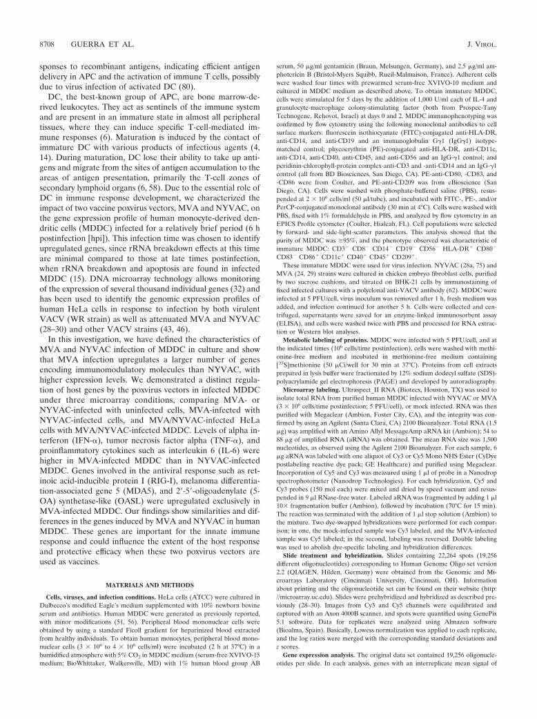
Infection with attenuated poxvirus vectors MVA andNYVAC causes extensive cell damage in human MDDC. Tocharacterize the impact of MVA and NYVAC infection onimmature human MDDC, we first defined the induced cyto-pathic effects by phase-contrast and immunofluorescence (IF)microscopy. At 6 hpi, MVA or NYVAC infection at 5 PFU/cellresulted in alterations in cell morphology characterized by cellrounding and cytoplasmic contraction; the cytopathic effectwas more pronounced in NYVAC than after MVA infection(Fig. 1A). The effects on cell morphology were severe by 16 hpiin MDDC infected with either virus strain (not shown). Thenumber of MVA- or NYVAC-infected MDDC was monitoredby IF after staining with a polyclonal antibody to VACV pro-teins. At 6 hpi, approximately 35% of MVA- or NYVAC-infected MDDC stained for viral proteins, which increased to45% by 16 hpi (Fig. 1B). Since the use of higher virus multi-
FIG. 1. Cellular and biochemical changes in human immature MDDC following infection with MVA or NYVAC poxvirus vectors. (A) Mor-phological changes in immature MDDC mock infected or infected with MVA or NYVAC (5 PFU/cell) in six-well plates; changes were examinedby phase-contrast microscopy at 6 hpi. The upper panels show representative fields (magnification, �4); the lower panels show the indicated areasat a higher magnification. (B) IF analysis. MDDC cultured on coverslips were infected with MVA or NYVAC (5 PFU/cell), and cells were fixedat 6 hpi and treated with a rabbit polyclonal antibody to VACV proteins (�-WR), followed by a Texas Red-labeled secondary antibody,phalloidin-FITC (for actin staining), and ToPro (for DNA staining) (left panels). Cells expressing viral antigens were quantified by IF in fourindependent experiments (right panel).
VOL. 81, 2007 INFECTION OF MDDC BY MVA AND NYVAC POXVIRUS VECTORS 8709

plicities caused extensive MDDC damage, we used 5 PFU/cellin these studies.
To examine viral protein synthesis in MDDC during MVAor NYVAC infection, we performed Western blotting usingantibodies specific for early p25 (E3L) and late viral proteinsp14 (A27L), p21 (A17L), p16 (A14L), p39 (A4L), and p42(B5R). Proteins encoded by the E3L, A4L, and A14L geneswere detected efficiently in lysates of cells infected with bothviruses (Fig. 2A). The late proteins encoded by the A17L andA27L genes were detected only in lysates of MVA-infectedcells, concurring with previous reports for HeLa cells (53). Theprotein encoded by the B5R gene was not detected in lysates ofcells infected with either virus, as determined by Western blot(Fig. 2A) and IF (not shown) analyses. MVA-infected MDDCgave a similar result for A36R gene expression, as previouslydescribed (15).
To document the overall protein expression pattern and theshutoff effect, MDDC were metabolically labeled with [35S]me-thionine at 2, 6, and 16 h after MVA or NYVAC infection andanalyzed by SDS-PAGE and autoradiography. We observed asevere translational block in protein synthesis by 6 hpi in cellsinfected with either virus (Fig. 2B). This blockade coincided
with an increase in phosphorylation of the small subunit of theinitiation factor eIF-2� (Fig. 2C), as described previously forNYVAC in HeLa cells (28, 53). These findings show that MVAor NYVAC infection of MDDC caused shutoff by 6 hpi,whereas the synthesis of early and some late viral proteinscontinued, indicating functional cell translational machinery. Ithas been previously reported that the viral C7L gene is able toprevent eIF-2� phosphorylation in NYVAC-infected HeLacells (53). The presence of this gene in the MVA genomemight explain the different levels in eIF-2� phosphorylationbetween MVA- and NYVAC-infected MDDC, although wecannot rule out the possibility of other mechanisms.
Since NYVAC infection of HeLa cells induces apoptosis(28a, 53), we used an antibody that recognizes both full-lengthand cleaved PARP-1 (73) to analyze whether apoptosis occursin infected MDDC. In both MVA and NYVAC infection,cleavage of 89-kDa PARP-1 was evident by 16 hpi, with noapoptosis at 6 hpi (Fig. 3A). After IF staining with the E3antibody for an early viral protein and ToPro for the ap-pearance of apoptotic bodies, we found similar numbers ofapoptotic cells for both viruses at 16 hpi (nearly 40% of totalinfected cells) (Fig. 3B). This result contrasts with the low
FIG. 2. Protein synthesis evaluation in MDDC after MVA or NYVAC infection. (A) Viral protein expression during NYVAC and MVAinfection. MDDC were mock infected (M) or infected with MVA or NYVAC (5 PFU/cell). At the indicated times postinfection, equal amountsof proteins from cell extracts were fractionated by SDS-PAGE, transferred onto nitrocellulose, and treated with antibodies to VACV proteins(WR) or specific virus early (p25) and late proteins (p21, p14, p39, p16, and p42). Molecular weight (MW) (in thousands) is indicated based onprotein standards. (B) Metabolic labeling of proteins during NYVAC and MVA infection. MDDC were mock infected (M) or infected with MVAor NYVAC (5 PFU/cell). At the times indicated, cells were labeled (30 min) with [35S]Met-Cys Promix (50 �Ci/ml), and equal amounts of proteinswere analyzed by SDS-PAGE (10%) and autoradiography. (C) The gel in B was transferred onto nitrocellulose and incubated with antibodies tototal eIF-2� or eIF-2� phosphorylated (P) at Ser51. Molecular weight (MW) (in thousands) is indicated based on protein standards.
8710 GUERRA ET AL. J. VIROL.

apoptotic effect induced by MVA in human HeLa cells(28a, 53).
We previously showed that NYVAC but not MVA infectionof HeLa cells triggers rRNA degradation late in infection, withthe same cleavage pattern observed during activation of theinterferon-induced 2-5A oligonucleotide synthetase/RNase Lsystem (53). We therefore examined rRNA integrity in MVA-and NYVAC-infected MDDC. Total RNA was isolated frominfected and mock-infected cells and fractionated by formal-dehyde-agarose gel electrophoresis. There was no rRNA deg-radation in uninfected cells or in infected cells at 6 hpi; incontrast, we observed a breakdown of 28S and 18S rRNA atlater times in cells infected with both viruses (Fig. 3C). Theseresults revealed that MVA and NYVAC infection of MDDCdoes not induce apoptosis or rRNA degradation at 6 hpi, butthese effects appeared later in infection.
Differential gene regulation following MVA or NYVAC in-fection of human MDDC compared to mock-infected cells.Since MDDC are the most potent APC and the only cells ableto activate naive T cells (47), we studied the impact of bothviruses on MDDC gene expression at a time when host rRNAhad not been degraded by virus infection. We used chips car-rying oligonucleotides from 19,256 human genes to profileMDDC gene expression and hybridized cDNA samples frominfected and uninfected (mock-infected) cells at 6 hpi. The
gene expression data were selected as described in Materialsand Methods. Compared to uninfected cells, we identified1,215 genes differentially expressed in MDDC after MVA in-fection (21.6% of the genes selected), 554 of which were up-regulated and 661 of which were downregulated (see examplesin Table S1 in the supplemental material). A similar experi-ment comparing NYVAC-infected with uninfected MDDCshowed variance in the regulation of 728 genes after NYVACinfection (13.5% of the genes selected), 360 of which wereupregulated and 368 of which were downregulated (see exam-ples in Table S2 in the supplemental material).
Host genes with altered expression in MVA- or NYVAC-infected MDDC belong to a number of functional categories(Fig. 4A). Both poxvirus vectors regulated similar numbers ofgenes involved in cell death, cancer, gene expression, cell de-velopment, and organism survival. NYVAC nonetheless selec-tively regulated more genes involved in cell growth, prolifera-tion, and morphology than MVA. In contrast, MVA regulateda larger number of genes involved in the immune response andimmune system development than NYVAC (Fig. 4A). Exam-ples of the differentially regulated genes are presented in Table1. Genes differentially expressed after poxvirus infection wererepresented using Venn diagrams to display differently andsimilarly regulated genes for MVA and NYVAC (Fig. 4B).MVA and NYVAC shared 195 genes that were upregulated
FIG. 3. Apoptosis induction and rRNA breakdown during MVA or NYVAC infection of MDDC. (A) Time course of PARP-1 cleavage duringMVA and NYVAC infection. MDDC were mock infected (M) or infected with MVA or NYVAC (5 PFU/cell); at the times indicated, totalproteins (100 �g) were fractionated by SDS-PAGE, transferred onto nitrocellulose, and immunoblotted with anti-PARP-1. An 89-kDa PARP-1cleavage product was observed at 16 hpi. Molecular weight standards (MW) (in thousands) are indicated (right). Equivalence of protein loadingwas confirmed using anti-actin controls. (B) Quantification of cells in apoptosis after MVA or NYVAC infection. MDDC were mock infected(M) or infected with MVA or NYVAC (5 PFU/cell); at 16 hpi, cells were fixed and processed to visualize apoptosis by IF using antibodies to theviral E3 protein (red) and ToPro for DNA staining (blue). The percentage of cells in apoptosis was determined by counting �2,000 cells in twoindependent experiments (bottom). (C) MVA or NYVAC infection of MDDC causes rRNA breakdown. Total rRNA was isolated from uninfectedMDDC (M) or MDDC infected with MVA or NYVAC at the indicated times postinfection; 2 �g of each sample was applied for electrophoresis,and the gel was stained with ethidium bromide. Arrows indicate bands corresponding to characteristic rRNA degradation products.
VOL. 81, 2007 INFECTION OF MDDC BY MVA AND NYVAC POXVIRUS VECTORS 8711

and 220 that were downregulated, whereas 359 genes wereupregulated only by MVA and 165 genes were upregulatedonly by NYVAC. In infected MDDC, MVA specifically down-regulated more genes than NYVAC (441 versus 148 genes).Both viruses produced an increase in specific immune mole-cules such as CXCL2, TNF-�, and several interferon-inducedproteins (IFIT1, IFIT4, ISG15, and ISG20), with higher ex-pression levels in MVA-infected MDDC than in NYVAC-infected MDDC. An alternative visualization using hierarchi-cal cluster analysis was also developed (not shown).
Comparison of MVA and NYVAC infection of humanMDDC shows specific differences in host gene expressionlevels. To further document specific differences between thetwo vectors, we performed microarrays with cDNAs preparedfrom MDDC infected with MVA and NYVAC for 6 h, but wenow compared NYVAC-infected samples with MVA-infectedsamples. We evaluated specific transcriptional differences be-tween the two vectors by removing those genes regulatedequally by both viruses from the processed data. We identified11,800 genes with similar expression levels for both viruses; 544genes showed expression pattern differences after poxvirus in-fection (4.4% of the genes selected), 283 of which had at leasttwofold-higher transcriptional levels after MVA than afterNYVAC infection. Human genes differentially expressed inMVA versus NYVAC infection of MDDC are shown in Table
2 and in Table S3 in the supplemental material. Genes includ-ing TNF-�, IFN-, and IL-12 were increased by fivefold orhigher after MVA infection compared to NYVAC infection.MVA and NYVAC infections produced 12- and 4-fold upregu-lation, respectively, of a recently described member of theNF-�B family, such as MAIL (molecule possessing ankyrinrepeats induced by lipopolysaccharide) (Tables 1 and 2).
The microarray findings indicate that both poxvirus vec-tors triggered similar but also distinct regulatory pathways inMDDC, with MVA upregulating more host genes thanNYVAC.
Validation of microarray data by quantitative real-time RT-PCR. To confirm the microarray results, we validated thechanges in transcript levels using real-time RT-PCR to verifythe transcriptional changes in 12 selected genes (TNF, IFN-,ISG15, NF-�B-2, IL12, IL7, IL6, IFN-�, OASL, ATF-3,ADORA, and H2AFY); HPRT was used as an internal control.The assay was performed with the same RNAs as those used inthe microarray experiment (Table 2). The expression patternand relative mRNA abundance of the selected genes con-curred with microarray data in all cases, with only slight vari-ations, validating the microarray findings (see Table S4 in thesupplemental material). Indeed, clear differences were ob-served in the expression levels of selected genes for both vi-ruses.
FIG. 4. Functional classification of genes with altered expression in MVA- or NYVAC-infected MDDC versus uninfected cells aftermicroarray analysis. Genes with altered expression in MVA- or NYVAC-infected MDDC were compared with uninfected cells. (A) The yaxis shows the most representative high-level functions associated with genes regulated in MVA- or NYVAC-infected MDDC, according toingenuity pathway analysis. The x axis represents the percentage of the total number of regulated genes associated with a given function.(B) Venn diagrams represent common or specific genes upregulated (2-fold) or downregulated (��2-fold) in MVA- or NYVAC-infectedMDDC compared to mock-infected cells.
8712 GUERRA ET AL. J. VIROL.

To further validate these results (see Table S4 in the sup-plemental material), we used cDNAs from MDDC obtainedfrom two other healthy volunteers. MDDC were infected withMVA or NYVAC for 6 h, cDNA was prepared, and RT-PCRwas performed as described above (Table 3). The results forthree donors revealed a similar pattern of cytokine gene ex-pression induced by the two poxvirus vectors (Table 3), vali-dating the microarray data.
Differences in host gene expression levels in MVA- andNYVAC-infected MDDC compared to infected HeLa cells.To provide further evidence for distinct gene regulation byNYVAC and MVA, we compared gene expression levelsbetween HeLa cells infected with MVA or NYVAC withMDDC infected with the same poxvirus vectors. Genes ex-pressed in 6-h NYVAC- or MVA-infected HeLa cells weresubtracted from the genes regulated by both viruses in in-fected MDDC. Thus, we selected those genes showing atleast twofold-higher expression levels in one cell type thanin the other. We identified 1,245 genes that were differen-tially expressed after MVA infection of MDDC compared toHeLa cells (34.5% of the genes selected); of these genes,
463 were upregulated and 782 were downregulated in in-fected MDDC compared to infected HeLa cells. In the caseof NYVAC infection, 1,970 genes were differentially ex-pressed in MDDC versus HeLa cells (51.1% of the selectedgenes); of these genes, 1,009 were upregulated and 961 weredownregulated in infected MDDC compared to HeLa cells.MVA- and NYVAC-infected MDDC shared 268 upregu-lated and 380 downregulated genes (data not shown). Func-tional analysis of genes with at least twofold-higher expres-sion levels in MVA- or NYVAC-infected MDDC showedsimilar percentages for the main functional categories in-cluding immune response, cell death, or cell signaling (datanot shown). Examples of these genes are shown in Table 4and in Tables S4 and S5 in the supplemental material.
Several genes that were upregulated in MVA-infectedMDDC also showed higher expression levels than their coun-terparts in MVA-infected HeLa cells or in NYVAC-infectedMDDC. These include TNF-�, IFN-, interferon-inducedIFIT1 and IFIT4 genes, cytokines such as GRO2 (CXCL2),and genes involved in the antiviral immune response (RIG-I,MDA5, and GBP5).
TABLE 1. Representative genes regulated by MVA or NYVAC in infected MDDC according to predicted biological function
Description GenBank accession no. Gene
Fold change intranscription
MVA NYVAC
IFN and IFN-induced genesIFN-induced protein with tetratricopeptide repeats 4 NM_001549 IFIT4 52.45 3.37IFN-induced protein with tetratricopeptide repeats 1 NM_001548 IFIT1 47.8 4.65IFN-induced, hepatitis C virus-associated microtubular
aggregate protein (44 kDa)NM_006417 MTAP44 20.95 2.63
IRF-2 NM_002199 IRF2 6.54 3.48IRF-7 NM_004031 IRF7 2.52 1.3IFN-�1 NM_024013 IFNA1 2.51 1.92Myxovirus (influenza virus) resistance 1, IFN-inducible
protein p78 (mouse)NM_002462 MX1 2.26 1.44
IRF-5 NM_002200 IRF5 �2.31 1.06IFN-�-inducible protein 27 NM_005532 IFI27 �2.46 �1.62IFN-�-inducible protein 30 NM_006332 IFI30 �2.79 �2.79
InterleukinsIL-6 (IFN, beta 2) NM_000600 IL6 4.05 1IL-1� NM_000575 IL1A �1.43 2.01IL-8 NM_000584 IL8 �2.9 �1.39IL-1 NM_000576 IL1B �2.25 1.39
TNF (TNF superfamily, member 2) NM_000594 TNF 52.21 6.79
Other cytokinesGRO2 oncogene (SCYB2), CXCL2 NM_002089 GRO2 6.58 2.22Small inducible cytokine A3 NM_002983 SCYA3 4.22 1.12Small inducible cytokine A5 (RANTES) NM_002985 SCYA5 4.17 �1.17Small inducible cytokine A4 NM_002984 SCYA4 3.74 �1.14Colony-stimulating factor 2 (granulocyte-macrophage) NM_000758 CSF2 3.13 �1.08
ApoptosisNuclear factor of kappa light polypeptide gene
enhancer in B cells inhibitor �NM_020529 NFKBIA 9.59 6.02
MAIL, NFKBIZ NM_031419 MAIL 12.99 4.91BCL2-associated athanogene 3 NM_004281 BAG3 8.42 1.28Calpain 1, (mu/I) large subunit NM_005186 CAPN1 4.1 1.78
Antiviral immune responseMelanoma differentiation-associated protein 5 NM_022168 MDA5 7.77 1.67
VOL. 81, 2007 INFECTION OF MDDC BY MVA AND NYVAC POXVIRUS VECTORS 8713

The comparative profiling of infected MDDC versus HeLacells provides additional evidence that both poxvirus vectorsaffect host gene expression differently.
Induction of immunomodulatory molecules and activationof IFN pathways in MVA- and NYVAC-infected MDDC. Whilethe above-described experiments indicate that both MVA andNYVAC produced an increase in gene expression of certaincytokines (see Tables S1 and S2 in the supplemental material),MVA elicited higher expression levels of specific immuno-modulatory molecules such as TNF-�, IFN-, CCL5, and IL-12. IL-1� and IL-1 expression levels were nonetheless slightlyenhanced after NYVAC infection. Genes involved in the an-tiviral response, such as OASL, RIG-I, and MDA5, were up-regulated after MVA infection. We therefore analyzed thecorrelation between transcription and translational levels ofspecific immunomodulatory molecules after poxvirus infectionof MDDC. Since we observed high transcriptional levels ofTNF-� after MVA infection of MDDC in the microarrays, weused ELISA to evaluate TNF-� levels in supernatants of in-fected MDDC from three healthy volunteers. High TNF-�
TABLE 2. Differential gene expression profiling of MVA-infected versus NYVAC-infected human DCa
Description GenBank accession no. Gene Fold change ofMVA/NYVAC
IFNs and IFN-induced genesIFN-induced protein with tetratricopeptide repeats 4 NM_001549 IFIT4 11.45IFN-induced protein with tetratricopeptide repeats 1 NM_001548 IFIT1 7.38IFN-induced, hepatitis C virus-associated microtubular aggregate
protein (44 kDa)NM_006417 MTAP44 5.52
Guanylate binding protein 5 NM_052942 GBP5 5.37IFN-1, fibroblast NM_002176 IFNB1 5.06IFN-stimulated protein, 15 kDa NM_005101 ISG15 2.56IFN-stimulated gene (20 kDa) BC016341 ISG20 2.51IRF-7 NM_004031 IRF7 2.31IFN-�1 NM_024013 IFNA1 1.18
InterleukinsIL-12 (NK cell-stimulatory factor 2) NM_002187 IL12B 5.44IL-6 (IFN-2) NM_000600 IL6 4.05IL-1� NM_000575 IL1A �2.42IL-1 NM_000576 IL1B �3.14
TNF and related genesTNF (TNF superfamily, member 2) NM_000594 TNF 10.38TNF receptor-associated factor 6 NM_004620 TRAF6 1.7Tumor necrosis factor receptor superfamily, member 10b AF016266 TNFRSF10B �1.07
Other cytokinesSmall inducible cytokine A4 NM_002984 SCYA4 5.61Small inducible cytokine A5 (RANTES) NM_002985 SCYA5 3.06GRO2 oncogene (SCYB2), CXCL2 NM_002089 GRO2 2.78Small inducible cytokine subfamily B member 10, CXCL10, IP-10 NM_001565 SCYB10 2.35Colony-stimulating factor 2 (granulocyte-macrophage) NM_000758 CSF2 1.94
ApoptosisBCL2-associated athanogene 3 NM_004281 BAG3 4.55MAIL, NFKBIZ NM_031419 MAIL 2.85Calpain 1 (mu/I) large subunit NM_005186 CAPN1 1.72
Antiviral immune responseRNA helicase NM_014314 RIG-I 5.72Melanoma differentiation-associated protein 5 NM_022168 MDA5 5.05OASL NM_003733 OASL 4.53
a Shown is a comparison of gene expression profiling between MVA- and NYVAC-infected MDDC. Representative human genes specifically regulated by eachvector according to predicted biological function are shown.
TABLE 3. Expression levels of selected genes byreal-time RT-PCRa
Gene product
Fold change by real-time RT-PCR
MVA vs mock NYVAC vs mock
DC 1 DC 2 DC 3 DC 1 DC 2 DC 3
TNF 36.63 25.45 44.32 2.23 4.95 4.48IFN- 43.86 28.87 27.76 2.15 4.98 1.85ISG15 3.82 4.62 3.81 2.15 2.27 2.26IL-12 7.91 5.89 7.98 2.08 1.98 1.79IL-7 1.78 0.90 2.07 1.27 1.55 0.81IL-6 2.01 2.36 3.03 1.16 1.41 2.54IFN-� 1.45 1.82 1.4 1.19 0.98 1NF�2 2.02 3.05 3.64 1.68 2.06 2OASL 27.66 37.66 26.63 2.13 1.43 0.519ATF-3 1.96 2.19 1.98 3.51 3.75 3.91H2AFY 1.44 1.89 1.98 3.73 0.75 0.91ADORAA2 0.714 1.18 3.6 1.28 1.18 0.63
a MDDC from three donors (DC1, DC2, and DC3) were mock, MVA, orNYVAC infected and processed for RT-PCR.
8714 GUERRA ET AL. J. VIROL.

levels were induced in MDDC from all volunteers after MVAinfection compared to NYVAC infection, with higher levels at6 than at 16 hpi (Fig. 5A).
Since high levels of type I IFN gene expression (Table 2)were produced after poxvirus infection of MDDC, and IFNexpression is regulated by IRFs (IRF-3 and IRF-7), we ana-lyzed the levels of these two factors involved in the IFN-responsive pathway. Western blot analysis showed increasedIRF-7 levels after MVA and NYVAC infection of MDDC butnot after infection of HeLa cells (Fig. 5B). To induce IFN geneexpression, IRF phosphorylation is needed (72). We thus usedWestern blotting to determine levels of IRF-3, both unphos-phorylated and phosphorylated, in MVA- or NYVAC-infectedMDDC. The results showed an increase in IRF-3 phosphory-lation in MVA- and NYVAC-infected compared to uninfectedcells (Fig. 5B). Phosphorylation levels were higher in NYVAC-than in MVA-infected MDDC, although this increase was notobserved in HeLa cells. Levels of the VACV protein E3, an
inhibitor of IRF-3 phosphorylation (72), were considerablyhigher after MVA infection than after NYVAC infection.
These findings indicate that the enhanced transcriptionallevels of genes encoding immunomodulatory molecules ob-served in microarray after MVA or NYVAC infection corre-late with protein levels, at least for TNF, and that the inductionof type I IFN gene expression may be mediated by poxvirus-induced expression of RIG-I/MDA5. This in turn would triggerthe phosphorylation of IRF-3, translocation to the nucleus,and the activation of IFN gene expression.
DISCUSSION
Efficient antiviral immunity involves both innate and adap-tive immune responses. Adaptive immunity leads to viral clear-ance and generates long-term immunological memory throughthe generation of specific T and B cells. DC found in animmature state in all tissues orchestrate the development of
TABLE 4. Differential gene expression in MVA/NYVAC-infected DC compared to that in MVA/NYVAC-infected HeLa cellsa
Description GenBank accession GeneFold change
MVA NYVAC
Immune response, inflammatory responseSmall inducible cytokine A3 NM_002983 SCYA3 66.81 10.63IFN-induced protein with tetratricopeptide repeats 4 NM_001549 IFIT4 32.52 10.87TNF (TNF superfamily, member 2) NM_000594 TNF 31.40 5.60IL-8 NM_000584 IL8 20.53 31.45V-fos FBJ murine osteosarcoma viral oncogene homolog NM_005252 FOS 20.49 1.75Guanylate binding protein 5 NM_052942 GBP5 15.21CD83 antigen (activated B lymphocytes, immunoglobulin superfamily) NM_004233 CD83 14.53 12.41IFN-1, fibroblast NM_002176 IFNB1 13.70 4.52GRO2 oncogene NM_002089 GRO2 13.21 1.64Pre-B-cell colony-enhancing factor NM_005746 PBEF 8.01 13.38MDA5 NM_022168 MDA5 6.66Nuclear factor of kappa light polypeptide gene enhancer in B cells 1 (p105) NM_003998 NFKB1 5.70 12.59RNA helicase NM_014314 RIG-I 5.42IFN-�-inducible protein (clone IFI-6–16) NM_022873 G1P3 4.69 2.77IRF-7 NM_004031 IRF7 4.24 3.37IFN-induced protein with tetratricopeptide repeats 1 NM_001548 IFIT1 3.88 8.63IFN-�-inducible protein 30 NM_006332 IFI30 3.51 5.85Myxovirus (influenza virus) resistance 1, IFN-inducible protein p78 (mouse) NM_002462 MX1 3.45Small inducible cytokine A4 NM_002984 SCYA4 14.94
ApoptosisTNF-�-induced protein 3 NM_006290 TNFAIP3 18.24 20.04Apolipoprotein E NM_000041 APOE 15.45 4.58TNF receptor-associated factor 1 NM_005658 TRAF1 2.43 4.92Death-associated protein 6 NM_001350 DAXX �1.22 6.17IL-1 NM_000576 IL1B 5.49Cathepsin B NM_001908 CTSB 11.43
Cell cycle, signalingCarcinoembryonic antigen-related cell adhesion molecule 6 M18216 CEACAM6 78.47 10.14Endothelial PAS domain protein 1 NM_001430 EPAS1 11.21 7.29Leupaxin NM_004811 LPXN 9.59 7.45Fibrinogen, gamma polypeptide NM_021870 FGG 7.79 13.30Prostaglandin E receptor 1 (subtype EP1), 42kD NM_000955 PTGER1 5.76 10.54Mitogen-activated protein kinase 6 NM_002748 MAPK6 5.33 8.48Wingless-type MMTV integration site family, member 10B NM_003394 WNT10B 4.25 6.44Pleckstrin NM_002664 PLEK 11.08CD53 antigen NM_000560 CD53 9.48RAP1B, member of RAS oncogene family NM_015646 RAP1B 15.26
a Both cell types were infected with MVA or NYVAC (5 PFU/cell), and total RNA was extracted at 6 hpi and processed for microarray and data analysis (seeMaterials and Methods and Results). FBJ, Finkel-Biskis-Junkins; PAS, Per-Arnt-Sim.
VOL. 81, 2007 INFECTION OF MDDC BY MVA AND NYVAC POXVIRUS VECTORS 8715

adaptive immunity (6). Following pathogen recognition, thereare changes in the expression of DC proinflammatory genes,including those coding for cytokines, chemokines, and costimu-latory molecules, in a process known as DC maturation (6, 58).These functional changes are necessary for initiating adaptiveimmunity. Immediately after viral infection, host innate im-mune defenses are induced. These immune responses are crit-ical for the activation of adaptive immunity. There is nonethe-less a 4- to 7-day delay before the adaptive immune response isinitiated, during which time the innate immune response hasan important role in early viral clearance (37). VACV attemptsto subvert the innate response by secreting proteins that inac-tivate complement (41), inhibit the IFN response (9, 74), pro-tect it from inflammatory responses, and prevent natural killer(NK) cell activation (2).
Poxvirus vectors are efficient activators of host immune re-sponses to virally expressed antigens of distinct origin and arebeing used as potential vaccines for several pathogens andtumors (49, 50, 55). It is therefore important to define thetranscriptional changes in human APC, particularly in MDDC,after VACV vector infection. Here, we showed that MDDCinfected with MVA or NYVAC at 5 PFU/cell caused extensivemorphological damage to the cells at late times postinfection.This is characterized by the severe inhibition of protein syn-thesis by 6 hpi and apoptosis induction together with rRNAcleavage at 16 hpi. In spite of a general translational blockinduced by the two vectors, some of the early (E3) and late(A4, A14, and A27) viral proteins examined were produced inthe infected cells, although differences were observed betweenthese vectors. In contrast to MVA infection, NYVAC-infected
MDDC do not produce A17 or A27 proteins, and both virusesdo not synthesize A36R or B5R proteins. The difference inviral gene expression between the two vectors might be relatedto the extent of eIF-2� phosphorylation and RNA degradationinduced during infection as well as the presence or absence ofcertain viral genes in the poxvirus vectors. In agreement withother investigators, MVA induced apoptosis late in the infec-tion and inhibition of some of the late proteins. In view of theseverity of the effect triggered by the two poxvirus vectors inMDDC late in the infection, we used 6 hpi and a multiplicity of5 PFU/cell for gene expression profiling. We applied genomicstudies using microarrays and various data analysis conditionsto define the impact of infection of immature human MDDCwith the two attenuated VACV strains MVA and NYVAC.We identified genes regulated by MVA and NYVAC infectionof MDDC at a postinfection time before rRNA breakdownand defined that these two vectors trigger some similar andsome distinct host gene expression profiles. For this study, weused three approaches. First, we identified genes that wereselectively expressed in MVA- or NYVAC-infected MDDCcompared to mock-infected cells. We subsequently definedgenes that were specifically regulated by MVA in comparisonwith NYVAC infection, and finally, we described genes regu-lated in MVA- or NYVAC-infected HeLa cells compared toMDDC infected by these viruses. Our analysis showed that ininfected human MDDC, MVA upregulates more genes thanNYVAC. Since the expression of genes involved in host de-fense is markedly enhanced after MVA infection, we discussthe contribution of some of these genes to MDDC responses,taking the viral genes that counteract host immune responses
FIG. 5. Validation of microarray data at the protein level. (A) TNF-� levels in cell supernatants after MVA or NYVAC infection. MDDC fromthree different donors were mock infected or infected with MVA or NYVAC (5 PFU/cell) for 2, 6, and 16 h, and TNF levels in supernatants weremeasured by ELISA. Values indicate duplicate samples in two independent experiments. An asterisk indicates a lack of data. (B) IRF-3 and IRF-7levels in virus-infected MDDC. Mock (lanes 1 and 4)-, MVA (lanes 2 and 5)-, or NYVAC (lanes 3 and 6)-infected (5 PFU/cell) MDDC or HeLacell extracts were fractionated by SDS-PAGE, transferred onto nitrocellulose, and incubated with antibodies to IRF-7, IRF-3, phosphorylatedIRF-3 (IRF-3-P), eIF-2� (as a protein loading control), and the viral protein E3L (as a virus infection control). The molecular weight of proteinstandards (MW) (in thousands) is indicated (right).
8716 GUERRA ET AL. J. VIROL.
into consideration. It should be noted that the effect of thepoxvirus vectors on host genome profiling is due to the contri-bution of the infected and noninfected cells (Fig. 1) togetherwith the immunomodulators released during infection acting inan autocrine and paracrine manner.
TLR. An efficient innate immune response is a prerequisitefor the activation of adaptive immune responses. Toll-like re-ceptors (TLR) are expressed predominantly in APC such asmacrophages and MDDC. TLR3 is a double-stranded RNAreceptor with a key role in antiviral and inflammatory re-sponses through cross-priming of several physiological path-ways including the activation of IFN responses, NF-�B,mitogen-activated protein kinase pathways, and the caspasecascade (54). We found that TLR3 expression was clearly up-regulated after MVA infection of MDDC, implying a possiblerole for TLR3 in double-stranded RNA-initiated antiviral andinflammatory responses. Altered TLR expression has beenlinked to enhanced responsiveness to viral infection, as re-ported previously for viruses such as respiratory syncytial virus
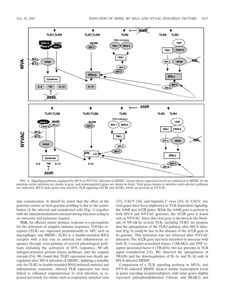
(27), VACV (34), and hepatitis C virus (45). In VACV, twoviral genes have been implicated in TLR-dependent signaling,the A46R and A52R genes. While the A46R gene is present inboth MVA and NYVAC genomes, the A52R gene is foundonly in NYVAC. Since this viral gene is involved in the block-ade of NF-�B by several TLR, including TLR3, we proposethat the upregulation of the TLR3 pathway after MVA infec-tion (Fig. 6) could be due to the absence of the A52R gene inits genome. This activation was not observed after NYVACinfection. The A52R gene has been described to associate withboth IL-1 receptor-associated kinase 2 (IRAK2) and TNF re-ceptor-associated factor 6 (TRAF6), two key proteins in TLRsignal transduction (34). We observed the upregulation ofTRAF6 and the downregulation of IL-1� and IL-1 only inMVA-infected MDDC.
Comparison of a TLR signaling pathway in MVA- andNYVAC-infected MDDC showed similar transcription levelsin genes encoding receptor/adaptors, with some genes slightlyrepressed (phosphatidylinositol 3-kinase and IRAK1) and
FIG. 6. Signaling pathways regulated by MVA or NYVAC infection in MDDC. Genes whose expression levels are enhanced in MDDC by thepoxvirus vector infection are shown in gray, and downregulated genes are shown in black. Viral genes known to interfere with selected pathwaysare indicated. MVA lacks genes that interfere TLR signaling (A52R and A53R), which are present in NYVAC.
VOL. 81, 2007 INFECTION OF MDDC BY MVA AND NYVAC POXVIRUS VECTORS 8717

some genes slightly enhanced (TRAF6 and TBK1). Transcrip-tion levels were similar in genes encoding transcription factorsand inflammatory cytokines, with some enhanced genes inMVA-infected (IFN-�, IFN-, TNF-�, IL-12, CCL3, CCL4,CCL5, and CXCL10) and in NYVAC-infected (IFN- andTNF-�) cells. Neither MVA nor NYVAC had any effect oncostimulatory molecule expression (CD40, CD80, and CD86).Transcription of genes involved in the antiviral immune re-sponse and cytokine production was clearly more enhanced inMVA-infected MDDC than in NYVAC-infected MDDC.
Type I IFN production and antiviral response. The mostconsistent changes in MDDC infection by the attenuated pox-virus vectors involved genes implicated in the type I IFN-�/response (Tables 1 to 3). By generating an intracellular envi-ronment that restricts viral replication, type I IFN represent afirst line of defense against virus infection (25). The IFN sig-naling system produces a broadly effective innate response bycreating an antiviral state in both an autocrine and a paracrinemanner (81). IFN gene expression is regulated by IRF-3 phos-phorylation, homodimerization, and nuclear translocation(67). Once in the nucleus, IRF-3 interacts with IRF-3-respon-sive promoters and the transcriptional coactivator histoneacetyltransferase CBP/p300, leading to the transcription ofIRF-responsive genes; together with NF-�B and AP-1, IRF-3also promotes IFN- transcription. The microarray experi-ments showed a clear upregulation of RIG-I and MDA5 levelsonly after MVA infection (Tables 1 to 3). As RIG-I andMDA-5 mRNA expression levels are strongly enhanced bytype I IFN (70), we propose that the elevated IFN levelsproduced after MVA infection might be involved in the up-regulation of RIG-I and MDA5, which can also activate theexpression of type I IFN in a feedback mechanism (Fig. 6).This is supported by the enhanced IRF-3 phosphorylation ob-served in MVA-infected MDDC cells (Fig. 5B).
RIG-I and MDA5 upregulation was not observed in previ-ous studies using mRNA microarrays from MVA-infectedHeLa cells (29). The specificity of RIG-I and MDA5 expres-sion in MVA-infected MDDC was also confirmed in a microar-ray experiment comparing the expression profiles of MVA-infected HeLa cells and MDDC at 6 hpi (Table 4; also seeTable S4 in the supplemental material). These observationsindicate a cell type-specific involvement of RIG-I in the anti-viral response to poxvirus infection. This concurs with theobservations made previously by Ichikawa et al., where type IIFN induction in fibroblasts and myeloid DC was RIG-I de-pendent, while type I IFN induction in periferical DC wasindependent of RIG-I (36). RNA virus recognition by RIG-Iand MDA5 is reported to involve distinct mechanisms (36), butlittle is known of the mechanisms used by their helicases ininfection with DNA viruses.
Type I IFN levels were notably higher during MVA infectionthan during NYVAC infection. There was no apparent RIG-Ior MDA-5 upregulation during NYVAC infection, possiblydue to low IFN levels or to specific differences between MVAand NYVAC genomes. VACV genes involved in inhibiting theIFN response include the E3L (16), B18R (17), K3L (12), andN1L (31) genes. Although MVA and NYVAC share commondeleted genes, including serpins (the B13R and B14R genes),the M2L and N1L genes, which are involved in signaling, andhost range genes (the K1L gene), most other deleted genes
differ between the two strains. The differences in deleted genesbetween MVA and NYVAC must play a role in the hostgenome expression pattern induced after MDDC infectionwith these vectors (Fig. 6).
Proinflammatory cytokine production (TNF-�, IL-12,and IL-6). Another important difference between MVA andNYVAC infection was the activation of TNF-�, IL-12, andIL-6, which were upregulated more than 10-, 5-, and 4-fold,respectively, in MVA versus NYVAC infection. Since NF-�Bplays an important role in the expression of inflammatory cy-tokines, including TNF-� and IL-12, our results suggest anactivation of this pathways by MVA, as previously describedfor HeLa cells (28, 29). Upregulation of these immunomodu-lators is likely to have an important role in MDDC function.TNF-� would be released by MDDC while in peripheral tis-sues to further recruit MDDC precursors and sustain antigencapture and presentation. On the contrary, IL-12 would bereleased by MDDC in lymph nodes to polarize Th cells towarda Th1 phenotype (42). The fact that viruses encode proteinsthat act to subvert nearly all aspects of TNF-� signaling (11)emphasizes the importance of the TNF-�/TNF receptor axis inantiviral immunity and virus-host interactions. Poxviruses haveevolved various strategies to prevent apoptosis, including theability to inhibit secreted TNF-� (57). In microarrays andWestern blots, we found an increase in TNF-� mRNA (Tables1 to 4) and protein levels (Fig. 5A) in MVA-infected but not inNYVAC-infected MDDC. As the A53R gene is deleted inMVA and is intact in NYVAC, we propose that by its high-affinity binding to human TNF (3), the A53R gene productmay be responsible for the decreased TNF levels observed inNYVAC-infected MDDC supernatants.
Apoptosis signaling. During our transcriptional profilinganalysis of MVA- and NYVAC-infected MDDC, we observeda clear rRNA breakdown associated with infection by MVAand NYVAC (Fig. 3). MVA infection produced high levels ofthe 5-OA synthetase-like messenger (Table 2) at 6 hpi, and thisis probably the reason for the apparent increase in RNA deg-radation by MVA compared to NYVAC infection. Activationof this enzyme mediates an antiviral and antitumor function bycleaving cellular and viral RNAs, promoting a general inhibi-tion of protein synthesis and apoptosis (53, 69). The rRNAbreakdown products are identical to those produced in cellsinfected with a VACV recombinant expressing RNase L (notshown), suggesting that MVA or NYVAC infection in MDDCtriggers the activation of the 5-OA synthetase/RNase L path-way at late times postinfection (Fig. 3C). Both MVA andNYVAC induced apoptosis in infected MDDC, indicating thata poxvirus-infected MDDC will eventually die. Taking intoconsideration that the viral E3 protein, a PKR inhibitor (7, 9,44, 65, 68, 79), is produced at high levels after infection ofMDDC with both viruses (Fig. 2), which should block PKRactivation, in view of the levels eIF-2� phosphorylation trig-gered by the poxvirus vectors, we cannot rule out the possibilityof another kinase being responsible for eIF-2� phosphoryla-tion and apoptosis induction after MDDC infection. The roleof apoptosis in the immune response is still unclear, but anti-gens produced by apoptotic cells have been reported to in-crease antigen immunogenicity, which is likely to be moreeffective in cross-priming (76).
In summary, we have identified important differences in host
8718 GUERRA ET AL. J. VIROL.

genome expression profiling of human immature MDDC afterinfection with the attenuated poxvirus vectors MVA andNYVAC. This has been achieved using microarray technologyand direct comparison of cDNAs from three different systems:MVA- or NYVAC-infected versus mock-infected MDDC,MVA- versus NYVAC-infected MDDC, and MVA- orNYVAC- infected MDDC versus infected HeLa cells. Weidentified a number of genes that were expressed similarly aswell as others that showed differential expression profiling bythese viral vectors. In general, MVA infection upregulatedmore genes than NYVAC infection. Of note are the differ-ences in TNF-� and IFN- levels, which were higher afterMVA infection of MDDC than after NYVAC infection. Thesedifferences in vector behavior might be related to the numberof genes in the genome of each vector, with MVA lacking moreimmunomodulatory genes than NYVAC. These poxvirus vec-tors induced apoptosis in MDDC, suggesting that a cell in-fected in vivo will not survive. Furthermore, the differences inhost gene expression and levels of immunomodulatory mole-cules produced in MDDC might affect the quality of the im-mune response induced by each of these vectors when used asvaccines against pathogens and tumors.
ACKNOWLEDGMENTS
We are indebted to R. Bablanian for critically reviewing the manu-script, V. Jimenez for expert technical assistance, and C. Mark forexcellent editorial help. We thank Alberto Pascual-Montano andIntegromics, SL, for help in clustering. We thank J. Tartaglia (Sanofi-Pasteur) for the generous gift of NYVAC, G. Sutter for MVA, andB. L. Jacobs for anti-E3 antibody.
This work was supported by grants from the Spanish Ministry ofEducation and Science (BIO2004-03954 and SAF2005-05566), theSpanish Ministry of Health (FIS2006-1259), the Spanish Foundationfor AIDS Research (FIPSE 36344/02 and FIPSE 36536-05), FundacionBotın, and the European Union (EuroVac QLRT-PL-1999-01321 andQLK2-CT-2002-01867). The Department of Immunology and Oncol-ogy was founded and is supported by the Spanish National ResearchCouncil (CSIC) and by Pfizer.
REFERENCES
1. Abendroth, A., G. Morrow, A. L. Cunningham, and B. Slobedman. 2001.Varicella-zoster virus infection of human dendritic cells and transmission toT cells: implications for virus dissemination in the host. J. Virol. 75:6183–6192.
2. Alcami, A. 2003. Viral mimicry of cytokines, chemokines and their receptors.Nat. Rev. Immunol. 3:36–50.
3. Alcami, A., A. Khanna, N. L. Paul, and G. L. Smith. 1999. Vaccinia virusstrains Lister, USSR and Evans express soluble and cell-surface tumournecrosis factor receptors. J. Gen. Virol. 80:949–959.
4. Aliprantis, A. O., R. B. Yang, M. R. Mark, S. Suggett, B. Devaux, J. D.Radolf, G. R. Klimpel, P. Godowski, and A. Zychlinsky. 1999. Cell activationand apoptosis by bacterial lipoproteins through Toll-like receptor-2. Science285:736–739.
5. Antoine, G., F. Scheiflinger, F. Dorner, and F. G. Falkner. 1998. The com-plete genomic sequence of the modified vaccinia Ankara strain: comparisonwith other orthopoxviruses. Virology 244:365–396.
6. Banchereau, J., and R. M. Steinman. 1998. Dendritic cells and the control ofimmunity. Nature 392:245–252.
7. Beattie, E., K. L. Denzler, J. Tartaglia, M. E. Perkus, E. Paoletti, and B. L.Jacobs. 1995. Reversal of the interferon-sensitive phenotype of a vacciniavirus lacking E3L by expression of the reovirus S4 gene. J. Virol. 69:499–505.
8. Beattie, E., E. B. Kauffman, H. Martinez, M. E. Perkus, B. L. Jacobs, E.Paoletti, and J. Tartaglia. 1996. Host-range restriction of vaccinia virusE3L-specific deletion mutants. Virus Genes 12:89–94.
9. Beattie, E., E. Paoletti, and J. Tartaglia. 1995. Distinct patterns of IFNsensitivity observed in cells infected with vaccinia K3L� and E3L� mutantviruses. Virology 210:254–263.
10. Belyakov, I. M., P. Earl, A. Dzutsev, V. A. Kuznetsov, M. Lemon, L. S. Wyatt,J. T. Snyder, J. D. Ahlers, G. Franchini, B. Moss, and J. A. Berzofsky. 2003.Shared modes of protection against poxvirus infection by attenuated and
conventional smallpox vaccine viruses. Proc. Natl. Acad. Sci. USA 100:9458–9463.
11. Benedict, C. A., P. S. Norris, and C. F. Ware. 2002. To kill or be killed: viralevasion of apoptosis. Nat. Immunol. 3:1013–1018.
12. Carroll, K., O. Elroy-Stein, B. Moss, and R. Jagus. 1993. Recombinantvaccinia virus K3L gene product prevents activation of double-strandedRNA-dependent, initiation factor 2 alpha-specific protein kinase. J. Biol.Chem. 268:12837–12842.
13. Cebere, I., L. Dorrell, H. McShane, A. Simmons, S. McCormack, C. Schmidt,C. Smith, M. Brooks, J. E. Roberts, S. C. Darwin, P. E. Fast, C. Conlon, S.Rowland-Jones, A. J. McMichael, and T. Hanke. 2006. Phase I clinical trialsafety of DNA- and modified virus Ankara-vectored human immunodefi-ciency virus type 1 (HIV-1) vaccines administered alone and in a prime-boostregime to healthy HIV-1-uninfected volunteers. Vaccine 24:417–425.
14. Cella, M., M. Salio, Y. Sakakibara, H. Langen, I. Julkunen, and A.Lanzavecchia. 1999. Maturation, activation, and protection of dendriticcells induced by double-stranded RNA. J. Exp. Med. 189:821–829.
15. Chahroudi, A., D. A. Garber, P. Reeves, L. Liu, D. Kalman, and M. B.Feinberg. 2006. Differences and similarities in viral life cycle progression andhost cell physiology after infection of human dendritic cells with modifiedvaccinia virus Ankara and vaccinia virus. J. Virol. 80:8469–8481.
16. Chang, H. W., J. C. Watson, and B. L. Jacobs. 1992. The E3L gene ofvaccinia virus encodes an inhibitor of the interferon-induced, double-stranded RNA-dependent protein kinase. Proc. Natl. Acad. Sci. USA 89:4825–4829.
17. Colamonici, O. R., B. Porterfield, P. Domanski, R. K. Handa, S. Flex, C. E.Samuel, R. Pine, and M. O. Diaz. 1994. Ligand-independent anti-oncogenicactivity of the alpha subunit of the type I interferon receptor. J. Biol. Chem.269:27275–27279.
18. Corona Gutierrez, C. M., A. Tinoco, T. Navarro, M. L. Contreras, R. R.Cortes, P. Calzado, L. Reyes, R. Posternak, G. Morosoli, M. L. Verde, and R.Rosales. 2004. Therapeutic vaccination with MVA E2 can eliminate precan-cerous lesions (CIN 1, CIN 2, and CIN 3) associated with infection byoncogenic human papillomavirus. Hum. Gene Ther. 15:421–431.
19. Deng, L., P. Dai, W. Ding, R. D. Granstein, and S. Shuman. 2006. Vacciniavirus infection attenuates innate immune responses and antigen presentationby epidermal dendritic cells. J. Virol. 80:9977–9987.
20. Drillien, R., D. Spehner, A. Bohbot, and D. Hanau. 2000. Vaccinia virus-related events and phenotypic changes after infection of dendritic cellsderived from human monocytes. Virology 268:471–481.
21. Engelmayer, J., M. Larsson, M. Subklewe, A. Chahroudi, W. I. Cox, R. M.Steinman, and N. Bhardwaj. 1999. Vaccinia virus inhibits the maturation ofhuman dendritic cells: a novel mechanism of immune evasion. J. Immunol.163:6762–6768.
22. Franchini, G., S. Gurunathan, L. Baglyos, S. Plotkin, and J. Tartaglia. 2004.Poxvirus-based vaccine candidates for HIV: two decades of experience withspecial emphasis on canarypox vectors. Expert Rev. Vaccines 3:S75–S88.
23. Gilbert, S. C., V. S. Moorthy, L. Andrews, A. A. Pathan, S. J. McConkey,J. M. Vuola, S. M. Keating, T. Berthoud, D. Webster, H. McShane, and A. V.Hill. 2006. Synergistic DNA-MVA prime-boost vaccination regimes formalaria and tuberculosis. Vaccine 24:4554–4561.
24. Gomez, C. E., J. L. Najera, V. Jimenez, K. Bieler, J. Wild, L. Kostic, S.Heidari, M. Chen, M. J. Frachette, G. Pantaleo, H. Wolf, P. Liljestrom, R.Wagner, and M. Esteban. 2007. Generation and immunogenicity of novelHIV/AIDS vaccine candidates targeting HIV-1 Env/Gag-Pol-Nef antigens ofclade C. Vaccine 25:1969–1992.
25. Grandvaux, N., B. R. tenOever, M. J. Servant, and J. Hiscott. 2002. Theinterferon antiviral response: from viral invasion to evasion. Curr. Opin.Infect. Dis. 15:259–267.
26. Grosjean, I., C. Caux, C. Bella, I. Berger, F. Wild, J. Banchereau, and D.Kaiserlian. 1997. Measles virus infects human dendritic cells and blocks theirallostimulatory properties for CD4� T cells. J. Exp. Med. 186:801–812.
27. Groskreutz, D. J., M. M. Monick, L. S. Powers, T. O. Yarovinsky, D. C. Look,and G. W. Hunninghake. 2006. Respiratory syncytial virus induces TLR3protein and protein kinase R, leading to increased double-stranded RNAresponsiveness in airway epithelial cells. J. Immunol. 176:1733–1740.
28. Guerra, S., L. A. Lopez-Fernandez, M. Angel Garcia, A. Zaballos, and M.Esteban. 2006. Human gene profiling in response to the active proteinkinase, interferon-induced serine/threonine protein kinase (PKR), in in-fected cells. Involvement of the transcription factor ATF-3 in PKR-inducedapoptosis. J. Biol. Chem. 281:18734–18745.
28a.Guerra, S., L. A. Lopez-Fernandez, A. Pascual-Montano, J. L. Najera, A.Zaballos, and M. Estaban. 2006. Host response to the attenuated poxvirusvector NYVAC: upregulation of apoptotic genes and NF-�B-responsivegenes in infected HeLa cells. J. Virol. 80:985–998.
29. Guerra, S., L. A. Lopez-Fernandez, R. Conde, A. Pascual-Montano, K.Harshman, and M. Esteban. 2004. Microarray analysis reveals characteristicchanges of host cell gene expression in response to attenuated modifiedvaccinia virus Ankara infection of human HeLa cells. J. Virol. 78:5820–5834.
30. Guerra, S., L. A. Lopez-Fernandez, A. Pascual-Montano, M. Munoz, K.Harshman, and M. Esteban. 2003. Cellular gene expression survey of vac-cinia virus infection of human HeLa cells. J. Virol. 77:6493–6506.
VOL. 81, 2007 INFECTION OF MDDC BY MVA AND NYVAC POXVIRUS VECTORS 8719

31. Haga, I. R., and A. G. Bowie. 2005. Evasion of innate immunity by vacciniavirus. Parasitology 130:S11–S25.
32. Harper, N., M. A. Hughes, S. N. Farrow, G. M. Cohen, and M. MacFarlane.2003. Protein kinase C modulates tumor necrosis factor-related apoptosis-inducing ligand-induced apoptosis by targeting the apical events of deathreceptor signaling. J. Biol. Chem. 278:44338–44347.
33. Harrop, R., N. Connolly, I. Redchenko, J. Valle, M. Saunders, M. G.Ryan, K. A. Myers, N. Drury, S. M. Kingsman, R. E. Hawkins, and M. W.Carroll. 2006. Vaccination of colorectal cancer patients with modifiedvaccinia Ankara delivering the tumor antigen 5T4 (TroVax) inducesimmune responses which correlate with disease control: a phase I/II trial.Clin. Cancer Res. 12:3416–3424.
34. Harte, M. T., I. R. Haga, G. Maloney, P. Gray, P. C. Reading, N. W. Bartlett,G. L. Smith, A. Bowie, and L. A. O’Neill. 2003. The poxvirus protein A52Rtargets Toll-like receptor signaling complexes to suppress host defense. J.Exp. Med. 197:343–351.
35. Hornemann, S., O. Harlin, C. Staib, S. Kisling, V. Erfle, B. Kaspers, G.Hacker, and G. Sutter. 2003. Replication of modified vaccinia virus Ankarain primary chicken embryo fibroblasts requires expression of the interferonresistance gene E3L. J. Virol. 77:8394–8407.
36. Ichikawa, T., K. Nakao, K. Nakata, K. Hamasaki, Y. Takeda, Y. Kajiya, S.Higashi, K. Ohkubo, Y. Kato, N. Ishii, and K. Eguchi. 2001. Geranylgeranyl-acetone induces antiviral gene expression in human hepatoma cells. Bio-chem. Biophys. Res. Commun. 280:933–939.
37. Janeway, C., Jr., and R. Medzhitov. 2000. Viral interference with IL-1 andToll signaling. Proc. Natl. Acad. Sci. USA 97:10682–10683.
38. Jenne, L., C. Hauser, J. F. Arrighi, J. H. Saurat, and A. W. Hugin. 2000.Poxvirus as a vector to transduce human dendritic cells for immunotherapy:abortive infection but reduced APC function. Gene Ther. 7:1575–1583.
39. Kanesa-thasan, N., J. J. Smucny, C. H. Hoke, D. H. Marks, E. Konishi, I.Kurane, D. B. Tang, D. W. Vaughn, P. W. Mason, and R. E. Shope. 2000.Safety and immunogenicity of NYVAC-JEV and ALVAC-JEV attenuatedrecombinant Japanese encephalitis virus-poxvirus vaccines in vaccinia-non-immune and vaccinia-immune humans. Vaccine 19:483–491.
40. Kastenmuller, W., I. Drexler, H. Ludwig, V. Erfle, C. Peschel, H. Bernhard,and G. Sutter. 2006. Infection of human dendritic cells with recombinantvaccinia virus MVA reveals general persistence of viral early transcriptionbut distinct maturation-dependent cytopathogenicity. Virology 350:276–288.
41. Kotwal, G. J., S. N. Isaacs, R. McKenzie, M. M. Frank, and B. Moss. 1990.Inhibition of the complement cascade by the major secretory protein ofvaccinia virus. Science 250:827–830.
42. Langenkamp, A., M. Messi, A. Lanzavecchia, and F. Sallusto. 2000. Kineticsof dendritic cell activation: impact on priming of TH1, TH2 and non-polarized T cells. Nat. Immunol. 1:311–316.
43. Langland, J. O., J. C. Kash, V. Carter, M. J. Thomas, M. G. Katze, and B. L.Jacobs. 2006. Suppression of proinflammatory signal transduction and geneexpression by the dual nucleic acid binding domains of the vaccinia virus E3Lproteins. J. Virol. 80:10083–10095.
44. Lee, S. B., and M. Esteban. 1994. The interferon-induced double-strandedRNA-activated protein kinase induces apoptosis. Virology 199:491–496.
45. Li, K., E. Foy, J. C. Ferreon, M. Nakamura, A. C. Ferreon, M. Ikeda, S. C.Ray, M. Gale, Jr., and S. M. Lemon. 2005. Immune evasion by hepatitis Cvirus NS3/4A protease-mediated cleavage of the Toll-like receptor 3 adaptorprotein TRIF. Proc. Natl. Acad. Sci. USA 102:2992–2997.
46. Ludwig, H., J. Mages, C. Staib, M. H. Lehmann, R. Lang, and G. Sutter.2005. Role of viral factor E3L in modified vaccinia virus Ankara infection ofhuman HeLa Cells: regulation of the virus life cycle and identification ofdifferentially expressed host genes. J. Virol. 79:2584–2596.
47. Mellman, I., and R. M. Steinman. 2001. Dendritic cells: specialized andregulated antigen processing machines. Cell 106:255–258.
48. Meyer, H., G. Sutter, and A. Mayr. 1991. Mapping of deletions in thegenome of the highly attenuated vaccinia virus MVA and their influence onvirulence. J Gen. Virol. 72:1031–1038.
49. Moss, B. 1996. Genetically engineered poxviruses for recombinant geneexpression, vaccination, and safety. Proc. Natl. Acad. Sci. USA 93:11341–11348.
50. Moss, B., M. W. Carroll, L. S. Wyatt, J. R. Bennink, V. M. Hirsch, S.Goldstein, W. R. Elkins, T. R. Fuerst, J. D. Lifson, M. Piatak, N. P. Restifo,W. Overwijk, R. Chamberlain, S. A. Rosenberg, and G. Sutter. 1996. Hostrange restricted, non-replicating vaccinia virus vectors as vaccine candidates.Adv. Exp. Med. Biol. 397:7–13.
51. Munoz-Fontela, C., M. Collado, E. Rodriguez, M. A. Garcia, A. Alvarez-Barrientos, J. Arroyo, C. Nombela, and C. Rivas. 2005. Identification of anuclear export signal in the KSHV latent protein LANA2 mediating itsexport from the nucleus. Exp. Cell Res. 311:96–105.
52. Myagkikh, M., S. Alipanah, P. D. Markham, J. Tartaglia, E. Paoletti, R. C.Gallo, G. Franchini, and M. Robert-Guroff. 1996. Multiple immunizationswith attenuated poxvirus HIV type 2 recombinants and subunit boosts re-quired for protection of rhesus macaques. AIDS Res. Hum. Retrovir. 12:985–992.
53. Najera, J. L., C. E. Gomez, E. Domingo-Gil, M. M. Gherardi, and M.Esteban. 2006. Cellular and biochemical differences between two attenuated
poxvirus vaccine candidates (MVA and NYVAC) and role of the C7L gene.J. Virol. 80:6033–6047.
54. Nishiya, T., E. Kajita, S. Miwa, and A. L. Defranco. 2005. TLR3 and TLR7are targeted to the same intracellular compartments by distinct regulatoryelements. J. Biol. Chem. 280:37107–37117.
55. Ockenhouse, C. F., P. F. Sun, D. E. Lanar, B. T. Wellde, B. T. Hall, K.Kester, J. A. Stoute, A. Magill, U. Krzych, L. Farley, R. A. Wirtz, J. C. Sadoff,D. C. Kaslow, S. Kumar, L. W. Church, J. M. Crutcher, B. Wizel, S. Hoff-man, A. Lalvani, A. V. Hill, J. A. Tine, K. P. Guito, C. de Taisne, R. Anders,W. R. Ballou, et al. 1998. Phase I/IIa safety, immunogenicity, and efficacytrial of NYVAC-Pf7, a pox-vectored, multiantigen, multistage vaccine can-didate for Plasmodium falciparum malaria. J. Infect. Dis. 177:1664–1673.
56. Pacheco, R., J. M. Martinez-Navio, M. Lejeune, N. Climent, H. Oliva, J. M.Gatell, T. Gallart, J. Mallol, C. Lluis, and R. Franco. 2005. CD26, adenosinedeaminase, and adenosine receptors mediate costimulatory signals in theimmunological synapse. Proc. Natl. Acad. Sci. USA 102:9583–9588.
57. Pomerantz, J., N. Schreiber-Agus, N. J. Liegeois, A. Silverman, L. Alland, L.Chin, J. Potes, K. Chen, I. Orlow, H. W. Lee, C. Cordon-Cardo, and R. A.DePinho. 1998. The Ink4a tumor suppressor gene product, p19Arf, interactswith MDM2 and neutralizes MDM2’s inhibition of p53. Cell 92:713–723.
58. Ridge, J. P., F. Di Rosa, and P. Matzinger. 1998. A conditioned dendritic cellcan be a temporal bridge between a CD4� T-helper and a T-killer cell.Nature 393:474–478.
59. Riegler, S., H. Hebart, H. Einsele, P. Brossart, G. Jahn, and C. Sinzger.2000. Monocyte-derived dendritic cells are permissive to the complete rep-licative cycle of human cytomegalovirus. J. Gen. Virol. 81:393–399.
60. Risco, C., J. R. Rodriguez, W. Demkowicz, R. Heljasvaara, J. L. Carrascosa,M. Esteban, and D. Rodriguez. 1999. The vaccinia virus 39-kDa proteinforms a stable complex with the p4a/4a major core protein early in morpho-genesis. Virology 265:375–386.
61. Rodriguez, D., M. Esteban, and J. R. Rodriguez. 1995. Vaccinia virus A17Lgene product is essential for an early step in virion morphogenesis. J. Virol.69:4640–4648.
62. Rodriguez, D., J. R. Rodriguez, J. F. Rodriguez, D. Trauber, and M. Esteban.1989. Highly attenuated vaccinia virus mutants for the generation of saferecombinant viruses. Proc. Natl. Acad. Sci. USA 86:1287–1291.
63. Rodriguez, J. F., R. Janeczko, and M. Esteban. 1985. Isolation and charac-terization of neutralizing monoclonal antibodies to vaccinia virus. J. Virol.56:482–488.
64. Rodriguez, J. R., C. Risco, J. L. Carrascosa, M. Esteban, and D. Rodriguez.1998. Vaccinia virus 15-kilodalton (A14L) protein is essential for assemblyand attachment of viral crescents to virosomes. J. Virol. 72:1287–1296.
65. Romano, P. R., F. Zhang, S.-L. Tan, M. T. Garcia-Barrio, M. G. Katze, T. E.Dever, and A. G. Hinnebusch. 1998. Inhibition of double-stranded RNA-dependent protein kinase PKR by vaccinia virus E3: role of complex forma-tion and the E3 N-terminal domain. Mol. Cell. Biol. 18:7304–7316.
66. Schneider, J., S. C. Gilbert, T. J. Blanchard, T. Hanke, K. J. Robson, C. M.Hannan, M. Becker, R. Sinden, G. L. Smith, and A. V. Hill. 1998. Enhancedimmunogenicity for CD8� T cell induction and complete protective efficacyof malaria DNA vaccination by boosting with modified vaccinia virusAnkara. Nat. Med. 4:397–402.
67. Servant, M. J., B. ten Oever, C. LePage, L. Conti, S. Gessani, I. Julkunen, R.Lin, and J. Hiscott. 2001. Identification of distinct signaling pathways leadingto the phosphorylation of interferon regulatory factor 3. J. Biol. Chem.276:355–363.
68. Sharp, T. V., F. Moonan, A. Romashko, B. Joshi, G. N. Barber, and R. Jagus.1998. The vaccinia virus E3L gene product interacts with both the regulatoryand the substrate binding regions of PKR: implications for PKR autoregu-lation. Virology 250:302–315.
69. Silverman, R. H. 2003. Implications for RNase L in prostate cancer biology.Biochemistry 42:1805–1812.
70. Siren, J., T. Imaizumi, D. Sarkar, T. Pietila, D. L. Noah, R. Lin, J. Hiscott,R. M. Krug, P. B. Fisher, I. Julkunen, and S. Matikainen. 2006. Retinoicacid inducible gene-I and mda-5 are involved in influenza A virus-inducedexpression of antiviral cytokines. Microbes Infect. 8:2013–2020.
71. Sivanandham, M., P. Shaw, S. F. Bernik, E. Paoletti, and M. K. Wallack.1998. Colon cancer cell vaccine prepared with replication-deficient vacciniaviruses encoding B7.1 and interleukin-2 induce antitumor response insyngeneic mice. Cancer Immunol. Immunother. 46:261–267.
72. Smith, E. J., I. Marie, A. Prakash, A. Garcia-Sastre, and D. E. Levy. 2001.IRF3 and IRF7 phosphorylation in virus-infected cells does not requiredouble-stranded RNA-dependent protein kinase R or Ikappa B kinase but isblocked by vaccinia virus E3L protein. J. Biol. Chem. 276:8951–8957.
73. Soldani, C., and A. I. Scovassi. 2002. Poly(ADP-ribose) polymerase-1 cleav-age during apoptosis: an update. Apoptosis 7:321–328.
74. Symons, J. A., A. Alcami, and G. L. Smith. 1995. Vaccinia virus encodes asoluble type I interferon receptor of novel structure and broad speciesspecificity. Cell 81:551–560.
75. Tartaglia, J., M. E. Perkus, J. Taylor, E. K. Norton, J. C. Audonnet, W. I.Cox, S. W. Davis, J. van der Hoeven, B. Meignier, M. Riviere, et al. 1992.NYVAC: a highly attenuated strain of vaccinia virus. Virology 188:217–232.
76. Terenzi, F., M. J. deVeer, H. Ying, N. P. Restifo, B. R. Williams, and R. H.
8720 GUERRA ET AL. J. VIROL.

Silverman. 1999. The antiviral enzymes PKR and RNase L suppress geneexpression from viral and non-viral based vectors. Nucleic Acids Res. 27:4369–4375.
77. Vanderplasschen, A., and P. P. Pastoret. 2003. The uses of poxviruses asvectors. Curr. Gene Ther. 3:583–595.
78. Wyatt, L. S., M. W. Carroll, C. P. Czerny, M. Merchlinsky, J. R. Sisler,and B. Moss. 1998. Marker rescue of the host range restriction defects ofmodified vaccinia virus Ankara. Virology 251:334–342.
79. Xiang, Y., R. C. Condit, S. Vijaysri, B. Jacobs, B. R. Williams, and R. H.Silverman. 2002. Blockade of interferon induction and action by the E3L
double-stranded RNA binding proteins of vaccinia virus. J. Virol. 76:5251–5259.
80. Yates, N. L., and M. A. Alexander-Miller. 2006. Vaccinia virus infectionof mature dendritic cells results in activation of virus-specific naıve CD8�
T cells: a potential mechanism for direct presentation. Virology 359:349–361.
81. Yoneyama, M., M. Kikuchi, T. Natsukawa, N. Shinobu, T. Imaizumi, M.Miyagishi, K. Taira, S. Akira, and T. Fujita. 2004. The RNA helicase RIG-Ihas an essential function in double-stranded RNA-induced innate antiviralresponses. Nat. Immunol. 5:730–737.
VOL. 81, 2007 INFECTION OF MDDC BY MVA AND NYVAC POXVIRUS VECTORS 8721


![Synthesis of Pyrrolo[1,3]-Diazepines and Potential Poxvirus ...](https://static.fdokumen.com/doc/165x107/63286568051fac18490eb53f/synthesis-of-pyrrolo13-diazepines-and-potential-poxvirus-.jpg)

