The myeloid 7/4-antigen defines recently generated inflammatory macrophages and is synonymous with...
13
The myeloid 7/4-antigen defines recently generated inflammatory macrophages and is synonymous with Ly-6B Marcela Rosas,* Benjamin Thomas, † Martin Stacey, ‡ Siamon Gordon, † and Philip R. Taylor* ,1 *Infection, Immunity and Biochemistry, Cardiff University School of Medicine, Cardiff, United Kingdom; † Sir William Dunn School of Pathology, University of Oxford, Oxford, United Kingdom; and ‡ Institute of Molecular and Cellular Biology, Faculty of Biological Science, University of Leeds, Leeds, United Kingdom RECEIVED AUGUST 12, 2009; REVISED MARCH 19, 2010; ACCEPTED MARCH 30, 2010. DOI: 10.1189/jlb.0809548 ABSTRACT This study aimed to identify the inflammation-associ- ated 7/4-antigen, which is highly expressed on neutro- phils, inflammatory monocytes, some activated macro- phages, as well as on bone marrow myeloid-restricted progenitors. The high expression on inflammatory cells is suggestive of a role in inflammation and makes the 7/4-antigen a potential target for the manipulation of in- flammatory cells. Consistent with this, the 7/4-antibody mediates specific depletion of 7/4-expressing neutro- phils and monocytes. We have identified the 7/4-anti- gen as a 25- to 30-kDa GPI-anchored glycoprotein syn- onymous with the Ly-6B.2 alloantigen. We character- ized the expression of Ly-6B during the inflammatory reaction induced by zymosan. During the later stages of an experimental, acute, self-resolving inflammatory response, we found that Ly-6B is differentially ex- pressed on macrophages. Ly-6B-expressing macro- phages also express more MHCII, CIITA, CCR2, Ly-6C, and CD62L than the Ly-6B-negative macrophages, which in turn, express more of the resident tissue mac- rophage marker SIGN-R1 and higher CD11b and F4/80. Ly-6B-expressing macrophages incorporate more BrdU than their Ly-6B-negative contemporaries when fed during the resolution phase of the acute inflamma- tory response. Thus, Ly-6B expression on mature mac- rophages defines a subset of recently generated in- flammatory macrophages that retain monocytic mark- ers and is hence a surrogate marker of macrophage turnover in inflammatory lesions. The definition of the 7/4:Ly-6B antigen will allow further characterization and specific modulation of Ly-6B-expressing cells in vivo. J. Leukoc. Biol. 88: 169 –180; 2010. Introduction Approximately 25 years ago, the reportedly “neutrophil- specific” rat mAb 7/4 was produced by immunization with nonadherent mouse bone marrow leukocytes [1]. It was noted that the 7/4-antigen was polymorphic and its expression con- trolled by a single dominant gene [1]. The 7/4-antigen was subsequently shown to be detectable on some populations of activated macrophages, such as those present during malaria and BCG infections [2, 3]. More recently, it has been appreci- ated that the 7/4-antigen is expressed on murine monocytes and seemingly lost during differentiation to macrophages in inflammatory lesions [4, 5], as it is during differentiation of bone marrow cultures in M-CSF [1, 6]. However, the 7/4-anti- gen is not expressed by mature tissue resident macrophages, and it has become extremely useful for the differential identi- fication of recruited inflammatory myeloid cells in experimen- tal models [4, 5, 7]. Its high and restricted expression on in- flammatory cells is indicative of a role in inflammation and makes the 7/4-antigen a potential target for the manipulation of inflammatory cells. However, the identity of the 7/4-antigen has remained elusive. The murine Ly-6/u-PAR family on mouse chromosome 15 [8] was classified initially, according to its reactivity with spe- cific antibodies, against Ly-6A–E [9 –11]. Following the same nomenclature, new members fitting into the mouse Ly-6 family have been identified on chromosome 15, including the my- eloid cell-expressed Ly-6G [12] and Ly-6I/M molecules [13, 14]. Like the 7/4-antigen, the Ly-6 genes are polymorphic. Two murine Ly-6 haplotypes have been defined based on the use of anti-Ly-6-sera. A2G, A/Sn, ASW, Balb/c, C3H/HEH, and CBA mouse strains were classified as Ly-6.1, whereas 129J, AKR, C57BL/6, C57BL/10, C58, DBA, and SJL were grouped 1 . Correspondence: Infection, Immunity and Biochemistry, Cardiff University School of Medicine, Tenovus Building, Heath Park, Cardiff, CF14 4XN, UK. E-mail: [email protected] This is an Open Access article distributed under the terms of the Creative Commons Attribution Non-Commercial License (http://creativecommons. org/licenses/by-nc/3.0/us/) which permits unrestricted non-commercial use, distribution, and reproduction in any medium, provided the original work is properly cited. Abbreviations: APCallophycocyanin, 2M2-microglobulin, BCGbacillus Calmette-Guerin, CIITAclass II, MHC, transactivator, CD62LCD62 ligand, FSCforward-scatter, MSmass spectromic, NZBNew Zealand Black, pI isoelectric point, PI-PLCphosphatidylinositol-specifc phospholipase C, PNGase Fpeptide N-glycanase F, SIGN-R1specific ICAM-3-grabbing nonin- tegrin-related 1, SSCside-scatter, u-PARurokinase-type plasminogen-acti- vated receptor Article 0741-5400/10/0088-169 © The Author(s) Volume 88, July 2010 Journal of Leukocyte Biology 169 Vol.88, No.1 , pp:169-180, October, 2016 Journal of Leukocyte Biology . 54.83.96.196 to IP www.jleukbio.org Downloaded from
-
Upload
independent -
Category
Documents
-
view
0 -
download
0
Transcript of The myeloid 7/4-antigen defines recently generated inflammatory macrophages and is synonymous with...
The myeloid 7/4-antigen defines recentlygenerated inflammatory macrophages and
is synonymous with Ly-6BMarcela Rosas,* Benjamin Thomas,† Martin Stacey,‡ Siamon Gordon,† and Philip R. Taylor*,1
*Infection, Immunity and Biochemistry, Cardiff University School of Medicine, Cardiff, United Kingdom; †Sir William DunnSchool of Pathology, University of Oxford, Oxford, United Kingdom; and ‡Institute of Molecular and Cellular Biology, Faculty
of Biological Science, University of Leeds, Leeds, United Kingdom
RECEIVED AUGUST 12, 2009; REVISED MARCH 19, 2010; ACCEPTED MARCH 30, 2010. DOI: 10.1189/jlb.0809548
ABSTRACTThis study aimed to identify the inflammation-associ-ated 7/4-antigen, which is highly expressed on neutro-phils, inflammatory monocytes, some activated macro-phages, as well as on bone marrow myeloid-restrictedprogenitors. The high expression on inflammatory cellsis suggestive of a role in inflammation and makes the7/4-antigen a potential target for the manipulation of in-flammatory cells. Consistent with this, the 7/4-antibodymediates specific depletion of 7/4-expressing neutro-phils and monocytes. We have identified the 7/4-anti-gen as a 25- to 30-kDa GPI-anchored glycoprotein syn-onymous with the Ly-6B.2 alloantigen. We character-ized the expression of Ly-6B during the inflammatoryreaction induced by zymosan. During the later stagesof an experimental, acute, self-resolving inflammatoryresponse, we found that Ly-6B is differentially ex-pressed on macrophages. Ly-6B-expressing macro-phages also express more MHCII, CIITA, CCR2, Ly-6C,and CD62L than the Ly-6B-negative macrophages,which in turn, express more of the resident tissue mac-rophage marker SIGN-R1 and higher CD11b and F4/80.Ly-6B-expressing macrophages incorporate moreBrdU than their Ly-6B-negative contemporaries whenfed during the resolution phase of the acute inflamma-tory response. Thus, Ly-6B expression on mature mac-rophages defines a subset of recently generated in-flammatory macrophages that retain monocytic mark-ers and is hence a surrogate marker of macrophageturnover in inflammatory lesions. The definition of the7/4:Ly-6B antigen will allow further characterization andspecific modulation of Ly-6B-expressing cells in vivo. J.Leukoc. Biol. 88: 169–180; 2010.
IntroductionApproximately 25 years ago, the reportedly “neutrophil-specific” rat mAb 7/4 was produced by immunization withnonadherent mouse bone marrow leukocytes [1]. It was notedthat the 7/4-antigen was polymorphic and its expression con-trolled by a single dominant gene [1]. The 7/4-antigen wassubsequently shown to be detectable on some populations ofactivated macrophages, such as those present during malariaand BCG infections [2, 3]. More recently, it has been appreci-ated that the 7/4-antigen is expressed on murine monocytesand seemingly lost during differentiation to macrophages ininflammatory lesions [4, 5], as it is during differentiation ofbone marrow cultures in M-CSF [1, 6]. However, the 7/4-anti-gen is not expressed by mature tissue resident macrophages,and it has become extremely useful for the differential identi-fication of recruited inflammatory myeloid cells in experimen-tal models [4, 5, 7]. Its high and restricted expression on in-flammatory cells is indicative of a role in inflammation andmakes the 7/4-antigen a potential target for the manipulationof inflammatory cells. However, the identity of the 7/4-antigenhas remained elusive.
The murine Ly-6/u-PAR family on mouse chromosome 15[8] was classified initially, according to its reactivity with spe-cific antibodies, against Ly-6A–E [9–11]. Following the samenomenclature, new members fitting into the mouse Ly-6 familyhave been identified on chromosome 15, including the my-eloid cell-expressed Ly-6G [12] and Ly-6I/M molecules [13,14]. Like the 7/4-antigen, the Ly-6 genes are polymorphic.Two murine Ly-6 haplotypes have been defined based on theuse of anti-Ly-6-sera. A2G, A/Sn, ASW, Balb/c, C3H/HEH,and CBA mouse strains were classified as Ly-6.1, whereas 129J,AKR, C57BL/6, C57BL/10, C58, DBA, and SJL were grouped
1 . Correspondence: Infection, Immunity and Biochemistry, Cardiff UniversitySchool of Medicine, Tenovus Building, Heath Park, Cardiff, CF14 4XN, UK.E-mail: [email protected]
This is an Open Access article distributed under the terms of the CreativeCommons Attribution Non-Commercial License (http://creativecommons.org/licenses/by-nc/3.0/us/) which permits unrestricted non-commercial use,distribution, and reproduction in any medium, provided the original work isproperly cited.
Abbreviations: APC�allophycocyanin, �2M��2-microglobulin, BCG�bacillusCalmette-Guerin, CIITA�class II, MHC, transactivator, CD62L�CD62 ligand,FSC�forward-scatter, MS�mass spectromic, NZB�New Zealand Black,pI�isoelectric point, PI-PLC�phosphatidylinositol-specifc phospholipase C,PNGase F�peptide N-glycanase F, SIGN-R1�specific ICAM-3-grabbing nonin-tegrin-related 1, SSC�side-scatter, u-PAR�urokinase-type plasminogen-acti-vated receptor
Article
0741-5400/10/0088-169 © The Author(s) Volume 88, July 2010 Journal of Leukocyte Biology 169 Vol.88, No.1 , pp:169-180, October, 2016Journal of Leukocyte Biology. 54.83.96.196 to IP www.jleukbio.orgDownloaded from
as Ly-6.2 [9, 11]. NZB and New Zealand White mice were un-usual in being Ly-6.1 at all test alloantigens except Ly-6B,where they were recorded as Ly-6B.2 [9].
Collectively, with particular regard to Ly-6C, Ly-6G, and a“common epitope” on these molecules recognized as “Gr-1”,these markers have been extremely useful for the characteriza-tion of myeloid cells from the bone marrow to inflammatorysettings [4, 5, 12, 15, 16], including the subsetting and identifi-cation of monocyte populations [4, 5, 12, 15], and for the def-inition of plasmacytoid dendritic cells [17]. Gr-1 has also beenused extensively to identify myeloid-derived suppressor cells(with a CD11b�Gr-1� phenotype), which are cells with mixedmyeloid phenotype, depending on the disease setting, with apotent ability to suppress T cell responses [18]. CD11b�Gr-1�-expressing myeloid cells also express the 7/4-antigen duringsteady-state, inflammation, and tumor challenge [5]. However,as mentioned, Gr-1 recognizes two epitopes (Ly-6G and Ly-6C)and can give complex staining patterns that require contextualinterpretation, which emphasizes the need for more specificantibodies that can perform more discrete functions, such astargeting of more select myeloid subsets in vivo.
Because of the potential for specific manipulation of inflam-matory myeloid cells in vivo, we adopted proteomic, genomic,and candidate techniques to identify the 7/4-antigen. We dem-onstrate that the 7/4-antigen is synonymous with the Ly-6B.2alloantigen, a previously uncharacterized member of the mu-rine Ly-6 family, closely related to Ly-6G and Ly-6C. The iden-tification of the Ly-6B gene in mice and its specific recogni-tion by a mAb make it a powerful, alternative target to modu-late the activity of specific myeloid subsets in vivo.
MATERIALS AND METHODS
Mice and general reagentsBalb/c, CBA, 129, and C57BL/6 mice and a Balb/c.7/4� congenic mousestrain created by six generations of backcross of the C57BL/6 7/4� pheno-type onto the Balb/c background were maintained within our institutesand handled in accordance with institutional and UK Home Office guide-lines. NZB/OlaHsd mice were purchased from Harlan (UK). The 7/4-anti-body as well as biotin-labeled CD204/scavenger receptor A (clone 2F8) andCD11b (clone 5C6) were produced in our laboratory. Anti-mouse IgG-PE,Gr-1-PE, CD93-PE, DX5-PE, CD200R-PE, CD11b-PerCP-Cy5.5, streptavidin-PerCP-Cy5.5, CD3-biotin, CD86-biotin, CD9-biotin, and MHCII-biotin(clone 2G9) were purchased from BD PharMingen (San Diego, CA, USA).F4/80-APC, CD11c-biotin (clone NH18), Ly-6C-AlexaFluor�488, 7/4-AlexaFluor�647, and CD68-AlexaFluor�647 were from Serotec (UK). Ly-6A.2-FITC was from BioLegend (San Diego, CA, USA), and SK38.86 ascitesfluid was a kind gift from Prof. Ulrich Hammerling (Memorial Sloan-KetteringCancer Center, New York, NY, USA). Percoll was obtained from Pharmacia(Uppsala, Sweden). PI-PLC was obtained from Sigma (Poole, UK).
Subcellular fractionationMice were injected with 1 ml 4% (w/v) thioglycollate broth and after 18 hperitoneal lavages, which are highly enriched in neutrophils, were collectedin PBS containing 5 mM EDTA. Subcellular fractionation of peritonealcells was performed by nitrogen cavitation and a Percoll density gradientaccording to Kjeldsen et al. [19]. Briefly, cells were spun down and resus-pended in Krebs-Ringer bicarbonate buffer with glucose (Sigma) and 0.5mM PMSF (Sigma). After 5 min incubation, cells were spun down, resus-pended in disruption buffer (100 mM KCl, 3 mM NaCl, 1 mM ATP Na2,
3.5 mM MgCl2, 10 mM PIPES, and 0.5 mM PMSF), and pressurized undernitrogen for 5 min at 380 pounds per square inch in a nitrogen bomb. Thecavitate was collected into EGTA (1.5 mM in 10 ml cell suspension) andspun down at 400 g for 15 min to pellet intact cells and nuclei. Superna-tant was collected and transferred slowly over a one-layer Percoll gradient(1 ml 1.12 g/ml Percoll under 20 ml 1.065 g/ml Percoll), followed by cen-trifugation at 37,000 g for 30 min. After centrifugation, the middle fraction(membranes) was collected and the Percoll removed by centrifugation at100,000 g for 45 min. The top fraction (cytoplasm) and the pellet contain-ing nuclei were kept for further analysis. Assessment of protein concentra-tion for all of the fractions was performed by the bicinchoninic acidmethod (Pierce, Rockford, IL, USA).
PNGase F treatmentMembranes obtained from subcellular fractionation (200 �g protein con-tent) were resuspended in glycoprotein denaturing buffer (New EnglandBioLabs, Beverly, MA, USA; 0.5% SDS and 1% �-ME) and boiled for 10min. G7 buffer (New England BioLabs; 50 mM sodium phospate, pH 7.5),10% Nonidet P-40, and PNGase (New England BioLabs) were added, andthe reaction was incubated at 37°C for 1 h. Separation of PNGase-treatedand untreated membranes was visualized by SDS-PAGE and by Western blotwith the 7/4-antibody.
SDS-PAGE and Western blotIntact cells or membranes were lysed in Laemmli sample buffer (4% SDS,20% glycerol, 0.12 M Tris-HCl, pH 6.8) and boiled for 5 min. Equalamounts of protein were separated by SDS-PAGE. For 7/4 or Gr-1 Westernblots, nitrocellulose membranes were blocked in 5% milk in PBS for 1 h atroom temperature. 7/4- or Gr-1 antibodies were diluted at 10 �g/ml in 5%milk (in PBS) and incubated overnight at 4°C. After washes with PBS-Tween (0.1% Tween-20), antibody binding was detected with anti-rat per-oxidase-conjugated antibody (Jackson Laboratory, Bar Harbor, ME, USA )and ECL (Amersham, UK).
PI-PLC treatmentMouse bone marrow was isolated as described previously [20] and resus-pended in PBS at 2 � 107 cells/ml, and 2 � 106 cells were incubated for30 min at 4°C or 37°C, with or without PI-PLC (Sigma) in the presence of150 mM NaCl and 10 mM Tris (pH 7.4). Cells were stained and analyzedby flow cytomtery for expression of markers as detailed below but gatingon bone marrow monocytes as F4/80�, CD11b�, SSClow cells and neutro-phils as F4/80–, CD11b�, SSChigh cells.
Flow cytometric analysisCells (bone marrow-derived or stable cell lines) were incubated in blockingbuffer (PBS containing 5% heat-inactivated rabbit serum, 0.5% BSA, 5 mMEDTA, 2 mM NaN3, 10 �g/ml 2.4G2) for 1 h at 4°C. FITC-labeled antibod-ies (7/4, Ly-6A.2) and Gr-1-PE were added at 10 �g/ml in a final volumeof 100 �l washing buffer (PBS containing 0.5% BSA, 5 mM EDTA, and 2mM NaN3) and incubated for 1 h at 4°C. SK38.86 ascites were used undi-luted and incubated as the other antibodies. Cells incubated with FITC- orPE-labeled antibodies were washed three times with washing buffer and re-suspended in 1% formaldehyde (in PBS). Cells incubated with SK38.86were washed twice with washing buffer, and anti-mouse-PE was added for afurther 1 h at 4°C. After this time, cells were treated as those incubatedwith fluorescent antibodies. Data were acquired on a FACSCalibur (BectonDickinson, San Diego, CA, USA) or CyAn ADP analyzer (Beckman-Coulter,Fullerton, CA, USA), and analysis was performed using FlowJo (Tree Star,Inc., Ashland, OR, USA) or Summit (Beckman-Coulter).
Blood from C57BL/6 mice was obtained by cardiac puncture with EDTAused as an anticoagulant. RBCs were lysed with ACK buffer (150 mMNH4Cl, 10 mM KHCO3, 0.1 mM Na2 EDTA), and only leukocytes wereused for staining with CD11b-PerCP-Cy5.5, F4/80-APC, 7/4-PE, and Ly-6C-FITC or DX5-PE (BD Biosciences, San Jose, CA, USA) and 7/4-Alexa647
170 Journal of Leukocyte Biology Volume 88, July 2010 www.jleukbio.org
Vol.88, No.1 , pp:169-180, October, 2016Journal of Leukocyte Biology. 54.83.96.196 to IP www.jleukbio.orgDownloaded from

and CD3-biotin with streptavidin-PerCP-Cy5.5. Data were acquired and ana-lyzed as indicated above.
Proteomics and ELISAThioglycollate-induced neutrophils from C57BL/6 mice were lysed using aSDS-based buffer, and protein extraction was carried out in 50% v/v aceto-nitrile and 0.1% trifluoroacetic acid buffer for proteomic analysis. Proteinswere separated by a ProteomeLab™ PF 2D protein fractionation system(Beckman-Coulter), which combines chromatofocusing (separation basedon pI) followed by nonporous reverse-phase chromatography (separationbased on protein size). More than 700 liquid fractions were collected andtested by ELISA for the presence of the 7/4-antigen by standard protocols.
Generation of stable cell lines expressing Ly-6proteinsRNA was isolated from bone marrow cells of Balb/c and C57BL/6, accord-ing to standard conditions of the RNAeasy spin protocol (Qiagen, Valencia,CA, USA), and cDNA synthesis was performed using the Advantage RT-PCR kit (BD Biosciences). The Ly-6-degenerate primers, GGACCACCATG-GACAVTWSTCAYRYKRYAARGWCCTG (5�) and GGGTCASAKMARG-GTCTGYAGRWNGACYGA (3�), were designed based on sequence similari-ties among Ly-6a, Ly-6c, Ly-6g, Ly-6e, and Ly-6i. Ly-6i was amplified using thefollowing specific primers: GGGGAATTCCACCATGGACACTTCTCACGC-GAT (5�) and GGGGAATTCTCACATCAGGGTCTGCAGGAGG (3�). PCRproducts were cloned into the pCR3.1 vector (Invitrogen Life Technolo-gies, Carlsbad, CA, USA) and sequenced (Sir William Dunn School of Pa-thology, Sequencing Service, Oxford, UK). Ly-6 proteins were subclonedfurther into the retroviral vector pFB(neo) (Stratagene, La Jolla, CA, USA)and transfected into the human embryo kidney 293T-based Phoenix eco-tropic packaging cells using FuGene 6 (Roche, Indianapolis, IN, USA). Ret-roviral supernatants were collected after 48 h and used to transduceNIH3T3, RAW264.7, and Ba/F3 cells. Stable cell lines were selected andmaintained in 0.6 mg/ml geneticin (Sigma).
ImmunofluorescenceNIH3T3 cells were plated at a density of 1 � 105 cells on glass coverslipsand incubated overnight at 37°C. The next day, cells were washed twicewith PBS and fixed with 2% paraformaldehyde (in PBS) for 10 min atroom temperature. Alternatively, Ba/F3 cells were cytospun onto glassslides prior to fixation as above. After three washes with washing buffer(PBS containing 0.5% BSA and 2 mM NaN3), cells were incubated inblocking buffer (PBS containing 5% heat-inactivated rabbit serum, 0.5%BSA, and 2 mM NaN3). 7/4- and rat-IgG2a control antibodies were dilutedin washing buffer at 10 �g/ml for 1 h at room temperature. Cells werewashed twice with washing buffer, incubated with anti-rat-Alexa488 (1:200)for 1 h at room temperature, and washed three more times. Coverslipswere mounted on a slide using fluorescent mounting media (Dako, Den-mark) and analyzed by fluorescence microscopy.
Zymosan-induced peritonitisZymosan was injected i.p. (2�106 particles in 100 �l PBS) into femaleC57BL/6 mice (6–8 weeks old), and peritoneal lavages were collected after4, 18, 72, and 168 h. Resident cells were harvested from uninjected mice.Cells were stained with a range of antibodies and processed and analyzedby flow cytomtery as indicated above.
Antibody-mediated cell depletionMice were injected i.p. with 100 �g antibody and were bled by cardiacpuncture after 16 h. Blood cells were immunostained (as above), and RBCswere removed by washing with ACK lysis buffer prior to flow cytometricanalysis.
Cell sortingPeritoneal lavages taken 7 days after the i.p. zymosan injection of C57BL/6female mice were spun down and resuspended in blocking buffer (see Flow
cytometric analysis above) at 2 � 107 cells/ml. After 30 min, 7/4-Alexa647and F4/80-PE were added and incubated for 45 min on ice. Cells werewashed twice with PBS containing 0.5% BSA and 5 mM EDTA and resus-pended in the same buffer at 1 � 107 cell/ml. Ly-6B� and Ly-6B– cellswere separated by the MoFloTM cell sorter (Beckman-Coulter), and singlepreparations were used for RNA isolation.
RT-PCRTotal RNA was extracted using Trizol (Invitrogen Life Technologies), ac-cording to the manufacturer’s instructions, and cDNA synthesis was pre-pared using the RETROscript� kit with an oligo(dT) primer (Ambion, Inc.,Austin, TX, USA), as described by the manufacturer. Primers were de-signed to carry out standard PCR for CCR2 (C-C motif; 5� TGGCTGT-GTTTGCCTCTCTA; 3� CCTACAGCGAA-ACAGGGTGT); CIITA (5� TCTC-CAGTGTCCTAATCTAC-CA; 3� AGATGTGTCCTCTGTCTCATTT); andSIGN-R1 (5� GCAGGAGAAGATCTA-CCAACA; 3� AGGCCCGGGCTAGC-CTTC-AGTGCATGGGG). �2M (5� TGACCGGCTTGTATGCTATC; 3� CAG-TGTCAGCCAGGATATAG) was used as a control housekeeping gene.
BrdU assayWhen required, mice were fed BrdU (Sigma) at 0.8 mg/ml in drinkingwater ad libitum. Cells from peritoneal lavages (106) were blocked and la-beled with F4/80-PE and 7/4-AlexaFluor�647 as mentioned above (seeFlow cytometric analysis). BrdU detection was performed subsequently us-ing the FITC BrdU flow kit (BD PharMingen).
RESULTS
Distribution of the 7/4-antigenTo identify the main tissue expression of the 7/4-antigen, we ob-tained samples from C57BL/6 mice including bone marrow, thy-mus, lung, heart, spleen, lymph nodes, liver, kidney, and smallintestine. We observed that the 7/4-antigen was abundant inbone marrow, and considerable expression was also evident inthe spleen, lymph nodes, and lung, although the tissues were notperfused prior to analysis. Flow cytometric analysis of collagenase-digested lymph nodes typically identified �0.5% of cells with 7/4expression and the phenotype of neutrophils or Ly-6C� mono-cytes (not shown), and this is consistent with the reduced West-ern blot signal compared with spleen, where �2% of cells are7/4 high expressors [5, 21]. However, thymus, heart, and liverhave low expression levels of the 7/4-antigen (Fig. 1A). This isconsistent with the reported expression of the 7/4-antigen onneutrophils and monocytes [1, 4, 5]. We [5] and others [4] havepreviously reported high 7/4 expression on monocytes, and now,we have confirmed that this expression was restricted to the Ly-6C� monocyte subset (Fig. 1B). Expression of the 7/4-antigenwas also evident on a small subset (15–40%) of CD3–DX5� NKcells (data not shown).
7/4-Mediated myeloid cell depletionAs the expression of the 7/4-antigen was relatively high onneutrophils and Gr-1� monocytes, we tested the capability ofthe antibody to deplete these cells in vivo. Mice given a singlei.p. injection with 100 �g 7/4- or rat IgG2a control antibody16 h previously were analyzed for the presence of neutrophilsand Gr-1� monocytes in their blood (Fig. 1C). The injectionof the 7/4-antibody caused a substantial reduction in the cir-
Rosas et al. Ly-6B defines recently generated inflammatory macrophages
www.jleukbio.org Volume 88, July 2010 Journal of Leukocyte Biology 171 Vol.88, No.1 , pp:169-180, October, 2016Journal of Leukocyte Biology. 54.83.96.196 to IP www.jleukbio.orgDownloaded from
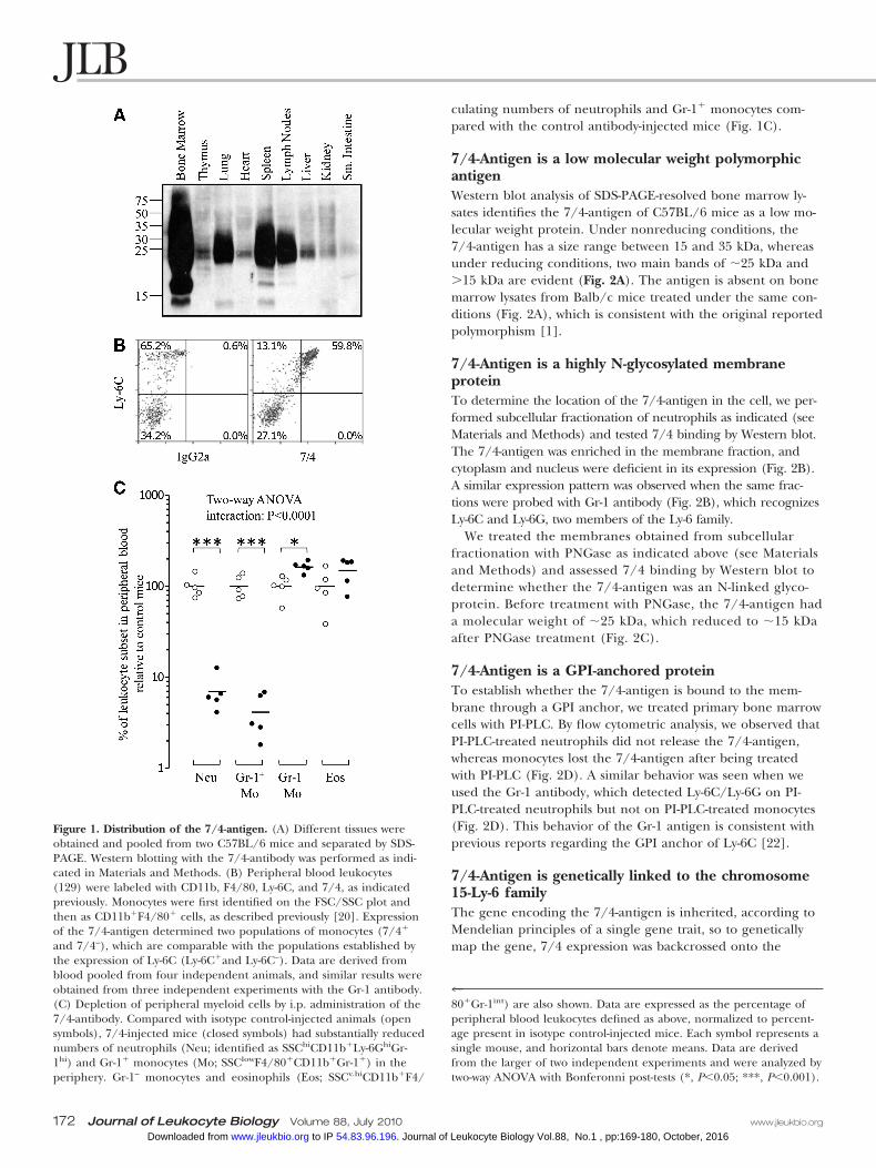
culating numbers of neutrophils and Gr-1� monocytes com-pared with the control antibody-injected mice (Fig. 1C).
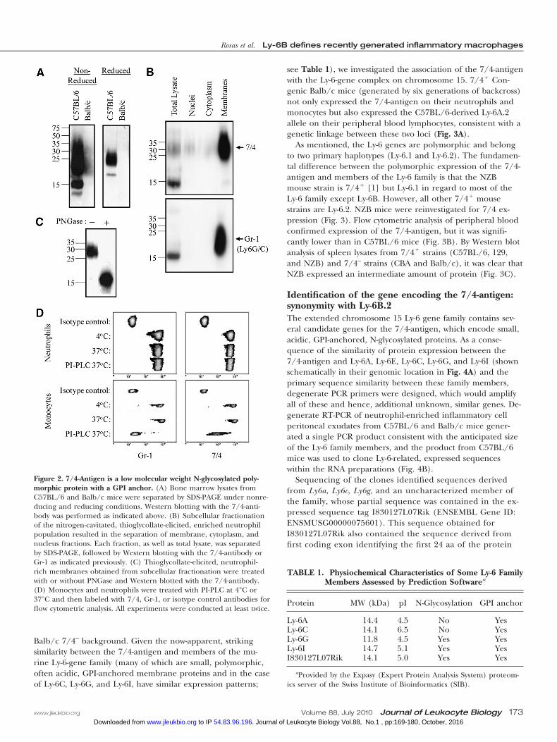
7/4-Antigen is a low molecular weight polymorphicantigenWestern blot analysis of SDS-PAGE-resolved bone marrow ly-sates identifies the 7/4-antigen of C57BL/6 mice as a low mo-lecular weight protein. Under nonreducing conditions, the7/4-antigen has a size range between 15 and 35 kDa, whereasunder reducing conditions, two main bands of �25 kDa and�15 kDa are evident (Fig. 2A). The antigen is absent on bonemarrow lysates from Balb/c mice treated under the same con-ditions (Fig. 2A), which is consistent with the original reportedpolymorphism [1].
7/4-Antigen is a highly N-glycosylated membraneproteinTo determine the location of the 7/4-antigen in the cell, we per-formed subcellular fractionation of neutrophils as indicated (seeMaterials and Methods) and tested 7/4 binding by Western blot.The 7/4-antigen was enriched in the membrane fraction, andcytoplasm and nucleus were deficient in its expression (Fig. 2B).A similar expression pattern was observed when the same frac-tions were probed with Gr-1 antibody (Fig. 2B), which recognizesLy-6C and Ly-6G, two members of the Ly-6 family.
We treated the membranes obtained from subcellularfractionation with PNGase as indicated above (see Materialsand Methods) and assessed 7/4 binding by Western blot todetermine whether the 7/4-antigen was an N-linked glyco-protein. Before treatment with PNGase, the 7/4-antigen hada molecular weight of �25 kDa, which reduced to �15 kDaafter PNGase treatment (Fig. 2C).
7/4-Antigen is a GPI-anchored proteinTo establish whether the 7/4-antigen is bound to the mem-brane through a GPI anchor, we treated primary bone marrowcells with PI-PLC. By flow cytometric analysis, we observed thatPI-PLC-treated neutrophils did not release the 7/4-antigen,whereas monocytes lost the 7/4-antigen after being treatedwith PI-PLC (Fig. 2D). A similar behavior was seen when weused the Gr-1 antibody, which detected Ly-6C/Ly-6G on PI-PLC-treated neutrophils but not on PI-PLC-treated monocytes(Fig. 2D). This behavior of the Gr-1 antigen is consistent withprevious reports regarding the GPI anchor of Ly-6C [22].
7/4-Antigen is genetically linked to the chromosome15-Ly-6 familyThe gene encoding the 7/4-antigen is inherited, according toMendelian principles of a single gene trait, so to geneticallymap the gene, 7/4 expression was backcrossed onto the
Figure 1. Distribution of the 7/4-antigen. (A) Different tissues wereobtained and pooled from two C57BL/6 mice and separated by SDS-PAGE. Western blotting with the 7/4-antibody was performed as indi-cated in Materials and Methods. (B) Peripheral blood leukocytes(129) were labeled with CD11b, F4/80, Ly-6C, and 7/4, as indicatedpreviously. Monocytes were first identified on the FSC/SSC plot andthen as CD11b�F4/80� cells, as described previously [20]. Expressionof the 7/4-antigen determined two populations of monocytes (7/4�
and 7/4–), which are comparable with the populations established bythe expression of Ly-6C (Ly-6C�and Ly-6C–). Data are derived fromblood pooled from four independent animals, and similar results wereobtained from three independent experiments with the Gr-1 antibody.(C) Depletion of peripheral myeloid cells by i.p. administration of the7/4-antibody. Compared with isotype control-injected animals (opensymbols), 7/4-injected mice (closed symbols) had substantially reducednumbers of neutrophils (Neu; identified as SSChiCD11b�Ly-6GhiGr-1hi) and Gr-1� monocytes (Mo; SSClowF4/80�CD11b�Gr-1�) in theperiphery. Gr-1– monocytes and eosinophils (Eos; SSCv.hiCD11b�F4/
480�Gr-1int) are also shown. Data are expressed as the percentage ofperipheral blood leukocytes defined as above, normalized to percent-age present in isotype control-injected mice. Each symbol represents asingle mouse, and horizontal bars denote means. Data are derivedfrom the larger of two independent experiments and were analyzed bytwo-way ANOVA with Bonferonni post-tests (*, P�0.05; ***, P�0.001).
172 Journal of Leukocyte Biology Volume 88, July 2010 www.jleukbio.org
Vol.88, No.1 , pp:169-180, October, 2016Journal of Leukocyte Biology. 54.83.96.196 to IP www.jleukbio.orgDownloaded from
Balb/c 7/4– background. Given the now-apparent, strikingsimilarity between the 7/4-antigen and members of the mu-rine Ly-6-gene family (many of which are small, polymorphic,often acidic, GPI-anchored membrane proteins and in the caseof Ly-6C, Ly-6G, and Ly-6I, have similar expression patterns;
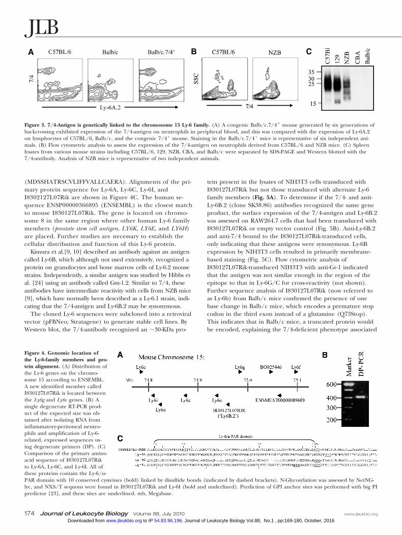
see Table 1), we investigated the association of the 7/4-antigenwith the Ly-6-gene complex on chromosome 15. 7/4� Con-genic Balb/c mice (generated by six generations of backcross)not only expressed the 7/4-antigen on their neutrophils andmonocytes but also expressed the C57BL/6-derived Ly-6A.2allele on their peripheral blood lymphocytes, consistent with agenetic linkage between these two loci (Fig. 3A).
As mentioned, the Ly-6 genes are polymorphic and belongto two primary haplotypes (Ly-6.1 and Ly-6.2). The fundamen-tal difference between the polymorphic expression of the 7/4-antigen and members of the Ly-6 family is that the NZBmouse strain is 7/4� [1] but Ly-6.1 in regard to most of theLy-6 family except Ly-6B. However, all other 7/4� mousestrains are Ly-6.2. NZB mice were reinvestigated for 7/4 ex-pression (Fig. 3). Flow cytometric analysis of peripheral bloodconfirmed expression of the 7/4-antigen, but it was signifi-cantly lower than in C57BL/6 mice (Fig. 3B). By Western blotanalysis of spleen lysates from 7/4� strains (C57BL/6, 129,and NZB) and 7/4– strains (CBA and Balb/c), it was clear thatNZB expressed an intermediate amount of protein (Fig. 3C).
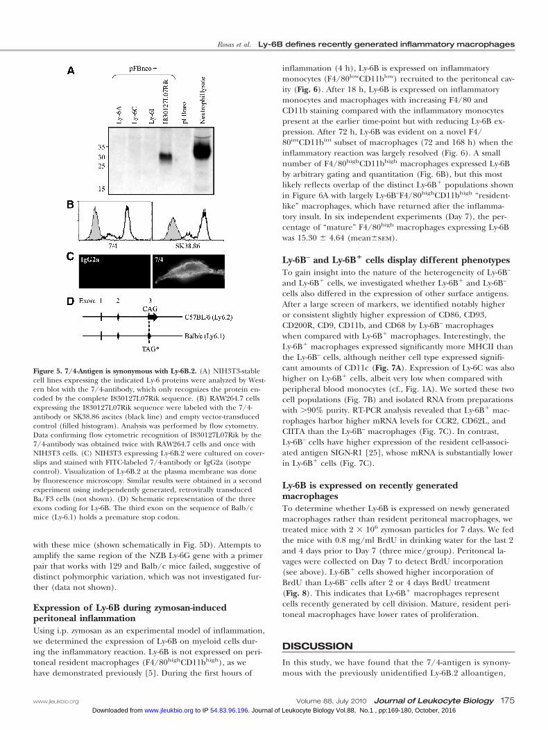
Identification of the gene encoding the 7/4-antigen:synonymity with Ly-6B.2The extended chromosome 15 Ly-6 gene family contains sev-eral candidate genes for the 7/4-antigen, which encode small,acidic, GPI-anchored, N-glycosylated proteins. As a conse-quence of the similarity of protein expression between the7/4-antigen and Ly-6A, Ly-6E, Ly-6C, Ly-6G, and Ly-6I (shownschematically in their genomic location in Fig. 4A) and theprimary sequence similarity between these family members,degenerate PCR primers were designed, which would amplifyall of these and hence, additional unknown, similar genes. De-generate RT-PCR of neutrophil-enriched inflammatory cellperitoneal exudates from C57BL/6 and Balb/c mice gener-ated a single PCR product consistent with the anticipated sizeof the Ly-6 family members, and the product from C57BL/6mice was used to clone Ly-6-related, expressed sequenceswithin the RNA preparations (Fig. 4B).
Sequencing of the clones identified sequences derivedfrom Ly6a, Ly6c, Ly6g, and an uncharacterized member ofthe family, whose partial sequence was contained in the ex-pressed sequence tag I830127L07Rik (ENSEMBL Gene ID:ENSMUSG00000075601). This sequence obtained forI830127L07Rik also contained the sequence derived fromfirst coding exon identifying the first 24 aa of the protein
TABLE 1. Physiochemical Characteristics of Some Ly-6 FamilyMembers Assessed by Prediction Softwarea
Protein MW (kDa) pI N-Glycosylation GPI anchor
Ly-6A 14.4 4.5 No YesLy-6C 14.1 6.5 No YesLy-6G 11.8 4.5 Yes YesLy-6I 14.7 5.1 Yes YesI830127L07Rik 14.1 5.0 Yes Yes
aProvided by the Expasy (Expert Protein Analysis System) proteom-ics server of the Swiss Institute of Bioinformatics (SIB).
Figure 2. 7/4-Antigen is a low molecular weight N-glycosylated poly-morphic protein with a GPI anchor. (A) Bone marrow lysates fromC57BL/6 and Balb/c mice were separated by SDS-PAGE under nonre-ducing and reducing conditions. Western blotting with the 7/4-anti-body was performed as indicated above. (B) Subcellular fractionationof the nitrogen-cavitated, thioglycollate-elicited, enriched neutrophilpopulation resulted in the separation of membrane, cytoplasm, andnucleus fractions. Each fraction, as well as total lysate, was separatedby SDS-PAGE, followed by Western blotting with the 7/4-antibody orGr-1 as indicated previously. (C) Thioglycollate-elicited, neutrophil-rich membranes obtained from subcellular fractionation were treatedwith or without PNGase and Western blotted with the 7/4-antibody.(D) Monocytes and neutrophils were treated with PI-PLC at 4°C or37°C and then labeled with 7/4, Gr-1, or isotype control antibodies forflow cytometric analysis. All experiments were conducted at least twice.
Rosas et al. Ly-6B defines recently generated inflammatory macrophages
www.jleukbio.org Volume 88, July 2010 Journal of Leukocyte Biology 173 Vol.88, No.1 , pp:169-180, October, 2016Journal of Leukocyte Biology. 54.83.96.196 to IP www.jleukbio.orgDownloaded from
(MDSSHATRSCVLIFFVALLCAERA). Alignments of the pri-mary protein sequence for Ly-6A, Ly-6C, Ly-6I, andI8302127L07Rik are shown in Figure 4C. The human se-quence ENSP00000366895 (ENSEMBL) is the closest matchto mouse I830127L07Rik. The gene is located on chromo-some 8 in the same region where other human Ly-6 familymembers (prostate stem cell antigen, LY6K, LY6E, and LY6H)are placed. Further studies are necessary to establish thecellular distribution and function of this Ly-6 protein.
Kimura et al.[9, 10] described an antibody against an antigencalled Ly-6B, which although not used extensively, recognized aprotein on granulocytes and bone marrow cells of Ly-6.2 mousestrains. Independently, a similar antigen was studied by Hibbs etal. [24] using an antibody called Gm-1.2. Similar to 7/4, theseantibodies have intermediate reactivity with cells from NZB mice[9], which have normally been described as a Ly-6.1 strain, indi-cating that the 7/4-antigen and Ly-6B.2 may be synonymous.
The cloned Ly-6 sequences were subcloned into a retroviralvector (pFBNeo; Stratagene) to generate stable cell lines. ByWestern blot, the 7/4-antibody recognized an �30-KDa pro-
tein present in the lysates of NIH3T3 cells transduced withI830127L07Rik but not those transduced with alternate Ly-6family members (Fig. 5A). To determine if the 7/4- and anti-Ly-6B.2 (clone SK38.86) antibodies recognized the same geneproduct, the surface expression of the 7/4-antigen and Ly-6B.2was assessed on RAW264.7 cells that had been transduced withI830127L07Rik or empty vector control (Fig. 5B). Anti-Ly-6B.2and anti-7/4 bound to the I830127L07Rik-transduced cells,only indicating that these antigens were synonymous. Ly-6Bexpression by NIH3T3 cells resulted in primarily membrane-based staining (Fig. 5C). Flow cytometric analysis ofI830127L07Rik-transduced NIH3T3 with anti-Gr-1 indicatedthat the antigen was not similar enough in the region of theepitope to that in Ly-6G/C for cross-reactivity (not shown).Further sequence analysis of I830127L07Rik (now referred toas Ly-6b) from Balb/c mice confirmed the presence of onebase change in Balb/c mice, which encodes a premature stopcodon in the third exon instead of a glutamine (Q73Stop).This indicates that in Balb/c mice, a truncated protein wouldbe encoded, explaining the 7/4-deficient phenotype associated
Figure 3. 7/4-Antigen is genetically linked to the chromosome 15 Ly-6 family. (A) A congenic Balb/c.7/4� mouse generated by six generations ofbackcrossing exhibited expression of the 7/4-antigen on neutrophils in peripheral blood, and this was compared with the expression of Ly-6A.2on lymphocytes of C57BL/6, Balb/c, and the congenic 7/4� mouse. Staining in the Balb/c.7/4� mice is representative of six independent ani-mals. (B) Flow cytometric analysis to assess the expression of the 7/4-antigen on neutrophils derived from C57BL/6 and NZB mice. (C) Spleenlysates from various mouse strains including C57BL/6, 129, NZB, CBA, and Balb/c were separated by SDS-PAGE and Western blotted with the7/4-antibody. Analysis of NZB mice is representative of two independent animals.
Figure 4. Genomic location ofthe Ly-6-family members and pro-tein alignment. (A) Distribution ofthe Ly-6 genes on the chromo-some 15 according to ENSEMBL.A new identified member calledI830127L07Rik is located betweenthe Ly6g and Ly6c genes. (B) Asingle degenerate RT-PCR prod-uct of the expected size was ob-tained after isolating RNA frominflammatory-peritoneal neutro-phils and amplification of Ly-6-related, expressed sequences us-ing degenerate primers (DP). (C)Comparison of the primary aminoacid sequence of I830127L07Rikto Ly-6A, Ly-6C, and Ly-6I. All ofthese proteins contain the Ly-6/u-PAR domain with 10 conserved cysteines (bold) linked by disulfide bonds (indicated by dashed brackets). N-Glycosylation was assessed by NetNG-lyc, and NXS/T sequons were found in I830127L07Rik and Ly-6I (bold and underlined). Prediction of GPI anchor sites was performed with big PIpredictor [23], and these sites are underlined. mb, Megabase.
174 Journal of Leukocyte Biology Volume 88, July 2010 www.jleukbio.org
Vol.88, No.1 , pp:169-180, October, 2016Journal of Leukocyte Biology. 54.83.96.196 to IP www.jleukbio.orgDownloaded from
with these mice (shown schematically in Fig. 5D). Attempts toamplify the same region of the NZB Ly-6G gene with a primerpair that works with 129 and Balb/c mice failed, suggestive ofdistinct polymorphic variation, which was not investigated fur-ther (data not shown).
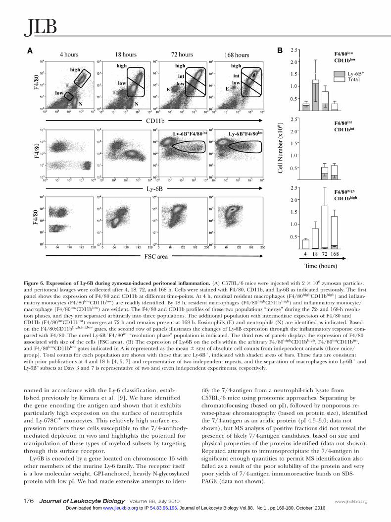
Expression of Ly-6B during zymosan-inducedperitoneal inflammationUsing i.p. zymosan as an experimental model of inflammation,we determined the expression of Ly-6B on myeloid cells dur-ing the inflammatory reaction. Ly-6B is not expressed on peri-toneal resident macrophages (F4/80highCD11bhigh), as wehave demonstrated previously [5]. During the first hours of
inflammation (4 h), Ly-6B is expressed on inflammatorymonocytes (F4/80lowCD11blow) recruited to the peritoneal cav-ity (Fig. 6). After 18 h, Ly-6B is expressed on inflammatorymonocytes and macrophages with increasing F4/80 andCD11b staining compared with the inflammatory monocytespresent at the earlier time-point but with reducing Ly-6B ex-pression. After 72 h, Ly-6B was evident on a novel F4/80intCD11bint subset of macrophages (72 and 168 h) when theinflammatory reaction was largely resolved (Fig. 6). A smallnumber of F4/80highCD11bhigh macrophages expressed Ly-6Bby arbitrary gating and quantitation (Fig. 6B), but this mostlikely reflects overlap of the distinct Ly-6B� populations shownin Figure 6A with largely Ly-6B–F4/80highCD11bhigh “resident-like” macrophages, which have returned after the inflamma-tory insult. In six independent experiments (Day 7), the per-centage of “mature” F4/80high macrophages expressing Ly-6Bwas 15.30 � 4.64 (mean�sem).
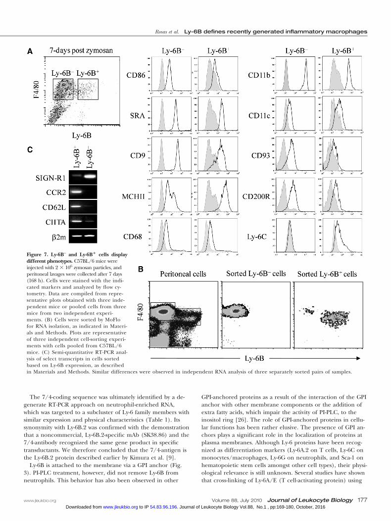
Ly-6B– and Ly-6B� cells display different phenotypesTo gain insight into the nature of the heterogeneity of Ly-6B–
and Ly-6B� cells, we investigated whether Ly-6B� and Ly-6B–
cells also differed in the expression of other surface antigens.After a large screen of markers, we identified notably higheror consistent slightly higher expression of CD86, CD93,CD200R, CD9, CD11b, and CD68 by Ly-6B– macrophageswhen compared with Ly-6B� macrophages. Interestingly, theLy-6B� macrophages expressed significantly more MHCII thanthe Ly-6B– cells, although neither cell type expressed signifi-cant amounts of CD11c (Fig. 7A). Expression of Ly-6C was alsohigher on Ly-6B� cells, albeit very low when compared withperipheral blood monocytes (cf., Fig. 1A). We sorted these twocell populations (Fig. 7B) and isolated RNA from preparationswith �90% purity. RT-PCR analysis revealed that Ly-6B� mac-rophages harbor higher mRNA levels for CCR2, CD62L, andCIITA than the Ly-6B– macrophages (Fig. 7C). In contrast,Ly-6B– cells have higher expression of the resident cell-associ-ated antigen SIGN-R1 [25], whose mRNA is substantially lowerin Ly-6B� cells (Fig. 7C).
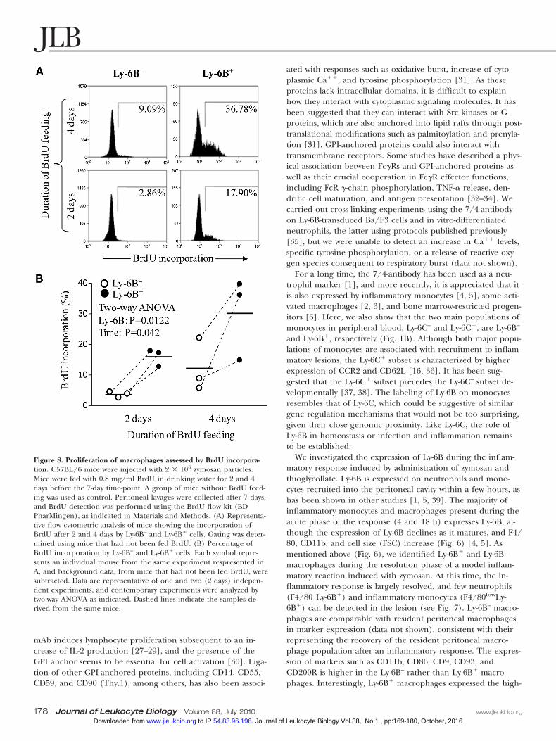
Ly-6B is expressed on recently generatedmacrophagesTo determine whether Ly-6B is expressed on newly generatedmacrophages rather than resident peritoneal macrophages, wetreated mice with 2 � 106 zymosan particles for 7 days. We fedthe mice with 0.8 mg/ml BrdU in drinking water for the last 2and 4 days prior to Day 7 (three mice/group). Peritoneal la-vages were collected on Day 7 to detect BrdU incorporation(see above). Ly-6B� cells showed higher incorporation ofBrdU than Ly-6B– cells after 2 or 4 days BrdU treatment(Fig. 8). This indicates that Ly-6B� macrophages representcells recently generated by cell division. Mature, resident peri-toneal macrophages have lower rates of proliferation.
DISCUSSION
In this study, we have found that the 7/4-antigen is synony-mous with the previously unidentified Ly-6B.2 alloantigen,
Figure 5. 7/4-Antigen is synonymous with Ly-6B.2. (A) NIH3T3-stablecell lines expressing the indicated Ly-6 proteins were analyzed by West-ern blot with the 7/4-antibody, which only recognizes the protein en-coded by the complete I830127L07Rik sequence. (B) RAW264.7 cellsexpressing the I830127L07Rik sequence were labeled with the 7/4-antibody or SK38.86 ascites (black line) and empty vector-transducedcontrol (filled histogram). Analysis was performed by flow cytometry.Data confirming flow cytometric recognition of I830127L07Rik by the7/4-antibody was obtained twice with RAW264.7 cells and once withNIH3T3 cells. (C) NIH3T3 expressing Ly-6B.2 were cultured on cover-slips and stained with FITC-labeled 7/4-antibody or IgG2a (isotypecontrol). Visualization of Ly-6B.2 at the plasma membrane was doneby fluorescence microscopy. Similar results were obtained in a secondexperiment using independently generated, retrovirally transducedBa/F3 cells (not shown). (D) Schematic representation of the threeexons coding for Ly-6B. The third exon on the sequence of Balb/cmice (Ly-6.1) holds a premature stop codon.
Rosas et al. Ly-6B defines recently generated inflammatory macrophages
www.jleukbio.org Volume 88, July 2010 Journal of Leukocyte Biology 175 Vol.88, No.1 , pp:169-180, October, 2016Journal of Leukocyte Biology. 54.83.96.196 to IP www.jleukbio.orgDownloaded from
named in accordance with the Ly-6 classification, estab-lished previously by Kimura et al. [9]. We have identifiedthe gene encoding the antigen and shown that it exhibitsparticularly high expression on the surface of neutrophilsand Ly-678C� monocytes. This relatively high surface ex-pression renders these cells susceptible to the 7/4-antibody-mediated depletion in vivo and highlights the potential formanipulation of these types of myeloid subsets by targetingthrough this surface receptor.
Ly-6B is encoded by a gene located on chromosome 15 withother members of the murine Ly-6 family. The receptor itselfis a low molecular weight, GPI-anchored, heavily N-glycosylatedprotein with low pI. We had made extensive attempts to iden-
tify the 7/4-antigen from a neutrophil-rich lysate fromC57BL/6 mice using proteomic approaches. Separating bychromatofocusing (based on pI), followed by nonporous re-verse-phase chromatography (based on protein size), identifiedthe 7/4-antigen as an acidic protein (pI 4.5–5.0; data notshown), but MS analysis of positive fractions did not reveal thepresence of likely 7/4-antigen candidates, based on size andphysical properties of the proteins identified (data not shown).Repeated attempts to immunoprecipitate the 7/4-antigen insignificant enough quantities to permit MS identification alsofailed as a result of the poor solubility of the protein and verypoor yields of 7/4-antigen immunoreactive bands on SDS-PAGE (data not shown).
Figure 6. Expression of Ly-6B during zymosan-induced peritoneal inflammation. (A) C57BL/6 mice were injected with 2 � 106 zymosan particles,and peritoneal lavages were collected after 4, 18, 72, and 168 h. Cells were stained with F4/80, CD11b, and Ly-6B as indicated previously. The firstpanel shows the expression of F4/80 and CD11b at different time-points. At 4 h, residual resident macrophages (F4/80highCD11bhigh) and inflam-matory monocytes (F4/80lowCD11blow) are readily identified. By 18 h, resident macrophages (F4/80highCD11bhigh) and inflammatory monocyte/macrophage (F4/80lowCD11blow) are evident. The F4/80 and CD11b profiles of these two populations “merge” during the 72- and 168-h resolu-tion phases, and they are separated arbitrarily into three populations. The additional population with intermediate expression of F4/80 andCD11b (F4/80intCD11bint) emerges at 72 h and remains present at 168 h. Eosinophils (E) and neutrophils (N) are identified as indicated. Basedon the F4/80:CD11bhigh,int,low gates, the second row of panels illustrates the changes of Ly-6B expression through the inflammatory response com-pared with F4/80. The novel Ly-6B�F4/80int “resolution phase” population is indicated. The third row of panels displays the expression of F4/80associated with size of the cells (FSC area). (B) The expression of Ly-6B on the cells within the arbitrary F4/80highCD11bhigh, F4/80intCD11bint,and F4/80lowCD11blow gates indicated in A is represented as the mean � sem of absolute cell counts from independent animals (three mice/group). Total counts for each population are shown with those that are Ly-6B�, indicated with shaded areas of bars. These data are consistentwith prior publications at 4 and 18 h [4, 5, 7] and representative of two independent repeats, and the separation of macrophages into Ly-6B� andLy-6B– subsets at Days 3 and 7 is representative of two and seven independent experiments, respectively.
176 Journal of Leukocyte Biology Volume 88, July 2010 www.jleukbio.org
Vol.88, No.1 , pp:169-180, October, 2016Journal of Leukocyte Biology. 54.83.96.196 to IP www.jleukbio.orgDownloaded from
The 7/4-coding sequence was ultimately identified by a de-generate RT-PCR approach on neutrophil-enriched RNA,which was targeted to a subcluster of Ly-6 family members withsimilar expression and physical characteristics (Table 1). Itssynonymity with Ly-6B.2 was confirmed with the demonstrationthat a noncommercial, Ly-6B.2-specific mAb (SK38.86) and the7/4-antibody recognized the same gene product in specifictransductants. We therefore concluded that the 7/4-antigen isthe Ly-6B.2 protein described earlier by Kimura et al. [9].
Ly-6B is attached to the membrane via a GPI anchor (Fig.3). PI-PLC treatment, however, did not remove Ly-6B fromneutrophils. This behavior has also been observed in other
GPI-anchored proteins as a result of the interaction of the GPIanchor with other membrane components or the addition ofextra fatty acids, which impair the activity of PI-PLC, to theinositol ring [26]. The role of GPI-anchored proteins in cellu-lar functions has been rather elusive. The presence of GPI an-chors plays a significant role in the localization of proteins atplasma membranes. Although Ly-6 proteins have been recog-nized as differentiation markers (Ly-6A.2 on T cells, Ly-6C onmonocytes/macrophages, Ly-6G on neutrophils, and Sca-1 onhematopoietic stem cells amongst other cell types), their physi-ological relevance is still unknown. Several studies have shownthat cross-linking of Ly-6A/E (T cell-activating protein) using
Figure 7. Ly-6B– and Ly-6B� cells displaydifferent phenotypes. C57BL/6 mice wereinjected with 2 � 106 zymosan particles, andperitoneal lavages were collected after 7 days(168 h). Cells were stained with the indi-cated markers and analyzed by flow cy-tometry. Data are compiled from repre-sentative plots obtained with three inde-pendent mice or pooled cells from threemice from two independent experi-ments. (B) Cells were sorted by MoFlofor RNA isolation, as indicated in Materi-als and Methods. Plots are representativeof three independent cell-sorting experi-ments with cells pooled from C57BL/6mice. (C) Semi-quantitative RT-PCR anal-ysis of select transcripts in cells sortedbased on Ly-6B expression, as describedin Materials and Methods. Similar differences were observed in independent RNA analysis of three separately sorted pairs of samples.
Rosas et al. Ly-6B defines recently generated inflammatory macrophages
www.jleukbio.org Volume 88, July 2010 Journal of Leukocyte Biology 177 Vol.88, No.1 , pp:169-180, October, 2016Journal of Leukocyte Biology. 54.83.96.196 to IP www.jleukbio.orgDownloaded from
mAb induces lymphocyte proliferation subsequent to an in-crease of IL-2 production [27–29], and the presence of theGPI anchor seems to be essential for cell activation [30]. Liga-tion of other GPI-anchored proteins, including CD14, CD55,CD59, and CD90 (Thy.1), among others, has also been associ-
ated with responses such as oxidative burst, increase of cyto-plasmic Ca��, and tyrosine phosphorylation [31]. As theseproteins lack intracellular domains, it is difficult to explainhow they interact with cytoplasmic signaling molecules. It hasbeen suggested that they can interact with Src kinases or G-proteins, which are also anchored into lipid rafts through post-translational modifications such as palmitoylation and prenyla-tion [31]. GPI-anchored proteins could also interact withtransmembrane receptors. Some studies have described a phys-ical association between Fc�Rs and GPI-anchored proteins aswell as their crucial cooperation in Fc�R effector functions,including FcR �-chain phosphorylation, TNF-� release, den-dritic cell maturation, and antigen presentation [32–34]. Wecarried out cross-linking experiments using the 7/4-antibodyon Ly-6B-transduced Ba/F3 cells and in vitro-differentiatedneutrophils, the latter using protocols published previously[35], but we were unable to detect an increase in Ca�� levels,specific tyrosine phosphorylation, or a release of reactive oxy-gen species consequent to respiratory burst (data not shown).
For a long time, the 7/4-antibody has been used as a neu-trophil marker [1], and more recently, it is appreciated that itis also expressed by inflammatory monocytes [4, 5], some acti-vated macrophages [2, 3], and bone marrow-restricted progen-itors [6]. Here, we also show that the two main populations ofmonocytes in peripheral blood, Ly-6C– and Ly-6C�, are Ly-6B–
and Ly-6B�, respectively (Fig. 1B). Although both major popu-lations of monocytes are associated with recruitment to inflam-matory lesions, the Ly-6C� subset is characterized by higherexpression of CCR2 and CD62L [16, 36]. It has been sug-gested that the Ly-6C� subset precedes the Ly-6C– subset de-velopmentally [37, 38]. The labeling of Ly-6B on monocytesresembles that of Ly-6C, which could be suggestive of similargene regulation mechanisms that would not be too surprising,given their close genomic proximity. Like Ly-6C, the role ofLy-6B in homeostasis or infection and inflammation remainsto be established.
We investigated the expression of Ly-6B during the inflam-matory response induced by administration of zymosan andthioglycollate. Ly-6B is expressed on neutrophils and mono-cytes recruited into the peritoneal cavity within a few hours, ashas been shown in other studies [1, 5, 39]. The majority ofinflammatory monocytes and macrophages present during theacute phase of the response (4 and 18 h) expresses Ly-6B, al-though the expression of Ly-6B declines as it matures, and F4/80, CD11b, and cell size (FSC) increase (Fig. 6) [4, 5]. Asmentioned above (Fig. 6), we identified Ly-6B� and Ly-6B–
macrophages during the resolution phase of a model inflam-matory reaction induced with zymosan. At this time, the in-flammatory response is largely resolved, and few neutrophils(F4/80–Ly-6B�) and inflammatory monocytes (F4/80lowLy-6B�) can be detected in the lesion (see Fig. 7). Ly-6B– macro-phages are comparable with resident peritoneal macrophagesin marker expression (data not shown), consistent with theirrepresenting the recovery of the resident peritoneal macro-phage population after an inflammatory response. The expres-sion of markers such as CD11b, CD86, CD9, CD93, andCD200R is higher in the Ly-6B– rather than Ly-6B� macro-phages. Interestingly, Ly-6B� macrophages expressed the high-
Figure 8. Proliferation of macrophages assessed by BrdU incorpora-tion. C57BL/6 mice were injected with 2 � 106 zymosan particles.Mice were fed with 0.8 mg/ml BrdU in drinking water for 2 and 4days before the 7-day time-point. A group of mice without BrdU feed-ing was used as control. Peritoneal lavages were collected after 7 days,and BrdU detection was performed using the BrdU flow kit (BDPharMingen), as indicated in Materials and Methods. (A) Representa-tive flow cytometric analysis of mice showing the incorporation ofBrdU after 2 and 4 days by Ly-6B– and Ly-6B� cells. Gating was deter-mined using mice that had not been fed BrdU. (B) Percentage ofBrdU incorporation by Ly-6B– and Ly-6B� cells. Each symbol repre-sents an individual mouse from the same experiment respresented inA, and background data, from mice that had not been fed BrdU, weresubtracted. Data are representative of one and two (2 days) indepen-dent experiments, and contemporary experiments were analyzed bytwo-way ANOVA as indicated. Dashed lines indicate the samples de-rived from the same mice.
178 Journal of Leukocyte Biology Volume 88, July 2010 www.jleukbio.org
Vol.88, No.1 , pp:169-180, October, 2016Journal of Leukocyte Biology. 54.83.96.196 to IP www.jleukbio.orgDownloaded from
est levels of MHCII and Ly-6C and at the mRNA level, CCR2,CD62L, and CIITA. The coexpression of the “Ly-6C� mono-cyte marker” Ly-6B with other markers associated with the Ly-6C� monocyte subset (CCR2 and CD62L) is suggestive of re-cent arrival and differentiation of these cells from inflamma-tory Ly-6B�Ly-6C� monocytes. Expression of MHCII isregulated by the transcriptional coactivator CIITA, whose re-pression has been associated with the terminal differentiationof B cells into plasma cells and maturation of dendritc cells[40, 41]. We observed that CIITA is expressed at lower levelsin Ly-6B– cells (Fig. 7C), and expression of MHCII is notablylower, which could be part of the maturation process of peri-toneal “resident” macrophages and again, would be consistentwith the hypothesis that Ly-6B-expressing macrophages are re-cently generated macrophages in the process of differentiatinginto, in this setting, resident peritoneal macrophages. The sug-gestion that these Ly-6B� cells are more mature comparedwith inflammatory monocytes is supported by their higher ex-pression of F4/80 and CD11b, lower expression of Ly-6C, andincreased cell size. The fact that they may be less mature thantheir Ly-6C– contemporaries is supported by BrdU incorpora-tion experiments, which indicate that the Ly-6B� cells are gen-erated more recently by cell division than their Ly-6B– contem-poraries. It is also supported by the observation that they ex-press less CD11b and F4/80 and are smaller than the Ly-6B–
cells but retain expression of monocytic markers (Ly-6C,CCR2, and CD62L). Collectively, this also supports the specu-lation that they are not derived from Ly-6B– macrophages bymarker regulation.
Taken in context with other studies about the distributionof the 7/4-antigen [4, 5] and the biology of the Ly-6C� mono-cyte subset, it seems likely that Ly-6B� monocytes are recruitedinto the peritoneal cavity, where they lose Ly-6B expression asthey differentiate [4, 5]. This differentiation can be into aninflammatory macrophage or dendritic cell phenotype or as areplacement for tissue resident macrophage populations (mostlikely during the resolution phase of the inflammatory re-sponse). Although the origins of the tissue resident macro-phages and dendritic cells are poorly characterized and likelyto differ under inflammatory circumstances, it seems unlikelythat these late inflammatory response Ly-6B� cells are becom-ing “resident peritoneal” dendritic cell populations, as themarker phenotype of the two cells is quite different (data notshown and ref. [42]). We speculate that Ly-6B expression bymacrophages in inflammatory lesions, such as seen during par-asite infection [3] and BCG granulomas [2] or on cells re-cruited to tumor challenge [5], represents the arrival of re-cently generated macrophages. As such, Ly-6B expression mayserve as a useful surrogate marker for the kinetics of mono-cyte/macrophage turnover in chronic inflammation, as it high-lights recently recruited monocyte-derived inflammatory mac-rophages and other potentially more obscure sources of mac-rophages in an inflammatory context. Also of interest is theexpression of Ly-6B by cells with the phenotype of myeloid-derived suppressor cells (Gr-1�CD11b�) during steady-stateinflammation and tumor challenge [5, 18]. Myeloid-derivedsuppressor cells are noted for their “immature”-like phenotype[18]. This would be consistent with the suggestion that my-
eloid-derived suppressor cells are also relatively, recently pro-duced cells retaining Ly-6B (and Gr-1) expression from theirneutrophilic/monocytic roots in a way directly analogous tothat proposed above for Ly-6B expression in our experimentalperitonitis model and with direct correlation to previous ma-larial and BCG infection models [2, 3]. The ability, by, for ex-ample, exploiting Ly-6B expression, to target these relativelyimmature, neutrophilic- and monocytic-like cells specifically invivo could represent a valuable, new approach to many aspectsof myeloid cell biology.
AUTHORSHIP
M. R. and P. R. T. designed and performed the experimentsand co-wrote the manuscript. B. T. and M. S. designed andconducted experiments. S. G. and P. R. T. conceived the proj-ect. All authors contributed to the interpretation of data.
ACKNOWLEDGMENTS
This work was funded by the Wellcome Trust (070579) andthe Medical Research Council (G0601617). We thank Alexan-dre Akoulitchev for his support and advice during the pro-teomics analysis. We also thank the staff of our animal facilitiesfor the care of the animals used in this study.
REFERENCES
1. Hirsch, S., Gordon, S. (1983) Polymorphic expression of a neutrophildifferentiation antigen revealed by monoclonal antibody 7/4. Immunoge-netics 18, 229–239.
2. Gordon, S., Keshav, S., Stein, M. (1994) BCG-induced granuloma forma-tion in murine tissues. Immunobiology 191, 369–377.
3. Lee, S. H., Crocker, P., Gordon, S. (1986) Macrophage plasma mem-brane and secretory properties in murine malaria. Effects of Plasmodiumyoelii blood-stage infection on macrophages in liver, spleen, and blood. J.Exp. Med. 163, 54–74.
4. Henderson, R. B., Hobbs, J. A., Mathies, M., Hogg, N. (2003) Rapid re-cruitment of inflammatory monocytes is independent of neutrophil mi-gration. Blood 102, 328–335.
5. Taylor, P. R., Brown, G. D., Geldhof, A. B., Martinez-Pomares, L., Gor-don, S. (2003) Pattern recognition receptors and differentiation antigensdefine murine myeloid cell heterogeneity ex vivo. Eur. J. Immunol. 33,2090–2097.
6. Bertoncello, I., Bartelmez, S. H., Bradley, T. R., Hodgson, G. S. (1989)Changes in cell surface antigen expression during murine bone marrowcell regeneration in vivo and proliferation in vitro. Immunol. Cell Biol. 67,127–133.
7. Taylor, P. R., Reid, D. M., Heinsbroek, S. E., Brown, G. D., Gordon, S.,Wong, S. Y. (2005) Dectin-2 is predominantly myeloid restricted and ex-hibits unique activation-dependent expression on maturing inflammatorymonocytes elicited in vivo. Eur. J. Immunol. 35, 2163–2174.
8. LeClair, K. P., Rabin, M., Nesbitt, M. N., Pravtcheva, D., Ruddle, F. H.,Palfree, R. G., Bothwell, A. (1987) Murine Ly-6 multigene family is lo-cated on chromosome 15. Proc. Natl. Acad. Sci. USA 84, 1638–1642.
9. Kimura, S., Tada, N., Liu-Lam, Y., Hammerling, U. (1984) Studies of themouse Ly-6 alloantigen system. II. Complexities of the Ly-6 region. Immu-nogenetics 20, 47–56.
10. Kimura, S., Tada, N., Nakayama, E., Hammerling, U. (1980) Studies ofthe mouse Ly-6 alloantigen system. I. Serological characterization ofmouse Ly-6 alloantigen by monoclonal antibodies. Immunogenetics 11,373–381.
11. Rock, K. L., Reiser, H., Bamezai, A., McGrew, J., Benacerraf, B. (1989)The LY-6 locus: a multigene family encoding phosphatidylinositol-an-chored membrane proteins concerned with T-cell activation. Immunol.Rev. 111, 195–224.
12. Fleming, T.J., O'hUigin, C., Malek, T. R. (1993) Characterization of twonovel Ly-6 genes. Protein sequence and potential structural similarity to�-bungarotoxin and other neurotoxins. J. Immunol. 150, 5379–5390.
13. Patterson, J. M., Johnson, M. H., Zimonjic, D. B., Graubert, T. A. (2000)Characterization of Ly-6M, a novel member of the Ly-6 family of hemato-poietic proteins. Blood 95, 3125–3132.
Rosas et al. Ly-6B defines recently generated inflammatory macrophages
www.jleukbio.org Volume 88, July 2010 Journal of Leukocyte Biology 179 Vol.88, No.1 , pp:169-180, October, 2016Journal of Leukocyte Biology. 54.83.96.196 to IP www.jleukbio.orgDownloaded from
14. Pflugh, D. L., Maher, S. E., Bothwell, A. L. (2000) Ly-6I, a new memberof the murine Ly-6 superfamily with a distinct pattern of expression.J. Immunol. 165, 313–321.
15. De Bruijn, M. F., Slieker, W. A., van der Loo, J. C., Voerman, J. S., vanEwijk, W., Leenen, P. J. (1994) Distinct mouse bone marrow macrophageprecursors identified by differential expression of ER-MP12 and ER-MP20antigens. Eur. J. Immunol. 24, 2279–2284.
16. Geissmann, F., Jung, S., Littman, D. R. (2003) Blood monocytes consistof two principal subsets with distinct migratory properties. Immunity 19,71–82.
17. Nakano, H., Yanagita, M., Gunn, M. D. (2001) CD11c(�)B220(�)Gr-1(�) cells in mouse lymph nodes and spleen display characteristics ofplasmacytoid dendritic cells. J. Exp. Med. 194, 1171–1178.
18. Gabrilovich, D. I., Nagaraj, S. (2009) Myeloid-derived suppressor cells asregulators of the immune system. Nat. Rev. Immunol. 9, 162–174.
19. Kjeldsen, L., Sengelov, H., Borregaard, N. (1999) Subcellular fraction-ation of human neutrophils on Percoll density gradients. J. Immunol.Methods 232, 131–143.
20. Taylor, P. R., Brown, G. D., Reid, D. M., Willment, J. A., Martinez-Po-mares, L., Gordon, S., Wong, S. Y. (2002) The �-glucan receptor, dec-tin-1, is predominantly expressed on the surface of cells of the mono-cyte/macrophage and neutrophil lineages. J. Immunol. 169, 3876–3882.
21. Taylor, P. R., Tsoni, S. V., Willment, J. A., Dennehy, K. M., Rosas, M.,Findon, H., Haynes, K., Steele, C., Botto, M., Gordon, S., Brown, G. D.(2007) Dectin-1 is required for �-glucan recognition and control of fun-gal infection. Nat. Immunol. 8, 31–38.
22. Jutila, D. B., Kurk, S., Jutila, M. A. (1994) Differences in the expressionof Ly-6C on neutrophils and monocytes following PI-PLC hydrolysis andcellular activation. Immunol. Lett. 41, 49–57.
23. Eisenhaber, B., Bork, P., Eisenhaber, F. (1999) Prediction of potentialGPI-modification sites in proprotein sequences. J. Mol. Biol. 292, 741–758.
24. Hibbs, M. L., Hogarth, P. M., Scott, B. M., Harris, R. A., McKenzie, I. F.(1984) Monoclonal antibody to murine neutrophils: identification of theGm-2.2 specificity. J. Immunol. 133, 2619–2623.
25. Taylor, P. R., Brown, G. D., Herre, J., Williams, D. L., Willment, J. A.,Gordon, S. (2004) The role of SIGNR1 and the �-glucan receptor (dec-tin-1) in the nonopsonic recognition of yeast by specific macrophages.J. Immunol. 172, 1157–1162.
26. Chatterjee, S., Mayor, S. (2001) The GPI-anchor and protein sorting. Cell.Mol. Life Sci. 58, 1969–1987.
27. Henderson, S. C., Kamdar, M. M., Bamezai, A. (2002) Ly-6A.2 expressionregulates antigen-specific CD4� T cell proliferation and cytokine produc-tion. J. Immunol. 168, 118–126.
28. Toulon, M., Palfree, R. G., Palfree, S., Dumont, F. J., Hammerling, U.(1988) Ly-6 A/E antigen of murine T cells is associated with a distinctpathway of activation. Requirements for interferon and exogenous inter-leukin 2. Eur. J. Immunol. 18, 937–942.
29. Yeh, E. T., Reiser, H., Daley, J., Rock, K. L. (1987) Stimulation of T cellsvia the TAP molecule, a member in a family of activating proteins en-coded in the Ly-6 locus. J. Immunol. 138, 91–97.
30. Su, B., Waneck, G. L., Flavell, R. A., Bothwell, A. L. (1991) The glycosylphosphatidylinositol anchor is critical for Ly-6A/E-mediated T cell activa-tion. J. Cell Biol. 112, 377–384.
31. Horejsi, V., Cebecauer, M., Cerny, J., Brdicka, T., Angelisova, P., Drbal,K. (1998) Signal transduction in leucocytes via GPI-anchored proteins: anexperimental artefact or an aspect of immunoreceptor function? Immu-nol. Lett. 63, 63–73.
32. Chuang, F. Y., Sassaroli, M., Unkeless, J. C. (2000) Convergence of Fc �receptor IIA and Fc � receptor IIIB signaling pathways in human neutro-phils. J. Immunol. 164, 350–360.
33. Ding, L., Shevach, E. M. (2001) Inhibition of the function of the Fc�RIIBby a monoclonal antibody to thymic shared antigen-1, a Ly-6 family anti-gen. Immunology 104, 28–36.
34. Hazenbos, W. L., Clausen, B. E., Takeda, J., Kinoshita, T. (2004) GPI-anchor deficiency in myeloid cells causes impaired Fc�R effector func-tions. Blood 104, 2825–2831.
35. Wang, G. G., Calvo, K. R., Pasillas, M. P., Sykes, D. B., Hacker, H.,Kamps, M. P. (2006) Quantitative production of macrophages or neutro-phils ex vivo using conditional Hoxb8. Nat. Methods 3, 287–293.
36. Auffray, C., Fogg, D., Garfa, M., Elain, G., Join-Lambert, O., Kayal, S.,Sarnacki, S., Cumano, A., Lauvau, G., Geissmann, F. (2007) Monitoringof blood vessels and tissues by a population of monocytes with patrollingbehavior. Science 317, 666–670.
37. Sunderkotter, C., Nikolic, T., Dillon, M. J., Van Rooijen, N., Stehling, M.,Drevets, D. A., Leenen, P. J. (2004) Subpopulations of mouse bloodmonocytes differ in maturation stage and inflammatory response. J. Im-munol. 172, 4410–4417.
38. Varol, C., Yona, S., Jung, S. (2009) Origins and tissue-context-dependentfates of blood monocytes. Immunol. Cell Biol. 87, 30–38.
39. Bertoncello, I., Bradley, T. R., Watt, S. M. (1991) An improved negativeimmunomagnetic selection strategy for the purification of primitive he-mopoietic cells from normal bone marrow. Exp. Hematol. 19, 95–100.
40. Landmann, S., Muhlethaler-Mottet, A., Bernasconi, L., Suter, T., Wald-burger, J. M., Masternak, K., Arrighi, J. F., Hauser, C., Fontana, A., Reith,W. (2001) Maturation of dendritic cells is accompanied by rapid tran-scriptional silencing of class II transactivator (CIITA) expression. J. Exp.Med. 194, 379–391.
41. LeibundGut-Landmann, S., Waldburger, J. M., Krawczyk, M., Otten, L. A.,Suter, T., Fontana, A., Acha-Orbea, H., Reith, W. (2004) Mini-review:specificity and expression of CIITA, the master regulator of MHC class IIgenes. Eur. J. Immunol. 34, 1513–1525.
42. Dioszeghy, V., Rosas, M., Maskrey, B. H., Colmont, C., Topley, N., Chaiti-dis, P., Kuhn, H., Jones, S. A., Taylor, P. R., O'Donnell, V. B. (2008) 12/15-Lipoxygenase regulates the inflammatory response to bacterial prod-ucts in vivo. J. Immunol. 181, 6514–6524.
KEY WORDS:monocyte � surface antigen � inflammation � u-PAR
180 Journal of Leukocyte Biology Volume 88, July 2010 www.jleukbio.org
Vol.88, No.1 , pp:169-180, October, 2016Journal of Leukocyte Biology. 54.83.96.196 to IP www.jleukbio.orgDownloaded from
10.1189/jlb.0809548Access the most recent version at doi:2010 88: 169-180 originally published online April 16, 2010J Leukoc Biol
Marcela Rosas, Benjamin Thomas, Martin Stacey, et al. macrophages and is synonymous with Ly-6BThe myeloid 7/4-antigen defines recently generated inflammatory
References
http://www.jleukbio.org/content/88/1/169.full.html#ref-list-1
This article cites 42 articles, 19 of which can be accessed free at:
Subscriptions
http://www.jleukbio.org/site/misc/Librarians_Resource.xhtml
is online at Journal of Leukocyte BiologyInformation about subscribing to
Permissions
http://www.jleukbio.org/site/misc/Librarians_Resource.xhtmlSubmit copyright permission requests at:
Email Alerts
http://www.jleukbio.org/cgi/alertsReceive free email alerts when new an article cites this article - sign up at
© 2010 Society for Leukocyte Biology
Vol.88, No.1 , pp:169-180, October, 2016Journal of Leukocyte Biology. 54.83.96.196 to IP www.jleukbio.orgDownloaded from