Secreted aspartic proteases of Candida albicans activate the NLRP3 inflammasome
Upload
independentCategory
view
1download
0
© 2009 ISHAM DOI: 10.3109/13693780802549594
Medical Mycology November 2009, 47, 681–689
Original Articles
Biofi lms of non- Candida albicans Candida species: quantifi cation, structure and matrix composition
SÓNIA SILVA*, MARIANA HENRIQUES*, ANTÓNIO MARTINS*, ROSÁRIO OLIVEIRA*, DAVID WILLIAMS† &
JOANA AZEREDO*
*Institute for Biotechnology and Bioengineering, Universidade do Minho, Braga, Portugal, and †School of Dentistry, Cardiff University, Heath Park, Cardiff, UK
Most cases of candidiasis have been attributed to C. albicans , but recently, non- Candida
albicans Candida (NCAC) species have been identifi ed as common pathogens. The abil-
ity of Candida species to form biofi lms has important clinical repercussions due to their
increased resistance to antifungal therapy and the ability of yeast cells within the biofi lms
to withstand host immune defenses. Given this clinical importance of the biofi lm growth
form, the aim of this study was to characterize biofi lms produced by three NCAC spe-
cies, namely C. parapsilosis, C. tropicalis and C. glabrata . The biofi lm forming ability of
clinical isolates of C. parapsilosis , C. tropicalis and C. glabrata recovered from different
sources, was evaluated by crystal violet staining. The structure and morphological charac-
teristics of the biofi lms were also assessed by scanning electron microscopy and the bio-
fi lm matrix composition analyzed for protein and carbohydrate content. All NCAC species
were able to form biofi lms although these were less extensive for C. glabrata compared
with C. parapsilosis and C. tropicalis . It was evident that C. parapsilosis biofi lm produc-
tion was highly strain dependent, a feature not evident with C. glabrata and C. tropica-lis . Scanning electron microscopy revealed structural differences for biofi lms with respect
to cell morphology and spatial arrangement. Candida parapsilosis biofi lm matrices had
large amounts of carbohydrate with less protein. Conversely, matrices extracted from C. tropicalis biofi lms had low amounts of carbohydrate and protein. Interestingly, C. glabrata
biofi lm matrix was high in both protein and carbohydrate content. The present work dem-
onstrates that biofi lm forming ability, structure and matrix composition are highly species
dependent with additional strain variability occurring with C. parapsilosis .
Keywords Biofi lm , non- Candida albicans Candida species
years, the number of infections caused by Candida species
has progressively increased [ 2 ]. This emergence is often
associated with the increasing incidence of human immu-
nodefi ciency virus (HIV) infection [ 3 ], the rise in the
elderly population base [ 4 ], a higher number of immuno-
compromised patients [ 5 ] and the more widespread use of
indwelling medical devices [ 6 ,7 ].
Although most cases of candidiases have been
attributed to Candida albicans , more recently, non-
Candida albicans Candida (NCAC) species ( Candida parapsilosis, Candida tropicalis and Candida glabrata )
have been identifi ed as common pathogens. The prevalence
of these species in human infection has been changing in
Received 4 July 2008; Received in fi nal revised form 29 August 2008;
Accepted 13 October 2008
Correspondence: Joana Azeredo, Institute for Biotechnology and
Bioengineering, Universidade do Minho, Campus de Gualtar 4710-
057, Braga, Portugal. Tel: +351 253604419; fax: +351 253678986;
E-mail: [email protected]
Introduction
Invasive fungal infections, such as candidiases, represent
a public health problem of major importance [ 1 ]. Candida
species normally exist as commensals but they are also
opportunistic pathogens, with the ability to cause a vari-
ety of superfi cial and systemic infections. In the past ten
Downloaded By: [B-on Consortium - 2007] At: 14:10 9 October 2009
© 2009 ISHAM, Medical Mycology, 47, 681–689
682 Silva et al.
recent years. In the 1980s, according to Kiehn et al . [ 8 ],
C. albicans constituted 68% of Candida isolates from sites
other than blood in cancer patients, while C. tropicalis, C. parapsilosis and C. glabrata only accounted for 12.3,
10.3 and 3.0% of isolates, respectively. Moreover, recently,
60% of the fungemia cases reported by Bassetti et al . [ 9 ]
were due to NCAC species.
Candida tropicalis has emerged as the second or third most
common agent of candidemia, mainly in oncology patients
[ 10 ,11 ]. Moreover, the increased incidence of C. tropicalis
as a causative agent of nosocomial urinary tract infections
has been reported [ 12 ]. Candida parapsilosis is generally
regarded as one of the less virulent yeast species, although
it is now a frequent cause of candidemia. Nosocomial out-
breaks of C. parapsilosis have also been described and have
been attributed to transfer of the yeast from the hands of
healthcare workers [ 13 ]. Candida glabrata has recently
emerged as an important nosocomial pathogen, yet little is
known about its epidemiology [ 14 ]. Candida glabrata is of
particular importance because of its innately high resistance
to certain antifungal agents, specifi cally the azoles [ 15 ].
One of the major contributions to Candida virulence is
its versatility in adapting to a variety of different habitats
and the formation of surface-attached microbial commu-
nities known as biofi lms [ 16 ]. Biofi lm cells are organized
into structured communities embedded within a matrix of
extracellular material that is produced by the biofi lm cells
[ 17 ]. Generally, the biofi lm matrix composition includes
(in addition to water), carbohydrates, proteins, phosphorus,
glucose and hexosamines. However, a large portion of
the biofi lm matrix still remains to be identifi ed [ 18 ]. The
formation of Candida biofi lms has important clinical reper-
cussions because of their increased resistance to antifungal
therapy and the protection afforded against host immune
defenses [ 17 ,19 ].
Many previous studies have focused on C. albicans
biofi lms [ 18, 20 – 24 ] due to its well-recognized virulence,
whereas only few studies of biofi lms generated by all
NCAC species have been reported [ 25 – 30 ]. Thus, the aims
of this work were fi rstly to assess the biofi lm formation
ability of clinical isolates of C. glabrata , C. tropicalis and
C. parapsilosis recovered from different body sites, and
secondly, to characterize the biofi lm structure and matrix
composition in terms of protein and carbohydrate content.
Materials and methods
Organisms
A total of 18 clinical strains ( Table 1 ) of C. tropicalis , C. glabrata and C. parapsilosis , recovered from different body
sites, were used in the course of this study. The majority
of isolates were recovered from vaginal and urinary tract
samples and were part of the collection at the Hospital of
S. Marcos, Braga, Portugal. C. tropicalis strains 12 and
75 (recovered from the vaginal tract), were obtained from
the archive collection of the University of Maringá, Brazil.
All oral isolates were stock isolates of the biofi lm group of
the Centre of Biological Engineering, and were originally
isolated in the Clinic of Dentistry, Congregados, Portugal.
Three reference strains of C. tropicalis , C. glabrata and
C. parapsilosis from the American Type Culture Collec-
tion (ATCC) were also examined. The identity of all isolates
was confi rmed using CHROMagar Candida (CHROMagar,
Paris, France) and by PCR-based sequencing using spe-
cifi c primers (ITS1 and ITS4) against the 5.8S subunit
gene reference. Genomic DNA was extracted following
previously described procedures [ 31 ]. The PCR products
were sequenced using the ABI-PRISM Big Dye terminator
cycle sequencing kit (Perkin Elmer, Applied Biosystems,
Warrington, UK).
Growth conditions
For each experiment, strains were subcultured on Sabou raud
dextrose agar (SDA) (Merck, Darmstadt, Germany) for 48 h
at 37°C. Cells were then inoculated in Sabouraud dextrose
broth (SDB) (Merck) and incubated for 18 h at 37°C under
agitation at 120 rev/min. After incubation, the cells were
harvested by centrifugation at 3000 g for 10 min at 4°C
and washed twice with ultra-pure sterile water. Pellets were
then suspended in SDB and the cellular density adjusted to
1 � 10 7 cells ml −1 using a Neubauer counting chamber.
Biofi lm biomass quantifi cation
Standardized cell suspensions (200 μl containing 1 � 10 7
cells ml −1 in SDB) were placed into selected wells of 96-well
polystyrene microtiter plates (Orange Scientifi c, Braine-
l’Alleud, Belgium) and incubated at 37°C on a shaker at
120 rev/min. At 24 h, 100 μl of SDB medium was removed
and an equal volume of fresh SDB added. The preparations
were then incubated for a further 48 h. After this step, the
medium was aspirated and non-adherent cells removed by
washing the biofi lms twice with sterile ultra-pure water.
Biofi lm forming ability was assessed through quanti-
fi cation of total biomass by crystal violet (CV) staining.
Thus, after washing, biofi lms were fi xed with 200 μl of
methanol, which was removed after 15 min of contact. The
microtiter plates were allowed to dry at room temperature,
and 200 μl of CV (1% v/v) added to each well and incu-
bated for 5 min. The wells were then gently washed with
sterile, ultra-pure water and 200 μl of acetic acid (33% v/v)
added to release and dissolve the stain. The absorbance of
the obtained solution was read in triplicate in a microtiter
plate reader (Bio-Tek Synergy HT, Izasa, Lisbon, Portugal)
Downloaded By: [B-on Consortium - 2007] At: 14:10 9 October 2009
© 2009 ISHAM, Medical Mycology, 47, 681–689
Biofi lms of non-Candida albicans Candida species 683
at 570 nm. Experiments were repeated as part of three to
fi ve independent assays.
Biofi lm structure
To examine the structure of biofi lms by scanning elec-
tron microscopy 2 ml of the standardized cell suspension
(1 � 10 7 cells ml −1 in SDB) was introduced into 24-well
polystyrene plates (Orange Scientifi c) and incubated for
48 h at 37°C and 120 rev/min. After 24 h, 1 ml of SDB
medium was removed and an equal volume of fresh SDB
added. At 48 h, the medium was aspirated and non-ad-
herent cells removed by washing the biofi lms twice with
sterile ultra-pure water. Samples were dehydrated with
alcohol (using 70% ethanol for 10 min, 95% ethanol for
10 min and 100% ethanol for 20 min) and air dried for 20
min. Samples were kept in a desiccator until the base of
the wells was removed for analysis. Prior to observation,
the bases of the wells were mounted onto aluminum stubs,
sputter coated with gold and observed with an S-360 scan-
ning electron microscope (Leo, Cambridge, USA).
Biofi lm matrix composition
Extraction method . Biofi lms for analysis of matrix mate-
rial were formed in 6-well polystyrene microtiter plates
(Orange Scientifi c). For this, inocula of 3 ml of yeast cell
suspension (1 � 10 7 cells ml −1 in SDB) were added to
each well and biofi lms were formed as described previ-
ously. After 48 h, the biofi lm matrix was extracted using
a slight modifi cation to a previously described protocol
[ 32 ]. Briefl y, biofi lm samples were scraped from the
6-well plates, resuspended with ultra-pure water, soni-
cated (Ultrasonic Processor, Cole-Parmer, Illinois, USA)
for 30s at 30 W, and then the suspension was vortexed
for 2 min. The suspension was centrifuged at 3000 g
for 10 min at 4°C and the supernatant fi ltered through a
0.2 μm nitrocellulose fi lter and stored at −20°C before
analysis. The pellets were dried at 60°C until a constant
dry biofi lm weight was determined. The experiments
were performed in triplicate and in three independent
assays.
Quantifi cation assays
Protein and carbohydrate quantifi cation . The protein con-
tent of the biofi lm matrix was measured using the BCA
Kit (Bicinchoninic Acid, Sigma-Aldrich, St Louis, USA),
using bovine serum albumin (BSA) as the standard.
Total carbohydrate content of the biofi lm matrix was
estimated according to the procedure of Dubois et al . [ 33 ],
using glucose as the standard.
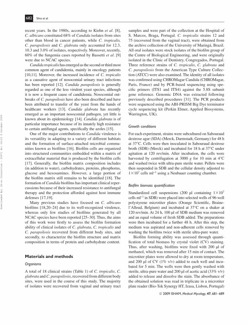
Table 1 Origin, reference and biofi lm matrix composition of non-Candida albicans Candida species. The values are means � standard deviations.
Matrix component (mg/g of biofi lm dry weight)
Species Origin Reference Protein Carbohydrate
C. parapsilosis Oral tract AD 35.9 � 7.2 748.8 � 43.8*
AM2 75.1 � 7.2* 926.8 � 144.9*
Urinary tract 534638 20.2 � 4.5* 263.7 � 13.2*
553877 46.8 � 16.6 592.6 � 93.4
Vaginal 491861 80.6 � 16.6* 555.2 � 238.5
513143 55.3 � 16.6 675.2 � 169.0
Reference ATCC 22019 42.2 � 10.3 516.4 � 219.1
C. tropicalis Oral tract AG1 46.3 � 3.5 22.2 � 5.8
T2.2 28.2 � 3.3* 21.5 � 4.0
Urinary tract 519468 34.2 � 9.3 15.7 � 1.9
544123 41.6 � 1.0 11.3 � 5.8
Vaginal 12 54.0 � 2.1* 58.7 � 7.4*
75 34.7 � 3.7 27.5 � 2.8
Reference ATCC 750 64.6 � 18.2* 15.5 � 2.8
C. glabrata Oral tract D1 325.2 � 31.4 572.8 � 111.2
AE2 226.7 � 84.1 241.8 � 52.2
Urinary tract 562123 181.7 � 28.7 409.5 � 112.4
513100 226.5 � 59.3 233.7 � 88.5
Vaginal 534784 136.4 � 38.5 398.3 � 130.8
585626 246.9 � 47.5 742.6 � 285.2*
Reference ATCC 2001 243.6 � 30.7 420.3 � 39.2
*Signifi cantly different (P � 0.05) for each species.
Downloaded By: [B-on Consortium - 2007] At: 14:10 9 October 2009
© 2009 ISHAM, Medical Mycology, 47, 681–689
684 Silva et al.
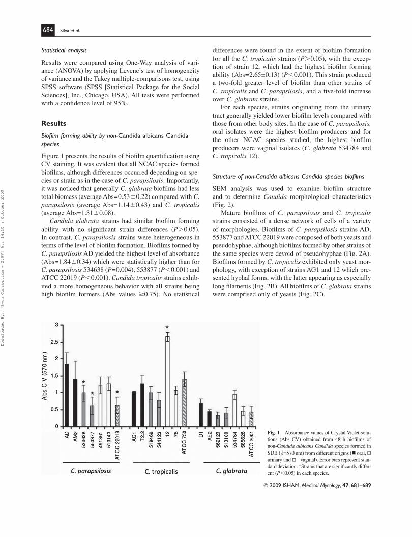
differences were found in the extent of biofi lm formation
for all the C. tropicalis strains ( P � 0.05), with the excep-
tion of strain 12, which had the highest biofi lm forming
ability (Abs = 2.65 0.13) ( P � 0.001). This strain produced
a two-fold greater level of biofi lm than other strains of
C. tropicalis and C. parapsilosis , and a fi ve-fold increase
over C. glabrata strains.
For each species, strains originating from the urinary
tract generally yielded lower biofi lm levels compared with
those from other body sites. In the case of C. parapsilosis, oral isolates were the highest biofi lm producers and for
the other NCAC species studied, the highest biofi lm
producers were vaginal isolates ( C. glabrata 534784 and
C. tropicalis 12).
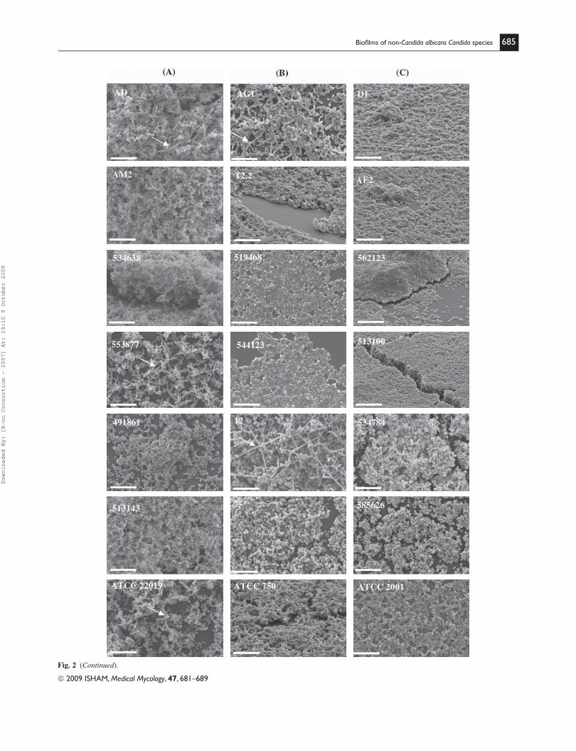
Structure of non-Candida albicans Candida species biofi lms
SEM analysis was used to examine biofi lm structure
and to determine Candida morphological characteristics
( Fig. 2 ).
Mature biofi lms of C. parapsilosis and C. tropicalis
strains consisted of a dense network of cells of a variety
of morphologies. Biofi lms of C. parapsilosis strains AD,
553877 and ATCC 22019 were composed of both yeasts and
pseudohyphae, although biofi lms formed by other strains of
the same species were devoid of pseudohyphae ( Fig. 2 A).
Biofi lms formed by C. tropicalis exhibited only yeast mor-
phology, with exception of strains AG1 and 12 which pre-
sented hyphal forms, with the latter appearing as especially
long fi laments ( Fig. 2 B). All biofi lms of C. glabrata strains
were comprised only of yeasts ( Fig. 2 C).
Statistical analysis
Results were compared using One-Way analysis of vari-
ance (ANOVA) by applying Levene’s test of homogeneity
of variance and the Tukey multiple-comparisons test, using
SPSS software (SPSS [Statistical Package for the Social
Sciences], Inc., Chicago, USA). All tests were performed
with a confi dence level of 95%.
Results
Biofi lm forming ability by non-Candida albicans Candida species
Figure 1 presents the results of biofi lm quantifi cation using
CV staining. It was evident that all NCAC species formed
biofi lms, although differences occurred depending on spe-
cies or strain as in the case of C. parapsilosis . Importantly,
it was noticed that generally C. glabrata biofi lms had less
total biomass (average Abs = 0.53 � 0.22) compared with C. parapsilosis (average Abs = 1.14 � 0.43) and C. tropicalis
(average Abs = 1.31 � 0.08).
Candida glabrata strains had similar biofi lm forming
ability with no signifi cant strain differences ( P � 0.05).
In contrast, C. parapsilosis strains were heterogeneous in
terms of the level of biofi lm formation. Biofi lms formed by
C. parapsilosis AD yielded the highest level of absorbance
(Abs = 1.84 � 0.34) which were statistically higher than for
C. parapsilosis 534638 ( P = 0.004), 553877 ( P � 0.001) and
ATCC 22019 ( P � 0.001). Candida tropicalis strains exhib-
ited a more homogeneous behavior with all strains being
high biofi lm formers (Abs values �0.75). No statistical
Fig. 1 Absorbance values of Crystal Violet solu-
tions (Abs CV) obtained from 48 h biofi lms of
non-Candida albicans Candida species formed in
SDB ( l=570 nm) from different origins ( oral,
urinary and vaginal). Error bars represent stan-
dard deviation. *Strains that are signifi cantly differ-
ent (P � 0.05) in each species.
Downloaded By: [B-on Consortium - 2007] At: 14:10 9 October 2009
© 2009 ISHAM, Medical Mycology, 47, 681–689
Biofi lms of non-Candida albicans Candida species 685
Fig. 2 (Continued).
Downloaded By: [B-on Consortium - 2007] At: 14:10 9 October 2009
© 2009 ISHAM, Medical Mycology, 47, 681–689
686 Silva et al.
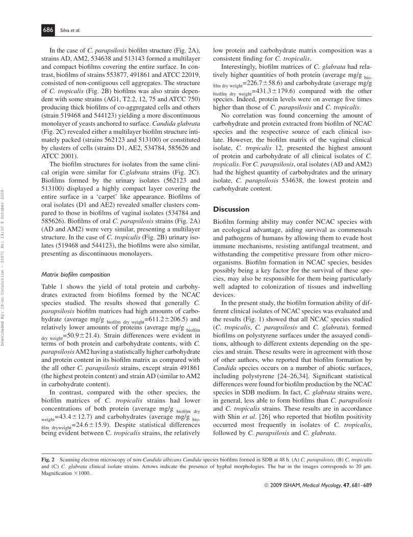
Fig. 2 Scanning electron microscopy of non-Candida albicans Candida species biofi lms formed in SDB at 48 h. (A) C. parapsilosis, (B) C. tropicalis
and (C) C. glabrata clinical isolate strains. Arrows indicate the presence of hyphal morphologies. The bar in the images corresponds to 20 μm.
Magnifi cation � 1000.
In the case of C. parapsilosis biofi lm structure ( Fig. 2 A),
strains AD, AM2, 534638 and 513143 formed a multilayer
and compact biofi lms covering the entire surface. In con-
trast, biofi lms of strains 553877, 491861 and ATCC 22019,
consisted of non-contiguous cell aggregates. The structure
of C. tropicalis ( Fig. 2 B) biofi lms was also strain depen-
dent with some strains (AG1, T2.2, 12, 75 and ATCC 750)
producing thick biofi lms of co-aggregated cells and others
(strain 519468 and 544123) yielding a more discontinuous
monolayer of yeasts anchored to surface. Candida glabrata
( Fig. 2 C) revealed either a multilayer biofi lm structure inti-
mately packed (strains 562123 and 513100) or constituted
by clusters of cells (strains D1, AE2, 534784, 585626 and
ATCC 2001).
The biofi lm structures for isolates from the same clini-
cal origin were similar for C. glabrata strains ( Fig. 2 C).
Biofi lms formed by the urinary isolates (562123 and
513100) displayed a highly compact layer covering the
entire surface in a ‘carpet’ like appearance. Biofi lms of
oral isolates (D1 and AE2) revealed smaller clusters com-
pared to those in biofi lms of vaginal isolates (534784 and
585626). Biofi lms of oral C. parapsilosis strains ( Fig. 2 A)
(AD and AM2) were very similar, presenting a multilayer
structure. In the case of C. tropicalis ( Fig. 2 B) urinary iso-
lates (519468 and 544123), the biofi lms were also similar,
presenting as discontinuous monolayers.
Matrix biofi lm composition
Table 1 shows the yield of total protein and carbohy-
drates extracted from biofi lms formed by the NCAC
species studied. The results showed that generally C. parapsilosis biofi lm matrices had high amounts of carbo-
hydrate (average mg/g biofi lm
dry
weight
= 611.2 � 206.5) and
relatively lower amounts of proteins (average mg/g biofi lm
dry weight
= 50.9 � 21.4). Strain differences were evident in
terms of both protein and carbohydrate contents, with C . parapsilosis AM2 having a statistically higher carbohydrate
and protein content in its biofi lm matrix as compared with
the all other C. parapsilosis strains, except strain 491861
(the highest protein content) and strain AD (similar to AM2
in carbohydrate content).
In contrast, compared with the other species, the
biofi lm matrices of C. tropicalis strains had lower
concentrations of both protein (average mg/g biofi lm
dry
weight = 43.4 � 12.7) and carbohydrates (average mg/g
bio-
fi lm dry
weight
= 24.6 � 15.9). Despite statistical differences
being evident between C . tropicalis strains, the relatively
low protein and carbohydrate matrix composition was a
consistent fi nding for C. tropicalis .
Interestingly, biofi lm matrices of C. glabrata had rela-
tively higher quantities of both protein (average mg/g bio-
fi lm dry
weight
= 226.7 � 58.6) and carbohydrate (average mg/g
biofi lm
dry
weight
= 431.3 � 179.6) compared with the other
species. Indeed, protein levels were on average fi ve times
higher than those of C. parapsilosis and C. tropicalis .
No correlation was found concerning the amount of
carbohydrate and protein extracted from biofi lm of NCAC
species and the respective source of each clinical iso-
late. However, the biofi lm matrix of the vaginal clinical
isolate, C. tropicalis 12, presented the highest amount
of protein and carbohydrate of all clinical isolates of C. tropicalis . For C. parapsilosis , oral isolates (AD and AM2)
had the highest quantity of carbohydrates and the urinary
isolate, C. parapsilosis 534638, the lowest protein and
carbohydrate content.
Discussion
Biofi lm forming ability may confer NCAC species with
an ecological advantage, aiding survival as commensals
and pathogens of humans by allowing them to evade host
immune mechanisms, resisting antifungal treatment, and
withstanding the competitive pressure from other micro-
organisms. Biofi lm formation in NCAC species, besides
possibly being a key factor for the survival of these spe-
cies, may also be responsible for them being particularly
well adapted to colonization of tissues and indwelling
devices.
In the present study, the biofi lm formation ability of dif-
ferent clinical isolates of NCAC species was evaluated and
the results ( Fig. 1 ) showed that all NCAC species studied
( C. tropicalis , C. parapsilosis and C. glabrata ) , formed
biofi lms on polystyrene surfaces under the assayed condi-
tions, although to different extents depending on the spe-
cies and strain. These results were in agreement with those
of other authors, who reported that biofi lm formation by
Candida species occurs on a number of abiotic surfaces,
including polystyrene [ 24 – 26 , 34 ]. Signifi cant statistical
differences were found for biofi lm production by the NCAC
species in SDB medium. In fact, C. glabrata strains were,
in general, less able to form biofi lms than C. parapsilosis
and C. tropicalis strains. These results are in accordance
with Shin et al . [ 26 ] who reported that biofi lm positivity
occurred most frequently in isolates of C. tropicalis ,
followed by C. parapsilosis and C. glabrata .
Downloaded By: [B-on Consortium - 2007] At: 14:10 9 October 2009
© 2009 ISHAM, Medical Mycology, 47, 681–689
Biofi lms of non-Candida albicans Candida species 687
a thick extracellular polymeric layer. In fact, almost all
microorganisms display structural heterogeneity within
their biofi lm architecture [ 38 ]. The present work indicates
that this heterogeneity appears to be common in biofi lms
formed by C. glabrata , C. tropicalis and C. parapsilosis
strains, revealing new important aspects on NCAC species
biofi lm ultrastructure.
One of the most important characteristics of both bacte-
rial and fungal biofi lms is the presence and composition
of the extracellular matrix [ 17 ,28 ]. There is a general con-
sensus that the biofi lm matrix acts as a barrier to diffusion
of antimicrobial agents, thereby limiting access of antimi-
crobials to organisms at the base of the biofi lm [ 39 ]. In
this study, biofi lm matrices were analyzed for carbohydrate
and protein content. Signifi cantly, consistent differences
were found in the composition of biofi lms of the NCAC
species. Matrices isolated from C. parapsilosis biofi lms
consisted of high amounts of carbohydrates and small
amounts of proteins, whilst C. tropicalis biofi lms were low
in both carbohydrate and protein content. These results are
in accordance with previous work [ 28 ] on C. tropicalis
biofi lm matrices which that they were mainly composed
of hexosamine, with smaller amounts of carbohydrate and
proteins. To the authors’ knowledge, this is the fi rst report
on the analysis of the biofi lm matrices of C. parapsilosis
and C. glabrata . Interestingly, the matrices recovered from
C. glabrata strains had higher amounts of both proteins
and carbohydrates. This is an interesting result, especially
when related to potential virulence of this species whose
infections yield both the highest mortality rate [ 40 ] and
resistance to antifungal agents [ 15 ].
The three different sources (body sites) for the clinical
isolates represent very diverse ecological niches and dif-
fer in many biotic and abiotic factors. Recent reports have
demonstrated that blood isolates produce greater quantities
of biofi lm compared with oral isolates [ 41 ]. In this current
study, no correlation was found in terms of biofi lm forming
ability and matrix composition with the origin of the iso-
late. However, biofi lm structure analysis did highlight some
interesting aspects. For C. glabrata , the biofi lm structure
for isolates from the same origin did appear to be similar.
This was also true for C. tropicalis urinary tract isolates.
It could readily be hypothesized that for certain body sites,
colonization requires a particular phenotype with respect
to biofi lm formation. Such a biofi lm phenotype might be
genetically rather than environmentally governed, thus
explaining why the biofi lm structural differences could be
detected in these in vitro studies. Through elucidating such
inherent differences, it might be possible to identify and
specifi cally combat strains adapted for infection at particu-
lar body sites. It must be emphasized, however, that further
investigations with isolates from specifi c environments are
required to confi rm this hypothesis.
It was noted that the biofi lm forming ability of
C. parapsilosis species was highly strain dependent, which
was less evident with both C. glabrata and C. tropicalis .
These observations corroborate previous reports for
C. albicans whose growth and virulence attributes, together
with biofi lm formation [ 23 ,25 ] have been shown to be
highly strain dependent. Such fi ndings undoubtedly
refl ect inherent physiological differences between strains
and could have signifi cance with respect to pathogenic
potential.
Despite the inherently destructive nature of SEM pro-
cessing, the method provided useful information on biofi lm
structure and on the different cellular morphologies. It is
known that biofi lm structure is dependent on environmen-
tal factors including growth conditions, nature of colonized
surface [ 22 ,25, 29 ] and importantly from the perspective
of this present study, the microbial species and strains
involved [ 22 ,25 , 29, 34, 35 ]. SEM did indeed reveal struc-
tural and morphological differences for the biofi lms of the
studied NCAC species and strains. Biofi lms of C. glabrata
( Fig. 2 C) presented as a multilayered structure with blasto-
conidia intimately packed, for some strains, and for others
as a biofi lm composed of cell clusters. As expected, there
was a total absence of pseudohyphae and hyphae since
C. glabrata is a non-hyphal species. Recently, Zaw et al . [ 36 ] also reported that after 48 h, the biofi lms of aerobi-
cally grown C. glabrata generally revealed a multilayer
structure packed with blastoconidia devoid of pseudohy-
phae and hyphae. In the presented study , C. parapsilosis
strains ( Fig. 2 A) yielded a multilayer biofi lm structure
that was comprised of a dense network of yeasts and
pseudohyphae. Although few studies on the biofi lm struc-
ture of C. parapsilosis strains have been reported, Kuhn
et al . [ 35 ] described that C. parapsilosis biofi lms con-
sisted of irregular groupings of blastoconidia on a basal
blastoconidia layer. Regarding C. tropicalis , its biofi lms
appeared as discontinuous layers of large blastoconidia
anchored to the surface, which was in accordance with
the fi ndings of Bizerra et al . [ 30 ]. The latter also reported
that C. tropicalis biofi lms formed in SDB medium, con-
tained only blastoconidia or generated a multilayer het-
erogeneous structure covering the entire surface as a thick
biofi lm. In the present study, large quantities of hyphal
elements were found in strain C. tropicalis 12 biofi lms
(vaginal clinical isolate). It has been suggested [ 21 ,37 ]
that the presence of such hyphae may have importance
in the structural integrity of multilayered biofi lms. The
present study reinforces and emphasizes a previous study
where one C. tropicalis strain formed a thin layer of hyphae
(in YNB) compared with other strains only presenting
blastoconidia [ 35 ]. Parahitiyawa et al . [ 34 ], reported that
on polystyrene surfaces, C. tropicalis biofi lms consisted
of large coaggregated microcolonies of blastoconidia with
Downloaded By: [B-on Consortium - 2007] At: 14:10 9 October 2009
© 2009 ISHAM, Medical Mycology, 47, 681–689
688 Silva et al.
Acknowledgements
The authors acknowledge ‘Fundação para a Ciência e
Tecnologia (FCT)’, Portugal, for supporting Sonia Silva
work through grant SFRH/BD/28341/2006 and project
PDTC/BIO/61112/2004.
Declaration of interest : The authors report no confl icts of
interest. The authors alone are responsible for the content
and writing of the paper.
References Fridkin SK, Jarvis WR. Epidemiology of nosocomial fungal infections. 1
Clin Microbiol Rev 1996; 9: 449– 511.
Odds FC. Candida and Candidosis, 2nd edn. London : Bailliere 2
Tindall, 1998 .
Samaranayake LP, Fidel PL, Naglik JR, 3 et al. Fungal infections
associated with HIV infection. Oral Dis 2002; 8: 151– 160 .
Fanello S, Bouchara JP, Jousset N, Delbos V, LeFlohic AM. Nosoco-4
mial Candida albicans acquisition in a geriatric unit: epidemiology
and evidence for person-to-person transmission. J Hosp Infect 2001;
47: 46– 52 .
Hargety JA, Ortiz J, Reich D, Manzarbeitia C. Fungal infections in 5
solid organ transplant patients. Surg Infect (Larchmt). 2003; 4: 263– 271 .
Kojic EM, Darouiche RO. 6 Candida infections of medical devices.
Clin Microbiol Rev 2004; 17: 255– 267 .
Kumamoto CA. 7 Candida biofi lms. Curr Opin Microbiol 2002; 5:
608– 611 .
Kiehn TE, Edwards FF, Armstrong D. The prevalence of yeasts in 8
clinical specimens from cancer patients. Amer J Clin Pathol 1980;
73: 518– 521 .
Bassetti M, Righi E, Costa A, 9 et al. Epidemiological trends in noso-
comial candidemia in intensive care. BMC Infect Dis 2006; 6: 21 .
Weinberger M, Leibovici L, Perez S, 10 et al. Characteristics of candi-
daemia with Candida albicans compared with non albicans Candida
species and predictors of mortality. J Hosp Infect 2005; 61: 146– 154 .
Nucci M, Colombo AL. Candidemia due to 11 Candida tropicalis : clini-
cal, epidemiologic and microbiologic characteristics of 188 episodes
occurring in tertiary care hospitals. Diagn Microbiol Infect Dis 2007;
58: 77– 82 .
Rho J, Shin JH, Song JW, 12 et al. Molecular investigation of two con-
secutive nosocomial clusters of Candida tropicalis candiduria using
pulsed-fi eld gel electrophoresis. J Microbiol 2004; 42: 80– 86 .
Bonassoli LA, Bertoli M, Svidzinski TIE. High frequency of 13 Candida parapsilosis on the hands of healthy hosts. J Hosp Infect 2005; 59:
159– 162 .
Hajjeh RA, Sofair AN, Harrison LH, 14 et al. Incidence of bloodstream
infections due to Candida species and in vitro susceptibilities of
isolates collected from 1998 to 2000 in a population-based active
surveillance program. J Clin Microbiol 2004; 42: 1519– 1527 .
Tsai H, Bobek LA. Studies of the mechanism of human salivary 15
histatin-5 candidacidal activity with histatin-5 variants and azole-
sensitive and -resistant Candida species. Antimicrob Agents Chemother 1997; 41: 2224– 2228 .
Costerton JW, Lewandowski Z, Caldwell DE, Korber DR, 16
Lappin-Scott HM. Microbial biofi lms. Annu Rev Microbial 1995; 49:
711– 745 .
Donlan RM, Costerton JW. Biofi lms: survival mechanisms of clinically 17
relevant microorganisms. Clin Microbiol Rev 2002; 15: 167– 193 .
Baillie GS, Douglas LJ. Matrix polymers of 18 Candida biofi lms and
their possible role in biofi lm resistance to antifungal agents. J Antimi-crob Chemother 2000; 46: 397– 403 .
Mukherjee PK, Chandra J. 19 Candida biofi lm resistance. Drug Resist Update 2004; 7: 301– 309 .
Hawser SP, Douglas LJ. Resistance of 20 Candida albicans biofi lms to
antifungal agents in vitro . Antimcrob Agents Chemother 1995; 39:
2128– 2131 .
Baillie GS, Douglas LJ. Role of dimorphism in the development of 21
Candida albicans biofi lm. J Med Microbiol 1999; 48: 671– 679 .
Chandra J, Kuhn DM, Mukherjee PK, 22 et al. Biofi lm formation by the
fungal pathogen Candida albicans : development, architecture and
drug resistance. J Bacteriol 2001; 183: 5385– 5394 .
Jin Y, Yip KH, Samaranayake YH, Yau JY, Samaranayake LP. 23
Biofi lm-forming ability of Candida albicans is unlikely to con-
tribute to high levels of oral yeast carriage in cases of human
immunodefi ciency virus infection. J Clin Microbiol 2003; 41:
2961– 2967 .
Ramage G, Martinez JP, Lopez-Ribot JL. 24 Candida biofi lms on im-
planted biomaterials: a clinically signifi cant problem. FEMS Yeast Res
2006; 6: 979– 986 .
Hawser SP, Douglas LJ. Biofi lm formation by 25 Candida species on
the surface of catheter materials in vitro . Infect Immun 1994; 62:
915– 921 .
Shin JH, Kee SJ, Shin MG, 26 et al. Biofi lm production by isolates of
Candida species recovered from nonneutropenic patients: comparison
of bloodstream isolates with isolates from other sources. J Clin Microbiol 2002; 40: 1244– 1248 .
Samaranayake YH, Ye J, Yau JYY, Cheung BPK, Samaranayake LP. 27
In vitro method to study antifungal perfusion in Candida biofi lms.
J Clin Microbiol 2005; 43: 818– 825 .
Al-Fattani MA, Douglas LJ. Biofi lm matrix of 28 Candida albicans and
Candida tropicalis : chemical composition and role in drug resistance.
J Med Microbiol 2006; 55: 999– 1008 .
Jain N, Kohli R, Cook E, 29 et al. Biofi lm formation by and antifungal
susceptibility of Candida isolates from urine. Appl Environ Microbiol 2007; 73: 1693– 1703 .
Bizerra CF, Nakamura CV, Poersch C, 30 et al. Characteristics of bio-
fi lm formation by Candida tropicalis and antifungal resistance. FEMS Yeast Res 2008; 8: 442– 450 .
Scherer S, Stevens DA. Application of DNA typing methods to epide-31
miology and taxonomy of Candida species . J Clin Microbiol 1987;
25: 675– 679 .
Azeredo J, Henriques M, Sillankorva S, Oliveira R. Extraction of 32
exopolymers from biofi lms: the protective effect of glutaraldehyde.
Water Sci Technol 2003; 47: 175– 179 .
Dubois M, Gilles KA, Hamilton JK, Rebers PA, Smith F. Colorimet-33
ric method for determination of sugars and related substances. Anal Chem 1956; 28: 350– 356 .
Parahitiyawa NB, Samaranayake YH, Samaranayake LP, 34 et al. Interspecies variation in Candida biofi lm formation studied using the
Calgary biofi lm device. APMIS 2006; 114: 298– 306 .
Kuhn DM, Chandra J, Mukherjee PK, Ghannoum MA. Comparison 35
of biofi lms formed by Candida albicans and Candida parapsilosis on
bioprosthetic surfaces. Infect Immun 2002; 70: 878– 888 .
Zaw MT, Samaranayake YH, Samaranayake LP. 36 In vitro biofi lm
formation of Candida albicans and non- albicans Candida species
under dynamic and anaerobic conditions. Arch Oral Biol 2007; 52:
761– 767 .
Ramage G, VandeWalle K, Bachmann SP, Wickes BL, Lopez-Ribot 37
JL. In vitro pharmacodynamic properties of three antifungal agents
against preformed Candida albicans biofi lms determined by time-kill
studies. Antimicrob Agents Chemother 2002; 46: 3634– 3636 .
Downloaded By: [B-on Consortium - 2007] At: 14:10 9 October 2009
© 2009 ISHAM, Medical Mycology, 47, 681–689
Biofi lms of non-Candida albicans Candida species 689
Wimpenny J, Manz W, Szewzyk U. Heterogeneity in biofi lms. 38 FEMS Microbiol Rev 2000; 24: 661– 671 .
Al-Fattani MA, Douglas LJ. Penetration of 39 Candida biofi lms by anti-
fungal agents. Antimicrob Agents Chemother. 2004; 48: 3291– 3297 .
Kcrmery K Jr. 40 Torulopsis glabrata an emerging pathogen in cancer
patients. Int J Antimicrob Agents 1999; 11: 1– 6 .
Kumar CPG, Menon T. Biofi lm production by clinical isolates of 41
Candida species. Med Mycol 2006; 44: 99– 101 .
This paper was fi rst published online on iFirst on 20 March 2009.
Downloaded By: [B-on Consortium - 2007] At: 14:10 9 October 2009
Copyright © 2022 FDOKUMEN