Rapamycin-coated expanded polytetrafluoroethylene bypass grafts exhibit decreased anastomotic...
9
From the Southern Association for Vascular Surgery Rapamycin-coated expanded polytetrafluoroethylene bypass grafts exhibit decreased anastomotic neointimal hyperplasia in a porcine model Catherine Cagiannos, MD, Omran R. Abul-Khoudoud, MD, Waldemar DeRijk, PhD, Daniel H. Shell IV, MD, Lisa K. Jennings, PhD, Elizabeth A. Tolley, PhD, Charles R. Handorf, MD, PhD, and Timothy C. Fabian, MD, Memphis, Tenn Objective: We tested the hypothesis that rapamycin coated onto, and eluted from, expanded polytetrafluoroethylene (ePTFE) grafts would diminish neointimal hyperplasia in a porcine model. Methods: Rapamycin (also called sirolimus) was coated onto the luminal surface of 6-mm-internal-diameter thin-walled ePTFE grafts by using an adhesive polymer that allows timed release of the drug. An adhesive polymer that allows timed release of rapamycin from ePTFE was developed with commercially available chemicals and applied on 6-mm ePTFE grafts. Graft integrity was characterized by scanning electron microscopy, and rapamycin levels were quantified by using high-performance liquid chromatography. Twenty-two mongrel pigs were randomized into three groups: untreated ePTFE (n 6), adhesive-only coated ePTFE (n 6), or adhesive- and rapamycin-coated ePTFE (n 10). End-to-side unilateral aortoiliac bypasses were performed by using 6-mm-internal-diameter ePTFE grafts and standardized anasto- motic lengths. Unilateral end-to-side aortoiliac ePTFE grafts (6-mm internal diameter) were inserted by using polypro- pylene sutures, 6-0 proximally and 7-0 distally; all anastomoses were 12 mm long. All animals received aspirin (325 mg orally) daily. All animals were given oral aspirin (325 mg) daily beginning on the day before surgery. At 28 days, the animals were killed, and the grafts were explanted in continuity with the adjacent aortic cuff and the outflow iliac artery. Variables compared between groups included graft patency, distal anastomotic length and cross-sectional narrowing, and intimal thickness at the arterial-graft junction indexed to the adjacent graft thickness. Microscopic analysis was performed with hematoxylin and eosin and Masson trichrome stains on paraffin sections. A pathologist blinded to experimental groups graded sections for collagen deposition, neointima formation, inflammatory cellular infiltrates, medial necrosis, and aneurysmal degeneration. Results: All animals survived until they were killed without clinical evidence of limb ischemia or graft infection. Preplanned t tests in the context of one-way analysis of variance showed no difference in outcome measures between the untreated ePTFE and adhesive-only coated ePTFE groups; therefore, they were combined in further comparisons with the adhesive- and rapamycin-coated ePTFE group. The Rapamycine eluting expanded polytetrafluoroethylene group had longer anastomoses (85.6% vs 60.6% of the initial anastomotic length maintained; P < .0001) and less cross-sectional narrowing in the outflow graft (16.2% vs 28.5%; P .0007) when compared with the other two groups by using two-tailed Student t tests. There was no evidence of medial necrosis or aneurysmal degeneration. All patent grafts had complete endothelialization on hematoxylin and eosin sections. Rapamycin was detectable and quantifiable in the arterial wall at 28 days after implantation. Conclusions: Rapamycin can be coated onto and eluted from ePTFE by using a nonionic polymer and a simple coating technique. At 4 weeks after implantation, the rapamycin-eluting ePTFE grafts demonstrate gross, pathologic, and morphometric features of diminished neointimal hyperplasia when compared with non– drug-eluting ePTFE. Four weeks after implantation in a porcine model, rapamycin-eluting ePTFE grafts demonstrated gross, pathologic, and morpho- metric features of diminished neointimal hyperplasia when compared with untreated and adhesive-only coated ePTFE grafts. ( J Vasc Surg 2005;42:980-8.) Clinical Relevance: Rapamycin-eluting ePTFE grafts decrease neointimal hyperplasia in a porcine model. Further studies are needed to evaluate whether patency will be improved. Rapamycin-eluting ePTFE grafts may allow the use of prosthetic grafts in situations in which autologous vein is unavailable and in which neointimal hyperplasia is pronounced, such as in small-diameter (<6-mm) vessels typical of infrapopliteal interventions. Although autologous saphenous vein is the best con- duit for peripheral arterial reconstruction, 30% of patients do not have this option as a result of prior vein harvest, trauma, or phlebitis. 1 Synthetic grafts made of expanded polytetrafluoroethylene (ePTFE) have been used as substi- tutes but have low patencies in vessels with diameters less than 6 mm because of early thrombosis or late graft failure from neointimal hyperplasia. 2 Infrapopliteal ePTFE grafts have primary patency rates at 4 years as low as 12%. 3 From the Department of Surgery, University of Tennessee Health Sciences Center. Competition of interest: none. Reprint requests: Timothy C. Fabian, MD, Department of Surgery, Univer- sity of Tennessee Health Sciences Center, 956 Court Ave, Suite G228, Memphis, TN 38163 (e-mail: [email protected]). 0741-5214/$30.00 Copyright © 2005 by The Society for Vascular Surgery. doi:10.1016/j.jvs.2005.06.018 980
-
Upload
independent -
Category
Documents
-
view
1 -
download
0
Transcript of Rapamycin-coated expanded polytetrafluoroethylene bypass grafts exhibit decreased anastomotic...

From the Southern Association for Vascular Surgery
Rapamycin-coated expandedpolytetrafluoroethylene bypass grafts exhibitdecreased anastomotic neointimal hyperplasiain a porcine modelCatherine Cagiannos, MD, Omran R. Abul-Khoudoud, MD, Waldemar DeRijk, PhD,Daniel H. Shell IV, MD, Lisa K. Jennings, PhD, Elizabeth A. Tolley, PhD,Charles R. Handorf, MD, PhD, and Timothy C. Fabian, MD, Memphis, Tenn
Objective: We tested the hypothesis that rapamycin coated onto, and eluted from, expanded polytetrafluoroethylene(ePTFE) grafts would diminish neointimal hyperplasia in a porcine model.Methods: Rapamycin (also called sirolimus) was coated onto the luminal surface of 6-mm-internal-diameter thin-walledePTFE grafts by using an adhesive polymer that allows timed release of the drug. An adhesive polymer that allows timedrelease of rapamycin from ePTFE was developed with commercially available chemicals and applied on 6-mm ePTFEgrafts. Graft integrity was characterized by scanning electron microscopy, and rapamycin levels were quantified by usinghigh-performance liquid chromatography. Twenty-two mongrel pigs were randomized into three groups: untreatedePTFE (n � 6), adhesive-only coated ePTFE (n � 6), or adhesive- and rapamycin-coated ePTFE (n � 10). End-to-sideunilateral aortoiliac bypasses were performed by using 6-mm-internal-diameter ePTFE grafts and standardized anasto-motic lengths. Unilateral end-to-side aortoiliac ePTFE grafts (6-mm internal diameter) were inserted by using polypro-pylene sutures, 6-0 proximally and 7-0 distally; all anastomoses were 12 mm long. All animals received aspirin (325 mgorally) daily. All animals were given oral aspirin (325 mg) daily beginning on the day before surgery. At 28 days, theanimals were killed, and the grafts were explanted in continuity with the adjacent aortic cuff and the outflow iliac artery.Variables compared between groups included graft patency, distal anastomotic length and cross-sectional narrowing, andintimal thickness at the arterial-graft junction indexed to the adjacent graft thickness. Microscopic analysis was performedwith hematoxylin and eosin and Masson trichrome stains on paraffin sections. A pathologist blinded to experimentalgroups graded sections for collagen deposition, neointima formation, inflammatory cellular infiltrates, medial necrosis,and aneurysmal degeneration.Results: All animals survived until they were killed without clinical evidence of limb ischemia or graft infection.Preplanned t tests in the context of one-way analysis of variance showed no difference in outcome measures between theuntreated ePTFE and adhesive-only coated ePTFE groups; therefore, they were combined in further comparisons withthe adhesive- and rapamycin-coated ePTFE group. The Rapamycine eluting expanded polytetrafluoroethylene group hadlonger anastomoses (85.6% vs 60.6% of the initial anastomotic length maintained; P < .0001) and less cross-sectionalnarrowing in the outflow graft (16.2% vs 28.5%; P � .0007) when compared with the other two groups by usingtwo-tailed Student t tests. There was no evidence of medial necrosis or aneurysmal degeneration. All patent grafts hadcomplete endothelialization on hematoxylin and eosin sections. Rapamycin was detectable and quantifiable in the arterialwall at 28 days after implantation.Conclusions: Rapamycin can be coated onto and eluted from ePTFE by using a nonionic polymer and a simple coatingtechnique. At 4 weeks after implantation, the rapamycin-eluting ePTFE grafts demonstrate gross, pathologic, andmorphometric features of diminished neointimal hyperplasia when compared with non–drug-eluting ePTFE. Four weeksafter implantation in a porcine model, rapamycin-eluting ePTFE grafts demonstrated gross, pathologic, and morpho-metric features of diminished neointimal hyperplasia when compared with untreated and adhesive-only coated ePTFEgrafts. ( J Vasc Surg 2005;42:980-8.)
Clinical Relevance: Rapamycin-eluting ePTFE grafts decrease neointimal hyperplasia in a porcine model. Further studiesare needed to evaluate whether patency will be improved. Rapamycin-eluting ePTFE grafts may allow the use ofprosthetic grafts in situations in which autologous vein is unavailable and in which neointimal hyperplasia is pronounced,
such as in small-diameter (<6-mm) vessels typical of infrapopliteal interventions.From the Department of Surgery, University of Tennessee Health SciencesCenter.
Competition of interest: none.Reprint requests: Timothy C. Fabian, MD, Department of Surgery, Univer-
sity of Tennessee Health Sciences Center, 956 Court Ave, Suite G228,Memphis, TN 38163 (e-mail: [email protected]).
0741-5214/$30.00Copyright © 2005 by The Society for Vascular Surgery.
doi:10.1016/j.jvs.2005.06.018980
Although autologous saphenous vein is the best con-duit for peripheral arterial reconstruction, 30% of patientsdo not have this option as a result of prior vein harvest,trauma, or phlebitis.1 Synthetic grafts made of expandedpolytetrafluoroethylene (ePTFE) have been used as substi-tutes but have low patencies in vessels with diameters lessthan 6 mm because of early thrombosis or late graft failurefrom neointimal hyperplasia.2 Infrapopliteal ePTFE grafts
have primary patency rates at 4 years as low as 12%.3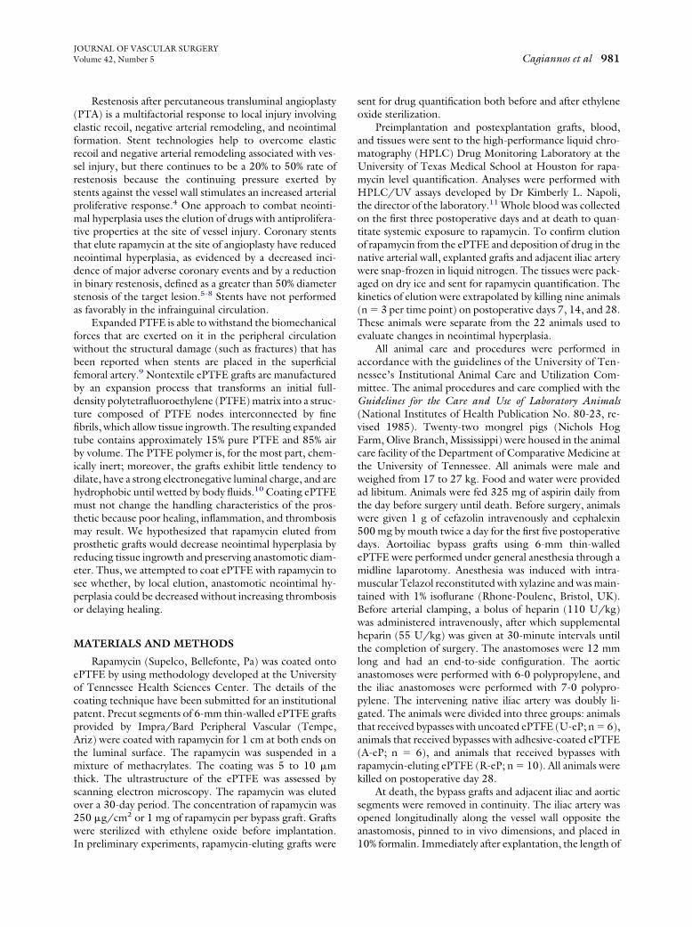
JOURNAL OF VASCULAR SURGERYVolume 42, Number 5 Cagiannos et al 981
Restenosis after percutaneous transluminal angioplasty(PTA) is a multifactorial response to local injury involvingelastic recoil, negative arterial remodeling, and neointimalformation. Stent technologies help to overcome elasticrecoil and negative arterial remodeling associated with ves-sel injury, but there continues to be a 20% to 50% rate ofrestenosis because the continuing pressure exerted bystents against the vessel wall stimulates an increased arterialproliferative response.4 One approach to combat neointi-mal hyperplasia uses the elution of drugs with antiprolifera-tive properties at the site of vessel injury. Coronary stentsthat elute rapamycin at the site of angioplasty have reducedneointimal hyperplasia, as evidenced by a decreased inci-dence of major adverse coronary events and by a reductionin binary restenosis, defined as a greater than 50% diameterstenosis of the target lesion.5-8 Stents have not performedas favorably in the infrainguinal circulation.
Expanded PTFE is able to withstand the biomechanicalforces that are exerted on it in the peripheral circulationwithout the structural damage (such as fractures) that hasbeen reported when stents are placed in the superficialfemoral artery.9 Nontextile ePTFE grafts are manufacturedby an expansion process that transforms an initial full-density polytetrafluoroethylene (PTFE) matrix into a struc-ture composed of PTFE nodes interconnected by finefibrils, which allow tissue ingrowth. The resulting expandedtube contains approximately 15% pure PTFE and 85% airby volume. The PTFE polymer is, for the most part, chem-ically inert; moreover, the grafts exhibit little tendency todilate, have a strong electronegative luminal charge, and arehydrophobic until wetted by body fluids.10 Coating ePTFEmust not change the handling characteristics of the pros-thetic because poor healing, inflammation, and thrombosismay result. We hypothesized that rapamycin eluted fromprosthetic grafts would decrease neointimal hyperplasia byreducing tissue ingrowth and preserving anastomotic diam-eter. Thus, we attempted to coat ePTFE with rapamycin tosee whether, by local elution, anastomotic neointimal hy-perplasia could be decreased without increasing thrombosisor delaying healing.
MATERIALS AND METHODS
Rapamycin (Supelco, Bellefonte, Pa) was coated ontoePTFE by using methodology developed at the Universityof Tennessee Health Sciences Center. The details of thecoating technique have been submitted for an institutionalpatent. Precut segments of 6-mm thin-walled ePTFE graftsprovided by Impra/Bard Peripheral Vascular (Tempe,Ariz) were coated with rapamycin for 1 cm at both ends onthe luminal surface. The rapamycin was suspended in amixture of methacrylates. The coating was 5 to 10 �mthick. The ultrastructure of the ePTFE was assessed byscanning electron microscopy. The rapamycin was elutedover a 30-day period. The concentration of rapamycin was250 �g/cm2 or 1 mg of rapamycin per bypass graft. Graftswere sterilized with ethylene oxide before implantation.
In preliminary experiments, rapamycin-eluting grafts weresent for drug quantification both before and after ethyleneoxide sterilization.
Preimplantation and postexplantation grafts, blood,and tissues were sent to the high-performance liquid chro-matography (HPLC) Drug Monitoring Laboratory at theUniversity of Texas Medical School at Houston for rapa-mycin level quantification. Analyses were performed withHPLC/UV assays developed by Dr Kimberly L. Napoli,the director of the laboratory.11 Whole blood was collectedon the first three postoperative days and at death to quan-titate systemic exposure to rapamycin. To confirm elutionof rapamycin from the ePTFE and deposition of drug in thenative arterial wall, explanted grafts and adjacent iliac arterywere snap-frozen in liquid nitrogen. The tissues were pack-aged on dry ice and sent for rapamycin quantification. Thekinetics of elution were extrapolated by killing nine animals(n � 3 per time point) on postoperative days 7, 14, and 28.These animals were separate from the 22 animals used toevaluate changes in neointimal hyperplasia.
All animal care and procedures were performed inaccordance with the guidelines of the University of Ten-nessee’s Institutional Animal Care and Utilization Com-mittee. The animal procedures and care complied with theGuidelines for the Care and Use of Laboratory Animals(National Institutes of Health Publication No. 80-23, re-vised 1985). Twenty-two mongrel pigs (Nichols HogFarm, Olive Branch, Mississippi) were housed in the animalcare facility of the Department of Comparative Medicine atthe University of Tennessee. All animals were male andweighed from 17 to 27 kg. Food and water were providedad libitum. Animals were fed 325 mg of aspirin daily fromthe day before surgery until death. Before surgery, animalswere given 1 g of cefazolin intravenously and cephalexin500 mg by mouth twice a day for the first five postoperativedays. Aortoiliac bypass grafts using 6-mm thin-walledePTFE were performed under general anesthesia through amidline laparotomy. Anesthesia was induced with intra-muscular Telazol reconstituted with xylazine and was main-tained with 1% isoflurane (Rhone-Poulenc, Bristol, UK).Before arterial clamping, a bolus of heparin (110 U/kg)was administered intravenously, after which supplementalheparin (55 U/kg) was given at 30-minute intervals untilthe completion of surgery. The anastomoses were 12 mmlong and had an end-to-side configuration. The aorticanastomoses were performed with 6-0 polypropylene, andthe iliac anastomoses were performed with 7-0 polypro-pylene. The intervening native iliac artery was doubly li-gated. The animals were divided into three groups: animalsthat received bypasses with uncoated ePTFE (U-eP; n � 6),animals that received bypasses with adhesive-coated ePTFE(A-eP; n � 6), and animals that received bypasses withrapamycin-eluting ePTFE (R-eP; n � 10). All animals werekilled on postoperative day 28.
At death, the bypass grafts and adjacent iliac and aorticsegments were removed in continuity. The iliac artery wasopened longitudinally along the vessel wall opposite theanastomosis, pinned to in vivo dimensions, and placed in
10% formalin. Immediately after explantation, the length of
JOURNAL OF VASCULAR SURGERYNovember 2005982 Cagiannos et al
the distal anastomosis was measured (heel to toe), and thepercentage of maintained anastomotic length was calcu-lated (explant anastomotic length/12). Two surgeons notblinded to experimental groups measured the explantedanastomotic length and averaged their values for each ani-mal. Specimens were not perfusion-fixed because sampleswere thin, usually 3 to 5 mm thick, which allowed for rapidformalin fixation. All specimens were collected and pro-cessed in a similar fashion. Absolute measurements werenot evaluated. Variables examined relative proportions orratios between groups. After a minimum of 24 hours ofsoaking in 10% formalin (Baxter Diagnostics, McGaw Park,Ill), the iliac segments were cross-sectioned at the heel andplaced in cassettes. Paraffin processing was performed bypathology technicians from the Department of Pathologyat the University of Tennessee Health Sciences Center.After paraffin embedding, two to three (5 �m each) sec-tions were stained with hematoxylin and eosin (H&E) orMasson trichrome and used for morphometric analysis orpathologic grading.
Morphometric comparisons were made by using ImageJ (version 1.30). This software was downloaded from theNational Institute of Health’s Web site (http://rsb.info.nih.gov/ij/). Measurements were made from cross sec-tions taken at the heel of the iliac anastomoses. Tissueblocks were generated from here because the hyperplasticresponse was most pronounced to the naked eye, and thesectioning resulted in a circular cross section of the ePTFEthat allowed for consistency during pathologic grading andmorphometric analysis. H&E-stained or Masson trichrome–stained paraffin sections were mounted on slides andviewed with the �2 objective on a Zeiss microscope (CarlZeiss Inc, Oberkochen, Germany). Fields were photo-graphed with a Camedia D-540 ZOOM digital camera(Olympus, Melville, New York). The digital images wereanalyzed with Image J to determine differences in morpho-metric criteria of neointimal hyperplasia. The morphometryanalysis was performed by a researcher blinded to experi-mental groups.
The percentage of cross-sectional narrowing attributedto neointimal hyperplasia and the degree of neointimalthickness at the arterial-graft junction indexed to ePTFEgraft thickness were compared between groups of animals.The percentage of cross-sectional narrowing was calculatedby dividing the neointimal area by the area bound by theinner table of the cross-sectioned ePTFE. The neointimalarea was calculated by subtracting the luminal area from thearea bound by the inner table of the cross-sectionedePTFE:
% Cross-sectional narrowing � internal ePTFE area� luminal area ⁄ internal ePTFE area
The intimal thickness index was calculated by dividingthe thickness of neointima at the heel of the iliac anasto-mosis by the cross-sectional thickness of the ePTFE graft.The measurement was performed where the internal elasticlamina of the native artery was disrupted by the polypro-
pylene suture used to perform the anastomosis. Measure-ments for percentage of cross-sectional narrowing and in-timal thickness index are presented as mean � SEM.
Semiquantitative histologic grading of H&E and Mas-son trichrome sections was performed by a pathologistblinded to experimental groups (C.R.H.). Features exam-ined included endothelialization, spindle cell ingrowth, andneointimal formation. Grading was performed as follows:0, none of the luminal circumference of the graft involved;1, less than 25% of the luminal circumference of the graftinvolved; 2, 25% to 75% of the luminal circumference of thegraft involved; and 3, greater than 75% of the luminalcircumference of the graft involved. Immunostains for�-actin and factor VIII–related antigen to identify smoothmuscle cells and endothelial cells were not performed. Thepathologist examined sections with the �40 power objec-tive and identified endothelial cells on the basis of surfacelocation and flattened cellular morphology. Spindle cellswere characterized with Masson trichrome as elongatedcells with purple cytoplasm that populated regions of extra-cellular matrix (ECM). Ingrowth of spindle cells was usedto correlate with vascular smooth muscle cell (VSMC)migration. The presence of spindle cells and ECM, whichstains blue or pink with Masson trichrome, depending oncollagen content, was used to designate areas of neointimalformation on the ePTFE inner surface.
Statistical analysis was performed with SAS 9.0 (SASInstitute Inc, Cary, NC) statistical software. First, pre-planned contrasts in the context of one-way analysis ofvariance (that are equivalent to two-tailed t tests with thesquare root of the mean square error used as the pooledstandard deviation) were used to demonstrate that therewas no statistical difference between the U-eP and the A-ePgroups. Next, the data were combined into one controlgroup (n � 12). Finally, data from the R-eP group (n � 10)were then compared with data from the combination of theU-eP and A-eP groups (n � 12; Table I) by using two-tailed unpaired t tests with equal variances. Differenceswere considered significant at P � .05. Variables are pre-sented as mean � SEM.
Reproducibility of measurements was assessed for the
Table I. Comparison of gross pathologic andmorphometric parameters (postoperative day 28) ofneointimal hyperplasia between animals treated withuncoated (U-eP) and adhesive-coated expandedpolytetrafluoroethylene (A-eP) and with rapamycin-eluting expanded polytetrafluoroethylene (R-eP)
VariableU-eP and
A-eP (n � 12)R-eP
(n � 10) P value
% Initial anastomoticlength 60.6% � 2.3% 85.6% � 2.5% �.0001
% Cross-sectionalnarrowing 28.5% � 2.7% 16.2% � 3.0% .007
Intimal thicknessindex 1.75 � 0.13 1.22 � 0.14 .01
percentage of cross-sectional narrowing, ePTFE graft thick-

(orig
JOURNAL OF VASCULAR SURGERYVolume 42, Number 5 Cagiannos et al 983
ness, and ePTFE internal graft area. The pooled within-animalstandard deviation for percentage of cross-sectional narrow-ing was 3.5%; the intraclass correlation coefficient was 0.84,indicating excellent reproducibility. For ePTFE graft thick-ness and area, the pooled within-animal standard deviationswere 0.08 mm and 1.40 mm2, respectively. For ePTFEgraft thickness, the reproducibility was poor; however, thereproducibility of the ePTFE internal graft area was mod-erate, with an intraclass correlation coefficient of 0.71.
RESULTS
Examining sections of coated grafts with scanning elec-tron microscopy, we found that the coating was adherent tothe nodal islands of the ePTFE and did not obliterate theinternodal fibrils (Fig 1). The scanning electron microscopystudies showed that rapamycin was encapsulated in thematrix of polymeric adhesives. Rapamycin appeared asspeckling on nodal islands. The scanning electron micros-copy studies did not show any other differences between
Fig 1. Scanning electron assessment of grafts. A, Uncoamagnification, �1000). B, Adhesive-coated ePTFE (AePTFE (R-eP; original magnification, �1000). D, R-eP
rapamycin-coated grafts and those coated with adhesive
alone. Handling characteristics of the graft material weremaintained during suturing and implantation.
All animals survived until they were killed, with palpa-ble pulses in the operated hindlimb and patent grafts. Noneof the animals developed wound or graft infections. Weightgain was similar. Aneurysmal degeneration was absent, andall grafts were well incorporated.
We compared animals that underwent bypass withuncoated ePTFE grafts with those that underwent by-pass with adhesive-coated ePTFE to see whether thevehicle for the rapamycin was responsible for any changesin the degree of neointimal hyperplasia. There were nodifferences in the length of the iliac anastomosis, thepercentage of cross-sectional narrowing, or the intimalthickness index between the two groups at explantation(Table II). The coating seemed inert and was not asso-ciated with a propensity for inflammation or thrombosis.The data for the two groups were then combined to formone control group (U-eP and A-eP) for comparison with
xpanded polytetrafluoroethylene (ePTFE; U-eP; originalriginal magnification, �1000). C, Rapamycin-eluting
inal magnification, �2500).
ted e-eP; o
the R-eP animals.

JOURNAL OF VASCULAR SURGERYNovember 2005984 Cagiannos et al
At death, animals treated with rapamycin-eluting ePTFEgrafts had longer explanted iliac anastomoses (R-eP, 10.3 �0.26 mm; U-eP and A-eP, 7.3 � 0.15 mm; P � .0001). Inaddition to having longer anastomoses (85.6% vs 60.6% ofinitial anastomotic length maintained; P � .0001), theR-eP animals had less cross-sectional narrowing in theoutflow graft (16.2% vs 28.5%; P � .0007) and decreasedintimal thickness indexed to ePTFE (Table I).
Complete endothelial coverage of ePTFE was noted inall groups. R-eP animals had less spindle cell ingrowth andneointimal formation. They received lower scores thanU-eP and A-eP animals (Table III). Medial necrosis andaneurysmal degeneration were absent, and there were nodifferences in cellular infiltration between the experimentalgroups.
Preliminary experiments with waterbaths showed thatthe adhesive coating was subject to aqueous attack withdissolution over a 30-day period. Scanning electron micros-copy samples of explanted anastomoses showed coverage ofthe internal surface of the ePTFE with cells and biologicdebris. The adhesive coating could not be visualized, andcomparisons of adhesive integrity and thickness betweenA-eP and R-eP samples could not be made. It was assumedthat the neointima formed on ePTFE as dissolution of theadhesive polymeric matrix was occurring.
The ethylene oxide sterilization process did not causeloss of rapamycin before implantation. Similar drug levelswere detected despite sterilization. Similarly, the rapamycinfraction was detected 18 minutes after instillation of the
Table II. Comparison of gross pathologic andmorphometric parameters (postoperative day 28) ofneointimal hyperplasia between animals treated withuncoated (U-eP) and adhesive-coated expandedpolytetrafluoroethylene (A-eP)
VariableU-eP
(n � 6)A-eP
(n � 6) P value
% Initial anastomoticlength 57.3% � 3.2% 63.8% � 3.2% .17
% Cross-sectionalnarrowing 27.4% � 4.0% 29.7% � 4.0% .68
Intimal thicknessindex 1.71 � 0.20 1.79 � 0.18 .78
Table III. Pathologic grading of anastomotic crosssections for neointimal hyperplasia
VariableU-eP and
A-eP (n � 12)R-eP
(n � 10) P value
Neointimal formation 2.5 � 0.2 0.8 � 0.2 �.0001Spindle cell ingrowth 2.3 � 0.3 0.8 � 0.2 .0007Endothelialization 3.0 � 0.0 3.0 � 0.0 1
U-eP, Uncoated expanded polytetrafluoroethylene; A-eP, adhesive-coatedexpanded polytetrafluoroethylene; R-eP, rapamycin-eluting expanded poly-tetrafluoroethylene; p � NS, numerical score.
sample onto the HPLC column, irrespective of steriliza-
tion. Table IV shows rapamycin levels obtained from grafts,explanted arteries, and blood sampled at various timepoints. Preliminary HPLC experiments showed that thecoating process resulted in loading of 1 mg of rapamycinper ePTFE graft before implantation. Results from 9 ani-mals that were killed temporally to help determine elutionkinetics showed that most rapamycin eluted off the graftsby 1 week. Levels decreased from 1000 �g before implan-tation to 26.7 �g at 1 week after explantation. Drug (2.9�g/g) was detected in the adjacent arterial wall on postop-erative day 7 and persisted until death. Despite beingpresent in the arterial wall on postoperative day 28, rapa-mycin did not reach levels associated with systemic toxicity.Rapamycin was not detectable in blood after postoperativeday 3.
DISCUSSION
Intimal hyperplasia is initiated by endothelial damage.Neointimal hyperplasia represents the response of VSMCsto physical, chemical, and humoral factors in regions ofdysfunctional endothelial regulation. VSMCs are inducedto migrate from the media to the intima, where theyproliferate and deposit ECM.12 Research on the develop-ment of neointimal hyperplasia has focused on the preven-tion of arterial restenosis after PTA and implantation ofvascular grafts. The endothelium is disrupted at vascularanastomoses and at sites of PTA. Use of stents can preventrecoil and remodeling in treated arteries but does noteliminate neointimal hyperplasia. Struts from implantedstents incite an inflammatory response in the adjacent ar-tery. This response perpetuates restenosis by initiating cy-tokine release from infiltrating cells. Vascular grafting withePTFE also elicits neointimal hyperplasia through alter-ations in wall shear, flow, and compliance mismatch be-tween the native artery and the prosthetic.12-14
Pharmacologic manipulation of VSMC migration andproliferation and ECM production is one approach in thetreatment of restenosis after vascular intervention. Rapamy-cin is a macrocyclic, lipophilic lactone with immunosup-pressive antibiotic activity derived from the actinomyceteStreptomyces hygroscopicus. Rapamycin is approved by theUS Food and Drug Administration for the prophylaxis ofrenal transplant rejection. Rapamycin has many propertiesthat make it a good agent to counteract neointimal hyper-plasia. Rapamycin binds to its cytosolic receptor FK506binding protein (FKBP-12) and inhibits the mammaliantarget of rapamycin. The mammalian target of rapamycin isa ubiquitous signal transduction kinase that is responsiblefor cell-cycle progression. Mammalian target of rapamycininactivation results in a reduction of cyclin-dependent ki-nases and increased levels of p27kip1, a cyclin-dependentkinase inhibitor. The net effect is to cause G1-S arrest inproliferating cells such as T cells and VSMCs.15,16 Inaddition to inhibiting cellular proliferation, rapamycin inhibitsthe migration of VSMCs into areas of vascular injury.17 Inpigs, rapamycin needs to be present in the vessel wall for 14days after injury to be effective.5,18 The rate of neointimal
proliferation in stented porcine coronary arteries is greatest
JOURNAL OF VASCULAR SURGERYVolume 42, Number 5 Cagiannos et al 985
at 14 days after injury. The neointima at this point begins tobecome populated by VSMCs in a proteoglycan-rich ma-trix. To be effective, rapamycin needs to be present duringthe time when the stimulus for VSMC migration andproliferation exists. Hydrophobic drugs such as rapamycinmay achieve higher mean tissue concentrations in the in-tima because they are less likely to diffuse back into thecirculation, thus facilitating longer exposure in the area ofinjury.19,20
Drug elution has been used with excellent results incoronary interventions. Paclitaxel- and rapamycin-elutingstents significantly reduce the incidence of restenosis andlate loss of arterial luminal diameter. Major adverse cardiacevents such as myocardial infarction, death, and targetlesion/vessel revascularization are also decreased with drug-eluting stents.21,22 Patients treated with bare metal stentsrequire more frequent coronary interventions. There is nodifference in mortality or incidence of acute myocardialinfarction, but studies to date have not included data todetect changes in these end points.22 Review of the litera-ture indicates that drug-eluting stents reduce event rates by40% to 60% at 12 months.22
Drug-eluting stents have not performed as well in theinfrainguinal circulation. Stenoses and occlusions are morecommon in the femoropopliteal region than in the coro-nary arteries. In addition, lesions here tend to be multiple,long, heavily calcified, and endophytic. Approximately 90%of the time, peripheral arteries can undergo successfulangioplasty, but recurrence is common: restenosis occurs inup to 80% of cases after 1 year. Stenting femoropoplitealvessels after balloon angioplasty has not substantially im-proved patency. Nitinol stents may improve these resultsbecause they are less prone to external compression andelicit less neointimal hyperplasia than more rigid balloon-expandable stents.23-26 Sirolimus-eluting (rapamycin-elut-ing), nitinol-expandable SMART (Cordis/Johnson &Johnson, Warren, New Jersey) stents in the peripheralcirculation have been evaluated in two trials. These trialshave the acronym SIROCCO, which stands for sirolimus-coated Cordis SMART nitinol self-expandable stent for thetreatment of obstructive superficial femoral artery disease.SIROCCO I had promising early results, with 0% restenosisin the drug-eluting arm at 6 months; however, stent frac-tures were reported in 6 of 33 patients (3 in each treatmentgroup).27 The 18-month results were mixed, with theslow-eluting rapamycin stent having 0% restenosis but thefast-eluting stent having 33% restenosis. By 24 months,both slow- and fast-eluting coated stents failed to show a
Table IV. Quantification of rapamycin drug levels in graft
Variable Day 0 Day 1 Day
Graft (�g) (n � 3) 1000 N/A N/Artery (�g/g) (n � 3) 0 N/A N/Blood (ng/mL) (n � 3) 16.9 � 1.8 9.3 � 0.2 2.0 �
N/A, Not applicable.
difference from uncoated stents and had a binary restenosis
rate of 40%.28 The SIROCCO II trial 18-month data alsofailed to show superiority, with a total in-stent binaryrestenosis rate of 20.7% for the rapamycin-eluting stent and17.9% for the uncoated stent arm. Stents in the peripheralarteries of the lower extremities treat longer, more calcifiedlesions in arteries with relatively low flow rates. Stents in theperiphery experience increased biomechanical forces in-cluding elongation, rotation, and radial compression as aresult of the anatomy of the femoropopliteal vasculature.The attendant stent deformation may result in stent frac-tures and neointimal proliferation.9 The greater propensityfor neointimal hyperplasia may require higher levels of drugthan can be eluted locally from coated stents. Also, the useof self-expanding stents presents new challenges for drugloading and delivery that do not pertain to coronary stenttechnology and that may limit the dose of available drug.Some might question the impetus to pursue improvementin peripheral circulation stenting in view of the relativesuccess that has been achieved by open surgery. The 70% to80% 5-year patency achieved with bypass surgery (vein andprosthetic) may be hard to surpass.29
Although metal alloy technology is optimized to de-crease the propensity for stent deformation and fracture,improving the performance of prosthetic grafts is still war-ranted. Prosthetic grafts are not prone to structural damageand maintain excellent handling characteristics, but small-diameter (�6-mm) grafts are prone to thrombosis. Lowshear and flow separation at prosthetic anastomoses causethe release of growth factors that result in VSMC prolifer-ation.12,14 Coating methods that exploit the hydrophobicnature of the graft and the electronegativity of the graftsurface while avoiding denaturation of the pharmacologicagent used to modify neointimal hyperplasia are necessaryso that the thromboresistance and biocompatibility of theePTFE can be maintained.
In these experiments, the reduction in neointimal hy-perplasia seen with the rapamycin-eluting ePTFE grafts wasencouraging because the maintenance of anastomoticlength and the decreased percentage of cross-sectional nar-rowing may translate to improved patency, especially insmall-diameter ePTFE grafts, which are most prone tofailure. Equally important is the fact that the coating pro-cess does not seem to influence thrombogenicity or alterarterial healing, as evidenced by complete endothelializa-tion in all experimental groups. The results need to beevaluated at longer intervals, because despite apparentsafety, the efficacy seen at 28 days may not persist. Carter etal30 found that rapamycin-eluting stents inhibited intimal
ery, and blood
Day 3 Day 7 Day 14 Day 28
N/A 26.7 � 1.4 12.8 � 5.0 0.005 � 0.001N/A 2.9 � 0.8 1.3 � 0.2 0.025 � 0.007
1.9 � 0.2 0 0 0
, art
2
AA0.1
hyperplasia for 30 days; however, long-term inhibition was

JOURNAL OF VASCULAR SURGERYNovember 2005986 Cagiannos et al
not sustained, presumably because cellular proliferationoccurred despite increased levels of p27kip1. In their work,rapamycin remained present in the arterial wall (0.32 ng/mg) at 90 days; however, although increased levels ofp27kip1 were detected, there was also increased expressionof proliferating cell nuclear antigen, and this raised thepossibility that factors stimulating neointimal formationwere not inhibited by rapamycin’s effects on the cellcycle.31-33 In the present experiment, rapamycin was de-posited in the adjacent arterial wall on postoperative day 7(2.9 ng/mg or 2.9 �g/g). At death, arterial levels ofrapamycin decreased to 0.025 ng/mg. The continued sup-pression of neointimal hyperplasia despite a 10-fold de-crease in drug concentration when compared with thestudy of Carter et al raises questions about the minimumdoses and durations of exposure that are required forinitiation and maintenance of rapamycin’s therapeutic ef-fect. It is not known what minimal tissue level of rapamycinneeds to be present to achieve a measurable decrease inneointimal hyperplasia. The current therapeutic rapamycinlevel (8-17 ng/mL) is derived from the blood of patientsreceiving prophylaxis against kidney transplant rejection.Low tissue levels, as evidenced by a lack of rapamycin in theblood after postoperative day 3, make systemic toxicityunlikely.
Another cautionary note pertains to the extrapolationof data from a porcine model to humans. The response ofperipheral porcine arteries to injury is not as well character-ized and seems less vigorous than that in coronary arter-ies.34 Humans and pigs exhibit differences in their re-sponses to rapamycin. Preclinical studies showed a 50%reduction in neointimal formation at 30 days, and earlyhuman trials showed 80% to 90% inhibition at 6 months.6
This discrepancy may be explained by differences in speciesarterial wall substrates. Human trials consisted of athero-sclerotic vessels, which have more abundant FKBP-12 re-ceptors, whereas porcine arteries were normal and hadlower levels of FKBP-12.30,35 Pigs may represent a toughermodel for demonstrating decreases in neointimal hyper-plasia with rapamycin because of their relative paucity ofrapamycin receptors. Paclitaxel-eluting stents used in por-cine models have also shown modest changes in neointimalhyperplasia. Preclinical porcine data for the TAXUS SR(Boston Scientific Corporation, Natick, Massachusetts)stent failed to show a reduction in neointimal hyperplasia at28 to 180 days, whereas human clinical data showed reduc-tions in restenosis at 9 months and a maintenance of effectup to 3 years after drug-eluting stent implantation.36 Re-gardless of the limitations, porcine coronary arteries re-spond to injury by producing a neointima within 28 daysthat is similar to that in humans. The amount of neointimaproduced is proportional to the degree of injury.37 Despitelimitations in establishing efficacy, the porcine model isgood for establishing safety of an intervention.37
In conclusion, the results of this study confirm thefeasibility and safety of coating ePTFE with rapamycin. At28 days, rapamycin-eluting ePTFE grafts demonstrate di-
minished gross, pathologic, and morphometric features ofneointimal hyperplasia. These results are with early evalua-tion of neointimal hyperplasia after implantation and needto be assessed with longer follow-up to confirm mainte-nance of efficacy. The data from this study support thedeposition of locally eluted rapamycin into the arterial walland its persistence in the artery even after drug has beeneluted from the prosthetic (Table IV). Pharmacologic in-hibition of VSMC function by rapamycin needs to bemaintained at least until endothelial coverage is achieved atanastomoses. Arterial injury causes endothelial dysfunc-tion, VSMC proliferation and migration, phenotype alter-ation, and ECM deposition during the first 2 to 4 weeks ina porcine model. In humans, the period of arterial healing islonger, and rapamycin needs to be present and active inperianastomotic tissue at least until endothelialization iscomplete. Alterations in the kinetics of rapamycin elutionmay be required to allow longer exposure of rapamycin tovessels adjacent to treated anastomoses. Unlike metal stents,which experience fractures and subsequent neointimal for-mation when exposed to the biomechanical forces in thefemoropopliteal circulation, rapamycin-eluting ePTFE graftsmay prove superior because handling characteristics andbiocompatibility are preserved. It remains to be seen whetherdecreased neointimal proliferation with rapamycin-elutingePTFE grafts will translate into improved patency that willallow more frequent use of prosthetic grafts in situations inwhich autologous material is not available and neointimalhyperplasia is prevalent.
The authors thank Dr Kimberly L. Napoli for expertisein quantification of rapamycin on ePTFE grafts and inarterial tissue.
REFERENCES
1. Sayers RD, Raptis S, Berce M, Miller JH. Long-term results of femo-rotibial bypass with vein or polytetrafluoroethylene. Br J Surg 1998;85:934-8.
2. Brewster DC, LaSalle AJ, Robinson JG, Strayhorn EC, Darling RC.Factors affecting patency of femorotibial bypass grafts. Surg GynecolObstet 1983;157:437-42.
3. Eagleton MJ, Ouriel K, Shortell C, Green RM. Femoral-infrapoplitealbypass with prosthetic grafts. Surgery 1999;126:759-65.
4. Beyar R. Novel approaches to reduce restenosis. Ann N Y Acad Sci2004;1015:367-78.
5. Sousa JE, Costa MA, Abizaid AC, Rensing BJ, Abizaid AS, Tanajura LF,et al. Sustained suppression of neointimal proliferation by sirolimus-eluting stents: one-year angiographic and intravascular ultrasoundfollow-up. Circulation 2001;104:2007-11.
6. Morice MC, Serruys PW, Sousa JE, Fajadet J, Ban Hayashi E, Perin M,et al. A randomized comparison of a sirolimus-eluting stent with astandard stent for coronary revascularization. N Engl J Med 2002;346:1773-80.
7. Moses WM, Leon MB, Popma JJ, Fitzgerald PJ, Holmes DR,O’Shaughnessy C, et al. Sirolimus-eluting stents versus standard stentsin patients with stenosis in a native coronary artery. N Engl J Med2003;349:1315-23.
8. Holmes DR, Leon MB, Moses JW, Popma JJ, Cutlip D, Fitzgerald PJ,et al. Analysis of 1-year clinical outcomes in the SIRIUS trial: a random-ized trial of a sirolimus-eluting stent versus a standard stent in patients athigh risk for coronary restenosis. Circulation 2004;109:634-40.
9. Allie DE, Hebert CJ, Walker CM. Nitinol stent fractures in the SFA.
Endovasc Today 2004;3:22-34.
JOURNAL OF VASCULAR SURGERYVolume 42, Number 5 Cagiannos et al 987
10. Cannon JA. The expanded reinforced polytetrafluoroethylene pros-thetic vascular graft (ERPTFEVG). In: Wright CB, Hobson RW,Hiratzka LF, Lynch TG, editors. Vascular grafting. Boston: JohnWright-PSG Inc; 1983. p. 31-42.
11. Napoli KL, Wang ME, Stepkowski SM, Kahan BD. Distribution ofsirolimus in rat tissue. Clin Biochem 1997;30:135-42.
12. Lemson MS, Tordoir JH, Daemen MJ, Kitslaar PJ. Intimal hyperplasiain vascular grafts. Eur J Vasc Endovasc Surg 2000;19:336-50.
13. Weston MW, Rhee K, Tarbell JM. Compliance and diameter mismatchaffect the wall shear rate distribution near an end-to-end anastomosis.J Biomech 1996;29:187-98.
14. Loth F, Jones SA, Zarins CK, Giddens DP, Nassar RF, Glagov S, et al.Relative contribution of wall shear stress and injury in experimentalintimal thickening at PTFE end-to-side anastomoses. J Biomech Eng2002;124:44-51.
15. Gallo R, Padurean A, Jayaraman T, Marx SO, Roque M, Adelman S, etal. Inhibition of intimal thickening after balloon angioplasty in porcinecoronary arteries by targeting regulators of the cell cycle. Circulation1999;99:2164-70.
16. Regar E, Sianos G, Serruys PW. Stent development and local drugdelivery. Br Med Bull 2001;59:227-48.
17. Suzuki T, Kopia G, Hayashi S, Bailey LR, Llanos G, Wilensky R, et al.Stent-based delivery of sirolimus reduces neointimal formation in aporcine coronary model. Circulation 2001;104:1188-93.
18. Virmani R, Kolodgie FD, Farb A, Lafont A. Drug eluting stents: arehuman and animal studies comparable? Heart 2003;89:133-8.
19. Hwang CW, Wu D, Edelman ER. Physiologic forces governing drugdistribution for stent-based delivery. Circulation 2001;104:600-5.
20. Ellozy SH, Carroccio A. Drug-eluting stents in peripheral vasculardisease: eliminating restenosis. Mt Sinai J Med 2003;70:417-9.
21. Shafiq N, Malhotra S, Pandhi P, Grover A, Uboweja A. A meta-analysisof clinical trials of paclitaxel- and sirolimus-eluting stents in patientswith obstructive coronary artery disease. Br J Clin Pharmacol 2004;59:94-101.
22. Hill RA, Dundar Y, Bakhai A, Dickson R, Walley T. Drug-elutingstents: an early systematic review to inform policy. Eur Heart J 2004;25:902-19.
23. Dorrucci V. Treatment of superficial femoral artery occlusive disease.J Cardiovasc Surg 2004;45:193-201.
24. Muradin GS, Bosch JL, Stijnen T, Hunink MG. Balloon dilatation andstent implantation for treatment of femoropopliteal arterial disease:meta-analysis. Radiology 2001;221:137-45.
25. Fontaine AB, Spigos DG, Eaton G, Das Passos S, Christoforidis G,
Khabiri H, et al. Stent-induced intimal hyperplasia: are there fundamen-hemodynamics and the biology there are much different. With this
tal differences between flexible and rigid stent designs? J Vasc IntervRadiol 1994;5:739-44.
26. Ruef J, Hofman M, Haase J. Endovascular intervention in iliac andinfrainguinal occlusive artery disease. J Interv Cardiol 2004;17:427-35.
27. Duda SH, Pusich B, Richter G, Landwehr P, Oliva VL, Tielbeek A, et al.Sirolimus-eluting stents for the treatment of obstructive superficial femoralartery disease: six-month results. Circulation 2002;106:1505-9.
28. Das T. Optimal therapeutic approaches to femoropopliteal artery inter-vention. Catheter Cardiovasc Interv 2004;63:21-30.
29. Comerata AJ. Endovascular and surgical revascularization for patientswith intermittent claudication. Am J Cardiol 2001;87:34D-43D.
30. Carter AJ, Meenakshi A, Kopia GA, Fermin T, Tsao PS, Kolata R, et al.Long-term effects of polymer-based, slow-release, sirolimus-elutingstents in a porcine coronary model. Cardiovasc Res 2004;63:617-24.
31. Braun-Dullaeus RC, Mann MJ, Seay U, Zhang L, von der Leyen HE,Morris RE, et al. Cell cycle protein expression in vascular smoothmuscle cells in vitro and in vivo is regulated through phosphatidylino-sitol 3-kinase and mammalian target of rapamycin. Arterioscler ThrombVasc Biol 2001;21:1152-8.
32. Sun J, Marx SO, Chen H-J, Poon M, Marks AR, Rabbani LE. Roleof p27kip1 in vascular smooth muscle cell migration. Circulation 2001;103:2967-72.
33. Diez-Juan A, Andres V. Coordinate control of proliferation and migra-tion by the p27kip1/cyclin-dependent kinase/retinoblastoma pathwayin the vascular smooth muscle cells and fibroblasts. Circ Res 2003;92:402-10.
34. Schwartz RS, Edelman ER, Carter AJ, Chronos NA, Rogers C, Robin-son KA, et al. Preclinical evaluation of drug-eluting stents for peripheralapplications: recommendations from an expert consensus group. Cir-culation 2004;110:2498-505.
35. Zohlnhofer D, Klein CA, Richter T, Brandl R, Murr A, Nuhrenberg T,et al. Gene expression profiling of human stent induced neointima bycDNA array analysis of microscopic specimens retrieved by helix cutteratherectomy: detection of FK506-binding protein 12 upregulation.Circulation 2001;103:1396-402.
36. Stone GW, Ellis SG, Cox DA, Hermiller J, O’Shaughnessy C, Mann JT,et al. A polymer-based, paclitaxel-eluting stent in patients with coronaryartery disease. N Engl J Med 2004;350:221-31.
37. Schwartz RS, Chronos NA, Virmani R. Preclinical restenosis modelsand drug-eluting stents—still important, still much to learn. J Am CollCardiol 2004;44:1373-85.
Submitted Mar 1, 2005; accepted Jun 20, 2005.
DISCUSSION
Dr Randolph Geary (Winston-Salem, NC). Thank you. Thiswas an interesting manuscript, and I enjoyed having the opportu-nity to look through it last night after the cigar reception. You areto be commended on a very nice presentation. I really did enjoythat.
Drug-eluting stents have clearly been one of the greatestadvances in interventional cardiology to date, with extraordinarilylow rates of restenosis even at late times in real-world patients withmultiple risk factors for restenosis and complex lesions. Thus, it isonly logical that this technology be applied to other vascular beds,such as the renal and superficial femoral artery and other vasculardevices beyond stents. Thus, the group from Memphis has taken alogical next step by coating the ends of PTFE grafts with a polymerthat elutes sirolimus, and they have shown reduced anastomoticintimal hyperplasia in a graft iliac anastomosis. The data appearsound, and they justify their conclusions in the context of anonatherosclerotic animal model with normal arteries. To help putthese data into clinical context, however, I have a few questions.
Do the authors feel that the aortoiliac model will accuratelypredict the biology below the inguinal ligament, where we have thegreatest problem with maintaining graft patency? Clearly, the
in mind, a second question, then. In looking at the coated stenttrials in the superior femoral artery, we see at late times that therestenosis rates are no better in sirolimus-eluting and in bare metalstents in the SIROCCO trial. This may be related in part to theduration of the drug elution or to a multitude of other factors, suchas stent fractures. The coating that you used and the amount ofdrug that you used—was it similar to what we see with thedrug-eluting stents? Or have you used a much higher dose, some-thing that is not directly comparable to those clinical data from thesuperficial . . . (end of cassette) . . . graft anastomosis but theincorporation of the PTFE, which is so important in graft healing,as well on the outside of the graft. Did you notice any differences inthe amount of inflammation either within the lesion or around thegraft?
Again, I enjoyed your presentation and thank you very muchfor the opportunity to discuss the paper.
Dr Cagiannos. Thank you for your comments. I am actuallygoing to start in reverse order.
With respect to inflammation, grafts were well incorporated,and, in fact, on visual examination at explantation, you couldn’treally tell differences between rapamycin-eluting grafts and our
uncoated grafts. We also did have our pathologists examine sec-
JOURNAL OF VASCULAR SURGERYNovember 2005988 Cagiannos et al
tions and count the number of cells per square millimeter. Thosedata are not presented, but there were no differences with respectto either the type of cell that infiltrated the graft and the tissuearound the graft or the number of cells that infiltrated. Thesefindings went against the anti-inflammatory nature of rapamycin.
We coated our grafts with 1 mg of rapamycin (sirolimus). TheSIROCCO trial utilized SMART stents coated with 2.5 mg ofrapamycin. The coronary stents are loaded with about 140 �g/cm2 of rapamycin, giving a range of about 185 to 200 �g ofsirolimus. Our grafts are in the mid range. We don’t have as muchsirolimus as the SIROCCO trial, and we don’t have as little as thecoronary stents. Some of the differences with respect to our graftare that we have a fairly rapid elution of rapamycin but still havemaintenance of drug in the artery. I think some of the coronaryliterature shows that the concentration of rapamycin in the artery ismore and that there is more sirolimus remaining on the stents.
With respect to the SIROCCO trial, I don’t think they havedone kinetics to see how much rapamycin is in the tissue or thegrafts, obviously, because the grafts are still in the patients.
With respect to the validity of this model in the infrainguinalcirculation, the greatest impetus for neointimal hyperplasia occursin ePTFE grafts, where you are less than 6 mm. We used 6-mmgrafts. To really test this model, one needs to use smaller grafts,about 4 mm, which can be done in pigs and can have a longerfollow-up. I think the end-to-side anastomotic model is a validmodel because it is normally performed in infrainguinal bypassesand elicits some of the shear stresses and compliance mismatchesthat are seen. This is a nonatherosclerotic model, but it does elicitneointimal hyperplasia in a relatively short period of time. Reviewof the literature shows that pigs may be more resistant to rapamycinbecause of a relative decrease in the cytosolic receptor, the FK506binding protein receptor. If one sees changes in this, it may meanthat there is the potential for, and in fact a greater chance of, seeingresults with humans.
I hope that these comments answer your questions and againthank you for your comments.
Dr Scott Berceli (Gainesville, Fla). Two questions. First, youfocused on the distal anastomosis. Did you look in the midgraftregion, and were there differences? If there weren’t, what are yourthoughts on that? Is it related to an interplay with the hemody-namics that occur that may sort of affect the healing, or is it in someway related to the endothelialization that may occur at the distal
anastomosis but not more in the center of the graft?As for my second question, I think the _____’s lab over the lastcouple of years has shown that with the amount of intimal hyper-plasia that develops in prosthetic grafts, as opposed to vein grafts,the healing occurs with a combination of both proliferation andsignificant apoptosis, which you do not see in vein grafts. Have yousort of looked to see whether the mechanism for decreased intimalhyperplasia is related to a difference in cell number, and if so, is itrelated to differences in proliferation or apoptosis?
Dr Cagiannos. We did do other studies that looked at ourproximal anastomosis and also had sections through the mid graft.We showed decreased neointimal hyperplasia both at the proximaland distal anastomoses, in the midgraft morphometrically, and ongross visual inspection. Our grafts were 6 cm long.
With respect to differences in apoptosis, we did not performterminal deoxynucleotidyl transferase–mediated deoxyuridinetriphosphate nick-end labeling staining to examine for apoptosis.However, we did do some bromodeoxyuridine immunostainingand showed that there are decreased regions of bromodeoxyuri-dine uptake in the hyperplastic tissue. Interestingly, the number ofcells that are actually proliferating per square millimeter is thesame, but there were decreased areas of proliferation. At this point,it appears that we do not block cellular proliferation with rapamy-cin, but that we decrease regions in the neointimal tissue where itoccurs. I hope those answer some of your questions.
Dr Mitchell Goldman (Knoxville, Tenn). You looked at theintimal hyperplasia of the graft, but did you look at the intimalhyperplasia of the vessel?
Dr Cagiannos. We did not do that directly. The equivalentmeasurement of that was looking at the maintenance of anasto-motic length. When we took those anastomoses and sectionedthem open, the anastomotic length is the result of the vesselgrowing inward, and I think that was our indirect measurement ofvessel neointimal hyperplasia. We did not look at neointimal for-mation in the outflow iliac vessel morphometrically, but grossly,we did not appreciate the white overgrowth of tissue typical of thislesion in this location.
Dr Goldman. Because, in fact, when we look at graft failures,the neointimal hyperplasia that vexes us most is really just distal tothe anastomosis and not in the graft.
Dr Cagiannos. That is a good point, and we do have sectionsthat can still be examined. I think that will be something that can
be done in the future.