Endoscopic Retrograde Cholangiography for Biliary Anastomotic Strictures After Liver Transplantation
16
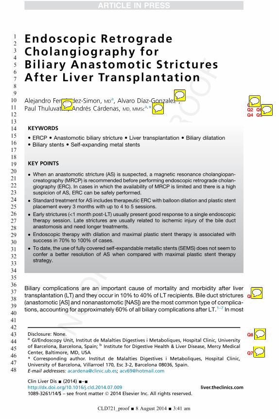
Endoscopic Retrograde Cholangiography for Biliary Anastomotic Strictures After Liver Transplantation Alejandro Fernández-Simon, MD a , Alvaro Díaz-Gonzalez a , Paul Thuluvath b , Andrés Cárdenas, MD, MMSc a, * Q2 Q3 Q4 Q5 Q6 Biliary complications are an important cause of mortality and morbidity after liver transplantation (LT) and they occur in 10% to 40% of LT recipients Q9 . Bile duct strictures (anastomotic [AS] and nonanastomotic [NAS]) are the most common type of complica- tions, accounting for approximately 60% of all biliary complications after LT. 1–7 In most Disclosure: None Q8 . a GI/Endoscopy Unit, Institut de Malalties Digestives i Metaboliques, Hospital Clinic, University of Barcelona, Barcelona, Spain; b Institute for Digestive Health & Liver Disease, Mercy Medical Center, Baltimore, MD, USA Q7 * Corresponding author. Institut de Malalties Digestives i Metaboliques, Hospital Clinic, University of Barcelona, Villarroel 170, Esc 3-2, Barcelona 08036, Spain. E-mail addresses: [email protected]; [email protected] KEYWORDS ERCP Anastomotic biliary stricture Liver transplantation Biliary dilatation Biliary stents Self-expanding metal stents KEY POINTS When an anastomotic stricture (AS) is suspected, a magnetic resonance cholangiopan- creatography (MRCP) is recommended before performing endoscopic retrograde cholan- giography (ERC). In cases in which the availability of MRCP is limited and there is a high suspicion of AS, ERC can be safely performed. Standard treatment for AS includes therapeutic ERC with balloon dilation and plastic stent placement every 3 months with up to 4 to 5 sessions. Early strictures (<1 month post-LT) usually present good response to a single endoscopic therapy session. Late strictures are usually related to ischemic injury of the bile duct anastomosis and need longer treatments. Endoscopic therapy with dilation and maximal plastic stent therapy is associated with success in 70% to 100% of cases. To date, the use of fully covered self-expandable metallic stents (SEMS) does not seem to confer a better resolution of AS when compared with maximal plastic stent therapy strategy. CLD721_proof ■ 8 August 2014 ■ 3:41 am Clin Liver Dis - (2014) -–- http://dx.doi.org/10.1016/j.cld.2014.07.009 liver.theclinics.com 1089-3261/14/$ – see front matter Ó 2014 Elsevier Inc. All rights reserved. 1 2 3 4 5 6 7 8 9 10 11 12 13 14 15 16 17 18 19 20 21 22 23 24 25 26 27 28 29 30 31 32 33 34 35 36 37 38 39 40 41 42 43 44 45 46 47 48
-
Upload
independent -
Category
Documents
-
view
3 -
download
0
Transcript of Endoscopic Retrograde Cholangiography for Biliary Anastomotic Strictures After Liver Transplantation
1
234
567
8910
11121314
151617
181920
212223
24252627
282930
313233
34353637
383940
414243
444546
4748
Endoscopic RetrogradeCholangiography for
Bil iary Anastomotic StricturesAfter Liver TransplantationQ2Q3
Q6
Alejandro Fernández-Simon, MDa, Alvaro Díaz-Gonzaleza,Paul Thuluvathb, Andrés Cárdenas, MD, MMSca,*
Q4 Q5
KEYWORDS
� ERCP � Anastomotic biliary stricture � Liver transplantation � Biliary dilatation� Biliary stents � Self-expanding metal stents
KEY POINTS
� When an anastomotic stricture (AS) is suspected, a magnetic resonance cholangiopan-creatography (MRCP) is recommended before performing endoscopic retrograde cholan-giography (ERC). In cases in which the availability of MRCP is limited and there is a highsuspicion of AS, ERC can be safely performed.
� Standard treatment for AS includes therapeutic ERC with balloon dilation and plastic stentplacement every 3 months with up to 4 to 5 sessions.
� Early strictures (<1 month post-LT) usually present good response to a single endoscopictherapy session. Late strictures are usually related to ischemic injury of the bile ductanastomosis and need longer treatments.
� Endoscopic therapy with dilation and maximal plastic stent therapy is associated withsuccess in 70% to 100% of cases.
� To date, the use of fully covered self-expandable metallic stents (SEMS) does not seem toconfer a better resolution of AS when compared with maximal plastic stent therapystrategy.
Q9
Biliary complications are an important cause of mortality and morbidity after livertransplantation (LT) and they occur in 10% to 40% of LT recipients. Bile duct strictures(anastomotic [AS] and nonanastomotic [NAS]) are themost common type of complica-tions, accounting for approximately 60%of all biliary complications after LT.1–7 Inmost
Disclosure: None Q8.a GI/Endoscopy Unit, Institut de Malalties Digestives i Metaboliques, Hospital Clinic, Universityof Barcelona, Barcelona, Spain; b Institute for Digestive Health & Liver Disease, Mercy MedicalCenter, Baltimore, MD, USA Q7
* Corresponding author. Institut de Malalties Digestives i Metaboliques, Hospital Clinic,University of Barcelona, Villarroel 170, Esc 3-2, Barcelona 08036, Spain.E-mail addresses: [email protected]; [email protected]
CLD721_proof ■ 8 August 2014 ■ 3:41 am
Clin Liver Dis - (2014) -–-http://dx.doi.org/10.1016/j.cld.2014.07.009 liver.theclinics.com1089-3261/14/$ – see front matter � 2014 Elsevier Inc. All rights reserved.
BOTERO MEJIA
Nota adhesiva
q5-Alvaro Diaz-Gonzalez, MD
BOTERO MEJIA
Nota adhesiva
q5-Paul J. Thuluvath, MD, FRCP
BOTERO MEJIA
Nota adhesiva
Q3-ok
BOTERO MEJIA
Nota adhesiva
Q4- MD, MMSc, AGAF. MMSc- stands for Masters in Medcial Science. It is a well known degree. I got this degree from Harvard Medical School
BOTERO MEJIA
Nota adhesiva
q6- ok
BOTERO MEJIA
Nota adhesiva
ok
BOTERO MEJIA
Nota adhesiva
disclosure - ok
BOTERO MEJIA
Nota adhesiva
q7-ok
web4C=FPO
Fernandez-Simon et al2
49
505152
535455
565758
59606162
636465
666768
697071
72737475
767778
798081
82838485
868788
899091
929394
95969798
99
cases, the anastomosis of the bile duct is performed as a duct-to-duct reconstruction,which makes endoscopic therapy with endoscopic retrograde cholangiography (ERC)feasible in most patients. Patients who undergo a Roux-en-Y choledochojejunostomycan also develop strictures at the anastomosiswith the bowel and inmost cases percu-taneous therapy by interventional radiology is performed. In high-volume LT centerswith experienced endoscopists, ERC can be successfully performed in patients witha Roux-en-Y choledochojejunostomy with small bowel enteroscopy; however, thistopic is out of the scope of this review and is discussed elsewhere.8–12 In this review,we discuss the diagnostic approach and the management of biliary anastomoticstrictures after LT because this type of complication accounts for most of the inter-ventional procedures performed in the biliary tract of LT recipients.
TYPES OF BILIARY STRICTURES
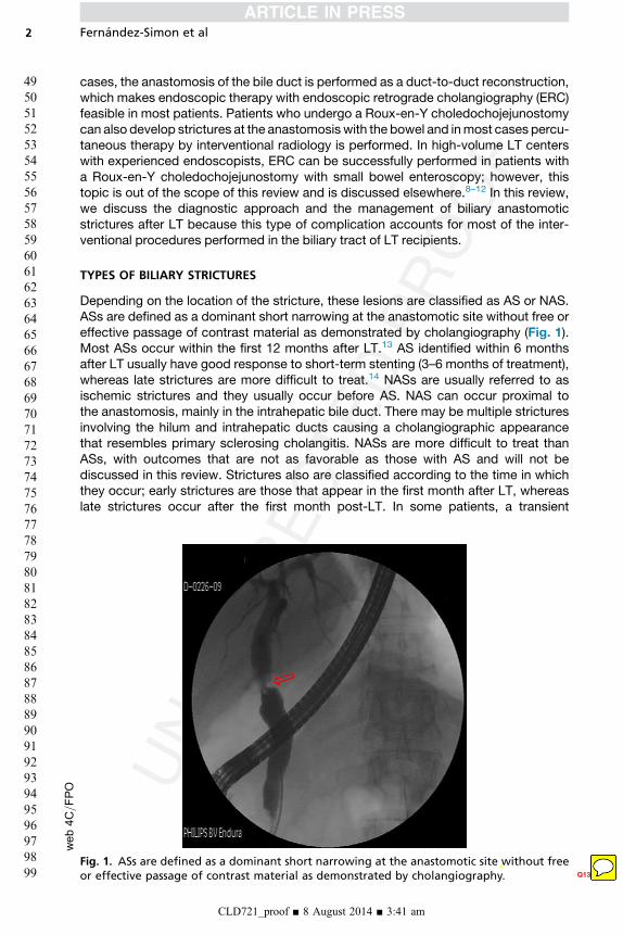
Depending on the location of the stricture, these lesions are classified as AS or NAS.ASs are defined as a dominant short narrowing at the anastomotic site without free oreffective passage of contrast material as demonstrated by cholangiography (Fig. 1).Most ASs occur within the first 12 months after LT.13 AS identified within 6 monthsafter LT usually have good response to short-term stenting (3–6 months of treatment),whereas late strictures are more difficult to treat.14 NASs are usually referred to asischemic strictures and they usually occur before AS. NAS can occur proximal tothe anastomosis, mainly in the intrahepatic bile duct. There may be multiple stricturesinvolving the hilum and intrahepatic ducts causing a cholangiographic appearancethat resembles primary sclerosing cholangitis. NASs are more difficult to treat thanASs, with outcomes that are not as favorable as those with AS and will not bediscussed in this review. Strictures also are classified according to the time in whichthey occur; early strictures are those that appear in the first month after LT, whereaslate strictures occur after the first month post-LT. In some patients, a transient
Fig. 1. ASs are defined as a dominant short narrowing at the anastomotic site without freeor effective passage of contrast material as demonstrated by cholangiography Q13.
CLD721_proof ■ 8 August 2014 ■ 3:41 am
BOTERO MEJIA
Texto insertado
(arrow)
BOTERO MEJIA
Nota adhesiva
ok

Q10
Q1Endoscopic Retrograde Cholangiography 3
100
101102103
104105106
107108109
110111112113
114115116
117118119
120121122
123124125126
127128129
130131132
133134135136
137138139
140141142
143144145
146147148149
150
narrowing of the anastomosis may become evident within the first 1 to 2 months afterLT due to postoperative edema and inflammation.15
RISK FACTORS
There are several known risk factors for the development of biliary AS after LT (Box 1).The most common risk factor is the development of hepatic artery thrombosis (HAT).The biliary system receives blood supply solely via the hepatic artery; thus, HAT canlead to complex anastomotic and hilar strictures. Hepatic artery stenosis can alsolead to both AS and NAS, particularly when associated with long cold ischemiatime.16 T-tubeplacement in LT is controversial. Originally T-tubeswere routinely placedas a prophylactic measure for AS development. The results of several comparativestudies, systematic reviews, and meta-analyses have favored the actual trend towardthe abandonment of the use of T-tubes after LT. A meta-analysis that included morethan 1000 patients showed that, although patients with T-tube in general presentedsuperior results in terms of biliary strictures, the overall complication rate was superiorcompared with patients without T-tube.17 A second meta-analysis of 5 randomizedcontrolled trials indicated that patientswith T-tubehadsignificantly fewerASs, but therewere no differences in the overall rate of complications.18 Despite these results, someinvestigators still defend the role of T-tube in LT. In a recent randomized single-centerprospective trial that evaluated 188 LTs, the use of T-tubes resulted in fewer ASs(n5 2, 2.1%) compared with the non–T-tube group (n5 13, 14.1%; P5 .002).19 Otherwell-described risk factors include bile leak after T-tube removal, technical factorsduring surgery (tight anastomosis, excessive dissection and electrocautery during thereconstruction, redundant bile duct), mismatched size between donor and recipientbile ducts, ischemia/reperfusion injury, presence of cytomegalovirus infection, dona-tion after cardiacdeath, ABObloodgroupmismatch, older ageof donor, graft steatosis,prolonged cold and warm ischemia times, and primary sclerosing cholangitis.16,20–31
DIAGNOSTIC APPROACH
Occasionally patients will present with nonspecific symptoms (fever and anorexia),abdominal pain (especially if associated with bile leaks), pruritus, or jaundice, butin most cases, a biliary stricture is usually suspected in asymptomatic LT recipients
Box 1
Risk factors for biliary anastomotic strictures after LT
Hepatic artery thrombosis
Hepatic artery stenosis
Bile leak
Technical factors
ABO mismatch
Ischemia/reperfusion injury
Cytomegalovirus infection
Donation after cardiac death
Primary sclerosing cholangitis
Ischemia
Age
CLD721_proof ■ 8 August 2014 ■ 3:41 am
BOTERO MEJIA
Nota adhesiva
Endoscopic Retrograde Cholangiography in liver transplant recipients
BOTERO MEJIA
Nota adhesiva
ok
web4C=FPO
Fernandez-Simon et al4
151
152153154
155156157
158159160
161162163164
165166167
168169170
171172173
174175176177
178179180
181182183
184185186187
188189190
191192193
194195196
197198199200
201
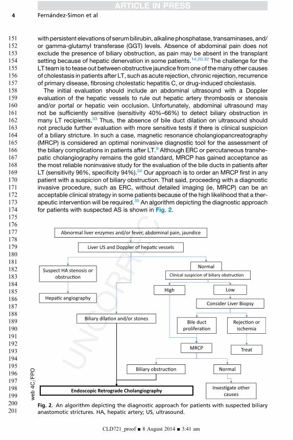
withpersistent elevationsof serumbilirubin, alkalinephosphatase, transaminases, and/or gamma-glutamyl transferase (GGT) levels. Absence of abdominal pain does notexclude the presence of biliary obstruction, as pain may be absent in the transplantsetting because of hepatic denervation in some patients.14,20,32 The challenge for theLT team is to teaseout betweenobstructive jaundice fromoneof themanyother causesof cholestasis in patients after LT, such as acute rejection, chronic rejection, recurrenceof primary disease, fibrosing cholestatic hepatitis C, or drug-induced cholestasis.The initial evaluation should include an abdominal ultrasound with a Doppler
evaluation of the hepatic vessels to rule out hepatic artery thrombosis or stenosisand/or portal or hepatic vein occlusion. Unfortunately, abdominal ultrasound maynot be sufficiently sensitive (sensitivity 40%–66%) to detect biliary obstruction inmany LT recipients.33 Thus, the absence of bile duct dilation on ultrasound shouldnot preclude further evaluation with more sensitive tests if there is clinical suspicionof a biliary stricture. In such a case, magnetic resonance cholangiopancreatography(MRCP) is considered an optimal noninvasive diagnostic tool for the assessment ofthe biliary complications in patients after LT.9 Although ERC or percutaneous transhe-patic cholangiography remains the gold standard, MRCP has gained acceptance asthe most reliable noninvasive study for the evaluation of the bile ducts in patients afterLT (sensitivity 96%, specificity 94%).34 Our approach is to order an MRCP first in anypatient with a suspicion of biliary obstruction. That said, proceeding with a diagnosticinvasive procedure, such as ERC, without detailed imaging (ie, MRCP) can be anacceptable clinical strategy in some patients because of the high likelihood that a ther-apeutic intervention will be required.35 An algorithm depicting the diagnostic approachfor patients with suspected AS is shown in Fig. 2.
Abnormal liver enzymes and/or fever, abdominal pain, jaundice
Liver US and Doppler of hepa�c vessels
Biliary dila�on and/or stones
Suspect HA stenosis or obstruc�on
Hepa�c angiography
Normal
Consider Liver Biopsy
Bile duct prolifera�on
Rejec�on or ischemia
MRCP
Biliary obstruc�on Normal
Inves�gate other causes
Endoscopic Retrograde Cholangiography
Treat
Clinical suspicion of biliary obstruc�on
High Low
Fig. 2. An algorithm depicting the diagnostic approach for patients with suspected biliaryanastomotic strictures. HA, hepatic artery; US, ultrasound.
CLD721_proof ■ 8 August 2014 ■ 3:41 am

Q11
Q12
print&web4C=FPO
Endoscopic Retrograde Cholangiography 5
202
203204205
206207208
209210211
212213214215
216217218
219220221
222223224
225226227228
229230231
232233234
235236237238
239240241
242243244
245246247
248249250251
252
TREATMENT STRATEGIESDilation and Plastic Stent Placement
Most patients with AS require ongoing ERC sessions every 3 months with balloondilation and long-term stenting (for 12–24 months). This technique consists of placinga guidewire across the stricture (Fig. 3), followed by a dilation of the AS using balloondiameters of 6 to 8 mm, and finally placing 8.0-Fr to 11.5-Fr plastic stents with anincreasing diameter and number if possible in each session (Fig. 4). The standardtechnique requires sphincterotomy of the papilla before stent placement. However,similar success rates and remission rates of the AS have been reported in patientswithout sphincterotomy, placing the stent above the intact sphincter of Oddi.36
Plastic stents need to be exchanged every 3 months to avoid occlusion and bacterialcholangitis. This approach has been reported to be more effective than dilationalone.37 Increasing the number of stents in each session improves success ratesand, thus, placing the maximal amount of stents possible in each session is recom-mended, in general up to 4 or 5 stents are needed (see Fig. 4). In a retrospectivestudy of 83 LT recipients with AS, treatment success was associated with thenumber of stents placed (8.0 in the success group vs 3.5 in those whose treatmentfailed).38 This approach usually requires several interventional sessions (mean of 3–5sessions per patient) to achieve long-term success rates of 70% to 100%.3,14,37,39–43
Table 1 describes the findings of different studies, including our own experience(unpublished data), evaluating the outcome of dilation and plastic stent placementof biliary AS.
Self-Expandable Metal Stents
Some studies have reported promising results using self-expandable metal stents(SEMS) for AS in LT recipients.49–53 Uncovered SEMS have an important drawback,and that is the inevitable reactive hyperplasia that may cause difficulty in the stentremoval, especially once it has been in place for more than 3 months54; thus, thesestents should not be placed in LT recipients. Fully covered SEMS are a more feasible
Fig. 3. (Left) A guidewire (green arrow) is pushed across the anastomotic stricture (redarrow). (Right) The guidewire allowed the dilation of the stricture and afterward, theplacement of 2 plastic stents (green arrows) across the stricture. Once placed, the stentspermitted the proper drainage of bile and contrast.
CLD721_proof ■ 8 August 2014 ■ 3:41 am
BOTERO MEJIA
Nota adhesiva
(Cardenas et al, 2014, unpublished data)
BOTERO MEJIA
Nota adhesiva
ok
web4C=FPO
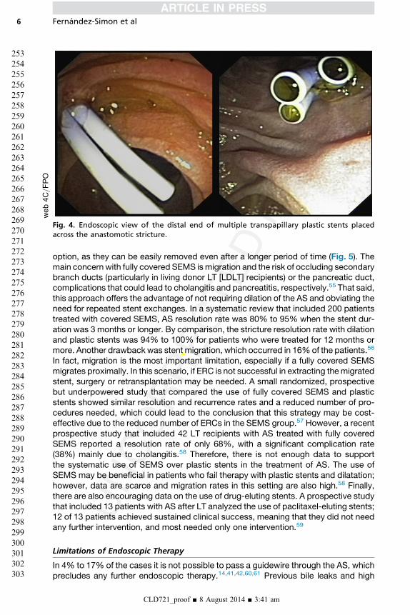
Fig. 4. Endoscopic view of the distal end of multiple transpapillary plastic stents placedacross the anastomotic stricture.
Fernandez-Simon et al6
253
254255256
257258259
260261262
263264265266
267268269
270271272
273274275
276277278279
280281282
283284285
286287288289
290291292
293294295
296297298
299300301302
303
option, as they can be easily removed even after a longer period of time (Fig. 5). Themain concern with fully covered SEMS ismigration and the risk of occluding secondarybranch ducts (particularly in living donor LT [LDLT] recipients) or the pancreatic duct,complications that could lead to cholangitis and pancreatitis, respectively.55 That said,this approach offers the advantage of not requiring dilation of the AS and obviating theneed for repeated stent exchanges. In a systematic review that included 200 patientstreated with covered SEMS, AS resolution rate was 80% to 95% when the stent dur-ation was 3 months or longer. By comparison, the stricture resolution rate with dilationand plastic stents was 94% to 100% for patients who were treated for 12 months ormore. Another drawbackwas stent migration, which occurred in 16%of the patients.56
In fact, migration is the most important limitation, especially if a fully covered SEMSmigrates proximally. In this scenario, if ERC is not successful in extracting themigratedstent, surgery or retransplantation may be needed. A small randomized, prospectivebut underpowered study that compared the use of fully covered SEMS and plasticstents showed similar resolution and recurrence rates and a reduced number of pro-cedures needed, which could lead to the conclusion that this strategy may be cost-effective due to the reduced number of ERCs in the SEMS group.57 However, a recentprospective study that included 42 LT recipients with AS treated with fully coveredSEMS reported a resolution rate of only 68%, with a significant complication rate(38%) mainly due to cholangitis.58 Therefore, there is not enough data to supportthe systematic use of SEMS over plastic stents in the treatment of AS. The use ofSEMS may be beneficial in patients who fail therapy with plastic stents and dilatation;however, data are scarce and migration rates in this setting are also high.58 Finally,there are also encouraging data on the use of drug-eluting stents. A prospective studythat included 13 patients with AS after LT analyzed the use of paclitaxel-eluting stents;12 of 13 patients achieved sustained clinical success, meaning that they did not needany further intervention, and most needed only one intervention.59
Limitations of Endoscopic Therapy
In 4% to 17% of the cases it is not possible to pass a guidewire through the AS, whichprecludes any further endoscopic therapy.14,41,42,60,61 Previous bile leaks and high
CLD721_proof ■ 8 August 2014 ■ 3:41 am
BOTERO MEJIA
Tachado
BOTERO MEJIA
Texto insertado
SEMS
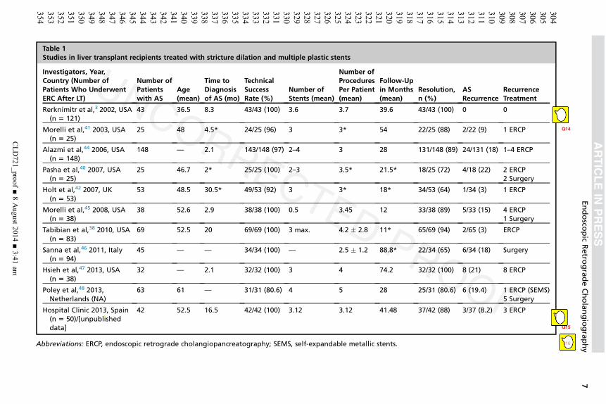
Table 1Studies in liver transplant recipients treated with stricture dilation and multiple plastic stents
Investigators, Year,Country (Number ofPatients Who UnderwentERC After LT)
Number ofPatientswith AS
Age(mean)
Time toDiagnosisof AS (mo)
TechnicalSuccessRate (%)
Number ofStents (mean)
Number ofProceduresPer Patient(mean)
Follow-Upin Months(mean)
Resolution,n (%)
ASRecurrence
RecurrenceTreatment
Rerknimitr et al,3 2002, USA(n 5 121)
43 36.5 8.3 43/43 (100) 3.6 3.7 39.6 43/43 (100) 0 0
Morelli et al,41 2003, USA(n 5 25)
25 48 4.5* Q1424/25 (96) 3 3* 54 22/25 (88) 2/22 (9) 1 ERCP
Alazmi et al,44 2006, USA(n 5 148)
148 — 2.1 143/148 (97) 2–4 3 28 131/148 (89) 24/131 (18) 1–4 ERCP
Pasha et al,40 2007, USA(n 5 25)
25 46.7 2* 25/25 (100) 2–3 3.5* 21.5* 18/25 (72) 4/18 (22) 2 ERCP2 Surgery
Holt et al,42 2007, UK(n 5 53)
53 48.5 30.5* 49/53 (92) 3 3* 18* 34/53 (64) 1/34 (3) 1 ERCP
Morelli et al,45 2008, USA(n 5 38)
38 52.6 2.9 38/38 (100) 0.5 3.45 12 33/38 (89) 5/33 (15) 4 ERCP1 Surgery
Tabibian et al,38 2010, USA(n 5 83)
69 52.5 20 69/69 (100) 3 max. 4.2 � 2.8 11* 65/69 (94) 2/65 (3) ERCP
Sanna et al,46 2011, Italy(n 5 94)
45 — — 34/34 (100) — 2.5 � 1.2 88.8* 22/34 (65) 6/34 (18) Surgery
Hsieh et al,47 2013, USA(n 5 38)
32 — 2.1 32/32 (100) 3 4 74.2 32/32 (100) 8 (21) 8 ERCP
Poley et al,48 2013,Netherlands (NA)
63 61 — 31/31 (80.6) 4 5 28 25/31 (80.6) 6 (19.4) 1 ERCP (SEMS)5 Surgery
Hospital Clinic 2013, Spain(n 5 50)/[unpublisheddata] Q15
42 52.5 16.5 42/42 (100) 3.12 3.12 41.48 37/42 (88) 3/37 (8.2) 3 ERCP
Abbreviations: ERCP, endoscopic retrograde cholangiopancreatography; SEMS, self-expandable metallic stents Q16.
Endosco
pic
Retro
gradeCholangiography
CLD721_proof
■8August
2014■3:41
am
7
304
305
306
307
308
309
310
311
312
313
314
315
316
317
318
319
320
321
322
323
324
325
326
327
328
329
330
331
332
333
334
335
336
337
338
339
340
341
342
343
344
345
346
347
348
349
350
351
352
353
354
BOTERO MEJIA
Tachado
BOTERO MEJIA
Texto insertado
2014
BOTERO MEJIA
Tachado
BOTERO MEJIA
Texto insertado
Cardenas et al, Hospital Clinic
BOTERO MEJIA
Nota adhesiva
NA: not available
BOTERO MEJIA
Nota adhesiva
* median value
BOTERO MEJIA
Nota adhesiva
ok
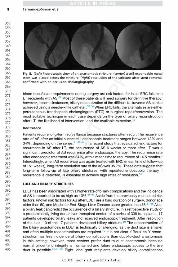
Fig. 5. (Left) Fluoroscopic view of an anastomotic stricture; (center) a self-expandable metalstent was placed across the stricture; (right) resolution of the stricture after stent removal,confirmed with an occlusion cholangiography.
Fernandez-Simon et al8
355
356357358
359360361
362363364
365366367368
369370371
372373374
375376377
378379380381
382383384
385386387
388389390391
392393394
395396397
398399400
401402403404
405
blood transfusion requirements during surgery are risk factors for initial ERC failure inLT recipients with AS.62 Most of these patients will need surgery for definitive therapy;however, in some instances, biliary recanalization of the difficult-to-traverse AS can beachieved using a needle-knife catheter.63,64 When ERC fails, the alternatives are eitherpercutaneous transhepatic cholangiogram (PTC) or surgical repair/conversion. Themost suitable technique in each case depends on the type of biliary reconstructionafter LT, the likelihood of intervention, and the available expertise.12
Recurrence
Patients require long-term surveillance because strictures often recur. The recurrencerate of AS after an initial successful endoscopic treatment ranges between 18% and34%, depending on the series.41,42,44 In a recent study that evaluated risk factors forrecurrence in AS after LT, the occurrence of AS 6 weeks or more after LT was asignificant predictor of AS recurrence after endoscopic therapy. The recurrence rateafter endoscopic treatment was 34%, with a mean time to recurrence of 14.5 months.7
Interestingly, when AS recurrence was again treated with ERC (mean time of follow-upof 151 weeks), long-term resolution rate of the ASwas 95.7%. These data suggest thatlong-term follow-up of late biliary strictures, with repeated endoscopic therapy ifrecurrence is detected, is essential to achieve high rates of resolution.13
LDLT AND BILIARY STRICTURES
LDLT has been associated with a higher rate of biliary complications and the incidenceof AS is reported to be as high as 40%.65,66 Aside from the previously mentioned riskfactors, known risk factors for AS after LDLT are a long duration of surgery, donor ageolder than 50, and Model for End-Stage Liver Disease score greater than 35.67,68 Also,a biliary leak can predict the occurrence of a biliary stricture. In a retrospective study ofa predominantly living donor liver transplant center, of a series of 338 transplants, 17patients developed biliary leaks and received endoscopic treatment. After resolutionof the leak, 10 of the 17 patients developed biliary stricture.69 The reconstruction ofthe biliary anastomosis in LDLT is technically challenging, as the duct size is smallerand often multiple reconstructions are required.70 It is not clear if Roux-en-Y recon-struction has less incidence of biliary complications than duct-to-duct anastomosisin this setting; however, most centers prefer duct-to-duct anastomosis becausenormal bilioenteric integrity is maintained and future endoscopic access to the bileduct is possible.66,71,72 Right lobe graft recipients develop biliary complications
CLD721_proof ■ 8 August 2014 ■ 3:41 am
Endoscopic Retrograde Cholangiography 9
406
407408409
410411412
413414415
416417418419
420421422
423424425
426427428
429430431432
433434435
436437438
439440441442
443444445
446447448
449450451
452453454455
456
more frequently compared with left lobe grafts, particularly if a duct-to-duct anasto-mosis has been used involving small-sized duct. Some investigators argue that ifduct size is smaller than 4 mm in diameter, a hepaticojejunostomy is preferred.73
Endoscopic treatment in LDLT is often more complex. Patients usually are treated insimilar fashion as deceased donor LT recipients, but require smaller-diameter stents(7.0–8.5 Fr) and balloon dilation. Resolution rates range between 75% and 80%, butrecurrence rate after initial endoscopic therapy can occur in up to 30%, especiallywhen stenting duration is short.74 Maximal stent therapy has been evaluated forthe treatment of AS in patients after LDLT. In a report of 110 patients, 38 of whomdeveloped AS, ERC was attempted as initial therapy in all patients. Thirty-two weremanaged entirely with endoscopic therapy, whereas 6 underwent PTC to traversethe stricture and proceed with ERC thereafter. Resolution rate was 79%, recurrentstrictures were reported in 21% of the patients, and all of them were successfullytreated with repeated endoscopic sessions.47 Recipients of liver from donors aftercardiac death also have a high incidence of biliary complications (up to 33%), espe-cially diffuse ischemic cholangiopathy. Focal strictures in this type of donor liverscan be treated successfully with endoscopic therapy, whereas diffuse stricturesusually require retransplantation.75
CHOLANGIOSCOPY
Single-operator cholangioscopy is a technique that can provide direct imaging of thebile duct mucosa, and can be useful in the management of biliary strictures after LT. Ina prospective study of 16 patients who developed biliary complications after LT,single-operator cholangioscopy (SOC) was performed and was feasible and tissuewas obtained in all cases. Twelve patients presented AS, among them 2 distinct visualpatterns were easily identified with SOC; one wasmild erythema and the other showedsignificant edema, ulceration, and sloughing. Those with the latter pattern required alonger period of stenting than patients with only mild erythema and scarring (457 vs167 days). In addition, in patients with mild erythema, resolution of strictures withendoscopic therapy was better than those with an edema, ulceration, and sloughingpattern (66% vs 33%). This distinction may help to predict outcomes of endoscopictherapy76; however more studies are need in this area.
COMPLICATIONS OF ERC
Although ERC is associated with significant complications, the rate of these complica-tions does not seem to differ from that of the general population. There is up to9% complication rate per procedure.4,5,77,78 The most common complications arepancreatitis, cholangitis, and postsphincterotomy bleeding. Other complicationsinclude bile leak, subcapsular hematoma, perforation, and stent migration. Biliarysphincterotomy, renal failure, repeated pancreatic duct injections, and therapy withmammalian target of rapamycin inhibitors may place patients at risk for complicationsafter ERC.78,79
SUMMARY
Biliary ASs are common in recipients of deceased donor and live donor livertransplants. Clinicians should have a high index of suspicion and promptly order anabdominal ultrasound. In those patients in whom there is a strong clinical suspicionof biliary obstruction, an ERC should be done, whereas an MRCP may be preferredif the index of suspicion for bile obstruction is lower. ERC is the preferred treatment
CLD721_proof ■ 8 August 2014 ■ 3:41 am
Fernandez-Simon et al10
457
458459460
461462463
464465466
467468469470
471472473
474475476
477478479
480481482483
484485486
487488489
490491492493
494495496
497498499
500501502
503504505506
507
for the management of most biliary AS. The combination of stricture dilation andplastic stent placement every 3 months is the best approach. Fully covered SEMSshould not be placed as first-line therapy, and their use should be considered on acase-by-case basis. The recurrence rate after therapy can be as high as 30%, butmost patients respond to repeat endoscopic therapy.
REFERENCES
1. Stratta RJ, Wood RP, Langnas AN, et al. Diagnosis and treatment of biliary tractcomplications after orthotopic liver transplantation. Surgery 1989;106:675–83[discussion: 683–4].
2. Greif F, Bronsther OL, Van Thiel DH, et al. The incidence, timing, and man-agement of biliary tract complications after orthotopic liver transplantation.Ann Surg 1994;219:40–5.
3. Rerknimitr R, Sherman S, Fogel EL, et al. Biliary tract complications after ortho-topic liver transplantation with choledochocholedochostomy anastomosis: endo-scopic findings and results of therapy. Gastrointest Endosc 2002;55:224–31.
4. PfauPR,KochmanML, Lewis JD, et al. Endoscopicmanagement of postoperativebiliary complications in orthotopic liver transplantation.Gastrointest Endosc2000;52:55–63.
5. Thuluvath PJ, Atassi T, Lee J. An endoscopic approach to biliary complicationsfollowing orthotopic liver transplantation. Liver Int 2003;23:156–62.
6. Thethy S, Thomson BN, Pleass H, et al. Management of biliary tract complica-tions after orthotopic liver transplantation. Clin Transplant 2004;18:647–53.
7. Zimmerman MA, Baker T, Goodrich NP, et al. Development, management, andresolution of biliary complications after living and deceased donor liver trans-plantation: a report from the adult-to-adult living donor liver transplantationcohort study consortium. Liver Transpl 2013;19:259–67.
8. Azeem N, Tabibian JH, Baron TH, et al. Use of a single-balloon enteroscopecompared with variable-stiffness colonoscopes for endoscopic retrograde chol-angiography in liver transplant patients with Roux-en-Y biliary anastomosis.Gastrointest Endosc 2013;77:568–77.
9. Arain MA, Attam R, FreemanML. Advances in endoscopic management of biliarytract complications after liver transplantation. Liver Transpl 2013;19:482–98.
10. Chua TJ, Kaffes AJ. Balloon-assisted enteroscopy in patients with surgicallyaltered anatomy: a liver transplant center experience (with video). GastrointestEndosc 2012;76:887–91.
11. Kawano Y, Mizuta K, Hishikawa S, et al. Rendezvous penetration method usingdouble-balloon endoscopy for complete anastomosis obstruction of hepaticoje-junostomy after pediatric living donor liver transplantation. Liver Transpl 2008;14:385–7.
12. Chahal P, Baron TH, Poterucha JJ, et al. Endoscopic retrograde cholangiog-raphy in post-orthotopic liver transplant population with Roux-en-Y biliary recon-struction. Liver Transpl 2007;13:1168–73.
13. Albert JG, Filmann N, Elsner J, et al. Long-term follow-up of endoscopic therapyfor stenosis of the biliobiliary anastomosis associated with orthotopic liver trans-plantation. Liver Transpl 2013;19:586–93.
14. Verdonk RC, Buis CI, Porte RJ, et al. Anastomotic biliary strictures after livertransplantation: causes and consequences. Liver Transpl 2006;12:726–35.
15. Verdonk RC, Buis CI, Porte RJ, et al. Biliary complications after liver trans-plantation: a review. Scand J Gastroenterol Suppl 2006;89–101.
CLD721_proof ■ 8 August 2014 ■ 3:41 am
Endoscopic Retrograde Cholangiography 11
508
509510511
512513514
515516517
518519520521
522523524
525526527
528529530
531532533534
535536537
538539540
541542543544
545546547
548549550
551552553
554555556557
558
16. Dacha S, Barad A, Martin J, et al. Association of hepatic artery stenosis andbiliary strictures in liver transplant recipients. Liver Transpl 2011;17:849–54.
17. Sotiropoulos GC, Sgourakis G, Radtke A, et al. Orthotopic liver transplantation:T-tube or not T-tube? Systematic review and meta-analysis of results. Transplan-tation 2009;87:1672–80.
18. Riediger C, Muller MW, Michalski CW, et al. T-Tube or no T-tube in the recon-struction of the biliary tract during orthotopic liver transplantation: systematicreview and meta-analysis. Liver Transpl 2010;16:705–17.
19. Lopez-Andujar R, Oron EM, Carregnato AF, et al. T-tube or No T-tube in cadav-eric orthotopic liver transplantation: the eternal dilemma: results of a prospectiveand randomized clinical trial. Ann Surg 2013;258:21–9.
20. Pascher A, Neuhaus P. Biliary complications after deceased-donor orthotopicliver transplantation. J Hepatobiliary Pancreat Surg 2006;13:487–96.
21. Sanchez-Urdazpal L, Gores GJ, Ward EM, et al. Ischemic-type biliary complica-tions after orthotopic liver transplantation. Hepatology 1992;16:49–53.
22. Sanchez-Urdazpal L, Gores GJ, Ward EM, et al. Diagnostic features and clinicaloutcome of ischemic-type biliary complications after liver transplantation.Hepatology 1993;17:605–9.
23. Busquets J, Figueras J, Serrano T, et al. Postreperfusion biopsies are useful inpredicting complications after liver transplantation. Liver Transpl 2001;7:432–5.
24. Sanchez-Urdazpal L, Batts KP, Gores GJ, et al. Increased bile duct complica-tions in liver transplantation across the ABO barrier. Ann Surg 1993;218:152–8.
25. Ludwig J, Wiesner RH, Batts KP, et al. The acute vanishing bile duct syndrome(acute irreversible rejection) after orthotopic liver transplantation. Hepatology1987;7:476–83.
26. Graziadei IW. Recurrence of primary sclerosing cholangitis after liver transplan-tation. Liver Transpl 2002;8:575–81.
27. Maheshwari A, Maley W, Li Z, et al. Biliary complications and outcomes of livertransplantation from donors after cardiac death. Liver Transpl 2007;13:1645–53.
28. Fung JJ, Eghtesad B, Patel-Tom K. Using livers from donation after cardiacdeath donors—a proposal to protect the true Achilles heel. Liver Transpl2007;13:1633–6.
29. Welling TH, Heidt DG, Englesbe MJ, et al. Biliary complications following livertransplantation in the model for end-stage liver disease era: effect of donor,recipient, and technical factors. Liver Transpl 2008;14:73–80.
30. Jay CL, Lyuksemburg V, Ladner DP, et al. Ischemic cholangiopathy aftercontrolled donation after cardiac death liver transplantation: a meta-analysis.Ann Surg 2011;253:259–64.
31. Brunner SM, Junger H, Ruemmele P, et al. Bile duct damage after cold storageof deceased donor livers predicts biliary complications after liver transplanta-tion. J Hepatol 2013;58:1133–9.
32. Thuluvath PJ, Pfau PR, Kimmey MB, et al. Biliary complications after liver trans-plantation: the role of endoscopy. Endoscopy 2005;37:857–63.
33. Sharma S, Gurakar A, Jabbour N. Biliary strictures following liver transplanta-tion: past, present and preventive strategies. Liver Transpl 2008;14:759–69.
34. Jorgensen JE, Waljee AK, Volk ML, et al. Is MRCP equivalent to ERCP for diag-nosing biliary obstruction in orthotopic liver transplant recipients? A meta-anal-ysis. Gastrointest Endosc 2011;73:955–62.
35. Elmunzer BJ, Debenedet AT, Volk ML, et al. Clinical yield of diagnostic endo-scopic retrograde cholangiopancreatography in orthotopic liver transplantrecipients with suspected biliary complications. Liver Transpl 2012;18:1479–84.
CLD721_proof ■ 8 August 2014 ■ 3:41 am

Fernandez-Simon et al12
559
560561562
563564565
566567568
569570571572
573574575
576577578
579580581
582583584585
586587588
589590591
592593594595
596597598
599600601
602603604
605606607608
609
36. Kurita A, Kodama Y, Minami R, et al. Endoscopic stent placement above theintact sphincter of Oddi for biliary strictures after living donor liver transplanta-tion. J Gastroenterol 2013;48:1097–104.
37. Zoepf T, Maldonado-Lopez EJ, Hilgard P, et al. Balloon dilatation vs. balloondilatation plus bile duct endoprostheses for treatment of anastomotic biliarystrictures after liver transplantation. Liver Transpl 2006;12:88–94.
38. Tabibian JH, Asham EH, Han S, et al. Endoscopic treatment of postorthotopicliver transplantation anastomotic biliary strictures with maximal stent therapy(with video). Gastrointest Endosc 2010;71:505–12.
39. Graziadei IW, Schwaighofer H, Koch R, et al. Long-term outcome of endoscopictreatment of biliary strictures after liver transplantation. Liver Transpl 2006;12:718–25.
40. Pasha SF, Harrison ME, Das A, et al. Endoscopic treatment of anastomoticbiliary strictures after deceased donor liver transplantation: outcomes aftermaximal stent therapy. Gastrointest Endosc 2007;66:44–51.
41. Morelli J, Mulcahy HE, Willner IR, et al. Long-term outcomes for patients withpost-liver transplant anastomotic biliary strictures treated by endoscopic stentplacement. Gastrointest Endosc 2003;58:374–9.
42. Holt AP, Thorburn D, Mirza D, et al. A prospective study of standardized nonsur-gical therapy in the management of biliary anastomotic strictures complicatingliver transplantation. Transplantation 2007;84:857–63.
43. Kulaksiz H, Weiss KH, Gotthardt D, et al. Is stenting necessary after balloon dila-tion of post-transplantation biliary strictures? Results of a prospective compara-tive study. Endoscopy 2008;40:746–51.
44. Alazmi WM, Fogel EL, Watkins JL, et al. Recurrence rate of anastomotic biliarystrictures in patients who have had previous successful endoscopic therapy foranastomotic narrowing after orthotopic liver transplantation. Endoscopy 2006;38:571–4.
45. Morelli G, Fazel A, Judah J, et al. Rapid-sequence endoscopic management ofposttransplant anastomotic biliary strictures. Gastrointest Endosc 2008;67:879–85.
46. Sanna C, Giordanino C, Giono I, et al. Safety and efficacy of endoscopic retro-grade cholangiopancreatography in patients with post-liver transplant biliarycomplications: results of a cohort study with long-term follow-up. Gut Liver2011;5:328–34.
47. Hsieh TH, Mekeel KL, Crowell MD, et al. Endoscopic treatment of anastomoticbiliary strictures after living donor liver transplantation: outcomes after maximalstent therapy. Gastrointest Endosc 2013;77:47–54.
48. Poley JW, Lekkerkerker MN, Metselaar HJ, et al. Clinical outcome of progressivestenting in patients with anastomotic strictures after orthotopic liver transplanta-tion. Endoscopy 2013;45:567–70.
49. Sauer P, Chahoud F, Gotthardt D, et al. Temporary placement of fully coveredself-expandable metal stents in biliary complications after liver transplantation.Endoscopy 2012;44:536–8.
50. Traina M, Tarantino I, Barresi L, et al. Efficacy and safety of fully covered self-expandable metallic stents in biliary complications after liver transplantation: apreliminary study. Liver Transpl 2009;15:1493–8.
51. Tarantino I, Traina M, Mocciaro F, et al. Fully covered metallic stents in biliary ste-nosis after orthotopic liver transplantation. Endoscopy 2012;44:246–50.
52. Cerecedo-Rodriguez J, Phillips M, Figueroa-Barojas P, et al. Self expandablemetal stents for anastomotic stricture following liver transplant. Dig Dis Sci2013;58:2661–6.
CLD721_proof ■ 8 August 2014 ■ 3:41 am

Endoscopic Retrograde Cholangiography 13
610
611612613
614615616
617618619
620621622623
624625626
627628629
630631632
633634635636
637638639
640641642
643644645646
647648649
650651652
653654655
656657658659
660
53. Chaput U, Scatton O, Bichard P, et al. Temporary placement of partially coveredself-expandablemetal stents for anastomoticbiliary stricturesafter liver transplan-tation: a prospective, multicenter study. Gastrointest Endosc 2010;72:1167–74.
54. Larghi A, Tringali A, Lecca PG, et al. Management of hilar biliary strictures. Am JGastroenterol 2008;103:458–73.
55. Luigiano C, Bassi M, Ferrara F, et al. Placement of a new fully covered self-expanding metal stent for postoperative biliary strictures and leaks not respond-ing to plastic stenting. Surg Laparosc Endosc Percutan Tech 2013;23:159–62.
56. Kao D, Zepeda-Gomez S, Tandon P, et al. Managing the post-liver transplanta-tion anastomotic biliary stricture: multiple plastic versus metal stents: a system-atic review. Gastrointest Endosc 2013;77:679–91.
57. Kaffes A, Griffin S, Vaughan R, et al. A randomized trial of a fully covered self-expandable metallic stent versus plastic stents in anastomotic biliary stricturesafter liver transplantation. Therap Adv Gastroenterol 2014;7:64–71.
58. Deviere J, Nageshwar Reddy D, Puspok A, et al. Successful management ofbenign biliary strictures with fully covered self-expanding metal stents. Gastro-enterology 2014;147:385–95.
59. Kabar I, Cicinnati VR, Beckebaum S, et al. Use of paclitaxel-eluting balloons forendotherapy of anastomotic strictures following liver transplantation. Endoscopy2012;44:1158–60.
60. Barriga J, Thompson R, Shokouh-Amiri H, et al. Biliary strictures after liver trans-plantation. Predictive factors for response to endoscopic management andlong-term outcome. Am J Med Sci 2008;335:439–43.
61. Weber A, Prinz C, Gerngross C, et al. Long-term outcome of endoscopic and/orpercutaneous transhepatic therapy in patients with biliary stricture after ortho-topic liver transplantation. J Gastroenterol 2009;44:1195–202.
62. Balderramo D, Sendino O, Burrel M, et al. Risk factors and outcomes of failedendoscopic retrograde cholangiopancreatography in liver transplant recipientswith anastomotic biliary strictures: a case-control study. Liver Transpl 2012;18:482–9.
63. Gupta K, Aparicio D, Freeman ML, et al. Endoscopic biliary recanalization byusing a needle catheter in patients with complete ligation or stricture of thebile duct: safety and feasibility of a novel technique (with videos). GastrointestEndosc 2011;74:423–8.
64. Martins FP, De Paulo GA, Macedo EP, et al. Endoscopic biliary recanalizationwith a needle-knife in post liver-transplant complete anastomotic stricture.Endoscopy 2012;44(Suppl 2):E304–5.
65. Takatsuki M, Eguchi S, Kawashita Y, et al. Biliary complications in recipients ofliving-donor liver transplantation. J Hepatobiliary Pancreat Surg 2006;13:497–501.
66. Wang SF, Huang ZY, Chen XP. Biliary complications after living donor liver trans-plantation. Liver Transpl 2011;17:1127–36.
67. Shah SA, Grant DR, McGilvray ID, et al. Biliary strictures in 130 consecutive rightlobe living donor liver transplant recipients: results of a Western center. Am JTransplant 2007;7:161–7.
68. Liu CL, Lo CM, Chan SC, et al. Safety of duct-to-duct biliary reconstruction inright-lobe live-donor liver transplantation without biliary drainage. Transplan-tation 2004;77:726–32.
69. Wadhawan M, Kumar A, Gupta S, et al. Post-transplant biliary complications: ananalysis from a predominantly living donor liver transplant center. J GastroenterolHepatol 2013;28:1056–60.
CLD721_proof ■ 8 August 2014 ■ 3:41 am
Fernandez-Simon et al14
661662663664665666667668669670671672673674675676677678679680681682683684685686687688689690
70. Gondolesi GE, Varotti G, Florman SS, et al. Biliary complications in 96 consec-utive right lobe living donor transplant recipients. Transplantation 2004;77:1842–8.
71. Freise CE, Gillespie BW, Koffron AJ, et al. Recipient morbidity after living anddeceased donor liver transplantation: findings from the A2ALL RetrospectiveCohort Study. Am J Transplant 2008;8:2569–79.
72. Soejima Y, Taketomi A, Yoshizumi T, et al. Biliary strictures in living donor livertransplantation: incidence, management, and technical evolution. Liver Transpl2006;12:979–86.
73. Hwang S, Lee SG, Sung KB, et al. Long-term incidence, risk factors, and man-agement of biliary complications after adult living donor liver transplantation.Liver Transpl 2006;12:831–8.
74. Seo JK, Ryu JK, Lee SH, et al. Endoscopic treatment for biliary stricture afteradult living donor liver transplantation. Liver Transpl 2009;15:369–80.
75. Croome KP, McAlister V, Adams P, et al. Endoscopic management of biliarycomplications following liver transplantation after donation from cardiac deathdonors. Can J Gastroenterol 2012;26:607–10.
76. Balderramo D, Sendino O, Miquel R, et al. Prospective evaluation of single-operator peroral cholangioscopy in liver transplant recipients requiring an eval-uation of the biliary tract. Liver Transpl 2013;19:199–206.
77. Rizk RS, McVicar JP, Emond MJ, et al. Endoscopic management of biliary stric-tures in liver transplant recipients: effect on patient and graft survival. Gastroint-est Endosc 1998;47:128–35.
78. Balderramo D, Bordas JM, Sendino O, et al. Complications after ERCP in livertransplant recipients. Gastrointest Endosc 2011;74:285–94.
79. Balderramo D, Navasa M, Cardenas A. Current management of biliary compli-cations after liver transplantation: emphasis on endoscopic therapy. Gastroen-terol Hepatol 2011;34:107–15.
CLD721_proof ■ 8 August 2014 ■ 3:41 am
Our reference: CLD 721 P-authorquery-v9
AUTHOR QUERY FORM
Journal: CLD
Article Number: 721
Dear Author,
Please check your proof carefully and mark all corrections at the appropriate place in the proof (e.g., by using on-screen
annotation in the PDF file) or compile them in a separate list. Note: if you opt to annotate the file with software other than
Adobe Reader then please also highlight the appropriate place in the PDF file. To ensure fast publication of your paper please
return your corrections within 48 hours.
For correction or revision of any artwork, please consult http://www.elsevier.com/artworkinstructions.
Any queries or remarks that have arisen during the processing of your manuscript are listed below and highlighted by flags in
the proof.
Location
in articleQuery / Remark: Click on the Q link to find the query’s location in text
Please insert your reply or correction at the corresponding line in the proof
Q1 Please approve the short title to be used in the running head at the top of each right-hand page.
Q2 This is how your name will appear on the contributor's list. Please add your academic title and any other
necessary titles and professional affiliations, verify the information, and OK
ALEJANDRO FERN�ANDEZ-SIMON, MD, GI/Endoscopy Unit, Institut de Malalties Digestives i
Metaboliques, Hospital Clinic, University of Barcelona, Barcelona, Spain
ALVARO D�IAZ-GONZALEZ, GI/Endoscopy Unit, Institut de Malalties Digestives i Metaboliques,
Hospital Clinic, University of Barcelona, Barcelona, Spain
PAUL THULUVATH, Institute for Digestive Health & Liver Disease, Mercy Medical Center, Baltimore,
Maryland
ANDR�ES C�ARDENAS, MD, MMSc, AGAF, GI/Endoscopy Unit, Institut de Malalties Digestives i
Metaboliques, Hospital Clinic, University of Barcelona, Barcelona, Spain
Q3 Are author names and order of authors OK as set?
Q4 Degree abbreviations are verified against a list of known degrees. MMsc is not yet on this list. Please verify
the degree.
Q5 Please provide professional degrees (eg, PhD, MD) for the authors 'Alvaro Dıaz-Gonzalez and Paul
Thuluvath'.
Q6 The following synopsis is the one that you supplied, but edited down to less than 100 words. Please confirm
OK, or submit a replacement (also less than 100 words). Please note that the synopsis will appear in
PubMed: Biliary complications after liver transplantation (LT) are an important cause of morbidity and
mortality. In most cases, an anastomosis of the bile duct is performed as a duct-to-duct reconstruction,
which makes endoscopic therapy with endoscopic retrograde cholangiography (ERC) feasible. Biliary
anastomotic strictures (AS) are the most common cause of biliary complications. The early detection of an
AS, which can sometimes be challenging given that its clinical presentation is often subtle, is of key
(continued on next page)

importance to obtain high treatment success. In this review, we focus on the management of AS after LT
with a special emphasis on ERC.
Q7 Please verify the affiliation addresses and provide the missing information (department name for affiliation
“b”, street name and zip code for both the affiliations).
Q8 As per the editorial remarks, "Please verify the disclosure statement."
Q9 If there are any drug dosages in your article, please verify them and indicate that you have done so by
initialing this query.
Q10 Nominal tables that are really just one-column lists are best represented as boxes, so Table 1 has been
converted to Box 1, and Table 2 has been renumbered as Table 1. Please verify.
Q11 Reference citations were not in sequential order. Hence, Refs. 44e79 have been renumbered both in text
and in reference list. Please verify.
Q12 Please provide the author name and year for unpublished data.
Q13 Fig. 1: Please indicate the arrow in the legend.
Q14 The designator “*” is present in Table 1 (originally Table 2). Please specify what this indicates.
Q15 Please provide author name and year for unpublished data in Table 1 (originally Table 2).
Q16 Please define NA in Table 1 legend.
Please check this box or indicate
your approval if you have no
corrections to make to the PDF file ,
Thank you for your assistance.