Codimension two bifurcation in a delayed neural network with unidirectional coupling

Cell Reports
Article
A Function for EHD Family Proteinsin Unidirectional Retrograde Dendritic Transportof BACE1 and Alzheimer’s Disease Ab ProductionVirginie Buggia-Prevot,1 Celia G. Fernandez,2 Vinod Udayar,4 Kulandaivelu S. Vetrivel,1 Aureliane Elie,1 Jelita Roseman,1
Verena A. Sasse,5 Margaret Lefkow,2 Xavier Meckler,1 Sohinee Bhattacharyya,6 Manju George,6 Satyabrata Kar,5
Vytautas P. Bindokas,3 Angele T. Parent,1 Lawrence Rajendran,4 Hamid Band,6 Robert Vassar,7 andGopal Thinakaran1,2,*1Departments of Neurobiology, Neurology, and Pathology, The University of Chicago, Chicago, IL 60637, USA2Committee on Neurobiology, The University of Chicago, Chicago, IL 60637, USA3Department of Pharmacological and Physiological Sciences, The University of Chicago, Chicago, IL 60637, USA4Systems and Cell Biology of Neurodegeneration, Division of Psychiatry Research, University of Zurich, 8008 Zurich, Switzerland5Centre for Prions and Protein Folding Diseases, Departments of Medicine and Psychiatry, University of Alberta, Edmonton, AB T6G 2M8,
Canada6Eppley Institute for Research in Cancer and Allied Diseases and Fred & Pamela Buffett Cancer Center, University of Nebraska Medical
Center, Omaha, NE 68198, USA7Department of Cell and Molecular Biology, Feinberg School of Medicine, Northwestern University, Chicago, IL 60611, USA
*Correspondence: [email protected]://dx.doi.org/10.1016/j.celrep.2013.12.006
This is an open-access article distributed under the terms of the Creative Commons Attribution-NonCommercial-No Derivative Works
License, which permits non-commercial use, distribution, and reproduction in any medium, provided the original author and source are
credited.
SUMMARY
Abnormal accumulation of b-secretase (BACE1) indystrophic neurites and presynaptic b-amyloid (Ab)production contribute to Alzheimer’s disease patho-genesis. Little, however, is known about BACE1sorting and dynamic transport in neurons. We inves-tigated BACE1 trafficking in hippocampal neuronsusing live-cell imaging and selective labeling. Wereport that transport vesicles containing internalizedBACE1 in dendrites undergo exclusive retrogradetransport toward the soma, whereas they undergobidirectional transport in axons. Unidirectional den-dritic transport requires Eps15-homology-domain-containing (EHD) 1 and 3 protein function. Further-more, loss of EHD function compromises dynamicaxonal transport and overall BACE1 levels in axons.EHD1/3 colocalize with BACE1 and APP b-C-termi-nal fragments in hippocampal mossy fiber terminals,and their depletion in neurons significantly attenu-ates Ab levels. These results demonstrate unidirec-tional endocytic transport of a dendritic cargo andreveal a role for EHD proteins in neuronal BACE1transcytosis and Ab production, processes that arehighly relevant for Alzheimer’s disease.
INTRODUCTION
Proteolytic processing of amyloid precursor protein (APP) by the
transmembrane aspartyl protease b-site APP cleaving enzyme 1
1552 Cell Reports 5, 1552–1563, December 26, 2013 ª2013 The Aut
(BACE1) initiates Ab production, a key step in Alzheimer’s
disease pathogenesis (Vassar et al., 1999; Yan et al., 1999). It
is extremely important to understand details of BACE1 trafficking
and processing of APP in neurons because a single amino acid
substitution adjacent to the BACE1 cleavage site of APP, which
significantly reduces BACE1 cleavage and thus Ab peptide
generation in cultured cells, has been recently found to protect
against disease onset as well as cognitive decline in the elderly
without Alzheimer’s disease (Jonsson et al., 2012). APP is a
type I transmembrane protein that undergoes secretory and
endocytic trafficking in neurons and is axonally transported
(reviewed in Haass et al., 2012). Conversion of APP to Ab
requires coordination of its intracellular itinerary with that of its
cleavage enzymes, which are also transmembrane proteins. In
cultured cell lines and primary neurons, a subset of full-length
APP is processed to generate Ab. Extensive studies have used
nonneuronal cells to identify the cellular organelles and sorting
pathways involved in amyloidogenic processing of APP.
Although a consensus has not yet emerged, there is a general
agreement on the importance of endocytic trafficking of APP
for Ab production (reviewed in Thinakaran and Koo, 2008; Rajen-
dran and Annaert, 2012; Haass et al., 2012). BACE1 activity is
optimal at acidic pH in vitro (Vassar et al., 1999), lending further
support to the notion that APP cleavage by BACE1 is initiated
during transit in acidic endocytic compartments.
In nonneuronal cells, BACE1 cycles between plasma
membrane and endosomes and shows predominant steady-
state localization in endocytic organelles (Vassar et al., 1999;
Huse et al., 2000; Chia et al., 2013). Two routes of BACE1 endo-
cytosis, a clathrin and adaptor protein-2 complex (AP-2) depen-
dent (clathrin-dependent) and an ADP-ribosylation factor 6
dependent (clathrin-independent), have been described (Prabhu
et al., 2012; Sannerud et al., 2011; Das et al., 2013). A C-terminal
hors

dual-function dileucine motif [495DDISLL500] appears to mediate
both modes of BACE1 internalization (Prabhu et al., 2012; He
et al., 2002). Although studies of BACE1 trafficking described
above have been informative, most of these were conducted in
nonneuronal cells. The sorting itinerary of transmembrane
proteins can be fundamentally different in polarized neurons
versus nonpolarized nonneuronal cells. Specifically, neuronal
protein sorting involves specialized and intricate transport mech-
anisms such as transcytosis and activity-dependent endocy-
tosis/recycling. Endosomal organelles are found distributed
throughout the soma, dendrites, and axons (Lasiecka and
Winckler, 2011). Neuronal endosomes traffic bidirectionally in
axons and dendrites, adding to the complexity of the mecha-
nisms that regulate endocytic transport in neurons. There is
some indication in the literature that BACE1 localizes to den-
drites and axons in neurons (Laird et al., 2005; Goldsbury
et al., 2006; Zhao et al., 2007; Sannerud et al., 2011; Das et al.,
2013). Compelling in vivo studies demonstrated that �70% of
Ab released in the brain requires ongoing endocytosis and that
synaptic activity regulates the vast majority of this endocy-
tosis-dependent Ab secretion (Cirrito et al., 2005, 2008). In
agreement, APP and BACE1 get routed to acidic endocytic or-
ganelles in dendrites upon induction of neuronal activity (Das
et al., 2013).
APP undergoes BACE1-mediated cleavage during antero-
grade axonal transport, and Ab can be generated and released
at or near presynaptic sites in vivo (Buxbaum et al., 1998;
Lazarov et al., 2002; Sheng et al., 2002; Cirrito et al., 2005; Harris
et al., 2010; Sokolow et al., 2012). Abnormal accumulation of
BACE1 in axon terminals has been documented in Alzheimer’s
disease brain (Zhao et al., 2007). Nevertheless, the trafficking
pathway(s) that account for dynamic sorting of BACE1 to axons
has not been investigated in any detail. Here, we characterize the
dynamics of endocytic BACE1 trafficking in hippocampal neu-
rons and report that BACE1 internalized in dendrites is exclu-
sively transported toward the soma, and this polarized transport
requires the function of Eps15 homology domain-containing
(EHD) 1 and EHD3 recycling regulatory proteins. Axonal sorting
and dynamic axonal transport of BACE1 are impaired in neurons
when EHD function is compromised. EHD1/3 colocalize with
BACE1 and APP b-C-terminal fragments in vivo, and loss of
EHD1 or 3 expression leads to diminution of Ab production in
hippocampal neurons. Together, these findings characterize uni-
directional endocytic BACE1 transport in neurons and identify a
role for EHD family proteins in neuronal BACE1 trafficking and Ab
production.
RESULTS
Selective Labeling of Internalized BACE1In order to selectively visualize endocytosed BACE1, we de-
signed a construct, termed BBS-BACE1-YFP, by introducing a
13-residue a-bungarotoxin binding site (BBS) near the N termi-
nus of BACE1 (Figure S1A). BBS-tagged receptors have been
shown to bind fluorescently labeled a-bungarotoxin (BTX) in
live transfected cells (Sekine-Aizawa and Huganir, 2004). Addi-
tion of the BBS site did not affect the ability of BACE1 to process
APP (Figure S1B). Transfected HeLa or HEK293 cells were
Cell Re
allowed to internalize Alexa-Fluor (AF)-coupled BTX for 60 min
at 37�C, at which point they were subjected to a brief acid
wash to remove surface-bound BTX (Figure 1A). This strategy
selectively labeled cells expressing BBS-BACE1-YFP, and there
was a strong correlation (R2 = 0.85; Pearson’s r = 0.92; p <
0.0001) between BTX labeling and YFP fluorescence intensity
(Figures 1B and 1C). In stably transfected HEK293 cells, BTX
uptake increased with time over a 3 hr period (Figure 1D). In cells
where only surface BACE1 was labeled with BTX at 12�C, acidwash quantitatively removed BTX binding (Figure S1C). We
then performed the BTX uptake assay in transfected hippocam-
pal neurons and imaged them after the acid wash. BTX labeling
was evident in neurons transfected with BBS-BACE1-YFP,
whereas it was virtually absent in untransfected neurons under
the conditions employed in our study (Figure 1E). In neurons
transfected with BBS-BACE1LL/AA-YFP, a mutant that predomi-
nantly accumulates at the cell surface (Huse et al., 2000; Prabhu
et al., 2012), even a brief, 2 min acid wash was sufficient to strip
nearly all BTX labeling (Figure S1D). Thus, the BTX uptake assay
allows us to selectively visualize endocytosed BACE1 in trans-
fected neurons.
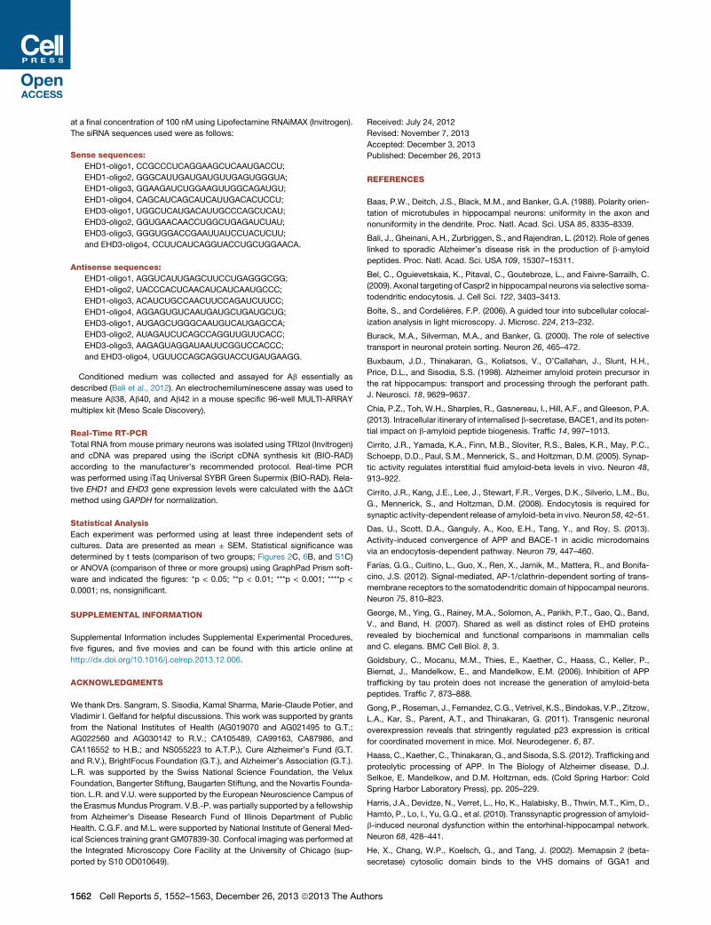
Internalized BACE1 Undergoes Exclusive RetrogradeTransport in DendritesIn normal human brain, BACE1 localizes to the neuronal soma,
dendrites, and axons; BACE1 distribution overlaps with that of
MAP2 in apical dendrites of hippocampal neurons (Figure S2A).
In cultured hippocampal neurons (12 days in vitro; DIV12),
BACE1-YFP localized to both the somatodendritic compart-
ments and the axons (Figures S2B and S2C). In dendrites,
BACE1 can be found along the dendritic surface and in internal
carriers as well as within dendritic spines, which are closely
apposed to presynaptic sites marked by Bassoon immunostain-
ing (Figures S2C and S2D). Endocytosed BACE1 also localized
to both somatodendritic compartments and axons of hippocam-
pal neurons, as determined by MAP2 immunostaining following
BTX uptake (Figure 2A). In order to visualize the dynamic trans-
port of total BACE1 (YFP fluorescence) or internalized BACE1
(BTX fluorescence) in dendrites, we performed live-cell imaging.
There was substantial variability in BACE1 motility in different
dendritic branches of each neuron during the period of image
acquisition, such that vesicles containing BACE1 underwent
active transport in some dendrites, whereas they remained
largely stationary in others. Therefore, we focused on dendrites
that exhibited the greatest BACE1 vesicle motility in each neuron
and generated kymographs to analyze the results (Figures 2B
and 2C; Movies S1 and S2). As expected, BACE1-YFP fluores-
cence was observed in vesicles that were transported in both
anterograde and retrograde directions along dendrites. Quanti-
tative analysis revealed that an approximately equal number of
BACE1-containing vesicles underwent anterograde and retro-
grade transport (Figure 2C). Intriguingly, we found that nearly
all of the motile vesicles (>95%) containing internalized BACE1
underwent retrograde transport, toward the cell body in proximal
(Figures 2B and 2C; Movie S1) as well as in distal dendritic
segments (Figure 2D; Movie S2). This vectorial retrograde move-
ment of BTX-labeled endocytic BACE1 in dendrites was unex-
pected because, to our knowledge, this has not previously
ports 5, 1552–1563, December 26, 2013 ª2013 The Authors 1553
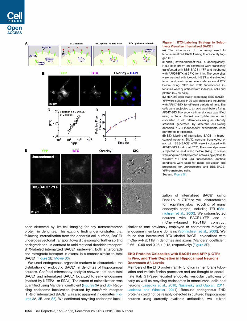
Figure 1. BTX-Labeling Strategy to Selec-
tively Visualize Internalized BACE1
(A) The schematics of the assay used to
label internalized BACE1 using fluorescently tag-
ged BTX.
(B and C) Development of the BTX-labeling assay.
HeLa cells grown on coverslips were transiently
transfected with BBS-BACE1-YFP and incubated
with AF555-BTX at 37�C for 1 hr. The coverslips
were washed with ice-cold HBSS and subjected
to an acid wash to remove surface-bound BTX
before fixing. YFP and BTX fluorescence in-
tensities were quantified from individual cells and
plotted (n = 50 cells).
(D) HEK293 cells stably expressing BBS-BACE1-
YFPwere cultured in 96-well dishes and incubated
with AF647-BTX for different periods of time. The
cells were subjected to an acid wash before fixing.
AF647-BTX fluorescence intensity was quantified
using a Tecan Safire2 microplate reader and
converted to fold differences using an intensity
standard generated by different cell-plating
densities. n = 3 independent experiments, each
performed in triplicates.
(E) BTX labeling of internalized BACE1 in hippo-
campal neurons. DIV12 neurons transfected or
not with BBS-BACE1-YFP were incubated with
AF647-BTX for 4 hr at 37�C. The coverslips were
subjected to acid wash before fixing. z stacks
were acquired and projected onto a single plane to
visualize YFP and BTX fluorescence. Identical
conditions were used for image acquisition and
processing for untransfected and BBS-BACE-
YFP-transfected cells.
See also Figure S1.
been observed by live-cell imaging for any transmembrane
protein in dendrites. This exciting finding demonstrates that
following internalization from the dendritic cell-surface, BACE1
undergoes vectorial transport toward the soma for further sorting
or degradation. In contrast to unidirectional dendritic transport,
BTX-labeled internalized BACE1 underwent both anterograde
and retrograde transport in axons, in a manner similar to total
BACE1 (Figure 2E; Movie S3).
We used endogenous organelle markers to characterize the
distribution of endocytic BACE1 in dendrites of hippocampal
neurons. Confocal microscopy analysis showed that both total
BACE1 and internalized BACE1 localized to early endosomes
(marked by NEEP21 or EEA1). The extent of colocalization was
quantified using Manders’ coefficient (Figures 3A and S3). Recy-
cling endosome localization (marked by transferrin receptor
[TfR]) of internalized BACE1 was also apparent in dendrites (Fig-
ures 3A, 3B, and S3). We confirmed recycling endosome locali-
1554 Cell Reports 5, 1552–1563, December 26, 2013 ª2013 The Authors
zation of internalized BACE1 using
Rab11b, a GTPase well characterized
for regulating slow recycling of many
endocytic cargos, including TfR (Son-
nichsen et al., 2000). We cotransfected
neurons with BACE1-YFP and a
mCherry-tagged Rab11B construct
similar to one previously employed to characterize recycling
endosome membrane domains (Sonnichsen et al., 2000). We
found that internalized BTX-labeled BACE1 colocalized with
mCherry-Rab11B in dendrites and axons (Manders’ coefficient
0.66 ± 0.09 and 0.28 ± 0.15, respectively) (Figure 3D).
EHD Proteins Colocalize with BACE1 and APP b-CTFsIn Vivo, and Their Depletion in Hippocampal NeuronsDecreases Ab LevelsMembers of the EHD protein family function in membrane tubu-
lation and vesicle fission processes and are thought to coordi-
nate Rab GTPase-mediated endocytic vesicular trafficking of
early as well as recycling endosomes in nonneuronal cells and
neurons (Lasiecka et al., 2010; Naslavsky and Caplan, 2011;
Lasiecka and Winckler, 2011). Because endogenous EHD
proteins could not be reliably detected in cultured hippocampal
neurons using currently available antibodies, we utilized

Figure 2. Dynamic Characteristics of Inter-
nalized BACE1
(A) Localization of internalized BACE1 in dendrites
and axons. DIV12 neurons were transfected with
BBS-BACE1-YFP and internalized BACE1 was
labeled by incubation with AF647-BTX for 4 hr
at 37�C followed by acid wash to remove surface-
bound BTX. Deconvolved images of a transfected
neuron depict the distribution of total and
internalized pool of BACE1 in the cell body and
dendrites (top) and the corresponding axonal
network (bottom).
(B) Unidirectional transport of internalized BACE1
in dendrites. Time-lapse images of total (YFP
fluorescence) and internalized BACE1 (BTX fluo-
rescence) were sequentially acquired following
AF647-BTX uptake, at the rate of one frame/sec
for 3 min. Images of a dendritic segment, with the
cell body on the left, and the corresponding ky-
mographs of total and internalized BACE1, are
shown.
(C) Quantification of total and internalized BACE1
motility in dendrites (n = 760 total and 699 inter-
nalized carriers from 18 neurons). Error bars
represent SEM. Unlike the bidirectional transport
of total BACE1 in dendrites, internalized BACE1
undergoes transport exclusively in the retrograde
direction.
(D) A still image from the first frame of Movie S2
depicting internalized BACE1 dynamic transport in
a distal dendrite. The retrograde transport tracks
of representative transport vesicles in different
branches of the same dendrite are indicated.
(E) Dynamic axonal transport of internalized
BACE1. Time-lapse images of total and internal-
ized BACE1 were sequentially acquired from
neurons following AF555-BTX uptake, at the rate
of one frame/sec for 3 min. Bidirectional axonal
transport of total and internalized BACE1 were
visualized by generating kymographs.
See also Figure S2.
previously described DsRed-tagged EHDs (George et al., 2007).
In transfected neurons, EHD1 and EHD3 colocalized with BACE1
in dendrites, dendritic spines, and neuronal soma, as well as in
axons (Figures 4A–4C). Internalized BACE1, labeled using BTX
uptake, also showed extensive colocalization with EHD1 and
EHD3 in dendrites and dendritic spines, and along the axons
(Figures 4A and 4B). Results of Manders’ coefficient of colocal-
ization are depicted in (Figure 4C). Together, the results
described above show that a significant fraction of BACE1 local-
izes to endocytic organelles along dendrites and axons.
Three of the four known mammalian EHD proteins (EHD1,
EHD3, and EHD4) were detected in mouse brain lysates by
immunoblot analysis (George et al., 2007), but the cellular and
subcellular localization of EHDs in the brain is unknown. To
determine whether EHD proteins are expressed in the hippo-
Cell Reports 5, 1552–1563, De
campus, we immunostained mouse brain
sections using antibodies that specifically
recognize individual EHD familymembers
(George et al., 2007) and performed
confocal microscopy analysis. Consistent with the immunoblot
data, expression of EHD1, EHD3, and EHD4 was readily detec-
tible by immunostaining in mouse brain, whereas EHD2 was
not detectable (Figures 5 and S3). Interestingly, EHD1 and
EHD3 prominently localized in hippocampal mossy fibers where
they colocalized with BACE1 (Figure 5A). EHD4 expression
was mainly restricted to the cell bodies of neurons in the dentate
gyrus and CA1–CA3 (Figure S4).
To assess whether EHD1 and 3 function in endosomal traf-
ficking affects amyloidogenic processing of APP, we analyzed
the effect of small interfering RNA (siRNA) knockdown of EHD1
and EHD3 in cultured neurons. In these experiments, we
achieved knockdown efficiencies of 64% (EHD1) and 61%
(EHD3) 72 hr after transfection with siRNAs, as assayed by quan-
titative PCR. Media conditioned by siRNA-treated neurons were
cember 26, 2013 ª2013 The Authors 1555

Figure 3. Subcellular Localization of Internalized BACE1
(A and B) Neurons transfected with BBS-BACE1-YFP were labeled by AF647-
BTX uptake, subjected to acid wash, and immunolabeled for EEA1, NEEP21,
and TfR. Manders’ coefficient of colocalization with endosome markers was
quantified by confocal microscopy for total BACE1 [YFP] (A; n = 11–16
neurons) and internalized BACE1 [BTX fluorescence] (B; n = 7–13 neurons).
Representative confocal images are depicted in Figure S3.
(C) Representative confocal images of a dendrite segment depicting coloc-
alization of internalized BACE1 dendrites and spines with endogenous TfR.
Scale bar, 10 mm.
(D) Neurons cotransfected with BACE1-YFP and mCherry-Rab11b were
labeled by AF647-BTX uptake, subjected to acid wash, and immunostained for
MAP2. The extent of colocalization of Rab11b and internalized BACE1 in
dendrites and axons was quantified (n = 8 neurons).
Error bars represent SEM. See also Figure S3.
assayed using a sensitive electrochemiluminescence assay to
quantify the levels of secreted Ab. We observed a significant
decrease of Ab38, Ab40, and Ab42 peptides secreted by neu-
rons following knockdown of EHD1 or EHD3 (Figure 4D).
To determine whether EHD’s influence on APP processing
might be relevant in vivo, we examined colocalization between
EHD and BACE1-cleaved APP b-C-terminal fragments (b-CTF)
in a transgenic mouse model of Alzheimer’s disease patho-
genesis. Confocal analysis revealed that both EHD1 and EHD3
show substantial overlap with APP b-CTF and/or Ab in mossy
fiber terminals in APPswe/PS1DE9 transgenic mouse brain
1556 Cell Reports 5, 1552–1563, December 26, 2013 ª2013 The Aut
(analyzed at 2 months, before the onset of overt extracellular
Ab deposition) (Figure 5B), suggesting that in mouse brain,
BACE1 in EHD-positive structures likely mediates the process-
ing of APP to b-CTF, the precursor of Ab. Together, these results
suggest a role for EHD1 and EHD3 in APP processing in neurons.
EHD1 and EHD3 Regulate Dynamic BACE1 Transport inHippocampal NeuronsThe results described above prompted us to investigate whether
EHD1 and EHD3 coordinate BACE1 transport in neurons. For
these studies, we chose to employ a dominant-negative (DN)
approach that has been successfully used in previous investi-
gations (George et al., 2007; Yap et al., 2010) because DN plas-
mids can be efficiently transfected into mature neurons to
compromise EHD function in less than 24 hr (unlike siRNA
approach, which requires 3 days to achieve knockdown of
EHD expression following transfection of young neurons at
DIV5). We performed live-cell imaging and generated kymo-
graphs to assess whether unidirectional retrograde transport of
internalized BACE1 was affected by expression of EHD DN
mutants that lack the EH domain (EHD-DEH) (Figure 6A; Movie
S4). Quantitative analysis revealed a significant decrease in the
fraction of retrograde transport of dendritic carriers containing
internalized BACE1 in neurons expressing DN mutant EHD1 or
EHD3 as compared with those expressing wild-type (WT)
proteins (Figure 6B). This effect was extremely robust such
that a marked reduction in retrograde transport of vesicles was
evident just from quantification of total BACE1 transport in
dendrites; nevertheless, anterograde transport of BACE1-YFP
remained unchanged by DN EHD expression (Figure 6B).
Together, these results reveal a functional requirement of
EHD1 and EHD3 for the dynamic retrograde transport of
BACE1-containing endosomes in dendrites.
We then asked whether EHD function was required for
membrane sorting of internalized BACE1 in endocytic organelles
and/or for the retrograde transport of BACE1 in endocytic
carriers. Coexpression of EHD1-DEH or EHD3-DEH with
BACE1 had no significant effect on steady-state BACE1 locali-
zation at the dendritic surface, as measured by surface BTX
labeling (the surface/total fluorescence ratios normalized to
vector controls were EHD1 WT, 1.0 ± 0.11; EHD1-DEH, 0.89 ±
0.12; EHD3 WT, 1.0 ± 0.21; EHD3-DEH, 1.05 ± 0.13 [p =
0.9272; n = 21–28 neurons each]). In agreement, localization
of endocytosed BACE1 in somatodendritic compartments
following BTX uptake was apparent in neurons transfected
with WT or DN EHDs (Figure 7A). Quantitative analysis of
organelle marker staining revealed no increase of internalized
BACE1 in endosomes positive for EEA1 or NEEP21, suggesting
that that the expression of EHD-DEH mutants did not cause a
retention of internalized BACE1 in early endosomes (Figure S5).
Furthermore, there was no decrease in the extent of colocaliza-
tion with TfR, suggesting that internalized BACE1 is able to
reach TfR-positive recycling endosomes (Figure S5). We noted
a small increase in the extent of BACE1 colocalization with TfR
in neurons expressing DN EHD proteins. This increase may be
mechanistically important because EHD1/3 could regulate
BACE1 exit from the TfR-positive endocytic recycling
compartment.
hors
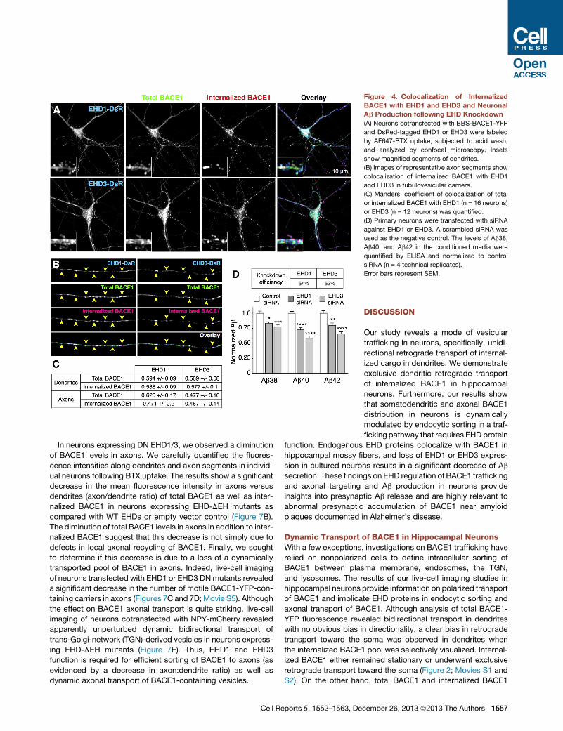
Figure 4. Colocalization of Internalized
BACE1 with EHD1 and EHD3 and Neuronal
Ab Production following EHD Knockdown
(A) Neurons cotransfected with BBS-BACE1-YFP
and DsRed-tagged EHD1 or EHD3 were labeled
by AF647-BTX uptake, subjected to acid wash,
and analyzed by confocal microscopy. Insets
show magnified segments of dendrites.
(B) Images of representative axon segments show
colocalization of internalized BACE1 with EHD1
and EHD3 in tubulovesicular carriers.
(C) Manders’ coefficient of colocalization of total
or internalized BACE1 with EHD1 (n = 16 neurons)
or EHD3 (n = 12 neurons) was quantified.
(D) Primary neurons were transfected with siRNA
against EHD1 or EHD3. A scrambled siRNA was
used as the negative control. The levels of Ab38,
Ab40, and Ab42 in the conditioned media were
quantified by ELISA and normalized to control
siRNA (n = 4 technical replicates).
Error bars represent SEM.
In neurons expressing DN EHD1/3, we observed a diminution
of BACE1 levels in axons. We carefully quantified the fluores-
cence intensities along dendrites and axon segments in individ-
ual neurons following BTX uptake. The results show a significant
decrease in the mean fluorescence intensity in axons versus
dendrites (axon/dendrite ratio) of total BACE1 as well as inter-
nalized BACE1 in neurons expressing EHD-DEH mutants as
compared with WT EHDs or empty vector control (Figure 7B).
The diminution of total BACE1 levels in axons in addition to inter-
nalized BACE1 suggest that this decrease is not simply due to
defects in local axonal recycling of BACE1. Finally, we sought
to determine if this decrease is due to a loss of a dynamically
transported pool of BACE1 in axons. Indeed, live-cell imaging
of neurons transfected with EHD1 or EHD3 DNmutants revealed
a significant decrease in the number of motile BACE1-YFP-con-
taining carriers in axons (Figures 7C and 7D;Movie S5). Although
the effect on BACE1 axonal transport is quite striking, live-cell
imaging of neurons cotransfected with NPY-mCherry revealed
apparently unperturbed dynamic bidirectional transport of
trans-Golgi-network (TGN)-derived vesicles in neurons express-
ing EHD-DEH mutants (Figure 7E). Thus, EHD1 and EHD3
function is required for efficient sorting of BACE1 to axons (as
evidenced by a decrease in axon:dendrite ratio) as well as
dynamic axonal transport of BACE1-containing vesicles.
Cell Reports 5, 1552–1563, De
DISCUSSION
Our study reveals a mode of vesicular
trafficking in neurons, specifically, unidi-
rectional retrograde transport of internal-
ized cargo in dendrites. We demonstrate
exclusive dendritic retrograde transport
of internalized BACE1 in hippocampal
neurons. Furthermore, our results show
that somatodendritic and axonal BACE1
distribution in neurons is dynamically
modulated by endocytic sorting in a traf-
ficking pathway that requires EHD protein
function. Endogenous EHD proteins colocalize with BACE1 in
hippocampal mossy fibers, and loss of EHD1 or EHD3 expres-
sion in cultured neurons results in a significant decrease of Ab
secretion. These findings on EHD regulation of BACE1 trafficking
and axonal targeting and Ab production in neurons provide
insights into presynaptic Ab release and are highly relevant to
abnormal presynaptic accumulation of BACE1 near amyloid
plaques documented in Alzheimer’s disease.
Dynamic Transport of BACE1 in Hippocampal NeuronsWith a few exceptions, investigations on BACE1 trafficking have
relied on nonpolarized cells to define intracellular sorting of
BACE1 between plasma membrane, endosomes, the TGN,
and lysosomes. The results of our live-cell imaging studies in
hippocampal neurons provide information on polarized transport
of BACE1 and implicate EHD proteins in endocytic sorting and
axonal transport of BACE1. Although analysis of total BACE1-
YFP fluorescence revealed bidirectional transport in dendrites
with no obvious bias in directionality, a clear bias in retrograde
transport toward the soma was observed in dendrites when
the internalized BACE1 pool was selectively visualized. Internal-
ized BACE1 either remained stationary or underwent exclusive
retrograde transport toward the soma (Figure 2; Movies S1 and
S2). On the other hand, total BACE1 and internalized BACE1
cember 26, 2013 ª2013 The Authors 1557

Figure 5. Colocalization of Endogenous EHD1 and EHD3 with BACE1 and APP b-CTFs in Mouse Brain
(A) Sagittal sections of P19 mouse brain were immunostained with antibodies against BACE1 and EHD1 or EHD3 and confocal images were acquired using 103
(top) and 1003 objectives (bottom). Insets show higher magnification of the boxed area within the terminal field of mossy fibers (MF).
(B) Sagittal sections of 2-month-old APP/PS1 transgenic mouse brain were stained with antibodies against APP b-CTF (monoclonal antibody [mAb] 3D6) and
EHD1 or EHD3 and analyzed by confocal microscopy. Lack of strongmAb 3D6 staining inmossy fibers of nontransgenicmice (NTg, inset) indicates the selectively
of this antibody for APP b-CTFs and/or Ab.
See also Figure S4.
underwent bidirectional axonal transport. The vectorial transport
of internalized BACE1 in dendrites shares some similarity with
the overall fate of receptors such as L1/NgCAM and TrkA, which
are axonally targeted following endocytosis from the somato-
dendritic cell surface (Yap and Winckler, 2012). However, our
live-cell imaging results of BACE1 endocytic transport charac-
terize the dynamic and exclusive retrograde transport of endocy-
tosed transmembrane protein cargo from dendrites.
The bidirectional and saltatory nature of BACE1 transport in
neurons are characteristics of microtubule-based transport.
Unlike the uniform orientation of microtubules in axons with their
plus ends distal to the cell body, dendrites of mature neurons
have an equal number of microtubules with their plus ends
oriented toward or away from the cell body (Baas et al., 1988).
Therefore, it is remarkable that dynamic transport of internalized
BACE1 is bidirectional in axons, whereas it is only retrograde in
dendrites. Not only do the transport vesicles containing internal-
ized BACE1 need to distinguish between the plus end and minus
end of microtubules in the near vicinity, they must also employ
long-range guidance cues in order to navigate through dendritic
branch points in a unidirectional manner and progressively move
toward the soma (see Movie S2). In comparison, TfR and inter-
1558 Cell Reports 5, 1552–1563, December 26, 2013 ª2013 The Aut
nalized transferrin are known to undergo bidirectional transport
in dendrites (Prekeris et al., 1999; Burack et al., 2000; Lasiecka
et al., 2010; Farıas et al., 2012; data not shown). How internalized
BACE1-containing endosomes in dendrites achieve unidirec-
tional retrograde movement toward the soma, despite microtu-
bule orientation in both directions, is an intriguing question that
will require further studies.
EHD Proteins Function in Transport of InternalizedBACE1The four mammalian EHD paralogs show partial overlap in intra-
cellular localization and function as homo- or hetero-oligomers.
Together, they regulate endocytic trafficking of a number of
receptors such as TfR, GLUT4 glucose transporter, AMPA
receptor, and L1/NgCAM (Naslavsky and Caplan, 2011). We
found that, in mouse brain, EHD1 and EHD3 colocalized with
BACE1 and APP b-C-terminal fragments in large mossy fiber
terminals. In cultured hippocampal neurons, expression of
EHD1-DEH or EHD3-DEHDNmutant resulted in the loss of retro-
grade transport of internalized BACE1 in dendrites. However,
because EHD DN expression did not cause aberrant retention
of internalized BACE1 in EEA1- or NEEP21-positive early
hors

Figure 6. EHD1 and EHD3 Are Required for
Retrograde Transport of Internalized
BACE1 in Dendrites
(A) Neurons cotransfected with BBS-BACE1-YFP
and the indicated DsRed-tagged EHDWT or EHD-
DEH construct were labeled by AF647-BTX up-
take. Representative kymographs of internalized
BACE1 transport in dendrites are shown.
(B) Quantification of total and internalized BACE1
motility in dendrites. EHD1WT n = 679 total (T) and
312 internalized (I) vesicles from 13 neurons;
EHD1-DEH n = 427 T- and 227 I-vesicles from
eight neurons; EHD3 WT n = 546 T and 291
I-vesicles from 12 neurons; EHD3-DEH n = 462 T-
and 182 I-vesicles from nine neurons.
Error bars represent SEM.
endosomes, it appears that the initial steps in membrane sorting
of BACE1 upon endocytosis in dendrites are EHD independent.
In axons, there was an overall decrease in the levels of total as
well as internalized BACE1 (Figures 7A and 7B). Interestingly,
the loss of the motile BACE1 pool in axons is particularly striking,
especially when transport of TGN-derived vesicles was unper-
turbed by DN EHD expression (Figures 7C and 7E). Together,
these findings indicate that EHD1 and EHD3 have a critical role
in endosomal transport of BACE1 and axonal targeting and/or
sorting of motile vesicles containing BACE1. It is known that
endosomes containing EHD1 traffic bidirectionally along den-
drites (Lasiecka et al., 2010). Internalized BACE1 shows clear
colocalization with EHD1 and EHD3 (Figure 4). Yet, nearly all
the motile vesicles containing internalized BACE1 in dendrites
Cell Reports 5, 1552–1563, De
undergo retrograde transport toward the
soma, raising the possibility that BACE1
might play an active role in vectorial
transport of a subset of endocytic vesi-
cles in hippocampal neurons.
Endocytic Transport of BACE1 IsDistinct from Transcytosis of CellAdhesion MoleculesThe characteristics of endocytic sorting
and the ultimate fate of internalized
BACE1 in hippocampal neurons share
some similarities, but also differ consider-
ably, when compared with trafficking of
cell adhesion molecules such as Caspr2
and L1/NgCAM, which also undergo
endocytic sorting following internalization
from the somatodendritic compartment.
Unlike Caspr2 or L1/NgCAM, BACE1 is
uniformly distributed on the surface of
dendrites and axons at steady state,
and internalized BACE1 is also readily
detected in both dendrites and axons.
Overexpression of either WT or DN EHD
proteins markedly reduces L1/NgCAM
endocytosis in dendrites (Yap et al.,
2010). However, this does not appear to
be the case for BACE1; colocalization of internalized BACE1
with EEA1, NEEP21, and TfR was not reduced by EHD DN
expression. Instead, retrograde motility of internalized BACE1-
containing vesicles in dendrites was compromised. In the case
of Caspr2, nonpolarized biosynthetic delivery to somatoden-
dritic and axonal plasma membrane is followed by compart-
ment-specific endocytosis and elimination of somatodendritic
Caspr2 (Bel et al., 2009). By contrast, biosynthetic L1/NgCAM
internalized from the somatodendritic plasma membrane is
transported in nondegradative endosomes and delivered to
axonal plasma membrane (Yap et al., 2008). Similar to L1/
NgCAM, endocytic elimination does not appear to be the reason
why BACE1 internalized from dendrites is transported to the
soma. Rather, loss of retrograde transport in dendrites is
cember 26, 2013 ª2013 The Authors 1559
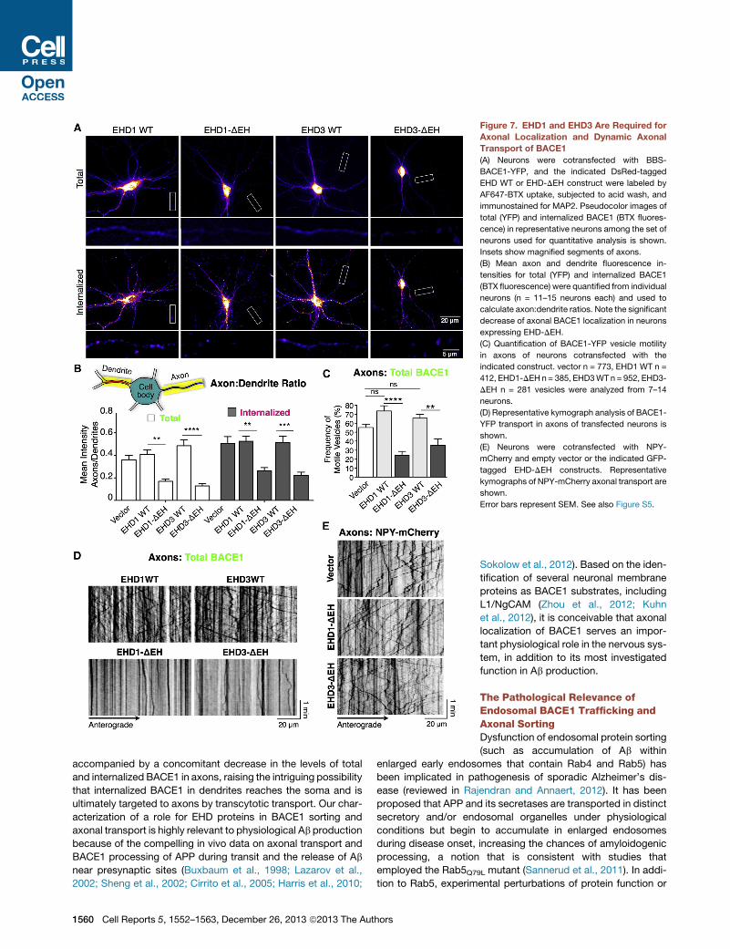
Figure 7. EHD1 and EHD3 Are Required for
Axonal Localization and Dynamic Axonal
Transport of BACE1
(A) Neurons were cotransfected with BBS-
BACE1-YFP, and the indicated DsRed-tagged
EHD WT or EHD-DEH construct were labeled by
AF647-BTX uptake, subjected to acid wash, and
immunostained for MAP2. Pseudocolor images of
total (YFP) and internalized BACE1 (BTX fluores-
cence) in representative neurons among the set of
neurons used for quantitative analysis is shown.
Insets show magnified segments of axons.
(B) Mean axon and dendrite fluorescence in-
tensities for total (YFP) and internalized BACE1
(BTX fluorescence) were quantified from individual
neurons (n = 11–15 neurons each) and used to
calculate axon:dendrite ratios. Note the significant
decrease of axonal BACE1 localization in neurons
expressing EHD-DEH.
(C) Quantification of BACE1-YFP vesicle motility
in axons of neurons cotransfected with the
indicated construct. vector n = 773, EHD1 WT n =
412, EHD1-DEH n= 385, EHD3WT n = 952, EHD3-
DEH n = 281 vesicles were analyzed from 7–14
neurons.
(D) Representative kymograph analysis of BACE1-
YFP transport in axons of transfected neurons is
shown.
(E) Neurons were cotransfected with NPY-
mCherry and empty vector or the indicated GFP-
tagged EHD-DEH constructs. Representative
kymographs of NPY-mCherry axonal transport are
shown.
Error bars represent SEM. See also Figure S5.
accompanied by a concomitant decrease in the levels of total
and internalized BACE1 in axons, raising the intriguing possibility
that internalized BACE1 in dendrites reaches the soma and is
ultimately targeted to axons by transcytotic transport. Our char-
acterization of a role for EHD proteins in BACE1 sorting and
axonal transport is highly relevant to physiological Ab production
because of the compelling in vivo data on axonal transport and
BACE1 processing of APP during transit and the release of Ab
near presynaptic sites (Buxbaum et al., 1998; Lazarov et al.,
2002; Sheng et al., 2002; Cirrito et al., 2005; Harris et al., 2010;
1560 Cell Reports 5, 1552–1563, December 26, 2013 ª2013 The Authors
Sokolow et al., 2012). Based on the iden-
tification of several neuronal membrane
proteins as BACE1 substrates, including
L1/NgCAM (Zhou et al., 2012; Kuhn
et al., 2012), it is conceivable that axonal
localization of BACE1 serves an impor-
tant physiological role in the nervous sys-
tem, in addition to its most investigated
function in Ab production.
The Pathological Relevance ofEndosomal BACE1 Trafficking andAxonal SortingDysfunction of endosomal protein sorting
(such as accumulation of Ab within
enlarged early endosomes that contain Rab4 and Rab5) has
been implicated in pathogenesis of sporadic Alzheimer’s dis-
ease (reviewed in Rajendran and Annaert, 2012). It has been
proposed that APP and its secretases are transported in distinct
secretory and/or endosomal organelles under physiological
conditions but begin to accumulate in enlarged endosomes
during disease onset, increasing the chances of amyloidogenic
processing, a notion that is consistent with studies that
employed the Rab5Q79L mutant (Sannerud et al., 2011). In addi-
tion to Rab5, experimental perturbations of protein function or

alterations in the expression of several mediators of endocytic
sorting, such as NEEP21, sorLA/LR11, GGA family members,
retromer complex subunits, ADP-ribosylation factor 6, sorting
nexin 17, and phosphatidylinositol clathrin assembly lymphoid-
myeloid leukemia protein (PICALM), have been reported to affect
APP processing and Ab production. We have identified EHD1
and EHD3 as regulators of BACE1 trafficking in neurons. Knock-
down of EHD expression in primary neurons significantly
reduced Ab secretion, implicating EHD function in neuronal
APP processing. These findings may also have functional rele-
vance to abnormal presynaptic accumulation of BACE1 near
amyloid plaques documented in Alzheimer’s disease (Zhao
et al., 2007). Interestingly, Rab11 GTPase, which forms a com-
plex with EHD proteins via Rab11FIP2, was independently iden-
tified as one of the two major regulators of Ab production in
mammalian cells in an unbiased paired RNAi screen of human
Rab GTPases and Rab GTPase-activating proteins (Udayar
et al., 2013). In conclusion, our studies demonstrate a role for
EHD proteins in regulating unidirectional dendritic BACE1 trans-
port and axonal sorting and provide insights into cellular mech-
anisms responsible for BACE1 localization and Ab release near
presynaptic sites.
EXPERIMENTAL PROCEDURES
cDNA Constructs
C-terminally EYFP-tagged BACE1 was generated by subcloning mouse
BACE1 cDNA (provided by Dr. Nabil G. Seidah) in-frame into the pEYFP-N1
vector. A similar strategy was used to generate BACE1-Cerulean. A 13-
amino-acid a-Bungaratoxin Binding Site (BBS) (Sekine-Aizawa and Huganir,
2004) was inserted after the prodomain of BACE1, to generate BBS-BACE1-
YFP. The dileucine mutation (L499A/L500A) was introduced by PCRmutagen-
esis. To generate mouse Rab11B, a mouse brain PCR product that codes for
amino acids 72 to 218 was exchanged for the corresponding region in HA-
tagged Rab11A construct (Ren et al., 1998). The cDNA inserts were then sub-
cloned in-frame into the pmCherry-C1 vector. All constructs were verified by
sequencing. DsRed-, GFP-, or myc-tagged human EHD1 and three plasmids
have been described (George et al., 2007). NPY-mCherry was provided by Dr.
Gary Banker.
BTX-Labeling Assay Development
HeLa cells plated on coverslips were transfected with BBS-BACE1WT-YFP
using Lipofectamine 2000. For the BTX uptake experiments, cells were incu-
bated the next day with 6.7 mg/ml AF555-BTX (Molecular Probes) at 37�C for
1 hr. Coverslips were then gently washed twice in cold Hanks’ balanced salt
solution with 10 mM HEPES (pH 7.3) (HBSS). BTX bound to cell-surface
BACE1 was removed by incubation in an acidic solution (0.5 M NaCl and
0.2 M acetic acid [pH 2.8]) for 5 min on ice (acid wash). For surface labeling,
transfected cells were incubated at 10�C for 20 min with 13.3 mg/ml AF555-
BTX in complete culture medium. The coverslips were washed twice in cold
HBSS, and one set of coverslips was further subjected to acid wash. Cells
were fixed in 4% PFA/4% sucrose for 20 min at room temperature, and nuclei
were stained with 0.25 mg/ml Hoechst 33342 (Molecular Probes) before
mounting the coverslips.
HEK cells stably expressing BBS-BACE1WT-YFP were plated in a 96-well
black glass-bottom plate (Costar) pretreated with poly-L-lysine (Sigma) to
optimize cell adhesion. Eighteen hours after plating, cells were incubated
at 37�C with 4 mg/ml AF647-BTX for a range of time points as indicated
(time course experiment in Figure 1D). After the incubation, cells were
gently washed three times on ice with PBS and fixed. Fluorescence inten-
sities were quantified a Tecan Safire2 microplate reader (Tecan). Nontrans-
fected HEK cells were assayed alongside as a control for background
subtraction. Three wells were used for each time point, and the experiment
Cell Re
was independently repeated three times. A standard curve was generated
in each experiment using a range of cell densities assayed by BTX uptake
for 1 hr.
Labeling of Surface and Internalized BACE1 in Neurons
All animal studies were approved by the Institutional Animal Care and Use
Committee and conducted in accordance with University of Chicago Animal
Care Guide lines. Hippocampal neurons were cultured from E17 mouse
embryos as previously described (Kaech and Banker, 2006). Dissociated
neurons were cultured on poly-D-lysine coated glass coverslips suspended
over a monolayer of primary astrocytes prepared from P0–P2 mouse pups.
Cultures were maintained in Neurobasal supplemented with B27 serum-free
and GlutaMAX-I supplement (Invitrogen). Neurons were transfected with
Lipofectamine 2000 (Invitrogen) on DIV11 and fixed for immunostaining or
used for live-cell imaging between DIV12 and 14. In BTX uptake experiments
AF647- or AF555-conjugated BTX (Invitrogen) was added to the culture
medium (6.6 or 2 mg/ml, respectively), at 37�C for 3–4 hr. Coverslips were
washed with ice-cold Hanks’ balanced salt solution with 10 mM HEPES
(pH 7.3) (HBSS), and BTX bound to cell-surface BACE1 was removed by a
2min acid wash. Subsequently neurons were fixed and stainedwith antibodies
against MAP2 and endosome markers (see Supplemental Experimental Pro-
cedures). For live-cell imaging, coverslips were washed after BTX uptake
with imaging medium (119 mM NaCl, 2.5 mM KCl, 2 mM CaCl2, 2 mM
MgCl2, 30 mM D-glucose, and 25 mM HEPES [pH 7.4]) before image acquisi-
tion. For surface staining, neurons were incubated on ice with culture medium
containing 13.33 mg/ml AF647-BTX for 20 min.
Image Acquisition and Analysis
Wide-field epifluorescence images of fixed neurons were acquired as 200 nm
z stacks using 203 (NA 0.75) or 603 (NA 1.49) objectives. Confocal images
were acquired using a Leica SP5 II STED-CW Superresolution Laser
Scanning Confocal microscope using 103 (NA 0.4) and 1003 (NA 1.4;
zoom 2.5) objectives. Quantitative analysis was performed using Metamorph
(Molecular Devices) and ImageJ software. Additional information on image
processing is detailed in the Supplemental Experimental Procedures. Axonal
and dendritic BACE1 fluorescence intensities were quantified on 603 z stack
projections of neurons using an established method (Sampo et al., 2003;
Farıas et al., 2012). Briefly, the average fluorescence intensities were
measured along 100–200 mm-long one-pixel-wide line segments traced on
two to three representative sections of dendrites and axons in each neuron
using ImageJ. The mean fluorescence intensity in the soma was quantified
by drawing a region around the soma. The average axon:dendrite ratio
was calculated for each neuron. Manders’ coefficient of colocalization of
BACE1 with organelles markers or tagged proteins was calculated on thresh-
olded confocal images of dendrites or 603 deconvolved z stack projections
of axons (identified by MAP2 staining) using JACoP ImageJ plugin (Bolte and
Cordelieres, 2006).
Live-Cell Imaging
Live-cell images were acquired on a motorized Nikon TE 2000 microscope
maintained at 37�C in a custom-designed environment chamber, at the rate
of one frame/s, using 603 (NA 1.49) objective and Cascade II:512CCD camera
(Photometrics). Image stacks were processed in ImageJ software. Kymo-
graphs were generated in Metamorph and used to determine the frequency
and directionality of movement of transport vesicles that moved at the rate
of >0.1 mm/sec.
Immunohistochemistry and Immunofluorescence Staining
The antibodies and methods used for staining human and mouse brain tissue
are described under Supplemental Experimental Procedures (Zhao et al.,
2007; Gong et al., 2011).
siRNA Transfection in Primary Neurons and Ab Quantification
Chemically synthesized stealth siRNAs from Invitrogen were used to knock
down endogenous EHD1 and EHD3 expression in primary mouse neuronal
cultures. A pool of four different siRNAs was transfected into DIV5 neurons
ports 5, 1552–1563, December 26, 2013 ª2013 The Authors 1561
at a final concentration of 100 nM using Lipofectamine RNAiMAX (Invitrogen).
The siRNA sequences used were as follows:
Sense sequences:
EHD1-oligo1, CCGCCCUCAGGAAGCUCAAUGACCU;
EHD1-oligo2, GGGCAUUGAUGAUGUUGAGUGGGUA;
EHD1-oligo3, GGAAGAUCUGGAAGUUGGCAGAUGU;
EHD1-oligo4, CAGCAUCAGCAUCAUUGACACUCCU;
EHD3-oligo1, UGGCUCAUGACAUUGCCCAGCUCAU;
EHD3-oligo2, GGUGAACAACCUGGCUGAGAUCUAU;
EHD3-oligo3, GGGUGGACCGAAUUAUCCUACUCUU;
and EHD3-oligo4, CCUUCAUCAGGUACCUGCUGGAACA.
Antisense sequences:
EHD1-oligo1, AGGUCAUUGAGCUUCCUGAGGGCGG;
EHD1-oligo2, UACCCACUCAACAUCAUCAAUGCCC;
EHD1-oligo3, ACAUCUGCCAACUUCCAGAUCUUCC;
EHD1-oligo4, AGGAGUGUCAAUGAUGCUGAUGCUG;
EHD3-oligo1, AUGAGCUGGGCAAUGUCAUGAGCCA;
EHD3-oligo2, AUAGAUCUCAGCCAGGUUGUUCACC;
EHD3-oligo3, AAGAGUAGGAUAAUUCGGUCCACCC;
and EHD3-oligo4, UGUUCCAGCAGGUACCUGAUGAAGG.
Conditioned medium was collected and assayed for Ab essentially as
described (Bali et al., 2012). An electrochemiluminescene assay was used to
measure Ab38, Ab40, and Ab42 in a mouse specific 96-well MULTI-ARRAY
multiplex kit (Meso Scale Discovery).
Real-Time RT-PCR
Total RNA from mouse primary neurons was isolated using TRIzol (Invitrogen)
and cDNA was prepared using the iScript cDNA synthesis kit (BIO-RAD)
according to the manufacturer’s recommended protocol. Real-time PCR
was performed using iTaq Universal SYBR Green Supermix (BIO-RAD). Rela-
tive EHD1 and EHD3 gene expression levels were calculated with the DDCt
method using GAPDH for normalization.
Statistical Analysis
Each experiment was performed using at least three independent sets of
cultures. Data are presented as mean ± SEM. Statistical significance was
determined by t tests (comparison of two groups; Figures 2C, 6B, and S1C)
or ANOVA (comparison of three or more groups) using GraphPad Prism soft-
ware and indicated the figures: *p < 0.05; **p < 0.01; ***p < 0.001; ****p <
0.0001; ns, nonsignificant.
SUPPLEMENTAL INFORMATION
Supplemental Information includes Supplemental Experimental Procedures,
five figures, and five movies and can be found with this article online at
http://dx.doi.org/10.1016/j.celrep.2013.12.006.
ACKNOWLEDGMENTS
We thank Drs. Sangram, S. Sisodia, Kamal Sharma, Marie-Claude Potier, and
Vladimir I. Gelfand for helpful discussions. This work was supported by grants
from the National Institutes of Health (AG019070 and AG021495 to G.T.;
AG022560 and AG030142 to R.V.; CA105489, CA99163, CA87986, and
CA116552 to H.B.; and NS055223 to A.T.P.), Cure Alzheimer’s Fund (G.T.
and R.V.), BrightFocus Foundation (G.T.), and Alzheimer’s Association (G.T.).
L.R. was supported by the Swiss National Science Foundation, the Velux
Foundation, Bangerter Stiftung, Baugarten Stiftung, and the Novartis Founda-
tion. L.R. and V.U. were supported by the European Neuroscience Campus of
the ErasmusMundus Program. V.B.-P. was partially supported by a fellowship
from Alzheimer’s Disease Research Fund of Illinois Department of Public
Health. C.G.F. and M.L. were supported by National Institute of General Med-
ical Sciences training grant GM07839-30. Confocal imaging was performed at
the Integrated Microscopy Core Facility at the University of Chicago (sup-
ported by S10 OD010649).
1562 Cell Reports 5, 1552–1563, December 26, 2013 ª2013 The Aut
Received: July 24, 2012
Revised: November 7, 2013
Accepted: December 3, 2013
Published: December 26, 2013
REFERENCES
Baas, P.W., Deitch, J.S., Black, M.M., and Banker, G.A. (1988). Polarity orien-
tation of microtubules in hippocampal neurons: uniformity in the axon and
nonuniformity in the dendrite. Proc. Natl. Acad. Sci. USA 85, 8335–8339.
Bali, J., Gheinani, A.H., Zurbriggen, S., and Rajendran, L. (2012). Role of genes
linked to sporadic Alzheimer’s disease risk in the production of b-amyloid
peptides. Proc. Natl. Acad. Sci. USA 109, 15307–15311.
Bel, C., Oguievetskaia, K., Pitaval, C., Goutebroze, L., and Faivre-Sarrailh, C.
(2009). Axonal targeting of Caspr2 in hippocampal neurons via selective soma-
todendritic endocytosis. J. Cell Sci. 122, 3403–3413.
Bolte, S., and Cordelieres, F.P. (2006). A guided tour into subcellular colocal-
ization analysis in light microscopy. J. Microsc. 224, 213–232.
Burack, M.A., Silverman, M.A., and Banker, G. (2000). The role of selective
transport in neuronal protein sorting. Neuron 26, 465–472.
Buxbaum, J.D., Thinakaran, G., Koliatsos, V., O’Callahan, J., Slunt, H.H.,
Price, D.L., and Sisodia, S.S. (1998). Alzheimer amyloid protein precursor in
the rat hippocampus: transport and processing through the perforant path.
J. Neurosci. 18, 9629–9637.
Chia, P.Z., Toh, W.H., Sharples, R., Gasnereau, I., Hill, A.F., and Gleeson, P.A.
(2013). Intracellular itinerary of internalised b-secretase, BACE1, and its poten-
tial impact on b-amyloid peptide biogenesis. Traffic 14, 997–1013.
Cirrito, J.R., Yamada, K.A., Finn, M.B., Sloviter, R.S., Bales, K.R., May, P.C.,
Schoepp, D.D., Paul, S.M., Mennerick, S., and Holtzman, D.M. (2005). Synap-
tic activity regulates interstitial fluid amyloid-beta levels in vivo. Neuron 48,
913–922.
Cirrito, J.R., Kang, J.E., Lee, J., Stewart, F.R., Verges, D.K., Silverio, L.M., Bu,
G., Mennerick, S., and Holtzman, D.M. (2008). Endocytosis is required for
synaptic activity-dependent release of amyloid-beta in vivo. Neuron 58, 42–51.
Das, U., Scott, D.A., Ganguly, A., Koo, E.H., Tang, Y., and Roy, S. (2013).
Activity-induced convergence of APP and BACE-1 in acidic microdomains
via an endocytosis-dependent pathway. Neuron 79, 447–460.
Farıas, G.G., Cuitino, L., Guo, X., Ren, X., Jarnik, M., Mattera, R., and Bonifa-
cino, J.S. (2012). Signal-mediated, AP-1/clathrin-dependent sorting of trans-
membrane receptors to the somatodendritic domain of hippocampal neurons.
Neuron 75, 810–823.
George, M., Ying, G., Rainey, M.A., Solomon, A., Parikh, P.T., Gao, Q., Band,
V., and Band, H. (2007). Shared as well as distinct roles of EHD proteins
revealed by biochemical and functional comparisons in mammalian cells
and C. elegans. BMC Cell Biol. 8, 3.
Goldsbury, C., Mocanu, M.M., Thies, E., Kaether, C., Haass, C., Keller, P.,
Biernat, J., Mandelkow, E., and Mandelkow, E.M. (2006). Inhibition of APP
trafficking by tau protein does not increase the generation of amyloid-beta
peptides. Traffic 7, 873–888.
Gong, P., Roseman, J., Fernandez, C.G., Vetrivel, K.S., Bindokas, V.P., Zitzow,
L.A., Kar, S., Parent, A.T., and Thinakaran, G. (2011). Transgenic neuronal
overexpression reveals that stringently regulated p23 expression is critical
for coordinated movement in mice. Mol. Neurodegener. 6, 87.
Haass, C., Kaether, C., Thinakaran, G., and Sisoda, S.S. (2012). Trafficking and
proteolytic processing of APP. In The Biology of Alzheimer disease, D.J.
Selkoe, E. Mandelkow, and D.M. Holtzman, eds. (Cold Spring Harbor: Cold
Spring Harbor Laboratory Press), pp. 205–229.
Harris, J.A., Devidze, N., Verret, L., Ho, K., Halabisky, B., Thwin, M.T., Kim, D.,
Hamto, P., Lo, I., Yu, G.Q., et al. (2010). Transsynaptic progression of amyloid-
b-induced neuronal dysfunction within the entorhinal-hippocampal network.
Neuron 68, 428–441.
He, X., Chang, W.P., Koelsch, G., and Tang, J. (2002). Memapsin 2 (beta-
secretase) cytosolic domain binds to the VHS domains of GGA1 and
hors

GGA2: implications on the endocytosis mechanism ofmemapsin 2. FEBS Lett.
524, 183–187.
Huse, J.T., Pijak, D.S., Leslie, G.J., Lee, V.M., and Doms, R.W. (2000). Matu-
ration and endosomal targeting of beta-site amyloid precursor protein-
cleaving enzyme. The Alzheimer’s disease beta-secretase. J. Biol. Chem.
275, 33729–33737.
Jonsson, T., Atwal, J.K., Steinberg, S., Snaedal, J., Jonsson, P.V., Bjornsson,
S., Stefansson, H., Sulem, P., Gudbjartsson, D., Maloney, J., et al. (2012). A
mutation in APP protects against Alzheimer’s disease and age-related cogni-
tive decline. Nature 488, 96–99.
Kaech, S., and Banker, G. (2006). Culturing hippocampal neurons. Nat. Protoc.
1, 2406–2415.
Kuhn, P.H., Koroniak, K., Hogl, S., Colombo, A., Zeitschel, U., Willem, M.,
Volbracht, C., Schepers, U., Imhof, A., Hoffmeister, A., et al. (2012). Secretome
protein enrichment identifies physiological BACE1 protease substrates in
neurons. EMBO J. 31, 3157–3168.
Laird, F.M., Cai, H., Savonenko, A.V., Farah, M.H., He, K., Melnikova, T., Wen,
H., Chiang, H.C., Xu, G., Koliatsos, V.E., et al. (2005). BACE1, a major determi-
nant of selective vulnerability of the brain to amyloid-beta amyloidogenesis, is
essential for cognitive, emotional, and synaptic functions. J. Neurosci. 25,
11693–11709.
Lasiecka, Z.M., and Winckler, B. (2011). Mechanisms of polarized membrane
trafficking in neurons — focusing in on endosomes. Mol. Cell. Neurosci. 48,
278–287.
Lasiecka, Z.M., Yap, C.C., Caplan, S., and Winckler, B. (2010). Neuronal early
endosomes require EHD1 for L1/NgCAM trafficking. J. Neurosci. 30, 16485–
16497.
Lazarov, O., Lee, M., Peterson, D.A., and Sisodia, S.S. (2002). Evidence that
synaptically released beta-amyloid accumulates as extracellular deposits in
the hippocampus of transgenic mice. J. Neurosci. 22, 9785–9793.
Naslavsky, N., and Caplan, S. (2011). EHD proteins: key conductors of endo-
cytic transport. Trends Cell Biol. 21, 122–131.
Prabhu, Y., Burgos, P.V., Schindler, C., Farıas, G.G., Magadan, J.G., and
Bonifacino, J.S. (2012). Adaptor protein 2-mediated endocytosis of the b-sec-
retase BACE1 is dispensable for amyloid precursor protein processing. Mol.
Biol. Cell 23, 2339–2351.
Prekeris, R., Foletti, D.L., and Scheller, R.H. (1999). Dynamics of tubulovesic-
ular recycling endosomes in hippocampal neurons. J. Neurosci. 19, 10324–
10337.
Rajendran, L., and Annaert, W. (2012). Membrane trafficking pathways in
Alzheimer’s disease. Traffic 13, 759–770.
Ren,M., Xu, G., Zeng, J., De Lemos-Chiarandini, C., Adesnik, M., and Sabatini,
D.D. (1998). Hydrolysis of GTP on rab11 is required for the direct delivery of
transferrin from the pericentriolar recycling compartment to the cell surface
but not from sorting endosomes. Proc. Natl. Acad. Sci. USA 95, 6187–6192.
Sampo, B., Kaech, S., Kunz, S., and Banker, G. (2003). Two distinct mecha-
nisms target membrane proteins to the axonal surface. Neuron 37, 611–624.
Sannerud, R., Declerck, I., Peric, A., Raemaekers, T., Menendez, G., Zhou, L.,
Veerle, B., Coen, K., Munck, S., De Strooper, B., et al. (2011). ADP ribosylation
Cell Re
factor 6 (ARF6) controls amyloid precursor protein (APP) processing by medi-
ating the endosomal sorting of BACE1. Proc. Natl. Acad. Sci. USA 108, E559–
E568.
Sekine-Aizawa, Y., and Huganir, R.L. (2004). Imaging of receptor trafficking
by using alpha-bungarotoxin-binding-site-tagged receptors. Proc. Natl.
Acad. Sci. USA 101, 17114–17119.
Sheng, J.G., Price, D.L., and Koliatsos, V.E. (2002). Disruption of corticocort-
ical connections ameliorates amyloid burden in terminal fields in a transgenic
model of Abeta amyloidosis. J. Neurosci. 22, 9794–9799.
Sokolow, S., Luu, S.H., Nandy, K., Miller, C.A., Vinters, H.V., Poon, W.W., and
Gylys, K.H. (2012). Preferential accumulation of amyloid-beta in presynaptic
glutamatergic terminals (VGluT1 and VGluT2) in Alzheimer’s disease cortex.
Neurobiol. Dis. 45, 381–387.
Sonnichsen, B., De Renzis, S., Nielsen, E., Rietdorf, J., and Zerial, M. (2000).
Distinct membrane domains on endosomes in the recycling pathway visual-
ized by multicolor imaging of Rab4, Rab5, and Rab11. J. Cell Biol. 149,
901–914.
Thinakaran, G., and Koo, E.H. (2008). Amyloid precursor protein trafficking,
processing, and function. J. Biol. Chem. 283, 29615–29619.
Udayar, V., Buggia-Prevot, V., Guerreiro, R.L., Siegel, G., Rambabu, N.,
Soohoo, A.L., Ponnuswamy, M., Siegenthaler, B., Bali, J., et al.; AESG
(2013). A paired RNAi and RabGAP overexpression screen identifies Rab11
as a regulator of b-amyloid production. Cell Rep. 5, this issue, 1536–1551.
Vassar, R., Bennett, B.D., Babu-Khan, S., Kahn, S., Mendiaz, E.A., Denis, P.,
Teplow, D.B., Ross, S., Amarante, P., Loeloff, R., et al. (1999). Beta-secretase
cleavage of Alzheimer’s amyloid precursor protein by the transmembrane
aspartic protease BACE. Science 286, 735–741.
Yan, R., Bienkowski, M.J., Shuck, M.E., Miao, H., Tory, M.C., Pauley, A.M.,
Brashier, J.R., Stratman, N.C., Mathews, W.R., Buhl, A.E., et al. (1999).
Membrane-anchored aspartyl protease with Alzheimer’s disease beta-secre-
tase activity. Nature 402, 533–537.
Yap, C.C., and Winckler, B. (2012). Harnessing the power of the endosome to
regulate neural development. Neuron 74, 440–451.
Yap, C.C., Wisco, D., Kujala, P., Lasiecka, Z.M., Cannon, J.T., Chang, M.C.,
Hirling, H., Klumperman, J., and Winckler, B. (2008). The somatodendritic
endosomal regulator NEEP21 facilitates axonal targeting of L1/NgCAM.
J. Cell Biol. 180, 827–842.
Yap, C.C., Lasiecka, Z.M., Caplan, S., and Winckler, B. (2010). Alterations of
EHD1/EHD4 protein levels interfere with L1/NgCAM endocytosis in neurons
and disrupt axonal targeting. J. Neurosci. 30, 6646–6657.
Zhao, J., Fu, Y., Yasvoina, M., Shao, P., Hitt, B., O’Connor, T., Logan, S.,
Maus, E., Citron, M., Berry, R., et al. (2007). Beta-site amyloid precursor
protein cleaving enzyme 1 levels become elevated in neurons around amyloid
plaques: implications for Alzheimer’s disease pathogenesis. J. Neurosci. 27,
3639–3649.
Zhou, L., Barao, S., Laga, M., Bockstael, K., Borgers, M., Gijsen, H., Annaert,
W., Moechars, D., Mercken, M., Gevaert, K., and De Strooper, B. (2012). The
neural cell adhesion molecules L1 and CHL1 are cleaved by BACE1 protease
in vivo. J. Biol. Chem. 287, 25927–25940.
ports 5, 1552–1563, December 26, 2013 ª2013 The Authors 1563

SUPPLEMENTAL INFORMATION
A Novel Function for EHD Family Proteins in Unidirectional Dendritic
Retrograde Transport of BACE1 and Alzheimer's Disease Aβ production
Virginie Buggia-Prévot1, Celia G. Fernandez3, Vinod Udayar4, Kulandaivelu S. Vetrivel1,
Aureliane Elie1, Jelita Roseman1, Verena A. Sasse5, Margaret Lefkow3, Xavier Meckler1,
Sohinee Bhattacharyya6, Manju George6, Satyabrata Kar5, Vytautas P. Bindokas2, Angèle T.
Parent1, Lawrence Rajendran4, Hamid Band6, Robert Vassar7, Gopal Thinakaran1,3,*

Figure S1 Buggia-Prevot et al.
D
C BTX SurfYFP Overlay + DAPI
- Aci
d W
ash
+ A
cid
Was
h
20 µm
- Aci
d W
ash
+ A
cid
Was
h
BTXYFPBTXYFP
BTX
Fluo
resc
ence
In
tens
ity (%
)
-Acid Wash
+0
100
50****
20 µm
B
BACE1
APP FL
sAPPβ
sAPPα
APP CTFs - β- +11- α
Vector
BACE1BACE1-YFP
BBS-BACE1-YFP
ABACE1-YFP
BBS-BACE1-YFP
BBS
N CTM YFP
TM YFP
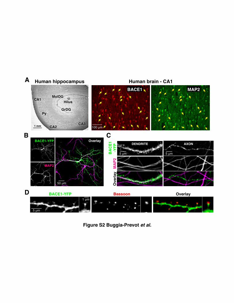
Figure S2 Buggia-Prevot et al.
A
HilusCA1
CA2CA3
GrDG
MolDG
Py
1 mm
Human hippocampusBACE1 MAP2Human brain - CA1
100 µm
B
50 µm
Overlay
50 µm
MAP2
BACE1-YFP
MA
P2O
verla
y
DENDRITE
BA
CE1
-YFP
AXON
5 µm5 µm
C
Bassoon OverlayBACE1-YFP
5 µm
1 µm
D

EEA1NEEP21TFR
0
0.2
0.4
0.6
0.8
EHD3-ΔEH
Vector
EHD1-ΔEH
EHD3-ΔEH
Vector
EHD1-ΔEH
EHD3-ΔEH
Vector
EHD1-ΔEH
Man
ders
’ coe
ffici
ent **
Internalized BACE1 colocalization in dendrites
Figure S5 Buggia-Prevot et al.

Supplemental Figure Legends:
Figure S1. Validation of the BTX-Labeling Assay to Visualize Internalized BACE1,
Related to Figure 1
(A) Schematic structure of BACE1-YFP and BBS-BACE1-YFP. The predicted transmembrane
domain (TM) and the site of BBS insertion after the prodomain are indicated.
(B) Analysis of APP processing by BACE1, BACE1-YFP and BBS-BACE1-YFP. HEK293 cells
stably expressing WT APP were transiently transfected with indicated BACE1 constructs. The
levels of BACE1, APP full-length (FL) and C-terminal fragments (CTFs) were assessed by
immunoblot analysis of cell lysates with BACE1-NT and CTM1 antibodies essentially as
described (Vetrivel et al., 2011). The levels of sAPPα and sAPPβ from the conditioned medium
were by analyzed by immunoblotting with mAb 26D6 and polyclonal sAPPβWT antibodies
(Vetrivel et al., 2011).
(C) HeLa cells transfected with BBS-BACE1-YFP were incubated with AF555-BTX at 10°C for
20 min to label BACE1 at the cell surface, and analyzed without or after acid wash. BTX
fluorescence intensities were quantified from individual cells (n=55 and 59 cells for – and + acid
wash, respectively) and the relative fluorescence intensity is plotted. Error bars are SEM.
(D) DIV12 neurons were transfected with BBS-BACE1LL/AA-YFP mutant and incubated with
AF647-BTX for 4 h at 37°C. Before fixing, one set of coverslips was subjected to acid wash
whereas another set was washed only with HBSS. Note that BBS-BACE1LL/AA-YFP is known to
accumulate predominantly at the surface, and surface BTX labeling is efficiently removed by the
acid wash.
Figure S2. Characterization of BACE1 Distribution in Hippocampal Neurons
in Human Brain and Mouse Primary Cultures, Related to Figure 2
(A) Left panel: Immunostaining of BACE1 in normal human hippocampus (stained with mAb
Clone 137612, R&D). Right panel: Immunofluorescence staining of BACE1 and MAP2 in CA1
region of normal human brain.
(B) Somato-dendritic and axonal distribution of BACE1-YFP in transfected DIV12 neurons that
were immunostained for the somato-dendritic marker MAP2.
(C) High magnification confocal images depict BACE1 localization in a dendrite and an axon.
(D) Confocal analysis of a dendrite segment of a transfected neuron adjacent to axonal segment
of an untransfected neuron immunostained for the presynaptic terminal marker Bassoon. Higher
magnification of a single spine is shown on the right.
Figure S3. Confocal Analysis of BACE1 Localization in Endosomes, Related to Figure 3
DIV12 neurons were transfected with BBS-BACE1-YFP and internalized BACE1 was labeled
by incubation with AF647-BTX for 4 h at 37°C followed by acid wash to remove surface-bound
BTX. After fixation, neurons were immunostained with antibodies against early endosomal
markers EEA1 or NEEP21, and the recycling endosome marker TfR. Insets show higher
magnification of the boxed area.
Figure S4. Absence of Colocalization of Endogenous EHD2 and EHD4 With BACE1 in
Mouse Brain, Related to Figure 5
(A) Sagittal sections of P19 mouse brain were immunostained with antibodies against BACE1
(mAb 3D5) and EHD2 or EHD4, and confocal images were acquired using 10X (top) and 100X
objectives (bottom). Inserts show higher magnifications of the boxed area within the terminal
field of mossy fibers (MF). EHD4 localizes to the soma and dendrites of the neurons in dentate
gyrus (DG), and CA1-CA3 region, but not in the axons of the hippocampal mossy fibers.
(B) Specificity of EHD1 and EHD3 antibodies. Sagittal brain sections of 11-month-old EHD1-/-,
EHD3-/-, or WT littermate mice were immunostained with EHD1 and EHD3 antibodies.
Confocal images along the mossy fibers were acquired using 100X objective using identical
settings and processed similarly for brightness and contrast.
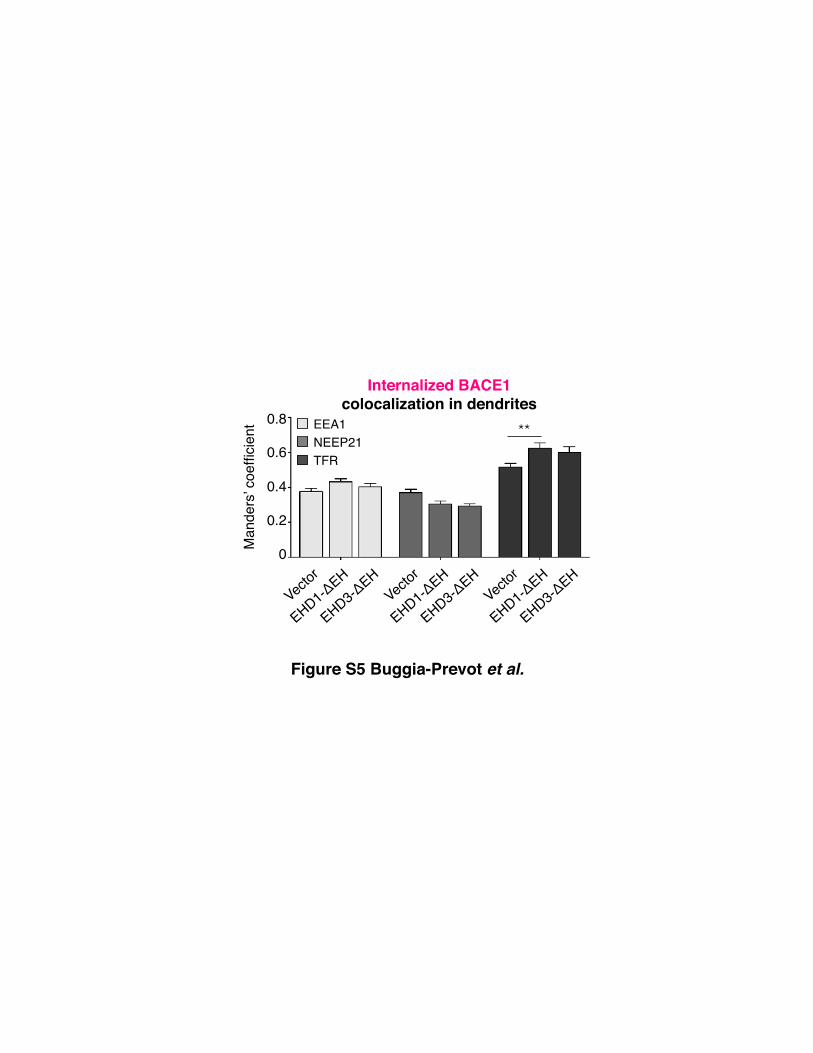
Figure S5. Confocal Analysis of Internalized BACE1 Localization in Endosomes Following
EHD-ΔEH Expression, Related to Figure 7
Neurons were co-transfected with BBS-BACE1-YFP and the indicated DsRed-tagged EHD WT
or EHD-ΔEH construct were labeled with AF647-BTX and subjected to acid wash. The neurons
were then immunostained for endosome markers EEA1, NEEP21, or TfR and analyzed by
confocal microscopy. The extent of colocalization between internalized BACE1 and endogenous
organelle markers was quantified in dendritic segments of transfected neurons (n=8-14 neurons
each). All error bars are SEM.

Supplemental Movie Legends:
Movie S1. Unidirectional Transport of Internalized BACE1 in Dendrites
Internalized BACE1 was labeled by incubation with Alexa555-BTX for 4 h at 37°C in a
neuron transfected with BBS-BACE1-YFP (DIV13), and washed for 5 min with warm
imaging medium prior to live imaging. Sequential time-lapse images of total BACE1 (top
panel) and internalized BACE1 (bottom panel) of the same dendrite were acquired at a
rate of 1 frame/sec for 3 min, with an exposure time of 300 ms, and displayed at 15
frames/sec. Note the bidirectional movement of total BACE1 and the unidirectional
movement of internalized BACE1 in the retrograde direction (toward the left). Scale bar
20 µm. Representative still images are shown in Figure 2B.
Movie S2. Unidirectional Transport of Internalized BACE1 in Distal Segments of
Dendrites
Internalized BACE1 was labeled by incubation with Alexa555-BTX for 4 h at 37°C in a
neuron transfected with BBS-BACE1-YFP (DIV13), and washed for 5 min with warm
imaging medium prior to live imaging. Sequential time-lapse images of internalized
BACE1 in distal segments of dendritic branches were acquired at a rate of 1 frame/sec,
with an exposure time of 300 ms, and displayed at 15 frames/sec. Note the unidirectional
movement of internalized BACE1 in the retrograde direction (toward the top). Four
transport vesicles along different branches of the same dendrite are tracked to highlight
their movement toward the soma.
Movie S3. Bidirectional Transport of Internalized BACE1 in Axons
Internalized BACE1 was labeled by incubation with Alexa555-BTX for 4 h at 37°C in a
neuron transfected with BBS-BACE1-YFP (DIV13), and washed for 5 min with warm
imaging medium prior to live imaging. Sequential time-lapse images of total BACE1 (top
panel) and internalized BACE1 (bottom panel) of the same axon were acquired at a rate
of 1 frame/sec for 3 min, with an exposure time of 300 ms, and displayed at 10
frames/sec. The direction of anterograde movement is toward the right. Note the
bidirectional movement of internalized BACE1 (red arrowheads point to an anterograde
vesicle; blue arrowheads point to a retrograde vesicle). Scale bar 20 µm. Representative
kymographs are shown in Figure 2D.
Movie S4. EHD1 and EHD3 are Required for Retrograde Transport of Internalized
BACE1 in Dendrites
Internalized BACE1 was labeled by incubation with Alexa647-BTX for 4 h at 37°C in
neurons (DIV12) cotransfected with BBS-BACE1-YFP and EHD1 WT (top left), EHD1-
ΔEH (bottom left), EHD3 WT (top right), or EHD3-ΔEH (bottom left). Time-lapse
images of internalized BACE1 were acquired in proximal dendrites at a rate of 1
frame/sec for 3 min, with an exposure time of 300 ms, and displayed at 15 frames/sec.
The direction of anterograde movement is toward the right. Note that the unidirectional
retrograde movement of internalized BACE1 is impaired by the expression of dominant-
negative EHD1 and EHD3 mutants. Scale bar 20 µm. Representative kymographs are
shown in Figure 7A.
Movie S5. EHD1 and EHD3 are Required for Dynamic Axonal Transport of BACE1.
DIV12 neurons cotransfected with BACE1-YFP and EHD1 WT (top left), ΔEH-EHD1
(bottom left), EHD3 WT (top right), or ΔEH-EHD3 (bottom left) were observed by live
cell imaging. Time-lapse images were acquired at a rate of 1 frame/sec for 3 min, with an
exposure time of 300 ms, and displayed at 10 frames/sec. Note the paucity of axonal
BACE1 motility in neurons coexpressing dominant-negative EHD1 and EHD3 mutants.
Scale bar 20 µm. Representative kymographs are shown in Figure 7C.

Supplemental Experimental Procedures:
Immunohistochemistry and immunofluorescence staining
Double immunofluorescence staining of human hippocampus was performed as
described (Kar et al., 2006). Briefly, paraffin embedded hippocampal sections (25 µm thick)
from control human brains were rehydrated, treated with citrate buffer for 20 min, and incubated
with mAb BACE1 and polyclonal MAP2 antibodies overnight. The sections were then washed,
incubated with appropriate secondary antibody for 2 h, treated with autofluorescence eliminator
for 5 min, and mounted using gold antifade medium.
Brains were harvested from bigenic BACE1-YFP mice (3-month), C57BL/6J (P19 WT;
11-month old EHD1-/-, EHD3-/-, and WT littermates), or APPswe/PS1ΔE9 transgenic mice (2-
month) were fixed overnight at 4°C in 4% paraformaldehyde and cryoprotected in PBS 30%
sucrose/PBS. Immunofluorescence staining was performed on 40 µm coronal sections or 30 µm
sagittal sections as described (Zhao et al., 2007; Vetrivel et al., 2008; Gong et al., 2011). Images
were acquired on Zeiss LSM 510 META laser scanning confocal microscope (BACE1-YFP
transgenic brain in Figure 1A and S1) or Leica SP5 II STED-CW Superresolution laser scanning
confocal microscope (all other brain sections), and processed using ImageJ software.
The following primary antibodies were used to stain brain sections: BACE1 mAb 3D5
(1:500), BACE1 mAb (1:100, R&D Systems), EHD1, EHD2, EHD3 or EHD4 pAb (1:500), Aβ1-
5 mAb 3D6 (83 ng/ml, Elan Pharmaceuticals), polyclonal MAP2 (1:100, SantaCruz). Alexa Fluor
488-, 555-, or 647-conjugated secondary antibodies (Molecular Probes) were used for detection.
Neurons were fixed with 4 % paraformaldehyde containing 4% sucrose for 20 min.
Neurons were then quenched with 50 mM NH4Cl for 10 min, permeabilized for 6 min on ice
with 0.2% Triton X-100 and blocked with PBS 3% BSA. Transfected COS cells were fixed with
4 % paraformaldehyde. After fixation, COS cells were permeabilized with 0.2% Triton X-100, 5
min at room temperature and blocked with PBS 3% BSA 50 mM NH4Cl, 10 mM glycine. The
coverslips were incubated for 1 h at room temperature with the primary antibodies for neuronal
immunostaining: MAP2 mAb (1:5,000; Sigma), NEEP21 pAb (1:150, NSG1, GenScript), EEA1

pAb (1:200, Millipore), TfR mAb (1:500, C2F2, Pharmingen), and Bassoon mAb (1:5,000,
SAP7F407, Enzo Life Sciences) diluted in PBS containing 3% BSA. Subsequently, the
coverslips were incubated with Alexa Fluor 350-, 555-, or 647-conjugated secondary antibodies
(Molecular Probes) for 1 h at room temperature and mounted using Permafluor (Thermo Fisher
Scientific).
Image processing
Image stacks of hippocampal neurons were processed in ImageJ software (Rasband,
1997-2012) by correcting (if necessary) image drift using TurboReg plugin (Thevenaz et al.,
1998), background subtraction, bleach correction using Enhance Contrast function, and using
Gaussian Blur and Unsharp Mask filters. Wide-field epifluorescence image stacks were
deconvolved using Huygens software (Scientific Volume Imaging). Extended Depth of Field
plugin of ImageJ was used to generate single plane projections from processed z-stacks (Forster
et al., 2004). For quantitative analysis, images were acquired and processed for background
subtraction and thresholding using identical settings. MTrackJ plug-in was used for manual
tracking of moving vesicles in dendrites (Meijering et al., 2012).

Supplemental References:
Forster, B., Van De Ville, D., Berent, J., Sage, D., and Unser, M. (2004). Complex wavelets for extended depth-of-field: a new method for the fusion of multichannel microscopy images. Microsc Res Tech 65, 33-42.
Kar, S., Poirier, J., Guevara, J., Dea, D., Hawkes, C., Robitaille, Y., and Quirion, R. (2006). Cellular distribution of insulin-like growth factor-II/mannose-6-phosphate receptor in normal human brain and its alteration in Alzheimer's disease pathology. Neurobiol Aging 27, 199-210.
Meijering, E., Dzyubachyk, O., and Smal, I. (2012). Methods for cell and particle tracking. Methods in enzymology 504, 183-200.
Rasband, W.S. (1997-2012). ImageJ. (Bethesda, Maryland, USA, U. S. National Institutes of Health).
Thevenaz, P., Ruttimann, U.E., and Unser, M. (1998). A pyramid approach to subpixel registration based on intensity. IEEE Trans Image Process 7, 27-41.
Vetrivel, K.S., Barman, A., Chen, Y., Nguyen, P.D., Wagner, S.L., Prabhakar, R., and Thinakaran, G. (2011). Loss of cleavage at beta'-site contributes to apparent increase in beta-amyloid peptide (Abeta) secretion by beta-secretase (BACE1)-glycosylphosphatidylinositol (GPI) processing of amyloid precursor protein. J. Biol. Chem. 286, 26166-26177.
Vetrivel, K.S., Kodam, A., Gong, P., Chen, Y., Parent, A.T., Kar, S., and Thinakaran, G. (2008). Localization and regional distribution of p23/TMP21 in the brain. Neurobiol. Dis. 32, 37-49.
Zhao, J., Fu, Y., Yasvoina, M., Shao, P., Hitt, B., O'Connor, T., Logan, S., Maus, E., Citron, M., Berry, R., et al. (2007). Beta-site amyloid precursor protein cleaving enzyme 1 levels become elevated in neurons around amyloid plaques: implications for Alzheimer's disease pathogenesis. J. Neurosci. 27, 3639-3649.
Copyright © 2022 FDOKUMEN