Self-assembled molecular rafts at liquid|liquid interfaces for four-electron oxygen reduction
Upload
independentCategory
view
5download
0
Journal of Alzheimer’s Disease 43 (2015) 1185–1198DOI 10.3233/JAD-141146IOS Press
1185
Biophysical Alterations in Lipid Rafts fromHuman Cerebral Cortex Associate withIncreased BACE1/A�PP Interaction in EarlyStages of Alzheimer’s Disease
Mario Dıaza,∗, Noemı Fabeloa, Virginia Martına, Isidre Ferrerb,c, Tomas Gomeza and Raquel Marınd
aDepartamento de Biologıa Animal, Universidad de La Laguna, Tenerife, SpainbInstitut de Neuropatologia, IDIBELL-Hospital Universitari de Bellvitge, Universitat de Barcelona,Hospitalet de LlobregatcCIBERNED (Centro de Investigacion Biomedica en Red de Enfermedades Neurodegenerativas), SpaindDepartamento de Fisiologıa, Universidad de La Laguna, Tenerife, Spain
Accepted 17 July 2014
Abstract. In the present study, we have assessed the biophysical properties of lipid rafts from different brain areas in subjectsexhibiting early neuropathological stages of Alzheimer’s disease (AD). By means of steady-state fluorescence polarizationanalyses using two environment-sensitive fluorescent probes, we demonstrate that lipid rafts from cerebellum, and frontaland entorhinal cortices, exhibit different biophysical behaviors depending on the stage of the disease. Thus, while membraneanisotropies were similar in the cerebellum along stages, lipid rafts from frontal and entorhinal cortices at AD stages I/II andAD III were significantly more liquid-ordered than in control subjects, both at the aqueous interface and hydrophobic core ofthe raft membrane. Thermotropic analyses demonstrated the presence of Arrhenius breakpoints between 28.3–32.0◦C, whichwere not influenced by the disease stage. However, analyses of membrane microviscosity (ηapp) demonstrate that frontal andentorhinal lipid rafts are notably more viscous and liquid-ordered all across the membrane from early stages of the disease. Thesephysicochemical alterations in lipid rafts do not correlate with changes in cholesterol or sphingomyelin levels, but to reducedunsaturation index and increased saturate/polyunsaturated ratios in phospholipid acyl chains. Moreover, we demonstrate that �-secretase/A�PP (amyloid-� protein precursor) interaction and lipid raft microviscosity are strongly, and positively, correlated inAD frontal and entorhinal cortices. These observations strengthens the hypothesis that physical properties of these microdomainsmodulate the convergence of amyloidogenic machinery toward lipid rafts, and also points to a critical role of polyunsaturatedfatty acids in amyloidogenic processing of A�PP.
Keywords: BACE1, �-secretase, cerebellum, docosahexaenoic acid, entorhinal cortex, fluorescence anisotropy, frontal cortex,lipid rafts, membrane viscosity, polyunsaturated fatty acids
∗Correspondence to: Dr. Mario Diaz, U.D.I. Fisiologıa Animal,Departamento de Biologıa Animal, Universidad de La Laguna,38206 Tenerife, Spain. Tel.: +34 922318343; Fax: +34 922318342;E-mail: [email protected].
INTRODUCTION
Compelling evidence accumulated over the last twodecades has pointed to lipid rafts, highly dynamicmembrane assemblies enriched in cholesterol, and sph-ingolipids, as critical membrane domains involvedin the regulation of A�PP (amyloid-� protein pre-cursor) processing, and in the generation of the
ISSN 1387-2877/15/$27.50 © 2015 – IOS Press and the authors. All rights reserved

1186 M. Dıaz et al. / Biophysical Alterations in AD Lipid Rafts
amyloid-� (A�) peptide, considered the drivingforce in Alzheimer’s disease (AD) pathology [1–5].Although the majority of full-length A�PP is com-partmentalized to non-raft regions, an important subsetof A�PP partitions into lipid rafts along with BACE1and �-secretase components [4–6]. Integration of thesemembrane proteins into lipid rafts is determined bytheir capacity to establish specific protein-lipid interac-tions. Thus, both BACE1 and the �-secretase subunitsundergo posttranslational S-palmitoylation which aidstheir targeting to lipid raft domains [4, 6]. Fur-thermore, A�PP contains a transmembrane domainresembling properties of canonical CRAC (Choles-terol Recognition/interaction Amino acid Consensussequence) [7, 8], which allows the direct interactionwith cholesterol—the major lipid component of lipidrafts [4, 7]. Additionally, Bhattacharyya et al. [9] haverecently reported that a fraction of A�PP undergoespost-translational palmitoylation and that this lipid-modified A�PP is specifically enriched in lipid rafts.
The singular lipid structure of lipid rafts makesthem liquid-ordered domains because the highly sat-urated phospholipid (including sphingomyelin) acylchains and high cholesterol contents, enable closerlipid packing and rather restricted lateral movement,than in the surrounding non-raft regions [10]. Mul-tiple lines of evidence, mostly based on selectivelipid depletion in cultured cells, have suggested thatthese physicochemical features of lipid rafts pro-vide the favorable environment for the amyloidogenicprocessing of A�PP by altering the clustering dynam-ics of A�PP-cleaving enzymes (reviewed in [1–5]).However, whether these alterations occur during theneuropathological progression leading to AD remainlargely unknown.
Recently, we have demonstrated that lipid rafts fromfrontal cortex in late stages of Alzheimer’s disease(Braak and Braak staging V/VI) display altered lipidprofiles compared to control subjects [11]. Most lipidalterations were detected in the fatty acid compositionof phospholipids, rather than in the raft lipid prototyp-ical lipid classes cholesterol or sphingomyelin [11].More recently, we have reported that alterations inlipid composition of lipid rafts from human cortexare detectable from very early stages of the disease,namely Braak and Braak stages I/II, where amyloidplaques and cognitive symptoms are still absent [12].In these lipid rafts, we found complex lipid changestaking place in entorhinal and frontal cortices, but notin cerebellum, consistent with both, reduction in longchain polyunsaturated fatty acids (LCPUFA) in phos-pholipids, and moderate cholesterol depletion, which
were accompanied by increased interaction of A�PPand BACE [12]. These changes in the lipid matrix oflipid rafts are expected to impact their physicochemicalproperties and to alter membrane microdomain envi-ronment during AD progression, which might facilitatethe co-segregation of A�PP and BACE, eventuallyleading to increased A� production.
Therefore, in the present study we have aimed toexplore 1) the impact of lipid changes in lipid raftsduring early stages of AD neuropathology, on thebiophysical properties of lipid rafts, and 2) the pos-sible relationships between biophysical changes andthe convergence and association of proteins involvedin the amyloidogenic processing of A�PP.
MATERIALS AND METHODS
Human brain tissue and lipid rafts isolation
Brain tissues were obtained from the Institute ofNeuropathology Brain Bank (Bellvitge UniversityHospital, Spain) following the guidelines of the Hos-pital Ethics Committee. Based on neuropathologicalexamination, 12 cases were classified into three cate-gories according to Braak and Braak [13]: AD stagesI/II (AD I group, average age 60.7 ± 2.9 years, of whichone case was AD II), AD stage III (AD III group, aver-age age 74.7 ± 3.2 years), and controls (CTRL group,average age 49.2 ± 4.64 years). A summary of all casesis shown in Table 1. With the sole exception of subject12, all cases were males. Regarding amyloid pathology,only one case (subject 7, staged I/A) displayed senileplaques in the neocortex (amyloid pathology stage A).Importantly, cases in this study were selected basedon the results of our previous study aimed to deter-mining the potential alterations of lipid compositionof lipid rafts in early stages of AD [12]. Therefore, indoing this, it was possible to establish the precise bio-physical correlates of lipid alterations. In every case,frontal cortex grey matter (area 8), entorhinal cortex,and cerebellar vermis were carefully dissected and sep-arated from the subcortical white matter immediatelyat autopsy, and stored at −80
◦C until use for lipid rafts
isolation.Lipid raft fractions were isolated following proce-
dures adapted for human brain lipid rafts detailed inMartın et al. [11] and Ramırez et al. [14]. Fractions inthe gradients were routinely tested for purity in westernblot assays using different lipid raft and non-raft pro-tein markers (i.e., anti-flotillin 1 and anti-caveolin-1 forlipid rafts, anti-�1 Na+/K+ ATPase subunit for non-raft plasma membrane, and anti-Hsp90 for cytosolic

M. Dıaz et al. / Biophysical Alterations in AD Lipid Rafts 1187
Table 1Summary of cases included in the study
Case Age Gender Postmortem delay Neuropathology
Control group1 40 Male 5 h 10 min NL2 44 Male 6 h 40 min NL3 52 Male 3 h NL4 61 Male 3 h 50 min NLAD I/II group5 67 Male 7 h 15 min II/06 61 Male 3 h 40 min I/07 53 Male 6 h 15 min I/A8 62 Male 5 h 45 min I/0AD III group9 66 Male 2 h 45 min III/010 75 Male 5 h 30 min III/011 77 Male 6 h 20 min III/012 81 Female 1 h 30 min III/0
NL, no lesions. Neuropathology was classified according to Braakand Braak stages of Alzheimer’s disease (stages I-III). 0-A indicatesstages of amyloid neuropathology.
proteins). Fractions containing lipid raft markersflotillin-1 and caveolin-1 were collected, resuspendedin isolation buffer and frozen at −80◦C until analyses.
Lipid analyses
Analyses of lipid composition of lipid rafts fractionswas carried out as described previously by our groupfor human brain [11, 12]. Total lipids were extractedwith chloroform/methanol (2:1 v/v) in the presenceof 0.01% of butylated hydroxytoluene. Lipid classeswere separated by one-dimensional double develop-ment high performance thin layer chromatographyand quantified by densitometry. A fraction of totallipids extracted from lipid rafts were subjected to acid-catalyzed transmethylation to form the correspondingfatty acid methyl esters (FAME) and dimethyl acetals(DMA), which were purified by thin layer chromatog-raphy (TLC) and quantified by gas chromatographusing flame ionization detection and identified by massspectrometry.
Immunoprecipitation assays
Lipid raft fractions were processed for immuno-precipitation as described in Fabelo et al. [12].Briefly, fractions diluted in immunoprecipitationbuffer (50 mM Tris-HCl pH 7.4; 150 mM NaCl,10% glycerol, 1% Nonidet-P 40, 1 mM PMSF, andRoche’s proteases inhibitor cocktail) were solubilizedin 2% octyl �-D-glucopyranoside solution containingNa3VO4 and EDTA, and immunoprecipitated with anexcess (5 �g) of rabbit polyclonal anti-APP antibody
(Y-188, Abcam) overnight at 4◦C. Samples were thenexposed to sheep anti-rabbit IgG dynabeads (Dynal).The resultant precipitated protein-dynabead immuno-complexes were disrupted using SDS loading bufferand resolved in 12.5% SDS-PAGE followed by westernblotting. Proteins were then transferred to Hybond-Pand blotted with rabbit anti-BACE polyclonal antibody(Chemicon International), and rabbit anti-Presenilin1 antibody (Sigma-Aldrich), both diluted 1:500 inBLOTTO. Visualization of specific immunosignalsdeveloped for anti-rabbit horseradish peroxidase-conjugated secondary antibodies (diluted 1:10,000 inBLOTTO) were obtained with ECL chemilumines-cence kit (Amersham, GE Healthcare). Densitometryvalues of specific signals were quantified using GS-800 calibrated densitometer (Bio-Rad, Madrid, Spain).Relative values of BACE immunosignals were normal-ized to the total A�PP content quantified in the samesample.
Steady-state fluorescence anisotropy of lipid rafts
Steady-state anisotropy (rs) measurements wereperformed using two different fluorescent probes:nonpolar 1,6-diphenyl-1,3,5-hexatriene (DPH) andpolar 1-[4-(trimethylammonium)phenyl]-6-phenyl-1,3,5-hexatriene (TMA-DPH). DPH and TMA-DPHwere dissolved in 1 tetrahydrofuran:1 ethanol (v/v)and stored as 200 �M stock solutions. Fluorescentprobes were used at 2 �M in TBS (20 mM Tris–HClbuffer, pH 7.5, containing 150 mM KCl) prepareddaily and stored protected from light until used. Lipidraft suspensions (250 �L, 50 �g protein/mL) wereincubated for 30 min at 37◦C in PBS solutions labelledwith the probes and under agitation. Fluorescencepolarization values were determined using 355 nmexcitation and 420 nm emission filters in an Appliskanmultiplate reader (Thermo Scientific) equipped withpolarizers. Unlabeled controls were simultaneouslyexamined to correct for light scattering and intrinsicfluorescence. Steady-state fluorescence anisotropieswere determined against temperature ramps from20◦C to 40◦C.
In some experiments, lipid rafts samples were prein-cubated with 5 mM methyl-�-cyclodextrine (M�CD)in TBS at 37◦C for 1 h, before incorporation of thefluorescent probes.
Microviscosity and thermodynamic calculations
Membrane microviscosity, η, is the measure of fric-tional resistance to rotational and translational motions

1188 M. Dıaz et al. / Biophysical Alterations in AD Lipid Rafts
of molecules within bilayers. Membrane microvis-cosity (the reciprocal to membrane fluidity) can beestimated by fluorescence anisotropy of rod-shapedprobes (as DPH or TMA-DPH), which reflects theirrotational motion inside the membrane [15, 16].Because in lipid membranes rotational mobility ofthese probes is not isotropic but restricted in space,it also reflects the level of lipid order (or lipid pack-ing) [15, 16]. These two parameters, microviscosityand lipid order, discriminate different membrane phasestates, such gel and liquid-ordered (which are believedto be responsible for functional lipid rafts domains),and fluid phase (in non-raft domains). In the presentstudy, apparent microviscosity (ηapp) coefficients werecomputed based on the adaptation of the Perrin equa-tion for rotational depolarization of DPH [17] andTMA-DPH [18] as described in Diaz et al. [19].Discontinuity breakpoints (Td), which inform on thetemperature where phase transitions occur, and acti-vation energies (Ea) for steady-state anisotropy werecomputed from Arrhenius plots [20], being the slopesof log(rs) or log(ηapp) versus 1/T equal to Ea/(2.3 R).Flow activation energy or activation energy of viscousflow (Eη) was derived by analogy to macroscopic fluidsfrom empirical determination of temperature depen-dence of ηapp, being the slope of log(ηapp) versus 1/Tplots equal to Eη/(2.3 R) [16, 21].
Statistics
Data were submitted to two-way ANOVA or one-way ANOVA followed by Student’s t-test, whereappropriate. Regression analyses were performed bynon-linear regression. Pearson correlation coefficients(R) and their corresponding p-values were calculatedfor all regression lines. Significance of regressionanalyses was assessed by one way ANOVA. Whenappropriate, 95% confidence intervals are indicated inFigs. 3 and 4.
RESULTS
Thermotropic behavior of lipid rafts anisotropy
We have used two different probes, the nonpolarprobe DPH to determine the physical order at thehydrophobic core of the lipid raft membrane, andthe polar TMA-DPH to assess rotational freedom atthe membrane plane [22, 23]. Results summarized inFig. 1A show the temperature response of TMA-DPHsteady-state anisotropy in lipid rafts from cerebellum(CB), entorhinal cortex (EC), and frontal cortex (FC)
in the control, AD I/II, and AD III groups. Anisotropycurves in the range 20–40◦C were fairly similar inthe cerebellum irrespective of the neuropathologicalstate, being the values measured at 37◦C nearly iden-tical in the three groups (CB, F2 ,5 = 1.82, p > 0.1).Conversely, in EC and FC, a clear upward shift inanisotropy curves was evident for controls as comparedto AD groups, indicating that TMA-DPH anisotropiesin lipid rafts from AD subjects are inherently higherin the whole range of temperatures than in controlrafts (EC: F2 ,5 = 2.31, p = 0.056; FC: F2 ,5 = 3.52,p < 0.05). Arrhenius plots of steady-state TMA-DPHanisotropies revealed the existence of thermotropicphase transitions around discontinuity points (Td),in the range 28.3–29.9◦C, which were quite similarbetween brain areas and also between disease stages(Fig. 1B). Similarly, computation of activation ener-gies (Ea) below Td indicated no differences betweenlipid rafts from either group.
Using a modified expression of Perrin equation fornon-spherical probes [18, 21], we have assessed thechanges in lipid raft microviscosity coefficients in thedifferent brain areas and conditions (Table 1). Apparentmicroviscosity (ηapp) and flow activation energies (Eη)in cerebellum were nearly constant (ηapp around 1.71poises at 37◦C and Eη around 2.5 kcal/mol) and wereunaffected by the disease (Fig. 1C). On the contrary,in EC and FC lipid rafts, ηapp values were significantlyhigher (average 1.82 poises at 37◦C) at AD stages I/IIand III/IV than in controls (Table 1), but these changeswere not followed by changes in Eη, which remainedin the range observed for cerebellum (Fig. 1C).
The thermotropic behavior of raft membranes atthe internal core of the bilayers was assessed usingDPH [22, 23]. As in the case of TMA-DPH, lipidrafts displayed temperature-induced changes in mem-brane anisotropy, which were unaffected by the diseasestage in CB (CB, F2,5 = 0.84, p > 0.1), but differed inEC and FC lipid rafts (EC: F2 ,5 = 5.53, p < 0.05; FC:F2,5 = 13.85, p < 0.05). Thus, in cortical lipid rafts atstages I/II and III, DPH anisotropies were significantlyhigher in the whole temperature range as comparedto controls (Fig. 2A). In order to get a deeper insightinto the characterization of the mechanisms leadingto these changes, we also explored the effects ofcholesterol depletion in the same rafts using M�CD.The preincubation protocol with M�CD used here(5 mM for 1 h), causes a 50% depletion of membranecholesterol [24]. Treatment with M�CD (+M�CD)brought about a considerable reduction of lipid raftprobe anisotropies in all groups compared with con-trols (TBS), but the effects were more remarkable in

M. Dıaz et al. / Biophysical Alterations in AD Lipid Rafts 1189
Fig. 1. Biophysical characterization of lipid rafts from cerebellum, entorhinal cortex, and frontal cortex in CTRL, AD I/II, and AD III brainsprobed with TMA-DPH. A) Temperature dependence of TMA-DPH fluorescence anisotropy of lipid rafts. B) Arrhenius plot for steady-stateanisotropy in lipid rafts in the different brain areas. Arrows indicate discontinuity points (Td in ◦C) and the range of activation energies (Eain kcal/mol) obtained below Td. C) Apparent microviscosity (ηapp ) analyses performed below Td based on the Perrin equation modified forrod-shaped probes. Slopes indicate the activation energy of viscous flow (Eη).

1190 M. Dıaz et al. / Biophysical Alterations in AD Lipid Rafts
Fig. 2. Biophysical characterization of lipid rafts from cerebellum, entorhinal cortex, and frontal cortex in CTRL, AD I/II, and AD III brainsprobed with DPH. A) Temperature dependence of DPH fluorescence anisotropy of lipid rafts treated with methyl-�-cyclodextrin (M�CD, 5mM) or without (TBS, TRIS-buffered saline). B) Arrhenius plot for steady-state anisotropy in lipid rafts in the different brain areas and inthe presence or absence of M�CD. Arrows indicate the position of discontinuity points (Td in ◦C) and the range of activation energies (Ea inkcal/mol) obtained below Td. C) Apparent microviscosity (ηapp ) analyses performed below Td. Slopes indicate the activation energy of viscousflow (Eη).
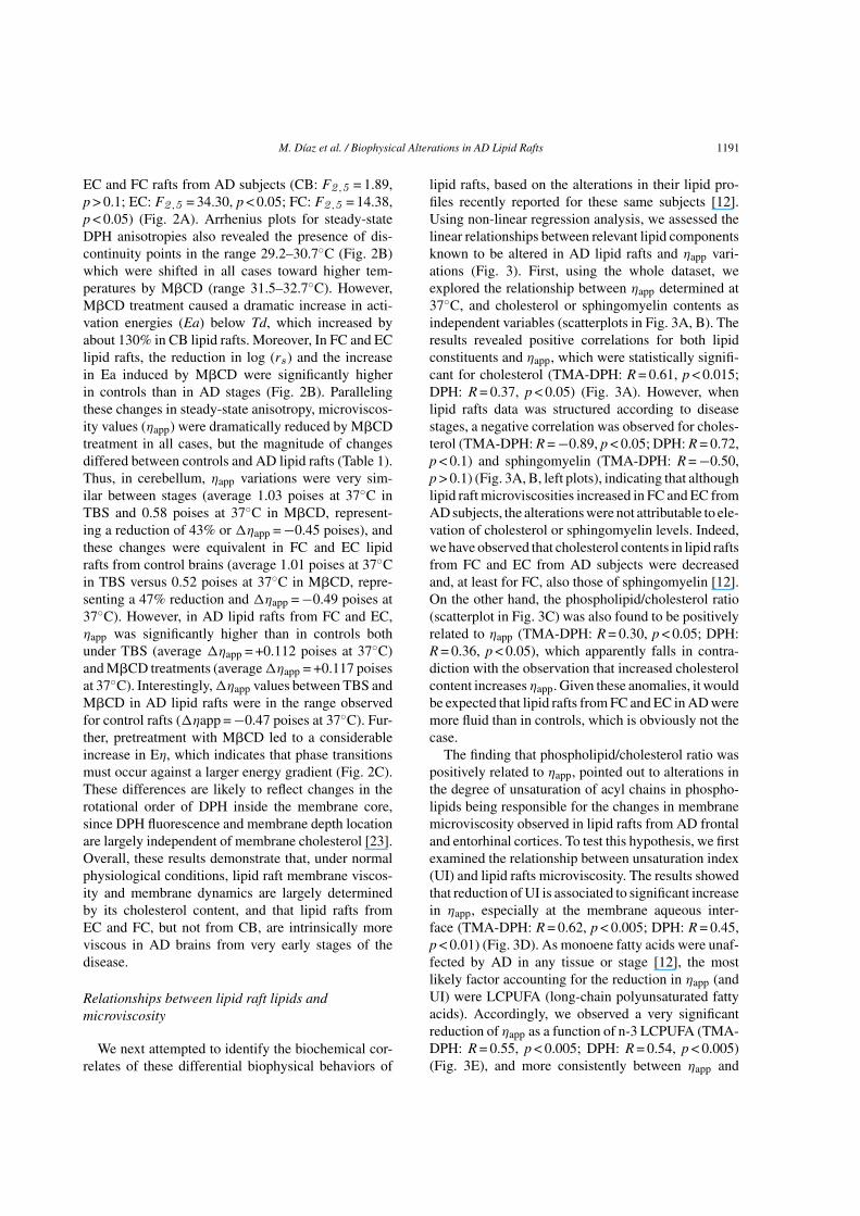
M. Dıaz et al. / Biophysical Alterations in AD Lipid Rafts 1191
EC and FC rafts from AD subjects (CB: F2 ,5 = 1.89,p > 0.1; EC: F2 ,5 = 34.30, p < 0.05; FC: F2 ,5 = 14.38,p < 0.05) (Fig. 2A). Arrhenius plots for steady-stateDPH anisotropies also revealed the presence of dis-continuity points in the range 29.2–30.7◦C (Fig. 2B)which were shifted in all cases toward higher tem-peratures by M�CD (range 31.5–32.7◦C). However,M�CD treatment caused a dramatic increase in acti-vation energies (Ea) below Td, which increased byabout 130% in CB lipid rafts. Moreover, In FC and EClipid rafts, the reduction in log (rs ) and the increasein Ea induced by M�CD were significantly higherin controls than in AD stages (Fig. 2B). Parallelingthese changes in steady-state anisotropy, microviscos-ity values (ηapp) were dramatically reduced by M�CDtreatment in all cases, but the magnitude of changesdiffered between controls and AD lipid rafts (Table 1).Thus, in cerebellum, ηapp variations were very sim-ilar between stages (average 1.03 poises at 37◦C inTBS and 0.58 poises at 37◦C in M�CD, represent-ing a reduction of 43% or �ηapp = −0.45 poises), andthese changes were equivalent in FC and EC lipidrafts from control brains (average 1.01 poises at 37◦Cin TBS versus 0.52 poises at 37◦C in M�CD, repre-senting a 47% reduction and �ηapp = −0.49 poises at37◦C). However, in AD lipid rafts from FC and EC,ηapp was significantly higher than in controls bothunder TBS (average �ηapp = +0.112 poises at 37◦C)and M�CD treatments (average �ηapp = +0.117 poisesat 37◦C). Interestingly, �ηapp values between TBS andM�CD in AD lipid rafts were in the range observedfor control rafts (�ηapp = −0.47 poises at 37◦C). Fur-ther, pretreatment with M�CD led to a considerableincrease in Eη, which indicates that phase transitionsmust occur against a larger energy gradient (Fig. 2C).These differences are likely to reflect changes in therotational order of DPH inside the membrane core,since DPH fluorescence and membrane depth locationare largely independent of membrane cholesterol [23].Overall, these results demonstrate that, under normalphysiological conditions, lipid raft membrane viscos-ity and membrane dynamics are largely determinedby its cholesterol content, and that lipid rafts fromEC and FC, but not from CB, are intrinsically moreviscous in AD brains from very early stages of thedisease.
Relationships between lipid raft lipids andmicroviscosity
We next attempted to identify the biochemical cor-relates of these differential biophysical behaviors of
lipid rafts, based on the alterations in their lipid pro-files recently reported for these same subjects [12].Using non-linear regression analysis, we assessed thelinear relationships between relevant lipid componentsknown to be altered in AD lipid rafts and ηapp vari-ations (Fig. 3). First, using the whole dataset, weexplored the relationship between ηapp determined at37◦C, and cholesterol or sphingomyelin contents asindependent variables (scatterplots in Fig. 3A, B). Theresults revealed positive correlations for both lipidconstituents and ηapp, which were statistically signifi-cant for cholesterol (TMA-DPH: R = 0.61, p < 0.015;DPH: R = 0.37, p < 0.05) (Fig. 3A). However, whenlipid rafts data was structured according to diseasestages, a negative correlation was observed for choles-terol (TMA-DPH: R = −0.89, p < 0.05; DPH: R = 0.72,p < 0.1) and sphingomyelin (TMA-DPH: R = −0.50,p > 0.1) (Fig. 3A, B, left plots), indicating that althoughlipid raft microviscosities increased in FC and EC fromAD subjects, the alterations were not attributable to ele-vation of cholesterol or sphingomyelin levels. Indeed,we have observed that cholesterol contents in lipid raftsfrom FC and EC from AD subjects were decreasedand, at least for FC, also those of sphingomyelin [12].On the other hand, the phospholipid/cholesterol ratio(scatterplot in Fig. 3C) was also found to be positivelyrelated to ηapp (TMA-DPH: R = 0.30, p < 0.05; DPH:R = 0.36, p < 0.05), which apparently falls in contra-diction with the observation that increased cholesterolcontent increases ηapp. Given these anomalies, it wouldbe expected that lipid rafts from FC and EC in AD weremore fluid than in controls, which is obviously not thecase.
The finding that phospholipid/cholesterol ratio waspositively related to ηapp, pointed out to alterations inthe degree of unsaturation of acyl chains in phospho-lipids being responsible for the changes in membranemicroviscosity observed in lipid rafts from AD frontaland entorhinal cortices. To test this hypothesis, we firstexamined the relationship between unsaturation index(UI) and lipid rafts microviscosity. The results showedthat reduction of UI is associated to significant increasein ηapp, especially at the membrane aqueous inter-face (TMA-DPH: R = 0.62, p < 0.005; DPH: R = 0.45,p < 0.01) (Fig. 3D). As monoene fatty acids were unaf-fected by AD in any tissue or stage [12], the mostlikely factor accounting for the reduction in ηapp (andUI) were LCPUFA (long-chain polyunsaturated fattyacids). Accordingly, we observed a very significantreduction of ηapp as a function of n-3 LCPUFA (TMA-DPH: R = 0.55, p < 0.005; DPH: R = 0.54, p < 0.005)(Fig. 3E), and more consistently between ηapp and

1192 M. Dıaz et al. / Biophysical Alterations in AD Lipid Rafts
Fig.
3.B
ivar
iate
rela
tions
hips
for
appa
rent
mic
rovi
scos
ityof
mem
bran
era
fts
aspr
obed
with
TM
A-D
PH(fi
lled
circ
les)
and
DPH
(em
pty
circ
les)
asde
pend
ent
vari
able
s,an
dch
oles
tero
l(A
),sp
hing
omye
lin(B
),ph
osph
olip
id/c
hole
ster
olra
tio(C
),un
satu
ratio
nin
dex
(D),
n-3
LC
PUFA
(E),
and
satu
rate
s/n-
3L
CPU
FAra
tio,a
sin
depe
nden
tvar
iabl
es,a
sob
tain
edin
the
thre
ebr
ain
area
san
ddi
seas
est
ages
.Rig
htpa
nels
inea
chlip
idva
riab
lesh
owth
esc
atte
rplo
tsfo
rsi
mul
tane
ous
mea
sure
men
tsin
the
who
leda
tase
t.U
nsat
urat
ion
inde
xw
asca
lcul
ated
asσ
mi*
n i,w
here
mii
sth
em
ole
perc
enta
gean
dn i
isth
enu
mbe
rof
carb
on-c
arbo
ndo
uble
bond
sin
each
fatty
acid
.Reg
ress
ion
(sol
id)
lines
are
indi
cate
d.95
%co
nfide
nce
inte
rval
s(d
otte
dlin
es)
are
show
nin
the
scat
ter
plot
s.

M. Dıaz et al. / Biophysical Alterations in AD Lipid Rafts 1193
Fig. 4. Relationships between apparent microviscosity (ηapp ) and amount of BACE1 and PSEN1 physically associated with A�PP, in lipid rafts.A, B) Scatterplots for simultaneous determinations of ηapp , as determined at 37◦C using TMA-DPH (solid circles) and DPH (empty circles),and BACE1 (A) and PSEN1 (B) immunoprecipated with A�PP, in the whole lipid rafts dataset. C, D) Regression analyses for the relationshipsbetween A�PP-immunoprecipitated BACE1 (circles) and PSEN1 (diamonds), and lipid rafts microviscosity as probed with TMA-DPH (C) andDPH (D), in the different brain areas and disease stages. Regression (solid) lines are indicated. In C, the two regression lines correspond tofrontal (CF, leftward line) and entorhinal cortex (EC, rightward line). 95% confidence intervals (dotted lines) are shown in the scatterplots. 95%Confidence intervals are indicated in A, B, and D.
sat/n-3 LCPUFA (TMA-DPH: R = 0.62, p < 0.001;DPH: R = 0.64, p < 0.001) (Fig. 3F).
Relationships between lipid raft microviscosityand AβPP and β/γ-secretase clustering
We finally assessed for the potential involvementof physicochemical alterations in lipid rafts on thedegree of physical association of A�PP and �- and�-secretases. First, by means of immunoprecipitationassays in the same lipid rafts, we observed that lipidrafts from entorhinal and frontal cortices at stagesI/II and III, exhibit significantly increased degree ofassociation between BACE1 and A�PP (Table 2).This augmented BACE1/A�PP interaction occurredonly in cortical lipid rafts, and was not detected inlipid rafts from cerebellum at any stage of the dis-
ease (Table 2). Further, the interaction was specificfor BACE1 and was not observed for PSEN1 beyondcontrol values, which is in agreement with the alter-ations recently reported for these same subjects [12].Moreover, BACE1/A�PP interaction seemed to beinfluenced by the lipid environment of lipid rafts sincea positive association was observed for the amount ofimmunoprecipitated BACE1 (but not PSEN1) and lipidraft microviscosity, especially at the aqueous interface(TMA-DPH: R = 0.62, p < 0.001) and hydrophobiccore (DPH: R = 0.59, p < 0.001) of the membrane(Fig. 4A, B). These relationships were extended tothe results obtained in lipid rafts from each brain areaand disease stage. The outcomes revealed that lipidraft microviscosity was positively correlated to thedegree of BACE1/A�PP association in cortical areas,but not in cerebellum. Indeed, lipid rafts from FC

1194 M. Dıaz et al. / Biophysical Alterations in AD Lipid Rafts
Table 2Apparent microviscosity values (ηapp ) at 37
◦C, calculated for the membrane plane and membrane hydrophobic core, as probed using TMA-DPH
and DPH, respectively, in lipid rafts from controls and AD subjects at neuropathological stages I/II and III
Brain area Stage TMA-DPH DPH
TBS M�CD
Cerebellum Ctrl 1.706 ± 0.035 1.021 ± 0.029 0.588 ± 0.021AD I/II 1.699 ± 0.036 1.040 ± 0.029 0.604 ± 0.016AD III 1.714 ± 0.034 1.025 ± 0.030 0.546 ± 0.010
Frontal cortex Ctrl 1.748 ± 0.025 1.014 ± 0.030 0.541 ± 0.015AD I/II 1.827 ± 0.038* 1.083 ± 0.028 0.659 ± 0.008*AD III 1.865 ± 0.046* 1.132 ± 0.029* 0.647 ± 0.009*
Entorhinal cortex Ctrl 1.686 ± 0.026 1.002 ± 0.029 0.515 ± 0.012AD I/II 1.805 ± 0.035* 1.105 ± 0.020* 0.605 ± 0.012*AD III 1.775 ± 0.034* 1.160 ± 0.029* 0.671 ± 0.007*
Data were submitted to one way ANOVA followed by Student’s t-test for comparisons with Ctrl group. * p < 0.05 compared to Ctrl.
Table 3Interactions of BACE1 and PSEN1 with A�PP in lipid rafts obtainedin brains from controls and AD subjects at neuropathological stages
I/II and III
Brain area Stage BACE1 PSEN1
Cerebellum Ctrl 1.10 ± 0.01 1.19 ± 0.04AD I/II 1.23 ± 0.08 1.18 ± 0.10AD III 1.21 ± 0.15 1.42 ± 0.12
Frontal cortex Ctrl 1.70 ± 0.25 1.54 ± 0.04AD I/II 4.20 ± 0.63** 1.39 ± 0.09AD III 4.26 ± 0.86** 1.28 ± 0.13
Entorhinal cortex Ctrl 1.81 ± 0.14 1.28 ± 0.04AD I/II 5.58 ± 1.03*** 1.09 ± 0.13AD III 3.67 ± 0.44* 1.10 ± 0.11
Data were submitted to ANOVA I followed by Student’s t-test forcomparisons with Ctrl group. *p < 0.05, **p < 0.01, and ***p < 0.005compared to Ctrl.
and EC at stages I/II and III, displaying highest lev-els of BACE1/A�PP interaction, are closely relatedto largest apparent viscosity values, especially at themembrane plane (Fig. 4C). Paralleling these findings,A�PP/PSEN association behaved rather independentlyof ηapp in lipid raft (Fig. 4C, D).
DISCUSSION
In the present study, we provide new evidencethat lipid rafts constitute subcellular targets seriouslyaffected in AD neurodegeneration. We show that evenat the earliest stages of AD, physicochemical prop-erties of raft membranes from entorhinal and frontalcortices exhibit severe alterations which might reflectinitial events in the progression of the disease. Itis widely accepted that lipid rafts are critical ele-ments in the amyloidogenic processing of A�PP [2,4–6], and that specific lipid alterations within thesedomains accelerate A� peptides generation thus facil-itating aggregation [1, 3–5, 25]. However, the extents to
which these lipid changes affect biophysical propertiesof lipid raft microenvironment and how this physic-ochemical scenario affect the dynamics of amyloidprocessing remain poorly understood.
By analyzing some of the biophysical propertiesof lipid rafts from control subjects, we show thatlipid rafts from all brain areas display equivalent ther-motropic behaviors, with phase transitions around adiscontinuity point at 29◦C, and similar activation ener-gies between brain areas as measured above Td. Thishomogeneity was observed both at the aqueous inter-face and hydrophobic core of the lipid rafts, beingthe viscosity of the internal core smaller and the flowactivation energy larger than at the aqueous interface.These observations indicate a great degree of packingof membrane phospholipids, which may provide moreabundant hydrophobic interactions between phospho-lipid fatty acids in the membrane plane, as it has beensuggested in other membrane systems, including lipidrafts [19, 26, 27]. However, in AD brains, we haveobserved that lipid rafts membrane domains are dif-ferentially affected by the disease stage depending onthe brain area considered. Thus, while cerebellar lipidrafts exhibit nearly constant biophysical and biochem-ical properties along AD neuropathological stages,rafts from enthorhinal and frontal cortices developsubstantial changes in lipid composition that impacttheir biophysical properties, particularly their micro-viscosity. Strikingly, even at stages I/II, lipid rafts fromfrontal and entorhinal cortices appear as more viscousand liquid-ordered structures than in control counter-parts. We also observed that these alterations projectthe whole transversal section of the lipid bilayer. Fromthe neuropathological point of view, these findings arerelevant since entorhinal cortex, which is one of thebrain areas where neurofibrillary tangles first appear,are devoid of amyloid plaques at stages I/II [13, 28].

M. Dıaz et al. / Biophysical Alterations in AD Lipid Rafts 1195
Therefore, biophysical changes must have occurred atinitial stages of AD neuropathology, well before amy-loid burden.
We have recently reported that lipid rafts fromentorhinal and frontal cortices of stages I/II andIII, contain significantly lower levels of n-3 and n-6 LCPUFA (specifically docosahexaenoic acid andarachidonic acid, respectively), which, in turn, deter-mine lower unsaturation and peroxidability indexes,and higher saturates/n-3 and saturates/n-6 indexes [12].Importantly, most of these modifications observed atearly stages are present, though to a different degree,in advanced stages (IV/V) of AD [11]. Noticeably,we have observed that lipid alterations also extend tolipid classes, since lipid rafts from AD I/II and ADIII cortex display significantly lower cholesterol lev-els as well as increased phospholipid/cholesterol ratio,as compared to controls. The reasons for these widelipid alterations are currently unknown, but they sug-gest that disruption of neuronal lipid homeostasis [2, 3,29–31], concomitantly with oxidative damage of mem-brane lipids [32–35], convey to overwhelm neuronalcapacity to adapt membrane biosynthetic/recyclingmechanisms needed to maintain lipid raft homeostasis.
It can be surmised that these biochemical changesare likely to severely affect physicochemical microen-vironment of lipid rafts. One strength in the presentstudy was that we have performed measurements ofbiophysical features within the same lipid rafts wherewe have previously identify lipid alterations associatedwith early stages of AD [12], which allowed assess-ment on the extent to which changes in each lipidvariable impacted membrane order. Thus, by analyzinglipid raft data as a whole, we demonstrate that increas-ing cholesterol or sphingomyelin contents in lipid raftscauses the expected increase in membrane viscosity[36–38], and that augmented levels of polyunsaturatedfatty acids or elevation of unsaturation index bringsabout more fluid raft membranes [19, 39, 40]. Takentogether, our data indicate that the increased micro-viscosity observed in lipid rafts from entorhinal andfrontal cortices in AD stages I/II and III, cannot beattributed to changes in cholesterol or sphingomyelinlevels (which in fact were reduced in these rafts). Inagreement, we show that forced depletion of choles-terol using M�CD, provoked a considerable reductionof microviscosity in lipid rafts from all origins, andthis effect was accompanied by a two-fold increase inEη, which highlights the central role of cholesterol insetting the thermodynamic properties of liquid-crystalphase characteristic of ordered lipid rafts [37, 41].Therefore, with cholesterol being reduced in untreated
FC and EC lipid rafts, but activation energies (Ea) andflow activation energies (Eη) being similar to controlrafts, some sort of alternative mechanisms must havebeen occurred to augment the microviscosity state ofthese lipid rafts, likely involving other components inthe membrane lipid matrix. We found the explanationon the reduction in the degree of unsaturation of lipidraft phospholipids, specifically for n-3 LCPUFA, andthe proportional increase in the saturates/n-3 LCPUFA.In agreement, best bivariate relationships for ηapp asdependent variable were observed for n-3 LCPUFAand saturates/n-3 LCPUFA relationship, especially atthe membrane plane. Interestingly, we have recentlydemonstrated a very similar physicochemical behav-ior in lipid rafts from frontal cortex of aged A�PP/PS1transgenic mice, a familial model of AD [19, 42].Therefore we conclude that lipid rafts from FC andEC in AD brains are intrinsically more viscous andordered than in control subjects due to the depletion inpolyunsaturated fatty acids and the secondary increasein saturates/n-3 LCPUFA ratio.
The pathological impact of the physiochemical alter-ations discussed above was demonstrated by the studyof the dynamics of associations of proteins directlyinvolved in amyloid production in the same lipid rafts.We had previously observed that BACE1 and PSENinteract with A�PP at the raft membranes, even in con-trol brains, but noticeably, that the degree of physicalassociation between A�PP and BACE1 was positivelymodulated in FC and EC of AD brains, even at the earli-est stages of the disease [12]. Here, we found that suchinteraction occurs in consonance with changes in thelipid raft microenvironment. Thus, simultaneous mea-surements of BACE1 or PSEN1 immunoprecipitatedwith A�PP and apparent microviscosity reveals thatthe amount of BACE1 present in lipid rafts increaseslinearly with ηapp, while the amount of PSEN1 inlipid rafts remained unaltered. A�PP/BACE1 asso-ciation was better correlated with the viscosity atthe aqueous interface than at hydrophobic core ofthe bilayer, suggesting interactions of their respectivetransmembrane segments with long chain saturatedfatty acids, which in turn are facilitated for inter-action upon reduction of membrane phospholipidspolyunsaturation. Obviously, factors promoting phys-ical contact between A�PP and �-secretase withinrafts are expected to enhance CTF�99 intermediateformation and to increase substrate availability forfurther cleavage by �-secretase [5, 41, 43]. There-fore, we postulate that it is the viscous lipid raftsenvironment, rather than their lipid components them-selves, what determines the ease of amyloidogenic
1196 M. Dıaz et al. / Biophysical Alterations in AD Lipid Rafts
processing of A�PP within rafts. In line with thishypothesis, our present data are also reconciling in rela-tion to conflicting evidence describing opposite effectsof cholesterol on the regulation of A�PP processing[1, 3, 25]. Thus, it has been solidly demonstrated thatcholesterol enrichment increases membrane viscosity[1, 37, 41], and this can explain the displacement ofA�PP processing toward the amyloidogenic cleavageand A� formation [5, 44, 45]. Conversely, work fromother laboratories have shown that neuronal membranecholesterol loss enhances amyloid peptide generation[25, 46], and this, as we show here, may also beassociated to reduced membrane fluidity as long asmembrane unsaturation is concurrently decreased, thuspaving a favorable environment for amyloidogenicA�PPc cleavage. Nonetheless, as we demonstrate inthe present study, the role of cholesterol in lipidrafts extends well beyond changes of membrane flu-idity, being also linked to thermodynamic behavior ofthese microdomains, including setting of flow activa-tion energies, determination of gel/liquid-crystallinephase transition, and stabilization of sphingomyelin-rich lipid matrix in the raft membranes.
The causes for the changes in the physicochem-ical properties of lipid rafts in human brain cortexat initial stages of AD neurodegeneration are cur-rently unknown. Certainly, membrane microviscosityis a homeostatic parameter resulting from complexlipid interactions within the bilayer under the influ-ence of physical parameters. The intricate lipid raftsnanoscopic dynamics, suggest that lipid alterationsmay well occur at different subcellular stages in thebiosynthetic pathway, from vesicle trafficking/sortingand phase segregation at the trans Golgi network, tophospholipid remodeling as consequence of acyla-tion/deacylation mechanisms [47–49]. However, giventhat one major determinant in the biophysical alter-ations observed here is the depletion in phospholipidpolyunsaturation, it is tempting to speculate thateither (lipo)peroxidative mechanisms alter the satu-rated/unsaturated equilibrium at the membrane or thatthe incorporation of LCPUFA into phospholipids isreduced at these initial stages of the disease. In anycase, it is likely that the extremely low capacity ofhuman brain to synthesize LCPUFA [50], especiallythose of the n-3 series, critically limits the ability ofnerve cells to correct such membrane defects.
In summary, our present study demonstrates thatA�PP/BACE1 association is significantly favored, ifnot promoted, by the physical microenvironment oflipid rafts. The finding that lipid rafts microviscosity ispositively related to the degree of physical association
of A�PP and BACE1 in lipid rafts where cholesterollevels are reduced, demonstrates essential role of n-3LCPUFA in the biophysical properties of lipid rafts inneuronal physiology. Understanding the mechanismsleading to changes in lipid raft membrane’s biophysicsand how they affect A�PP processing, should providenew clues for early detection and insights to tailoringnew therapeutic strategies for prevention and treatmentof AD.
ACKNOWLEDGMENTS
This study was supported by research grantsSAF2010-22114-C02-01/02 from Ministerio deEconomıa y Competitividad (MINECO, SPAIN)and by the Seventh Framework Programme of theEuropean Commission (grant agreement 278486:DEVELAGE). We thank Dr. Fernando Lahoz(Departmento de Fısica Fundamental y Experimental,Universidad de La Laguna) for helpful commentsand discussion on the anisotropy analyses. We aregrateful to CEI-Canarias, Campus Atlantico Triconti-nental (Universidad de La Laguna) and to BiosearchLife-Puleva Biotech for continuous support to theproject at the Laboratory of Membrane Physiologyand Biophysics.
Authors’ disclosures available online (http://www.j-alz.com/disclosures/view.php?id=2456).
REFERENCES
[1] Wood WG, Schroeder F, Igbavboa U, Avdulov NA, ChochinaSV (2002) Brain membrane cholesterol domains, agingand amyloid beta-peptides. Neurobiol Aging 23, 685-694.
[2] Cordy JM, Hooper NM, Turner AJ (2006) The involvementof lipid rafts in Alzheimer’s disease. Mol Membr Biol 23,111-122.
[3] Di Paolo G, Kim TW (2011) Linking lipids to Alzheimer’sdisease: Cholesterol and beyond. Nat Rev Neurosci 12, 284-296.
[4] Rushworth JV, Hooper NM (2011) Lipid rafts: LinkingAlzheimer’s amyloid-� production, aggregation, and toxi-city at neuronal membranes. Int J Alzheimers Dis 2011,603052.
[5] Hicks DA, Nalivaeva NN, Turner AJ (2012) Lipid rafts andAlzheimer’s disease: Protein-lipid interactions and perturba-tion of signaling. Front Physiol 3, 189.
[6] Vetrivel KS, Thinakaran G (2010) Membrane rafts inAlzheimer’s disease beta-amyloid production. Biochim Bio-phys Acta 1801, 860-867.
[7] Beel AJ, Sakakura M, Barrett PJ, Sanders CR (2010) Directbinding of cholesterol to the amyloid precursor protein: Animportant interaction in lipid-Alzheimer’s disease relation-ships? Biochim Biophys Acta 1801, 975-982.
[8] Fantini J, Barrantes FJ (2013) How cholesterol interacts withmembrane proteins: An exploration of cholesterol-binding

M. Dıaz et al. / Biophysical Alterations in AD Lipid Rafts 1197
sites including CRAC, CARC, and tilted domains. Front Phys-iol 4, 31.
[9] Bhattacharyya R, Barren C, Kovacs DM (2013) Palmitoy-lation of amyloid precursor protein regulates amyloidogenicprocessing in lipid rafts. J Neurosci 33, 11169-11183.
[10] Pike LJ (2006) Rafts defined: A report on the Keystone Sym-posium on Lipid Rafts and Cell Function. J Lipid Res 47,1597-1598.
[11] Martın V, Fabelo N, Santpere G, Puig B, Marın R, Fer-rer I, Dıaz M (2010) Lipid alterations in lipid rafts fromAlzheimer’s disease human brain cortex. J Alzheimers Dis19, 489-502.
[12] Fabelo N, Martın V, Marın R, Moreno D, Ferrer I, Dıaz M(2014) Altered lipid composition in cortical lipid rafts occursat early stages of sporadic Alzheimer’s disease and facilitatesAPP/BACE1 interactions. Neurobiol Aging 35, 1801-1812.
[13] Braak H, Braak E (1991) Neuropathological stageing ofAlzheimer-related changes. Acta Neuropathol 82, 239-259.
[14] Ramırez CM, Gonzalez M, Dıaz M, Alonso R, Ferrer I,Santpere G, Puig B, Meyer G, Marin R (2009) VDAC andER� interaction in caveolae from human cortex is altered inAlzheimer’s disease. Mol Cell Neurosci 42, 172-183.
[15] Demchenko AP, Mely Y, Duportail G, Klymchenko AS (2009)Monitoring biophysical properties of lipid membranes byenvironment-sensitive fluorescent probes. Biophys J 96, 3461-3470.
[16] Szollosi J (1994) Fluidity/viscosity of biological membranes.In Mobility and Proximity in Biological Membranes, Dam-janovich S, Edidin M, Szollosi J, Tron L, eds. CRC Press,Boca Raton, FL, pp. 137-208.
[17] Laat SW, Saag PT, Shinitzky M (1977) Microviscosity modu-lation during the cell cycle of neuroblastoma cells. Proc NatlAcad Sci U S A 10, 4458-4461.
[18] Chazotte B (1994) Comparisons of the relative effects of poly-hydroxyl compounds on local versus long-range motions inthe mitochondrial inner membrane. Fluorescence recoveryafter photobleaching, fluorescence lifetime and fluorescenceanisotropy studies. Biochim Biophys Acta 1194, 315-328.
[19] Diaz ML, Fabelo N, Marın R (2012) Genotype-inducedchanges in biophysical properties of frontal cortex lipid raftfrom APP/PS1 transgenic mice. Front Physiol 3, 454.
[20] Almansa E, Sanchez JJ, Cozzi S, Rodrıguez C, Dıaz M(2003) Temperature activity relationship for the intestinalNa+-K+-ATPase of Sparus aurata. A role for the phospho-lipid microenvironment? J Comp Physiol B 173, 231-237.
[21] Shinitzky M, Barenholz Y (1974) Dynamics of the hydro-carbon layer in liposomes of lecithin and sphingomyelincontaining dicetylphosphate. J Biol Chem 249, 2652-2657.
[22] Lentz BR (1993) Use of fluorescent probes to monitor molec-ular order and motions within liposome bilayers. Chem PhysLipids 64, 99-116.
[23] Kaiser RD, London E (1998) Location of diphenylhexatriene(DPH) and its derivatives within membranes: Comparisonof different fluorescence quenching analyses of membranedepth. Biochemistry 37, 8180-8190.
[24] Zidovetzki R, Levitan I (2007) Use of cyclodextrins tomanipulate plasma membrane cholesterol content: Evidence,misconceptions and control strategies. Biochim Biophys Acta1768, 1311-1324.
[25] Abad-Rodriguez J, Ledesma MD, Craessaerts K, Perga S,Medina M, Delacourte A, Dingwall C, De Strooper B, DottiCG (2004) Neuronal membrane cholesterol loss enhancesamyloid peptide generation. J Cell Biol 167, 953-960.
[26] Sonnino S, Prinetti A (2010) Lipids and membrane lateralorganization. Front Physiol 1, 153.
[27] Van Blitterswijk WJ, van der Meer BW, Hilkmann H (1987)Quantitative contributions of cholesterol and the individ-ual classes of phospholipids and their degree of fatty acyl(un)saturation to membrane fluidity measured by fluorescencepolarization. Biochemistry 26, 1746-1756.
[28] Serrano-Pozo A, Frosch MP, Masliah E, Hyman BT (2011)Neuropathological alterations in Alzheimer disease. ColdSpring Harb Perspect Med 1, a006189.
[29] Bennett SA, Valenzuela N, Xu H, Franko B, Fai S, Figeys D(2013) Using neurolipidomics to identify phospholipid medi-ators of synaptic (dys)function in Alzheimer’s Disease. FrontPhysiol 4, 168.
[30] Corrigan FM, Horrobin DF, Skinner ER, Besson JA, CooperMB (1998) Abnormal content of n-6 and n-3 long-chain unsat-urated fatty acids in the phosphoglycerides and cholesterolesters of parahippocampal cortex from Alzheimer’s diseasepatients and its relationship to acetyl CoA content. Int JBiochem Cell Biol 30, 197-207.
[31] Piccinini M, Scandroglio F, Prioni S, Buccinna B, LobertoN, Aureli M, Chigorno V, Lupino E, DeMarco G, LomartireA, Rinaudo MT, Sonnino S, Prinetti A (2010) Deregulatedsphingolipid metabolism and membrane organization in neu-rodegenerative disorders. Mol Neurobiol 41, 314-340.
[32] Zampagni M, Evangelisti E, Cascella R, Liguri G, BecattiM, Pensalfini A, Uberti D, Cenini G, Memo M, Bagnoli S,Nacmias B, Sorbi S, Cecchi C (2010) Lipid rafts are primarymediators of amyloid oxidative attack on plasma membrane.J Mol Med 88, 597-608.
[33] Ferrer I (2009) Altered mitochondria, energy metabolism,voltage-dependent anion channel, and lipid rafts convergeto exhaust neurons in Alzheimer’s disease. J BioenergBiomembr 41, 425-431.
[34] Sultana R, Perluigi M, Allan Butterfield D (2013) Lipid perox-idation triggers neurodegeneration: A redox proteomics viewinto the Alzheimer disease brain. Free Radic Biol Med 62,157-169.
[35] Aso E, Lomoio S, Lopez-Gonzalez I, Joda L, CarmonaM, Fernandez-Yague N, Moreno J, Juves S, Pujol A, Pam-plona R, Portero-Otın M, Martın V, Diaz M, Ferrer I (2012)Amyloid generation and dysfunctional immunoproteasomeactivation with disease progression in animal model of famil-ial Alzheimer’s disease. Brain Pathol 22, 636-653.
[36] Barenholz Y, Thompson TE (1980) Sphingomyelins in bilay-ers and biological membranes. Biochim Biophys Acta 604,129-158.
[37] Ohvo-Rekila H, Ramstedt B, Leppimaki P, Slotte JP (2002)Cholesterol interactions with phospholipids in membranes.Prog Lipid Res 41, 66-97.
[38] Finegold L (1993) Cholesterol in Membrane Models. CRCPress, Boca Raton, FL, USA.
[39] Shaikh SR (2012) Biophysical and biochemical mechanismsby which dietary N-3 polyunsaturated fatty acids from fish oildisrupt membrane lipid rafts. J Nutr Biochem 23, 101-105.
[40] Corsetto PA, Cremona A, Montorfano G, Jovenitti IE, OrsiniF, Arosio P, Rizzo AM (2012) Chemical-physical changesin cell membrane microdomains of breast cancer cells afteromega-3 PUFA incorporation. Cell Biochem Biophys 64, 45-59.
[41] Askarova S, Yang X, Lee JC (2011) Impacts of membranebiophysics in Alzheimer’s disease: From amyloid precursorprotein processing to A� peptide-induced membrane changes.Int J Alzheimers Dis 2011, 134971.
[42] Fabelo N, Martın V, Marın R, Santpere G, Aso E, Fer-rer I, Dıaz M (2012) Evidence for premature “Lipid raftaging” in APP/PS1 double transgenic mice, a familial model

1198 M. Dıaz et al. / Biophysical Alterations in AD Lipid Rafts
of Alzheimer’s disease. J Neuropathol Exp Neurol 71,868-881.
[43] von Arnim CA, von Einem B, Weber P, Wagner M, Schwan-zar D, Spoelgen R, Strauss WL, Schneckenburger H (2008)Impact of cholesterol level upon APP and BACE proxim-ity and APP cleavage. Biochem Biophys Res Commun 370,207-212.
[44] Simons M, Keller P, De Strooper B, Beyreuther K, Dotti CG,Simons K (1998) Cholesterol depletion inhibits the generationof beta-amyloid in hippocampal neurons. Proc Natl Acad SciU S A 95, 6460-6464.
[45] Ehehalt R1, Keller P, Haass C, Thiele C, Simons K (2003)Amyloidogenic processing of the Alzheimer beta-amyloidprecursor proteindepends on lipid rafts. J Cell Biol 160, 113-123.
[46] Ledesma MD, Dotti CG (2005) The conflicting role of braincholesterol in Alzheimer’s disease: Lessons from the brainplasminogen system. Biochem Soc Symp 72, 129-138.
[47] Diaz-Rohrer BB, Levental KR, Simons K, Levental I (2014)Membrane raft association is a determinant of plasma mem-brane localization. Proc Natl Acad Sci U S A 111, 8500-8505.
[48] Sonnino S, Prinetti A (2013) Membrane domains and the“lipid raft” concept. Curr Med Chem 20, 4-21.
[49] Kitson AP, Stark KD, Duncan RE (2012) Enzymes in brainphospholipid docosahexaenoic acid accretion: A PL-ethora ofpotential PL-ayers. Prostaglandins Leukot Essent Fatty Acids87, 1-10.
[50] Alessandri JM, Guesnet P, Vancassel S, Astorg P, Denis I,Langelier B, Aıd S, Poumes-Ballihaut C, Champeil-PotokarG, Lavialle M (2004) Polyunsaturated fatty acids in the cen-tral nervous system: Evolution of concepts and nutritionalimplications throughout life. Reprod Nutr Dev 44, 509-538.
Copyright © 2022 FDOKUMEN